Neurological Examination, clinical cases and neuropsychological ...
Neurological Examination, clinical cases and neuropsychological ...
Neurological Examination, clinical cases and neuropsychological ...

Create successful ePaper yourself
Turn your PDF publications into a flip-book with our unique Google optimized e-Paper software.
23/07/54<br />
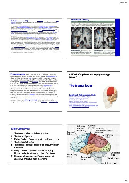
The fusiform face area (FFA) is a part of the human visual system which might be specialized for facial<br />
recognition, although there is some evidence that it also processes categorical information about other objects,<br />
particularly familiar ones.<br />
Localization<br />
The FFA is located in the ventral stream on the ventral surface of the temporal lobe on the fusiform gyrus. It is<br />
adjacent to the parahippocampal place area <strong>and</strong> near the putative extrastriate body area. It is in a slightly different<br />
place for each human <strong>and</strong> displays some lateralization, usually being larger in the right hemisphere.<br />
The FFA was discovered <strong>and</strong> continues to be investigated in humans using positron emission tomography (PET) <strong>and</strong><br />
functional magnetic resonance imaging (fMRI) studies. Usually, a participant views images of faces, objects, places,<br />
bodies, scrambled faces, scrambled objects, scrambled places <strong>and</strong> scrambled bodies. This is called a functional<br />
localizer. Comparing the neural response between faces <strong>and</strong> scrambled faces will reveal areas that are faceresponsive,<br />
while comparing cortical activation between faces <strong>and</strong> objects will reveal areas that are face‐selective.<br />
Functional role<br />
The human FFA was first described by Justine Sergent in 1992 <strong>and</strong> more recently by Nancy Kanwisher in 1997 who<br />
proposed that the existence of the FFA is evidence for domain specificity in the visual system. More recently, it has<br />
been suggested that the FFA processes more than just faces. Some groups, including Isabel Gauthier <strong>and</strong> others,<br />
maintain that the FFA is an area for recognizing fine distinctions between well‐known objects. Gauthier et al. tested<br />
both car <strong>and</strong> bird experts, <strong>and</strong> found some activation in the FFA when car experts were identifying cars <strong>and</strong> when bird<br />
experts were identifying birds. A recent paper by Kalanit Grill‐Spector et al. also suggests that processing in the FFA is<br />
not exclusive to faces, although an erratum was later published which brought to light some errors.The debate about<br />
the functional role of the FFA is ongoing.<br />
A 2009 magnetoencephalography study found that objects incidentally perceived as faces, an example of pareidolia,<br />
evoke an early (165 ms) activation in the FFA, at a time <strong>and</strong> location similar to that evoked by faces, whereas other<br />
common objects do not evoke such activation. This activation is similar to a slightly earlier peak at 130 ms seen for<br />
images of real faces. The authors suggest that face perception evoked by face‐like objects is a relatively early process,<br />
<strong>and</strong> not a late cognitive reinterpretation phenomenon.<br />
Fusiform Face Area (FFA)<br />
N & NJ Kotchabhakdi 2008 260<br />
Prosopagnosia (Greek: "prosopon" = "face", "agnosia" = "inabilty to<br />
recognise/identify familiar people or objects") is a disorder of face perception<br />
where the ability to recognize faces is impaired, while the ability to recognize<br />
other objects may be relatively intact. The term originally referred to a condition<br />
following acute brain damage, but a congenital form of the disorder has been<br />
proposed, which may be inherited by about 2.5% of the population. The specific<br />
brain area usually associated with prosopagnosia is the fusiform gyrus.<br />
Few successful therapies have so far been developed for affected people,<br />
although individuals often learn to use 'piecemeal' or 'feature by feature'<br />
recognition strategies. This may involve secondary clues such as clothing, gait,<br />
hair color, body shape, <strong>and</strong> voice. Because the face seems to function as an<br />
important identifying feature in memory, it can also be difficult for people with<br />
this condition to keep track of information about people, <strong>and</strong> socialize normally<br />
with others.<br />
Some also use the term prosophenosia, which refers to the inability to<br />
recognize faces following extensive damage of both occipital <strong>and</strong> temporal lobes<br />
415703 Cognitive Neuropsychology<br />
Week 6:<br />
The Frontal lobes<br />
Naiphinich Kotchabhakdi, Ph.D.<br />
Director, Salaya Stem Cell R & D Project,<br />
Research Center for Neuroscience,<br />
Institute of Molecular Biosciences,<br />
Mahidol University Salaya Campus,<br />
999 Phutthamonthol 4 Road, Salaya, Phutthamonthol,<br />
Nakornpathom 73170 Thail<strong>and</strong><br />
Email: scnkc@mahidol.ac.th or naiphinich@gmail.com<br />
Web: www.neuroscience.mahidol.ac.th<br />
Main Objectives:<br />
1. The Frontal lobes <strong>and</strong> their functions<br />
2. The Motor System<br />
3. Motor Cortical Organization in the Frontal Lobe<br />
4. The Prefrontal cortex<br />
5. The Frontal lobes <strong>and</strong> higher or executive brain<br />
functions<br />
6. Deep brain structures in Frontal lobe, e.g.,<br />
Limbic brain structures <strong>and</strong> their functions<br />
7. Neuropsychology of the Frontal lobes <strong>and</strong><br />
executive brain function disorders.<br />
44