Neurological Examination, clinical cases and neuropsychological ...
Neurological Examination, clinical cases and neuropsychological ...
Neurological Examination, clinical cases and neuropsychological ...

You also want an ePaper? Increase the reach of your titles
YUMPU automatically turns print PDFs into web optimized ePapers that Google loves.
23/07/54<br />
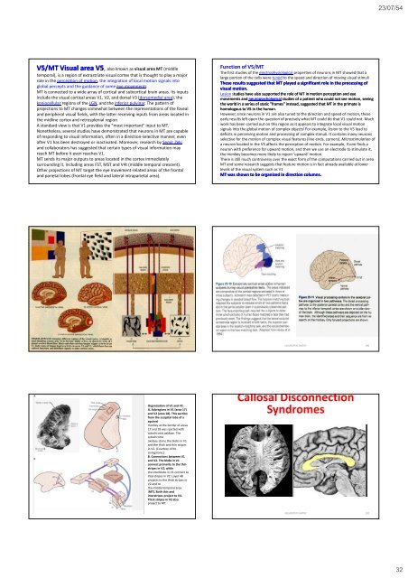
V5/MT Visual area V5, V also known as visual area MT (middle<br />
temporal), is a region of extrastriate visual cortex that is thought to play a major<br />
role in the perception of motion, the integration of local motion signals into<br />
global percepts <strong>and</strong> the guidance of some eye movements<br />
MT is connected to a wide array of cortical <strong>and</strong> subcortical brain areas. Its inputs<br />
include the visual cortical areas V1, V2, <strong>and</strong> dorsal V3 (dorsomedial area), the<br />
koniocellular regions of the LGN, <strong>and</strong> the inferior pulvinar. The pattern of<br />
projections to MT changes somewhat between the representations of the foveal<br />
<strong>and</strong> peripheral visual fields, with the latter receiving inputs from areas located in<br />
the midline ecortex <strong>and</strong> retrosplenial ospe region<br />
A st<strong>and</strong>ard view is that V1 provides the "most important" input to MT.<br />
Nonetheless, several studies have demonstrated that neurons in MT are capable<br />
of responding to visual information, often in a direction‐selective manner, even<br />
after V1 has been destroyed or inactivated. Moreover, research by Semir Zeki<br />
<strong>and</strong> collaborators has suggested that certain types of visual information may<br />
reach MT before it even reaches V1.<br />
MT sends its major outputs to areas located in the cortex immediately<br />
surrounding it, including areas FST, MST <strong>and</strong> V4t (middle temporal crescent).<br />
Other projections of MT target the eye movement‐related areas of the frontal<br />
<strong>and</strong> parietal lobes (frontal eye field <strong>and</strong> lateral intraparietal area).<br />
Function of V5/MT<br />
The first studies of the electrophysiological properties of neurons in MT showed that a<br />
large portion of the cells were tuned to the speed <strong>and</strong> direction of moving visual stimuli<br />
These results suggested that MT played a significant role in the processing of<br />
visual motion.<br />
Lesion studies have also supported the role of MT in motion perception <strong>and</strong> eye<br />
movements <strong>and</strong> <strong>neuropsychological</strong> studies of a patient who could not see motion, seeing<br />
the world in a series of static "frames" instead, suggested that MT in the primate is<br />
homologous to V5 in the human.<br />
However, since neurons in V1 are also tuned to the direction <strong>and</strong> speed of motion, these<br />
early results left open the question of precisely what MT could do that V1 could not. Much<br />
work has been carried out on this region as it appears to integrate local visual motion<br />
signals into the global motion of complex objects ] For example, lesion to the V5 lead to<br />
deficits in perceiving motion <strong>and</strong> processing of complex stimuli. It contains many neurons<br />
selective for the motion of complex visual features (line ends, corners). Microstimulation of<br />
a neuron located in the V5 affects the perception of motion. For example, if one finds a<br />
neuron with preference for upward motion, <strong>and</strong> then we use an electrode to stimulate it,<br />
the monkey becomes more likely to report 'upward' motion.<br />
There is still much controversy over the exact form of the computations carried out in area<br />
MT <strong>and</strong> some research suggests that feature motion is in fact already available at lower<br />
levels of the visual system such as V1<br />
MT was shown to be organized in direction columns.<br />
NEUROPSYCHIATRY 190<br />
Organization of V1 <strong>and</strong> V2.<br />
A. Subregions in V1 (area 17)<br />
<strong>and</strong> V2 (area 18). This section<br />
from the occipital lobe of a<br />
squirrel<br />
monkey at the border of areas<br />
17 <strong>and</strong> 18 was reacted with<br />
cytochrome oxidase. The<br />
cytochrome<br />
oxidase stains the blobs in V1<br />
<strong>and</strong> the thick <strong>and</strong> thin stripes<br />
in V2. (Courtesy of M.<br />
Livingstone.)<br />
B. Connections between V1<br />
<strong>and</strong> V2. The blobs in V1<br />
connect primarily to the thin<br />
stripes in V2, while<br />
the interblobs in V1 connect to<br />
interstripes in V2. Layer 4B<br />
projects to the thick stripes in<br />
V2 <strong>and</strong> to<br />
the middle temporal area<br />
(MT). Both thin <strong>and</strong><br />
interstripes project to V4.<br />
Thick stripes in V2 also<br />
project to MT.<br />
Callosal Disconnection<br />
Syndromes<br />
NEUROPSYCHIATRY 192<br />
32