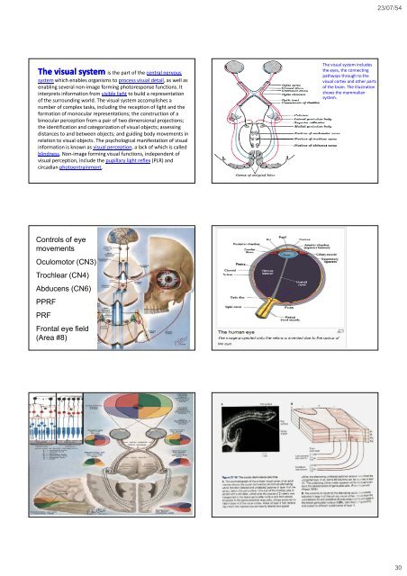
23/07/54 The visual system is the part of the central nervous system which enables organisms to process visual detail, as well as enabling several non‐image forming photoresponse functions. It interprets information from visible light to build a representation of the surrounding world. The visual system accomplishes a number of complex tasks, including the reception of light <strong>and</strong> the formation of monocular representations; the construction of a binocular perception from a pair of two dimensional projections; the identification <strong>and</strong> categorization of visual objects; assessing distances to <strong>and</strong> between objects; <strong>and</strong> guiding body movements in relation to visual objects. The psychological manifestation of visual information is known as visual perception, a lack of which is called blindness. Non‐image forming visual functions, independent of visual perception, include the pupillary light reflex (PLR) <strong>and</strong> circadian photoentrainment. The visual system includes the eyes, the connecting pathways through to the visual cortex <strong>and</strong> other parts of the brain. The illustration shows the mammalian system. Controls of eye movements Oculomotor (CN3) Trochlear (CN4) Abducens (CN6) PPRF PRF Frontal eye field (Area #8) NEUROPSYCHIATRY 177 30
23/07/54 Primary visual cortex (V1) The primary visual cortex is the best studied visual area in the brain. In all mammals studied, it is located in the posterior pole of the occipital cortex (the occipital cortex is responsible for processing visual stimuli). It is the simplest, earliest cortical visual area. It is highly specialized for processing information about static <strong>and</strong> moving objects <strong>and</strong> is excellent in pattern recognition. The functionally defined primary visual cortex is approximately equivalent to the anatomically defined striate cortex. The name "striate cortex" is derived from the stria of Gennari, a distinctive stripe visible to the naked eye that represents myelinated aonsfrom axons the lateral geniculate body terminating in layer 4 of the gray matter. The primary visual cortex is divided into six functionally distinct layers, labeled 1 through 6. Layer 4, which receives most visual input from the lateral geniculate nucleus (LGN), is further divided into 4 layers, labelled 4A, 4B, 4Cα, <strong>and</strong> 4Cβ. Sublamina 4Cα receives most magnocellular input from the LGN, while layer 4Cβ receives input from parvocellular pathways. The average number of neurons in the adult human primary visual cortex, in each hemisphere, has been estimated at around 140 million (Leuba & Kraftsik, Anatomy <strong>and</strong> Embryology, 1994). V1 has a very well‐defined map of the spatial information in vision. For example, in humans the upper bank of the calcarine sulcus responds strongly to the lower half of visual field (below the center), <strong>and</strong> the lower bank of the calcarine to the upper half of visual field. Conceptually, this retinotopic mapping is a transformation of the visual image from retina to V1. The correspondence between a given location in V1 <strong>and</strong> in the subjective visual field is very precise: even the blind spots are mapped into V1. Evolutionarily, this correspondence is very basic <strong>and</strong> found in most animals that possess a V1. In human <strong>and</strong> animals with a fovea in the retina, a large portion of V1 is mapped to the small, central portion of visual field, a phenomenon known as cortical magnification. Perhaps for the purpose of accurate spatial encoding, neurons in V1 have the smallest receptive field size of any visual cortex microscopic regions. The tuning properties of V1 neurons (what the neurons respond to) differ greatly over time. Early in time (40 ms <strong>and</strong> further) individual V1 neurons have strong tuning to a small set of stimuli. That is, the neuronal responses can discriminate small changes in visual orientations, spatial frequencies <strong>and</strong> colors. Furthermore, individual V1 neurons in human <strong>and</strong> animals with binocular vision have ocular dominance, namely tuning to one of the two eyes. In V1, <strong>and</strong> primary sensory cortex in general, neurons with similar tuning properties tend to cluster together as cortical columns. David Hubel <strong>and</strong> Torsten Wiesel proposed the classic ice‐cube cube organization model of cortical columns for two tuning properties: ocular dominance <strong>and</strong> orientation. However, this model cannot accommodate the color, spatial frequency <strong>and</strong> many other features to which neurons are tuned. The exact organization of all these cortical columns within V1 remains a hot topic of current research. Current consensus seems to be that early responses of V1 neurons consists of tiled sets of selective spatiotemporal filters. In the spatial domain, the functioning of V1 can be thought of as similar to many spatially local, complex Fourier transforms, or more accurately, Gabor transforms. Theoretically, these filters together can carry out neuronal processing of spatial frequency, orientation, motion, direction, speed (thus temporal frequency), <strong>and</strong> many other spatiotemporal features. Experiments of neurons substantiate these theories, but also raise new questions. Later in time (after 100 ms) neurons in V1 are also sensitive to the more global organisation of the scene (Lamme & Roelfsema, 2000). These response properties probably stem from recurrent processing (the influence of higher‐tier cortical areas on lower‐tier cortical areas) <strong>and</strong> lateral connections from pyramidal neurons (Hupe et al. 1998). While feedforward connections are mainly driving, feedback connections are mostly modulatory in their effects (Angelucci et al., 2003; Hupe et al., 2001). Evidence shows that feedback originating in higher level areas such as V4, IT or MT, with bigger <strong>and</strong> more complex receptive fields, can modify <strong>and</strong> shape V1 responses, accounting for contextual or extra‐classical receptive field effects (Guo et al., 2007; Harrison et al., 2007; Huang et al., 2007; Sillito et al., 2006). The visual information relayed to V1 is not coded in terms of spatial (or optical) imagery, but rather as the local contrast. As an example, for an image comprising half side black <strong>and</strong> half side white, the divide line between black <strong>and</strong> white has strongest local contrast <strong>and</strong> is encoded, while few neurons code the brightness information (black or white per se). As information is further relayed to subsequent visual areas, it is coded as increasingly non‐local frequency/phase signals. Importantly, at these early stages of cortical visual processing, spatial location of visual information is well preserved amid the local contra trast encoding. Visual area V2, also called , also called prestriate cortex, is the second major area in the visual cortex, <strong>and</strong> the first region within the visual association area. It receives strong feedforward connections from V1 (direct <strong>and</strong> via the pulvinar) <strong>and</strong> sends strong connections to V3, V4, <strong>and</strong> V5. It also sends strong feedback connections to V1. Anatomically, V2 is split into four quadrants, a dorsal <strong>and</strong> ventral representation in the left <strong>and</strong> the right hemispheres. Together these four regions provide a complete map of the visual world. Functionally, V2 has many properties in common with V1. Cells are tuned to simple properties such as orientation, spatial frequency, <strong>and</strong> color. The responses of many V2 neurons are also modulated by more complex properties, such as the orientation of illusory contours <strong>and</strong> whether the stimulus is part of the figure or the ground (Qiu <strong>and</strong> von der Heydt, 2005). Recent research has shown that V2 cells show a small amount of attentional modulation (more than V1, less than V4), are tuned for moderately complex patterns, <strong>and</strong> may be driven by multiple orientations at different subregions within a single receptive field. It is argued that the entire ventral visual‐to‐hippocampal stream is important for visual memory. This theory, unlike the dominant one, predicts that object‐recognition memory (ORM) alterations could result from the manipulation in V2, an area that is highly interconnected within the ventral stream of visual cortices. In the monkey brain, this area receives strong feedforward connections from the primary visual cortex (V1) <strong>and</strong> sends strong projections to other secondary visual cortices (V3, V4, <strong>and</strong> V5) . Most of the neurons of this area are tuned to simple visual characteristics such as orientation, spatial frequency, size, color, <strong>and</strong> shape <strong>and</strong> V2 cells also respond to various complex shape characteristics, such as the orientation of illusory contours <strong>and</strong> whether the stimulus is part of the figure or the ground . Anatomical studies implicate layer 3 of area V2 in visual‐information processing. In contrast to layer 3, layer 6 of the visual cortex is composed of many types of neurons, <strong>and</strong> their response to visual stimuli is more complex. In a recent study, the Layer 6 cells of the V2 cortex were found to play a very important role in the storage of Object Recognition Memory as well as the conversion of short‐term object memories into long‐term memories. Third visual complex, including area V3 The term third visual complex refers to the region of cortex located immediately in front of V2, which includes the region named visual area V3 in humans. The "complex" nomenclature is justified by the fact that some controversy still exists regarding the exact extent of area V3, with some researchers proposing that the cortex located in front of V2 may include two or three functional subdivisions. For example, David Van Essen <strong>and</strong> others (1986) have proposed that the existence of a "dorsal V3" in the upper part of the cerebral hemisphere, which is distinct from the "ventral V3" (or ventral posterior area, VP) located in the lower part of the brain. Dorsal <strong>and</strong> ventral V3 have distinct connections with other parts of the brain, appear different in sections stained with a variety of methods, <strong>and</strong> contain neurons that respond to different combinations of visual stimulus (for example, colour‐selective neurons are more common in the ventral V3). Additional subdivisions, including V3A <strong>and</strong> V3B have also been reported in humans. These subdivisions are located near dorsal V3, but do not adjoin V2. Dorsal V3 is normally considered to be part of the dorsal stream, receiving inputs from V2 <strong>and</strong> from the primary visual area <strong>and</strong> projecting to the posterior parietal cortex. It may be anatomically located in Brodmann area 19. Recent work with fMRI has suggested that area V3/V3A may play a role in the processing of global motion Other studies prefer to consider dorsal V3 as part of a larger area, named the dorsomedial area (DM), which contains a representation of the entire visual field. Neurons in area DM respond to coherent motion of large patterns covering extensive portions of the visual field (Lui <strong>and</strong> collaborators, 2006). Ventral V3 (VP), has much weaker connections from the primary visual area, <strong>and</strong> stronger connections with the inferior temporal cortex. While earlier studies proposed that VP only contained a representation of the upper part of the visual field (above the point of fixation), more recent work indicates that this area is more extensive than previously appreciated, <strong>and</strong> like other visual areas it may contain a complete visual representation. The revised, more extensive VP is referred to as the ventrolateral posterior area (VLP) by Rosa <strong>and</strong> Tweedale. Visual area V4 is one of the visual areas in the extrastriate visual cortex. It is located anterior to V2 <strong>and</strong> posterior to posterior inferotemporal area (PIT). It comprises at least four regions (left <strong>and</strong> right V4d, left <strong>and</strong> right V4v), <strong>and</strong> some groups report that it contains rostral <strong>and</strong> caudal subdivisions as well. It is unknown what the human homologue of V4 is, <strong>and</strong> this issue is currently the subject of much scrutiny. V4 is the third cortical area in the ventral stream, receiving strong feedforward input from V2 <strong>and</strong> sending strong connections to the PIT. It also receives direct inputs from V1, especially for central space. In addition, it has weaker connections to V5 <strong>and</strong> dorsal prelunate gyrus (DP). V4 is the first area in the ventral stream to show strong attentional modulation. Most studies indicate that selective attention can change firing rates in V4 by about 20%. A seminal paper by Moran <strong>and</strong> Desimone characterizing these effects was the first paper to find attention effects anywhere in the visual cortex. Like V1, V4 is tuned for orientation, spatial frequency, <strong>and</strong> color. Unlike V1, V4 is tuned for object features of intermediate complexity, like simple geometric shapes, although no one has developed a full parametric description of the tuning space for V4. Visual area V4 is not tuned for complex objects such as faces, as areas in the inferotemporal cortex are. The firing properties of V4 were first described by Semir Zeki in the late 1970s, who also named the area. Before that, V4 was known by its anatomical description, the prelunate gyrus. Originally, Zeki argued that the purpose of V4 was to process color information. Work in the early 1980s proved that V4 was as directly involved in form recognition as earlier cortical areas. This research supported the Two Streams hypothesis, first presented by Ungerleider <strong>and</strong> Mishkin in 1982. Recent work has shown that V4 exhibits long‐term plasticity, encodes stimulus salience, is gated by signals coming from the frontal eye fields <strong>and</strong> shows changes in the spatial profile of its receptive fields with attention. 31