Neurological Examination, clinical cases and neuropsychological ...
Neurological Examination, clinical cases and neuropsychological ...
Neurological Examination, clinical cases and neuropsychological ...

You also want an ePaper? Increase the reach of your titles
YUMPU automatically turns print PDFs into web optimized ePapers that Google loves.
23/07/54<br />
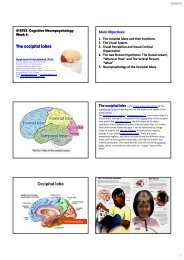
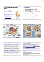
The parietal lobe is defined by four anatomical boundaries: the central<br />
sulcus separates the parietal lobe from the frontal lobe; the parieto‐occipital<br />
sulcus separates the parietal <strong>and</strong> occipital lobes; the lateral sulcus (sylvian<br />
fissure) is the most lateral boundary separating it from the temporal lobe;<br />
<strong>and</strong> the medial longitudinal fissure divides the two hemispheres.<br />
Immediately posterior to the central sulcus, <strong>and</strong> the most anterior part of<br />
the parietal lobe, is the postcentral gyrus (Brodmann area 3), the secondary<br />
somatosensory cortical area. Dividing this <strong>and</strong> the posterior parietal cortex<br />
is the postcentral sulcus.<br />
The posterior parietal cortex can be subdivided into the superior parietal<br />
lobule (Brodmann areas 5 + 7) <strong>and</strong> the inferior parietal lobule (39 + 40),<br />
separated by the intraparietal sulcus (IPS). The intraparietal sulcus <strong>and</strong><br />
adjacent gyri are essential in guidance of limb <strong>and</strong> eye movement, <strong>and</strong><br />
based on cytoarchitectural <strong>and</strong> functional differences is further divided into<br />
medial (MIP), lateral (LIP), ventral (VIP), <strong>and</strong> anterior (AIP) areas<br />
Parietal lobe Function<br />
The parietal lobe plays important roles in integrating sensory information from various<br />
parts of the body, knowledge of numbers <strong>and</strong> their relations, <strong>and</strong> in the manipulation of<br />
objects. Portions of the parietal lobe are involved with visuospatial processing. Although<br />
multisensory in nature, the posterior parietal cortex is often referred to by vision scientists<br />
as the dorsal stream of vision (as opposed to the ventral stream in the temporal lobe). This<br />
dorsal stream has been called both the 'where' stream (as in spatial vision) <strong>and</strong> the 'how'<br />
stream (as in vision for action).<br />
Various studies in the 1990s found that different regions of the posterior parietal cortex in<br />
Macaques represent different parts of space.<br />
► The lateral intraparietal (LIP) contains a map of neurons (retinotopically‐coded when<br />
the eyes are fixed) representing the saliency of spatial locations, <strong>and</strong> attention to these<br />
spatial locations. It can be used by the oculomotor system for targeting eye movements,<br />
when appropriate.<br />
►The ventral intraparietal (VIP) area receives input from a number of senses (visual,<br />
somatosensory, auditory, <strong>and</strong> vestibular). Neurons with tactile receptive fields represented<br />
space in a head‐centered reference frame. The cells with visual receptive fields also fire<br />
with head‐centered reference frames but possibly also with eye‐centered coordinates<br />
► The medial intraparietal (MIP) area neurons encode the location of a reach target in<br />
nose‐centered coordinates.<br />
► The anterior intraparietal (AIP) area contains neurons responsive to shape, size, <strong>and</strong><br />
orientation of objects to be grasped as well as for manipulation of h<strong>and</strong>s themselves, both<br />
to viewed <strong>and</strong> remembered stimuli.<br />
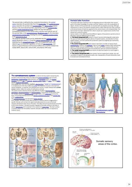
The somatosensory system is a diverse sensory system comprising the<br />
receptors <strong>and</strong> processing centres to produce the sensory modalities such as touch,<br />
temperature, proprioception (body position), <strong>and</strong> nociception (pain). The sensory<br />
receptors cover the skin <strong>and</strong> epithelia, skeletal muscles, bones <strong>and</strong> joints, internal organs,<br />
<strong>and</strong> the cardiovascular system.<br />
While touch (also, more formally, tactition; adjectival form: "tactile" or "somatosensory")<br />
is considered one of the five traditional senses, the impression of touch is formed from<br />
several modalities. In medicine, the colloquial term touch is usually replaced with somatic<br />
senses to better reflect the variety of mechanisms involved.<br />
The system reacts to diverse stimuli using different receptors: thermoreceptors,<br />
nociceptors, mechanoreceptors <strong>and</strong> chemoreceptors. Transmission of information from<br />
the receptors passes via sensory nerves through htracts in the spinal cord <strong>and</strong> into the<br />
brain. Processing primarily occurs in the primary somatosensory area in the parietal lobe<br />
of the cerebral cortex.<br />
The cortical homunculus was devised by Wilder Penfield.<br />
At its simplest, the system works when activity in a sensory neuron is triggered by a<br />
specific stimulus such as heat; this signal eventually passes to an area in the brain<br />
uniquely attributed to that area on the body—this allows the processed stimulus to be felt<br />
at the correct location. The point‐to‐point mapping of the body surfaces in the brain is<br />
called a homunculus <strong>and</strong> is essential in the creation of a body image. This brain‐surface<br />
("cortical") map is not immutable, however. Dramatic shifts can occur in response to<br />
stroke or injury.<br />
Somatosensory system<br />
Dermatome<br />
Somatic sensory<br />
areas of the cortex.<br />
34