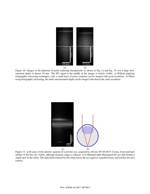
Figure 9: Rendering of a volume of a fly’s eye, that was acquired by <strong>full</strong>-<strong>field</strong> <strong>swept</strong>-<strong>source</strong> OCT.10mm) the point scatterers are visible, which corresponds to about 15mm imaging in air. The DC part of signal is notsuppressed well and can clearly be seen in the middle of the imaging depth. Signal of the point scatterers is highest inthe central part of the image, and degrades outwards due to the limited <strong>coherence</strong> length of the laser (signal roll-off). Inthe entire B-scan, only in a small layer the point scatterers are shown with a good resolution due to the limited depth offocus. Fig. 10b shows the effect of holographic refoc<strong>using</strong>, by resampling in Fourier-space to obtain diffraction limitedresolution over the entire measurement depth. Using this technique, lateral and axial resolution of about 10 µm in themedium (n ≈ 1.5), could be extended to the entire image which covered almost 10mm (about 15mm in air) imaging depth.With the corresponding Rayleigh length of 2z R ≈ 125 µm imaging was performed over 80 Rayleigh lengths.Fig. 11a demonstrates off-<strong>axis</strong> FF-SS-OCT at NA 0.04 for imaging of the anterior segment of a porcine eye. The sweeprange of the laser was reduced to 30nm. Although cornea and front surface of the lens are clearly visible, contrast anddynamic range of the image is limited. As illustrated in Fig. 11b the collimated light on the eye illuminates only a singlespot on the retina. Light scattered from here reaches also the camera, due to the lack of a confocal gating, and causesincoherent noise. By further extending the measurement depth, reducing the laser sweep range to 25nm, and additionallysqueezing the eye in axial direction, the entire eye including retina was imaged (Fig. 12). Now the light, scattered by theretina was no longer incoherent, and the dynamic range of the images increased significantly. Front and back surface of thelens were clearly seen. The loss in signal in the front surface is caused by the roll-off due to the limited <strong>coherence</strong> lengthof the laser. The resulting image corresponds to an imaging depth of more than 30mm in air.5. CONCLUSIONIn conclusion, we demonstrated an off-<strong>axis</strong> setup for <strong>full</strong>-<strong>field</strong> <strong>swept</strong>-<strong>source</strong> OCT, which abolishes many shortcomingsof scanning and <strong>full</strong>-<strong>field</strong> SS-OCT. It extends the focal range, sensitivity and lateral resolution are maintained over anextended depth spanning approximately 80 Rayleigh lengths. The off-<strong>axis</strong> setup allows for removing autocorrelationterms and artifacts. Additionally, it resolves the complex conjugate ambiguity and thus allows <strong>full</strong>-range imaging, withoutrequiring any moving parts in the setup.Proc. of SPIE Vol. 8571 857104-6
5 mm5 mm0.5 mm0.5 mm(a)(b)Figure 10: Images of the phantom of point scattering nanoparticles as shown in Fig. 1a and Fig. 1b over a large measurementdepth of almost 10 mm. The DC signal in the middle of the images is clearly visible. a) Without applyingholographic refoc<strong>using</strong> techniques, only a small layer of point scatterers can be imaged with good resolution. b) When<strong>using</strong> holographic refoc<strong>using</strong>, the entire measurement depth can be imaged with almost the same resolution.5 mm1 mm(a)(b)Figure 11: a) B-scans of the anterior segment of a porcine eye, acquired by off-<strong>axis</strong> FF-SS-OCT. Cornea, front and backsurface of the lens are visible, although dynamic range is reduced. b) Collimated light illuminated the eye and formed asingle spot on the retina. The light backscattered by the retina leaves the eye again as a parallel beam, and reaches the areacamera.Proc. of SPIE Vol. 8571 857104-7