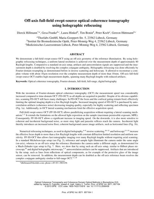
Off-axis full-field swept-source optical coherence tomography using ...
Off-axis full-field swept-source optical coherence tomography using ...
Off-axis full-field swept-source optical coherence tomography using ...
- No tags were found...

You also want an ePaper? Increase the reach of your titles
YUMPU automatically turns print PDFs into web optimized ePapers that Google loves.
<strong>Off</strong>-<strong>axis</strong> <strong>full</strong>-<strong>field</strong> <strong>swept</strong>-<strong>source</strong> <strong>optical</strong> <strong>coherence</strong> <strong>tomography</strong><strong>using</strong> holographic refoc<strong>using</strong>Dierck Hillmann *,a , Gesa Franke b,c , Laura Hinkel b , Tim Bonin b , Peter Koch a , Gereon Hüttmann b,ca Thorlabs GmbH, Maria-Goeppert-Str. 5, 23562 Lübeck, Germanyb Institut für Biomedizinische Optik, Peter-Monnig-Weg 4, 23562 Lübeck, Germanyc Medizinisches Laserzentrum Lübeck, Peter-Monnig-Weg 4, 23562 Lübeck, GermanyABSTRACTWe demonstrate a <strong>full</strong>-<strong>field</strong> <strong>swept</strong>-<strong>source</strong> OCT <strong>using</strong> an off-<strong>axis</strong> geometry of the reference illumination. By <strong>using</strong> holographicrefoc<strong>using</strong> techniques, a uniform lateral resolution is achieved over the measurement depth of approximately 80Rayleigh lengths. Compared to a standard on-<strong>axis</strong> setup, artifacts and autocorrelation signals are suppressed and the measurementdepth is doubled by resolving the complex conjugate ambiguity. Holographic refoc<strong>using</strong> was done efficiently byFourier-domain resampling as demonstrated before in inverse scattering and holoscopy. It allowed to reconstruct a completevolume with about 10 µm resolution over the complete measurement depth of more than 10mm. <strong>Off</strong>-<strong>axis</strong> <strong>full</strong>-<strong>field</strong><strong>swept</strong>-<strong>source</strong> OCT enables high measurement depths, spanning many Rayleigh lengths with reduced artifacts.Keywords: Optical <strong>coherence</strong> <strong>tomography</strong>, Fourier-domain, <strong>full</strong>-<strong>field</strong>, <strong>full</strong>-range, digital holography1. INTRODUCTIONWith the invention of Fourier-domain <strong>optical</strong> <strong>coherence</strong> <strong>tomography</strong> (OCT) the measurement speed was considerablyincreased compared to time-domain OCT (TD-OCT) as all depths are acquired in parallel. Despite of its obvious capabilities,scanning FD-OCT still faces many challenges: In FD-OCT the focus and the confocal gating remain fixed, effectivelylimiting the optimal imaging depth to a few Rayleigh lengths. Increased imaging speed of FD-OCT is purchased by autocorrelationartifacts (<strong>coherence</strong> noise) decreasing imaging quality, especially for highly scattering and reflecting specimen(Fig. 1a). Additionally, in OCT lateral scanning mechanisms limit the effective acquisition speed.Full-<strong>field</strong> <strong>swept</strong>-<strong>source</strong> OCT (FF-SS-OCT) allows parallelizing acquisition without requiring a lateral scanning mechanism.1, 2 It extends the limitations on the allowed light exposition on the sample (maximum permissible exposure, MPE).Consequently, FF-SS-OCT allows a significant increase in imaging speed. On the downside, it is also more sensitive tocoherent and incoherent background noise, as more stray light and parasitic reflexes reach the camera. Incoherent lightthereby introduces an increased noise floor, coherent background causes image artifacts, such as horizontal lines (Fig. 1b).Numerical refoc<strong>using</strong> techniques, as used in digital holography, 4–9 inverse scattering, 10–12 and holoscopy 13–15 increasethe effective focus depth to more than a few Rayleigh lengths with constant diffraction limited resolution and uniform sensitivity.FF-SS-OCT thus allows fast tomographic imaging over many Rayleigh lengths without requiring axial scanning.In a standard Michelson-type setup (see Fig. 2), reference and sample light illuminate the camera under the same angle(on-<strong>axis</strong>), whereas in an off-<strong>axis</strong> setup the reference illuminates the camera under a different angle, as demonstrated fora Mach-Zehnder type setup in Fig. 7. Here, we show that by <strong>using</strong> such an off-<strong>axis</strong> setup, similar to Hilbert phase microscopy16 and digital holographic microscopy, 4, 5 autocorrelation artifacts can be suppressed. Artifacts that are introducedby the setup, such as lines from reflections of <strong>optical</strong> components, as for example of the protective glass of the imagesensor, can be removed. In addition, the measurement depth can be doubled as the off-<strong>axis</strong> reference beam resolves thecomplex conjugate ambiguity similar to <strong>full</strong>-range OCT.17, 18*dhillmann@thorlabs.com; phone +49 451 2903256Optical Coherence Tomography and Coherence Domain Optical Methods in Biomedicine XVII,edited by James G. Fujimoto, Joseph A. Izatt, Valery V. Tuchin, Proc. of SPIE Vol. 8571, 857104© 2013 SPIE · CCC code: 1605-7422/13/$18 · doi: 10.1117/12.2006436Proc. of SPIE Vol. 8571 857104-1
DC signalAutocorrelationtermsComplex conjugatedsignal termlog ISignal termzFigure 3: A typical A-scan of an FD-OCT system. The different terms of the interference signals can be distinguished,but they influence imaging quality and cause artifacts. The complex conjugate signal term can overlay the signal term anddistort the image information.yxFigure 4: Image, as acquired by the off-<strong>axis</strong> <strong>full</strong>-<strong>field</strong> setup (left). The carrier frequency due to the off-<strong>axis</strong> illuminationbecomes visible in the zoomed image (right).2. PRINCIPLEOCT works by acquiring the interference signal I between waves originating from the sample O and a reference waveR. In FD-OCT, this signal is acquired spectrally resolved, and after a Fourier transform the various signal terms can bedistinguished (see also Fig. 3). The interference term is given byI ∝ |R + O| 2 = |R| 2 + |O| 2 + R ∗ O + RO ∗ ,where |R| 2 +|O| 2 denotes DC and autocorrelation terms, R ∗ O is the signal term and RO ∗ its complex conjugate. In generalthe DC and autocorrelation term do not carry the desired A-scan information. The signal term and the conjugate signalterm can only be distinguished, if sample arm path-lengths are always ensured to be longer than the reference path lengthor vice versa. If this is not in the case, the signals above and below the reference plane will overlap with each other, andimaged structures can not be clearly identified.With the proposed setup, there is a small angle between reference and sample light reaching the camera. As the lightinterferes, this causes interference fringes and effectively results in a carrier frequency on the sample light <strong>field</strong>. The carrierfrequency, which is demonstrated in Fig. 4, causes the signal term R ∗ O, and its complex conjugate to be shifted away fromthe DC-component into opposite directions after a two-dimensional Fourier transform (Fig. 5). Combining this lateraldiscrimination of the signal terms with the axial discrimination of the OCT signal terms, as shown in Fig. 6, the terms areseparated. The complex conjugate ambiguity is resolved by filtering one of the two signal terms, effectively increasingthe measurement depth. As DC and autocorrelation signals are still centered and not shifted, they do not overlay with thesignal terms and are also removed, which reduces coherent imaging artifacts.Without restrictions to the measuring depth by confocal gating, and by resolving the complex conjugate ambiguity andtherefore halving the influence of the limited instantaneous <strong>coherence</strong> length of the laser, the achievable imaging depth islargely increased. Finally, by <strong>using</strong> holographic refoc<strong>using</strong>, 12, 15, 19 the limitations on the optimal lateral resolution to theRayleigh length is effectively removed.Proc. of SPIE Vol. 8571 857104-3
k ySignal termk x-_itDC signalAutoccorelationtermComplex conjugatedsignal termFigure 5: Fourier transforming the acquired images, the different signal terms can be distinguished. The DC and autocorrelationterms are centered in the image, the signal term and its complex conjugate are visible as circles in the upper rightand lower left corner, respectively. The circular shape of the signal terms is caused by the aperture placed in the Fourierconjugated plane of the image/object.zk yComplex conjugatedsignal termk xAutoccorelationtermDC signalSignal termFigure 6: Combining two-dimensional FFT of acquired images as shown in Fig. 5 with the axial discrimination in an OCTA-scan (Fig. 3) shows that signal term, its complex conjugate, and the autocorrelation term can clearly be distinguished.Suitable filtering can select one of the two signal terms and suppress other signals.3. SETUPThe setup we used for off-<strong>axis</strong> <strong>full</strong>-<strong>field</strong> <strong>swept</strong>-<strong>source</strong> OCT was a Mach-Zehnder type setup as shown in Fig. 7. As tunablelaser we used a Broadsweeper BS-840-1 (Superlum, Ireland) with a tuning range from 867nm to 816nm and 3mW outputpower. Its light was split by a fiber coupler in reference and sample arm. The reference light was collimated to a beam withabout 16mm diameter and illuminated the camera under a slight angle of approximately 3 ◦ . The sample was illuminatedby collimated light and the backscattered light was imaged onto the camera <strong>using</strong> a two lens telescope, with a circularaperture in the Fourier plane of the imaging lens. The circular aperture effectively limited the NA of the setup and ensuredcorrect sampling of interference fringes on the camera. A Basler ACE camera (acA2040-180km, Basler, Germany) with2048 × 2048 pixels of size 5.5 µm × 5.5 µm each was used. Different areas of interest (AOI) were selected to adjustacquisition speed and <strong>field</strong> of view. The sweep of the laser and the acquisition of the camera were hardware synchronized,by triggering the laser with the camera. Images were acquired with a numerical apertures (NA) of up to 0.08.4. RESULTSThe effect of the lateral filtering is shown in Fig. 8. A scotch tape was acquired at approximately NA 0.04 by the off-<strong>axis</strong>FF-SS-OCT setup, and reconstructed without applying the lateral filtering for removing the autocorrelation and conjugatedterm (Fig. 8a). The images are thus comparable to images, acquired with a standard Michelson-type setup. Fig. 8b showsthe same dataset with the lateral filter applied. Here, horizontal lines and <strong>coherence</strong> noise are significantly reduced. Finally,Fig. 8c shows the scotch tape with the complex conjugate ambiguity resolved, measurement depth could be increased,although the central DC line is still visible. The proposed technique can also be used to image more complex structures, asshown in Fig. 9. Here, a 3D-rendering of an off-<strong>axis</strong> FF-SS-OCT volume of a bug is shown.Fig. 10a shows the increased measurement depth for a scattering phantom, containing 300nm to 800nm sized ironoxide nanoparticles embedded in polyurethane resin, acquired at NA 0.08. Almost in the entire depth of the sample (aboutProc. of SPIE Vol. 8571 857104-4
Swept-<strong>source</strong>CameraApertureSampleFigure 7: Setup for an off-<strong>axis</strong> <strong>full</strong>-<strong>field</strong> <strong>swept</strong>-<strong>source</strong> system. The collimated reference light illuminates the camera undera slight angle.1 mm(a)(b)(c)Figure 8: Images of scotch tape, acquired by off-<strong>axis</strong> FF-SS-OCT. a) B-scan as obtained when no filtering is applied.b) B-scan with lateral filtering applied. c) B-scan showing positive and negative path length differences of the sample light.The complex conjugate ambiguity of FD-OCT is resolved by filtering the signal term.1 mm1 mmProc. of SPIE Vol. 8571 857104-5
Figure 9: Rendering of a volume of a fly’s eye, that was acquired by <strong>full</strong>-<strong>field</strong> <strong>swept</strong>-<strong>source</strong> OCT.10mm) the point scatterers are visible, which corresponds to about 15mm imaging in air. The DC part of signal is notsuppressed well and can clearly be seen in the middle of the imaging depth. Signal of the point scatterers is highest inthe central part of the image, and degrades outwards due to the limited <strong>coherence</strong> length of the laser (signal roll-off). Inthe entire B-scan, only in a small layer the point scatterers are shown with a good resolution due to the limited depth offocus. Fig. 10b shows the effect of holographic refoc<strong>using</strong>, by resampling in Fourier-space to obtain diffraction limitedresolution over the entire measurement depth. Using this technique, lateral and axial resolution of about 10 µm in themedium (n ≈ 1.5), could be extended to the entire image which covered almost 10mm (about 15mm in air) imaging depth.With the corresponding Rayleigh length of 2z R ≈ 125 µm imaging was performed over 80 Rayleigh lengths.Fig. 11a demonstrates off-<strong>axis</strong> FF-SS-OCT at NA 0.04 for imaging of the anterior segment of a porcine eye. The sweeprange of the laser was reduced to 30nm. Although cornea and front surface of the lens are clearly visible, contrast anddynamic range of the image is limited. As illustrated in Fig. 11b the collimated light on the eye illuminates only a singlespot on the retina. Light scattered from here reaches also the camera, due to the lack of a confocal gating, and causesincoherent noise. By further extending the measurement depth, reducing the laser sweep range to 25nm, and additionallysqueezing the eye in axial direction, the entire eye including retina was imaged (Fig. 12). Now the light, scattered by theretina was no longer incoherent, and the dynamic range of the images increased significantly. Front and back surface of thelens were clearly seen. The loss in signal in the front surface is caused by the roll-off due to the limited <strong>coherence</strong> lengthof the laser. The resulting image corresponds to an imaging depth of more than 30mm in air.5. CONCLUSIONIn conclusion, we demonstrated an off-<strong>axis</strong> setup for <strong>full</strong>-<strong>field</strong> <strong>swept</strong>-<strong>source</strong> OCT, which abolishes many shortcomingsof scanning and <strong>full</strong>-<strong>field</strong> SS-OCT. It extends the focal range, sensitivity and lateral resolution are maintained over anextended depth spanning approximately 80 Rayleigh lengths. The off-<strong>axis</strong> setup allows for removing autocorrelationterms and artifacts. Additionally, it resolves the complex conjugate ambiguity and thus allows <strong>full</strong>-range imaging, withoutrequiring any moving parts in the setup.Proc. of SPIE Vol. 8571 857104-6
5 mm5 mm0.5 mm0.5 mm(a)(b)Figure 10: Images of the phantom of point scattering nanoparticles as shown in Fig. 1a and Fig. 1b over a large measurementdepth of almost 10 mm. The DC signal in the middle of the images is clearly visible. a) Without applyingholographic refoc<strong>using</strong> techniques, only a small layer of point scatterers can be imaged with good resolution. b) When<strong>using</strong> holographic refoc<strong>using</strong>, the entire measurement depth can be imaged with almost the same resolution.5 mm1 mm(a)(b)Figure 11: a) B-scans of the anterior segment of a porcine eye, acquired by off-<strong>axis</strong> FF-SS-OCT. Cornea, front and backsurface of the lens are visible, although dynamic range is reduced. b) Collimated light illuminated the eye and formed asingle spot on the retina. The light backscattered by the retina leaves the eye again as a parallel beam, and reaches the areacamera.Proc. of SPIE Vol. 8571 857104-7
5 mm1 mm(a)(b)Figure 12: a) B-scans of the eye of a porcine eye, acquired by off-<strong>axis</strong> FF-SS-OCT. Compared to Fig. 11a, the entire eyeis visible and thus backscattered light from the retina reaching the camera is coherent and forms an image at the correctdepth. Consequently imaging quality is improved; cornea, front and back surface of the lens are clearly visible. b) Toimage the complete eye it had to be is squeezed.REFERENCES[1] Považay, B., Unterhuber, A., Hermann, B., Sattmann, H., Arthaber, H., and Drexler, W., “Full-<strong>field</strong> time-encodedfrequency-domain <strong>optical</strong> <strong>coherence</strong> <strong>tomography</strong>,” Opt. Express 14(17), 7661–7669 (2006).[2] Bonin, T., Franke, G., Hagen-Eggert, M., Koch, P., and Hüttmann, G., “In vivo Fourier-domain <strong>full</strong>-<strong>field</strong> OCT of thehuman retina with 1.5 million A-lines/s,” Opt. Lett. 35(20), 3432–3434 (2010).[3] Woolliams, P. D., Ferguson, R. A., Hart, C., Grimwood, A., and Tomlins, P. H., “Spatially deconvolved <strong>optical</strong><strong>coherence</strong> <strong>tomography</strong>,” Appl. Opt. 49, 2014–2021 (Apr 10 2010).[4] Schnars, U. and Jueptner, W., [Digital holography: digital hologram recording, numerical reconstruction, and relatedtechniques], Springer (2005).[5] Kim, M., [Digital Holographic Microscopy: Principles, Techniques, and Applications], Springer Series in OpticalSciences, Springer (2011).[6] Zvyagin, A. V., “Fourier-domain <strong>optical</strong> <strong>coherence</strong> <strong>tomography</strong>: optimization of signal-to-noise ratio in <strong>full</strong> space,”Opt. Commun. 242(1-3), 97–108 (2004).[7] Kim, M. K., “Wavelength-scanning digital interference holography for <strong>optical</strong> section imaging,” Opt. Lett. 24, 1693–1695 (Dec 1999).[8] Potcoava, M. C. and Kim, M. K., “Optical <strong>tomography</strong> for biomedical applications by digital interference holography,”Meas. Sci. Technol. 19(7), 074010 (2008).[9] Moiseev, A. A., Gelikonov, G. V., Shilyagin, P. A., Terpelov, D. A., and Gelikonov, V. M., “Digital refoc<strong>using</strong> in<strong>optical</strong> <strong>coherence</strong> <strong>tomography</strong>,” Proc. SPIE 8213(1), 82132C (2012).[10] Ralston, T. S., Marks, D. L., Scott Carney, P., and Boppart, S. A., “Interferometric synthetic aperture microscopy,”Nat. Phys. 3(2), 129–134 (2007). 10.1038/nphys514.[11] Ralston, T. S., Marks, D. L., Carney, P. S., and Boppart, S. A., “Real-time interferometric synthetic aperture microscopy,”Opt. Express 16, 2555–2569 (Feb 18 2008).[12] Marks, D. L., Ralston, T. S., Boppart, S. A., and Carney, P. S., “Inverse scattering for frequency-scanned <strong>full</strong>-<strong>field</strong><strong>optical</strong> <strong>coherence</strong> <strong>tomography</strong>,” J. Opt. Soc. Am. A 24(4), 1034–1041 (2007).[13] Hillmann, D., Lührs, C., Bonin, T., Koch, P., and Hüttmann, G., “Holoscopy—holographic <strong>optical</strong> <strong>coherence</strong> <strong>tomography</strong>,”Opt. Lett. 36, 2390–2392 (Jul 2011).[14] Franke, G. L., Hillmann, D., Claußen, T., Lührs, C., Koch, P., and Hüttmann, G., “High resolution holoscopy,” Proc.SPIE 8213(1), 821324 (2012).Proc. of SPIE Vol. 8571 857104-8
[15] Hillmann, D., Franke, G., Lührs, C., Koch, P., and Hüttmann, G., “Efficient holoscopy image reconstruction,” Opt.Express 20, 21247–21263 (Sep 2012).[16] Ikeda, T., Popescu, G., Dasari, R. R., and Feld, M. S., “Hilbert phase microscopy for investigating fast dynamics intransparent systems,” Opt. Lett. 30, 1165–1167 (May 2005).[17] Yasuno, Y., Makita, S., Endo, T., Aoki, G., Itoh, M., and Yatagai, T., “Simultaneous B-M-mode scanning method forreal-time <strong>full</strong>-range Fourier domain <strong>optical</strong> <strong>coherence</strong> <strong>tomography</strong>,” Appl. Opt. 45, 1861–1865 (Mar 2006).[18] Jungwirth, J., Baumann, B., Pircher, M., Götzinger, E., and Hitzenberger, C. K., “Extended in vivo anterior eyesegmentimaging with <strong>full</strong>-range complex spectral domain <strong>optical</strong> <strong>coherence</strong> <strong>tomography</strong>,” J. Biomed. Opt. 14(5),050501–050503 (2009).[19] Davis, B. J., Marks, D. L., Ralston, T. S., Carney, P. S., and Boppart, S. A., “Interferometric synthetic aperturemicroscopy: Computed imaging for scanned coherent microscopy,” Sensors 8(6), 3903–3931 (2008).Proc. of SPIE Vol. 8571 857104-9