Chapter 10 - Molecular Biology of the Gene
Chapter 10 - Molecular Biology of the Gene
Chapter 10 - Molecular Biology of the Gene
You also want an ePaper? Increase the reach of your titles
YUMPU automatically turns print PDFs into web optimized ePapers that Google loves.
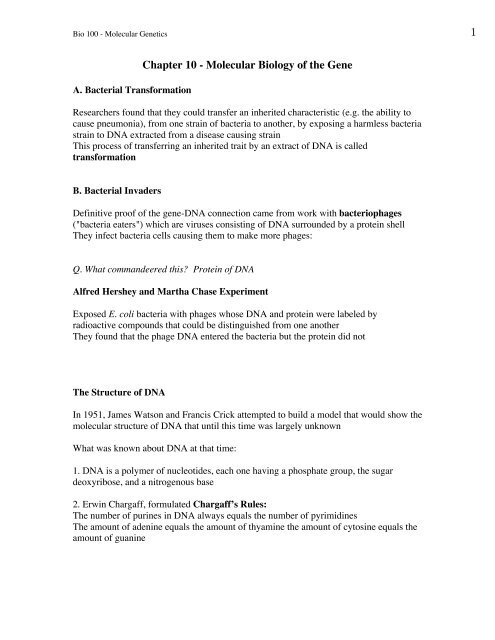
Bio <strong>10</strong>0 - <strong>Molecular</strong> <strong>Gene</strong>tics 1A. Bacterial Transformation<strong>Chapter</strong> <strong>10</strong> - <strong>Molecular</strong> <strong>Biology</strong> <strong>of</strong> <strong>the</strong> <strong>Gene</strong>Researchers found that <strong>the</strong>y could transfer an inherited characteristic (e.g. <strong>the</strong> ability tocause pneumonia), from one strain <strong>of</strong> bacteria to ano<strong>the</strong>r, by exposing a harmless bacteriastrain to DNA extracted from a disease causing strainThis process <strong>of</strong> transferring an inherited trait by an extract <strong>of</strong> DNA is calledtransformationB. Bacterial InvadersDefinitive pro<strong>of</strong> <strong>of</strong> <strong>the</strong> gene-DNA connection came from work with bacteriophages("bacteria eaters") which are viruses consisting <strong>of</strong> DNA surrounded by a protein shellThey infect bacteria cells causing <strong>the</strong>m to make more phages:Q. What commandeered this? Protein <strong>of</strong> DNAAlfred Hershey and Martha Chase ExperimentExposed E. coli bacteria with phages whose DNA and protein were labeled byradioactive compounds that could be distinguished from one ano<strong>the</strong>rThey found that <strong>the</strong> phage DNA entered <strong>the</strong> bacteria but <strong>the</strong> protein did notThe Structure <strong>of</strong> DNAIn 1951, James Watson and Francis Crick attempted to build a model that would show <strong>the</strong>molecular structure <strong>of</strong> DNA that until this time was largely unknownWhat was known about DNA at that time:1. DNA is a polymer <strong>of</strong> nucleotides, each one having a phosphate group, <strong>the</strong> sugardeoxyribose, and a nitrogenous base2. Erwin Chargaff, formulated Chargaff’s Rules:The number <strong>of</strong> purines in DNA always equals <strong>the</strong> number <strong>of</strong> pyrimidinesThe amount <strong>of</strong> adenine equals <strong>the</strong> amount <strong>of</strong> thyamine <strong>the</strong> amount <strong>of</strong> cytosine equals <strong>the</strong>amount <strong>of</strong> guanine
Bio <strong>10</strong>0 - <strong>Molecular</strong> <strong>Gene</strong>tics 23. Rosalind Franklin and Maurice Wilkins had prepared an X-ray defraction photograph<strong>of</strong> DNA indicating that DNA was long and skinny, helical and consisted <strong>of</strong> 2 parallelstrandsThe model proposed by Watson and Crick suggested <strong>the</strong> following features <strong>of</strong> DNA:1. The DNA molecule is composed <strong>of</strong> 2 nucleotide chains oriented in opposite directionsIn each strand each P group is attached to one sugar at <strong>the</strong> 5’ position (<strong>the</strong> 5th C) and to<strong>the</strong> o<strong>the</strong>r at <strong>the</strong> 3’ position2. The DNA molecule is analogous to <strong>the</strong> structure <strong>of</strong> a ladderThe bases on <strong>the</strong> 2 strands are directed inward and form <strong>the</strong> rung <strong>of</strong> <strong>the</strong> ladder, while <strong>the</strong>sugar-phosphate face outside and act as <strong>the</strong> sides <strong>of</strong> <strong>the</strong> ladder3. Complimentary base pairing - bases A and T always paired and G and C alwayspaired4. They suggested that <strong>the</strong> two DNA strands are twisted toge<strong>the</strong>r to form intertwinnedhelices = double helixThe Structure <strong>of</strong> RNAThe pentose sugar in RNA is riboseThe pyrimidine thyamine is replaced with uracilThus, <strong>the</strong> 4 bases are: A, U, C, and GRNA is single stranded, and does not form <strong>the</strong> double helix like DNAThere are 3 different classes <strong>of</strong> RNA: mRNA, rRNA, and tRNADNA has 2 primary functions:1. To replicate <strong>the</strong> stored information it contains2. Use <strong>the</strong> stored information in <strong>the</strong> syn<strong>the</strong>sis <strong>of</strong> proteinsCentral Dogma <strong>of</strong> molecular genetics:DNA ---------------------------> RNA --------------------> proteintranscriptiontranslation
Bio <strong>10</strong>0 - <strong>Molecular</strong> <strong>Gene</strong>tics 3DNA Replication: A Closer LookThe double stranded DNA molecule lends itself to replication because each strand canserve as a template for <strong>the</strong> formation <strong>of</strong> a complimentary strandDNA replication requires <strong>the</strong> following steps:1. The double helix unwinds and unzipsThe unwinding is largely due to <strong>the</strong> activity <strong>of</strong> helicaseUnzipping occurs at a distinct site along <strong>the</strong> chromosome and creates a replication bubbleThe y-shaped junction at <strong>the</strong> spot where <strong>the</strong> 2 strands are unwinding is called <strong>the</strong>replication fork2. Complimentary Base PairingThe exposed sequence <strong>of</strong> bases are paired with free nucleotides that drift into <strong>the</strong> area3. JoiningBase pairs are joined toge<strong>the</strong>r in a process called polymerizationIt is catalyzed by DNA polymeraseNote:DNA polymerase moves in one direction on <strong>the</strong> DNA (e.g. toward <strong>the</strong> replication fork)Thus nucleotides are added to <strong>the</strong> newly formed DNA chain in <strong>the</strong> 5’ - 3’ directionDNA replication is continuous on this side <strong>of</strong> <strong>the</strong> templateDNA replication is discontinuous on <strong>the</strong> o<strong>the</strong>r templateAn enzyme, ligase, will later link <strong>the</strong>se stretchesIn <strong>the</strong> end you get 2 double stranded DNA molecules, identical to one ano<strong>the</strong>r;replication is semiconservativeAccuracy <strong>of</strong> <strong>the</strong> ReplicationIn some cases, bases pairs do not pair correctly with one ano<strong>the</strong>r during replicationNone<strong>the</strong>less, <strong>the</strong> error rate is minimal because DNA polymerase has a "pro<strong>of</strong>reading"functionOn average <strong>the</strong>re is one mistake per billion nucleotides, or 3 mistakes per mitosis
Bio <strong>10</strong>0 - <strong>Molecular</strong> <strong>Gene</strong>tics 4Transcription (DNA ----> RNA)During transcription, <strong>the</strong> DNA code is passed to mRNA1. A segment <strong>of</strong> <strong>the</strong> DNA helix unwinds and unzips, and complimentary RNAnucleotides pair with <strong>the</strong> DNA nucleotides <strong>of</strong> one strand2. Initiation - RNA nucleotides are joined by <strong>the</strong> enzyme RNA polymerase, and <strong>the</strong>mRNA molecule is formedRNA polymerase must be must be instructed where to start and where to stop <strong>the</strong>transcribing processThe “start transcribing” signal is a nucleotide sequence called a promoter – located in<strong>the</strong> DNA next to <strong>the</strong> beginning <strong>of</strong> <strong>the</strong> gene3. After initiation, <strong>the</strong> RNA (mRNA) strand is elongatesFinally, <strong>the</strong>re is termination – where <strong>the</strong> RNA polymerase reaches a special sequence <strong>of</strong>bases in <strong>the</strong> DNA template called <strong>the</strong> terminatorRNA ProcessingIn eukaryotes, after <strong>the</strong> mRNA strand is formed, it passes from <strong>the</strong> nucleus to <strong>the</strong>cytoplasm where it becomes associated with ribosomesBut, before it leaves <strong>the</strong> nucleus, <strong>the</strong> mRNA is modified or processedOne kind <strong>of</strong> processing is <strong>the</strong> addition <strong>of</strong> extra nucleotides (The “cap” and “tail” ) to <strong>the</strong>ends <strong>of</strong> <strong>the</strong> RNA transcriptAno<strong>the</strong>r type <strong>of</strong> processing results from <strong>the</strong> fact that DNA is comprised <strong>of</strong> long stretches<strong>of</strong> noncoding regions (=introns) that interrupt nucleotide sequences that code for aminoacidsThe coding regions - <strong>the</strong> parts <strong>of</strong> <strong>the</strong> gene that are expressed - are called exonsBoth exons and introns are transcribed from DNA into RNARNA splicing - before <strong>the</strong> RNA leaves <strong>the</strong> nucleus, <strong>the</strong> introns are removed and <strong>the</strong>exons are joined to produce <strong>the</strong> mRNA with a continuous coding sequence
Bio <strong>10</strong>0 - <strong>Molecular</strong> <strong>Gene</strong>tics 5Translation (mRNA -----> protein)During translation <strong>the</strong> sequence <strong>of</strong> bases <strong>of</strong> <strong>the</strong> mRNA is translated into a sequence <strong>of</strong>amino acids3 nucleotide units <strong>of</strong> <strong>the</strong> mRNA that codes for a amino acid is called a codone.g., CGU codes for <strong>the</strong> aa ArginineAlso, <strong>the</strong>re are 3 codons called stop codons, that serve as a signal polypeptide chainterminatione.g., UAA, UAG, UGAOne codon is a start codon that signals to begin polypeptidee.g., AUGTranslation: A Closer LookDuring translation, <strong>the</strong> sequence <strong>of</strong> codons in mRNA dictates <strong>the</strong> order <strong>of</strong> <strong>the</strong> aminoacids in <strong>the</strong> polypeptideTranslation requires <strong>the</strong> efforts <strong>of</strong> several enzymes, and 2 o<strong>the</strong>r types <strong>of</strong> RNA: rRNA andtRNArRNAIt makes up <strong>the</strong> ribosomes; <strong>the</strong>y consist <strong>of</strong> 2 subunits, each <strong>of</strong> which is comprised <strong>of</strong>rRNA and proteinsrRNA is transcribed from DNA in <strong>the</strong> area <strong>of</strong> <strong>the</strong> nucleolusRibosomes function in coordinating protein syn<strong>the</strong>sisEach ribosome has a binding site for mRNA on its small subunitIts large subunit serves as a binding sites for tRNA: <strong>the</strong> P site holds <strong>the</strong> tRNA carrying<strong>the</strong> growing polypepetide chain; <strong>the</strong> A site holds a tRNA carrying <strong>the</strong> next amino acid tobe added to <strong>the</strong> chaintRNAtRNA is also transcribed in <strong>the</strong> nucleus and later moves to <strong>the</strong> cytoplasmtRNA brings amino acids from <strong>the</strong> cytoplasm to <strong>the</strong> ribosomes located in <strong>the</strong> cytoplasmAmino acids attach to one end <strong>of</strong> <strong>the</strong> tRNA molecule (CCA sequence)The o<strong>the</strong>r end <strong>of</strong> <strong>the</strong> molecule has a codon complimentary to <strong>the</strong> mRNA codon and iscalled <strong>the</strong> anticodon
Bio <strong>10</strong>0 - <strong>Molecular</strong> <strong>Gene</strong>tics 6There are 3 important steps in translation: initiation, elongation, and termination1. InitiationThe mRNA molecule binds (5’ end) to a small ribosomal subunitInitiation begins at <strong>the</strong> area <strong>of</strong> <strong>the</strong> start codon (AUG) <strong>of</strong> m RNAThe first tRNA pairs with <strong>the</strong> start codon <strong>of</strong> mRNAA large ribosomal subunit <strong>the</strong>n attaches to <strong>the</strong> smaller unitThis completes <strong>the</strong> ribosomal structureIt possess 2 active sites <strong>of</strong> attachment for tRNA2. ElongationThe ribosome moves down <strong>the</strong> length <strong>of</strong> <strong>the</strong> mRNA (5' ---> 3')It stops at <strong>the</strong> second and third mRNA codonsTwo tRNAs are <strong>the</strong>n received by <strong>the</strong>se codons, each carrying a specific amino acidThe ribosome moves again, occupying <strong>the</strong> third and fourth codon, etc.As <strong>the</strong> ribosome moves past an mRNA codon with a tRNA, <strong>the</strong> tRNA are released andre-enter <strong>the</strong> cytoplasms tRNA poolWhen each <strong>of</strong> <strong>the</strong> tRNAs with an amino acid are attached to <strong>the</strong> ribosome, <strong>the</strong> enzymepeptidyl transferase forges a peptide bond between 2 amino acids3. TerminationThe process continues until <strong>the</strong> ribosome encounters one <strong>of</strong> three possible stop codonse.g. UAG, UAA, UGAThe is no tRNA to recognize <strong>the</strong>se codons; instead a release factor will bind to <strong>the</strong> stopcodonThe last tRNA is released and <strong>the</strong> polypetide chain is freedThe polypeptides may <strong>the</strong>n go to <strong>the</strong> ER where fur<strong>the</strong>r syn<strong>the</strong>sis occurs (e.g. secondary,tertiary structure)Some goes to <strong>the</strong> cellular organelles e.g. mitochondria and chloroplasts


