Yersinia Virulence Depends on Mimicry of Host Rho ... - ResearchGate
Yersinia Virulence Depends on Mimicry of Host Rho ... - ResearchGate
Yersinia Virulence Depends on Mimicry of Host Rho ... - ResearchGate

You also want an ePaper? Increase the reach of your titles
YUMPU automatically turns print PDFs into web optimized ePapers that Google loves.
<str<strong>on</strong>g>Yersinia</str<strong>on</strong>g> <str<strong>on</strong>g>Virulence</str<strong>on</strong>g> <str<strong>on</strong>g>Depends</str<strong>on</strong>g> <strong>on</strong><strong>Mimicry</strong> <strong>of</strong> <strong>Host</strong> <strong>Rho</strong>-FamilyNucleotide Dissociati<strong>on</strong> InhibitorsGerd Prehna, 1 Maya I. Ivanov, 2 James B. Bliska, 2 and C. Erec Stebbins 1, *1 Laboratory <strong>of</strong> Structural Microbiology, Rockefeller University, New York, NY 10021, USA2 Department <strong>of</strong> Molecular Genetics and Microbiology and Center for Infectious Diseases, St<strong>on</strong>y Brook University,St<strong>on</strong>y Brook, NY 11794, USA*C<strong>on</strong>tact: stebbins@rockefeller.eduDOI 10.1016/j.cell.2006.06.056SUMMARY<str<strong>on</strong>g>Yersinia</str<strong>on</strong>g> spp. cause gastroenteritis and theplague, representing historically devastatingpathogens that are currently an important biodefenseand antibiotic resistance c<strong>on</strong>cern. Acritical virulence determinant is the <str<strong>on</strong>g>Yersinia</str<strong>on</strong>g> proteinkinase A, or YpkA, a multidomain proteinthat disrupts the eukaryotic actin cytoskelet<strong>on</strong>.Here we solve the crystal structure <strong>of</strong> a YpkA-Rac1 complex and find that YpkA possessesa Rac1 binding domain that mimics host guanidinenucleotide dissociati<strong>on</strong> inhibitors (GDIs) <strong>of</strong>the <strong>Rho</strong> GTPases. YpkA inhibits nucleotide exchangein Rac1 and <strong>Rho</strong>A, and mutati<strong>on</strong>s thatdisrupt the YpkA-GTPase interface abolish thisactivity in vitro and impair in vivo YpkA-inducedcytoskeletal disrupti<strong>on</strong>. In cell culture experiments,the kinase and the GDI domains <strong>of</strong> YpkAact synergistically to promote cytoskeletal disrupti<strong>on</strong>,and a Y. pseudotuberculosis mutantlacking YpkA GDI activity shows attenuated virulencein a mouse infecti<strong>on</strong> assay. We c<strong>on</strong>cludethat virulence in <str<strong>on</strong>g>Yersinia</str<strong>on</strong>g> depends str<strong>on</strong>gly up<strong>on</strong>mimicry <strong>of</strong> host GDI proteins by YpkA.INTRODUCTIONNearly 200 milli<strong>on</strong> people are estimated to have died in theplague epidemics that devastated the ancient world (Perryand Fetherst<strong>on</strong>, 1997), and the successful weap<strong>on</strong>izati<strong>on</strong><strong>of</strong> plague in the former Soviet Uni<strong>on</strong> bioweap<strong>on</strong>s programhas made this pathogen a primary biodefense c<strong>on</strong>cern(Henders<strong>on</strong>, 1999; Inglesby et al., 2000). Additi<strong>on</strong>al medicalc<strong>on</strong>cerns arise from the evoluti<strong>on</strong> <strong>of</strong> multidrug resistantstrains <strong>of</strong> the plague bacterium that have beenreported in several locati<strong>on</strong>s from patient isolates (Galimandet al., 1997; McCormick, 1998). The plague iscaused by the bacterial pathogen <str<strong>on</strong>g>Yersinia</str<strong>on</strong>g> pestis, which,al<strong>on</strong>g with the food-borne agents <strong>of</strong> gastroenteritis, Y. enterocolitica,and Y. pseudotuberculosis, shares the highlyc<strong>on</strong>served <str<strong>on</strong>g>Yersinia</str<strong>on</strong>g> virulence plasmid, or pYV, which isnecessary to cause disease (Cornelis et al., 1998). Thisplasmid harbors numerous genes, a large number <strong>of</strong>which are associated with a type III protein secreti<strong>on</strong> system(T3SS) that c<strong>on</strong>fers the ability <strong>of</strong> diverse pathogens todeliver virulence factors into host cells (Galán and Collmer,1999). The translocated virulence factors <strong>of</strong> <str<strong>on</strong>g>Yersinia</str<strong>on</strong>g> includea protein tyrosine phosphatase, a cysteine protease,and a GTPase-activating protein, am<strong>on</strong>g others,and collaborate to disrupt and impair the normal cytoskeletalregulati<strong>on</strong> in macrophages and polymorph<strong>on</strong>uclearneutrophils (PMNs) to achieve an antiphagocytic effect(Viboud and Bliska, 2005; Aepfelbacher et al., 1999;Aepfelbacher, 2004; Cornelis, 2000, 2002).An important virulence factor <strong>of</strong> <str<strong>on</strong>g>Yersinia</str<strong>on</strong>g> spp. is the<str<strong>on</strong>g>Yersinia</str<strong>on</strong>g> protein kinase A, or YpkA (also called YopO inY. enterocolitica), a substrate <strong>of</strong> the T3SS, which was firstidentified through its important c<strong>on</strong>tributi<strong>on</strong> to diseaseprogressi<strong>on</strong> and a regi<strong>on</strong> <strong>of</strong> sequence homology toeukaryotic serine/thre<strong>on</strong>ine kinases (Galyov et al., 1993).Y. pseudotuberculosis mutants with disrupti<strong>on</strong>s in YpkAhave shown severely attenuated virulence in mouse infecti<strong>on</strong>models (Galyov et al., 1993, 1994). YpkA is an 82 kDamultidomain protein, which, in additi<strong>on</strong> to the proteinkinase, possesses a domain that binds to the smallGTPases <strong>Rho</strong>A and Rac1, a domain required for activati<strong>on</strong>by actin, and a relatively uncharacterized N-terminaldomain (Barz et al., 2000; Dukuzumuremyi et al., 2000;Galyov et al., 1993, 1994; Hakanss<strong>on</strong> et al., 1996; Juriset al., 2000).In cultured cells, YpkA appears to functi<strong>on</strong> primarily indisrupting the host actin cytoskelet<strong>on</strong> (Dukuzumuremyiet al., 2000; Häkanss<strong>on</strong> et al., 1996; Juris et al., 2000).Cells transfected with YpkA, or exposed to strains <strong>of</strong> <str<strong>on</strong>g>Yersinia</str<strong>on</strong>g>preferentially translocating YpkA over other virulencefactors, lose their actin stress fibers and tend to round upand detach up<strong>on</strong> prol<strong>on</strong>ged exposure (Häkanss<strong>on</strong> et al.,1996; Juris et al., 2000; Nejedlik et al., 2004). Interestingly,this effect appears to be <strong>on</strong>ly partially associated with thekinase activity, as mutati<strong>on</strong>s in the active site attenuate,Cell 126, 869–880, September 8, 2006 ª2006 Elsevier Inc. 869
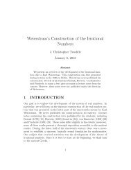
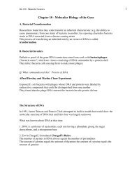
Figure 1. Overall Structure <strong>of</strong> the YpkA-Rac1 Complex(A) 2:2 YpkA:Rac1 c<strong>on</strong>tents <strong>of</strong> the asymmetric unit shown as ribb<strong>on</strong> diagrams. The two separate c<strong>on</strong>tacts <strong>of</strong> YpkA to Rac1 are shown, as is thenucleotide present, GDP.(B) A Ribb<strong>on</strong> diagram <strong>of</strong> YpkA (434–732) is shown and structural elements labeled. The domain break between the N-terminal GTPase binding domainand the C-terminal actin activati<strong>on</strong> domain is indicated by the divide between the two domain labels.(C) Sequence-structural analysis <strong>of</strong> YpkA (434–732). Sec<strong>on</strong>dary structure for given residues is indicated above the sequence. The relative solubility <strong>of</strong>each residue in the structure is denoted by a colored box beneath it, spanning dark blue (solvent exposed) to white (buried from solvent). The residuesinvolved in each charged patch <strong>of</strong> YpkA are labeled in violet and red for basic and acidic, respectively. Disordered regi<strong>on</strong>s are shown in yellow.but do not abolish, cytoskeletal alterati<strong>on</strong>s, and large internaldeleti<strong>on</strong>s in the protein that are C terminal to the kinasedomain are unable to cause cytoskeletal disrupti<strong>on</strong>s(Dukuzumuremyi et al., 2000; Juris et al., 2000). YpkA localizesto the plasma membrane in cultured cells (Dukuzumuremyiet al., 2000; Häkanss<strong>on</strong> et al., 1996; Nejedliket al., 2004), and mutati<strong>on</strong>s in the protein that abolish bindingto the <strong>Rho</strong> GTPases do not affect membrane localizati<strong>on</strong>(Dukuzumuremyi et al., 2000).Biochemically, the kinase activity <strong>of</strong> YpkA is regulated ina host dependent fashi<strong>on</strong> (Barz et al., 2000; Dukuzumuremyiet al., 2000; Juris et al., 2000). Recombinant and <str<strong>on</strong>g>Yersinia</str<strong>on</strong>g>-secretedYpkA are inactive but are activated in thepresence <strong>of</strong> heat-sensitive host cell extract (Barz et al.,2000; Dukuzumuremyi et al., 2000; Juris et al., 2000), andin particular in the presence <strong>of</strong> actin al<strong>on</strong>e (Juris et al.,2000). A 20 amino acid peptide at the COOH terminus <strong>of</strong>YpkA was shown by deleti<strong>on</strong> mutagenesis to be criticalfor YpkA binding to actin and for kinase autophosphorylati<strong>on</strong>in vitro (Juris et al., 2000). Therefore, like many eukaryoticprotein kinases, the <str<strong>on</strong>g>Yersinia</str<strong>on</strong>g> kinase appears to interactwith and be dependent <strong>on</strong> several regulatory elements inorder to achieve full activity (Huse and Kuriyan, 2002).We have solved the high-resoluti<strong>on</strong> crystal structure <strong>of</strong>the <strong>Rho</strong>-GTPase binding domain <strong>of</strong> YpkA with its smallGTPase host target Rac1. Analysis <strong>of</strong> this structure andbiochemical data reveals that YpkA mimics host guanidinenucleotide dissociati<strong>on</strong> inhibitors (GDIs) for the Rh<strong>of</strong>amily <strong>of</strong> small GTPases. This interacti<strong>on</strong> with Rac1 iscritical for the cytoskeletal disrupti<strong>on</strong> induced by YpkA incell culture and is also crucial for virulence in mice.RESULTSOverall Structure <strong>of</strong> the YpkA-Rac1 ComplexA C-terminal regi<strong>on</strong> <strong>of</strong> YpkA, residues 434–732 (Figure S1),is highly soluble and binds to Rac1 and <strong>Rho</strong>A, although, inagreement with previous studies (Barz et al., 2000; Dukuzumuremyiet al., 2000), not to Cdc42 (Figures S2, 3D, and3E). Although YpkA (434–732) and Rac1 form a dimer insoluti<strong>on</strong> (Figure S2), the complex crystallized as a 2:2packing (YpkA:Rac1) in the asymmetric unit with a n<strong>on</strong>crystallographic2-fold axis <strong>of</strong> symmetry (Figure 1A). Eachmolecule <strong>of</strong> YpkA (434–732) therefore makes c<strong>on</strong>tacts(‘‘A’’ and ‘‘B’’) to two different Rac1 molecules (Figure 1A).C<strong>on</strong>tact A interacts nearly exclusively with the regulatorySwitch I and Switch II regi<strong>on</strong>s <strong>of</strong> the GTPase, whereasC<strong>on</strong>tact B interacts with two C-terminal helices adjacentto the <strong>Rho</strong>GTPase ‘‘inserti<strong>on</strong>’’ (relative to the Ras-familysmall GTPases), distal to the nucletotide and Switch regi<strong>on</strong>s.Four facts argue for C<strong>on</strong>tact A being the biologicallyrelevant interacti<strong>on</strong>: (1) it has nearly twice the buried surfacearea as C<strong>on</strong>tact B, (2) it c<strong>on</strong>tacts the critical SwitchI and Switch II regi<strong>on</strong>s <strong>of</strong> Rac1, whereas C<strong>on</strong>tact B doesnot, (3) mutati<strong>on</strong>s <strong>of</strong> C<strong>on</strong>tact A disrupt YpkA-Rac1 interacti<strong>on</strong>s,whereas mutati<strong>on</strong>s in C<strong>on</strong>tact B do not (see below),870 Cell 126, 869–880, September 8, 2006 ª2006 Elsevier Inc.
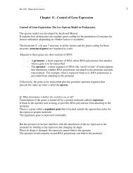
Figure 2. The YpkA-Rac1 Interface(A) Ribb<strong>on</strong> diagram view <strong>of</strong> the C<strong>on</strong>tact A interacti<strong>on</strong> between YpkA and Rac1. Switch I (yellow) and Switch II (red) are highlighted, and GDP is noted.(B) The image in panel (A) rotated by 90 about a horiz<strong>on</strong>tal axis.(C) Close up <strong>of</strong> the Switch I interacti<strong>on</strong>s with the a6 helix <strong>of</strong> YpkA. Hydrogen b<strong>on</strong>ds are denoted by dashed red lines.(D) Close up <strong>of</strong> the Switch II interacti<strong>on</strong>s with the a5 and a6 helices <strong>of</strong> YpkA. Hydrogen b<strong>on</strong>ds are denoted by dashed red lines.(E) Schematic <strong>of</strong> the interacti<strong>on</strong>s <strong>of</strong> the a5 and a6 helices <strong>of</strong> YpkA with Switch I and Switch II <strong>of</strong> Rac1. Hydrogen b<strong>on</strong>ds are indicated by red linesbetween the interacting residues, and hydrophobic interacti<strong>on</strong>s are shown with a yellow background.and (4) a heterodimer appears to be the biological unit asjudged by biochemical experiments (Figures 3 and S2).The overall structure <strong>of</strong> YpkA (434–732) reveals an el<strong>on</strong>gated,all-helical molecule c<strong>on</strong>sisting <strong>of</strong> two distinct subdomainsc<strong>on</strong>nected by a 65 Å l<strong>on</strong>g ‘‘backb<strong>on</strong>e’’ or ‘‘linkerhelix’’ (Figures 1A and 1B). The N-terminal subdomainc<strong>on</strong>tains most <strong>of</strong> the sequence-identified, ACC fingerlikerepeats that resemble, at the sequence level, elementsrequired in host factors for small GTPase binding,whereas the C-terminal subdomain c<strong>on</strong>tains the sequenceimplicated in actin activati<strong>on</strong>. The overall surface<strong>of</strong> the molecule is highly charged, c<strong>on</strong>taining a large basicpatch in the GTPase binding domain and a large acidicpatch in the actin-activati<strong>on</strong> domain (Figure S3).Despite apparent sequence similarities, the N-terminalsubdomain <strong>of</strong> YpkA (residues 434–615) does not sharethe coiled-coil fold <strong>of</strong> host ACC fingers, but instead it c<strong>on</strong>sists<strong>of</strong> six helices organized into two three-helix bundlespacked against each other. Each <strong>of</strong> the bundles is stabilizedby hydrophobic zippering in the core, and extensivehydrophobic packing is observed between the bundles.The previously identified regi<strong>on</strong>s harboring sequencesimilarity with ACC fingers (Dukuzumuremyi et al., 2000;Maesaki et al., 1999a, 1999b) are not involved in theYpkA-Rac1 interface. The far C-terminal subdomain <strong>of</strong>YpkA, c<strong>on</strong>taining the polypeptide implicated in actin activati<strong>on</strong>(residues 705–732, corresp<strong>on</strong>ding to helix a10), isa novel and el<strong>on</strong>gated fold c<strong>on</strong>sisting <strong>of</strong> four helices clusteredinto two pairs that <strong>on</strong>ly moderately interact with eachother. The a10 helix (residues 705–730) encompasses thepeptide that had been predicted to play a role in the interacti<strong>on</strong><strong>of</strong> YpkA with actin, and past work has shown thatthe deleti<strong>on</strong> <strong>of</strong> the regi<strong>on</strong> eliminates both the ability <strong>of</strong>YpkA to bind to actin and kinase activity (Juris et al.,2000). This helix forms an integral part <strong>of</strong> the fold <strong>of</strong> thissubdomain, c<strong>on</strong>tributing a large number <strong>of</strong> n<strong>on</strong>polarresidues to its hydrophobic core.There are very few c<strong>on</strong>formati<strong>on</strong>al changes betweenYpkA (434–732) crystallized al<strong>on</strong>e and YpkA (434–732) incomplex with Rac1 (Figure S3). Most differences arelocated in the C-terminal subdomain <strong>of</strong> YpkA and involvea slight overall displacement in the positi<strong>on</strong>ing <strong>of</strong> the a7and a8 helices, as well as alterati<strong>on</strong>s in the c<strong>on</strong>formati<strong>on</strong>and relative disorder <strong>of</strong> solvent exposed loops. Rac1 islittle altered by the binding <strong>of</strong> YpkA except for in theSwitch regi<strong>on</strong>s as discussed below.The YpkA-Rac1 InterfaceYpkA and Rac1 form an interface burying roughly 1600 Å 2and limited to residues 573–601 <strong>of</strong> YpkA (spanning thehelices a5 and a6) c<strong>on</strong>tacting the key regulatory Switch Iand Switch II regi<strong>on</strong>s <strong>of</strong> Rac1 (Figures 2A–2E). Switch Iand Switch II together create a c<strong>on</strong>cave pocket into whichthe a6 (backb<strong>on</strong>e) helix <strong>of</strong> YpkA inserts (Figure 2B), which,al<strong>on</strong>g with the clustering <strong>of</strong> the Switch II helix with the a5and a6 helices, cement the interacti<strong>on</strong> tightly.Cell 126, 869–880, September 8, 2006 ª2006 Elsevier Inc. 871
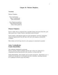
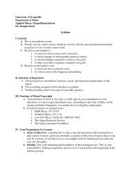
Figure 5. YpkA Inhibits Nucleotide Exchange in Rac1 and <strong>Rho</strong>AThe intrinsic and GEF catalyzed rate <strong>of</strong> mant-GTP exchange into the small GTPases Rac1, <strong>Rho</strong>A, and Cdc42 was m<strong>on</strong>itored as described in ExperimentalProcedures. The left column shows the intrinsic rate inhibiti<strong>on</strong> data, and the right column shows the rate inhibiti<strong>on</strong> data when challenged withthe Salm<strong>on</strong>ella GEF SopE. Panel (A) is Rac1, panel (B) is <strong>Rho</strong>A, and panel (C) is Cdc42. The experiments are shown with the increase <strong>of</strong> relativefluorescence units (RFU) over time in sec<strong>on</strong>ds. The labels are described in a legend below the figure, where 1.53 and 33 are relative molar c<strong>on</strong>centrati<strong>on</strong>s<strong>of</strong> YpkA or YpkA C<strong>on</strong>tact A mutant above the small GTPase. Error bars (panels A-C) represent the standard deviati<strong>on</strong> (from the mean valueplotted) calculatd from a minimum <strong>of</strong> three independent experiments.Cell 126, 869–880, September 8, 2006 ª2006 Elsevier Inc. 875
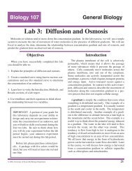
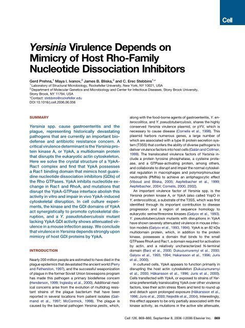
Figure 6. GDI Activity Is Critical for YpkAto Promote Cytoskeletal Alterati<strong>on</strong>s(A) The bar graph represents the percentage <strong>of</strong>transfected cells with cytoskeletal alterati<strong>on</strong>sand the c<strong>on</strong>tributi<strong>on</strong> <strong>of</strong> the observed wildtypephenotype and the observed intermediatephenotype to the total percentage. The wildtypephenotype is indicated in gray and theintermediate phenotype in white. Bacterial AlkalinePhosphatase, BAP, is the negative c<strong>on</strong>trol;1–732 is wild-type YpkA; 1–732 K272A isa kinase inactive mutant <strong>of</strong> YpkA; 1–732 C<strong>on</strong>tactA is YpkA with the C<strong>on</strong>tact A mutant;1–732 K272A C<strong>on</strong>tact A is the kinase inactivemutant plus the C<strong>on</strong>tact A mutant; 434–732 isthe crystallized c<strong>on</strong>struct; 434–732 C<strong>on</strong>tact Ais the C<strong>on</strong>tact A mutant (<strong>Rho</strong>A/Rac1 bindingdeficient structure-based mutant). Error bars(panels A-C) represent the standard deviati<strong>on</strong>(from the mean value plotted) calculated froma minimum <strong>of</strong> three independent experiments.(B) Examples <strong>of</strong> the visualizati<strong>on</strong> <strong>of</strong> YpkA by theimmunostaining <strong>of</strong> transfected Henle407 cells.Each <strong>of</strong> the three observed phenotypes isshown with staining for both the epitope,FLAG-tagged YpkA and for the actin cytoskelet<strong>on</strong>(Experimental Procedures). Each row islabeled by the c<strong>on</strong>struct transfected. The firstcolumn shows the presence <strong>of</strong> expressi<strong>on</strong> <strong>of</strong>the FLAG-tagged YpkA c<strong>on</strong>structs and is coloredin green. The sec<strong>on</strong>d column shows theimmunostaining <strong>of</strong> the actin cytoskelet<strong>on</strong> byrhodamine phalloidin in red, and the lastcolumn is the overlay <strong>of</strong> the fluorescent signals.strains showed that the null mutant did not secrete YpkAprotein, while the C<strong>on</strong>tact A mutant secreted a full-lengthpolypeptide (Figure S5A). Immunoblot analysis <strong>of</strong> secretedYops showed that YopT and YopE were secretedat native levels by the null mutant and C<strong>on</strong>tact A mutant(Figures S5B and S5C). In additi<strong>on</strong>, the results <strong>of</strong> a HeLacell rounding assay indicated that the null mutant andC<strong>on</strong>tact A mutant translocated YopT and YopE into hostcells at normal levels (Figure S6). Mice were infected intragastricallywith <strong>on</strong>e <strong>of</strong> the mutants, or the isogenic parentalstrain, and the animals were m<strong>on</strong>itored for survivalover a 14 day period. As shown in Figure 7, all eightmice infected with the wild-type strain succumbed to theinfecti<strong>on</strong> by day 9, while <strong>on</strong>ly two mice infected with theC<strong>on</strong>tact A mutant died, <strong>on</strong>e <strong>on</strong> day 7 and <strong>on</strong>e <strong>on</strong> day 12.The survival curves for the mice infected with the wildtypeor C<strong>on</strong>tact A mutant strains were significantly different(P = 0.0003), as determined by a logrank test. Interestingly,all mice infected with the ypkA null mutant died byday 8 (Figure 7), a survival rate not significantly differentfrom the wild-type c<strong>on</strong>trol (P = 0.7333). Other groupsstudying pathogenesis <strong>of</strong> <str<strong>on</strong>g>Yersinia</str<strong>on</strong>g> ypkA null mutants inmouse infecti<strong>on</strong> assays have recently reported similarfindings, in that strains lacking the ypkA gene appear to876 Cell 126, 869–880, September 8, 2006 ª2006 Elsevier Inc.
Figure 7. The GDI Activity <strong>of</strong> YpkA Is Critical for <str<strong>on</strong>g>Yersinia</str<strong>on</strong>g><str<strong>on</strong>g>Virulence</str<strong>on</strong>g>Groups <strong>of</strong> mice were infected intragastrically with 5 3 10 9 CFU <strong>of</strong> wildtypeY. pseudotuberculosis, a ypkA null mutant, or a ypkA C<strong>on</strong>tact Amutant. Survival <strong>of</strong> the mice was recorded over a 14 day period.Results shown are compiled from two independent experimentsthat were performed with groups <strong>of</strong> four mice (logrank value <strong>of</strong> P =0.0003 between the wild-type and C<strong>on</strong>tact A mutant curves).be as virulent as parental strains (Logsd<strong>on</strong> and Mecsas,2003; Trülzsch et al., 2004). In c<strong>on</strong>trast, Y. pseudotuberculosisstrains that express the altered YpkA proteinlacking the GDI activity are clearly attenuated for virulence(Figure 7). Taken together, these findings indicate that theGDI activity <strong>of</strong> YpkA is critical for <str<strong>on</strong>g>Yersinia</str<strong>on</strong>g> virulence.DISCUSSIONA <strong>Host</strong> GDI Mimic Crucial to <str<strong>on</strong>g>Virulence</str<strong>on</strong>g> in <str<strong>on</strong>g>Yersinia</str<strong>on</strong>g><str<strong>on</strong>g>Virulence</str<strong>on</strong>g> in <str<strong>on</strong>g>Yersinia</str<strong>on</strong>g> pseudotuberculosis depends up<strong>on</strong>the translocated virulence factor YpkA (Galyov et al.,1993), a protein that is very highly c<strong>on</strong>served in the plaguepathogen, <str<strong>on</strong>g>Yersinia</str<strong>on</strong>g> pestis. Despite the known importanceto virulence, little has been forthcoming in understandingthe mechanism <strong>of</strong> activity <strong>of</strong> YpkA. This has been particularlytrue <strong>of</strong> the C-terminal domain <strong>of</strong> the protein, which,while known to bind <strong>Rho</strong> GTPases, has remained enigmaticin terms <strong>of</strong> functi<strong>on</strong>.The cocrystal structure <strong>of</strong> a C-terminal domain <strong>of</strong> YpkAand Rac1 reveals that this bacterial virulence factor isa mimic <strong>of</strong> host <strong>Rho</strong>-family GDI proteins in its binding tothe GTPase and also in its ability to inhibit nucleotide exchange.Loss-<strong>of</strong>-c<strong>on</strong>tact mutati<strong>on</strong>s in YpkA that impairRac1 and <strong>Rho</strong>A binding abolish this GDI-like activity,and severely diminish the cytoskeletal disrupti<strong>on</strong> inducedby this domain. Furthermore, these mutati<strong>on</strong>s severely decrease<str<strong>on</strong>g>Yersinia</str<strong>on</strong>g> virulence in a mouse model <strong>of</strong> infecti<strong>on</strong>.Altogether, these data str<strong>on</strong>gly suggest that YpkA mimicshost GDI proteins by acting as an ‘‘<strong>of</strong>f switch’’ to modulatethe Rac1-associated signaling pathways that regulatehost cytoskeletal structure.Collaborative Inactivati<strong>on</strong> <strong>of</strong> Small GTPasesby <str<strong>on</strong>g>Yersinia</str<strong>on</strong>g> Outer ProteinsInterestingly, despite the fact that YpkA mimics key aspects<strong>of</strong> GDI functi<strong>on</strong>, it does not possess all the activities<strong>of</strong> its host cell counterparts. <strong>Host</strong> cell <strong>Rho</strong>GDIs are alsoable to slow the intrinsic rate <strong>of</strong> GTP hydrolysis by smallGTPases and, more importantly, have a specialized domainthat the protein uses to bind the isoprenylated tail<strong>of</strong> small GTPases to remove them from the cell membrane.Although YpkA can bind to both the GDP andGTP c<strong>on</strong>formati<strong>on</strong>s <strong>of</strong> <strong>Rho</strong>A and Rac1 (Dukuzumuremyiet al., 2000) and inhibit nucleotide release, it <strong>on</strong>ly hasa moderate effect <strong>on</strong> the intrinsic rate <strong>of</strong> GTP hydrolysis(data not shown). Moreover, YpkA does not possess anyclear analog <strong>of</strong> the b sheet motif necessary to bind an isoprenylgroup, and it seems likely that YpkA has no suchfuncti<strong>on</strong>. This may in fact be reas<strong>on</strong>able, as another <str<strong>on</strong>g>Yersinia</str<strong>on</strong>g>virulence factor that is translocated into host cellsal<strong>on</strong>g with YpkA, YopT, removes any selective pressurefor such a membrane removing GDI-like activity. This isbecause YopT, a cysteine protease that specificallycleaves <strong>of</strong>f the C-terminal tail residues <strong>of</strong> the <strong>Rho</strong>GTPasesCdc42, Rac1, and <strong>Rho</strong>A, removes the small GTPasesfrom the membrane in a highly efficient manner (Shaoet al., 2002). Moreover, <str<strong>on</strong>g>Yersinia</str<strong>on</strong>g> also translocates intohost cells YopE, a GAP or GTPase activating protein essentialfor virulence, that quickly catalyzes the hydrolysis<strong>of</strong> GTP bound to small GTPases <strong>of</strong> the <strong>Rho</strong> family (Blackand Bliska, 2000). Therefore, between YpkA and YopT,two <strong>of</strong> the physiological effects <strong>of</strong> a <strong>Rho</strong>GDI can be recapitulated.The final activity seen in many host <strong>Rho</strong>GDIs,the inhibiti<strong>on</strong> <strong>of</strong> GAP activity <strong>on</strong> the <strong>Rho</strong>GTPases, wouldbe counterproductive and interfere with the functi<strong>on</strong> <strong>of</strong>YopE. Thus, YpkA appears to be perfectly engineered towork in c<strong>on</strong>cert with YopE and YopT.Synergy between Kinase and GDI DomainsOur data suggest an explanati<strong>on</strong> <strong>of</strong> previous resultsobtained in the work by Juris et al., 2000. In their research,HeLa cells were transfected with vectors expressing wildtypeYpkA, a YpkA kinase inactivated mutant (K269A <strong>of</strong><str<strong>on</strong>g>Yersinia</str<strong>on</strong>g> entercolitica YopO, equivalent to K272A <strong>of</strong> <str<strong>on</strong>g>Yersinia</str<strong>on</strong>g>pseudotuberculosis YpkA), and a C-terminal deleti<strong>on</strong>c<strong>on</strong>struct that prevented kinase activity, reporting that,compared to the wild-type phenotype, the kinase inactivemutant exhibited an intermediate phenotype where actinstress fiber formati<strong>on</strong> was disrupted, but the actin micr<strong>of</strong>ilamentsystem partially remained. This observati<strong>on</strong> isc<strong>on</strong>sistent with our observati<strong>on</strong>s that YpkA (434–732)al<strong>on</strong>e, as well as full-length YpkA K272A, are sufficientto disrupt stress fiber formati<strong>on</strong> as well as to cause alow level <strong>of</strong> cellular deformati<strong>on</strong>. Results with the C-terminal deleti<strong>on</strong> c<strong>on</strong>structs show that no cytoskeletaldisrupti<strong>on</strong> activity is observed, although they c<strong>on</strong>tain theelements necessary for Rac1/<strong>Rho</strong>A binding. Our biochemicaland structural results suggest that this may beexplained by the aggregated and misfolded nature <strong>of</strong>these c<strong>on</strong>structs (Figure S1) and not simply a lack <strong>of</strong> bindingto actin. Taking into account our observati<strong>on</strong>s and thework by Juris et al., 2000, it is apparent that the full effect<strong>of</strong> YpkA functi<strong>on</strong> is achieved by both the kinase activityand the GDI activity <strong>of</strong> the C-terminal domain, althoughCell 126, 869–880, September 8, 2006 ª2006 Elsevier Inc. 877
our point mutants in cell culture argue that the GDI-likeactivity is the greater c<strong>on</strong>tributor.<str<strong>on</strong>g>Virulence</str<strong>on</strong>g> Phenotypes <strong>of</strong> ypkA MutantsIn the initial work that reported the identificati<strong>on</strong> <strong>of</strong> YpkA,Galyov et al., 1993 dem<strong>on</strong>strated that two different Y.pseudotuberculosis ypkA mutants were attenuated for virulencein a mouse infecti<strong>on</strong> model. One attenuated mutantresulted from an in-frame deleti<strong>on</strong> <strong>of</strong> ypkA cod<strong>on</strong>s 207–388, which removed a major porti<strong>on</strong> <strong>of</strong> the kinase domainand produced a protein that was secreted by the T3SS butlacked kinase activity (Galyov et al., 1993). The other mutantproduced a protein truncated after residue 548 thatwas also competent for secreti<strong>on</strong> by the T3SS (Galyovet al., 1993). Taken at face value, these results suggestedthat both the kinase activity and a C-terminal regi<strong>on</strong> <strong>of</strong>YpkA were important for <str<strong>on</strong>g>Yersinia</str<strong>on</strong>g> virulence. However, it remainedunclear how the C-terminal regi<strong>on</strong> <strong>of</strong> YpkA c<strong>on</strong>tributedto <str<strong>on</strong>g>Yersinia</str<strong>on</strong>g> virulence. Moreover, in recent studies,little or no role for YpkA in virulence could be found when<str<strong>on</strong>g>Yersinia</str<strong>on</strong>g> mutants c<strong>on</strong>taining a larger in-frame deleti<strong>on</strong> <strong>of</strong>ypkA (cod<strong>on</strong>s 21–712) (Logsd<strong>on</strong> and Mecsas, 2003), ora complete deleti<strong>on</strong> <strong>of</strong> the yopO reading frame (Trülzschet al., 2004), were utilized in mouse infecti<strong>on</strong> assays. Byc<strong>on</strong>structing a Y. pseudotuberculosis point mutant specificallydefective for GDI functi<strong>on</strong>, we now dem<strong>on</strong>strate thatthis activity within the C-terminal domain <strong>of</strong> YpkA is criticalfor <str<strong>on</strong>g>Yersinia</str<strong>on</strong>g> virulence. We further c<strong>on</strong>firmed the findingsthat mutati<strong>on</strong>s that remove most, or all, <strong>of</strong> the ypkA readingframe do not lead to attenuati<strong>on</strong> <strong>of</strong> virulence in a mouseinfecti<strong>on</strong> model (Logsd<strong>on</strong> and Mecsas, 2003; Trülzschet al., 2004). One possible explanati<strong>on</strong> for these resultsis that null mutati<strong>on</strong>s in ypkA result in increased translocati<strong>on</strong><strong>of</strong> other Yop virulence factors in vivo, which compensatesfor the loss <strong>of</strong> YpkA functi<strong>on</strong>. If true, this type <strong>of</strong>phenomen<strong>on</strong> further underscores the power and importance<strong>of</strong> employing mutati<strong>on</strong>s that ablate the activity,and not the expressi<strong>on</strong>, <strong>of</strong> a suspected bacterial virulencedeterminant.A Widespread Theme <strong>of</strong> <strong>Host</strong> <strong>Mimicry</strong>It is becoming increasingly clear that a comm<strong>on</strong> strategyused by bacterial pathogens to modulate host cell biologyis the mimicry <strong>of</strong> eukaryotic biochemical processes(Stebbins and Galán, 2001). This has been especially true<strong>of</strong> virulence factors that target the <strong>Rho</strong> GTPases in orderto manipulate host cytoskeletal structure. We have presenteddata here that the <str<strong>on</strong>g>Yersinia</str<strong>on</strong>g> virulence factor YpkA,in additi<strong>on</strong> to its host-like serine/thre<strong>on</strong>ine kinase activity,possesses an additi<strong>on</strong>al host mimicry by harboringkey functi<strong>on</strong>s <strong>of</strong> the <strong>Rho</strong>GDI proteins, preventing nucleotideexchange in <strong>Rho</strong>A and Rac1 and thereby disruptingthe host cytoskelet<strong>on</strong>. This fascinating interacti<strong>on</strong> weshow to exert a virulence effect, revealing another example<strong>of</strong> host mimicry in the virulence strategies <strong>of</strong> bacterialpathogens.EXPERIMENTAL PROCEDURESCl<strong>on</strong>ing, Mutagenesis, and Protein Purificati<strong>on</strong>The Y. pseudotuburculosis YpkA gene was cl<strong>on</strong>ed from the <str<strong>on</strong>g>Yersinia</str<strong>on</strong>g>virulence plasmid as an N-terminal fusi<strong>on</strong> with glutathi<strong>on</strong>e-S-transferase(GST). To create the YpkA mutants, the amino acid substituti<strong>on</strong>swere introduced by PCR using primers c<strong>on</strong>taining the appropriatebase changes and subsequent removal <strong>of</strong> the template plasmid bydigesti<strong>on</strong> with Dpn I prior to transformati<strong>on</strong> and verified by DNAsequencing. YpkA cl<strong>on</strong>es were transformed into BL21 (DE3) cells,and expressi<strong>on</strong> was induced with 1mM IPTG for 16 hr at 20 C. YpkAc<strong>on</strong>structs were affinity purified using a glutathi<strong>on</strong>e-sepharose resin,separated from GST by site-specific proteolytic cleavage and furtherpurified by i<strong>on</strong> exchange and gel filtrati<strong>on</strong> chromatography.CrystallographyYpkA (434–732) was reductively methylated following published protocols(Rypniewski et al., 1993) and crystallized at 4 C by hanging dropvapor diffusi<strong>on</strong> using a 1:1 mixture <strong>of</strong> protein and well soluti<strong>on</strong>, whichc<strong>on</strong>sisted <strong>of</strong> 100 mM CAPs pH 10.5, 140–180 mM NaCl, and 16% to18% PEG1500. Rac1 was purified using a glutathi<strong>on</strong>e-sepharoseresin, separated from GST by site-specific proteolytic cleavage, andfurther purified by i<strong>on</strong> exchange and gel filtrati<strong>on</strong> chromatography.The complex between methylated YpkA (434–732), and Rac1 1–184bound to GDP was formed by first combining purified YpkA with an excess<strong>of</strong> Rac1 and then by isolati<strong>on</strong> by gel filtrati<strong>on</strong> chromatography,and crystallized at 22 C by hanging drop-vapor diffusi<strong>on</strong> using a 1:1or 3:2 mixture <strong>of</strong> complex and well soluti<strong>on</strong>, which c<strong>on</strong>sisted <strong>of</strong>100 mM HEPES pH 6.5–7.5, and 5% to 8% PEGMME2000. Phasesfor the m<strong>on</strong>omer structure were determined using the anomaloussignal from selenomethi<strong>on</strong>ine-substituted protein crystals using theSOLVE/RESOLVE package (Terwilliger, 2004). The partial model builtby RESOLVE was then rebuilt and initially refined using ARP/wARP(Perrakis et al., 1999) with the native data set. The model generatedby ARP/wARP was then refined using REFMAC5 (Murshudov et al.,1997) from the CCP4 suite <strong>of</strong> programs (CCP4, 1994; Pottert<strong>on</strong>et al., 2003). The final model has an R/R free <strong>of</strong> 20.9/23.8, with 96.4%<strong>of</strong> the residues in the most favored regi<strong>on</strong>s <strong>of</strong> the Ramchandran plotwith no outliers. For the YpkA (434–732) and Rac1 1–184 GDP complexphases were determined by molecular replacement (search model1MH1). The soluti<strong>on</strong> was then refined using REFMAC5 (Murshudovet al., 1997) from the CCP4 suite <strong>of</strong> programs (1994; Pottert<strong>on</strong> et al.,2003; Winn et al., 2001, 2003). The final model has an R/R free <strong>of</strong>22.4/25.9.Binding and Exchange AssaysThe complex between YpkA (434–732) and Rac1 (1–184) was formedby first combining purified YpkA with an excess <strong>of</strong> Rac1, examining bygel filtrati<strong>on</strong> chromatography, and finally visualizing by SDS-PAGE.The C<strong>on</strong>tact A mutant and C<strong>on</strong>tact B mutant were purified as describedfor YpkA (434–732), and the complex with Rac1 was formedand analyzed as described for YpkA (434–732). Binding experimentswith <strong>Rho</strong>A (F25N 1–181) and Cdc42 (1–184) were carried out in thesame manner as Rac1. Both <strong>Rho</strong>A and Cdc42 were purified by GSTaffinity chromatography followed by gel filtrati<strong>on</strong>. Purified Rac1 or<strong>Rho</strong>A was incubated al<strong>on</strong>e with buffer (20 mM Tris, pH 7.5, 50 mMNaCl, 5 mM MgCl 2 ) or with purified YpkA (434–732), YpkA (434–732)C<strong>on</strong>tact A mutant for 10 min <strong>on</strong> ice. Both YpkA (434–732) and YpkA(434–732) C<strong>on</strong>tact A mutant were added in either a 1.5-fold or in a3-fold molar ratio access <strong>of</strong> Rac1. Purified Cdc42 was incubated al<strong>on</strong>ewith buffer or with purified YpkA (434–732) in a 3-fold molar ratioaccess. To initiate the intrinsic exchange reacti<strong>on</strong>, the stock incubati<strong>on</strong>swere then added to reacti<strong>on</strong> buffer (20 mM Tris, pH 7.5, 50 mMNaCl, 5 mM MgCl 2 , and mant-GTP) resulting in a final c<strong>on</strong>centrati<strong>on</strong><strong>of</strong> Rac1 at 40 mM and mant-GTP at 100 mM. Reacti<strong>on</strong>s were then transferredto a 96-well plate and left for 5 min to bring to room temperature.The fluorescence <strong>of</strong> each reacti<strong>on</strong> was measured every 5 min for a total878 Cell 126, 869–880, September 8, 2006 ª2006 Elsevier Inc.
<strong>of</strong> 1 hr using a SpectraMax GeminiXS fluorimeter from Molecular Devices.An excitati<strong>on</strong> wavelength <strong>of</strong> 355 nm and an emissi<strong>on</strong> wavelength<strong>of</strong> 448 nm with a cut<strong>of</strong>f <strong>of</strong> 435 nm was used to detect the presence <strong>of</strong>bound mant-GTP. SopE was added to each reacti<strong>on</strong> after the 5 minincubati<strong>on</strong> to room temperature at a final c<strong>on</strong>centrati<strong>on</strong> <strong>of</strong> 0.22 mMfor Rac1 or <strong>Rho</strong>A and 0.04 mM for Cdc42.Transfecti<strong>on</strong>sYpkA c<strong>on</strong>structs 1–732, 434–732, and 434–732 C<strong>on</strong>tact A mutant(Tyr591A, Asn595A, Glu599A), were cl<strong>on</strong>ed into the pFLAG-CMV-4mammalian expressi<strong>on</strong> vector using the restricti<strong>on</strong> sites NotI andXbaI resulting in N-terminally tagged c<strong>on</strong>structs. Henle407 cells weregrown to c<strong>on</strong>fluence in DMEM media c<strong>on</strong>taining 10% Fetal Bovine Serum(FBS) and penicillin/streptomycin. The cells were then transferredto six-well tissue culture dishes c<strong>on</strong>taining a glass coverslip. Approximately200,000 cells were added to each well with a total volume <strong>of</strong>2 ml and grown overnight. The transfecti<strong>on</strong>s were carried out usingthe Geneporter2 reagent and protocol (Genlantis). A total <strong>of</strong> 6 mg <strong>of</strong>DNA was added to each reacti<strong>on</strong>, and reacti<strong>on</strong>s were performed intriplicate. Reacti<strong>on</strong>s were allowed to proceed for 24 hr before beingfixed and stained. Cells were fixed with 3% Formaldyde, permeabilizedwith 0.5% Trit<strong>on</strong>, and blocked with 3% BSA in PBS before exposureto antibodies. The YpkA c<strong>on</strong>structs were visualized by primaryantibody staining with mouse a-FLAG antibody (Sigma) and then bythe sec<strong>on</strong>dary Alexa-Fluor goat a-mouse antibody (Molecular Probes).The actin cytoskelet<strong>on</strong> was visualized by staining with <strong>Rho</strong>daminePhalloidin (Molecular Probes). To quantify the effect <strong>of</strong> each c<strong>on</strong>struct<strong>on</strong> the host cytoskel<strong>on</strong> each reacti<strong>on</strong> was counted blind and in triplicate.For each reacti<strong>on</strong>, between 75 and 200 transfected cells werecounted for each <strong>of</strong> the triplicate reacti<strong>on</strong>s (a total <strong>of</strong> 225–450 cellsfor each c<strong>on</strong>struct) and the state <strong>of</strong> their cytoskelet<strong>on</strong> scored aswild-type or intermediate effect. The cells were visualized by the use<strong>of</strong> an Axioplan2 upright microscope with Attoattic fluorescent filtersand a Hammatsu Digital Camera or by the use <strong>of</strong> a Zeiss LSM510c<strong>on</strong>focal microscope.<str<strong>on</strong>g>Yersinia</str<strong>on</strong>g> Strains and Mouse Infecti<strong>on</strong> AssaysIP2777 is a virulent serogroup O1 strain <strong>of</strong> Y. pseudotuberculosis obtainedfrom Michel Sim<strong>on</strong>et (Sim<strong>on</strong>et and Falkow, 1992). The LD50<strong>of</strong> IP2777 in C57BL/6 mice challenged intragastrically is 5 3 10 8CFU. YpkA null and C<strong>on</strong>tact A mutants were c<strong>on</strong>structed as describedin the Supplemental Experimental Procedures. Two independentmouse infecti<strong>on</strong> experiments were carried out, and the results presentedare compiled from the two experiments. Eight-week-old femaleC57BL/6 mice (Tac<strong>on</strong>ic) were challenged by the intragastric route usinga 20 gauge feeding needle. Bacterial inocula were prepared fromcultures grown in Luria Broth (LB) with shaking at 26 C. Bacteriawere inoculated into LB, grown overnight, and subcultured in LB toan OD600 <strong>of</strong> 0.1. After a sec<strong>on</strong>d overnight growth, the cultures werecentrifuged, the bacterial pellets were washed <strong>on</strong>ce in Hank’s balancedsalt soluti<strong>on</strong> (HBSS), and suspended HBSS to yield 5 3 10 9CFU per 0.2 ml. C57BL/6 mice were fasted for 18 hr prior to infecti<strong>on</strong>.Groups <strong>of</strong> four animals were infected with 0.2 ml <strong>of</strong> suspended bacteria(a dose equivalent to 10 LD50s). Infected mice were provided foodand water and carefully observed three times a day over a 14 dayperiod. Mice exhibiting severe signs <strong>of</strong> disease (ruffled fur, hunchedposture, and immobility) were humanely euthanized by CO 2 inhalati<strong>on</strong>.Survival curves were plotted using Prism (GraphPad) and analyzed forsignificant differences using the Mantel-Haenszel logrank test. Theseexperiments were carried out in compliance with protocols approvedby the IACUC at St<strong>on</strong>y Brook University.Supplemental DataSupplemental Data include six figures, two tables, and references andcan be found with this article <strong>on</strong>line at http://www.cell.com/cgi/c<strong>on</strong>tent/full/126/5/869/DC1/.ACKNOWLEDGMENTSProtein N-terminal sequencing and mass-spectroscopic analysis wereperformed by the Protein Resource Center <strong>of</strong> the Rockefeller Universityunder the directi<strong>on</strong> <strong>of</strong> H. Deng. We thank H. Mueller at RockefellerUniversity, R. Udupi <strong>of</strong> Brookhaven beamline X9A, and W. Shi <strong>of</strong> BrookhavenBeamline X29 for access to and assistance with crystallographicequipment. We also thank Yue Zhang at St<strong>on</strong>y Brook Universityfor help in developing c<strong>on</strong>diti<strong>on</strong>s for the mouse infecti<strong>on</strong> assay andLance Palmer at St<strong>on</strong>y Brook University for help with statistical analysis.This work was funded in part by research funds to C.E.S. from theRockefeller University and PHS grants 1U19AI056510 (to C.E.S) andRO1AI433890 (to J.B.B) from the Nati<strong>on</strong>al Institute <strong>of</strong> Allergy and InfectiousDiseases. Coordinates and structure factors have been depositedin the protein data bank under accessi<strong>on</strong> code 2H7O and 2H7Vfor the YpkA and YpkA-Rac1 structures, respectively. Protein biochemistry,structural biology, and cytoskeletal assays were performedby G.P. <str<strong>on</strong>g>Yersinia</str<strong>on</strong>g> strain c<strong>on</strong>structi<strong>on</strong>s were performed by M.I.I. Mouse,and HeLa cell infecti<strong>on</strong>s were performed by M.I.I. and J.B.B.Received: November 9, 2005Revised: April 20, 2006Accepted: June 16, 2006Published: September 7, 2006REFERENCESAepfelbacher, M. (2004). Modulati<strong>on</strong> <strong>of</strong> <strong>Rho</strong> GTPases by type III secreti<strong>on</strong>system translocated effectors <strong>of</strong> <str<strong>on</strong>g>Yersinia</str<strong>on</strong>g>. Rev. Physiol. Biochem.Pharmacol. 152, 65–77.Aepfelbacher, M., Zumbihl, R., Ruckdeschel, K., Jacobi, C.A., Barz, C.,and Heesemann, J. (1999). The tranquilizing injecti<strong>on</strong> <strong>of</strong> <str<strong>on</strong>g>Yersinia</str<strong>on</strong>g> proteins:a pathogen’s strategy to resist host defense. Biol. Chem. 380,795–802.Barz, C., Abahji, T.N., Trülzsch, K., and Heesemann, J. (2000). The <str<strong>on</strong>g>Yersinia</str<strong>on</strong>g>Ser/Thr protein kinase YpkA/YopO directly interacts with thesmall GTPases <strong>Rho</strong>A and Rac-1. FEBS Lett. 482, 139–143.Black, D.S., and Bliska, J.B. (2000). The <strong>Rho</strong>GAP activity <strong>of</strong> the <str<strong>on</strong>g>Yersinia</str<strong>on</strong>g>pseudotuberculosis cytotoxin YopE is required for antiphagocyticfuncti<strong>on</strong> and virulence. Mol. Microbiol. 37, 515–527.Buchwald, G., Friebel, A., Galán, J.E., Hardt, W.D., Wittingh<strong>of</strong>er, A.,and Scheffzek, K. (2002). Structural basis for the reversible activati<strong>on</strong><strong>of</strong> a <strong>Rho</strong> protein by the bacterial toxin SopE. EMBO J. 21, 3286–3295.CCP4 (Collaborative Computati<strong>on</strong>al Project, Number 4 (1994). TheCCP4 suite: programs for protein crystallography. Acta Crystallogr.D Biol. Crystallogr. 50, 760–763.Cornelis, G.R. (2000). Molecular and cell biology aspects <strong>of</strong> plague.Proc. Natl. Acad. Sci. USA 97, 8778–8783.Cornelis, G.R. (2002). The <str<strong>on</strong>g>Yersinia</str<strong>on</strong>g> Ysc-Yop ‘type III’ weap<strong>on</strong>ry. Nat.Rev. Mol. Cell Biol. 3, 742–752.Cornelis, G.R., Boland, A., Boyd, A.P., Geuijen, C., Iriarte, M., Neyt, C.,Sory, M.P., and Stainier, I. (1998). The virulence plasmid <strong>of</strong> <str<strong>on</strong>g>Yersinia</str<strong>on</strong>g>,an antihost genome. Microbiol. Mol. Biol. Rev. 62, 1315–1352.Dukuzumuremyi, J.M., Rosqvist, R., Hallberg, B., Åkerström, B., Wolf-Watz, H., and Schesser, K. (2000). The <str<strong>on</strong>g>Yersinia</str<strong>on</strong>g> protein kinase A is ahost factor inducible <strong>Rho</strong>A/Rac-binding virulence factor. J. Biol.Chem. 275, 35281–35290.Dvorsky, R., and Ahmadian, M.R. (2004). Always look <strong>on</strong> the bright site<strong>of</strong> <strong>Rho</strong>: structural implicati<strong>on</strong>s for a c<strong>on</strong>served intermolecular interface.EMBO Rep. 5, 1130–1136.Friebel, A., and Hardt, W.D. (2000). Purificati<strong>on</strong> and biochemical activity<strong>of</strong> Salm<strong>on</strong>ella exchange factor SopE. Methods Enzymol. 325,82–91.Cell 126, 869–880, September 8, 2006 ª2006 Elsevier Inc. 879
Galán, J.E., and Collmer, A. (1999). Type III secreti<strong>on</strong> machines:bacterial devices for protein delivery into host cells. Science 284,1322–1328.Galimand, M., Guiyoule, A., Gerbaud, G., Rasoamanana, B., Chanteau,S., Carniel, E., and Courvalin, P. (1997). Multidrug resistance in<str<strong>on</strong>g>Yersinia</str<strong>on</strong>g> pestis mediated by a transferable plasmid. N. Engl. J. Med.337, 677–680.Galyov, E.E., Häkanss<strong>on</strong>, S., Forsberg, Å., and Wolf-Watz, H. (1993). Asecreted protein kinase <strong>of</strong> <str<strong>on</strong>g>Yersinia</str<strong>on</strong>g> pseudotuberculosis is an indispensablevirulence determinant. Nature 361, 730–732.Galyov, E.E., Häkanss<strong>on</strong>, S., and Wolf-Watz, H. (1994). Characterizati<strong>on</strong><strong>of</strong> the oper<strong>on</strong> encoding the YpkA Ser/Thr protein kinase and theYopJ protein <strong>of</strong> <str<strong>on</strong>g>Yersinia</str<strong>on</strong>g> pseudotuberculosis. J. Bacteriol. 176,4543–4548.Grizot, S., Faure, J., Fieschi, F., Vignais, P.V., Dagher, M.C., and Pebay-Peyroula, E. (2001). Crystal structure <strong>of</strong> the Rac1-<strong>Rho</strong>GDI complexinvolved in nadph oxidase activati<strong>on</strong>. Biochemistry 40, 10007–10013.Hakanss<strong>on</strong>, S., Galyov, E.E., Rosqvist, R., and Wolf-Watz, H. (1996).The <str<strong>on</strong>g>Yersinia</str<strong>on</strong>g> YpkA Ser/Thr kinase is translocated and subsequentlytargeted to the inner surface <strong>of</strong> the HeLa cell plasma membrane.Mol. Microbiol. 20, 593–603.Hardt, W.D., Chen, L.M., Schuebel, K.E., Bustelo, X.R., and Galán, J.E.(1998). S. typhimurium encodes an activator <strong>of</strong> <strong>Rho</strong> GTPases thatinduces membrane ruffling and nuclear resp<strong>on</strong>ses in host cells. Cell93, 815–826.Henders<strong>on</strong>, D.A. (1999). The looming threat <strong>of</strong> bioterrorism. Science283, 1279–1282.Huse, M., and Kuriyan, J. (2002). The c<strong>on</strong>formati<strong>on</strong>al plasticity <strong>of</strong> proteinkinases. Cell 109, 275–282.Inglesby, T.V., Dennis, D.T., Henders<strong>on</strong>, D.A., Bartlett, J.G., Ascher,M.S., Eitzen, E., Fine, A.D., Friedlander, A.M., Hauer, J., Koerner,J.F., et al. (2000). Plague as a biological weap<strong>on</strong>: medical and publichealth management. Working group <strong>on</strong> civilian biodefense. JAMA283, 2281–2290.Juris, S.J., Rudolph, A.E., Huddler, D., Orth, K., and Dix<strong>on</strong>, J.E. (2000).A distinctive role for the <str<strong>on</strong>g>Yersinia</str<strong>on</strong>g> protein kinase: actin binding, kinaseactivati<strong>on</strong>, and cytoskelet<strong>on</strong> disrupti<strong>on</strong>. Proc. Natl. Acad. Sci. USA97, 9431–9436.Logsd<strong>on</strong>, L.K., and Mecsas, J. (2003). Requirement <strong>of</strong> the <str<strong>on</strong>g>Yersinia</str<strong>on</strong>g>pseudotuberculosis effectors YopH and YopE in col<strong>on</strong>izati<strong>on</strong> and persistencein intestinal and lymph tissues. Infect. Immun. 71, 4595–4607.Maesaki, R., Ihara, K., Shimizu, T., Kuroda, S., Kaibuchi, K., and Hakoshima,T. (1999a). The structural basis <strong>of</strong> <strong>Rho</strong> effector recogniti<strong>on</strong>revealed by the crystal structure <strong>of</strong> human <strong>Rho</strong>A complexed with theeffector domain <strong>of</strong> PKN/PRK1. Mol. Cell 4, 793–803.Maesaki, R., Shimizu, T., Ihara, K., Kuroda, S., Kaibuchi, K., andHakoshima, T. (1999b). Biochemical and crystallographic characterizati<strong>on</strong><strong>of</strong> a <strong>Rho</strong> effector domain <strong>of</strong> the protein serine/thre<strong>on</strong>ine kinaseN in a complex with <strong>Rho</strong>A. J. Struct. Biol. 126, 166–170.McCormick, J.B. (1998). Epidemiology <strong>of</strong> emerging/re-emergingantimicrobial-resistant bacterial pathogens. Curr. Opin. Microbiol. 1,125–129.Murshudov, G.N., Vagin, A.A., and Dods<strong>on</strong>, E.J. (1997). Refinement <strong>of</strong>macromolecular structures by the maximum-likelihood method. ActaCrystallogr. D Biol. Crystallogr. 53, 240–255.Nejedlik, L., Pierfelice, T., and Geiser, J.R. (2004). Actin distributi<strong>on</strong> isdisrupted up<strong>on</strong> expressi<strong>on</strong> <strong>of</strong> <str<strong>on</strong>g>Yersinia</str<strong>on</strong>g> YopO/YpkA in yeast. Yeast 21,759–768.Perrakis, A., Morris, R., and Lamzin, V.S. (1999). Automated proteinmodel building combined with iterative structure refinement. Nat.Struct. Biol. 6, 458–463.Perry, R.D., and Fetherst<strong>on</strong>, J.D. (1997). <str<strong>on</strong>g>Yersinia</str<strong>on</strong>g> pestis–etiologicagent <strong>of</strong> plague. Clin. Microbiol. Rev. 10, 35–66.Pottert<strong>on</strong>, E., Briggs, P., Turkenburg, M., and Dods<strong>on</strong>, E. (2003).A graphical user interface to the CCP4 program suite. Acta Crystallogr.D Biol. Crystallogr. 59, 1131–1137.Rudolph, M.G., Weise, C., Mirold, S., Hillenbrand, B., Bader, B., Wittingh<strong>of</strong>er,A., and Hardt, W.D. (1999). Biochemical analysis <strong>of</strong> SopEfrom Salm<strong>on</strong>ella typhimurium, a highly efficient guanosine nucleotideexchange factor for <strong>Rho</strong>GTPases. J. Biol. Chem. 274, 30501–30509.Rypniewski, W.R., Holden, H.M., and Rayment, I. (1993). Structuralc<strong>on</strong>sequences <strong>of</strong> reductive methylati<strong>on</strong> <strong>of</strong> lysine residues in hen eggwhite lysozyme: an X-ray analysis at 1.8-A resoluti<strong>on</strong>. Biochemistry32, 9851–9858.Scheffzek, K., Stephan, I., Jensen, O.N., Illenberger, D., and Gierschik,P. (2000). The Rac-<strong>Rho</strong>GDI complex and the structural basis for theregulati<strong>on</strong> <strong>of</strong> <strong>Rho</strong> proteins by <strong>Rho</strong>GDI. Nat. Struct. Biol. 7, 122–126.Shao, F., Merritt, P.M., Bao, Z., Innes, R.W., and Dix<strong>on</strong>, J.E. (2002).A <str<strong>on</strong>g>Yersinia</str<strong>on</strong>g> effector and a Pseudom<strong>on</strong>as avirulence protein definea family <strong>of</strong> cysteine proteases functi<strong>on</strong>ing in bacterial pathogenesis.Cell 109, 575–588.Sim<strong>on</strong>et, M., and Falkow, S. (1992). Invasin expressi<strong>on</strong> in <str<strong>on</strong>g>Yersinia</str<strong>on</strong>g>pseudotuberculosis. Infect. Immun. 60, 4414–4417.Stebbins, C.E., and Galán, J.E. (2001). Structural mimicry in bacterialvirulence. Nature 412, 701–705.Terwilliger, T. (2004). SOLVE and RESOLVE: automated structure soluti<strong>on</strong>,density modificati<strong>on</strong> and model building. J. Synchrotr<strong>on</strong> Radiat.11, 49–52.Trülzsch, K., Sporleder, T., Igwe, E.I., Rüssmann, H., and Heesemann,J. (2004). C<strong>on</strong>tributi<strong>on</strong> <strong>of</strong> the major secreted Yops <strong>of</strong> <str<strong>on</strong>g>Yersinia</str<strong>on</strong>g> enterocoliticaO:8 to pathogenicity in the mouse infecti<strong>on</strong> model. Infect.Immun. 72, 5227–5234.Viboud, G.I., and Bliska, J.B. (2005). <str<strong>on</strong>g>Yersinia</str<strong>on</strong>g> outer proteins: role inmodulati<strong>on</strong> <strong>of</strong> host cell signaling resp<strong>on</strong>ses and pathogenesis. Annu.Rev. Microbiol. 59, 69–89.Wei, Y., Zhang, Y., Derewenda, U., Liu, X., Minor, W., Nakamoto, R.K.,Somlyo, A.V., Somlyo, A.P., and Derewenda, Z.S. (1997). Crystalstructure <strong>of</strong> <strong>Rho</strong>A-GDP and its functi<strong>on</strong>al implicati<strong>on</strong>s. Nat. Struct.Biol. 4, 699–703.Winn, M.D., Isupov, M.N., and Murshudov, G.N. (2001). Use <strong>of</strong> TLSparameters to model anisotropic displacements in macromolecularrefinement. Acta Crystallogr. D Biol. Crystallogr. 57, 122–133.Winn, M.D., Murshudov, G.N., and Papiz, M.Z. (2003). MacromolecularTLS refinement in REFMAC at moderate resoluti<strong>on</strong>s. Methods Enzymol.374, 300–321.880 Cell 126, 869–880, September 8, 2006 ª2006 Elsevier Inc.