Biology 107 General Biology Labs 10 and 11 - University of Evansville
Biology 107 General Biology Labs 10 and 11 - University of Evansville
Biology 107 General Biology Labs 10 and 11 - University of Evansville

You also want an ePaper? Increase the reach of your titles
YUMPU automatically turns print PDFs into web optimized ePapers that Google loves.
60the pipettor ARE sterile. This means that when youslowly insert the sterile tip into the growth medium toremove 0.99 ml <strong>of</strong> sterile broth <strong>and</strong> set up your dilutions,only the tip can touch the liquid or any part <strong>of</strong>the inside <strong>of</strong> the flask. If you accidentally “clunk” anypart <strong>of</strong> the micropipettor onto a sterile surface, such asthe inside <strong>of</strong> the flask containing the sterile broth, yourisk contaminating the broth for everyone else in yourlab. If you make this mistake, tell your instructor. Tominimize this potential problem, one member <strong>of</strong> eachgroup should carefully watch the persons using themicropipettor <strong>and</strong> visually help guide them as theyenter the flask.During this lab exercise, you will not refer toyour lab manual. Before class each week, eachstudent will individually prepare a protocol for thelab exercise. This protocol is a typed document thatlists the steps <strong>of</strong> your experiment, <strong>and</strong> explains whyeach step is important. Your protocol does not haveto be a long document, but it does have to be complete.Think back to the experiment in section B <strong>of</strong>the enzyme lab (page 39). A student protocol for thisexercise might read as follows.1. We will be carrying out four reactions, each withits own blank, so we will need eight test tubes foreach group. Blank tubes will contain potato extract,but no substrate (catechol) so that the color due to theextract can be subtracted from the total color in thereaction tubes to give the color due to product formation.Mark the tubes as indicated in the data table.2. Using glass pipettes to measure liquid volumes,make blank tubes containing water <strong>and</strong> potato extractaccording to the data table. Seal with parafilm <strong>and</strong>mix the blank tubes so that they are the same colorthroughout.3. Set the spectrophotometer to 540 nm. This wavelength<strong>of</strong> light is absorbed by the product. The moreabsorbance we read, the more product is in the tube.4. Zero the spectrophotometer with Blank 1. Anyadditional absorbance in tube 1 is due to product formation.5. Begin making tube 1 by adding water <strong>and</strong> substrateto the tube. Do not add the extract (contains catecholase)until everything is ready.6. Start the reaction by adding potato extract, sealing,<strong>and</strong> mixing the tube. Note the time the reactionbegan. Allow the reaction to continue converting substrateinto product for three minutes.7. As the three-minute time approaches, mix the tubeagain, <strong>and</strong> read the absorbance at exactly three minutes.Record the absorbance <strong>and</strong> time in the datatable. The absorbance is in indication <strong>of</strong> how muchproduct was formed.8. Repeat steps 4 through 7 using tubes with differentamounts <strong>of</strong> potato extract. Be sure to use the correctblank with each reaction tube. Adding more enzymeshould increase the amount <strong>of</strong> product formed.This protocol, along with the data table, wouldallow you to do the experiment correctly. Writing itout beforeh<strong>and</strong> would help you think about what youwere going to do <strong>and</strong> why. Your protocol is not completewithout an explanation <strong>of</strong> your experiment.You will need two protocols for week one -Counting Bacteria (including a dilution diagram),<strong>and</strong> Transformation. For the second lab week, youwill need a data table for transformation resultsas well as Plasmid Purification <strong>and</strong> Gel Electrophoresisprotocols. Remember, you will be usingyour written protocols, not your lab manual, so doa good job!Section A - Counting BacteriaHow many bacteria are in a given sample? Thisquestion arises in the clinical microbiology laboratory,in public health debates, <strong>and</strong> in many molecular biologyexperiments. How might you answer this question?The most convenient method is to spread thecells on an agar plate <strong>and</strong> then incubate the plate overnight.Wherever a cell l<strong>and</strong>s on the plate, a bacterialcolony is present the next morning. For an example <strong>of</strong>plating cells, see page 378 in your textbook.Competent cells have been prepared by yourinstructor, <strong>and</strong> are stored on ice in the tube labeled“Cells”. The cells must be kept ice cold at all times.
If they are allowed to warm even slightly, your experimentwill not work.Since your tube contains hundreds <strong>of</strong> millions <strong>of</strong>cells, counting each <strong>and</strong> every one is not really possible.Instead, we will dilute the cells a known amount,count the cells in the diluted sample, <strong>and</strong> calculatethe number <strong>of</strong> cells in the original tube. The dilutioncalculations in the following example should becarefully reviewed before lab. If you want additionalexamples <strong>of</strong> dilution calculations, review your workfrom the first lab exercise.DILUTION SAMPLE CALCULATIONAssume that a tube has 1 ml <strong>of</strong> mediumcontaining <strong>10</strong> 8 cells per ml. If you spreadall these cells on a plate, they would beimpossible to count. Since we can onlyreally count about <strong>10</strong>0 (<strong>10</strong> 2 ) cells on eachplate, we must dilute the cells in the originaltube one million (<strong>10</strong> 6 ) fold (<strong>10</strong> 8 cells inthe original tube divided by <strong>10</strong> 6 = <strong>10</strong> 2 cellson the plate) so that we can count them.Plating <strong>and</strong> counting the cells in a <strong>10</strong> 6 folddilution <strong>of</strong> our original cells will allow usto multiply by one million to get back tothe number <strong>of</strong> cells per ml we started with.(<strong>10</strong>0 cells on the plate multiplied by <strong>10</strong> 6 =<strong>10</strong> 8 cells in the original tube)A one-million-fold dilution is impossible to carryout in a single step without using large quantities <strong>of</strong>liquid. For example, diluting your original 0.1 ml onemillion fold in one step would require 99,999.9 mlor approximately <strong>10</strong>0 liters <strong>of</strong> medium. Instead, wewill use serial dilutions. Before beginning your dilution,you should watch the instructor demonstrate theuse <strong>of</strong> the micropipettors. Using these devices is animportant skill in the biology laboratory, <strong>and</strong> yourgrade depends on careful measurement.At the worktable, select three sterile microtubesfor each group <strong>and</strong> label them <strong>10</strong> -2 , <strong>10</strong>-4, <strong>and</strong> <strong>10</strong> -6 .Working sterilely, use the micropipettor to place 0.99ml (990 µl) <strong>of</strong> sterile medium into each <strong>of</strong> the threesterile tubes. Remember that any bacteria introducedfrom your h<strong>and</strong>s or elsewhere will throw <strong>of</strong>f yourcount. Then, place <strong>10</strong> µl <strong>of</strong> your bacterial sample intoyour tube marked <strong>10</strong> -2 . Note that this is a <strong>10</strong>0 (<strong>10</strong> 2 )fold dilution <strong>of</strong> your original tube. Mix this tube well.Then, using a new sterile tip (why?) take <strong>10</strong> µl fromthis tube <strong>and</strong> place it in the tube marked <strong>10</strong> -4 , for a <strong>10</strong> 4dilution. Mix, <strong>and</strong> place <strong>10</strong> µl from this tube into thetube marked <strong>10</strong> -6 for a <strong>10</strong> 6 dilution.Now, still working sterilely, spread 200 µl <strong>of</strong> your<strong>10</strong> -6 dilution onto an agar plate using the hockeystickmethod demonstrated by your instructor. Sinceyou want to assure that you can count the cells on theplate, make another counting plate with 20 µl <strong>of</strong> your<strong>10</strong> -6 dilution.For your protocol, make a diagram explainingyour dilutions. Draw the tubes <strong>and</strong> plates you willuse, the amounts <strong>of</strong> liquid in each transfer, <strong>and</strong> howmany cells you expect in each tube or on each plate ifyour original solution has <strong>10</strong> 9 cells per ml.Make sure the plates are properly labeled, <strong>and</strong>give them to your instructor for overnight incubation.Arrange for your lab group to visit the lab tomorrowto count the bacterial colonies on the plate, <strong>and</strong> if necessaryto redo the experiment.Before you leave class, use your dilution diagramto think about how you will answer the followingquestion: what was the number <strong>of</strong> cells per ml in theoriginal tube? For example, if you count 17 cells in<strong>10</strong>0 μl <strong>of</strong> your <strong>10</strong> -6 dilution, how many cells are ineach ml <strong>of</strong> this dilution? How many cells are in eachml <strong>of</strong> the <strong>10</strong> -4 dilution? How many cells per ml in theoriginal tube?IN YOUR LAB REPORT, STATE THENUMBER OF CELLS PER ml IN THEORIGINAL TUBE.Section B - Transformation61DNA has been prepared for you by your instructor<strong>and</strong> 1 µl <strong>of</strong> DNA solution will be placed into asterile 1.5 ml tube for each group. (One µl is 0.001ml <strong>and</strong> pronounced microliter. It is also referred to asλ or lambda.) Label the tube with your group number<strong>and</strong> get ready to place 0.1 ml (<strong>10</strong>0 µl) <strong>of</strong> cells fromthe ice water bath into the tube. It is essential that thecells stay ice cold <strong>and</strong> sterile at all times. Quicklyremove the tube labeled “Cells” from the ice bath,invert gently to mix, carefully open the cap <strong>and</strong>, using
the micropipettor, remove exactly 0.1 ml (<strong>10</strong>0 µl)from the tube <strong>and</strong> place it in your labeled tube containingDNA. Gently mix the contents <strong>of</strong> the tube byflicking the side with your fingernail. Do not shake thetube or you will damage the bacterial cells.Immediately place the tube back into the ice bath<strong>and</strong> incubate it there for thirty minutes. This incubationallows the DNA to bind to the outside <strong>of</strong> the cell,<strong>and</strong> would be a good time to start your dilution experiment.After thirty minutes, heat shock the cells at exactly42°C for exactly ninety seconds, then return to icefor two minutes. This causes the cells to take upthe plasmid DNA. Then, add 0.9 ml <strong>of</strong> medium,<strong>and</strong> incubate at 37°C for a thirty to forty-five minuteoutgrowth. During the outgrowth, cells have anopportunity to express genes on the plasmid, makingthe protein responsible for conferring ampicillin resistance.Plate 0.1 ml <strong>of</strong> your transformation mix ON APLATE CONTAINING AMPICILLIN using the samemethod you used in your dilution. Plate 0.01 ml onanother plate, in case your first plate has too manycolonies to count. Count the transformed coloniestomorrow.IN YOUR LAB REPORT, STATE: 1)THE NUMBER OF CELLS IN YOURTRANSFORMATION TUBE; 2) THENUMBER THAT TOOK UP THEPLASMID; AND 3) THE TRANSFOR-MATION EFFICIENCY (EXPRESSEDAS A DECIMAL).Section C - Counting ColoniesWhen you return to class to count your cells,remember that wherever a single cell l<strong>and</strong>ed on thesurface <strong>of</strong> the plate, a colony <strong>of</strong> cells will be presentthe next day. When you count these colonies, you arecounting all <strong>of</strong> the cells that were able to live on theplate where they were spread. You are estimating twoquantities: the number <strong>of</strong> cells in the “Cells” tube, <strong>and</strong>the number <strong>of</strong> cells that took up the plasmid. For each<strong>of</strong> these estimates, you have two plates. You shouldsee a <strong>10</strong>-fold difference between similar plates.Example: If you count 37 colonies on the ampicillinplate which received <strong>10</strong> µl <strong>of</strong> transformed cells,you would expect about 370 colonies on the platewhich received <strong>10</strong>0 µl. If, however, you counted <strong>11</strong>colonies on the plate which received <strong>10</strong>0 µl <strong>of</strong> transformedcells, you may find no colonies on the platewith the smaller volume. You expect a <strong>10</strong>-fold difference,but you won’t always find it. In this lab, youshould count all <strong>of</strong> the colonies that are possible tocount Enter the number <strong>of</strong> colonies you count in thespace below. If there are so many colonies on a platethat they are too close together to distinguish, make anote <strong>of</strong> that on your data sheet <strong>and</strong> use the count fromthe companion plate for your calculations.After you count the colonies on your plates, besure to WASH YOUR HANDS WITH SOAP beforeleaving lab. Save your plates for next week’s labexercise (See Section D).PlateCells, <strong>10</strong> 6 dilution200µlCells, <strong>10</strong> 6 dilution20µlTransformation<strong>10</strong>0µlTransformation<strong>10</strong>µlNumber <strong>of</strong> ColoniesIn order to know how well your experimentworked, you will need to compare your results to theresults <strong>of</strong> others. Design a table to collect the transformation<strong>and</strong> cell count results from all <strong>of</strong> the groupsin your lab section. Bring it to class next week.Section D - Plasmid Purification62When biologists introduce a plasmid into E. coli, itis usually because they want to use the bacterium asa means <strong>of</strong> storing <strong>and</strong> amplifying the plasmid. If theplasmid carries a DNA insert, then the insert is amplifiedalong with the rest <strong>of</strong> the plasmid. This is a goodway to make a large number <strong>of</strong> copies <strong>of</strong> an insert forfurther analysis. It is only useful, though, if there is a
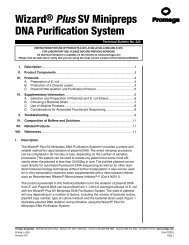
63convenient way to recover the plasmid from the bacteria.Remember, in the transformation experiment,you introduced plasmid molecules into bacterial cells.Each cell that took up a plasmid became a colony containingmillions <strong>of</strong> cells after overnight growth. Sinceeach <strong>of</strong> these cells has between ten <strong>and</strong> one hundredplasmid molecules, large amounts <strong>of</strong> plasmid (<strong>and</strong>insert) can be prepared quickly.During the second week <strong>of</strong> this lab exercise youwill grow bacterial cells containing your plasmid,purify the plasmid DNA <strong>and</strong> visualize it using anagarose gel. Plasmid purification involves a number<strong>of</strong> steps, as described below, but the principle is verysimple. Cells are collected, broken open, <strong>and</strong> plasmidDNA is separated from all <strong>of</strong> the other moleculesin the cell. This procedure is outlined in Figure 3.Figure 4 on the following page shows an illustratedversion <strong>of</strong> the steps outlined in Figure 3.The day before your lab period, someone from yourgroup should come to the laboratory at the scheduledtime. Make sure you have prepared for this meeting- you should know all the details about what you aredoing, <strong>and</strong> why. Your instructor will have prepareda number <strong>of</strong> culture tubes containing sterile liquidmedium that contains ampicillin. Working sterilely,inoculate a culture tube with a single bacterial colony.Simply touch the inoculating loop first to the colonythen to the liquid in the tube. It is not necessary tomove the whole colony, or even a visible clump <strong>of</strong>cells. Place the tube in the shaking water bath asdirected, <strong>and</strong> allow to shake overnight at 37° C.At the beginning <strong>of</strong> your lab period, each groupshould take one 1.5 ml microtube, label it with theirgroup number, <strong>and</strong> use a pipette to transfer 1.5 ml <strong>of</strong>the bacterial culture into the tube. Then, place thetube in the microcentrifuge, <strong>and</strong> your instructor willspin the tubes so that the cells form a pellet on thebottom <strong>of</strong> the tube. Each group should then recovertheir tube, pour <strong>of</strong>f the medium without disturbing thepellet, <strong>and</strong> add another 1.5 ml <strong>of</strong> the same bacterialculture to the tube containing the pellet. After anothercentrifugation <strong>and</strong> complete removal <strong>of</strong> the medium(also called the supernatant) the tube will contain onlythe cells from 3 ml <strong>of</strong> bacterial culture.Next, we will add 250 µl <strong>of</strong> cell resuspension solutionto the pellet. Use the vortex machine to getall <strong>of</strong> the cells back into suspension. When you areready, add 250 µl <strong>of</strong> cell lysis solution <strong>and</strong> mix byinverting the tube four to five times. This solution hasa very high pH <strong>and</strong> contains sodium dodecyl sulfate, astrongly amphipathic detergent that dissolved the cellmembrane, releasing the contents. When you add thisreagent, you should see the solution clear somewhat<strong>and</strong> become very viscous. The viscosity is caused bythe bacterial chromosome, which we will precipitate<strong>and</strong> discard shortly. Treat the contents <strong>of</strong> the tubegently after lysis - DO NOT VORTEX. After yourlysate has cleared, add <strong>10</strong> µl <strong>of</strong> alkaline protease solutionto digest cellular protein, <strong>and</strong> incubate for fiveminutes at room temperature.Figure 3. Outline <strong>of</strong> Plasmid Purification.Add 350µl <strong>of</strong> neutralization solution to the lysate,<strong>and</strong> mix gently (do not vortex). This lowers the pH<strong>and</strong> most lipids precipitate (come out <strong>of</strong> solution).
64Since the bacterial chromosome is attached to themembrane, it also precipitates in this step. Yourinstructor will centrifuge the tube for ten minutes,<strong>and</strong> this will again result in a pellet <strong>and</strong> a supernatant.This time it is the supernatant that contains theplasmid DNA. Move to the vacuum manifold (Figure4 on the next page), obtain <strong>and</strong> label a minicolumnwith your group number, install the minicolumn ontothe vacuum manifold as directed, <strong>and</strong> carefully pouryour cleared lysate into the column. The column containsresin that binds plasmid DNA. You will attachyour plasmid DNA to the column <strong>and</strong> then wash awayother impurities.Apply vacuum using the stopcock on the vacuumstem so that your lysate is drawn through the column.When all the liquid has been drawn through, release thevacuum. Then add 750 µl <strong>of</strong> column wash solution,apply vacuum to draw it over the column, <strong>and</strong> releasethe vacuum. Repeat the wash step, using only 250 µl<strong>of</strong> column wash solution for the second wash. Whenall <strong>of</strong> the wash solution has been removed from thecolumn, release the vacuum, <strong>and</strong> transfer the columnto a spin collection tube. Your instructor will spin thecolumn briefly in the microcentrifuge to remove anyremaining liquid. Then, transfer the column to a new1.5 ml microtube (labeled with your group number),add <strong>10</strong>0 µl <strong>of</strong> water to the column, <strong>and</strong> allow yourinstructor to centrifuge the water through the column.Since the DNA does not bind to the column resin inthe presence <strong>of</strong> pure water, it is released, <strong>and</strong> travelswith the water into the centrifuge tube. You may nowdiscard the column.Figure 4. Wizard SV Illustrated Protocol. Reproduced with permission from Promega Notes 59:<strong>10</strong> (1996).
65Section E - Gel ElectrophoresisCAUTION: UV light <strong>and</strong> ethidium bromide areboth hazardous. Listen carefully when your instructorexplains how to minimize your exposure.A glance at the tube with your plasmid shouldconvince you that a small volume <strong>of</strong> water containingplasmid DNA is not easy to distinguish from any othersmall volume <strong>of</strong> water. We need a way to visualizethe DNA. To determine whether you have correctlypurified your plasmid, combine <strong>10</strong> µl <strong>of</strong> your plasmidsolution with 5 µl <strong>of</strong> blue loading buffer in a new500µl microtube. Be careful, this buffer will stainyour skin <strong>and</strong> clothing. Mix gently, centrifuge for 5seconds, <strong>and</strong> load all 15 µl into a well <strong>of</strong> the agarosegel your instructor has prepared. Once all groups haveloaded their DNA on the gel, an electric field will beapplied, <strong>and</strong> negatively charged DNA will movetoward the positive electrode. The gel matrix retardsthis movement, with larger DNA molecules retardedmore than small molecules. The result is separation<strong>of</strong> DNA molecules by size (Figure 5). Your instructorwill include on the gel a sample <strong>of</strong> the correct plasmid,<strong>and</strong> yours should run at the same size.DNA will be visualized using a UV transilluminator.The gel matrix prepared by your instructor containeda compound called ethidium bromide. Thismolecule binds strongly to DNA, <strong>and</strong> the bound complexfluoresces orange in the presence <strong>of</strong> UV light. Wewill place the gel on a UV light source, <strong>and</strong> any DNAin the gel will begin to glow brightly. Your instructorwill post a photo <strong>of</strong> your gel on the course web site foryou to include in your lab report.Figure 5. DNA separated by size on an agarosegel. This gel contains 20 lanes. Each sample isloaded into a well near the top <strong>of</strong> the gel. An electricfield is applied, <strong>and</strong> the molecules move downwardin each lane, with small molecules movingfastest. DNA is visualized under UV light in thepresence <strong>of</strong> ethidium bromide. The far right <strong>and</strong>left lanes contain st<strong>and</strong>ards <strong>of</strong> known size.Lab AssignmentThis assignment is to be h<strong>and</strong>ed in the week <strong>of</strong> November 28, at the beginning <strong>of</strong> lab (beforeyour quiz). Each student must complete this assignment individually.Write a brief introduction <strong>and</strong> a methods section explaining what you did in this experiment. Make surethese sections cover transformation, selection, plasmid purification <strong>and</strong> gel electrophoresis. Then, write a resultssection in which you report the number <strong>of</strong> cells per ml in the original tube <strong>of</strong> competent cells (the tube labeled“Cells”). Use this number to determine how many cells you used for the transformation. Calculate how many <strong>of</strong>these cells were transformed. Why must you use data from other lab groups in your section to get an idea <strong>of</strong> howwell your experiment worked? Your lab report will not be complete without an analysis <strong>of</strong> your data compared toclass results. Return to section D <strong>of</strong> the enzyme lab exercise (pages 45-46). How can you use the class mean <strong>and</strong>st<strong>and</strong>ard deviation to tell whether your results make sense? Include in your results section a printed photo <strong>of</strong> youragarose gel showing that you recovered the same plasmid you received from the instructor in the first lab period.All text material for this assignment must be submitted to www.turnitin.com.