Essential Revision Notes for MRCP Third Edition - PasTest
Essential Revision Notes for MRCP Third Edition - PasTest
Essential Revision Notes for MRCP Third Edition - PasTest
- No tags were found...

You also want an ePaper? Increase the reach of your titles
YUMPU automatically turns print PDFs into web optimized ePapers that Google loves.
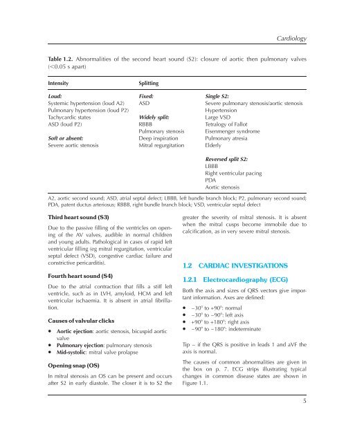
CardiologyTable 1.2. Abnormalities of the second heart sound (S2): closure of aortic then pulmonary valves(,0.05 s apart)IntensitySplittingLoud: Fixed: Single S2:Systemic hypertension (loud A2)Pulmonary hypertension (loud P2)ASDSevere pulmonary stenosis/aortic stenosisHypertensionTachycardic states Widely split: Large VSDASD (loud P2) RBBB Tetralogy of FallotPulmonary stenosis Eisenmenger syndromeSoft or absent: Deep inspiration Pulmonary atresiaSevere aortic stenosis Mitral regurgitation ElderlyReversed split S2:LBBBRight ventricular pacingPDAAortic stenosisA2, aortic second sound; ASD, atrial septal defect; LBBB, left bundle branch block; P2, pulmonary second sound;PDA, patent ductus arteriosus; RBBB, right bundle branch block; VSD, ventricular septal defect<strong>Third</strong> heart sound (S3)Due to the passive filling of the ventricles on openingof the AV valves, audible in normal childrenand young adults. Pathological in cases of rapid leftventricular filling (eg mitral regurgitation, ventricularseptal defect (VSD), congestive cardiac failure andconstrictive pericarditis).Fourth heart sound (S4)Due to the atrial contraction that fills a stiff leftventricle, such as in LVH, amyloid, HCM and leftventricular ischaemia. It is absent in atrial fibrillation.Causes of valvular clicks• Aortic ejection: aortic stenosis, bicuspid aorticvalve• Pulmonary ejection: pulmonary stenosis• Mid-systolic: mitral valve prolapseOpening snap (OS)In mitral stenosis an OS can be present and occursafter S2 in early diastole. The closer it is to S2 thegreater the severity of mitral stenosis. It is absentwhen the mitral cusps become immobile due tocalcification, as in very severe mitral stenosis.1.2 CARDIAC INVESTIGATIONS1.2.1 Electrocardiography (ECG)Both the axis and sizes of QRS vectors give importantin<strong>for</strong>mation. Axes are defined:• –308 to +908: normal• –308 to –908: left axis• +908 to +1808: right axis• –908 to –1808: indeterminateTip – if the QRS is positive in leads 1 and aVF theaxis is normal.The causes of common abnormalities are given inthe box on p. 7. ECG strips illustrating typicalchanges in common disease states are shown inFigure 1.1.5