THE ART OF DENTISTRY - School of Dental Medicine - Case ...
THE ART OF DENTISTRY - School of Dental Medicine - Case ...
THE ART OF DENTISTRY - School of Dental Medicine - Case ...

You also want an ePaper? Increase the reach of your titles
YUMPU automatically turns print PDFs into web optimized ePapers that Google loves.
2<br />
<strong>THE</strong> SCHOOL<br />
<strong>OF</strong> DENTAL<br />
MEDICINE TODAY<br />
CBCT <strong>OF</strong>FERS REAL-TIME, REAL-WORLD 3D IMAGES<br />
BY MARGARET MULLIGAN<br />
The Crani<strong>of</strong>acial Imaging Center (CIC)<br />
in the <strong>School</strong> <strong>of</strong> <strong>Dental</strong> <strong>Medicine</strong><br />
(http://ImagingCenter.case.edu) is using<br />
cone-beam computed tomography<br />
(CBCT), which allows practitioners to<br />
study a patient’s 3-dimensional (3D)<br />
image. The technology and its use is the<br />
primary focus <strong>of</strong> the work <strong>of</strong> J. Martin<br />
Palomo, D.D.S., M.S.D. ’97, Associate<br />
Pr<strong>of</strong>essor, Department <strong>of</strong> Orthodontics.<br />
Dr. Palomo is one <strong>of</strong> the few people who<br />
explored computer applications in the<br />
days when they were less than user-friendly.<br />
“I was a computer kid,” he says. “As a<br />
child I watched as technology made<br />
information easier to attain and process<br />
for many different uses. Later, I asked<br />
myself, ‘How could I use this in my specialty<br />
<strong>of</strong> orthodontics? How can technology<br />
make clinical work more efficient?’ I have<br />
worked since 1996 creating and using 3D<br />
images <strong>of</strong> the head and neck. In the past,<br />
the use <strong>of</strong> 3D images was not practical or<br />
user-friendly, which prevented broader<br />
use <strong>of</strong> such images. The development <strong>of</strong><br />
CBCT solved many problems not only<br />
for dental medicine, but also for other<br />
clinicians using head and neck images.<br />
This allowed me to merge my interest in<br />
computers with my work in orthodontia.<br />
With the support <strong>of</strong> my department and<br />
the <strong>School</strong> <strong>of</strong> <strong>Dental</strong> <strong>Medicine</strong>, we<br />
jumped into the game early.<br />
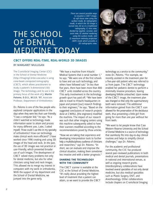
There are several possible ways<br />
to visualize a CBCT image.<br />
A) s<strong>of</strong>t tissue view using the<br />
surface mode, B) radiographic<br />
mode, in which the 3D image is<br />
turned into 2D as in a traditional<br />
radiography, C) slice view,<br />
divided by sagittal, coronal, and<br />
axial view, D) skeletal rendering<br />
using the surface mode, and<br />
E) maximum intensity projection<br />
(MIP) mode which provides a<br />
3D radiographic view.<br />
“We have a machine from Hitachi<br />
Medical Systems that is serial number 1,”<br />
he says. “We were one <strong>of</strong> the first schools<br />
to have and use such technology, and<br />
others have followed suit.” In less than<br />
five years, there have been more than 800<br />
CBCT units installed across the country.<br />
This early involvement in the technology<br />
growth spurt has paid <strong>of</strong>f. “We have been<br />
able to travel to Hitachi headquarters in<br />
Japan and present [our] research findings<br />
to their engineers,” he says. “Based on the<br />
suggested conclusions <strong>of</strong> research projects<br />
done at CWRU, [the engineers] modified<br />
the machine. The impact <strong>of</strong> our research<br />
was such that other imaging centers using<br />
this machine subsequently asked to have<br />
their scanners modified according to the<br />
recommendations posed by those articles.<br />
“Now we are taking that experience and<br />
developing interpretation tools to further<br />
help a multidisciplinary audience <strong>of</strong> clinicians<br />
and researchers,” says Dr. Palomo. “In<br />
short, we can evaluate and improve the<br />
clinical situation, making them sometimes<br />
less invasive and with a better prognosis.”<br />
SHARING <strong>THE</strong> TECHNOLOGY<br />
WITH <strong>THE</strong> COMMUNITY<br />
The CBCT scanner is available at the<br />
CIC at the <strong>School</strong> <strong>of</strong> <strong>Dental</strong> <strong>Medicine</strong>.<br />
“It’s really about providing the highest<br />
level <strong>of</strong> treatment that a patient can<br />
attain, so we want to provide the CBCT<br />
technology as a service to the community,”<br />
notes Dr. Palomo. “For example, we<br />
recently assisted in the treatment plan for<br />
a five-year-old patient who was referred to<br />
us from Japan. The CBCT technology<br />
enabled her pediatric dentist to perform a<br />
minimally invasive procedure, leaving<br />
developing follicles untouched. Upon review<br />
<strong>of</strong> the CBCT image, the treatment plan<br />
was changed so that only the supernumerary<br />
teeth were removed. The additional<br />
information gained from the CBCT scan<br />
allowed for the preservation <strong>of</strong> the deciduous<br />
teeth, thus preventing a little girl from<br />
going for more than one year without her<br />
front teeth.<br />
“We want to let people know that <strong>Case</strong><br />
Western Reserve University and the <strong>School</strong><br />
<strong>of</strong> <strong>Dental</strong> <strong>Medicine</strong> is a source <strong>of</strong> technology<br />
that seamlessly fits into day-to-day clinical<br />
routines and helps to simplify clinical<br />
challenges,” says Dr. Palomo.<br />
For the academic and pr<strong>of</strong>essional<br />
community, the CIC has produced<br />
several peer-reviewed publications in both<br />
medical and dental journals, presentations<br />
in national and international venues, as<br />
well as ongoing research grants.<br />
Interdisciplinary collaborations have<br />
involved several specialties <strong>of</strong> not only dental<br />
medicine, but also medical specialties<br />
such as Plastic Surgery, ENT, and<br />
Anesthesiology. Other collaborations<br />
include chapters on Crani<strong>of</strong>acial Imaging