


Value of ECG-gated Thallium-201 Dipyridamole SPECT in ...
Value of ECG-gated Thallium-201 Dipyridamole SPECT in ...
Value of ECG-gated Thallium-201 Dipyridamole SPECT in ...
- No tags were found...

Create successful ePaper yourself
Turn your PDF publications into a flip-book with our unique Google optimized e-Paper software.
Orig<strong>in</strong>al Article<strong>ECG</strong>-<strong>gated</strong> <strong>SPECT</strong> and <strong>Thallium</strong>-<strong>201</strong> Myocardial PerfusionActa Cardiol S<strong>in</strong> 2006;22:2430Nuclear Medic<strong>in</strong>e<strong>Value</strong> <strong>of</strong> <strong>ECG</strong>-<strong>gated</strong> <strong>Thallium</strong>-<strong>201</strong> <strong>Dipyridamole</strong><strong>SPECT</strong> <strong>in</strong> Borderl<strong>in</strong>e Cases <strong>of</strong> MyocardialPerfusion ScanYueh-Chung Chen, 1 Kun-Chou Ch<strong>in</strong> 2 and J<strong>in</strong>g Tang Ko 3Background: <strong>Thallium</strong>-<strong>201</strong> myocardial perfusion scan is a commonly utilized method for detection <strong>of</strong> coronaryartery disease. Although this technique is generally reliable, results can be <strong>in</strong>conclusive <strong>in</strong> some cases. The purpose<strong>of</strong> this study was to assess whether candidates for coronary angiogram could be better identified with the addition <strong>of</strong>an electrocardiographic (<strong>ECG</strong>)-<strong>gated</strong> myocardial perfusion scan to a thallium scan.Method: A total <strong>of</strong> 512 patients with suspected coronary artery disease were <strong>in</strong>cluded <strong>in</strong> this study and received athallium scan. In 69 patients, the results were borderl<strong>in</strong>e, and 32 <strong>of</strong> these 69 patients also received an <strong>ECG</strong>-<strong>gated</strong>scan and underwent coronary angiogram with<strong>in</strong> one month.Results: We found that 21 <strong>of</strong> the 32 patients who underwent coronary angiogram had significant coronary arterydisease. However, when an <strong>ECG</strong>-<strong>gated</strong> perfusion scan was also performed, 25 <strong>of</strong> the 32 patients had abnormalregional wall motion follow<strong>in</strong>g the stress test. Twenty-one <strong>of</strong> these 25 patients were found to have significantcoronary artery disease. Statistical analysis revealed that the <strong>ECG</strong>-<strong>gated</strong> perfusion scan <strong>in</strong> comb<strong>in</strong>ation with thethallium scan was significantly more accurate than the thallium scan alone <strong>in</strong> borderl<strong>in</strong>e cases <strong>of</strong> myocardial perfusionscan (p = 0.0166), with 100% sensitivity, 63.6% specificity, an 84% positive predictive rate, and a 100% negativepredictive rate.Conclusions: The <strong>ECG</strong>-<strong>gated</strong> perfusion scan <strong>in</strong> comb<strong>in</strong>ation with a thallium scan was more accurate <strong>in</strong> predict<strong>in</strong>gthe presence <strong>of</strong> coronary artery disease than a thallium scan alone <strong>in</strong> borderl<strong>in</strong>e cases <strong>of</strong> myocardial perfusion scan.Key Words:Gated myocardial <strong>SPECT</strong> <strong>Thallium</strong>-<strong>201</strong> Myocardial perfusion scan Stunn<strong>in</strong>g Coronary artery diseaseINTRODUCTION<strong>Thallium</strong>-<strong>201</strong> myocardial perfusion s<strong>in</strong>gle-photonemission computed tomography (<strong>SPECT</strong>) is a commonlyReceived: November 29, 2005 Accepted: February 10, 20061Division <strong>of</strong> Cardiology, Department <strong>of</strong> Internal Medic<strong>in</strong>e, Taipei CityHospital, Ren-Ai Branch, 2 Department <strong>of</strong> Nuclear Medic<strong>in</strong>e, M<strong>in</strong>-ShengGeneral Hospital, and 3 Division <strong>of</strong> Cardiology, De- partment <strong>of</strong> InternalMedic<strong>in</strong>e,Taipei City Hospital, Zhong-Xiao Branch.Address correspondence and repr<strong>in</strong>t requests to: Dr. Yueh-ChungChen, Division <strong>of</strong> Cardiology, Department <strong>of</strong> Internal Medic<strong>in</strong>e,Taipei City Hospital, Ren-Ai Branch, No. 10, Section 4, Ren-AiRoad, Taipei 106, Taiwan. Tel: 886-2-2709-3600 ext. 3741; Fax:886-2-2704-9399; E-mail: chenyuehchung.tw@yahoo.com.twutilized method to detect coronary artery disease. 1 Althoughthis technique is generally reliable, <strong>in</strong>conclusiveresults can be obta<strong>in</strong>ed when try<strong>in</strong>g to detect perfusiondefects <strong>in</strong>volv<strong>in</strong>g a small territory, 2 reverse redistribution,3 or fixed defects caused by an attenuation effect 4,5(e.g. a diaphragm shadow <strong>in</strong> males or a breast shadow <strong>in</strong>females). The electrocardiographic (<strong>ECG</strong>) <strong>gated</strong> myocardialperfusion scan is utilized for assessment <strong>of</strong> regionaland global motion <strong>of</strong> the left ventricular wall. This testmay also be useful <strong>in</strong> differentiat<strong>in</strong>g attenuation artifactsfrom myocardial scars.The purpose <strong>of</strong> this study was to assess the efficacy<strong>of</strong> perform<strong>in</strong>g an <strong>ECG</strong>-<strong>gated</strong> perfusion scan <strong>in</strong> comb<strong>in</strong>ationwith a thallium scan. We hypothesized that theActa Cardiol S<strong>in</strong> 2006;22:2430 24
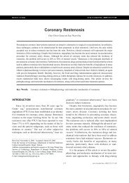
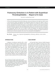
<strong>ECG</strong>-<strong>gated</strong> <strong>SPECT</strong> and <strong>Thallium</strong>-<strong>201</strong> Myocardial Perfusioncomb<strong>in</strong>ation <strong>of</strong> the two tests could help to clarify <strong>in</strong>conclusiveresults from a thallium scan, 6,7 and be useful <strong>in</strong>determ<strong>in</strong><strong>in</strong>g if an <strong>in</strong>vasive coronary angiogram wasneeded.METHODSPatient PopulationFrom January to November <strong>of</strong> 2004, 512 patients <strong>in</strong>Ren-Ai Hospital with suspected coronary artery diseasewere <strong>in</strong>cluded <strong>in</strong> the study and underwent stress thallium-<strong>201</strong>myocardial perfusion <strong>SPECT</strong>. Exclusion criteria<strong>in</strong>cluded a history <strong>of</strong> documented myocardial <strong>in</strong>farction,coronary bypass surgery, or percutaneous coronary <strong>in</strong>tervention.A positive result was def<strong>in</strong>ed as a perfusiondefect <strong>in</strong> an area larger than 6% <strong>of</strong> the total left ventriclewith the exception <strong>of</strong> the <strong>in</strong>ferior wall <strong>in</strong> male patients,the anterior wall <strong>in</strong> female patients, or areas with uptakereduced by 65%. A negative result was def<strong>in</strong>ed as nopre- or post-stress perfusion defect. Borderl<strong>in</strong>e resultswere def<strong>in</strong>ed as areas <strong>of</strong> reversible redistribution smallerthan 6% <strong>of</strong> the total left ventricle, or areas <strong>of</strong> the <strong>in</strong>feriorwall <strong>in</strong> male patients and anterior wall <strong>in</strong> female patientswith 65% to 75% reduced uptake. Accord<strong>in</strong>g to thesedef<strong>in</strong>itions, 191 patients had a positive result, 252 patientshad a negative result, and 69 patients had a borderl<strong>in</strong>eresult. All borderl<strong>in</strong>e case received <strong>ECG</strong>-<strong>gated</strong> scansimultaneously. Of the 69 cases with a borderl<strong>in</strong>e result,32 patients received a coronary angiogram with<strong>in</strong> onemonth <strong>of</strong> the scan. Of the 32 cases received angiogram,there were 44% women and 56% men with a mean bodyweight <strong>of</strong> 64 16 kilogram and a mean age <strong>of</strong> 61 14years. Of the 69 cases with a borderl<strong>in</strong>e result, therewere 41% women and 59% men with a mean bodyweight <strong>of</strong> 62 14 kilogram and a mean age <strong>of</strong> 62 15years.We selected 32 patients who underwent a coronaryangiogram for further evaluation <strong>in</strong> order to assess if use<strong>of</strong> an <strong>ECG</strong>-<strong>gated</strong> scan was useful <strong>in</strong> clarify<strong>in</strong>g <strong>in</strong>conclusiveresults <strong>of</strong> a thallium scan and identify<strong>in</strong>g patientsfor an <strong>in</strong>vasive coronary angiogram <strong>in</strong> cl<strong>in</strong>ical practice(Figure 1).ProcedureAll patients were scanned <strong>in</strong> the sup<strong>in</strong>e position.Figure 1. Algorithm <strong>of</strong> the exam<strong>in</strong>ation procedure.25 Acta Cardiol S<strong>in</strong> 2006;22:2430
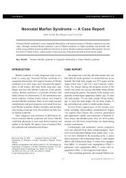
Yueh-Chung Chen et al.<strong>Dipyridamole</strong> 0.56 mg/kg was <strong>in</strong>travenously <strong>in</strong>jected,and patients were <strong>in</strong>structed to raise each leg or do ahandgrip maneuver as the adjuvant stress exercise. Thiswas followed by <strong>in</strong>jection <strong>of</strong> 2.5-3.0 mCi <strong>of</strong> thallium-<strong>201</strong> followed by <strong>SPECT</strong> imag<strong>in</strong>g. Imag<strong>in</strong>g at rest wasconducted after 3-4 hours.The sc<strong>in</strong>tigraphic imag<strong>in</strong>es were acquired with anADAC dual-head <strong>SPECT</strong> camera (ADAC Laboratories,Milpitas,CA) equipped with a low-energy, high-resolusioncollimator. Thirty-two projections (40 secondsperprojection) were obta<strong>in</strong>ed over a 180 semicircular arc(45 right anterior oblique to 45 left posterior oblique).Image resolution was 64 64 pixels. Filtered back projectionwas performed by use <strong>of</strong> a low-pass Butterworthfilter with a frequency cut<strong>of</strong>f <strong>of</strong> 4 and an order <strong>of</strong> 5.An <strong>ECG</strong> R wave gat<strong>in</strong>g detector was used to acquire8 frames <strong>of</strong> each cardiac cycle. No attenuation or scattercorrection was used. <strong>ECG</strong>-<strong>gated</strong> <strong>SPECT</strong> images were visuallyscored for motion us<strong>in</strong>g a 20-segment model <strong>of</strong>the left ventricle (Figure 2). Five representative slices (3distal, middle and basal short-axis slices and 1 verticaland 1 horizontal mid-ventricular long axis slice) wereautomatically selected for this purpose.All images were processed and <strong>in</strong>terpreted by thesame experienced technologist. All stress and rest imageswere displayed simultaneously on an ADAC Pegasysworkstation (ADAC Laboratories) through use <strong>of</strong>the commercially available Autoquant QGS (QuantitativeGated <strong>SPECT</strong>) s<strong>of</strong>tware program. 8,21 Images wereanalyzed us<strong>in</strong>g a l<strong>in</strong>ear color scale, with each color transitionrepresent<strong>in</strong>g a 10% difference <strong>in</strong> uptake activity.Regional wall motion <strong>in</strong> the left ventricle was scored us<strong>in</strong>ga 20-segment model follow<strong>in</strong>g the stress test.Statistical AnalysisDifferences <strong>in</strong> data among two protocols were testedus<strong>in</strong>g analysis <strong>of</strong> standard deviation. The hypothesis testabout the difference between two population propositionswas used to evaluate the significance <strong>of</strong> successfulexam<strong>in</strong>ations, us<strong>in</strong>g thallium scan alone or with the addition<strong>of</strong> the <strong>ECG</strong>-<strong>gated</strong> scan for detection <strong>of</strong> coronaryartery disease. The level for statistical significance waspredeterm<strong>in</strong>ed at p < 0.05. All comparisons were onetailed.RESULTSWe identified 69 patients with a borderl<strong>in</strong>e result onthe thallium scan, and 32 <strong>of</strong> these patients underwent acoronary angiogram with<strong>in</strong> one month follow<strong>in</strong>g thescan. Of the patients undergo<strong>in</strong>g a coronary angiogram,21 <strong>of</strong> 32 patient exhibited significant coronary artery disease(> 50% stenosis <strong>of</strong> one or more vessels) and therema<strong>in</strong><strong>in</strong>g 11 patients showed normal or <strong>in</strong>significantcoronary artery disease. However, if <strong>ECG</strong>-<strong>gated</strong> perfusionscan was used simultaneously, 25 <strong>of</strong> the 32 patientsexhibited abnormal regional wall motion follow<strong>in</strong>g thestress test. Furthermore, 21 <strong>of</strong> these 25 patients exhibitedsignificant coronary artery disease, with the rema<strong>in</strong><strong>in</strong>g 4patients exhibit<strong>in</strong>g no coronary artery disease. F<strong>in</strong>ally, 7<strong>of</strong> the 32 patients with normal regional wall motion follow<strong>in</strong>gthe stress test exhibited normal or <strong>in</strong>significantcoronary artery disease.Statistical analysis revealed that the <strong>ECG</strong>-<strong>gated</strong> perfusionscan <strong>in</strong> comb<strong>in</strong>ation with the thallium scan wassignificantly more accurate than the thallium scan alone<strong>in</strong> borderl<strong>in</strong>e cases <strong>of</strong> myocardial perfusion scan (p =0.0166), with 100% sensitivity, 63.6% specificity, an84% positive predictive rate, and a 100% negative predictiverate. Patient data and characteristics are presented<strong>in</strong> Table 1.DISCUSSIONFigure 2.ventricle.Schematic representation <strong>of</strong> the 20-segment model <strong>of</strong> the leftThe post-stress perfusion defects caused by myocardialischemia are similar to the so-called phenomena <strong>of</strong>transient ischemia stunn<strong>in</strong>g (TIS), <strong>in</strong>dicat<strong>in</strong>g that thedefects may be both transient and reversible. 9 The technique<strong>of</strong> technetium-99m <strong>SPECT</strong> <strong>of</strong>fers a superior imagequality compared to thallium-<strong>201</strong> <strong>SPECT</strong> due to a higherActa Cardiol S<strong>in</strong> 2006;22:2430 26
Yueh-Chung Chen et al.other hand, is better suited for TIS detection 12 but has ahigher rate <strong>of</strong> borderl<strong>in</strong>e results. 13,14 Reverse redistribution<strong>in</strong> a rest<strong>in</strong>g thallium scan <strong>in</strong> patients with coronaryartery disease also has a close relationship with coronaryanatomy and ventricular function. 15 However, the anatomiccorrelation <strong>in</strong> our study seemed not very identical.We believed this is not uncommon <strong>in</strong> cl<strong>in</strong>ical practice.The addition <strong>of</strong> an <strong>ECG</strong>-<strong>gated</strong> scan to technetium-99m scan can clarify the uncerta<strong>in</strong>ty <strong>of</strong> borderl<strong>in</strong>e resultsand <strong>in</strong>crease the success rate <strong>of</strong> diagnos<strong>in</strong>g coronary arterydisease. 16 Gated <strong>SPECT</strong> imag<strong>in</strong>g is also <strong>of</strong> value <strong>in</strong>recogniz<strong>in</strong>g questionable perfusion defects, 4 <strong>in</strong>clud<strong>in</strong>gattenuation artifacts, which are ubiquitous, particularlywhen persistent defects raise the question <strong>of</strong> myocardialscar versus defect. The presence <strong>of</strong> correspond<strong>in</strong>g normalor abnormal wall motion before and after stressimag<strong>in</strong>g with an <strong>ECG</strong>-<strong>gated</strong> scan may resolve this problem,and may be even more accurate than the treadmillexercise test (The change <strong>of</strong> regional wall motion observedby echocardiography pre- and post-stress may bemore accurate than treadmill exercise test 22 for the diagnosis<strong>of</strong> coronary artery disease). The complementary<strong>in</strong>formation can also be useful to evaluate the severity <strong>of</strong>TIS and the extent <strong>of</strong> myocardial viability. Furthermore,the ejection fraction <strong>of</strong> the left ventricle can be accuratelyassessed by <strong>gated</strong> <strong>SPECT</strong> with either technetium-99m or thallium scan <strong>in</strong> properly selected patients (e.g.patients who are not overweight) with normal or depressedleft ventricular function. 17,18 Assessment <strong>of</strong> globalfunction is useful for determ<strong>in</strong>g prognosis and <strong>in</strong> differentiat<strong>in</strong>gischemic from non-ischemic cardiomyopathy. 19In the current study, we evaluated the efficacy <strong>of</strong>coronary artery disease detection by us<strong>in</strong>g the same QGS(Quantitative Gated <strong>SPECT</strong>) method <strong>in</strong> comb<strong>in</strong>ationwith a thallium scan. Based on the f<strong>in</strong>d<strong>in</strong>gs <strong>of</strong> the coronaryangiograms, more than half <strong>of</strong> the borderl<strong>in</strong>e casesidentified by thallium scan revealed significant coronaryartery disease, <strong>in</strong>dicat<strong>in</strong>g the value <strong>of</strong> follow<strong>in</strong>g the borderl<strong>in</strong>ecases with a coronary angiogram. However, ourstudy showed, if QGS (e.g. <strong>ECG</strong>-<strong>gated</strong> scan) was added<strong>in</strong> these borderl<strong>in</strong>e cases, the coronary artery disease detectionrate by coronary angiogram <strong>in</strong>creased. Thesef<strong>in</strong>d<strong>in</strong>gs suggest that borderl<strong>in</strong>e patients with abnormalpresentation follow<strong>in</strong>g QGS should receive an angiogram.There rema<strong>in</strong>s a number <strong>of</strong> issues that require furtherclarification, <strong>in</strong>clud<strong>in</strong>g the diagnostic value <strong>of</strong> QGSwhen the thallium scan is positive or when the thalliumscan is negative. We believe that <strong>ECG</strong>-<strong>gated</strong> <strong>SPECT</strong>also improves diagnosis <strong>in</strong> those populations, and thatwill be the subject <strong>of</strong> further <strong>in</strong>vestigation.F<strong>in</strong>ally, we should note that our study has some limitation.M<strong>in</strong>imal bias may still exist <strong>in</strong> the <strong>in</strong>terpretationeven though we used the same experienced technologist.Besides, there was no absolute standard for determ<strong>in</strong><strong>in</strong>gthe contribution <strong>of</strong> collateral flow to regions with an anatomicobstruction. Furthermore, these catheterized patientswere not randomly selected, and the total number<strong>of</strong> cases exam<strong>in</strong>ed was relatively small.CONCLUSIONThe addition <strong>of</strong> an <strong>ECG</strong>-<strong>gated</strong> perfusion scan mayreduce the uncerta<strong>in</strong>ty <strong>in</strong> <strong>in</strong>terpret<strong>in</strong>g results <strong>of</strong> a thalliumscan alone, especially with respect to <strong>in</strong>terpretation<strong>of</strong> borderl<strong>in</strong>e results. Measur<strong>in</strong>g rest<strong>in</strong>g and post-stressleft ventricular ejection fraction and changes <strong>in</strong> regionalwall motion, 7,20 can make it possible to detect coronaryartery disease more precisely <strong>in</strong> cl<strong>in</strong>ical practice.REFERENCES1. Chen ML, Chao IM, Chen CH. Diagnostic accuracy and safety <strong>of</strong>dipyridamole thallium-<strong>201</strong> s<strong>in</strong>gle photon emission computedtomography <strong>in</strong> coronary artery disease. Acta Cardiol S<strong>in</strong> 1996;12:126-133.2. Van Tra<strong>in</strong> KF, Maddahi J, Berman DS, et al. Quantitative analysis<strong>of</strong> tomographic stress thallium-<strong>201</strong> myocardial sc<strong>in</strong>tigraphy: amulticenter trial. J Nucl Med 1990;31:1168-1179.3. Popma JJ, Smitherman TC, Walker BS, et al. Reverse redistribution<strong>of</strong> thallium-<strong>201</strong> detected by <strong>SPECT</strong> imag<strong>in</strong>g afterdypiridamole <strong>in</strong> ang<strong>in</strong>a pectoris. Am J Cardiol 1990;65:1176-1180.4. DePuey EG, Rozanski A. Us<strong>in</strong>g <strong>gated</strong> technetium-99m sestamibi<strong>SPECT</strong> to characterize fixed myocardial defects as <strong>in</strong>farct orartifact. J Nucl Med 1995;36;952-955.5. Wacker FJ, Bodenheimer M, Fleiss JL, Brown M. Factorsaffect<strong>in</strong>g uniformity <strong>in</strong> <strong>in</strong>terpretation <strong>of</strong> planar thallium-<strong>201</strong>imag<strong>in</strong>g <strong>in</strong> a multicenter trial. J Am Coll Cardiol 1993;21:1064-1074.6. Maddahi J, Van Tra<strong>in</strong> K, Prigent F, et al. Quantitative s<strong>in</strong>glephoton emission computed thallium-<strong>201</strong> tomography for detectionand localized <strong>of</strong> coronary artery disease: optimization andprospective validation <strong>of</strong> a new technique. J Am Coll CardiolActa Cardiol S<strong>in</strong> 2006;22:2430 28
<strong>ECG</strong>-<strong>gated</strong> <strong>SPECT</strong> and <strong>Thallium</strong>-<strong>201</strong> Myocardial Perfusion1989;14:1689-1699.7. Germano G, Kavanagh PB, Waechter PB, et al. A new algorithmfor the quantitation <strong>of</strong> myocardial perfusion <strong>SPECT</strong>. I. Theoreticalaspects. J Nucl Med. 2000;41:712-719.8. Achter AD, K<strong>in</strong>g MA, Dahlberg ST, Hendrick P, LaCroix KJ,Tsui BMW. An <strong>in</strong>vestigation <strong>of</strong> the estimation <strong>of</strong> ejectionfractions and cardiac volumes by quantitative <strong>gated</strong> <strong>SPECT</strong>s<strong>of</strong>tware package <strong>in</strong> simulated <strong>gated</strong> <strong>SPECT</strong> images. J NuclCardiol 1998;5:144-152.9. He ZX, Cwaig E, Preslar JS, et al. Accuracy <strong>of</strong> left ventricularejection fraction determ<strong>in</strong>ed by <strong>gated</strong> myocardial perfusion<strong>SPECT</strong> with Tl-<strong>201</strong> and Tc-99m sestamibi: comparison withfirst-pass radionuclide angiography. J Nucl Cardiol 1999;6:412-417.10. Manrique A, Fraggi M, Verra P, et al. Tl-<strong>201</strong> and Tc99m MIBI<strong>gated</strong> <strong>SPECT</strong> <strong>in</strong> patients with large perfusion defects and leftventricular dysfunction: comparison with equilibrium radionuclideangiography. J Nucl Med 1999;40:805-809.11. Johnson LL, Verdesca SA, Aude WY, et al. Postischemic stunn<strong>in</strong>gcan affect left ventricular ejection fraction and regional wallmotion on post-stress <strong>gated</strong> sestamibi tomograms. J Am CollCardiol 1997;30:1641-1648.12. Paul AK, Hasegawa S, Yoshioka J, Tsujimura E, Yamaguchi H,Tokita N, et al. Exercise-<strong>in</strong>duced stunn<strong>in</strong>g cont<strong>in</strong>ues for at leastone hour: evaluation with quantitative <strong>gated</strong> s<strong>in</strong>ple-photon emissiontomography. Eur J Nucl Med 1999;26:410-415.13. Hendel RC, Parker M, Wackers FJ, Ragio P, Lahiri A, Zaret BL.Reduced variability <strong>of</strong> <strong>in</strong>terpretation and improved image qualitywith a technetium-99m myocardial perfusion agent: comparison<strong>of</strong> thallium-<strong>201</strong> and technetium-99m-labelled tetr<strong>of</strong>osm<strong>in</strong>. J NuclCardiol 1994;1:509-514.14. Tailefer R, Depurey EG, Udelson JE, Beller GA, Latour Y,Reeves F. Comparative diagnostic accurancy <strong>of</strong> Tl-<strong>201</strong> and Tc-99m sestamibi <strong>SPECT</strong> imag<strong>in</strong>g (perfusion and <strong>ECG</strong>-<strong>gated</strong> <strong>SPECT</strong>)<strong>in</strong> detect<strong>in</strong>g coronary artery disease <strong>in</strong> women. J Am Coll Cardiol1997;29:69-77.15. Pace L, Cuocolo A, Maurea S, et al. Reverse redistribution <strong>in</strong>rest<strong>in</strong>g thallium-<strong>201</strong> myocardial sc<strong>in</strong>tigraphy <strong>in</strong> patients withcoronary artery disease: relation to coronary anatomy and ventricularfunction. J Nucl Med 1993;34:1688-1692.16. Sharir T, Berman DS, Waechter PB, et al. Quantitative analysis <strong>of</strong>regional motion and thicken<strong>in</strong>g by <strong>gated</strong> myocardial perfusion<strong>SPECT</strong>: normal heterogeneity and criteria for abnormality. J NuclMed 2001;42:1630-1638.17. Bolli R, Marban E. Molecular and cellular mechanisms <strong>of</strong> myocardialstunn<strong>in</strong>g. Physiol Rev 1999;79:609-634.18. Ambrosio G, Betocchi S, Pace L, et al. Prolonged impairment <strong>of</strong>regional contractile function after resolution <strong>of</strong> exercise-<strong>in</strong>ducedang<strong>in</strong>a: evidence <strong>of</strong> myocardial stunn<strong>in</strong>g <strong>in</strong> patients with coronarydisease. Circulation 1996;94:2455-2464.19. Danias PG, Ahlberg AW, Clark BA, Mess<strong>in</strong>eo F, Lev<strong>in</strong>e MG,Mcgill CC, et al. Comb<strong>in</strong>ed assessment <strong>of</strong> myocardial perfusionand left ventricular function with exercise technetium-99msestamibi <strong>gated</strong> s<strong>in</strong>gle-photon emission computed tomographycan differentiate ischemic and non-ischemic dilated cardiomyopathy.Am J Cardiol 1998;82:1253-1258.20. Vallejo E, Dione DP, Bruni WL, Constable RT, Borek PP, SoaresJP, et al. Reproducibility and ccuracy <strong>of</strong> <strong>gated</strong> <strong>SPECT</strong> for determ<strong>in</strong>ation<strong>of</strong> left ventricular volumes and ejection fraction:experimental validation us<strong>in</strong>g MRI. J Nucl Med 2000;41:874- 882.21. Germano G, Kavanagh P, Su HT, et al. Automatic reorientation <strong>of</strong>three-dimensional transaxial myocardial perfusion <strong>SPECT</strong> images.J Nucl Med 1995;36:1107-1114.22. Severi S, Picano E, Michelassi C, et al. Diagnostic and prognosticvalue <strong>of</strong> dipyridamole echocardiography <strong>in</strong> patients with suspectedcoronary artery disease. Circulation 1994;89:1160-1173.29 Acta Cardiol S<strong>in</strong> 2006;22:2430
Orig<strong>in</strong>al ArticleActa Cardiol S<strong>in</strong> 2006;22:24−30心 節 律 門 閥 鉈 壓 力 攝 影 對 傳 統 造 影 邊 緣 型 病 例 之 價 值1陳 鉞 忠2秦 公 焯3柯 景 塘1台 北 市 市 立 聯 合 醫 院 仁 愛 分 院 心 臟 內 科2桃 園 縣 敏 盛 醫 院 核 子 醫 學 科3台 北 市 市 立 聯 合 醫 院 忠 孝 分 院 心 臟 內 科背 景 鉈 -<strong>201</strong> 心 肌 灌 注 攝 影 為 一 常 用 於 冠 心 症 診 斷 之 工 具 , 然 而 常 有 一 些 情 形 , 例 如 小 區域 之 缺 氧 、 可 逆 性 再 分 佈 及 衰 減 效 應 造 成 之 灌 流 不 足 , 均 可 能 使 判 讀 發 生 困 難 。方 法 我 們 希 望 使 用 心 節 律 門 閥 鉈 壓 力 攝 影 加 入 原 來 檢 查 顯 示 邊 緣 性 結 果 之 病 例 中 , 看 是否 可 以 增 加 冠 心 症 之 正 確 診 斷 。2004 年 1 月 至 11 月 共 512 位 受 檢 病 人 中 , 有 69 位 呈 現 邊緣 性 結 果 , 其 中 32 位 接 受 了 心 導 管 檢 查 , 本 實 驗 即 取 此 32 名 病 例 做 分 析 。結 果 鉈 -<strong>201</strong> 心 肌 灌 注 攝 影 32 名 邊 緣 性 病 例 中 ,21 位 有 顯 著 冠 心 症 ,11 位 無 ; 此 32 名病 例 若 再 加 上 心 節 律 門 閥 鉈 壓 力 攝 影 檢 查 後 , 不 正 常 變 化 之 病 例 共 25 位 , 當 中 21 位 有 顯著 冠 心 症 ,4 位 無 , 而 無 不 正 常 變 化 之 7 名 病 例 中 均 無 冠 心 症 ; 換 言 之 , 本 研 究 從 邊 緣 病例 中 , 找 出 32 名 , 將 這 32 名 病 例 皆 實 施 心 導 管 手 術 , 其 中 有 11 名 將 被 誤 作 心 導 管 手 術 ,但 多 加 了 一 套 檢 驗 手 續 , 此 32 名 病 例 中 僅 4 名 可 能 被 誤 作 心 導 管 手 術 , 可 見 後 者 比 前 者 更能 降 低 誤 判 的 機 率 。 我 們 使 用 兩 母 體 比 率 差 異 之 假 設 檢 驗 來 測 試 此 一 結 果 , 顯 示 假 設 有 意義 ,p 值 = 0.0166 (p < 0.05)。結 論 心 節 律 門 閥 鉈 壓 力 攝 影 對 鉈 -<strong>201</strong> 心 肌 灌 注 攝 影 邊 緣 性 結 果 之 病 人 , 於 冠 心 症 之 診 斷上 有 極 佳 的 參 考 效 果 。關 鍵 詞 : 心 節 律 門 閥 鉈 壓 力 攝 影 、 鉈 -<strong>201</strong>、 心 肌 灌 注 攝 影 、 心 肌 昏 迷 、 冠 心 症 。30


![Full Text [Download PDF]](https://img.yumpu.com/22999153/1/190x260/full-text-download-pdf.jpg?quality=85)