Radiographic findings in several joints of nine bears
Radiographic findings in several joints of nine bears
Radiographic findings in several joints of nine bears

You also want an ePaper? Increase the reach of your titles
YUMPU automatically turns print PDFs into web optimized ePapers that Google loves.
Institut für Genetik, Ernährung und Haltung von Haustieren der Vetsuisse-Fakultät der<br />
Universität Bern, Abteilung Tierhaltung und Tierschutz<br />
(Leiter: Pr<strong>of</strong>. Dr. A. Steiger)<br />
Zoo Basel<br />
(Direktor: Dr. O. Pagan)<br />
Departement für kl<strong>in</strong>ische Veter<strong>in</strong>ärmediz<strong>in</strong>, Abteilung für bildgebende Verfahren der<br />
Vetsuisse-Fakultät der Universität Bern<br />
(Leiter: Pr<strong>of</strong>. Dr. J. Lang)<br />
Arbeit unter der Leitung von<br />
Dr. C. Wenker und Dr. U. Geissbühler<br />
Symptoms, radiographic exam<strong>in</strong>ations and pathologies:<br />
development <strong>of</strong> a scor<strong>in</strong>g system to evaluate physical condition<br />
and quality <strong>of</strong> life <strong>in</strong> geriatric zoo mammals<br />
Inaugural-Dissertation<br />
zur Erlangung der Doktorwürde<br />
der Vetsuisse-Fakultät der Universität Bern<br />
vorgelegt von<br />
Jérôme Föllmi<br />
von Genf-Stadt, GE<br />
2005
Von der Vetsuisse-Fakultät der Universität Bern auf Antrag<br />
von Pr<strong>of</strong>. Dr. A. Steiger als Dissertation genehmigt.<br />
Bern, Der Dekan der<br />
Vetsuisse-Fakultät der Universität Bern<br />
3
Table <strong>of</strong> contents<br />
Summary 7<br />
I. A scor<strong>in</strong>g system to evaluate physical condition and<br />
quality <strong>of</strong> life <strong>in</strong> geriatric zoo mammals 11<br />
Summary 11<br />
Résumé 12<br />
Introduction 12<br />
Animals, Materials and Methods 13<br />
Animals 13<br />
Symptoms 13<br />
Retrospective and prospective cases 14<br />
Age 14<br />
Pathologies 14<br />
Evaluation <strong>of</strong> pa<strong>in</strong>, discomfort and quality <strong>of</strong> life 15<br />
Scor<strong>in</strong>g system 15<br />
Statistics 15<br />
Table scor<strong>in</strong>g system 18<br />
Results 21<br />
Symptom and pathology 21<br />
Age and pathology 22<br />
<strong>Radiographic</strong> exam<strong>in</strong>ation <strong>of</strong> liv<strong>in</strong>g animals 22<br />
Evaluation <strong>of</strong> pa<strong>in</strong>, discomfort and quality <strong>of</strong> life 23<br />
Scor<strong>in</strong>g system 23<br />
Discussion 24<br />
Acknowledgements 26<br />
References 26<br />
Figure 1 29<br />
Figure 2 29<br />
Figure 3 29<br />
Figure 4 30<br />
Figure 5 30<br />
Figure 6 30<br />
Figure 7 31<br />
Figure 8 31<br />
Figure 9 31<br />
II. <strong>Radiographic</strong> <strong>f<strong>in</strong>d<strong>in</strong>gs</strong> <strong>in</strong> <strong>several</strong> jo<strong>in</strong>ts <strong>of</strong> n<strong>in</strong>e <strong>bears</strong> 35<br />
Summary 35<br />
Résumé 36<br />
Introduction 36<br />
Material and Methods 36<br />
Results 39<br />
Discussion 40<br />
Acknowledgments 41<br />
References 41<br />
Figure 1 43<br />
Figure 2 43<br />
Figure 3 44<br />
Figure 4 44<br />
Figure 5 45<br />
Figure 6 45<br />
III. Remerciements 47<br />
5
Summary<br />
Nowadays, the longevity <strong>of</strong> captive zoo animals has been <strong>in</strong>creas<strong>in</strong>g. Consequently, agerelated<br />
diseases are becom<strong>in</strong>g more common. The diagnosis and the treatment can be difficult,<br />
so welfare might be compromised. The aim <strong>of</strong> this study was to improve decision mak<strong>in</strong>g for<br />
euthanasia <strong>in</strong> geriatric zoo animals at an appropriate time us<strong>in</strong>g a scor<strong>in</strong>g system.<br />
The first paper “A scor<strong>in</strong>g system to evaluate physical condition and quality <strong>of</strong> life <strong>in</strong><br />
geriatric zoo mammals” deals with the decision to perform euthanasia by means <strong>of</strong> a scor<strong>in</strong>g<br />
system. This scor<strong>in</strong>g system was created with the aid <strong>of</strong> medical records, necropsy reports,<br />
evaluation <strong>of</strong> pa<strong>in</strong>, discomfort and quality <strong>of</strong> life <strong>of</strong> 60 retrospective cases and 10 prospective<br />
cases <strong>of</strong> old zoo mammals belong<strong>in</strong>g to 24 different species <strong>in</strong> five European zoos. A<br />
radiographic exam<strong>in</strong>ation <strong>of</strong> the 10 prospective cases was <strong>in</strong>cluded. It was found that 79.3%<br />
<strong>of</strong> animals with an age equal or superior to 100% <strong>of</strong> the life span <strong>in</strong> the wild suffered from<br />
dysfunction <strong>of</strong> the musculoskeletal system (41.4%) or had developed neoplasia (37.9%). This<br />
scor<strong>in</strong>g system utilizes the age, symptoms, therapy, evaluation <strong>of</strong> pa<strong>in</strong>, discomfort and quality<br />
<strong>of</strong> life, radiological exam<strong>in</strong>ation and the importance <strong>of</strong> the genetical value <strong>of</strong> the animal.<br />
The paper will be submitted to Animal Welfare. The study will be submitted as a<br />
presentation at the meet<strong>in</strong>g <strong>of</strong> the European Association <strong>of</strong> Zoo and Wildlife Veter<strong>in</strong>arians <strong>in</strong><br />
Budapest, Hungary, <strong>in</strong> May 2006.<br />
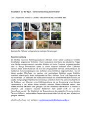
The second paper “<strong>Radiographic</strong> <strong>f<strong>in</strong>d<strong>in</strong>gs</strong> <strong>in</strong> <strong>several</strong> jo<strong>in</strong>ts <strong>of</strong> n<strong>in</strong>e <strong>bears</strong>” deals with the<br />
alterations <strong>of</strong> jo<strong>in</strong>ts <strong>in</strong> n<strong>in</strong>e <strong>bears</strong> (brown bear, Ursus arctos; black bear, Ursus americanus;<br />
polar bear, Ursus maritimus; sun bear, Helarctos malayanus). Radiographs <strong>of</strong> the elbows,<br />
hips, stifles and tarsal jo<strong>in</strong>ts were performed. The radiographs were scored accord<strong>in</strong>g to the<br />
presence <strong>of</strong> signs <strong>of</strong> osteoarthritis. All <strong>bears</strong> without and with gait abnormality showed<br />
osteoarthritic changes <strong>in</strong> their jo<strong>in</strong>ts. Not only the age seem to be responsible for these<br />
osteoarthritic changes, but the nutrition and / or the enclosures could play a role. We would<br />
like to cont<strong>in</strong>ue to make radiographic exam<strong>in</strong>ations <strong>of</strong> <strong>bears</strong> and to try to f<strong>in</strong>d a correlation<br />
between nutrition, ground <strong>of</strong> enclosures and osteoarthritic changes. The results <strong>of</strong> this study<br />
was used for the part “radiographic exam<strong>in</strong>ation” <strong>of</strong> the scor<strong>in</strong>g system <strong>of</strong> the first paper.<br />
The paper will be submitted to Veter<strong>in</strong>ary Radiology &Ultrasound. The study will be<br />
presented at the jo<strong>in</strong>t scientific conference <strong>of</strong> the International Veter<strong>in</strong>ary Radiology<br />
Association <strong>in</strong> Vancouver, Canada, <strong>in</strong> August 2006.<br />
7
Part I<br />
A scor<strong>in</strong>g system to evaluate physical condition and<br />
quality <strong>of</strong> life <strong>in</strong> geriatric zoo mammals<br />
9
A SCORING SYSTEM TO EVALUATE PHYSICAL CONDITION AND<br />
QUALITY OF LIFE IN GERIATRIC ZOO MAMMALS<br />
J Föllmi 1 , A Steiger 1 , C Walzer 2 , N Robert 3 , U Geissbühler 4 , M G Doherr 5 and C Wenker 6<br />
1 Institute <strong>of</strong> Animal Genetics, Nutrition and Hous<strong>in</strong>g, Division <strong>of</strong> Animal Hous<strong>in</strong>g and<br />
Welfare, Vetsuisse Faculty, University <strong>of</strong> Berne, Bremgartenstrasse 109a, CH 3001 Berne,<br />
Switzerland<br />
2Research Institute <strong>of</strong> Wildlife Ecology, University <strong>of</strong> Veter<strong>in</strong>ary Medic<strong>in</strong>e,<br />
Savoyenstrasse 1, A 1160 Vienna, Austria<br />
3Centre for Fish and Wildlife Health, Institute <strong>of</strong> Animal Pathology, Vetsuisse Faculty,<br />
University <strong>of</strong> Berne, Länggassstrasse 122, CH 3001 Berne, Switzerland<br />
4 Radiology Section, Department <strong>of</strong> Cl<strong>in</strong>ical Veter<strong>in</strong>ary Medic<strong>in</strong>e, Vetsuisse Faculty,<br />
University <strong>of</strong> Berne, Länggassstrasse 124, CH 3001 Berne, Switzerland<br />
5Department <strong>of</strong> Cl<strong>in</strong>ical Veter<strong>in</strong>ary Science, Vetsuisse Faculty, University <strong>of</strong> Berne,<br />
Bremgartenstrasse 109a, CH 3001 Berne, Switzerland<br />
6Zoo Basel, B<strong>in</strong>n<strong>in</strong>gerstrasse 40, CH 4054 Basel, Switzerland<br />
Correspondence: Jérôme Föllmi 1 , e-mail: jerome.foellmi@itz.unibe.ch<br />
Tel: +41.31.305.90.50, Fax: +41.31.631.26.40<br />
Keywords: euthanasia, geriatrics, pathology, scor<strong>in</strong>g system, symptom, zoo mammals<br />
Abstract<br />
The decision to perform euthanasia <strong>in</strong> geriatric zoo mammals is usually a highly complex<br />
procedure <strong>in</strong>volv<strong>in</strong>g ethical, medical, emotional and sometimes political factors. However,<br />
subsequent necropsies <strong>of</strong>ten show that the pathological changes <strong>of</strong> organs and / or the<br />
musculoskeletal system are already advanced. Therefore we hypothesize that euthanasia is<br />
<strong>of</strong>ten delayed to the detriment <strong>of</strong> the animal’s welfare. The purpose <strong>of</strong> this study was to<br />
facilitate and establish an <strong>in</strong>itial objective decision mak<strong>in</strong>g framework for the euthanasia <strong>of</strong><br />
geriatric zoo mammals. A scor<strong>in</strong>g-system to assess the physical condition and quality <strong>of</strong> life<br />
<strong>in</strong> old zoo mammals is presented, based on retrospective and prospective <strong>in</strong>vestigation <strong>of</strong> 70<br />
geriatric zoo mammals <strong>in</strong> five European zoos. Medical records and necropsy reports were<br />
studied <strong>in</strong> retrospective cases. Symptoms were monitored and recorded <strong>in</strong> prospective cases.<br />
<strong>Radiographic</strong> <strong>in</strong>vestigations under general anesthesia or at necropsy were performed<br />
additionally. A significant association between symptoms and pathological <strong>f<strong>in</strong>d<strong>in</strong>gs</strong> revealed<br />
that 63.1% <strong>of</strong> the exam<strong>in</strong>ed animals (n = 41/65) had pathological alterations <strong>of</strong> the<br />
musculoskeletal system (36.9%), or suffered from neoplasia (26.2%). Based on the <strong>in</strong>dividual<br />
reports, 28 veter<strong>in</strong>arians from different fields <strong>of</strong> veter<strong>in</strong>ary medic<strong>in</strong>e concluded that these<br />
animals had mild to severe pa<strong>in</strong>, discomfort and a significantly reduced quality <strong>of</strong> life, thus<br />
welfare was strongly reduced. The proposed scor<strong>in</strong>g system <strong>in</strong>cludes all these factors and<br />
<strong>of</strong>fers a simple and reliable tool to support decision-mak<strong>in</strong>g for euthanasia <strong>in</strong> geriatric zoo<br />
mammals.<br />
11
Mots-clé: euthanasie, gériatrie, mammifères de zoo, pathologie, scor<strong>in</strong>g system, symptôme<br />
Résumé<br />
La décision d’effectuer une euthanasie d’un animal gériatrique est une procédure très<br />
complexe <strong>in</strong>cluant des facteurs éthiques, médicaux, émotionnels et parfois politiques.<br />
Cependant, la nécropsie ultérieure de ces animaux met souvent en évidence des changements<br />
pathologiques déjà bien avancés des organes et / ou du système locomoteur. C’est pourquoi<br />
nous faisons l’hypothèse que l’euthanasie est souvent retardée au détriment du bien-être de<br />
l’animal. Le but de cette étude est de faciliter et d’établir un cadre pour une décision <strong>in</strong>itiale<br />
objective lors d’euthanasie chez les animaux gériatriques. Un « scor<strong>in</strong>g system » sur la<br />
condition physique et la qualité de vie des vieux animaux de zoo y est présenté, basé sur une<br />
<strong>in</strong>vestigation rétrospective et prospective sur 70 mammifères gériatriques de zoo dans c<strong>in</strong>q<br />
zoos européens. Les dossiers médicaux et les rapports de nécropsie ont été étudiés pour les<br />
cas rétrospectifs. Des symptômes ont été observés et archivés pour les cas prospectifs. Des<br />
<strong>in</strong>vestigations radiographiques sous anesthésie générale ou avant nécropsie ont été effectuées<br />
en plus. Une association significative entre les symptômes et les pathologies a révélé que<br />
63.1% des animaux exam<strong>in</strong>és (n = 41/65) présentaient des pathologies du système locomoteur<br />
(36.9%) ou souffraient de néoplasie (26.2%). Sur la base de rapports <strong>in</strong>dividuels, 28<br />
vétér<strong>in</strong>aires de différents doma<strong>in</strong>es en médec<strong>in</strong>e vétér<strong>in</strong>aire ont par ailleurs conclu que ces<br />
animaux présentaient des douleurs et un <strong>in</strong>confort allant de léger à sévère a<strong>in</strong>si qu’une qualité<br />
de vie significativement réduite, entraînant un bien-être fortement restre<strong>in</strong>t de l’animal. Le<br />
« scor<strong>in</strong>g system » proposé <strong>in</strong>clut tous ces facteurs et <strong>of</strong>fre un outil simple et efficace pour<br />
soutenir la décision d’effectuer une euthanasie chez les mammifères gériatriques de zoo.<br />
Introduction<br />
In the last years <strong>of</strong> the 20 th century zoos became <strong>in</strong>creas<strong>in</strong>gly focused on conservation<br />
(through captive breed<strong>in</strong>g), research and education. This has been <strong>in</strong> part due to chang<strong>in</strong>g<br />
public attitudes towards zoos and the appropriate methods <strong>of</strong> keep<strong>in</strong>g wild animals <strong>in</strong><br />
captivity (Young 2003). In addition, the conditions <strong>of</strong> captivity <strong>of</strong> zoo animals have<br />
improved and the animal longevity has been <strong>in</strong>creased. As a result, problems aris<strong>in</strong>g from<br />
the management <strong>of</strong> long-lived species and age-related diseases are <strong>in</strong>creas<strong>in</strong>g. Some <strong>of</strong> the<br />
problems may be difficult to diagnose and treat, thereby potentially compromis<strong>in</strong>g animal<br />
welfare (Kitchener 2004). In many cases, the subjects are apparently healthy, but old zoo<br />
animals at necropsy are found to suffer from a range <strong>of</strong> health problems that may not have<br />
been apparent when they were alive (Richardson 2000). Animals <strong>in</strong> captivity <strong>in</strong>variably live<br />
longer than their wild counterparts (Nowak 1999; Richardson 2000; Erw<strong>in</strong> et al 2002).<br />
Enabl<strong>in</strong>g zoo animals to live <strong>in</strong> better conditions for a long period is presently viewed as<br />
important. However, zoos <strong>of</strong>ten unwitt<strong>in</strong>gly condemn their animals to long pa<strong>in</strong>ful lives. In<br />
some <strong>in</strong>stance this may benefit the conservation <strong>of</strong> genetic l<strong>in</strong>eages. However, <strong>in</strong> other<br />
<strong>in</strong>stances the already well-represented animal occupies much needed space well beyond<br />
mak<strong>in</strong>g a significant contribution to the breed<strong>in</strong>g program. A good zoo strives to improve<br />
the quality <strong>of</strong> its animals’ lives, not necessarily their length <strong>of</strong> life (Richardson 2001).<br />
Suffer<strong>in</strong>g is an unpleasant state <strong>of</strong> m<strong>in</strong>d that disrupts the quality <strong>of</strong> life. It is the mental<br />
state associated with unpleasant experiences such as pa<strong>in</strong>, malaise, distress, <strong>in</strong>jury and<br />
emotional numbness (Gregory 2004). In the present study we focused on pa<strong>in</strong>, because pa<strong>in</strong>,<br />
probably more than any other state, directly reduces welfare (Duncan 2004).<br />
Generally, it is very difficult to determ<strong>in</strong>e the appropriate time when to perform<br />
euthanasia. Besides the medical aspect, the zoo veter<strong>in</strong>arian must take <strong>in</strong>to consideration the<br />
different <strong>in</strong>terests <strong>of</strong> zoo managers and staff, visitors, animal welfare organizations, sponsors<br />
and possibly the needs <strong>of</strong> the respective breed<strong>in</strong>g programs (e.g. European Endangered<br />
12
Species Program). The f<strong>in</strong>al decision to perform euthanasia is greatly <strong>in</strong>fluenced by the<br />
general cl<strong>in</strong>ical condition <strong>of</strong> the animal. In this regard, the correct <strong>in</strong>terpretation <strong>of</strong> cl<strong>in</strong>ical<br />
symptoms <strong>in</strong> zoo animals plays a central role <strong>in</strong> the evaluation <strong>of</strong> the animal’s welfare.<br />
Persistence <strong>of</strong> cl<strong>in</strong>ical signs <strong>in</strong> spite <strong>of</strong> medical treatment should rapidly give rise to<br />
discussions on the future quality <strong>of</strong> life <strong>in</strong> the <strong>in</strong>dividual. In some cases a change <strong>of</strong> drug may<br />
be helpful, sometimes a surgical <strong>in</strong>tervention under general anesthesia may be <strong>in</strong>dicated and<br />
<strong>in</strong> other <strong>in</strong>stances the decision for euthanasia must be taken. Treatment <strong>in</strong>variably raises many<br />
questions about its efficacy, and each veter<strong>in</strong>arian must exercise the highest standards <strong>of</strong><br />
animal welfare (EAZA 1999; WAZA 1999). Interventions <strong>in</strong>variably cause stress and are a<br />
risk for the animals. Additionally, this mobilizes personnel and material, takes time, and<br />
generates costs.<br />
Based on the correlation <strong>of</strong> symptoms, pr<strong>of</strong>essional perceptions and necropsy results<br />
we propose an <strong>in</strong>itial scor<strong>in</strong>g-system to evaluate physical condition and quality <strong>of</strong> life <strong>in</strong><br />
geriatric zoo mammals, with the aim <strong>of</strong> improv<strong>in</strong>g the decision process for euthanasia at an<br />
appropriate time.<br />
Animals, Materials and Methods<br />
Animals<br />
This study <strong>in</strong>cluded 70 old zoo mammals belong<strong>in</strong>g to 24 different species (Table 1). Sixtyfive<br />
animals died or were euthanized between 1979 and 2005 <strong>in</strong> five European zoos and five<br />
animals were still alive at the time <strong>of</strong> manuscript submission. Only cases whose symptoms<br />
(Table 2) were documented reliably and with pathological reports were chosen for this study.<br />
Sixty animals were retrospective cases and 10 were prospective cases.<br />
The age <strong>of</strong> the zoo animals is expressed <strong>in</strong> percentage <strong>of</strong> the reported maximal age <strong>in</strong><br />
the wild when available and <strong>of</strong> the maximal reported longevity <strong>in</strong> captivity. (Haltenorth 1977;<br />
Grzimek 1993; Nowak 1999; Cat Specialist Group 2004; International Association for Bear<br />
Research and Management 2004).<br />
Symptoms<br />
A variable factor <strong>of</strong> multiplication which depends on the frequency <strong>of</strong> the symptoms observed<br />
is allocated <strong>in</strong> the scor<strong>in</strong>g system. Frequency between 11% and 33% for each symptom<br />
obta<strong>in</strong>ed a factor 2 (group 1), frequency between 5% and 6% for each symptom obta<strong>in</strong>ed a<br />
factor 1.5 (group 2) and frequency between 1.0% and 4.5% for each symptom obta<strong>in</strong>ed a<br />
factor 1 (group 3).<br />
Table 2 The 18 symptoms observed <strong>in</strong> all animals, two symptoms observed only<br />
<strong>in</strong> <strong>bears</strong> and three symptoms observed only <strong>in</strong> primates <strong>in</strong> this study.<br />
All animals, <strong>in</strong>clud<strong>in</strong>g<br />
Observations <strong>in</strong> <strong>bears</strong> Observations <strong>in</strong><br />
<strong>bears</strong> and primates<br />
only<br />
primates only<br />
Vomit<strong>in</strong>g Diarrhoea Reduced bath<strong>in</strong>g Social pressure<br />
Dysorexia Reluctance to move Increased bath<strong>in</strong>g Bite wounds<br />
Cachexia Pa<strong>in</strong> when stand<strong>in</strong>g up Isolation from group<br />
Anorexia Lameness<br />
Weakness Muscle atrophy<br />
Apathy Polydipsia/Polyuria<br />
Dehydration Position <strong>in</strong> hierarchy ↓<br />
Dyspnoea Hypersalivation<br />
Cough<strong>in</strong>g Recumbency<br />
13
Retrospective and prospective cases<br />
For the retrospective cases, the medical records <strong>of</strong> the participat<strong>in</strong>g zoos were used and<br />
persons (veter<strong>in</strong>arians, keepers) <strong>in</strong>volved <strong>in</strong> the care <strong>of</strong> the concerned animals were<br />
<strong>in</strong>terviewed to precisely def<strong>in</strong>e the symptoms, which the animals had shown before their<br />
euthanasia or natural death. The symptoms, which were taken <strong>in</strong>to consideration, were<br />
def<strong>in</strong>ed as the predom<strong>in</strong>at<strong>in</strong>g symptoms dur<strong>in</strong>g the last year <strong>of</strong> life. In addition, the necropsy<br />
report was analyzed for each case.<br />
For the prospective cases, a similar procedure was performed <strong>in</strong> six <strong>bears</strong> (U. arctos,<br />
U. maritimus), one giraffe (G. camelopardalis tippelskirchi), one okapi (O. johnstoni) and one<br />
camel (C. bactrianus) (Table 3); symptoms were monitored, and, after euthanasia or death <strong>of</strong><br />
the animal, necropsy was performed either on site or at the Institute for Animal Pathology,<br />
University <strong>of</strong> Berne. Each prospective case was exam<strong>in</strong>ed radiographically, five <strong>of</strong> them <strong>in</strong><br />
general anesthesia <strong>in</strong>clud<strong>in</strong>g four adult brown <strong>bears</strong> (U. arctos) and one polar bear (U.<br />
maritimus).<br />
Prior to necropsies, postmortem radiographic exam<strong>in</strong>ation (Siemens Polydoros 100<br />
system, with a performance <strong>of</strong> 80 kW at 100 kV) was performed <strong>in</strong> four animals: one giraffe<br />
(G. camelopardalis tippelskirchi), one okapi (O. johnstoni), one Bactrian camel (C.<br />
bactrianus) and one brown bear (U. arctos).<br />
Table 3 Symptoms <strong>of</strong> the prospective cases (n = 10) <strong>of</strong> this study<br />
Animal Age Sex Symptom<br />
Brown bear 29 Female mild to moderate weakness, mild lameness, mild<br />
(U. arctos)<br />
reluctance to move, mild dysorexia<br />
Brown bear<br />
(U. arctos)<br />
Brown bear<br />
(U. arctos)<br />
Brown bear<br />
(U. arctos)<br />
Polar bear<br />
(U. maritimus)<br />
Sun bear<br />
(H. malayanus)<br />
Brown bear<br />
(U. arctos)<br />
Giraffe<br />
(G. camelopardalis)<br />
Okapi<br />
(O. johnstoni)<br />
Bactrian camel<br />
(C. bactrianus)<br />
23 Female severe lameness, severe reluctance to move, pa<strong>in</strong> when<br />
stand<strong>in</strong>g up, mild dysorexia, cachexia<br />
33 Female mild lameness, stiff neck, mild dysorexia<br />
33 Female gait abnormality, mild dysorexia<br />
27 Male dysorexia, paraparesis<br />
21 Male No cl<strong>in</strong>ical symptoms<br />
28 Female severe lameness, severe reluctance to move<br />
18 Female Moderate lameness, moderate reluctance to move<br />
13 Female colic, dysorexia, reluctance to move, anorexia dur<strong>in</strong>g<br />
eight days<br />
19 Female severe lameness, severe reluctance to move,severe pa<strong>in</strong><br />
when stand<strong>in</strong>g up, moderate cachexia, moderate to<br />
severe muscle atrophy<br />
Pathologies<br />
Pathological <strong>f<strong>in</strong>d<strong>in</strong>gs</strong> were classified per system or per organ <strong>in</strong>clud<strong>in</strong>g pancreas,<br />
hepatobiliary system, musculoskeletal system, respiratory system, genital system, lymphatic<br />
system, cardiovascular system, kidney and adrenal gland, gastro<strong>in</strong>test<strong>in</strong>al tract, sk<strong>in</strong>, oral<br />
diseases <strong>in</strong>clud<strong>in</strong>g teeth, and trauma. Pathologies were classified <strong>in</strong>to three groups: neoplasia,<br />
dysfunction <strong>of</strong> the musculoskeletal system and other pathologies (Table 4). Further, we<br />
subdivided neoplasia <strong>in</strong>to two groups: “neoplasia <strong>of</strong> visceral organs” and “neoplasia <strong>of</strong> non<br />
14
visceral organs”. Neoplasia and dysfunctions <strong>of</strong> the musculoskeletal system were the most<br />
common diseases recorded <strong>in</strong> our study.<br />
Evaluation <strong>of</strong> pa<strong>in</strong>, discomfort and quality <strong>of</strong> life<br />
A data card was created for each animal (n = 65), <strong>in</strong>clud<strong>in</strong>g species, name, age, symptoms and<br />
the diagnosed pathologies <strong>of</strong> the animal followed by three scales for evaluation <strong>of</strong> pa<strong>in</strong>,<br />
discomfort and quality <strong>of</strong> life (Table 5). Each scale was graduated from zero (no pa<strong>in</strong>, no<br />
discomfort, good quality <strong>of</strong> life) to ten (high level <strong>of</strong> pa<strong>in</strong>, high level <strong>of</strong> discomfort, poor<br />
quality <strong>of</strong> life). The sum <strong>of</strong> the three scales gave the general assessment factor (from 0 to 30).<br />
Twenty-eight veter<strong>in</strong>arians evaluated these data cards. Groups were created accord<strong>in</strong>g to the<br />
specialty <strong>of</strong> each veter<strong>in</strong>arian: zoo veter<strong>in</strong>arians (n = 12), residents (n = 7) (two <strong>in</strong> cl<strong>in</strong>ic and<br />
five <strong>in</strong> pathology), veter<strong>in</strong>ary pathologists (n = 2), <strong>in</strong>ternists (n = 2), surgeons (n = 3) and<br />
anesthetists (n = 2). Two data cards were double <strong>in</strong> each set <strong>of</strong> 65 cards to control the<br />
objectivity <strong>of</strong> the veter<strong>in</strong>arians’ evaluation. No veter<strong>in</strong>arians were <strong>in</strong>formed <strong>of</strong> these doubled<br />
cards.<br />
Scor<strong>in</strong>g system<br />
The proposed scor<strong>in</strong>g system is based on the scor<strong>in</strong>g system “4 a vet” (association 4avet)<br />
created to evaluate pa<strong>in</strong> <strong>in</strong> small companion animals which is used at the Veter<strong>in</strong>ary Faculty<br />
<strong>of</strong> the Berne University, and has been adapted to evaluate the physical condition and quality<br />
<strong>of</strong> life <strong>in</strong> geriatric zoo mammals. It <strong>in</strong>cludes five parts (Table Scor<strong>in</strong>g system). The first part is<br />
the “history assessment” with age and cl<strong>in</strong>ical symptoms. The second part ”therapy” conta<strong>in</strong>s<br />
the response on the therapy with specific and tentative therapy. The third part is the<br />
“evaluation <strong>of</strong> pa<strong>in</strong>, discomfort and quality <strong>of</strong> life”. The fourth part conta<strong>in</strong>s the “radiographic<br />
exam<strong>in</strong>ation”. The fifth part conta<strong>in</strong>s the “additional assessment” <strong>in</strong>clud<strong>in</strong>g breed<strong>in</strong>g <strong>in</strong>terest,<br />
gender, ability to reproduce and h<strong>in</strong>drance <strong>of</strong> new breeder. In the result section we<br />
differentiate the evaluation without or with “radiographic exam<strong>in</strong>ations”.<br />
Statistics<br />
Descriptive statistics <strong>in</strong>clude frequency tables, measures <strong>of</strong> central tendency and spread,<br />
and the respective graphs. The association between the specific symptoms and<br />
pathologies was assessed us<strong>in</strong>g cross-tabulation and the chi-square test or, when any <strong>of</strong><br />
the expected cell frequencies was below 5, the Fisher’s Exact test. In addition, odds<br />
ratios (OR) with 95% confidence <strong>in</strong>tervals were calculated. All statistical procedures<br />
were performed with NCSS 2001 s<strong>of</strong>tware (Number Cruncher Statistical Systems,<br />
Kaysville, UT, USA) and Micros<strong>of</strong>t Excel. The overall level <strong>of</strong> statistical significance<br />
was set to P < 0.05.<br />
Table 4 Pathologies classified <strong>in</strong> three pathological groups found <strong>in</strong> different familiae <strong>of</strong> animals (n=65)<br />
<strong>of</strong> this study<br />
Familiae Neoplasia (n = 17) Dysfunction <strong>of</strong> the musculoskeletal<br />
system (n = 24)<br />
Other pathologies (n = 24)<br />
Ursidae Hepatocellular carc<strong>in</strong>oma (n=1)<br />
Pancreas adenocarc<strong>in</strong>oma (n=1)<br />
Bile duct carc<strong>in</strong>oma and<br />
adenocarc<strong>in</strong>oma <strong>of</strong> the<br />
duodenum(n=1)<br />
Arthrosis and spondylarthropathy,<br />
moderate to severe (n=5)<br />
15<br />
Pancreas necrosis (n=1)<br />
Myokardiose, severe (n=1)<br />
Bronchopneumonie (n=1) Enteritis,<br />
colitis and Glomerulonephritis<br />
(n=1)
Familiae Neoplasia (n = 17) Dysfunction <strong>of</strong> the musculoskeletal<br />
system (n = 24)<br />
Felidae Pancreas adenocarc<strong>in</strong>oma with Arthrosis, severe (n=2)<br />
metastasis (n=1)<br />
Adenocarc<strong>in</strong>oma <strong>of</strong> the duodenum<br />
(n=1)<br />
Bile duct carc<strong>in</strong>oma (n=1)<br />
Phaeochromozytom (n=1)<br />
Chondroma (n=1)<br />
Chondr<strong>of</strong>ibrosarcoma with metastasis<br />
(n=1)<br />
Leiomyoma cervix (n=1)<br />
Lymphosarcoma (n=1)<br />
Leukose (n=1)<br />
Squamous cell carc<strong>in</strong>oma <strong>of</strong> the<br />
omentum (n=1)<br />
Spondylarthropathy (n=2)<br />
Canidae Haemangiosarcoma (n=1) Spondylarthropathy (n=1)<br />
Other pathologies (n = 24)<br />
Infarction (n=1)<br />
Perforation <strong>of</strong> the stomach (n=1)<br />
Pyelonephritis (n=1)<br />
Necrotiz<strong>in</strong>g Hepatitis (n=1)<br />
Camelidae Cholangiocarc<strong>in</strong>oma (n=1) Arthrosis, moderate to severe (n=3) Sand impaction (n=1)<br />
Giraffidae Fracture, distal phalanx (n=2) Kidney <strong>in</strong>farct (n=1)<br />
Deformation <strong>of</strong> claws (n=1) Congestion <strong>of</strong> the abomasum(n=1)<br />
Elephantidae Arthritis, severe (n=1)<br />
Equidae Trauma <strong>of</strong> the cannon bone(n=1)<br />
Lam<strong>in</strong>itis (n=1)<br />
Fracture <strong>of</strong> the pelvis (n=1)<br />
Tapiridae Anomaly <strong>of</strong> teeth, severe (n=1)<br />
Pneumonia, severe (n=1)<br />
Rh<strong>in</strong>ocerotidae Arthrosis, moderate to severe (n=2) Pancreatitis (n=1)<br />
Sand impaction (n=1)<br />
Moschidae Adenocarc<strong>in</strong>oma <strong>of</strong> the ileum (n=1)<br />
Bovidae Arthrosis, severe (n=1) Bronchopneumonia, severe (n=2)<br />
Cebidae Squamous cell carc<strong>in</strong>oma <strong>of</strong> the<br />
stomach (n=1)<br />
Fracture, distal phalanx (n=1) Infected sk<strong>in</strong> lesion (n=1)<br />
Cercopithecidae Bite wounds (n=1)<br />
Colitis, chronic (n=1)<br />
G<strong>in</strong>givitis and periodonditis,<br />
chronic (n=1)<br />
Hernia (n=1)<br />
Amyloidosis <strong>of</strong> the liver (n=1)<br />
Hepatitis (n=1)<br />
16
Table 5 Animal card filled out by veter<strong>in</strong>arian for each retrospective and prospective cases (n = 65) to evaluate<br />
pa<strong>in</strong>, discomfort and quality <strong>of</strong> life with the aid <strong>of</strong> observed symptoms and pathologic <strong>f<strong>in</strong>d<strong>in</strong>gs</strong> by animals<br />
N°Ref.: ________________ Name: ______________<br />
Species: __________ Sex: ______________<br />
Age: ________________<br />
Symptom<br />
Vomit<strong>in</strong>g Dehydration Reluctance to move Isolation from the group Recumbency<br />
Dysorexia Dyspnoea Pa<strong>in</strong> when stand<strong>in</strong>g up Social pressure<br />
Cachexia Cough<strong>in</strong>g Lameness Bite wounds<br />
Anorexia Diarrhoea Muscle atrophy Influenced by the season<br />
Weakness Bath<strong>in</strong>g ↑ Polydipsia/Polyuria Spr<strong>in</strong>g/Summer<br />
Apathy Bath<strong>in</strong>g ↓ Hierarchy↓ Autumn/W<strong>in</strong>ter<br />
Pathology<br />
Pancreas Respiratory System Kidney & Adrenal gland<br />
Pancreas adenocarc<strong>in</strong>oma Bronchopneumonia Chronic nephritis<br />
Pancreas necrosis _____________________________ Pyelonephritis<br />
__________________________________ Phaeochromozytom<br />
__________________________________ Trauma ________________________________<br />
_____________________________ ________________________________<br />
Hepathobiliary system _____________________________<br />
Hepatocellular carc<strong>in</strong>oma Gastro<strong>in</strong>test<strong>in</strong>al tract<br />
Hepatitis Oral diseases Squamous cell carc<strong>in</strong>oma stomach<br />
Cyst Abscess Adenocarc<strong>in</strong>oma <strong>of</strong> duodenum<br />
Bile duct carc<strong>in</strong>oma Periodontitis Colitis<br />
Cholangiocarc<strong>in</strong>oma Anomaly <strong>of</strong> teeth Gastritis<br />
__________________________________ _____________________________ Perforation <strong>of</strong> stomach<br />
__________________________________ _ Squamous cell carc<strong>in</strong>oma omentum<br />
________________________________<br />
Musculoskeletal System Genital System ________________________________<br />
Arthrosis Leiomyoma <strong>of</strong> cervix<br />
Spondyloarthropathy Sk<strong>in</strong><br />
Arthritis Lymphatic System Infected sk<strong>in</strong> lesion<br />
Erosion Leucosis ________________________________<br />
Synovialitis Lymphosarcoma ________________________________<br />
Fracture _____________________________<br />
Chondr<strong>of</strong>ibrosarcoma<br />
Chondroma Cardiovascular System<br />
Deformation <strong>of</strong> claws Cardiac <strong>in</strong>sufficiency<br />
Lam<strong>in</strong>itis Haemangiosarcoma<br />
_________________________________ Infarction<br />
_________________________________ _____________________________<br />
_________________________________ _____________________________<br />
_________________________________<br />
Evaluation<br />
Scale is graduated from 0 to 10. 0 = no, 5 = moderate und 10 = high, severe<br />
0_____1_____2_____3_____4_____5_____6_____7_____8_____9_____10<br />
Pa<strong>in</strong> 0___________________________________________________________10<br />
Discomfort 0___________________________________________________________10<br />
Quality <strong>of</strong> life good___________________________________________________________very bad<br />
17<br />
Fiche/JF-Diss-ZooBasel
Scor<strong>in</strong>g system to evaluate physical condition and quality <strong>of</strong> life<br />
<strong>in</strong> old zoo mammals<br />
Animal: Species:<br />
Age: Maximal age <strong>in</strong> the wild:<br />
HISTORY ASSESSMENT<br />
Age 100% corresponds to the maximal age estimated for an animal <strong>in</strong> the wild<br />
< 80% – 2<br />
80–100% – 1<br />
101–120% +2<br />
>120% +4<br />
Symptom Scale is graduated from 0 to 10 0 = no symptoms and 10 = severe symptoms<br />
EXAMPLE<br />
Card<strong>in</strong>al symptoms yes /no If yes, severity po<strong>in</strong>ts<br />
Vomit<strong>in</strong>g<br />
1cm = 1; here 3.6cm = 3.6<br />
Dur<strong>in</strong>g the last two months<br />
Card<strong>in</strong>al symptoms yes /no If yes, severity<br />
Other symptoms yes /no If yes, severity<br />
Date<br />
0 10<br />
Score<br />
Po<strong>in</strong>ts<br />
✕2<br />
Po<strong>in</strong>ts<br />
Vomit<strong>in</strong>g 0 10 ✕2<br />
Dysorexia 0 10 ✕2<br />
Anorexia 0 10 ✕2<br />
Cachexia 0 10 ✕2<br />
Lameness 0 10 ✕2<br />
Diarrhoea 0 10 ✕1.5<br />
Polydipsia/Polyuria 0 10 ✕1.5<br />
Dyspnoea 0 10 ✕1<br />
Cough<strong>in</strong>g 0 10 ✕1<br />
Bite wounds 0 10 ✕1<br />
Social pressure 0 10 ✕1<br />
Isolation from group 0 10 ✕1<br />
Other: 0 10 ✕1<br />
Other: 0 10 ✕1<br />
Total<br />
Po<strong>in</strong>ts<br />
Apathy 0 10 ✕2<br />
Pa<strong>in</strong> when stand<strong>in</strong>g up 0 10 ✕2<br />
Reluctance to move 0 10 ✕2<br />
Weakness 0 10 ✕2<br />
Muscle atrophy 0 10 ✕1.5<br />
Position <strong>in</strong> hierarchy 0 10 ✕1.5<br />
Dehydratation 0 10 ✕1<br />
Bath<strong>in</strong>g 0 10 ✕1<br />
Bath<strong>in</strong>g 0 10 ✕1<br />
Other: 0 10 ✕1<br />
Other: 0 10 ✕1<br />
18<br />
Total<br />
Subtotal 1<br />
© jföllmi_ZooBasel
THERAPY<br />
Specific therapy yes yes, go to next two questions<br />
no no, go to tentative therapy<br />
Scale is graduated from 0 to 10 0=good, yes and 10=bad, no<br />
Efficiency <strong>of</strong> the treatment was 0 10 ✕1<br />
After the treatment, did the<br />
symptoms disappear?<br />
0 10 ✕1<br />
Tentative therapy yes /no yes, go to next two questions<br />
Efficiency <strong>of</strong> treatment was 0 10 ✕1<br />
After the treatment, did the 0 10 ✕1<br />
symptoms disappear?<br />
EVALUATION<br />
Evaluation <strong>of</strong> Pa<strong>in</strong> 0 10 ✕1<br />
Evaluation <strong>of</strong> Quality <strong>of</strong> life 0 10 ✕1<br />
Evaluation <strong>of</strong> Discomfort 0 10 ✕1<br />
RADIOGRAPHIC EXAMINATION<br />
Scale is graduated from 0 to 10 0=no, good and 10=severe, bad<br />
Exam<strong>in</strong>ation yes /no yes, go to next question<br />
Subtotal 2<br />
Subtotal 3<br />
Elbow jo<strong>in</strong>t, left 0 none +5 mild +10 moderate/severe signs <strong>of</strong> alterations<br />
Elbow jo<strong>in</strong>t, right 0 none +5 mild +10 moderate/severe signs <strong>of</strong> alterations<br />
Hip jo<strong>in</strong>t, left 0 none +5 mild +10 moderate/severe signs <strong>of</strong> alterations<br />
Hip jo<strong>in</strong>t, right 0 none +5 mild +10 moderate/severe signs <strong>of</strong> alterations<br />
Knee jo<strong>in</strong>t, left 0 none +5 mild +10 moderate/severe signs <strong>of</strong> alterations<br />
Knee jo<strong>in</strong>t, right 0 none +5 mild +10 moderate/severe signs <strong>of</strong> alterations<br />
Tarsal jo<strong>in</strong>t, left 0 none +5 mild +10 moderate/severe signs <strong>of</strong> alterations<br />
Tarsal jo<strong>in</strong>t, right 0 none +5 mild +10 moderate/severe signs <strong>of</strong> alterations<br />
Shoulder jo<strong>in</strong>t, left 0 none +5 mild +10 moderate/severe signs <strong>of</strong> alterations<br />
Shoulder jo<strong>in</strong>t, right 0 none +5 mild +10 moderate/severe signs <strong>of</strong> alterations<br />
Vertebrate column 0 none +5 mild +10 moderate/severe signs <strong>of</strong> alterations<br />
Other: 0 none +5 mild +10 moderate/severe signs <strong>of</strong> alterations<br />
Other: 0 none +5 mild +10 moderate/severe signs <strong>of</strong> alterations<br />
19<br />
Total<br />
Subtotal 4<br />
Score<br />
Score<br />
Score<br />
© jföllmi_ZooBasel
ADDITIONAL ASSESSMENT<br />
Breed<strong>in</strong>g <strong>in</strong>terest<br />
Gender<br />
Able to reproduce<br />
H<strong>in</strong>der the arrival <strong>of</strong> new breeder<br />
RESULTS<br />
yes –5<br />
no +1<br />
important –5<br />
not important +1<br />
yes –5<br />
no +5<br />
yes +5<br />
no 0<br />
Without «radiographic exam<strong>in</strong>ation» Sum <strong>of</strong> the four subtotals<br />
From 1 to 30 Treatment<br />
From 31 to 51 Doubtful<br />
Over 51 Euthanasia recommended<br />
With «radiographic exam<strong>in</strong>ation» Sum <strong>of</strong> the five subtotals<br />
From 1 to 45 Treatment<br />
From 46 to 66 Doubtful<br />
Over 66 Euthanasia recommended<br />
20<br />
Subtotal 5<br />
Score<br />
© jföllmi_ZooBasel
Results<br />
Symptoms and pathology<br />
The symptoms <strong>in</strong>cluded <strong>in</strong> group 1 (frequency between 11 and 33%) were shown <strong>in</strong> 95.7% <strong>of</strong><br />
the cases (n = 67/70). The animals show<strong>in</strong>g one symptom 20% (n = 14/70), two symptoms<br />
30% (n = 21/70), three symptoms 22.9% (n = 16/70) or four symptoms 11.4% (n = 8/70)<br />
represented 84.3%. The animals show<strong>in</strong>g either five symptoms (5.7%, n = 4/70), six<br />
symptoms (4.3%, n = 3/70), seven symptoms (2.9%, n = 2/70) or eight symptoms (2.9%, n =<br />
2/70) represented 15.7%. We found significant associations between five symptoms and five<br />
pathologies (Table 6). In the follow<strong>in</strong>g we will focus on the results <strong>of</strong> the five most frequently<br />
observed symptoms.<br />
Table 6 Observed symptoms significantly associated<br />
with pathologic <strong>f<strong>in</strong>d<strong>in</strong>gs</strong> <strong>in</strong> this sample.<br />
Symptoms Pathologies (p-value)<br />
vomit<strong>in</strong>g neoplasia P = 0.02<br />
apathy neoplasia P = 0.007<br />
anorexia neoplasia P = 0.01<br />
lameness arthrosis P < 0.000001<br />
pa<strong>in</strong> when stand<strong>in</strong>g up spondyloarthropathy P = 0.009<br />
Vomit<strong>in</strong>g<br />
In this sample vomit<strong>in</strong>g was observed <strong>in</strong> <strong>bears</strong>, <strong>in</strong> felids and <strong>in</strong> one tapir (T. terrestris). This<br />
symptom was significantly associated with neoplasia (P = 0.02; OR = 6.2): 29.4% <strong>of</strong> all<br />
animals with neoplasia (n = 5/17) but only 6.3% <strong>of</strong> all animals (n = 3/48) without tumors<br />
vomited.<br />
Anorexia<br />
In this sample anorexia was observed <strong>in</strong> all species and was a consistent <strong>f<strong>in</strong>d<strong>in</strong>gs</strong> <strong>in</strong> <strong>bears</strong> and<br />
felids (72.2%). This symptom was significantly associated with neoplasia (P = 0.01; OR =<br />
4.3): 52.9% <strong>of</strong> all animals (n = 9/17) with neoplasia but 20.8% <strong>of</strong> all animals (n = 10/48)<br />
without neoplasia showed anorexia.<br />
Apathy<br />
In this sample apathy was ma<strong>in</strong>ly observed <strong>in</strong> <strong>bears</strong> and felids (87.5%, n = 8/9) and was<br />
significantly associated with neoplasia (P = 0.007; OR = 8.2): 35.3% <strong>of</strong> all animals (n = 6/17)<br />
with neoplasia but only 6.3% <strong>of</strong> all animals (n = 3/48) without neoplasia showed apathy.<br />
Lameness<br />
In this sample lameness was the symptom with the highest observed frequency and was<br />
recorded <strong>in</strong> all species. It was found <strong>in</strong> 37.1% <strong>of</strong> all studied animals (n = 26/70). Lameness<br />
was significantly associated with dysfunction <strong>of</strong> the musculoskeletal system (P < 0.000001;<br />
OR = 27.8) and arthrosis (P < 0.000001): 75% <strong>of</strong> all animals (n = 18/24) with dysfunction <strong>of</strong><br />
the musculoskeletal system but only 9.8% <strong>of</strong> all animals (n = 4/41) without dysfunction <strong>of</strong> the<br />
musculoskeletal system showed lameness; 100% <strong>of</strong> all animals (n = 14/14) with arthrosis but<br />
15.7% <strong>of</strong> all animals (n = 8/51) without arthrosis showed lameness.<br />
Pa<strong>in</strong> when stand<strong>in</strong>g up<br />
In this sample we found a significant association between pa<strong>in</strong> when stand<strong>in</strong>g up and<br />
spondylosis / spondylarthrosis (P = 0.009; OR = 8.9): 62.5% <strong>of</strong> all animals (n = 5/8) with<br />
spondylosis / spondylarthrosis but only 15.8% <strong>of</strong> all animals (n = 9/57) without spondylosis<br />
showed pa<strong>in</strong> when stand<strong>in</strong>g up.<br />
21
Age and pathology<br />
Animals with neoplasia represented 26.2% (n = 17/65) <strong>of</strong> the total set, animals suffer<strong>in</strong>g from<br />
dysfunction <strong>of</strong> the musculoskeletal system represented 36.9% (n = 24/65) and animals<br />
suffer<strong>in</strong>g from other diseases represented 36.9% (n = 24/65) (Table 7 and 8). In the group<br />
“neoplasia” 50% <strong>of</strong> the animals (n = 5/10) with “neoplasia <strong>of</strong> visceral organ” and 87.5% <strong>of</strong><br />
the animals (n = 6/7) with “neoplasia <strong>of</strong> non visceral organ” had an age equal and superior to<br />
100% <strong>of</strong> the life expectancy <strong>in</strong> the wild. In the group “other pathologies”, 100% <strong>of</strong> the<br />
animals (n = 7/7) with an age equal or superior to 100% <strong>of</strong> the life expectancy <strong>in</strong> the wild<br />
suffered either from dysfunction <strong>of</strong> the cardiovascular system or from dysfunction <strong>of</strong> the<br />
kidneys or from the liver.<br />
Table 7 Frequency <strong>of</strong> the three pathological groups <strong>of</strong> this study <strong>in</strong> correlation<br />
with an age <strong>of</strong> animals ≥ 100% <strong>of</strong> the life expectancy <strong>in</strong> the wild (%) and <strong>in</strong><br />
correlation with an age ≥ 50% <strong>of</strong> the maximal age <strong>in</strong> captivity (%)<br />
Pathologies Animals (n = 29) with an age Animals (n = 62) with an age ≥<br />
≥ 100% <strong>of</strong> the life expectancy 50% <strong>of</strong> the maximal age <strong>in</strong><br />
<strong>in</strong> the wild (%)<br />
captivity (%)<br />
Neoplasia 36.6% 25.0%<br />
Dysfunction <strong>of</strong> the<br />
musculoskeletal system<br />
39.0% 34.4%<br />
Other pathologies 24.4% 40.6%<br />
Table 8 Frequency <strong>of</strong> all animals <strong>of</strong> this study with the same group <strong>of</strong> pathology <strong>in</strong><br />
correlation with an age <strong>of</strong> animals ≥ 100% <strong>of</strong> the life expectancy <strong>in</strong> the wild (%) and<br />
<strong>in</strong> correlation with an age ≥ 50% <strong>of</strong> the maximal age <strong>in</strong> captivity (%)<br />
Pathologies Animals (n = 29) with an age ≥<br />
100% <strong>of</strong> the life expectancy <strong>in</strong> the<br />
wild (%)<br />
All animals with neoplasia 73.3% 81%<br />
All animals with dysfunction <strong>of</strong><br />
the musculoskeletal system<br />
All animals with other<br />
pathologies<br />
75% 81.8%<br />
60% 84.6%<br />
Animals(n = 62) with an age ≥<br />
50% <strong>of</strong> the maximal age <strong>in</strong><br />
captivity (%)<br />
<strong>Radiographic</strong> exam<strong>in</strong>ation <strong>of</strong> anesthetized animals<br />
Four brown <strong>bears</strong> (U. arctos) and one polar bear (U. maritimus) showed mild lameness, gait<br />
abnormalities or paraparesis, and one brown bear (U. arctos) showed severe lameness.<br />
Radiographs showed none to mild osteoarthritic changes <strong>in</strong> the elbow jo<strong>in</strong>ts <strong>of</strong> four <strong>bears</strong>. In<br />
the brown bear (U. arctos) with severe lameness severe osteoarthritis was diagnosed <strong>in</strong> both<br />
elbow jo<strong>in</strong>ts. Concern<strong>in</strong>g the hip jo<strong>in</strong>t, five <strong>bears</strong> with mild lameness showed none<br />
radiographic alterations, and the radiographs <strong>of</strong> the brown bear (U. arctos) with severe<br />
lameness revealed severe osteoarthritic alterations. The stifle jo<strong>in</strong>t <strong>in</strong> one brown bear (U.<br />
arctos) was not exam<strong>in</strong>ed. Three <strong>bears</strong> had mild to moderate osteoarthritis <strong>in</strong> the stifle jo<strong>in</strong>t<br />
and the bear with severe lameness had moderate osteoarthritic changes. Furthermore, three<br />
<strong>bears</strong> showed enthesiophytes <strong>in</strong> the right patella proximally. One bear showed enthesiophytes<br />
<strong>in</strong> the left patella proximally and one other bear showed enthesiophytes <strong>in</strong> the left patella<br />
proximally and distally. One stifle <strong>in</strong> a brown bear (U. arctos) with gait abnormalities showed<br />
changes consistent with osteochondromatosis. The same bear showed an extraarticular bone<br />
lesion consistent with an enostosis-like lesion <strong>in</strong> the tibia. Radiographs showed mild<br />
22
osteoarthritic changes <strong>of</strong> the tarsal jo<strong>in</strong>t <strong>in</strong> three <strong>bears</strong>, mild to moderate osteoarthritic<br />
alterations <strong>in</strong> four <strong>bears</strong> and <strong>in</strong> two <strong>bears</strong> moderate osteoarthritic changes. Two <strong>bears</strong> with<br />
lameness and/or gait abnormalities showed enthesiophytes, one bear dorsally <strong>in</strong> the right<br />
metatarsus and the other one <strong>in</strong> the plantar ligament. In one bear with lameness marked cufflike,<br />
periarticular m<strong>in</strong>eralizations were detected <strong>in</strong> the left tarsal jo<strong>in</strong>t (Table 9) (publication <strong>in</strong><br />
preparation).<br />
Table 9 <strong>Radiographic</strong> exam<strong>in</strong>ations <strong>of</strong> anesthetized animals (n = 6) <strong>in</strong> this study<br />
Jo<strong>in</strong>ts Brown bear Brown bear Brown bear Brown bear Polar bear Malayan bear<br />
Maxilla (<strong>in</strong>let) x x x x<br />
Mandible (<strong>in</strong>let) x x x x x<br />
Cervical vertebrae (ll) x x<br />
Thoracal vertebrae (ll) x x<br />
Thoraco-lumbar vertebrae (ll) x x x x<br />
Lumbar vertebrae (ll) x x<br />
Elbow left (ml) x x x x x<br />
Elbow right (ml) x x x x x x<br />
Elbow left (dp) x x x x x<br />
Elbow right (dp) x x x x<br />
Hip (vd) x x x x x x<br />
Stifle left (ml) x x x x<br />
Stifle right (ml) x x x x x<br />
Stifle left (dp) x x<br />
Stifle right (dp) x x<br />
Tarsal left (ml) x x x x x x<br />
Tarsal right (ml) x x x x x<br />
Tarsal left (dp) x x x<br />
Tarsal right (dp) x x<br />
ll = latero-lateral; ml = medio-lateral; dp = dorso-palmar/-plantar; vd = ventro-dorsal<br />
x = jo<strong>in</strong>t radiographed<br />
Evaluation <strong>of</strong> pa<strong>in</strong>, discomfort and quality <strong>of</strong> life<br />
The scor<strong>in</strong>g for pa<strong>in</strong>, discomfort, quality <strong>of</strong> life and general assessment between the groups <strong>of</strong><br />
veter<strong>in</strong>arians differed. In the group “zoo veter<strong>in</strong>arians” the median score was 8 for pa<strong>in</strong> (<strong>in</strong> a<br />
scale from 1 to 10), 8 for discomfort (<strong>in</strong> a scale from 1 to 10), 8.5 for quality <strong>of</strong> life (<strong>in</strong> a scale<br />
from 1 to 10) and 24 for the general assessment (<strong>in</strong> a scale from 0 to 30). The scores were<br />
very similar <strong>in</strong> the group “pathologists” and <strong>in</strong> the group “residents” with a median score <strong>of</strong> 7<br />
and 7.5 for pa<strong>in</strong>; 8 and 8 for discomfort; 8 and 8.5 for quality <strong>of</strong> life; and 23 and 24 for the<br />
general assessment, respectively. The scores obta<strong>in</strong>ed by the “<strong>in</strong>ternists”, “surgeons” and<br />
“anesthetists” were 5, 6 and 6 for pa<strong>in</strong>; equal with 7 for discomfort and quality <strong>of</strong> life, and 18,<br />
19 and 18.5 for the general assessment, respectively.<br />
In the group “zoo veter<strong>in</strong>arians” the control with the two double cards resulted with a low<br />
variation <strong>of</strong> the evaluation <strong>of</strong> the general assessment with mean difference <strong>of</strong> 0.9. In the group<br />
“residents” the mean difference is <strong>of</strong> 1.6, <strong>in</strong> the group “anesthetist” the mean difference is <strong>of</strong><br />
2.2 and <strong>in</strong> groups “pathologist”, “<strong>in</strong>ternists” and “surgeons” the mean difference is <strong>of</strong> 2.5.<br />
Scor<strong>in</strong>g system<br />
The section “results <strong>of</strong> the scor<strong>in</strong>g system” was established as follows: for each symptom<br />
observed <strong>in</strong> an animal five po<strong>in</strong>ts were allocated and multiplied with a variable factor (as<br />
def<strong>in</strong>ed under “Symptoms”). An evaluation <strong>of</strong> the therapy could not be established <strong>in</strong> each<br />
case. Therefore a mean value <strong>of</strong> five po<strong>in</strong>ts was allocated for each question concern<strong>in</strong>g<br />
23
efficacy and disappearance <strong>of</strong> symptoms dur<strong>in</strong>g treatment. The result <strong>of</strong> the zoo veter<strong>in</strong>arian<br />
with the highest evaluation as well as <strong>of</strong> the zoo veter<strong>in</strong>arian with the lowest evaluation <strong>of</strong><br />
pa<strong>in</strong>, quality <strong>of</strong> life and discomfort was used to calculate the mean for each animal. M<strong>in</strong>us<br />
five po<strong>in</strong>ts were allocated to the part “additional assessment”, and plus 15 po<strong>in</strong>ts to the part<br />
“radiographic exam<strong>in</strong>ation”. A mean was calculated for each animal. On this basis, treatment<br />
can be recommended when the obta<strong>in</strong>ed result lies between one to 30 po<strong>in</strong>ts for result without<br />
additional exam<strong>in</strong>ation, and between one to 45 po<strong>in</strong>ts with additional exam<strong>in</strong>ation. The<br />
prognosis is considered to be doubtful when the result lies between 31 to 51 or 46 to 66,<br />
respectively. Euthanasia should be considered with a score over 51 or 66, respectively.<br />
Discussion<br />
The aim <strong>of</strong> this study was to develop a systematic approach to def<strong>in</strong><strong>in</strong>g the appropriate time to<br />
euthanize a geriatric zoo mammal. For this purpose, we studied retrospective and prospective<br />
cases from five different European zoos <strong>in</strong> order to evaluate a correlation between symptoms<br />
and pathologic <strong>f<strong>in</strong>d<strong>in</strong>gs</strong>. Additionally, 28 veter<strong>in</strong>arians evaluated pa<strong>in</strong>, discomfort and quality<br />
<strong>of</strong> life <strong>of</strong> these animals. Comb<strong>in</strong><strong>in</strong>g these results the study presents a scor<strong>in</strong>g procedure to<br />
allow the zoo veter<strong>in</strong>arian to evaluate the general condition <strong>of</strong> an old animal and if necessary<br />
to support the decision for euthanasia.<br />
In the geriatric zoo mammals, similar symptoms were described <strong>in</strong>dependently <strong>of</strong> the<br />
zoo and <strong>of</strong> the species. Of the five predom<strong>in</strong>ant cl<strong>in</strong>ical symptoms, vomit<strong>in</strong>g, anorexia and<br />
apathy were significantly associated with neoplasia whereas lameness, pa<strong>in</strong> when stand<strong>in</strong>g up<br />
and anorexia were significantly associated with “dysfunction <strong>of</strong> the musculoskeletal system”.<br />
These five symptoms were very frequent. Neoplasia was diagnosed <strong>in</strong> 26.2% <strong>of</strong> all exam<strong>in</strong>ed<br />
animals, but if we consider only the species from which life expectancy <strong>in</strong> the wild is known,<br />
this value reached 37.9% (with an age equal or superior to 100% <strong>of</strong> the life expectancy <strong>in</strong> the<br />
wild). Dysfunction <strong>of</strong> the musculoskeletal system was recorded <strong>in</strong> 36.9% <strong>of</strong> all exam<strong>in</strong>ed<br />
animals, but <strong>in</strong> 41.4% consider<strong>in</strong>g only species with known maximal age <strong>in</strong> the wild , and<br />
other diseases were diagnosed <strong>in</strong> 36.9% <strong>of</strong> all exam<strong>in</strong>ed animals, but this value dim<strong>in</strong>ished to<br />
20.7% consider<strong>in</strong>g only species with known maximal age <strong>in</strong> the wild. The groups neoplasia<br />
and dysfunction <strong>of</strong> the musculoskeletal system represented 63.1% <strong>of</strong> all animals <strong>in</strong> this study,<br />
but this value reached 79.3% consider<strong>in</strong>g only species with known maximal age <strong>in</strong> the wild.<br />
As far as neoplasia is concerned, it is known that age has an <strong>in</strong>fluence on the risk <strong>of</strong><br />
develop<strong>in</strong>g neoplasia (Kennedy & Strafuss 1976; Port et al 1981; Gage 1999; de Magalhaes &<br />
Toussa<strong>in</strong>t 2002; Nimmervoll et al 2005). The present study showed that an animal with<br />
symptoms and with an age equal and superior to 100% <strong>of</strong> the maximal age reached <strong>in</strong> wild<br />
animals had a probability <strong>of</strong> 33.3 % to develop neoplasia. In addition, tumors were either<br />
malignant (82.3%, n = 14/17) and / or surgery was impossible due to their localization<br />
(recurr<strong>in</strong>g leiomyoma <strong>of</strong> the cervix, chondroma <strong>of</strong> thoracic vertebra).<br />
Old animals are <strong>of</strong>ten affected with dysfunctions <strong>of</strong> the musculoskeletal system (Jack<br />
& Thacker 1985; George et al 1990; Canfield & Spencer 1993; Kompanje & Klaver 1998;<br />
Kompanje et al 2000; Colman & B<strong>in</strong>kley 2002; Erw<strong>in</strong> et al 2002; Morbeck et al 2002;<br />
Nichols & Zihlman 2002; Kitchener et al 2003, Kitchener 2004). Degenerative skeletal<br />
lesions represent a major portion <strong>of</strong> old zoo mammal pathologies. In a study <strong>in</strong>volv<strong>in</strong>g 27 bear<br />
skeletons <strong>of</strong> different bear species that were about 15 years <strong>of</strong> age, 96% had evidence <strong>of</strong><br />
skeletal pathologies (Kitchener 2004). Lions (Panthera leo), tigers (Panthera tigris), gorillas<br />
(Gorilla gorilla), orangutans (Pongo pygmeus), babirusas (Babyrousa babyrussa) and pygmy<br />
hippos (Hexaprotodon liberiensis) suffer from similar problems as <strong>bears</strong>, but with vary<strong>in</strong>g<br />
degrees (Kitchener 2004). The pathological alterations were considered significant and <strong>in</strong><br />
human terms these would be regarded as very pa<strong>in</strong>ful (Kitchener 2003; Gregory 2004). In the<br />
present study, animals suffer<strong>in</strong>g from arthrosis and / or spondylarthropathy represent 66.7%<br />
(n = 16/24) <strong>of</strong> all animals with dysfunction <strong>of</strong> the musculoskeletal system. Lameness and<br />
24
difficulty when stand<strong>in</strong>g up are significantly associated with arthrosis and spondylarthropathy,<br />
imply<strong>in</strong>g that these animals have pa<strong>in</strong>. In these cases treatment only has a palliative character.<br />
As far as other pathologies are concerned, the results show that age has an <strong>in</strong>fluence<br />
on the development <strong>of</strong> pathologies, such as, for <strong>in</strong>stance, pancreas or liver necrosis,<br />
dysfunction <strong>of</strong> the kidney or dysfunction <strong>of</strong> the cardiovascular system.<br />
The median score for the general assessment <strong>of</strong> pa<strong>in</strong>, discomfort and quality <strong>of</strong> life<br />
was 24 (scale from 0 to 30) <strong>in</strong> the group “zoo veter<strong>in</strong>arians”. This means that the pathologic<br />
<strong>f<strong>in</strong>d<strong>in</strong>gs</strong> were assumed by the veter<strong>in</strong>arians to be <strong>of</strong> moderate to severe cl<strong>in</strong>ical significance,<br />
imply<strong>in</strong>g a high degree <strong>of</strong> pa<strong>in</strong>, discomfort as well as a poor quality <strong>of</strong> life. The pathologists<br />
and the residents made a similar estimation. The anesthetists, <strong>in</strong>ternists and surgeons had a<br />
lower median score, equal to 18.5. A reason for this difference probably is that they are not<br />
used to work with zoo or wild animals.<br />
Thorough observation <strong>of</strong> the cl<strong>in</strong>ical signs is very important as they give significant<br />
<strong>in</strong>formation about the general condition <strong>of</strong> the animal. When an old animal shows symptoms,<br />
it is <strong>of</strong>ten too late for an effective treatment. Our study shows that an animal with an age equal<br />
or superior to 100% <strong>of</strong> the maximum age <strong>in</strong> wild animals with one or more symptoms had a<br />
poor prognosis. If a treatment turns out to be <strong>in</strong>effective it should be stopped and euthanasia<br />
considered, as the animal has a high (79.3% accord<strong>in</strong>g to this study) risk to suffer from<br />
neoplasia or a dysfunction <strong>of</strong> the musculoskeletal system.<br />
Comb<strong>in</strong><strong>in</strong>g the results, a scor<strong>in</strong>g system is proposed to evaluate the general condition<br />
and the quality <strong>of</strong> life <strong>of</strong> geriatric zoo mammals. The scor<strong>in</strong>g system <strong>in</strong>cludes five parts: the<br />
first part refers to the “history assessment” with po<strong>in</strong>ts attributed for age and for symptoms.<br />
The symptoms have a variable factor <strong>of</strong> multiplication, which depends on the frequency <strong>of</strong> the<br />
symptoms. This variable factor has been created <strong>in</strong> order to differentiate the value <strong>of</strong> the<br />
symptom accord<strong>in</strong>g to its frequency. The second part <strong>in</strong>cludes the “therapy” with po<strong>in</strong>ts for<br />
the efficacy <strong>of</strong> the treatment. For <strong>in</strong>stance the treatment <strong>of</strong> neoplasia, osteoarthritis, chronic<br />
nephritis, cardiac <strong>in</strong>sufficiency or stomach perforation has a poor efficacy or are difficult to<br />
apply. The third part refers to the “evaluation <strong>of</strong> pa<strong>in</strong>, discomfort, and quality <strong>of</strong> life” and<br />
represents a subjective evaluation performed by the exam<strong>in</strong><strong>in</strong>g veter<strong>in</strong>arian, based on the<br />
observed symptoms, therapy, radiology, other potential <strong>in</strong>formation and on the experience <strong>of</strong><br />
the veter<strong>in</strong>arian. The fourth part <strong>in</strong>volves the “radiographic exam<strong>in</strong>ation”. In this part the<br />
radiographic exam<strong>in</strong>ation <strong>of</strong> the jo<strong>in</strong>ts is <strong>in</strong>cluded, consider<strong>in</strong>g that dysfunction <strong>of</strong> the<br />
musculoskeletal system represents a major problem <strong>in</strong> geriatric animals (37%). F<strong>in</strong>ally, the<br />
fifth part refers to the “additional assessment” which <strong>in</strong>cludes further factors such as breed<strong>in</strong>g<br />
<strong>in</strong>terest, gender, and ability to reproduce.<br />
For the establishment <strong>of</strong> the section “results <strong>of</strong> the scor<strong>in</strong>g system”, <strong>in</strong> the part “history<br />
assessment” we allocated five po<strong>in</strong>ts for each symptom observed. This value was def<strong>in</strong>ed due<br />
to the fact that the degree <strong>of</strong> symptom was described with words and not with numbers <strong>in</strong> the<br />
medical records. In all retrospective and prospective cases <strong>of</strong> this study, the efficacy and / or<br />
response to treatment played an important role <strong>in</strong> the euthanasia decision. Consequently, <strong>in</strong><br />
the part “therapy” <strong>of</strong> the scor<strong>in</strong>g system, five po<strong>in</strong>ts were allocated for the efficacy <strong>of</strong> the<br />
treatment and five po<strong>in</strong>ts for the disappearance <strong>of</strong> symptoms. In the part “additional<br />
assessment” an animal can receive positive or negative po<strong>in</strong>ts depend<strong>in</strong>g, for example, on its<br />
breed<strong>in</strong>g <strong>in</strong>terest or its gender. A proven breeder male black rh<strong>in</strong>oceros (D. bicornis), has<br />
much more “value” than, for example, a male European wolf (C. lupus), which is over<br />
represented <strong>in</strong> the population. In the part “radiographic exam<strong>in</strong>ation” po<strong>in</strong>ts were given based<br />
on the type <strong>of</strong> alteration <strong>in</strong> a jo<strong>in</strong>t, the number <strong>of</strong> jo<strong>in</strong>ts affected <strong>in</strong> each animal and the<br />
localization <strong>in</strong> the jo<strong>in</strong>t.<br />
The mean total score for each animal was 52.72. Therefore, based on this sample set<br />
we would recommend euthanasia when the total score is over 51 po<strong>in</strong>ts. An animal’s<br />
prognosis with a score between 31 and 51 is considered doubtful. In this category we had few<br />
animals (n = 5/65) which had treatment options and euthanasia might not have been justified<br />
<strong>in</strong> this moment, for example a bear with colitis or a leopard with pyelonephritis. Therefore,<br />
25
we cannot justify euthanasia <strong>in</strong> all cases with a score from 31 to 51 but we would suggest<br />
treatment for all animals with a score below 30.<br />
The scor<strong>in</strong>g system was tested on n<strong>in</strong>e animals for evaluation. Based on these results<br />
five animals were euthanized. Three animals had a score between 67 and 102 without<br />
radiographic exam<strong>in</strong>ation and two animals had a score <strong>of</strong> 81 and 140 with radiographic<br />
exam<strong>in</strong>ation. Subsequent necropsy and pathologic <strong>f<strong>in</strong>d<strong>in</strong>gs</strong> proved that euthanasia was<br />
justified <strong>in</strong> all five cases. Based on the results <strong>of</strong> the scor<strong>in</strong>g system four animals are still<br />
alive. Three animals have a score between 20 and 28 without radiographic exam<strong>in</strong>ation. One<br />
animal with a score <strong>of</strong> 42 without radiographic exam<strong>in</strong>ation is <strong>in</strong> category “doubtful”. This<br />
animal will be reevaluated on a regular basis.<br />
In conclusion, a geriatric zoo mammal show<strong>in</strong>g one or more <strong>of</strong> the described<br />
symptoms has a reduced quality <strong>of</strong> life. With the support <strong>of</strong> the proposed scor<strong>in</strong>g system, the<br />
general condition <strong>of</strong> the old animal can be systematically and rapidly evaluated. Furthermore,<br />
it allows evaluation <strong>of</strong> therapeutic efficacy at any stage. This scor<strong>in</strong>g system provides a<br />
decision aid to the veter<strong>in</strong>arian for appropriate euthanasia <strong>in</strong> geriatric zoo mammals.<br />
Acknowledgements<br />
The authors thank Pr<strong>of</strong>. Jean-Michel Hatt, Zoo Zürich, Switzerland; Dr. Christelle Vitaud,<br />
Safari Peaugres, France; Dr. Pierre Moisson and Dr. David Gomis, Zoo Mulhouse, France,<br />
and Dr. Willi Häfeli, Tierpark Dählhölzli, Switzerland, for their contributions to this study.<br />
We thank all the veter<strong>in</strong>arians who evaluated pa<strong>in</strong>, discomfort and quality <strong>of</strong> life <strong>of</strong> the study<br />
animals.<br />
References<br />
Bourne D and Boardman SI 2004 Consideration for pa<strong>in</strong> management <strong>in</strong> rum<strong>in</strong>ants.<br />
European Association <strong>of</strong> Zoo- and Wildlife Veter<strong>in</strong>arians (EAZWV), Ebelt<strong>of</strong>t, Denmark pp<br />
177-182.<br />
Canfield PJ and Spencer AJ 1993 Secondary degenerative arthropathy (osteoarthrosis) <strong>of</strong><br />
the hip jo<strong>in</strong>ts <strong>in</strong> ag<strong>in</strong>g, free-liv<strong>in</strong>g koalas. Australian Veter<strong>in</strong>ary Journal 70(10): 394-395<br />
Colman RJ and B<strong>in</strong>kley N 2002 Skeletal ag<strong>in</strong>g <strong>in</strong> macaque monkeys. Interdiscipl<strong>in</strong>ary Top<br />
Gerontology 31: 32-47<br />
Cat Specialist Group 2004 http://lynx.uio.no/catfolk/<br />
de Magalhaes JP and Toussa<strong>in</strong>t O 2002 The evolution <strong>of</strong> mammalian ag<strong>in</strong>g. Experimental<br />
Gerontology 37: 769-775<br />
Duncan IJH 2004 Pa<strong>in</strong>, fear and distress. Global conference on animal welfare: an OIE<br />
<strong>in</strong>itiative, Paris, France pp 163-178. World Organisation for Animal Health: Luxembourg<br />
European Association <strong>of</strong> Zoos and Aquariums EAZA 1999 Code <strong>of</strong> ethics and animal<br />
welfare.http://www.eazwv.org<br />
Erw<strong>in</strong> JM, H<strong>of</strong> PR, Ely JJ and Perl DP 2002 One gerontology: advanc<strong>in</strong>g understand<strong>in</strong>g<br />
<strong>of</strong> ag<strong>in</strong>g through studies <strong>of</strong> great apes and other primates. Interdiscipl<strong>in</strong>ary Top Gerontology<br />
31: 1-21<br />
26
Föllmi J, Wenker C, Steiger A, Geissbühler U (<strong>in</strong> preparation) <strong>Radiographic</strong> <strong>f<strong>in</strong>d<strong>in</strong>gs</strong> <strong>in</strong><br />
<strong>several</strong> jo<strong>in</strong>ts <strong>of</strong> n<strong>in</strong>e <strong>bears</strong><br />
Gage LJ 1999 Geriatric medic<strong>in</strong>e <strong>in</strong> aged captive p<strong>in</strong>nipeds. International Association for<br />
Aquatic Animal Medic<strong>in</strong>e, Boston, USA pp 130-131.<br />
George PO, Rajan A, Varkey CA, Balagopalan TP and Rajankutty K 1990 Osteoarthritis<br />
<strong>in</strong> an elephant (Elephas Maximus Indicus). Journal <strong>of</strong> Veter<strong>in</strong>ary and Animal<br />
Sciences 21(1): 157-159<br />
Gregory NG 2004 Physiology and behaviour <strong>of</strong> animal suffer<strong>in</strong>g. Oxford: Blackwell<br />
publish<strong>in</strong>g<br />
Grzimek B 1993 Grzimeks Enzyklopädie des Tierreichs. München, Germany: Deutscher<br />
Taschenbuch Verlag GmbH & Co. KG<br />
Haltenorth T 1977 Säugetiere Afrikas und Madagaskars. BLV Verlagsgesellschaft.<br />
München, Germany<br />
International Association for Bear Research and Management (IBA) 2004<br />
http://www.bearbiology.com<br />
International Veter<strong>in</strong>ary Information Service (www.ivis.org) 2002 A core curriculum for<br />
pr<strong>of</strong>essional education for management <strong>of</strong> animal pa<strong>in</strong>.P.-M. J. Ludders JW, Robertson S,<br />
Gaynor J, Hellyer PW, Wong P, and Barakatt C (eds)<br />
Jack SW and Thacker HL 1985 Degenerative jo<strong>in</strong>t disease <strong>in</strong> a Nile hippopotamus.<br />
American Veter<strong>in</strong>ary Medical Association. Journal 187(11): 1235<br />
Kennedy GA and Strafuss AC 1976 Multiple neoplasia <strong>in</strong> an aged cougar. Journal <strong>of</strong> zoo<br />
Animal Medic<strong>in</strong>e 7(1): 24-26<br />
Kitchener AC 2004 The problems <strong>of</strong> old <strong>bears</strong> <strong>in</strong> zoos. International Zoo News 51(5): 282-<br />
293<br />
Kitchener AC, Kolter L and Brownste<strong>in</strong> D 2003 Problems with old <strong>bears</strong> <strong>in</strong> zoos. EEP<br />
Yearbook 1999 / 2000 <strong>in</strong>clud<strong>in</strong>g Proceed<strong>in</strong>gs <strong>of</strong> the 17th EAZA Conference, Aalborg 19 - 24<br />
September 2000 pp 625-628.<br />
Kompanje EJO and Klaver PSJ 1998 Spondarthritis (spondyloarthropathy) and<br />
osteoarthrosis <strong>in</strong> an old female sloth bear (ursus urs<strong>in</strong>us cuvier, 1823), a case report.<br />
European Association <strong>of</strong> Zoo- and Wildlife Veter<strong>in</strong>arians (EAZWV), Chester, United<br />
K<strong>in</strong>gdom pp 473 - 479.<br />
Kompanje EJO, Klaver PSJ and de Vries GT 2000 Spondyloarthropathy and<br />
osteoarthrosis <strong>in</strong> three <strong>in</strong>domalayan <strong>bears</strong>: ursus urs<strong>in</strong>us Cuvier, 1823, ursus thibetanus<br />
Raffles, 1821, and ursus malayanus Shaw & Nodder, 1791 (Mammalia:Carnivora: Ursidae).<br />
Contributions to Zoology 69(4)<br />
Morbeck ME, Galloway A and Sumner DR 2002 Gett<strong>in</strong>g old at gombe: skeletal ag<strong>in</strong>g <strong>in</strong><br />
wild-rang<strong>in</strong>g chimpanzees. Interdiscipl<strong>in</strong>ary Top Gerontology 31: 48-62<br />
Nichols KA and Zihlman AL 2002 Skeletal and dental evidence <strong>of</strong> ag<strong>in</strong>g <strong>in</strong> captive western<br />
lowland gorillas: a prelim<strong>in</strong>ary report. Interdiscipl<strong>in</strong>ary Top Gerontology 31: 22-31<br />
27
Nimmervoll H, Eggenberger U and Robert N 2005 Metastaz<strong>in</strong>g mammary carc<strong>in</strong>oma and<br />
nephrolithiasis <strong>in</strong> a Rothschild's giraffe (giraffa camelopardalis rothshildi). Erkrankungen der<br />
Zootiere, Verhandlungsbericht des 42. Internationalen Symposiums über die Erkrankungen<br />
der Zoo- und Wildtiere, Prague, Czech Republic pp 264. Institute for Zoo and Wildlife<br />
Research: Berl<strong>in</strong>, Germany<br />
Nowak RM 1999 Walker's Mammals <strong>of</strong> the world. 6th Edition. Baltimore, USA: The Johns<br />
Hopk<strong>in</strong>s University Press<br />
Port CD, Maschgan ER, Pond J and Scarpelli DG 1981 Multiple neoplasia <strong>in</strong> a jaguar<br />
(Panthera onca). Journal <strong>of</strong> Comparative Pathology 91(1): 115-122<br />
Richardson DM 2000 Euthanasia: a nettle we need to grasp Bioparco Rome Zoo, Italy<br />
Richardson DM 2001 Is management euthanasia a necessary tool for realis<strong>in</strong>g an<br />
<strong>in</strong>stitutional or a regional collection plan? Bioparco Rome Zoo, Italy<br />
World Association <strong>of</strong> Zoos and Aquariums WAZA 1999 Code <strong>of</strong> ethics and animal<br />
welfare.http://www.waza.org<br />
Young RJ 2003 Environmental enrichment for captive animals. UFAW Animal Welfare<br />
Series, Oxford, UK: Blackwell Science<br />
28
Pathologies <strong>of</strong> the group “Neoplasia”<br />
Fig. 1. Polar bear (Ursus maritimus), 25 yr-old. Hepatocellular carc<strong>in</strong>oma.<br />
Fig. 2a<br />
Fig. 2 a and b. Brown bear (Ursus arctos), 32 yr-old. Adenocarc<strong>in</strong>oma <strong>of</strong> the<br />
exocr<strong>in</strong>e pancreas (red arrow) with severe dilation <strong>of</strong> the ductus choledocus and <strong>of</strong><br />
the gallbladder due to obstruction <strong>of</strong> the bile flow (white arrows).<br />
Fig. 3a<br />
Fig. 2b<br />
Fig. 3b Fig. 3c<br />
Fig. 3 a, b and c. Muskox (Ovibos moschatus), 15 yr-old. Ileal adenocarc<strong>in</strong>oma (red<br />
arrow) with <strong>in</strong>vasion <strong>in</strong>to the wall <strong>of</strong> an artery (white arrow) and metastasis <strong>in</strong> the<br />
rum<strong>in</strong>al serosa (green arrow).<br />
29
Pathologies <strong>of</strong> the group “Dysfunction <strong>of</strong> the musculoskeletal system”<br />
Fig. 4a<br />
Fig. 4b Fig. 4c<br />
Fig. 4 a, b and c. Brown bear (Ursus arctos), 28 yr-old. Fig. 4a: osteoarthrosis with loss <strong>of</strong> the<br />
articular cartilage and presence <strong>of</strong> l<strong>in</strong>ear groov<strong>in</strong>g <strong>in</strong> subchondral plate (arrow) <strong>of</strong> the processus<br />
anconaeus. Fig. 4b: osteoarthrosis with focal-extensive loss <strong>of</strong> the articular cartilage <strong>of</strong> the distal<br />
humerus (arrow). Fig. 4c: Degeneration <strong>of</strong> the <strong>in</strong>tervertebral disk and sclerosis <strong>of</strong> the vertebral bone<br />
(Th2-Th3).<br />
Fig. 5a<br />
Fig. 5b<br />
Fig. 5c<br />
Fig. 5 a, b and c. Leopard (Panthera pardus), 17 yr-old. Fig. 5a: osteoarthrosis with flatten<strong>in</strong>g <strong>of</strong><br />
the caput humeri. Osteophyte proliferation around caput humeri and <strong>in</strong> sulcus <strong>in</strong>tertubercularis.<br />
Fig. 5b: complete loss <strong>of</strong> articular cartilage and eburnification <strong>of</strong> the condylus lateralis and severe<br />
deep l<strong>in</strong>ear groov<strong>in</strong>g <strong>in</strong> the epiphyseal bone <strong>of</strong> the condylus medialis <strong>of</strong> the right humerus. Fig. 5c:<br />
severe deep l<strong>in</strong>ear groov<strong>in</strong>g and osteophyte proliferation <strong>of</strong> the ulna head with fracture <strong>of</strong> the<br />
<strong>in</strong>cisura trochlearis.<br />
Fig. 6a Fig. 6b<br />
Fig. 6c<br />
Fig. 6 a, b and c. Giraffe (Giraffa camelopardalis), 18 yr-old. Third phalanx <strong>of</strong> the medial claw <strong>of</strong> the<br />
right forelimb with articular fracture (arrows). 30
Pathologies <strong>of</strong> the group “Others pathologies”<br />
Fig. 7a Fig. 7b<br />
Fig. 7c<br />
Fig. 7d<br />
Fig. 8a<br />
Fig. 8b<br />
Fig. 7 a, b, c and d. Okapi, 13 yr-old. Fig. 7a: okapi<br />
(Okapia johnstoni) with signs <strong>of</strong> colic. Fig. 7b:<br />
impaction <strong>of</strong> the abomasum. Fig. 7c: dilation <strong>of</strong><br />
abomasum and <strong>of</strong> ostium omaso-abomasicum (white<br />
arrows). Fig. 7d: obstruction <strong>in</strong> small <strong>in</strong>test<strong>in</strong>e with<br />
compacted food content.<br />
Fig. 8 a and b. Crab-eat<strong>in</strong>g macaque (Macaca fascicularis), 20 yr-old. Liver, diaphragmatical and<br />
visceral face, with cysts <strong>of</strong> ecch<strong>in</strong>ococcus multilocularis.<br />
Fig. 9a Fig. 9b<br />
Fig. 9 a and b. Crab-eat<strong>in</strong>g macaque (Macaca fascicularis), 19 yr-old. Abdomen with<br />
severe splenomegalie. Pale and firm liver due to chronic amyloidosis.<br />
31
Part II<br />
<strong>Radiographic</strong> <strong>f<strong>in</strong>d<strong>in</strong>gs</strong> <strong>in</strong> <strong>several</strong> jo<strong>in</strong>ts <strong>of</strong> n<strong>in</strong>e <strong>bears</strong><br />
33
<strong>Radiographic</strong> <strong>f<strong>in</strong>d<strong>in</strong>gs</strong> <strong>in</strong> <strong>several</strong> jo<strong>in</strong>ts <strong>of</strong> n<strong>in</strong>e <strong>bears</strong><br />
Jérôme Föllmi, Christian Wenker, Andreas Steiger, Urs Geissbühler<br />
From the Institute <strong>of</strong> Animal Genetics, Nutrition and Hous<strong>in</strong>g, Division <strong>of</strong> Animal Hous<strong>in</strong>g<br />
and Welfare (Jérôme Föllmi, Andreas Steiger) and from the Department <strong>of</strong> Cl<strong>in</strong>ical<br />
Veter<strong>in</strong>ary Medic<strong>in</strong>e, Radiology Section (Urs Geissbühler), Vetsuisse Faculty, University <strong>of</strong><br />
Berne, Bremgartenstrasse 109a, CH 3001 Berne, Switzerland.<br />
From Zoo Basel B<strong>in</strong>n<strong>in</strong>gerstrasse 40, CH 4011 Basel, Switzerland. (Christian Wenker)<br />
Correspondence: Jérôme Föllmi, Institute <strong>of</strong> Animal Genetics, Nutrition and Hous<strong>in</strong>g,<br />
Division <strong>of</strong> Animal Hous<strong>in</strong>g and Welfare, Vetsuisse Faculty, University <strong>of</strong> Berne,<br />
Bremgartenstrasse 109a, CH 3001 Berne, Switzerland. Phone: +41.31.305.90.50,<br />
Fax: +41.31.631.26.40, Email: jerome.foellmi@itz.unibe.ch<br />
Urs Geissbühler, Department <strong>of</strong> Cl<strong>in</strong>ical Veter<strong>in</strong>ary Medic<strong>in</strong>e, Section<br />
<strong>of</strong> Radiology, Vetsuisse Faculty, University <strong>of</strong> Berne, Länggass-Strasse 124, CH 3001 Bern,<br />
Switzerland. Phone: +41.31. Fax: +41., Email: urs.geissbuehler@.unibe.ch<br />
Summary<br />
<strong>Radiographic</strong> exam<strong>in</strong>ations <strong>of</strong> the elbows, hips, stifles and tarsal jo<strong>in</strong>ts <strong>of</strong> n<strong>in</strong>e captive <strong>bears</strong><br />
<strong>of</strong> four genera rang<strong>in</strong>g from 11 to 33 years were performed <strong>in</strong> five European zoos . Five<br />
<strong>in</strong>dividuals showed lameness and/or gait abnormality, four <strong>in</strong>dividuals did not suffer from any<br />
gait abnormality. The radiographs were scored by the presence <strong>of</strong> signs <strong>of</strong> osteoarthritis.<br />
Degenerative changes were detected <strong>in</strong> both groups. Lameness or gait abnormality seemed to<br />
be associated with the radiographic <strong>f<strong>in</strong>d<strong>in</strong>gs</strong> only <strong>in</strong> two <strong>bears</strong>. Osteoarthritis was present<br />
radiographically <strong>in</strong> most <strong>of</strong> the jo<strong>in</strong>ts. Osteoarthritis appears to be very prevalent <strong>in</strong> the<br />
population <strong>of</strong> <strong>bears</strong> exam<strong>in</strong>ed <strong>in</strong> this report. However, correlation <strong>of</strong> the radiographic <strong>f<strong>in</strong>d<strong>in</strong>gs</strong><br />
to cl<strong>in</strong>ical disease is not always evident.<br />
Key words: ag<strong>in</strong>g, bear, lameness, osteoarthritis, radiography, zoo, degenerative jo<strong>in</strong>t disease<br />
Résumé<br />
Etude radiographique des coudes, des hanches, des genoux et des tarses chez neuf ours en<br />
captivité de quatre espèces différentes avec un âge allant de 11 à 33 ans. Cette étude a été<br />
effectuée dans c<strong>in</strong>q zoos européens. C<strong>in</strong>q <strong>in</strong>dividus ont montré une boiterie et / ou une allure<br />
anormale et quatre <strong>in</strong>dividus ne présentaient pas de signe cl<strong>in</strong>ique. Les radiographies ont reçu<br />
des po<strong>in</strong>ts lors de présence d’ostéoarthrite. Des changements dégénératifs ont été trouvés dans<br />
les deux groupes. Une boiterie ou une allure anormale semble être associée par diagnostic<br />
radiographique seulement chez deux ours. L’ostéoarthrite était présente radiographiquement<br />
35
dans la plupart des articulations. La prévalence de l’ostéoarthrite se révèle être importante<br />
dans la population d’ours exam<strong>in</strong>ée de cette étude. Cependant, la corrélation entre le<br />
diagnostic radiographique et la cl<strong>in</strong>ique ne se révèle pas toujours évident.<br />
Mots-clé: ours, boiterie, ostéoarthrite, radiographie, zoo, maladie dégénérative des<br />
articulations, vieillissement<br />
Introduction<br />
The conditions <strong>of</strong> keep<strong>in</strong>g wild animals <strong>in</strong> captivity are improv<strong>in</strong>g and the longevity <strong>of</strong> these<br />
animals is <strong>in</strong>creas<strong>in</strong>g. Consequently, management <strong>of</strong> long-liv<strong>in</strong>g species and age-related<br />
diseases are becom<strong>in</strong>g common, some <strong>of</strong> which may be difficult to diagnose and treat, thereby<br />
potentially compromis<strong>in</strong>g welfare. 1 Animals <strong>in</strong> captivity <strong>in</strong>variably live longer than their wild<br />
counterparts. 2-4 There are many consequences <strong>of</strong> old age <strong>in</strong> captive mammals 1 <strong>in</strong>clud<strong>in</strong>g<br />
problems <strong>of</strong> skeleton and teeth. 1,2,5-13 This is also one major problem <strong>of</strong> captive <strong>bears</strong>, because<br />
they usually live a long life <strong>in</strong> zoos. Individuals <strong>of</strong> most bear species may live more than 20<br />
years and for the larger species, some <strong>in</strong>dividuals get till 30 years old and beyond. 14<br />
The care <strong>of</strong> these animals and especially the decision to perform euthanasia <strong>of</strong> geriatric<br />
zoo <strong>bears</strong> is usually a highly complex procedure <strong>in</strong>volv<strong>in</strong>g ethical, medical, emotional and<br />
sometimes political factors.. In old animals, euthanasia has to be considered, but it is very<br />
difficult to evaluate the appropriate time. Besides the medical aspect, the zoo veter<strong>in</strong>arian<br />
must take <strong>in</strong>to consideration the different <strong>in</strong>terests <strong>of</strong> zoo managers and staff, visitors, animal<br />
welfare organizations, sponsors and possibly the needs <strong>of</strong> the respective breed<strong>in</strong>g programs<br />
(e.g. European Endangered Species Program). Consecutive necropsy <strong>of</strong> animals <strong>of</strong>ten shows<br />
that the pathological changes <strong>of</strong> organs and / or the musculoskeletal system are already<br />
advanced. Therefore we hypothesize that the decision to perform euthanasia might be <strong>of</strong>ten<br />
delayed to the detriment <strong>of</strong> the animal’s welfare. The aim <strong>of</strong> this study was to ga<strong>in</strong><br />
radiographic data from jo<strong>in</strong>ts <strong>of</strong> zoo <strong>bears</strong> <strong>in</strong> order to evaluate the <strong>in</strong>cidence <strong>of</strong> osteoarthritis.<br />
The radiographic <strong>f<strong>in</strong>d<strong>in</strong>gs</strong> then serve as one parameter <strong>of</strong> a scor<strong>in</strong>g system to support the<br />
decision mak<strong>in</strong>g for euthanasia <strong>in</strong> geriatric zoo <strong>bears</strong> and zoo mammals (publication <strong>in</strong><br />
preparation).<br />
Material and Methods<br />
Several jo<strong>in</strong>ts <strong>of</strong> n<strong>in</strong>e zoo <strong>bears</strong> (<strong>in</strong> the follow<strong>in</strong>g text numbered from 1 to 9) <strong>of</strong> four genera,<br />
exam<strong>in</strong>ed for health problems, were radiographed. The <strong>bears</strong> live <strong>in</strong> five European zoos. Four<br />
adult brown <strong>bears</strong> (Ursus arctos) (1-4) and one polar bear ( Ursus maritimus) (5) suffered<br />
from lameness and / or gait abnormalities or paraparesis. One sun bear (Helarctos malayanus)<br />
(6) and three black <strong>bears</strong> (Ursus americanus) (7-9) revealed no gait abnormalities and have<br />
been anesthetized for other reasons (Table 1).<br />
36
Table 1 Age, sex and cl<strong>in</strong>ical signs <strong>of</strong> exam<strong>in</strong>ed <strong>bears</strong> (n = 9)<br />
Bears Age Sex Cl<strong>in</strong>ical signs<br />
Brown bear (Ursus arctos) (1) 29 F mild lameness forelimb right, mild dysorexia, mild weakness, mild<br />
reluctance to move, apathy<br />
Brown bear (Ursus arctos) (2) 23 F severe lameness <strong>of</strong> the left forelimb and the right h<strong>in</strong>d limb, severe<br />
reluctance to move, pa<strong>in</strong> when stand<strong>in</strong>g up, mild dysorexia,<br />
cachexia<br />
Brown bear (Ursus arctos) (3) 33 F mild gait abnormalities, mild dysorexia<br />
Brown bear (Ursus arctos) (4) 33 F mild lameness <strong>of</strong> the forelimb right, stiff neck, mild dysorexia<br />
Polar bear (Ursus maritimus) (5) 27 M paraparesis s<strong>in</strong>ce one week, mild dysorexia<br />
Sun bear (Helarctos malayenus) (6) 21 M Control <strong>of</strong> the oro-nasal s<strong>in</strong>us tract, control <strong>of</strong> the eyes<br />
Black bear (Ursus americanus) (7) 17 M no, check-up<br />
Black bear (Ursus americanus) (8) 15 M no, check-up<br />
Black bear (Ursus americanus) (9) 11 M no, check-up<br />
A brown bear (1) was immobilized with etorph<strong>in</strong>e/acepromaz<strong>in</strong>e (0,02 mg/kg IM, Large<br />
Animal Immobilon ®, C-Vet Veter<strong>in</strong>ary Products, Leyland, UK); two brown <strong>bears</strong> (3, 4) were<br />
immobilized with tiletam<strong>in</strong>e/zolazepame (2,5 mg/kg IM, Zoletil 100 ®, Laboratories Read<strong>in</strong>g-<br />
Virbac, Carros, England) and with medetomid<strong>in</strong>e (42,5 µg/kg IM, Domitor®, Orion Corporation<br />
Animal Health, Turku, F<strong>in</strong>land). Another brown bear (2) and the polar bear (5) were<br />
immobilized with S-ketam<strong>in</strong>e (1,5 mg/kg IM, S-Ketam<strong>in</strong>, non-commercial preparation, Graeub<br />
AG, Berne, Switzerland) and with medetomid<strong>in</strong>e (20 µg/kg IM, Zalop<strong>in</strong>e ®, Orion Corporation<br />
Animal Health, Turku, F<strong>in</strong>land). Follow<strong>in</strong>g endotracheal <strong>in</strong>tubation, anesthesia was ma<strong>in</strong>ta<strong>in</strong>ed<br />
with is<strong>of</strong>lurane (IsoFlo ®, Abott AG, Baar, Switzerland) delivered <strong>in</strong> O2 100% us<strong>in</strong>g a large<br />
animal circle system (Matrx VML Large Animal, USA). The sun bear (6) was immobilized with<br />
ketam<strong>in</strong>e (3 mg/kg IM) and medetomid<strong>in</strong>e (0,07 mg/Kg IM, Zalop<strong>in</strong>e ®, Orion Corporation<br />
Animal Health, Turku, F<strong>in</strong>land). Two bolus <strong>of</strong> ketam<strong>in</strong>e (1,25 mg/kg, Ketasol-100 ®, Graeub<br />
AG, Berne, Switzerland) were adm<strong>in</strong>istered IM to prolong anesthesia. The three black <strong>bears</strong> (7-<br />
9) were also immobilized with tiletam<strong>in</strong>e/zolazepame (5mg/kg IM, Zoletil 100 ®, Laboratories<br />
Read<strong>in</strong>g-Virbac, Carros, England).<br />
Radiographs <strong>of</strong> all <strong>bears</strong> were performed <strong>in</strong> autumn 2004 and spr<strong>in</strong>g 2005 with three<br />
different mobile x-ray equipments. The systems consisted <strong>of</strong> a Mobilett® (Siemens,<br />
Germany) with a performance <strong>of</strong> 20 kW at 100 kV for four brown <strong>bears</strong> (1-4) and one sun<br />
bear (6), a Unimax 200® (Generay, Italy) with a performance <strong>of</strong> 32 kW at 100 kV for the<br />
polar bear (5) and a Gierth HF 80/15 plus® (Gierth, Germany) with a performance <strong>of</strong> 24 kW<br />
at 80 kV for the three black <strong>bears</strong> (7-9).<br />
All radiographs were registered by fifth generation phosphor storage plates<br />
(ST V, 18 x 24 / 24 x 30 / 35 x 43 cm) from Fuji. For the read<strong>in</strong>g out <strong>of</strong> the plates two<br />
different systems were used: a Fuji FCR 5000R (Veter<strong>in</strong>ary Cl<strong>in</strong>ic at the University <strong>of</strong> Berne)<br />
and a Fuji XG1 (Human Hospital <strong>of</strong> the University <strong>of</strong> Basel).<br />
The radiographs were scored by the presence <strong>of</strong> either none to mild, moderate and<br />
severe signs <strong>of</strong> osteoarthritis. In three <strong>bears</strong> (7-9) the radiographic exam<strong>in</strong>ation <strong>of</strong> the hip<br />
jo<strong>in</strong>ts and <strong>of</strong> the stifle jo<strong>in</strong>ts were not performed (Table 2).<br />
37
Table 2. Radiological exam<strong>in</strong>ation <strong>of</strong> jo<strong>in</strong>ts <strong>in</strong> the n<strong>in</strong>e <strong>bears</strong><br />
Bears<br />
Jo<strong>in</strong>ts Brown <strong>bears</strong> (1) Brown <strong>bears</strong> (2) Brown <strong>bears</strong> (3) Brown <strong>bears</strong> (4) Polar bear (5) Malayan bear (6) Black bear (7) Black bear (8) Black bear (9)<br />
Maxilla (<strong>in</strong>let) x x x x<br />
Mandible (<strong>in</strong>let) x x x x x<br />
Cervical vertebrae (ll) x x<br />
Thoracal vertebrae (ll) x x<br />
Thoraco-lumbar vertebrae (ll) x x x x<br />
Lumbar vertebrae (ll)<br />
x x<br />
Elbow left (ml) x x x x x x x x<br />
Elbow right (ml) x x x x x x x x x<br />
Elbow left (dp) x x x x x<br />
Elbow right (dp)<br />
x x x x<br />
Hip (vd) x x x x x x<br />
Stifle left (ml) x x x x<br />
Stifle right (ml) x x x x x<br />
Stifle left (dp) x x<br />
Stifle right (dp) x x<br />
Tarsus left (ml) x x x x x x x x x<br />
Tarsus right (ml) x x x x x x<br />
Tarsus left (dp) x x x<br />
Tarsus right (dp) x x<br />
ll = latero-lateral; ml = medio-lateral; dp = dorso-palmar/-plantar; vd = ventro-dorsal<br />
x = jo<strong>in</strong>t radiographed<br />
38
Results<br />
Radiographs showed none osteoarthritic alterations <strong>in</strong> the elbow <strong>of</strong> one bear (5), none to mild<br />
osteoarthritic changes <strong>in</strong> six <strong>bears</strong> (1, 3, 4, 7-9), moderate osteoarthritic changes <strong>in</strong> one bear<br />
(6) and severe osteoarthritic alterations <strong>in</strong> one bear (2). This bear with severe osteoarthritis<br />
showed a severe lameness (Figure 1). Moreover, two <strong>bears</strong> with lameness and/or gait<br />
abnormalities showed enthesiophytes, one bear (1) <strong>in</strong> the medial and the other one(3) <strong>in</strong> the<br />
lateral epicondyle <strong>of</strong> the left elbow jo<strong>in</strong>t. The jo<strong>in</strong>t <strong>of</strong> the same both <strong>bears</strong> and a third brown<br />
bear (9) showed osteophytes <strong>in</strong> the left elbow. Marked cuff-like lesions (periarticular<br />
m<strong>in</strong>eralization) were detected <strong>in</strong> both elbow jo<strong>in</strong>ts <strong>of</strong> one and <strong>in</strong> the right elbow <strong>of</strong> another<br />
bear (9) (Figure 2).<br />
The radiographs <strong>of</strong> the hip jo<strong>in</strong>t showed none bone modifications <strong>in</strong> five <strong>bears</strong> (1, 3-6).<br />
Severe osteoarthritic changes were detected <strong>in</strong> one bear (2) with severe lameness (Figure 3).<br />
In three <strong>bears</strong> (7-9) without lameness and/or gait abnormalities, radiographs <strong>of</strong> the hip jo<strong>in</strong>ts<br />
were not performed.<br />
Radiographs <strong>of</strong> the stifle jo<strong>in</strong>t were performed <strong>in</strong> six <strong>bears</strong>; four <strong>bears</strong> (1-4) with<br />
lameness or gait abnormality, one bear (5) with paraparesis and one bear (6) without<br />
lameness. Five <strong>bears</strong> (1, 3, 4-6) have none to mild osteoarthritic changes <strong>in</strong>clud<strong>in</strong>g the bear<br />
without lameness hav<strong>in</strong>g mild osteoarthritis <strong>in</strong> the stifle jo<strong>in</strong>t (Figure 4). One bear (2) with<br />
severe lameness has moderate osteoarthritic alterations. In one stifle jo<strong>in</strong>t <strong>of</strong> a bear (3) with<br />
gait abnormality we detected changes consistent with osteochondromatosis (Figure 5). The<br />
same bear showed an extraarticular bone lesion consistent with an enostosis-like alteration <strong>in</strong><br />
the tibia.<br />
Radiographs show none to mild osteoarthritic changes <strong>of</strong> the tarsal jo<strong>in</strong>ts <strong>in</strong> seven<br />
<strong>bears</strong> (1, 2, 4-6, 8, 9) <strong>in</strong>clud<strong>in</strong>g three animals without gait abnormality and / or lameness. Two<br />
<strong>bears</strong> (3, 7), <strong>in</strong>clud<strong>in</strong>g one without lameness, delivered moderate osteoarthritis. One bear with<br />
lameness and / or gait abnormality showed an enthesiophyte <strong>in</strong> the orig<strong>in</strong> <strong>of</strong> the left plantar<br />
ligament. In one bear with lameness marked cuff-like, periarticular m<strong>in</strong>eralizations were<br />
detected <strong>in</strong> the left tarsal jo<strong>in</strong>t. In two <strong>bears</strong> without lameness and <strong>in</strong> two <strong>bears</strong> with lameness<br />
and/or gait abnormality, signs <strong>of</strong> a bilateral partial collapse <strong>of</strong> the central tarsal bone (Figure<br />
6) and concurrent degenerative jo<strong>in</strong>t disease were present. In another bear (7) without signs <strong>of</strong><br />
lameness marked l<strong>in</strong>ear m<strong>in</strong>eralizations <strong>in</strong> the s<strong>of</strong>t tissue <strong>of</strong> the crural region were present.<br />
Radiographs <strong>of</strong> the brown bear (1) with mild lameness <strong>of</strong> the right forelimb and with<br />
mild reluctance to move show none to mild osteoarthritic changes <strong>in</strong> both elbow jo<strong>in</strong>ts and <strong>in</strong><br />
both tarsal jo<strong>in</strong>ts. An association between the cl<strong>in</strong>ical symptoms (severe lameness and severe<br />
reluctance to move) and osteoarthritic alterations is suspected <strong>in</strong> a brown bear (2).<br />
Radiographs show <strong>in</strong> this case mild osteoarthritic changes <strong>in</strong> the tarsal jo<strong>in</strong>ts, moderate<br />
osteoarthritic changes <strong>in</strong> both stifle jo<strong>in</strong>ts and severe osteoarthritic alterations <strong>in</strong> the elbow<br />
and <strong>in</strong> the hip jo<strong>in</strong>ts. In two brown <strong>bears</strong> (3, 4) with the same age, one with mild lameness <strong>of</strong><br />
the right forelimb and the other one with gait abnormality, the radiographs show nearly the<br />
same alterations: None to mild osteoarthritic changes <strong>in</strong> the left elbow jo<strong>in</strong>t and none<br />
osteoarthritic alterations <strong>in</strong> the right elbow jo<strong>in</strong>t. Radiographs show <strong>in</strong> one tarsal jo<strong>in</strong>t <strong>of</strong> one<br />
bear moderate osteoarthritic changes, thus that none to mild osteoarthritic changes <strong>in</strong> the stifle<br />
jo<strong>in</strong>ts. In the other one, the left tarsal jo<strong>in</strong>t has moderate osteoarthritic alterations and the right<br />
tarsal jo<strong>in</strong>t has mild changes. Radiographs <strong>of</strong> the stifle jo<strong>in</strong>ts <strong>of</strong> this bear show none to mild<br />
osteoarthritic changes. The polar bear (5) with paraparesis presents none alterations <strong>of</strong> both<br />
forelimbs and <strong>of</strong> the hip jo<strong>in</strong>ts. On the other hand, radiographs show none to mild<br />
osteoarthritic changes <strong>in</strong> tarsal and <strong>in</strong> the stifle jo<strong>in</strong>ts. The radiographs <strong>of</strong> the sun bear (6)<br />
without lameness, show moderate osteoarthritic changes <strong>in</strong> both elbow jo<strong>in</strong>ts and <strong>in</strong> both stifle<br />
jo<strong>in</strong>ts. The left tarsal jo<strong>in</strong>t presents mild osteoarthritic alterations. The three black <strong>bears</strong> (7-9)<br />
without lameness present mild osteoarthritic changes <strong>of</strong> the right elbow jo<strong>in</strong>ts. Radiographs <strong>of</strong><br />
one bear show mild osteoarthritic changes <strong>of</strong> the left elbow. In three black <strong>bears</strong> the tarsal<br />
jo<strong>in</strong>ts have mild osteoarthritic alterations.<br />
39
Discussion<br />
In the literature skeletal pathologies <strong>in</strong> captive <strong>bears</strong> are described. These observations were<br />
made, without any exception, directly at necropsy or on skeletons <strong>in</strong> collections <strong>of</strong> museums<br />
and <strong>in</strong> addition they were recorded <strong>in</strong> old adults. The patients <strong>of</strong> our study consisted <strong>of</strong> n<strong>in</strong>e<br />
captive <strong>bears</strong> with an age from 11 to 33 years and the exam<strong>in</strong>ations were performed under<br />
general anesthesia.<br />
Considerable problems arose dur<strong>in</strong>g the radiographic exam<strong>in</strong>ation. An important<br />
factor was the security (flight tracks) <strong>of</strong> all <strong>in</strong>volved people (technicians, veter<strong>in</strong>arians, animal<br />
keepers, but also zoo visitors). In most <strong>in</strong>stances we decided therefore to leave the <strong>bears</strong><br />
with<strong>in</strong> the stable. Where possible, we moved them to the room <strong>in</strong> front <strong>of</strong> the stable. The<br />
exam<strong>in</strong>ation room was quite conf<strong>in</strong>ed <strong>in</strong> almost every zoo. Under these conditions,<br />
position<strong>in</strong>g <strong>of</strong> the animals, the cassette and the x-ray-tube was generally difficult. Some <strong>bears</strong><br />
weighted up to 280 kg and at least four strong men were necessary to move and elevate the<br />
patients. The exam<strong>in</strong>ations were additionally h<strong>in</strong>dered by the <strong>in</strong>halation anesthesia and the<br />
consideration <strong>of</strong> the radiation protection. We were not able to f<strong>in</strong>ish our exam<strong>in</strong>ation protocol<br />
<strong>in</strong> one case due to the unexpected early recovery <strong>of</strong> a patient anesthetized with an <strong>in</strong>jection<br />
protocol. Due to the limited number <strong>of</strong> phosphor storage plates sometimes we were forced to<br />
read out some plates dur<strong>in</strong>g the exam<strong>in</strong>ation <strong>in</strong> a local human hospital. The limited<br />
performance <strong>of</strong> mobile systems furthermore disabled acquir<strong>in</strong>g hip jo<strong>in</strong>t radiographs <strong>in</strong> some<br />
<strong>in</strong>stances. And sometimes the wet wrap <strong>of</strong> <strong>bears</strong> created artifacts when it was ra<strong>in</strong><strong>in</strong>g.<br />
A great number <strong>of</strong> secondary causes <strong>of</strong> osteoarthritis are discussed <strong>in</strong> the literature<br />
such as trauma, metabolic disorder, immune-mediated arthritis or crystal <strong>in</strong>duced arthritis. 15<br />
One <strong>of</strong> them is “physiological” ag<strong>in</strong>g. Other factors are genetic predispositions (jo<strong>in</strong>t<br />
dysplasia, osteochondrosis), captivity (characteristic <strong>of</strong> floor <strong>of</strong> the enclosure), nutrition and<br />
<strong>in</strong>fection. 15, 16 Degenerative jo<strong>in</strong>t disease are described <strong>in</strong> humans and a lot <strong>of</strong> mammals such<br />
as horse and dog. 17, 18 The disease <strong>in</strong> these species may be primary or secondary to a<br />
developmental or acquired disorder. The aetiopathogenesis, the specific locations and the<br />
radiographic signs <strong>of</strong> this jo<strong>in</strong>t disease are quite well known.<br />
16, 17, 18, 19<br />
In our patientload we detected changes resembl<strong>in</strong>g to alterations described <strong>in</strong> other mammals.<br />
In analogy to alterations found <strong>in</strong> horses for example, we detected a collapse <strong>of</strong> the small<br />
tarsal bones 16 , changes <strong>in</strong> the strong part <strong>of</strong> the tarsus resembl<strong>in</strong>g to bone spav<strong>in</strong> 20 and<br />
osteochondromatosis-like lesions <strong>in</strong> one stifle jo<strong>in</strong>t 21 . Other changes resembled to<br />
radiographic <strong>f<strong>in</strong>d<strong>in</strong>gs</strong> <strong>in</strong> dogs. The type <strong>of</strong> radiographic changes <strong>in</strong> some stifle jo<strong>in</strong>ts, for<br />
example, are comparable to changes found <strong>in</strong> cranial cruciate ligament lesions and some<br />
alterations <strong>in</strong> hip jo<strong>in</strong>ts were similar to dogs with bilateral hip displasia and concurrent<br />
coxarthrosis. 18, 22 The cuff-like type <strong>of</strong> lesion <strong>in</strong> two elbow jo<strong>in</strong>ts, however, resembles to the<br />
changes detected <strong>in</strong> cats suffer<strong>in</strong>g from a chronic hypervitam<strong>in</strong>osis A. 15 And comparable<br />
enostotic lesions as detected <strong>in</strong> the tibia <strong>of</strong> one <strong>of</strong> the brown <strong>bears</strong> have been described <strong>in</strong><br />
horses and dogs (endostal reaction).<br />
15, 16, 17<br />
The nutrition can play a role at two levels <strong>in</strong> zoo <strong>bears</strong>. Large quantities <strong>of</strong> food can<br />
<strong>in</strong>duce an overweight and afterwards can cause osteoarthritic changes as described <strong>in</strong> other<br />
animals. An <strong>in</strong>adequate food for <strong>bears</strong>, might <strong>in</strong>fluence the upset <strong>of</strong> skeletal disorders such as<br />
it is described <strong>in</strong> osteochondrosis <strong>of</strong> dogs and pigs with possible consequent osteoarthritis. 18,<br />
23<br />
Polyarthritis is thought to be followed by osteoarthritic changes. In the literature<br />
<strong>in</strong>fectious and non-<strong>in</strong>fectious causes are mentioned. Some authors 8, 10, 11 suggest that<br />
osteoarthritis <strong>in</strong> <strong>bears</strong> and <strong>in</strong> felids could be caused by reactive arthritis, which is described <strong>in</strong><br />
humans as a sequale to <strong>in</strong>fectious diarrhea due to Salmonella, Shigella, Yers<strong>in</strong>ia,<br />
Campylobacter or to transmitted <strong>in</strong>fection (Chlamydia trachomatis). 24 This type <strong>of</strong><br />
polyarthritis is called Reiter’s Syndrome <strong>in</strong> human medic<strong>in</strong>e. Many enthesiophytes were<br />
visible on the radiographs <strong>of</strong> the tarsal and the stifle jo<strong>in</strong>ts <strong>in</strong> our patientload, which have<br />
comparable characteristic to Reiter’s syndrome. But a human be<strong>in</strong>g with the Reiter’s<br />
syndrome shows <strong>of</strong>ten asymmetrical peripheral nonpurulent arthritis <strong>in</strong> more than five jo<strong>in</strong>ts,<br />
40
what we didn’t observe <strong>in</strong> the <strong>bears</strong> <strong>of</strong> our study. Furthermore, we do not have <strong>in</strong> one history<br />
assessment <strong>of</strong> these <strong>bears</strong> a mention <strong>of</strong> an <strong>in</strong>fectious diarrhea.<br />
A correlation between the radiographic <strong>f<strong>in</strong>d<strong>in</strong>gs</strong> and cl<strong>in</strong>ical symptoms is not evident,<br />
but the degree <strong>of</strong> radiographic change does not necessarily correlate with the cl<strong>in</strong>ical signs. 16<br />
A correlation was found <strong>in</strong> a bear (2) with severe lameness show<strong>in</strong>g moderate to severe signs<br />
<strong>of</strong> osteoarthritis <strong>in</strong> elbow jo<strong>in</strong>ts, hip jo<strong>in</strong>ts and <strong>in</strong> stifle jo<strong>in</strong>ts. But the radiographs <strong>of</strong> all <strong>bears</strong><br />
(1, 3-5) with mild lameness, gait abnormality or parapraresis showed radiographically none,<br />
mild and moderate alterations <strong>of</strong> the jo<strong>in</strong>ts. One possible explication is that a gait abnormality<br />
is caused by osteoarthritic changes <strong>in</strong> other jo<strong>in</strong>ts, which we did not exam<strong>in</strong>e <strong>in</strong> our study, like<br />
the shoulder or the vertebral column. These <strong>bears</strong> presented alterations <strong>in</strong> the left and the right<br />
jo<strong>in</strong>ts, and therefore the lameness was not as pronounced as expected. The cl<strong>in</strong>ical<br />
localization <strong>of</strong> a lameness is very difficult <strong>in</strong> zoo animals because the unique possibility to<br />
make palpation or provocation test is under general anesthesia. The weight <strong>of</strong> the animal can<br />
cause some problems to perform these provocation tests.<br />
It was our <strong>in</strong>tention to use the radiographic <strong>f<strong>in</strong>d<strong>in</strong>gs</strong> as one factor <strong>in</strong> a scor<strong>in</strong>g system,<br />
which provides an aid to the responsible persons <strong>in</strong> decision mak<strong>in</strong>g <strong>of</strong> euthanasia <strong>in</strong> geriatric<br />
zoo animals (publication <strong>in</strong> preparation). The scor<strong>in</strong>g system aids to evaluate the general<br />
condition and the quality <strong>of</strong> life <strong>in</strong> geriatric zoo mammals. It <strong>in</strong>cludes five parts. The fourth<br />
part <strong>in</strong>volves the “radiographic exam<strong>in</strong>ation”. In this part the radiographic exam<strong>in</strong>ation <strong>of</strong> the<br />
jo<strong>in</strong>ts is <strong>in</strong>cluded, consider<strong>in</strong>g that dysfunction <strong>of</strong> the musculoskeletal system represents a<br />
major problem <strong>in</strong> geriatric animals (37%). On the basis <strong>of</strong> this study <strong>in</strong> the part “radiographic<br />
exam<strong>in</strong>ation” po<strong>in</strong>ts for the scor<strong>in</strong>g system were given based on the type <strong>of</strong> alteration <strong>in</strong> a<br />
jo<strong>in</strong>t, the number <strong>of</strong> jo<strong>in</strong>ts affected <strong>in</strong> each animal and the localization <strong>in</strong> the jo<strong>in</strong>t.<br />
In conclusion all <strong>bears</strong> without and with gait abnormality show osteoarthritic changes<br />
<strong>in</strong> jo<strong>in</strong>ts. We noticed that the three youngest <strong>bears</strong> <strong>of</strong> this study showed osteoarthritic<br />
modifications but no cl<strong>in</strong>ical signs. For this reason we would like to compare our results with<br />
x-rays <strong>of</strong> wild <strong>bears</strong>. It would be <strong>in</strong>terest<strong>in</strong>g to pursue further radiographic exam<strong>in</strong>ations to<br />
correlate them with the cl<strong>in</strong>ical signs.<br />
Acknowledgments<br />
The authors thank Dr. Christelle Vitaud, Safari Peaugres, France; Dr. Pierre Moisson<br />
and Dr. David Gomis, Zoo Mulhouse, France; Dr. Willi Häfeli, Tierpark Dählhölzli,<br />
Switzerland; and René Strickler, Zoo Subigen, Switzerland, for their k<strong>in</strong>d hospitality <strong>in</strong> their<br />
zoos, as well as Dr. Christian Walzer and Dr. Nadia Robert for their scientific contributions to<br />
this study.<br />
We thank Mr. Kamm from Fuji, Switzerland for the technical support and the<br />
technicians at the section <strong>of</strong> radiology at University <strong>of</strong> Basel<br />
References<br />
1. Kitchener AC. The problems <strong>of</strong> old <strong>bears</strong> <strong>in</strong> zoos. International Zoo News 2004; 5(5):<br />
282-293<br />
2. Erw<strong>in</strong> JM, H<strong>of</strong> PR, Ely JJ, Perl DP. One gerontology: advanc<strong>in</strong>g understand<strong>in</strong>g <strong>of</strong><br />
ag<strong>in</strong>g through studies <strong>of</strong> great apes and other primates. Interdiscipl<strong>in</strong>ary Top Gerontology<br />
2002; 31: 1-21<br />
3. Richardson D. Euthanasia: a nettle we need to grasp, Bioparco Rome Zoo, Italy. 2000<br />
4. Nowak RM. Walker's mammals <strong>of</strong> the world. Baltimore, USA: The Johns Hopk<strong>in</strong>s<br />
University Press; 1999<br />
41
5. Colman RJ, B<strong>in</strong>kley N. Skeletal ag<strong>in</strong>g <strong>in</strong> macaque monkeys. Interdiscipl<strong>in</strong>ary Top<br />
Gerontology 2002; 31: 32-47<br />
6. Nichols KA, Zihlman AL. Skeletal and dental evidence <strong>of</strong> ag<strong>in</strong>g <strong>in</strong> captive western<br />
lowland gorillas: a prelim<strong>in</strong>ary report. Interdiscipl<strong>in</strong>ary Top Gerontology 2002; 31: 22-31<br />
7. Rotschild BM, Rotschild C, Woods RJ. Inflammatory arthritis <strong>in</strong> canids:<br />
spondyloarthropathy. Journal <strong>of</strong> Zoo and Wildlife Medic<strong>in</strong>e 2001; 32(1): 58-64<br />
8. Rotschild BM, Rotschild C, Woods RJ. Inflammatory arthritis <strong>in</strong> large cats: an<br />
expanded spectrum <strong>of</strong> spondyloarthropathy. Journal <strong>of</strong> Zoo and Wildlife Medic<strong>in</strong>e 1998;<br />
29(3): 279-284<br />
9. Rotschild BM, Rotschild C. No laugh<strong>in</strong>g matter: spondyloarthropathy and osteoarthritis<br />
<strong>in</strong> hyaenidae. Journal <strong>of</strong> Zoo and Wildlife Medic<strong>in</strong>e 1994; 25(2): 259-263<br />
10. Kompanje EJO, Klaver PSJ, De Vries GT. Spondyloarthropathy and osteoarthrosis <strong>in</strong><br />
three <strong>in</strong>domalayan <strong>bears</strong>: ursus urs<strong>in</strong>us Cuvier, 1823, ursus thibetanus Raffles, 1821, and<br />
ursus malayanus Shaw & Nodder, 1791 (Mammalia: Carnivora: Ursidae). Contributions to<br />
Zoology 2000; 69(4)<br />
11. Kompanje EJO, Klaver PSJ. Spondarthritis (spondyloarthropathy) and osteoarthritis <strong>in</strong><br />
an old female sloth bear (ursus urs<strong>in</strong>us Cuvier, 1823), a case report. European Association <strong>of</strong><br />
Zoo- and Wildlife Veter<strong>in</strong>arians (EAZWV), Chester, UK 1998: 473-479<br />
12. Wenker CJ, Müller M, Berger M, et al. Dental health status and endodontic treatment<br />
<strong>of</strong> captive brown <strong>bears</strong> (ursus arctos ssp.) liv<strong>in</strong>g <strong>in</strong> the bernese bear pit. Journal <strong>of</strong> veter<strong>in</strong>ary<br />
dentistery 1998; 15(1): 27-34<br />
13. Jack SW, Thacker HL. Degenerative jo<strong>in</strong>t disease <strong>in</strong> a nile hippopotamus. Journal <strong>of</strong><br />
American Veter<strong>in</strong>ary Medical Association 1985; 187(11): 135<br />
14. Kitchener AC, Kolter L, Brownste<strong>in</strong> D. Problems with old <strong>bears</strong> <strong>in</strong> zoos. In: *EEP<br />
Yearbook 1999/2000 <strong>in</strong>clud<strong>in</strong>g Proceed<strong>in</strong>gs <strong>of</strong> the 17th EAZA Conference, Aalborg 19-24<br />
September 2000 2001: 625-628<br />
15. Kealy JK, Mc Allister H. Bones and jo<strong>in</strong>ts. In: Diagnostic radiology and<br />
ultrasonography <strong>of</strong> the dog and cat. 3rd ed. Philadelphia: W.B. Saunders Company; 2000:<br />
253-338<br />
16. Thrall DE. <strong>Radiographic</strong> signs <strong>of</strong> jo<strong>in</strong>t disease. In: Textbook <strong>of</strong> veter<strong>in</strong>ary diagnostic<br />
radiology. 4th ed. Philadelphia: W. B. Saunders Company; 2002: 187-208<br />
17. Auer JA, Strick JA. Angular limb deformities. In: Equ<strong>in</strong>e surgery. 2nd ed.<br />
Philadelphia: W.B. Saunders Company; 1999: 736-751<br />
18. Slatter D. Osteoarthritis. In: Textbook <strong>of</strong> small animal surgery. 3 rd ed. Philadelphia:<br />
W.B. Saunders Company; 2003: 2208-2245<br />
19. Cotran RS, Kuman V, Coll<strong>in</strong>s T. Bones, jo<strong>in</strong>ts and s<strong>of</strong>t tissue tumors. In: Pathologic<br />
basis <strong>of</strong> diseases. 6 th ed. Philadelphia: W.B. Saunders Company; 1999:1246-1259<br />
20. Auer JA, Strick JA. The tarsus. In: Equ<strong>in</strong>e surgery. 2nd ed. Philadelphia: W.B.<br />
Saunders Company; 1999: 848-861<br />
21. Auer JA, Strick JA. The radius and ulna. In: Equ<strong>in</strong>e surgery. 2nd ed. Philadelphia:<br />
W.B. Saunders Company; 1999: 831-841<br />
22. Kealy JK, Mc Allister H. Hip dysplasia: pathogenesis. In: Diagnostic radiology and<br />
ultrasonography <strong>of</strong> the dog and cat. 3rd ed. Philadelphia: W.B. Saunders Company; 2000:<br />
2009-2018<br />
23. Straw BE, D’allaire S, Mengel<strong>in</strong>g WL, et al. Diseases <strong>of</strong> the nervous and locomotor<br />
systems. In: Diseases <strong>of</strong> sw<strong>in</strong>e. 8 th ed. Ames, Iowa: Iowa state university press; 1999: 861-882<br />
24. Khan MA. Arthritis and autoimmunity. Report on the 15th Sigrid Jusélius Symposium<br />
Rheumatology Int 1995; 15: 39-42<br />
42
Figure 1. Brown bear, 23 year-old, mediolateral view <strong>of</strong> the right elbow. Severe cufflike<br />
lesions (articular and periarticular m<strong>in</strong>eralisations), comparable to changes seen <strong>in</strong><br />
elbow jo<strong>in</strong>ts <strong>of</strong> cats with hypervitam<strong>in</strong>osis A.<br />
Figure 2. Black bear, 17 year-old, mediolateral radiograph <strong>of</strong> the right elbow. Ovoid,<br />
vertically oriented s<strong>of</strong>t tissue m<strong>in</strong>eralization cranially to the radial head (arrow).<br />
Concurrent osteophyte formation dorsoproximally at the radial head.<br />
43
Figure 3. Brown bear, 23 year-old, ventrodorsal flexionview <strong>of</strong> both hip jo<strong>in</strong>ts. The<br />
marked bilateral <strong>in</strong>congruence <strong>of</strong> the articular contours, subluxation and marked<br />
osteophytic and periarticular reactions are consistent with bilateral osteoarthritic<br />
alterations <strong>of</strong> the hip jo<strong>in</strong>ts as a sequale <strong>of</strong> hip displasia. The changes are comparable<br />
to hip displasia <strong>in</strong> dogs.<br />
Figure 4. Polar bear, 27 years-old, mediolateral view <strong>of</strong> the left stifle. Presence <strong>of</strong><br />
entesiophytes and osteophytes proximally and distally at the patella, cranially and<br />
caudally at the tibial plateau and caudally at the femoral condyles. The <strong>in</strong>frapatellar fat<br />
pad is partially dislocated or opacified by a s<strong>of</strong>t tissue structure. A s<strong>of</strong>t tissue opaque<br />
structure is bow<strong>in</strong>g <strong>in</strong> caudal direction <strong>of</strong> the femorotibial jo<strong>in</strong>t (articular border).<br />
44
Figure 5. Brown bear, 33 years-old, mediolateral radiograph <strong>of</strong> the left stifle jo<strong>in</strong>t.<br />
Multiple m<strong>in</strong>eral-opaque, ovoid structures follow<strong>in</strong>g the outl<strong>in</strong><strong>in</strong>g <strong>of</strong> the caudal part<br />
<strong>of</strong> the femorotibial jo<strong>in</strong>t comparable to an osteochondromatosis seen <strong>in</strong> other animals<br />
(arrow). Formation <strong>of</strong> small enthesiophytes at the proximal border <strong>of</strong> the patella.<br />
Figure 6. Black bear, 11 years-old, mediolateral view <strong>of</strong> the left tarsus. Presence <strong>of</strong> a<br />
marked l<strong>in</strong>ear, shell-like m<strong>in</strong>eralization <strong>in</strong> the s<strong>of</strong>t tissue <strong>of</strong> the crural region (white<br />
arrow). Partial collapse <strong>of</strong> the central (third?) tarsal bone (red arrow) and osteo- and<br />
enthesiophytic rections dorsally at the distal <strong>in</strong>tertarsal and tarsometatarsal jo<strong>in</strong>t.<br />
Similar changes have been detected <strong>in</strong> foals after premature birth.<br />
45
III Remerciements<br />
J’aimerais remercier tous ceux qui pendant ces deux années ont permis la réalisation de ce<br />
travail, a<strong>in</strong>si que ceux qui ont cru en ce travail et qui m’ont fait confiance.<br />
Tout particulièrement :<br />
Dr. Christian Wenker, Dr. Christian Walzer et Dr. Nadia Robert pour leurs conseils<br />
scientifiques et compétents, pour leur <strong>in</strong>itiative, pour leur confiance, pour leur soutien, pour le<br />
climat de travail agréable et amicale et aussi pour leur conviction en ce travail.<br />
Pr<strong>of</strong>. Andreas Steiger pour son <strong>in</strong>térêt, son enthousiasme et son soutien dans ce travail.<br />
Dr. Urs Geissbühler pour ses conseils scientifiques et compétents, pour sa confiance, pour son<br />
soutien, pour ses <strong>in</strong>itiatives et pour son enthousiasme.<br />
Dr. Marcus Doherr pour ses conseils et son aide pour la statistique.<br />
La direction du zoo de Bâle, plus particulièrement le Dr. Olivier Pagan, pour l’<strong>in</strong>térêt et le<br />
soutien dans ce travail, a<strong>in</strong>si que le Dr. Jürg Völlm et le Dr. Friederike von Houwald pour leur<br />
<strong>in</strong>térêt, leur collaboration et leur sympathie.<br />
Dr. Jean-Michel Hatt, Dr. Christelle Vitaud, Dr Pierre Moisson et Dr. David Gomis, Dr. Willi<br />
Häfeli, Dr. Mart<strong>in</strong> Wehrle et René Strickler pour leur <strong>in</strong>térêt, leur collaboration et leur<br />
hospitalité lors de la venue dans leurs zoos.<br />
Pr<strong>of</strong>. Ewald Isenbügel, Dr. Hanspeter Ste<strong>in</strong>metz and Dr. Gaby Hürlimann du Zoo Zürich, Dr.<br />
Willem Schaftenaar du Zoo Rotterdam pour leur collaboration.<br />
Tous les soigneurs des différents zoos pour leur aide et leur collaboration.<br />
Les différentes cl<strong>in</strong>iques et sections du Tierspital de Berne pour leur collaboration, à savoir :<br />
Dr. Claudia Spadavecchia, Dr. Alessandra Bergadano, Dr. Peter Kronen, Dr. Olivier<br />
Leviennois, Dr. Christoph Peterbauer et Dr. Shannon Gerber de l’anesthésiologie ; Dr. Peter<br />
Schawalder, Dr. David Spreng, Dr. Kar<strong>in</strong> Hurter, Dr. Judith Howard de la cl<strong>in</strong>ique des petits<br />
animaux ; Dr. Adrian Ste<strong>in</strong>er et Dr. Mireille Meylan de la cl<strong>in</strong>ique des rum<strong>in</strong>ants ; Dr. Ana<br />
Overmann, Dr. Stefan Hoby, Med vet. Veronika Sieber, Med. vet Helena Nimmervoll, Med<br />
vet Valeria Cafe-Marcal de la pathologie.<br />
Paul Schmid, du musée d’histoire naturelle de Berne, Burkart Engesser du musée d’histoire<br />
naturelle de Bâle et Jacquel<strong>in</strong>e Studer du musée d’histoire naturelle de Genève pour leur<br />
hospitalité et pour le temps consacré à me guider dans les souterra<strong>in</strong>s de leurs musées<br />
respectifs.<br />
Tous mes amis et tout particulièrement Grégoire Blaudszun, Gilles Ste<strong>in</strong>er, Stefan Hoby,<br />
Edoardo Giani, Michael Marti pour leur amitié et les bons moments passés ensemble.<br />
Eva Scheidegger pour son soutien, sa confiance et son sourire quotidien.<br />
Ma famille et plus particulièrement mes parents, Mart<strong>in</strong>e et Marco Föllmi pour le soutien<br />
moral et f<strong>in</strong>ancier.<br />
47