Review Article10.11.12.13.14.15.16.17.18.19.20.21.22.cell granulomas of the oral cavity. J Oral Sci 1998; 40: 57-60.Kfir Y, Buchner A, Hansen LS. Reactive lesions of the gingiva.A clinicopathological study of 741 cases. J Periodontol 1980;51: 655-61.Hirshberg A, Kozlovsky A, Schwartz-Arad D, Mardinger O,Kaplan I. Peripheral giant cell granuloma associated withdental implants. J Periodontol 2003; 74: 1381-84.Regezi JA, Sciubba JJ, Jordan RCK. Red-Blue lesions. InRegezi JA, Sciubba JJ, Jordan RCK, eds. Oral Pathology.Clinical Pathologic Correlations 5th ed. St. Louis: Saunders;2009: 107-25.Eversole LR, Rovin S. Reactive lesions of gingiva. J OralPathol 1972; 1: 30-38.Dayan D, Buchner A, Spirer S. Bone formation in Peripheralgiant cell granuloma. J Periodontol 1990; 61: 444-46.Motamedi MHK, Eshghyar N, Jafari SM et al. Peripheraland central giant cell granulomas of the jaws: A demographicstudy. Oral Surg Oral Med Oral Pathol Oral Radiol Endod2007; 103: e39-e43.Kruse-Lӧsler B, Diallo R, Gaertner C, Mischke K-L, JoosU, Kleinheinz J. Central giant cell granuloma of the jaws: Aclinical, radiologic and histopathologic study of 26 cases. OralSurg Oral Med Oral Pathol Oral Radiol Endod 2006; 101:346-54.Jaffe HL. Giant cell reparative granuloma, traumatic bonecyst and fibrous (fibro-osseous) dysplasia of jaw bones. OralSurg 1953; 6:159-75.Carranza FA, Hogan EL. Gingival Enlargements. In NewmanMG, Takei HH, Klokkevold PR, eds. Carranza’s ClinicalPeriodontology 10th ed. St Louis: Saunders; 2009: 373-390.Carvalho YR, Lyola AM, Gomez RS, Ariyo VC. Peipheral giantcell granuloma. An immunohistochemical and ultrastructuralstudy. Oral Dis 1995; 1: 20-25.Regezi JA, Zarbo RJ, Lyoyd RV. Muramidase, alpha 1-antitrypsin, alpha 1-antichemotrypsin and S-100 proteinimmunoreactivity in giant cell lesions. Cancer 1987; 59: 64-68.Dayan D, Buchner A, David D. Myofibroblasts in peripheralgiant cell granuloma. Light and electron microscopic study.Int J Oral Maxillofac Surg 1989; 18: 258-61.Soames JV, Southam JC. Hyperplastic, neoplastic, and relateddisorders of oral mucosa. In Soames JV, Southam JC, eds.Oral Pathology 4th ed. New Delhi: Oxford University Press;2005: 101-115.23. Parbatani R, Tinsley GF, Danford MH. Primaryhyperparathyroidism presenting as a giant-cell epulis. OralSurg Oral Med Oral Pathol Oral Radiol Endod 1998; 85:282-84.24. Reichart PA, Philipsen HP. Gingiva. In Reichart PA, PhilipsenHP, eds. Color Atlas of Dental Medicine. Oral Pathology.New York: Thieme; 2000: 148-75.25. Bodner L, Peist M, Gatot A, Fliss DM. Growth potential ofperipheral giant granuloma. Oral Surg Oral Med Oral PatholOral Radiol Endod 1997; 83: 548-51.26. Zarei MR, Chamani G, Amanpoor S. Reactive hyperplasiaof the oral cavity in Kerman province, Iran: A review of 172cases British Journal of Oral and Maxillofacial Surgery 2007;45: 288-92.27. Salum FG, Yurgel LS, Cherubini K, De Figueiredo MA,Medeiros IC, Nicola FS. Pyogenic granuloma, peripheral giantcell granuloma and peripheral ossifying fibroma: retrospectiveanalysis of 138 cases. Minerva Stomatol. 2008; 57: 227-32.28. Chaparro-Avendano AV, Berini-Aytés L, Gay Escoda C.Peripheral giant cell granuloma. A report of five cases andreview of literature. Med Oral Patol Oral Cir Bucal 2005; 10:48-57.29. Peralles PG, Viana APB, Azevedo ALR, Pires FR. Gingivaland alveolar hyperplastic reactive lesions: clinicopathologicalstudy of 90 cases. Braz J Oral Sci 2006; 5: 1085-89.30. Esmeili T, Lozada-Nur F, Epstein J. Common benign oral softtissue masses. Dent Clin N Am 2005; 49: 223-40.31. Cawson RA, Odell EW. Common benign mucosal swellings.In Cawson RA, Odell EW, eds. Cawson’s Essentials ofOral Pathology & Oral Medicine 7th ed. Spain: ChurchillLivingstone; 2002: 275-80.32. Lucas RB. Giant cell lesions. In Lucas RB eds. Pathologyof Tumors of the Oral Tissues. 4th ed. London: ChurchillLivingston; 1984: 259-73.33. Whitaker SB, Waldron CA. Central giant cell lesions of thejaws: a clinical, radiologic and histopathologic study. OralSurg Oral Pathol Oral Med 1993; 75: 199-208.34. Bonetti F, Pelosi G, Martignoni G et al. Peripheral giant cellgranuloma: Evidence for osteoclastic differentiation. OralSurg Oral Med Oral Pathol 1990; 70: 471-75.35. Choi C, Terzian E, Schneider R, Trochesset DA. Peripheralgiant cell granuloma associated with hyperparathyroidismsecondary to end-stage renal disease: a case report. J OralMaxillofac Surg 2008; 66: 1063-66.440Indian Journal of Multidisciplinary Dentistry, Vol. 2, <strong>Issue</strong> 2, February-April 2012
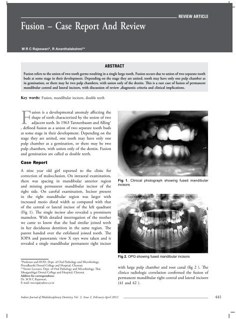
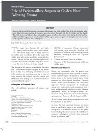
Fusion – Case Report And ReviewReview articleM R C Rajeswari*, R Ananthalakshmi**AbstractFusion refers to the union of two tooth germs resulting in a single large tooth. Fusion occurs due to union of two separate toothbuds at some stage in their development. Depending on the stage they are united, tooth may have only one pulp chamber asin gemination, or there may be two pulp chambers, with union only of the dentin. This is a rare case of fusion of permanentmandibular central and lateral incisors, with discussion of review ,diagnostic criteria and clinical implications.Key words: Fusion, mandibular incisors, double teethFusion is a developmental anomaly affecting theshape of teeth characterized by the union of twoadjacent teeth. In 1963 Tannenbaum and Alling 1, defined fusion as a union of two separate tooth budsat some stage in their development. Depending on thestage they are united, one tooth may have only onepulp chamber as a gemination, or there may be twopulp chambers, with union only of the dentin. Fusionand gemination are called as double teeth.Case ReportA nine year old girl reported to the clinic forcorrection of malocclusion. On intraoral examination,there was spacing in mandibular anterior regionand missing permanent mandibular incisor of theright side. On careful examination, Incisor presentin the right mandibular region was larger withincreased mesio distal width as compared with thatof the central or lateral incisor of the left quadrant(Fig 1). The single incisor also revealed a prominentmamelon. With detailed interrogation of the motherwe came to know that she had similar joined teethin her deciduous dentition in the same region. Theparent handed over the exfoliated joined tooth. TheIOPA and panoramic view X rays were taken and itrevealed a single mandibular permanent right incisorFig 1. Clinical photograph showing fused mandibularincisors*Professor and HOD, Dept. of Oral Pathology and Microbiology,Priyadharshi Dental College and Hospital, Chennai.**Senior Lecturer, Dept. of Oral Pathology and Microbiology, ThaiMoogambigai Dental College and Hospital, Chennai.Address for correspondenceDr. M R C Rajeswari,E-mail: mrcraj@yahoo.co.inFig 2. OPG showing fused mandibular incisorswith large pulp chamber and root canal (fig 2 ). Theclinico radiologic correlation confirmed the fusion ofpermanent mandibular right central and lateral incisors(41 and 42 ).Indian Journal of Multidisciplinary Dentistry, Vol. 2, <strong>Issue</strong> 2, February-April 2012441