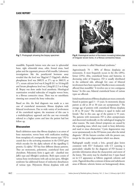
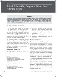
Case ReportFig 7. Photograph showing the biopsy specimenFig 8. Histological section of the lesion revealing trabeculaeof irregular woven bone, in a fibrous connective tissue.mandible. Expansile lesions were also seen in sphenoidalbone, right ethmoidal sinus, orbit, frontal sinus, hardpalate and the zygomatic process of left maxilla. Laboratoryinvestigations like the parathyroid hormone assayrevealed that the level was 58pg/ml (7-53pg/ml), alkalinephosphatase level was 300U/L at 37’c up to 306U/L at37’c, serum calcium level was 8.2mg/dl (8.1 to 10.4mg/dl)and Serum phosphorus level was 2.8mg/dl (2.5 to 4.8mg/dl. Biopsy was done under local anesthesia. Histologicalexamination revealed trabeculae of irregular woven bone,in a fibrous connective tissue. There was no osteoblasticrimming seen around the bony trabeculae.Based on this, the final diagnosis was made as a rarecase of craniofacial monostotic fibrous dysplasia withbilateral involvement. Due to wide variety of involvementof the craniofacial regions, the treatment of this case isa multidisciplinary approach and the case was eventuallyreferred to a higher centre and later the patient had lostfollow up.DiscussionReed’s definition states that fibrous dysplasia is an arrest ofbone maturation, woven bone with ossification resultingfrom metaplasia of a nonspecific fibro osseous type. 2 FD iscaused due to the activating mutations in the GNAS genewhich encodes for the alpha subunit of the signalling Gprotein, Gs alpha. 3 FD has four different disease patterns.They are monostotic, polyostotic, craniofacial form andcherubism. Polyostotic form is again sub - classified intoJaffe’s type and Albright’s syndrome. Both types consist ofvarious bony involvements with cafe-au-lait spots. Albrightsyndrome has additional feature of endocrine disturbancesof various type. Polyostotic fibrous dysplasia with softtissue myxomas is called Mazabraud syndrome. 4Approximately 70 – 80% of fibrous dysplasias aremonostotic. It most frequently occurs in the ribs (28%),femur (23%), tibia, craniofacial bones and humerus, indecreasing order of frequency. FD is usually distributedin the unilateral side, although few cases of bilateralinvolvement have been reported. Maxilla is more commonlyaffected than mandible. 4 It involves one or two contiguousbones. 5 In this case, bilateral craniofacial bones of varioustypes are affected.Initial manifestations of fibrous dysplasia are most commonlyfound in patients aged 3 – 15 years. In monostotic diseasepatient as old as 20 or 30 years are asymptomatic. 4 Theaverage age of patients with craniofacial fibrous dysplasiais 20 to 30 years. 6 The incidence is equal in males andfemale. 2 But in this case, age of the patient is 55 years.The patients with monostotic FD is often asymptomaticand discovered incidentally on the radiological imaging forother reasons. The main clinical symptoms are caused bythe expanding mass, which may result in bony deformity,and nasal or sinus obstruction. 5 Cystic degeneration mayoccur spontaneously in the FD lesions years after the initialdiagnosis. 7 All the features correlate with the present case,except for the cystic degeneration which was absent.Radiograph usually reveals a lytic, ground glass lesionconsistent with FD. 8 Evaluation with CT scanning isparticularly valuable for the diagnosis of FD because itprovides information about the characteristic and extent ofthe lesion. This entity may be classified into 3 types basedon its CT appearance as follows: pagetoid, sclerotic andcystic. Pagetoid describes a mixture of dense and radiolucentareas of fibrosis; sclerotic lesions are homogenously dense,462Indian Journal of Multidisciplinary Dentistry, Vol. 2, <strong>Issue</strong> 2, February-April 2012
Case Reportwhereas cystic describes one or more spherical or ovoidlucencies that are surrounded by a dense periphery. 9 MRIis useful in identifying fluid filled non FD-filled cysticlesions. 8 This case makes the second category of sclerotictype.FD is histologically characterized by a benign– appearing spindle cell fibrous stroma containingscattered, irregularly shaped trabeculae of immature( woven ) bone, lacking osteoblast, that evolve directlyfrom the stroma. 10 FD may occasionally contain cartilage,the amount of which is quite variable. This correlates withthe present case.Treatment for FD is always multidisciplinary, due to varyingsomatic mosaicism of craniofacial involvement of fibrousdysplasia, indicating need for a neuro- ophthalmologicaland an audiological testing. An early non surgical attempt oftreatment of FD includes glucocorticosteroids, calcitonin,and external beam radiation. 11 The only medications thathave shown any efficiency in the treatment of FD arebisphophonates. The most rigorously conducted study todate using high dose, intravenous pamidronate (1 – 1.5mg/kg per day on three consecutive days, given every 4months) was by Plotkin et al. 8 Generally, a conservativesurgical approach including facial osseous debulking andcontouring has been recommended for stable disease withminor symptoms. In progressive and more symptomaticcases with functional impairment and cosmeticdeformity, radical resection of diseased tissue, optic nervedecompression, and reconstruction of the facial areaswith autogenous bone grafts may be needed. 12 Althoughthe case of total resection of the craniofacial bones havebeen reported, the complete excision may result in thedeformity and loss of function of the surgically involvedpart and consequently lower the patient’s quality of lifecaused by the significant complications of the surgery suchas serious infection, leakage of cerebrospinal fluid, orbitaldamage, and intracranial injury. 13 Therefore endoscopicsinus surgery (ESS) is done to resect most of the diseasedbone. Stem cell treatment for FD is attractive. As such, asa stem cell disease, it may be a candidate for mesenchymalcell tissue engineering also. 14ConclusionFibrous dysplasia can be a good example of somaticmosaicism as a wide variety of disease is possible. Thegeneral dental practitioner can be the first to detect suchconditions especially when the only affected areas are in themaxilla-mandibular region, so sufficient knowledge on thiscondition is important for the proper diagnosis, treatmentand prevention of further complications. For obtaining thedefinite diagnosis, treatment and further management offibrous dysplasia, imaging studies, histological examinationand laboratory tests are considered mandatory. Effortsshould be made to define the optimal approach to thediagnosis and treatment of this challenging disease.ACKNOWLEDGEMENTDr. Dinakaran. J, MDS, Professor and Head of theDepartment, Department of Oral Pathology, SriRamakrishna Dental College and Hospital, Coimbatore.Dr. Jeyaraj J M, Principal, Head of the Department ofOrthodontics, Sri Ramakrishna Dental College andHospital, Coimbatore.ABBREVATIONSFD – fibrous dysplasiaBMSCs – bone marrow stromal cellsGNAS – guanine nucleotide binding protein, alphastimulatingcAMP – cyclic adenosine monophosphateOPG – orthopantamogramCT – computed tomographyMRI – magnetic resonance imagingReferences1. Ben hadj Hamida F, Jlaiel R, Ben Rayana N, MahjoubH, Mellouli T, Ghorbel M, Krifa F. Craniofacial fibrousdysplasia: A case report. J Fr. Ophthalmol, (2005);Oct;28(8):e6.2. Cholakova. R,Kanasirska. P, Kanasirska. N, Chenchev. Iv,Dinkova. A. Fibrous dysplasia in the maxillomandibularregion – A case report, Journal of IMAB – AnnualProceeding (Scientific Paper) (2010); vol. 16, book 4.3. Bianco P, Riminucci M, Gronthos S, Robey PG Bonemarrow stromal stem cells: nature, biology, and potentialapplications. Stem cells (2001); 19:180-192.4. Suneedh Gupta, Umesh. K, Warad N. M and ShakeelAhmed. Fibrous dysplasia of maxillary bone: A casereport. Al Ameen J Med Sci (2011); 4(1):92-97.5. Zhaowei Gu, Zhiwei Cao, Yunxiu Wang. EndoscopicManagement in Fibrous Dysplasia of Ethmoid Sinus:One case report and literature review.. Clin OncolCancer Res (2009); 6:152-154.Indian Journal of Multidisciplinary Dentistry, Vol. 2, <strong>Issue</strong> 2, February-April 2012463