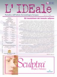
Histological evaluation of prostatic tissue following transurethral laser resection (TULaR) using the 980 nm diode laserFigure 4.Stromal oedema associated with ectasic vessels butwithout extravasation of red blood cells after treatmentwith the diode 980 nm laser at 100 W (H & E; 100X).Figure 7.Extravasation of red blood cells and hemorrhagicareas after transurethral resection of the prostate(TURP) (H & E; 100X).Figure 5.Occlusion of small vessels after treatmentwith the diode 980 nm laser at 100 W (H & E; 400X).Figure 6.Histological samples following transurethral resectionof the prostate (TURP) showed a coagulation rimof 0.1 mm (H & E; 100X).ulation rim of 0.5 mm (range: 0.2-1 mm) (Figure 2) andadjacent to the vaporized tissue, coagulated connective tissueand glandular epithelia were seen. Beyond this zone acomplete detachment of glandular epithelia from the connectivetissue was observed (Figure 3). Stromal oedemaassociated with ectasic vessels but without extravasation ofred blood cells, haemosiderin deposition and haemorrhagicareas were also retrieved (Figure 4). All cases showedocclusion of small vessels beyond the zone of coagulatedtissue (Figure 5).Collections of lymphocytes, probably related to a previouschronic prostatitis, were an occasional finding.Samples obtained from TURP ranged in size from 5 mmto 20 mm and showed black, irregular margins.Histological evaluation showed a coagulation rim of 0.3mm (range: 0.1-0.5 mm) (Figure 6). Next to the vaporizedtissue, coagulated connective tissue and glandularepithelia were seen, but beyond this zone no detachmentof glandular epithelia from the connective tissue wasobserved. Unlike laser treatment, samples obtained fromTURP showed extravasation of red blood cells,haemosiderin deposition and haemorrhagic areas (Figu -re 7). No incidence of adenocarcinoma was noted in anyof the prostate tissue samples.DISCUSSIONThis study is the first report on the histological effectson prostate tissue following ablation with the 980 nmdiode laser and the first time that such effects have beencompared with the morphological changes caused byTURP. Data show that the coagulation depth with the980 nm laser is consistently under 1 mm with 100 Wpulsed power when used in contact mode, providinglow thermal diffusion into the tissue. At the same time,the occlusion of small vessels justifies the haemostaticpro<strong>per</strong>ties with minimal bleeding during the procedure.Histological evaluation showed two distinct zones withinthe ablated samples. The zone where the completedetachment of glandular epithelia from the connectiveArchivio Italiano di Urologia e Andrologia 2010; 82, 13
R. Leonardi, R. Caltabiano, S. Lanzafametissue was observed may result from lower thermal damagedue to the wavelength used and to the techniqueitself. Unlike laser treatment, samples obtained fromTURP did not show detachment of glandular epitheliafrom the connective tissue. This finding may be particularlyrelevant because the glandular epithelial detachmentwith minimal necrotic damage obtained with thelaser treatment could lead to fibroblast activation andsubsequent scarring in the prostate. This should facilitatea quickly recovery of the patient with few irritativesymptoms after the treatment; however, further studiesare needed in order to evaluate biopsy samples a fewweek after laser treatment to confirm our hypothesis.An histological study of the changes induced duringholmium laser resection of the prostate (HoLRP)revealed that changes that took place could be mistakenfor malignant change. Thermal injury was moreextensive than previously considered and artifactsobserved under low power consisted of glandular distortionand contraction with crowding. Higher magnificationrevealed clumping of the chromatin of the nucleus,resulting in hy<strong>per</strong>chromasia and irregularity of thenucleus and loss of polarity (6). A later study has comparedHoLRP and TURP and showed that HoLRPcaused significant tissue vaporization and greater thermaldamage than TURP (7). However, prostatic architecturewas maintained in the majority of histologicalspecimens. A comparative study looked at the histologicaleffects on prostate tissue induced with HoLEP andTURP (8). Tissue samples that were removed duringHoLEP revealed major histological alterations resultingfrom resection and coagulation on the external circumferenceof the enucleated tissue. Similar architecturaland cytological artifacts were observed in HoLEP andTURP tissue specimens. These included distortion ofthe glandular structure with artifactual cellular detachmentfrom the underlying basement membrane. The tissueobtained by TULaR can be used for histologicaldiagnostic examination. Histological examination onthe specimen obtained by laser resection is reallyimportant because it will confirm the BPH and excludethe presence of a malignant neoplasm not identified byprevious biopsies usually <strong>per</strong>formed before the lasertreatment. In the comparative study on HoLEP andTURP, incidental carcinomas were identified in 7.5% ofHoLEP samples and 10% of TURP specimens; highgrade PIN was shown in 10% of samples in each treatmentgroup (8). Although in the current study no casesof adenocarcinoma were reported, the sampling of tissueduring TULaR leaves open the possibility of detectingmalignant tissue.CONCLUSIONSThe 980 nm diode laser provides high rates of tissueablation, associated with excellent haemostasis. It hasbeen shown that tissue samples can be obtained withthis technique, which allow a histological diagnosis ofBPH to be made. The current method involving the 980nm diode laser induces a vaporesection of prostate tissueand the acronym of TULaR (transurethral laserresection) has therefore been created to describe thistechnique.REFERENCES1. Lytton B, Emery JM, Harvard B. The incidence of benign prostaticobstruction. J Urol 1968; 99:639.2. Montorsi F, Guazzoni G, Bergamaschi F et al. Long term clinicalreliability of transurethral and open prostatectomy for benign prostaticobstruction: A term of comparison for nonsurgical procedure.Eur Urol 1993; 23:262.3. de la Rosette JJ, Collins E, Bachmann A, et al. Historical aspectsof laser therapy for benign prostatic hy<strong>per</strong>plasia. Eur Urol Suppl2008; 7:363.4. Reich O, Gratzke C, Stief CG. Techniques and long-term resultsof surgical procedures for BPH. Eur Urol 2006; 49:970.5. Leonardi R. Preliminary results on selective light vaporizationwith the side-firing 980 nm diode laser in benign prostatic hy<strong>per</strong>plasia:an ejaculation sparing technique. Prostate Cancer ProstaticDis 2009; 12:277.6. Gan E, Costello A, Slavin J, Stillwell RG. Pitfalls in the diagnosisof prostate adenocarcinoma from holmium resection of the prostate.Tech Urol 2000; 6:185.7. Das A, Kennett KM, Sutton T, Fraundorfer MR, Gilling PJ.Histologic effects of holmium:YAG laser resection versustransurethral resection of the prostate. J Endourol 2000; 14:459.8. Naspro R, Freschi M, Salonia A et al. Holmium laser enucleationversus transurethral resection of the prostate. Are histological findingscomparable. J Urol 2004; 171:1203.CorrespondenceRosario Leonardi, MDDepartment of Urology, Clinica BasileVia Odorico da Pordenone 5 - 95128 Catania, Italyleonardi.r@tiscali.itRosario Caltabiano, MDDepartment GF IngrassiaSection of PathologyUniversity of CataniaVia Santa Sofia 87, 93123 Catania, Italyrosario.caltabiano@unict.itSalvatore Lanzafame, MDDepartment GF IngrassiaSection of PathologyUniversity of CataniaVia Santa Sofia 87, 93123 Catania, Italylanzafa@unict.it4Archivio Italiano di Urologia e Andrologia 2010; 82, 1
- Page 2 and 3: Official Journal of the SIEUN, the
- Page 4 and 5: ContentsHistological evaluation of
- Page 7: R. Leonardi, R. Caltabiano, S. Lanz
- Page 11 and 12: F. Galasso, R. Giannella, P. Bruni,
- Page 13 and 14: F. Galasso, R. Giannella, P. Bruni,
- Page 15 and 16: ORIGINAL PAPERSurgery for renal cel
- Page 17 and 18: S.D. Dyakov, G. Lucarelli, A.I. Hin
- Page 19 and 20: S.D. Dyakov, G. Lucarelli, A.I. Hin
- Page 21 and 22: M. Aza, S.S. Iqbal, M.V. Muhammad,
- Page 23 and 24: Archivio Italiano di Urologia e And
- Page 25 and 26: The Clavien classification system t
- Page 27 and 28: PRESENTATIONPercutaneous nephrolith
- Page 29 and 30: Percutaneous nephrolithotomy: An ex
- Page 31 and 32: PCNL in ItalyTable 1.Number and hos
- Page 33 and 34: PCNL in ItalyFigure 3.Comparison be
- Page 35 and 36: The patient position for PNL: Does
- Page 37 and 38: PCNL: Tips and tricks in targeting,
- Page 39 and 40: Tubeless percutaneous nephrolithoto
- Page 41 and 42: PRESENTATIONHigh burden and complex
- Page 43 and 44: High burden and complex renal calcu
- Page 45 and 46: PRESENTATIONEndoscopic combined int
- Page 47 and 48: PRESENTATIONHigh burden stones: The
- Page 49 and 50: PRESENTATIONStone treatment in chil
- Page 51 and 52: Stone treatment in children: Where
- Page 53 and 54: PRESENTATIONExtracorporeal shock wa
- Page 55 and 56: PRESENTATIONPercutaneous nephrolith
- Page 57 and 58: PRESENTATIONFlexible ureteroscopy f
- Page 59 and 60:
Flexible ureteroscopy for kidney st
- Page 61 and 62:
Indications, prediction of success
- Page 63 and 64:
Indications, prediction of success
- Page 65 and 66:
Indications, prediction of success
- Page 67 and 68:
Indications, prediction of success
- Page 69 and 70:
Laparoscopic and open stone surgery
- Page 71 and 72:
Laparoscopic and open stone surgery
- Page 73 and 74:
Laparoscopic and open stone surgery
- Page 75:
Laparoscopic and open stone surgery