Cloning Cytochrome b PCR Fragment into pGEM-T - Cornell College
Cloning Cytochrome b PCR Fragment into pGEM-T - Cornell College
Cloning Cytochrome b PCR Fragment into pGEM-T - Cornell College

Create successful ePaper yourself
Turn your PDF publications into a flip-book with our unique Google optimized e-Paper software.
1/12<strong>Cloning</strong> & Sequencing the <strong>Cytochrome</strong> b Gene using <strong>pGEM</strong>-T VectorsI. Gel Analysis and Purification using IBI’s Gel/<strong>PCR</strong> DNA <strong>Fragment</strong> Extraction Kit1. Make a 1.2% (w/v) low-melting agarose gel in 1X TAE (Tris, acetate, EDTA) buffer bydiluting the 50X concentrated TAE stock solution.50X Tris-acetate EDTA buffer (for 500 mL)a. 2.0M Tris – 121gb. 28.6ml of glacial acetic acidc. 50ml of 0.5M EDTA (pH 8.0) - 9.3g EDTA/50mld. Adjust pH to 8.0e. autoclave2. Place the flask onto a ring stand and gently swirl the flask while heating the agarose solutionover a Bunsen burner. Bring the solution to a boil and remove the flask from the burner. BECAREFUL NOT TO LET THE AGAROSE SOLUTION BOIL OUT OF THE FLASK.3. Allow the agarose to cool to 60˚C and add 0.5 g/mL of ethidium bromide (our stock ofethidium bromide is 10 mg/mL) and gently mix. WEAR GLOVES FROM THIS POINTON BECAUSE ETHIDIUM BROMIDE IS A MUTAGEN! Wash the pipette tip well inwater before you dispose of the tip.4. Pour the agarose <strong>into</strong> the gel tray. Make sure the gel comb is inserted <strong>into</strong> the tray. Fill thetray with agarose until the volume reaches the top of the comb’s teeth. Allow the gel to hardenfor approximately 20-30 minutes.5. Add 4 μl of sterile DNA tracking dye to each sample. Mix well and centrifuge for 5 seconds.Run UNDIGESTED as a control (0.3 g) and add tracking dye to this sample. Prepare asample containing a 100 bp ladder (5-6 l) with tracking dye. Place the samples back on ice.6. After your gel has hardened, gently remove the comb and the side supports from the gel. Placethe gel <strong>into</strong> the chamber and add enough 1X TAE buffer to cover the gel.7. Carefully load your samples <strong>into</strong> the gel using a micropipette. Load the entire sample <strong>into</strong>the gel. Be sure to record what wells contain your samples.8. Place the cover onto the electrophoretic chamber and connect the electrodes to the powersupply. Run the gel between 50-60 volts.9. Analyze your results under an ultraviolet light source and photograph.10. Excise the DNA fragment(s) of interest (remove as much agarose as possible) using a cleanrazor blade. Transfer the up to 300 mg of gel slice (from the 4 <strong>PCR</strong> reactions) <strong>into</strong> amicrocentrifuge tube. (The maximum capacity of each column is about 300 mg of gel mass.).11. Add 500 l of DF buffer to the sample and mix by vortexing.1
12. Incubate the sample at 55-60 ˚C for 15 minutes or until the gel slice(s) are completelydissolved. During the incubation invert the tube every 2-3 minutes. Cool the dissolvedsample mixture to room temperature. (Once the agarose gel is melted, the gel will notresolidify at room temperature.)13. Place one DF column in a 2 mL collection tube for each tube containing dissolved gel slices.14. Transfer the dissolved gel mixture (up to 800 l) to the DF column.15. Centrifuge the DF column assembly in a microcentrifuge (use the larger microcentrifuge) attop speed for 30 seconds. Discard the flow-through and place the DF column back in the 2mL collection tube. (If the samplex mixture was more than 800 l, transfer the remaining gelmixture to the DF column and repeat step 15.)16. Add 400 l of W1 buffer <strong>into</strong> the DF column and microcentrifuge (top speed) for 30 seconds.Discard the flow-through. .17. Place the DF column back <strong>into</strong> the 2 mL collection tube. Add 600 l of Wash buffer (be surethat ethanol has been added to the Wash buffer when the kit was first opened) <strong>into</strong> the DFcolumn and allow to incubate for 1 minute at room temperature.18. Microcentrifuge the sample for 30 seconds (top speed) and discard the flow-through.19. Place the DF column back <strong>into</strong> the 2 mL collection tube. Microcentrifuge (top speed) againfor 3 minutes in order to dry the matrix.20. Transfer the dried DF column to a new 1.5 mL microcentrifuge tube. Add 15-50 l ofElution buffer <strong>into</strong> the center of the column matrix. Let the column sit for 2 minutes or untilthe Elution buffer is absorbed by the matrix.21. Centrifuge (top speed) for 2 minutes to elute the purified DNA. If you started with multiplegel slices of the SAME sample that required multiple columns, combine your eluted DNA<strong>into</strong> one tube.22. Determine the mass and concentration of your purified <strong>PCR</strong> product by scanning the DNAfrom 310-220 nm in the spectrophotometer.II.<strong>Cloning</strong> <strong>PCR</strong> Products using the <strong>pGEM</strong>-T <strong>Cloning</strong> KitA. Ligation of <strong>PCR</strong> Product <strong>into</strong> <strong>pGEM</strong>-T1. Briefly centrifuge the <strong>pGEM</strong>-T tube to bring the contents to the bottom of the tube.2. Add the following to a 0.5 ml <strong>PCR</strong> microcentrifuge tube:5 μl 2X Rapid Ligation Buffer, (Vortex the 2X Rapid Ligation Buffervigorously before each use.)2
1 μl <strong>pGEM</strong>-T Vector (50 ng)<strong>PCR</strong> product (see calculation**) Use a 3:1 insert to vector ratio.3 Weiss units T4 DNA LigaseSterile dH 2 O to a final volume of 10 μl**Calculation of <strong>PCR</strong> product mass:(ng of vector x kb size of insert) / (kb size of vector) x (insert:vector molar ratio) = ng of insertExample a 3:1 ratio of insert:vector (assuming the <strong>PCR</strong> product is 0.6 kb) is(50 ng vector)(0.6 kb insert) / (3.0 kb vector) x (3/1) = 30.0 ng of insert3. Mix the reactions by pipetting. Incubate the reactions overnight at 4˚C.B. Preparation of LB Plates with IPTG and X-Gal1. Inoculate 2 LB plates (containing ampicillin at 50 g/mL) with IPTG and X-Gal per ligationreaction.2. LB platesBacto-tryptone10.0 g/LBacto-yeast5.0 g/LNaCl10.0 g/LAdd dH 2 O to 800 mL.Adjust the pH to 7.5Bring the volume to 1 LAdd 1.5% (w/v) Bacto-AgarAutoclave and allow media to coolAdd 50 µg/ml of dry ampicillin, mix well and pour about 20-25 mL <strong>into</strong> each Petri dish.3. IPTG/X-Gal Platesa. Mix 4 µl of 200 µg/µl IPTG stock with 16 µl of X-Gal (the X-Gal stock is 50mg/ml) per plate.b. Then add 20 µl onto the center of each LB agar plate and spread evenly (usesterile technique).c. Incubate the plates at 37˚C until the fluid disappears.d. Equilibrate the plates to room temperature prior to plating.C. Transformation of the Ligation Mixture <strong>into</strong> JM109 using Blue/White Selection1. Briefly centrifuge the samples containing the ligation reactions. Add 5 μl of each ligationreaction to a microcentrifuge tube containing 100 l of competent JM109 cells on ice.2. Plate 50 μl of the transformation culture onto duplicate pre-warmed LB/ampicillin/IPTG/X-Gal plates (two plates per transformation).3
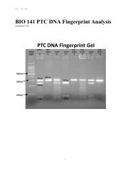
3. Incubate the plated cells overnight at 37˚C. Approximately 20-50 colonies per plate areroutinely seen. White colonies generally contain cloned insert DNA while blue colonies donot. Blue colonies should also be checked for cloned insert DNA.III.Plasmid Clone Isolation for Characterization using Zyppy Plasmid Miniprep KitA. E. coli Inoculation1. In order to determine if your transformants contain plasmids with <strong>PCR</strong> amplifiedmitochondria cytochrome b gene, inoculate a single white colony (blue colonies do notcontain <strong>PCR</strong> products) from your transformation plate <strong>into</strong> sterile liquid LB (10 mL)containing 50 g/mL of ampicillin. Inoculate up to 6 separate white colonies per plate(inoculated <strong>into</strong> 6 different tubes of LB; one colony per tube of LB). Grow thetransformants overnight at 37°C in the shaking incubator (200 RPM).LB liquid cultureBacto-tryptone10.0 g/LBacto-yeast5.0 g/LNaCl10.0 g/LAdd dH 2 O to 800 mL.Adjust the pH to 7.5Bring the volume to 1 LAliquot the liter of solution <strong>into</strong> appropriate volumesAutoclaveB. Buffer Preparations.Before beginning the protocol, make sure the following preparations have been completed.1. Add RNase A to the Neutralization Buffer: aliquot 1 mL of Neutralization Buffer <strong>into</strong> the tubecontaining the lyophilized RNase A, mix and transfer the solution back <strong>into</strong> the NeutralizationBuffer bottle. This buffer must be stored at 4° C.2. Add 24 mL of 100% ethanol (or 26 mL of 95% ethanol) to the 6 mL Zyppy Wash Bufferbefore use.3. The 7X Lysis Buffer may have a precipitate. If you notice a precipitate in this buffer, heat thebuffer at 37°C for 15 minutes and mix the bottle by gentle inversion.C. Plasmid Isolation Protocol1. Mix your overnight culture of cells completely and pipette 1.5 mL of cells <strong>into</strong> amicrocentrifuge tube. Centrifuge the culture in the microcentrifuge for 30 seconds. Discardthe supernatant.4
2. Repeat step one using the same microcentrifuge tube. After you discard the supernatant,completely resuspend the cells in 600 l of sterile dH 2 O.DO NOT DISCARD THE ORIGINAL OVERNIGHT CULTURE. IF THE CLONINGAND TRANSFORMATION ARE SUCCESSFUL, YOU WILL NEED THIS CULTURETO MAKE STOCKS OF THE TRANSFORMANTS.3. Add 100 l of 7X Lysis Buffer and mix the sample by inverting the tube 4-6 times. Thesolution should change from opaque to clear blue indicating complete lysis. You mustproceed to the next step within 2 minutes.4. Add 350 l of cold Neutralization Buffer and mix thoroughly. The sample will turn yellowwhen the neutralization is complete and a yellowish precipitate will form. Invert the samplean additional 2-3 times to ensure complete neutralization.5. Centrifuge the sample at top speed in the Marathon microcentrifuge for 4 minutes.6. Transfer the supernatant (~900 l) <strong>into</strong> a Zymo-Spin II column. Avoid disturbing the celldebris pellet.7. Place the column in a collection tube (from the kit) and centrifuge for 15 seconds.8. Discard the flow-through and place the column back <strong>into</strong> the same collection tube.9. Add 200 l of Endo-Wash Buffer to the column. Centrifuge for 15 seconds. Empty thecollection tube.10. Add 400 l of Zyppy Wash Buffer to the column. Centrifuge for 30 seconds.11. Transfer the column <strong>into</strong> a clean 1.5 mL microcentrifuge tube. Add 30 l of Zyppy ElutionBuffer directly to the column matrix and let the column stand for 1 minute at roomtemperature.12. Centrifuge the column for 15 seconds to elute the plasmid DNA.13. Scan your DNA sample from 310-225 nm on the spectrophotometer. You will need to have avolume of 50 l. Your peak absorbance should be between 257-260 nm.DNA Mass = (OD @ 257 nm) (50 g/mL) (Volume – mL).V. Endonuclease Analysis of Plasmid DNA Isolated from Transformants1. Add the proper amount of DNA (0.75 g of molecular weight markers pre-digested withHind III and/or Eco RI, 6 l of 100 base pair ladder, 0.5 g of isolated plasmid that you planto digest) <strong>into</strong> separate sterile microcentrifuge tubes. Include one sample of undigestedplasmid DNA as a negative control.5
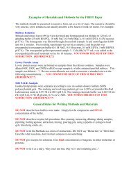
Mix well. Add water last and mix the components well by pipetting the reaction up anddown several times with the same tip. Simply tapping the tube is not sufficient to mix.**One of the perennial problems of DNA sequencing occurs when analyzing DNA segments thatare G-rich. Basically, DNA structure dictated by Watson-Crick base pairing is disrupted in G-rich segments because of their ability to create inter and intra strand hydrogen bonding. Thisaggregation causes enzymatic disruption so sequencing and <strong>PCR</strong> experiments become highlyproblematical. The problem arises from extra hydrogen bonding forming at the N7 position ofG. Traditionally, 7-deaza-G has been used to overcome these problems with some successbecause it eliminates intra strand hydrogen bonding.7-deaza dGTPdGTP3. Label a set of four 0.2 mL tubes A, T, G and C for each template/primer combination.4. Add 4.0 µl of the A Termination mix (purple-capped tube) to the A tube(s), and the TTermination mix to the T tube(s). Do the same for the G and T tubes.5. Add 4.0 µl of the appropriate template/primer combination (master mix from step 2) to eachA, T, G, and C tube and mix well.6. Carefully seal the tubes, place in thermal cycler and start the program.7. Pour a 5.5% acrylamide sequencing gel (8M urea).8. At the completion of the cycling program, add 3 µl of Stop Solution to each tube.9. Denature samples at 92°C for 3 minutes and place on ice prior to loading.10. Pre-run the gel in 0.8X TBE and load the samples (between 0.5 l, and 1.0 l) <strong>into</strong> the gel.The gel runs for 9 hours.7