Info - Leica Biosystems
Info - Leica Biosystems
Info - Leica Biosystems
You also want an ePaper? Increase the reach of your titles
YUMPU automatically turns print PDFs into web optimized ePapers that Google loves.
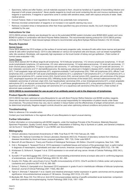
• Specimens, before and after fixation, and all materials exposed to them, should be handled as if capable of transmitting infection anddisposed of with proper precautions 2 . Never pipette reagents by mouth and avoid contacting the skin and mucous membranes withreagents or specimens. If reagents or specimens come in contact with sensitive areas, wash with copious amounts of water. Seekmedical advice.• Consult Federal, State or local regulations for disposal of any potentially toxic components.• Minimize microbial contamination of reagents or an increase in non-specific staining may occur.• Retrieval, incubation times or temperatures other than those specified may give erroneous results. Any such change must bevalidated by the user.Instructions for UseCD10 (56C6) primary antibody was developed for use on the automated BOND system (includes <strong>Leica</strong> BOND-MAX system and <strong>Leica</strong>BOND-III system) in combination with Bond Polymer Refine Detection. The recommended staining protocol for CD10 (56C6) primaryantibody is IHC Protocol F. Heat induced epitope retrieval is recommended using Bond Epitope Retrieval Solution 2 for 20 minutes.Results ExpectedNormal TissuesClone 56C6 detects the CD10 antigen on the surface of normal early progenitor cells, immature B cells within bone marrow and germinalcenter B cells within lymphoid tissue. CD10 is also detected on various non-lymphoid cells and tissues, such as breast myoepithelialcells, bile canaliculi, fibroblasts, with especially high expression on the brush border of kidney and gut epithelium. (Total number ofnormal cases evaluated = 85).Tumor TissuesClone 56C6 stained 26/116 diffuse large B-cell lymphomas, 10/15 follicular lymphomas, 1/12 chronic lymphocytic lymphomas, 1/1 B-cellacute lymphoblastic lymphoma, 2/2 seminomas, 2/2 colon adenocarcinomas, 1/2 rectal adenocarcinomas, 1/2 renal cell carcinomas, 1/1brain choroid plexus papilloma, 1/1 larynx squamous cell carcinoma, 1/1 soft tissue fibromatosis, 1/1 lung non-small cell carcinoma, 1/1liver metastatic carcinoma and 1/1 dermatofibrosarcoma. No staining was observed in Hodgkin’s disease (0/27), mantle cell lymphomas(0/7), T-cell anaplastic large cell lymphomas (0/7), angioimmunoblastic T-cell lymphomas (0/4), T/NK cell lymphomas (0/3), diffuse T-celllymphomas (0/2), a primitive B/T cell acute lymphoblastic lymphoma (0/1), a peripheral T-cell lymphoma (0/1), a T-cell lymphoma (0/1), amarginal zone lymphoma (0/1), ovarian tumors (0/4), thyroid tumors (0/4), cervical tumors (0/2), squamous cell carcinomas of the tongue(0/2), esophageal squamous cell carcinomas (0/2), infiltrating ductal carcinomas of the breast (0/2), stomach adenocarcinomas (0/2),metastatic carcinomas of unknown origin (0/2), liver hepatocellular carcinomas (0/2), a liver cholangiocarcinoma (0/1), a brain anaplasticastrocytoma (0/1), an atypical carcionoid of the thymus (0/1), a soft tissue ganglioneuroma (0/1), a lung adenocarcinoma (0/1), a lungsquamous cell carcinoma (0/1), a lung large cell carcinoma (0/1) or a squamous cell carcinoma of the skin (0/1). (Total number ofabnormal cases evaluated = 242).CD10 (56C6) is recommended for use as part of an antibody panel to aid in the diagnosis of lymphomas.Product Specific LimitationsCD10 (56C6) has been optimized at <strong>Leica</strong> <strong>Biosystems</strong> for use with Bond Polymer Refine Detection and BOND ancillary reagents.Users who deviate from recommended test procedures must accept responsibility for interpretation of patient results under thesecircumstances. The protocol times may vary, due to variation in tissue fixation and the effectiveness of antigen enhancement, and mustbe determined empirically. Negative reagent controls should be used when optimizing retrieval conditions and protocol times.TroubleshootingRefer to reference 3 for remedial action.Contact your local distributor or the regional office of <strong>Leica</strong> <strong>Biosystems</strong> to report unusual staining.Further <strong>Info</strong>rmationFurther information on immunostaining with BOND reagents, under the headings Principle of the Procedure, Materials Required,Specimen Preparation, Quality Control, Assay Verification, Interpretation of Staining, Key to Symbols on Labels, and General Limitationscan be found in “Using BOND Reagents” in your BOND user documentation.Bibliography1. Clinical Laboratory Improvement Amendments of 1988, Final Rule 57 FR 7163 February 28, 1992.2. Villanova PA. National Committee for Clinical Laboratory Standards (NCCLS). Protection of laboratory workers from infectiousdiseases transmitted by blood and tissue; proposed guideline. 1991; 7(9). Order code M29-P.3. Bancroft JD and Stevens A. Theory and Practice of Histological Techniques. 4th Edition. Churchill Livingstone, New York. 1996.4. Ordi J, Romagosa C, Tavassoli FA et al. CD10 expression in epithelial tissues and tumours of the gynecologic tract: a useful markerin diagnosis of mesonephric, trophoblastic and clear cell tumors. American Journal of Surgical Pathology 2003 27(2), 178–186.5. Kristiansen G, Schluns K, Yongwei Y et al. CD10 expression in non-small cell lung cancer. Annals of Cellular Pathology 2002 24(1),41–46.6. Sumathi V P and McCluggage WG. CD10 is useful in demonstrating endometrial stroma at ectopic sites and in confirming diagnosisof endometriosis. Journal of Clinical Pathology 2002 55, 391–392.7. Dunphy CH, Polsky JM, Lance Evans H et al. Paraffin immunoreactivity of CD10, CDw75 and Bcl-6 in follicle center cell lymphoma.Leukaemia and Lymphoma 2001 41(5–6), 585–592.8. Eshoa C, Perkins S, Kampalath et al. Decreased CD10 expression in grade III and in interfollicular infiltrates of follicular lymphomas.American Journal of Clinical Pathology 2001 115(6), 862–867.9. Tajima Y, Shimoda T, Nakanishi Y et al. Gastric and intestinal phenotypic marker expression in gastric carcinomas and its prognosticsignificance: immunohistochemical analysis of 136 lesions. Oncology 2001 61(3), 212–220.10. Bavikatty NR, Ross CW, Finn WG et al. Anti-CD10 immunoperoxidase staining of paraffin-embedded acute leukemias: comparisonwith flow cytometric immunophenotyping. Human Pathology 2000 31(9), 1051–1054.PA0270Page 3


