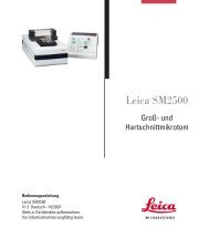
Info - Leica Biosystems
Info - Leica Biosystems
Info - Leica Biosystems

You also want an ePaper? Increase the reach of your titles
YUMPU automatically turns print PDFs into web optimized ePapers that Google loves.
Bond Ready-to-Use Primary AntibodyCD10 (56C6)Catalog No: PA0270<strong>Leica</strong> <strong>Biosystems</strong> Newcastle LtdBalliol Business Park WestBenton LaneNewcastle Upon Tyne NE12 8EWUnited Kingdom( +44 191 215 4242Instructions for UsePlease read before using this product.Mode d’emploiÁ lire avant d’utiliser ce produit.Istruzioni per l’usoSi prega di leggere, prima di usare il prodotto.GebrauchsanweisungBitte vor der Verwendung dieses Produkts lesen.Instrucciones de usoPor favor, leer antes de utilizar este producto.Instruções de UtilizaçãoLeia estas instruções antes de utilizar este produto.BruksanvisningVar god läs innan ni använder produkten.Οδηγίες ΧρήσηςΠαρακαλούμε διαβάστε τις οδηγίες πριν χρησιμοποιήσετε το προϊόν αυτό.BrugsanvisningLæs venligst før produktet tages i brug.GebruiksinstructiesLezen vóór gebruik van dit product.BruksanvisningVennligst les denne før du bruker produktet.Kullanım TalimatlarıLütfen bu ürünü kullanmadan önce okuyunuz.Check the integrity of the packaging before use.Vérifier que le conditionnement est en bon état avant l’emploi.Prima dell’uso, controllare l’integrità della confezione.Vor dem Gebrauch die Verpackung auf Unversehrtheit überprüfen.Comprobar la integridad del envase, antes de usarlo.Verifique a integridade da embalagem antes de utilizar o produto.Kontrollera att paketet är obrutet innan användning.Ελέγξτε την ακεραιότητα της συσκευασίας πριν από τη χρήση.Kontroller, at pakken er ubeskadiget før brug.Controleer de verpakking vóór gebruik.Sjekk at pakningen er intakt før bruk.Kullanmadan önce ambalajın bozulmamış olmasını kontrol edin.www.<strong>Leica</strong><strong>Biosystems</strong>.comEN FR IT DE ES PTSV EL DA NL NO TR
PA0270Page 1
Bond Ready-To-Use Primary AntibodyCD10 (56C6)Catalog No: PA0270Intended UseThis reagent is for in vitro diagnostic use.CD10 (56C6) monoclonal antibody is intended to be used for the qualitative identification by light microscopy of the human CD10molecule in formalin-fixed, paraffin-embedded tissue by immunohistochemical staining using the automated BOND system (includes<strong>Leica</strong> BOND-MAX system and <strong>Leica</strong> BOND-III system).The clinical interpretation of any staining or its absence should be complemented by morphological studies and proper controls andshould be evaluated within the context of the patient’s clinical history and other diagnostic tests by a qualified pathologist.Summary and ExplanationImmunohistochemical techniques can be used to demonstrate the presence of antigens in tissue and cells (see “Using BOND Reagents”in your BOND user documentation). CD10 (56C6) primary antibody is a ready to use product that has been specifically optimized for usewith Bond Polymer Refine Detection. The demonstration of the human CD10 molecule is achieved by first allowing the binding of CD10(56C6) to the section, and then visualizing this binding using the reagents provided in the detection system. The use of these products, incombination with the automated BOND system (includes <strong>Leica</strong> BOND-MAX system and <strong>Leica</strong> BOND-III system), reduces the possibilityof human error and inherent variability resulting from individual reagent dilution, manual pipetting and reagent application.Reagents ProvidedCD10 (56C6) is a mouse anti-human monoclonal antibody produced as a tissue culture supernatant, and supplied in Tris buffered salinewith carrier protein, containing 0.35 % ProClin 950 as a preservative.Total volume = 7 mL.Clone56C6ImmunogenProkaryotic recombinant fusion protein corresponding to the external domain of the human CD10 glycoprotein.SpecificityHuman CD10 molecule, also known as common acute lymphocytic leukemia antigen (CALLA).Ig ClassIgG1.Total Protein ConcentrationApprox 10 mg/mL.Antibody ConcentrationGreater than or equal to 1 mg/L as determined by ELISA.Dilution and MixingCD10 (56C6) primary antibody is optimally diluted for use on the BOND system (includes <strong>Leica</strong> BOND-MAX system and <strong>Leica</strong> BOND-IIIsystem). Reconstitution, mixing, dilution or titration of this reagent is not required.Materials Required But Not ProvidedRefer to “Using BOND Reagents” in your BOND user documentation for a complete list of materials required for specimen treatment andimmunohistochemical staining using the BOND system (includes <strong>Leica</strong> BOND-MAX system and <strong>Leica</strong> BOND-III system).Storage and StabilityStore at 2–8 °C. Do not use after the expiration date indicated on the container label.The signs indicating contamination and/or instability of CD10 (56C6) are: turbidity of the solution, odor development, and presence ofprecipitate.Return to 2–8 °C immediately after use.Storage conditions other than those specified above must be verified by the user 1 .Precautions• This product is intended for in vitro diagnostic use.• The concentration of ProClin 950 is 0.35 %. It contains the active ingredient 2-methyl-4-isothiazolin-3-one, and may cause irritationto the skin, eyes, mucous membranes and upper respiratory tract. Wear disposable gloves when handling reagents.• To obtain a copy of the Material Safety Data Sheet contact your local distributor or regional office of <strong>Leica</strong> <strong>Biosystems</strong>, or alternatively,visit the <strong>Leica</strong> <strong>Biosystems</strong>’ Web site, www.<strong>Leica</strong><strong>Biosystems</strong>.comPA0270Page 2
• Specimens, before and after fixation, and all materials exposed to them, should be handled as if capable of transmitting infection anddisposed of with proper precautions 2 . Never pipette reagents by mouth and avoid contacting the skin and mucous membranes withreagents or specimens. If reagents or specimens come in contact with sensitive areas, wash with copious amounts of water. Seekmedical advice.• Consult Federal, State or local regulations for disposal of any potentially toxic components.• Minimize microbial contamination of reagents or an increase in non-specific staining may occur.• Retrieval, incubation times or temperatures other than those specified may give erroneous results. Any such change must bevalidated by the user.Instructions for UseCD10 (56C6) primary antibody was developed for use on the automated BOND system (includes <strong>Leica</strong> BOND-MAX system and <strong>Leica</strong>BOND-III system) in combination with Bond Polymer Refine Detection. The recommended staining protocol for CD10 (56C6) primaryantibody is IHC Protocol F. Heat induced epitope retrieval is recommended using Bond Epitope Retrieval Solution 2 for 20 minutes.Results ExpectedNormal TissuesClone 56C6 detects the CD10 antigen on the surface of normal early progenitor cells, immature B cells within bone marrow and germinalcenter B cells within lymphoid tissue. CD10 is also detected on various non-lymphoid cells and tissues, such as breast myoepithelialcells, bile canaliculi, fibroblasts, with especially high expression on the brush border of kidney and gut epithelium. (Total number ofnormal cases evaluated = 85).Tumor TissuesClone 56C6 stained 26/116 diffuse large B-cell lymphomas, 10/15 follicular lymphomas, 1/12 chronic lymphocytic lymphomas, 1/1 B-cellacute lymphoblastic lymphoma, 2/2 seminomas, 2/2 colon adenocarcinomas, 1/2 rectal adenocarcinomas, 1/2 renal cell carcinomas, 1/1brain choroid plexus papilloma, 1/1 larynx squamous cell carcinoma, 1/1 soft tissue fibromatosis, 1/1 lung non-small cell carcinoma, 1/1liver metastatic carcinoma and 1/1 dermatofibrosarcoma. No staining was observed in Hodgkin’s disease (0/27), mantle cell lymphomas(0/7), T-cell anaplastic large cell lymphomas (0/7), angioimmunoblastic T-cell lymphomas (0/4), T/NK cell lymphomas (0/3), diffuse T-celllymphomas (0/2), a primitive B/T cell acute lymphoblastic lymphoma (0/1), a peripheral T-cell lymphoma (0/1), a T-cell lymphoma (0/1), amarginal zone lymphoma (0/1), ovarian tumors (0/4), thyroid tumors (0/4), cervical tumors (0/2), squamous cell carcinomas of the tongue(0/2), esophageal squamous cell carcinomas (0/2), infiltrating ductal carcinomas of the breast (0/2), stomach adenocarcinomas (0/2),metastatic carcinomas of unknown origin (0/2), liver hepatocellular carcinomas (0/2), a liver cholangiocarcinoma (0/1), a brain anaplasticastrocytoma (0/1), an atypical carcionoid of the thymus (0/1), a soft tissue ganglioneuroma (0/1), a lung adenocarcinoma (0/1), a lungsquamous cell carcinoma (0/1), a lung large cell carcinoma (0/1) or a squamous cell carcinoma of the skin (0/1). (Total number ofabnormal cases evaluated = 242).CD10 (56C6) is recommended for use as part of an antibody panel to aid in the diagnosis of lymphomas.Product Specific LimitationsCD10 (56C6) has been optimized at <strong>Leica</strong> <strong>Biosystems</strong> for use with Bond Polymer Refine Detection and BOND ancillary reagents.Users who deviate from recommended test procedures must accept responsibility for interpretation of patient results under thesecircumstances. The protocol times may vary, due to variation in tissue fixation and the effectiveness of antigen enhancement, and mustbe determined empirically. Negative reagent controls should be used when optimizing retrieval conditions and protocol times.TroubleshootingRefer to reference 3 for remedial action.Contact your local distributor or the regional office of <strong>Leica</strong> <strong>Biosystems</strong> to report unusual staining.Further <strong>Info</strong>rmationFurther information on immunostaining with BOND reagents, under the headings Principle of the Procedure, Materials Required,Specimen Preparation, Quality Control, Assay Verification, Interpretation of Staining, Key to Symbols on Labels, and General Limitationscan be found in “Using BOND Reagents” in your BOND user documentation.Bibliography1. Clinical Laboratory Improvement Amendments of 1988, Final Rule 57 FR 7163 February 28, 1992.2. Villanova PA. National Committee for Clinical Laboratory Standards (NCCLS). Protection of laboratory workers from infectiousdiseases transmitted by blood and tissue; proposed guideline. 1991; 7(9). Order code M29-P.3. Bancroft JD and Stevens A. Theory and Practice of Histological Techniques. 4th Edition. Churchill Livingstone, New York. 1996.4. Ordi J, Romagosa C, Tavassoli FA et al. CD10 expression in epithelial tissues and tumours of the gynecologic tract: a useful markerin diagnosis of mesonephric, trophoblastic and clear cell tumors. American Journal of Surgical Pathology 2003 27(2), 178–186.5. Kristiansen G, Schluns K, Yongwei Y et al. CD10 expression in non-small cell lung cancer. Annals of Cellular Pathology 2002 24(1),41–46.6. Sumathi V P and McCluggage WG. CD10 is useful in demonstrating endometrial stroma at ectopic sites and in confirming diagnosisof endometriosis. Journal of Clinical Pathology 2002 55, 391–392.7. Dunphy CH, Polsky JM, Lance Evans H et al. Paraffin immunoreactivity of CD10, CDw75 and Bcl-6 in follicle center cell lymphoma.Leukaemia and Lymphoma 2001 41(5–6), 585–592.8. Eshoa C, Perkins S, Kampalath et al. Decreased CD10 expression in grade III and in interfollicular infiltrates of follicular lymphomas.American Journal of Clinical Pathology 2001 115(6), 862–867.9. Tajima Y, Shimoda T, Nakanishi Y et al. Gastric and intestinal phenotypic marker expression in gastric carcinomas and its prognosticsignificance: immunohistochemical analysis of 136 lesions. Oncology 2001 61(3), 212–220.10. Bavikatty NR, Ross CW, Finn WG et al. Anti-CD10 immunoperoxidase staining of paraffin-embedded acute leukemias: comparisonwith flow cytometric immunophenotyping. Human Pathology 2000 31(9), 1051–1054.PA0270Page 3
11. Chen CC, Raikow RB, Sonmez-Alpan E et al. Classification of small B-cell lymphoid neoplasms using a paraffin sectionimmunohistochemical panel. Applied Immunohistochemistry & Molecular Morphology 2000 8(1), 1–11.12. Chu P and Arber DA. Paraffin-section detection of CD10 in 505 nonhematopoietic neoplasms. Frequent expression in renal cellcarcinoma and endometrial stromal sarcoma. American Journal of Clinical Pathology 2000 113(3), 374–382.13. Chu PG, Chang KL, Weiss LM et al. Immunohistochemical detection of CD10 in paraffin sections of hematopoietic neoplasms: acomparison with flow cytometry detection in 56 cases. Applied Immunohistochemistry & Molecular Morphology 2000 8(4), 257–262.14. Conde-Sterling DA, Aguilera NS, Nandedkar MA et al. Immunoperoxidase detection of CD10 in Precursor T-lymphoblastic lymphoma/leukemia: a clinicopathologic study of 24 cases. Archives of Pathology & Laboratory Medicine 2000 124(5), 704-708.15. Watson P, Wood KM, Lodge A et al. Monoclonal antibodies recognising CD5, CD10 and CD23 in formalin-fixed, paraffin-embeddedtissue: production and assessment of their value in the diagnosis of small B-cell lymphoma. Histopathology 2000 36(2), 145–150.16. Endoh Y, Tamura G, Motoyama T et al. Well-differentiated adenocarcinoma mimicking complete-type intestinal metaplasia in thestomach. Human Pathology 1999 30(7), 826-832.17. Kaufmann O, Flath B, Späth-Schwalbe E et al. Immunohistochemical detection of CD10 with monoclonal antibody 56C6 on paraffinsections. American Journal of Clinical Pathology 1999 111(1), 117-122.18. McIntosh GG, Lodge AJ, Watson P et al. NCL-CD10-270: a new monoclonal antibody recognising CD10 in paraffin-embedded tissue.American Journal of Pathology 1999 154(1), 77–82.19. Millar E K, Waldron S, Spencer A et al. CD10 positive thyroid marginal zone non-Hodgkin lymphoma. Journal of Clinical Pathology1999 52, 849-850.Date of Issue10 July 2013PA0270Page 4
Anticorps Primaire Prêt À L’emploi Bond CD10 (56C6)Référence: PA0270Utilisation PrévueCe réactif est destiné au diagnostic in vitro.CD10 (56C6) est un anticorps monoclonal destiné à l’identification qualitative par microscopie optique de l’antigène humain CD10 dansdes tissus fixés au formol et inclus dans la paraffine par coloration immunohistochimique en utilisant le système automatisé BOND (quiinclut le système <strong>Leica</strong> BOND-MAX et le système <strong>Leica</strong> BOND-III).L’interprétation clinique de tout marquage ou de son absence doit être complétée par des études morphologiques utilisant des contrôlesappropriés et évaluée dans le contexte des antécédents cliniques du patient et des autres tests diagnostiques par un pathologistequalifié.Résumé et ExplicationsLes techniques immunohistochimiques peuvent être utilisées pour la mise en évidence d’antigènes sur tissus ou cellules(voir « Utilisation des réactifs BOND » dans votre manuel d’utilisation BOND). L’anticorps primaire CD10 (56C6) est prêt à l’emploi et aété spécialement optimisé pour Bond Polymer Refine Detection. La démonstration de l’antigène humain CD10 est obtenue en laissantd’abord CD10 (56C6) se lier à la coupe, puis en visualisant cette liaison au moyen des réactifs fournis dans le système de détection.L’utilisation de ces produits, en combinaison avec le système BOND automatisé (qui comprend les systèmes <strong>Leica</strong> BOND-MAX et <strong>Leica</strong>BOND-III), réduit le risque d’erreurs humaines et la variabilité inhérente résultant de la dilution des réactifs individuels, du pipetagemanuel et de l’application des réactifs.Réactifs FournisCD10 (56C6) est un anticorps monoclonal anti-humain de souris, produit par surnageant de culture de tissu et conditionné dans dutampon salin Tris avec une protéine de transport, contenant 0,35 % de ProClin 950 comme conservateur.Volume total = 7 ml.Clone56C6ImmunogèneProtéine de fusion recombinante procaryote correspondant au domaine externe de la glycoprotéine CD10 humaine.SpécificitéMolécule CD10 humaine, également appelée antigène typique de la leucémie lymphocytaire aigüe (CALLA: common acute lymphocyticleukemia antigen).Classe d’IgIgG1Concentration Totale en ProtéineEnviron 10 mg/ml.Concentration en AnticorpsSupérieure ou égale à 1 mg/l déterminée par ELISA.Dilution et MélangeL’anticorps primaire CD10 (56C6) est dilué de manière optimale pour une utilisation sur le système BOND (qui comprend les systèmes<strong>Leica</strong> BOND-MAX et <strong>Leica</strong> BOND-III). Reconstitution, mélange, dilution et titration de ce réactif non nécessaires.Matériel Nécessaire Mais Non FournisVeuillez vous référer à la section “Utilisation des réactifs BOND” dans votre mode d’emploi BOND pour obtenir une liste détaillée desmatériaux requis pour le traitement des échantillons et la coloration immunohistochimique via le système BOND (qui comprend lessystèmes <strong>Leica</strong> BOND-MAX et <strong>Leica</strong> BOND-III).Conservation et StabilitéConserver entre 2 et 8 °C. Ne pas utiliser après la date de péremption indiquée sur l’étiquette du récipient.Une turbidité de la solution, une présence d’odeurs ou de précipité sont des signes indicateurs d’une contamination et/ou d’uneinstabilité de CD10 (56C6).Remettre à 2–8 °C immédiatement après usage.Des conditions de stockage différentes de celles ci-dessus doivent être contrôlées par l’utilisateur 1 .Précautions• Ce produit est conçu pour le diagnostic in vitro.• La concentration de ProClin 950 est de 0,35 %. Contient du 2-méthyl-4-isothiazoline-3-one (principe actif) et peut entraîner desirritations de la peau, des yeux, des muqueuses et des voies aériennes supérieures. Porter des gants jetables lors de la manipulationdes réactifs.PA0270Page 5
• Pour obtenir une copie de la fiche technique des substances dangereuses, contactez votre distributeur local ou le bureau régional de<strong>Leica</strong> <strong>Biosystems</strong>, ou allez sur le site Web de <strong>Leica</strong> <strong>Biosystems</strong>, www.<strong>Leica</strong><strong>Biosystems</strong>.com• Les échantillons, avant et après fixation, et tous les matériels ayant été en contact avec eux, devraient être manipulés comme s’ilsétaient à risque infectieux et éliminés avec les précautions adéquates 2 . Ne jamais pipeter les réactifs à la bouche et éviter le contactde la peau et des muqueuses avec les réactifs ou les échantillons. Si des réactifs ou des échantillons entrent en contact avec deszones sensibles, rincer abondamment à l’eau. Consultez un médecin.• Renseignez-vous sur les règlements fédéraux, nationaux et locaux pour l’élimination des composés potentiellement toxiques.• Éviter une contamination microbienne des réactifs qui peut entraîner un marquage non spécifique.• Des durées ou températures de démasquage ou d’incubation autres que celles spécifiées peuvent donner des résultats erronés. Toutchangement doit être validé par l’utilisateur.Mode d’emploiL’anticorps primaire CD10 (56C6) a été développé pour être utilisé sur le système BOND automatisé (qui comprend les systèmes <strong>Leica</strong>BOND-MAX et <strong>Leica</strong> BOND-III) en combinaison avec le Bond Polymer Refine Detection. Le protocole de marquage recommandé pourl’anticorps primaire CD10 (56C6) est IHC Protocol F. Un démasquage d’épitope par la chaleur est recommandé avec Bond EpitopeRetrieval Solution 2 durant 20 minutes.Résultats AttendusTissus sainsLe clone 56C6 détecte l’antigène CD10 à la surface des cellules progénitrices précoces normales, des cellules B immatures de la moelleosseuse et des lymphocytes B des centres germinatifs dans le tissu lymphoïde. Le CD10 est également exprimé dans une variété decellules et de tissus non lymphoïdes, comme les cellules myoépithéliales du sein, les canalicules biliaires, les fibroblastes, avec uneexpression particulièrement élevée dans la bordure en brosse du rein et les cellules épithéliales de l’intestin. (Nombre total de casnormaux évalués = 85).Tissus tumorauxLe clone 56C6 a coloré 26/116 lymphomes diffus à grandes cellules B, 10/15 lymphomes folliculaires, 1/12 lymphomeslymphocytaires chroniques, 1/1 lymphome lymphoblastique aigu à lymphocytes B, 2/2 séminomes, 2/2 adénocarcinomes ducolon, 1/2 adénocarcinomes du rectum, 1/2 carcinomes des cellules rénales, 1/1 papillome des plexus choroïdes cérébraux, 1/1carcinome à cellules squameuses du larynx, 1/1 fibromatose des tissus mous, 1/1 carcinome non à petites cellules du poumon,1/1 carcinome métastatique du foie et 1/1 dermatofibrosarcome. Aucune coloration n’a été observée dans la maladie de Hodgkin(0/27), des lymphomes du manteau (0/7), des lymphomes anaplasiques à grandes cellules T (0/7), des lymphomes à cellules Tangioimmunoblastiques (0/4), des lymphomes à cellules T/NK (0/3), des lymphomes diffus à lymphocytes T (0/2), un lymphomelymphoblastique à cellules B/T primitives (0/1), un lymphome à lymphocytes T périphériques (0/1), un lymphome à lymphocytes T (0/1),un lymphome des zones marginales (0/1), des tumeurs ovariennes (0/4), des tumeurs de la thyroïde (0/4), des tumeurs du col utérin(0/2), des carcinomes spinocellulaires de la langue (0/2), des carcinomes à cellules squameuses de l’œsophage (0/2), des carcinomescanalaires infiltrants du sein (0/2), des adénocarcinomes de l’estomac (0/2), des carcinomes métastatiques d’origine inconnue (0/2),des carcinomes hépatocellulaires (0/2), un cholangiocarcinome du foie (0/1), un astrocytome anaplasique du cerveau (0/1), une tumeurcarcinoïde atypique du thymus (0/1), un ganglioneurome des tissus mous (0/1), un adénocarcinome du poumon (0/1), un carcinome àcellules squameuses du poumon (0/1), un carcinome à grandes cellules du poumon (0/1) ou un carcinome spinocellulaire de la peau(0/1). (Nombre total de cas anormaux évalués = 242).CD10 (56C6) est recommandé pour utilisation au sein d’un panel d’anticorps pour l’aide au diagnostic des lymphomes.Limites Spécifiques du ProduitCD10 (56C6) a été optimisé chez <strong>Leica</strong> <strong>Biosystems</strong> pour une utilisation avec Bond Polymer Refine Detection et les réactifs auxiliairesBOND. Les utilisateurs qui ne respectent pas les procédures de test recommandées prennent la responsabilité de l’interprétation desrésultats des patients dans ces conditions. Les durées du protocole doivent être déterminées empiriquement, à cause des variationsde fixation des tissus et d’efficacité du renforcement antigénique. Des contrôles négatifs des réactifs devraient être réalisés lors del’optimisation des conditions de démasquage et des durées du protocole.Identification des ProblèmesVoir la référence 3 pour connaître les actions correctrices.Prenez contact avec votre distributeur local ou avec le bureau régional de <strong>Leica</strong> <strong>Biosystems</strong> pour signaler tout marquage inattendu.<strong>Info</strong>rmations ComplémentairesDes informations complémentaires sur l’immunomarquage avec les réactifs BOND, les principes de la méthode, le matériel nécessaire,la préparation des échantillons, le contrôle qualité, les vérifications d’analyse, l’interprétation du marquage, les légendes et symbolessur les étiquettes et les limites générales, peuvent être obtenues dans « Utilisation des réactifs BOND » dans votre manuel d’utilisationBOND.Bibliographie1. Clinical Laboratory Improvement Amendments of 1988, Final Rule 57 FR 7163 February 28, 1992.2. Villanova PA. National Committee for Clinical Laboratory Standards (NCCLS). Protection of laboratory workers from infectiousdiseases transmitted by blood and tissue; proposed guideline. 1991; 7(9). Order code M29-P.3. Bancroft JD and Stevens A. Theory and Practice of Histological Techniques. 4th Edition. Churchill Livingstone, New York. 1996.4. Ordi J, Romagosa C, Tavassoli FA et al. CD10 expression in epithelial tissues and tumours of the gynecologic tract: a useful markerin diagnosis of mesonephric, trophoblastic and clear cell tumors. American Journal of Surgical Pathology 2003 27(2), 178–186.5. Kristiansen G, Schluns K, Yongwei Y et al. CD10 expression in non-small cell lung cancer. Annals of Cellular Pathology 2002 24(1),41–46.6. Sumathi V P and McCluggage WG. CD10 is useful in demonstrating endometrial stroma at ectopic sites and in confirming diagnosisof endometriosis. Journal of Clinical Pathology 2002 55, 391–392.PA0270Page 6
7. Dunphy CH, Polsky JM, Lance Evans H et al. Paraffin immunoreactivity of CD10, CDw75 and Bcl-6 in follicle center cell lymphoma.Leukaemia and Lymphoma 2001 41(5–6), 585–592.8. Eshoa C, Perkins S, Kampalath et al. Decreased CD10 expression in grade III and in interfollicular infiltrates of follicular lymphomas.American Journal of Clinical Pathology 2001 115(6), 862–867.9. Tajima Y, Shimoda T, Nakanishi Y et al. Gastric and intestinal phenotypic marker expression in gastric carcinomas and its prognosticsignificance: immunohistochemical analysis of 136 lesions. Oncology 2001 61(3), 212–220.10. Bavikatty NR, Ross CW, Finn WG et al. Anti-CD10 immunoperoxidase staining of paraffin-embedded acute leukemias: comparisonwith flow cytometric immunophenotyping. Human Pathology 2000 31(9), 1051–1054.11. Chen CC, Raikow RB, Sonmez-Alpan E et al. Classification of small B-cell lymphoid neoplasms using a paraffin sectionimmunohistochemical panel. Applied Immunohistochemistry & Molecular Morphology 2000 8(1), 1–11.12. Chu P and Arber DA. Paraffin-section detection of CD10 in 505 nonhematopoietic neoplasms. Frequent expression in renal cellcarcinoma and endometrial stromal sarcoma. American Journal of Clinical Pathology 2000 113(3), 374–382.13. Chu PG, Chang KL, Weiss LM et al. Immunohistochemical detection of CD10 in paraffin sections of hematopoietic neoplasms: acomparison with flow cytometry detection in 56 cases. Applied Immunohistochemistry & Molecular Morphology 2000 8(4), 257–262.14. Conde-Sterling DA, Aguilera NS, Nandedkar MA et al. Immunoperoxidase detection of CD10 in Precursor T-lymphoblastic lymphoma/leukemia: a clinicopathologic study of 24 cases. Archives of Pathology & Laboratory Medicine 2000 124(5), 704-708.15. Watson P, Wood KM, Lodge A et al. Monoclonal antibodies recognising CD5, CD10 and CD23 in formalin-fixed, paraffin-embeddedtissue: production and assessment of their value in the diagnosis of small B-cell lymphoma. Histopathology 2000 36(2), 145–150.16. Endoh Y, Tamura G, Motoyama T et al. Well-differentiated adenocarcinoma mimicking complete-type intestinal metaplasia in thestomach. Human Pathology 1999 30(7), 826-832.17. Kaufmann O, Flath B, Späth-Schwalbe E et al. Immunohistochemical detection of CD10 with monoclonal antibody 56C6 on paraffinsections. American Journal of Clinical Pathology 1999 111(1), 117-122.18. McIntosh GG, Lodge AJ, Watson P et al. NCL-CD10-270: a new monoclonal antibody recognising CD10 in paraffin-embedded tissue.American Journal of Pathology 1999 154(1), 77–82.19. Millar E K, Waldron S, Spencer A et al. CD10 positive thyroid marginal zone non-Hodgkin lymphoma. Journal of Clinical Pathology1999 52, 849-850.Date de Publication10 juillet 2013PA0270Page 7
Anticorpo Primario Pronto All’uso Bond CD10 (56C6)N. catalogo: PA0270Uso PrevistoReagente per uso diagnostico in vitro.L’anticorpo monoclonale CD10 (56C6) è progettato per l’utilizzo nell’identificazione qualitativa in microscopia ottica dell’a molecolaumana CD10 in tessuti fissati in formalina e inclusi in paraffina, sottoposti a colorazione immunoistochimica con sistema automatizzatoBOND (include i sistemi <strong>Leica</strong> BOND-MAX e <strong>Leica</strong> BOND-III).L’interpretazione clinica di un’eventuale colorazione, o della sua assenza, deve avvalersi di studi morfologici e di opportuni controlli edessere effettuata da patologi qualificati, nel contesto dell’anamnesi clinica del paziente e di altri test diagnostici.Sommario e SpeigazioneGrazie alle tecniche di immunoistochimica è possibile dimostrare la presenza di antigeni nel tessuto e nelle cellule (vedere “Uso deireagenti BOND” nella documentazione per l’utente BOND). L’anticorpo primario CD10 (56C6) è un prodotto pronto per l’uso che èstato ottimizzato in modo specifico per l’impiego con il Bond Polymer Refine Detection. La dimostrazione delle molecole CD10 umanesi ottiene in primo luogo consentendo il legame di CD10 (56C6) alla sezione, quindi visualizzando tale legame per mezzo dei reagentiforniti nel sistema di rilevazione. L’uso di questi prodotti in combinazione con il sistema automatizzato BOND (include il sistema <strong>Leica</strong>BOND-MAX e il sistema <strong>Leica</strong> BOND-III), riduce la possibilità di errori umani e la variabilità inerente derivante dalla diluizione deireagenti, dal pipettaggio manuale e dall’applicazione dei reagenti.Reagenti FornitiIl CD10 (56C6) è un anticorpo monoclonale murino anti-umano prodotto come surnatante di coltura tissutale e fornito in soluzione salinatamponata Tris con proteina carrier, contenente 0,35 % di ProClin 950 come conservante.Volume totale = 7 ml.Clone56C6ImmunogenoProteina ricombinante procariotica di fusione corrispondente al dominino esterno della glicoproteina CD10 umana.SpecificitàMolecola di CD10 umana, anche nota come CALLA (common acute lymphocytic leukemia antigen, antigene della leucemia linfociticaacuta comune).Classe IgIgG1Concentrazione Proteica TotaleCirca 10 mg/ml.Concentrazione Dell’anticorpoUguale o superiore a 1 mg/l, determinata mediante ELISA.Diluizione e MiscelazioneL’anticorpo primario CD10 (56C6) è diluito in modo ottimale per essere usato con il sistema BOND (include il sistema <strong>Leica</strong> BOND-MAXe il sistema <strong>Leica</strong> BOND-III). Non è necessario ricostituire, miscelare, diluire o titolare il reagente.Materiale Necessario Non FornitoPer una lista completa dei materiali necessari al trattamento dei campioni e alla colorazione immunoistochimica usando il sistema BOND(include il sistema <strong>Leica</strong> BOND-MAX e il sistema <strong>Leica</strong> BOND-III), consultare “L’uso dei reagenti BOND” nel proprio manuale utenteBOND.Conservazione e StabilitàConservare a 2–8 °C. Non utilizzare dopo la data di scadenza indicata sull’etichetta del contenitore.I segni di contaminazione e/o instabilità del CD10 (56C6) sono: torbidità della soluzione, formazione di odori e presenza di un precipitato.Riportare a 2–8 °C immediatamente dopo l’uso.L’utente deve verificare eventuali condizioni di conservazione diverse da quelle specificate 1 .Precauzioni• Il prodotto è destinato all’uso diagnostico in vitro.• La concentrazione del ProClin 950 è 0,35 %. Esso contiene il principio attivo 2-metil-4-isotiazolin-3-one e può causare irritazionealla cute, agli occhi, alle membrane mucose e alle alte vie respiratorie. Per la manipolazione dei reagenti usare guanti monouso.• Una copia della Scheda di sicurezza può essere richiesta al distributore locale o all’ufficio di zona di <strong>Leica</strong> <strong>Biosystems</strong> o, inalternativa, visitando il sito di <strong>Leica</strong> <strong>Biosystems</strong> www.<strong>Leica</strong><strong>Biosystems</strong>.comPA0270Page 8
• I campioni, prima e dopo la fissazione, e tutti i materiali esposti ad essi devono essere manipolati come potenziali vettori di infezionee smaltiti con le opportune precauzioni 2 . Non pipettare mai i reagenti con la bocca ed evitare il contatto dei reagenti o dei campionicon la pelle e le membrane mucose. Se un reagente o un campione viene a contatto con zone sensibili, lavare abbondantemente conacqua. Consultare un medico.• Consultare la normativa nazionale, regionale o locale vigente per lo smaltimento dei componenti potenzialmente tossici.• Ridurre al minimo la contaminazione microbica dei reagenti per evitare il rischio di una colorazione non specifica.• Tempi o temperature di incubazione diversi da quelli specificati possono fornire risultati erronei. Ogni eventuale modifica deve esserevalidata dall’utente.Istruzioni per L’usoL’anticorpo primario CD10 (56C6) è stato sviluppato per l’uso nei sistemi automatizzati BOND (include il sistema <strong>Leica</strong> BOND-MAX e ilsistema <strong>Leica</strong> BOND-III) in combinazione con il Bond Polymer Refine Detection. Il protocollo di colorazione consigliato per l’anticorpoprimario CD10 (56C6) è l’IHC Protocol F. Per lo smascheramento termoindotto dell’epitopo si consiglia l’uso della Bond Epitope RetrievalSolution 2 per 20 minuti.Risultati AttesiTessuti normaliIl clone 56C6 rileva l’antigene CD10 sulla superficie delle cellule progenitrici, di cellule B immature nel midollo osseo e di cellule Bdi centri germinali all’interno del sistema linfatico. La CD10 viene rilevata anche su diverse cellule e tessuti non linfoidi, come cellulemioepiteliali della mammella, canalicoli della bile, fibroblasti, con un’espressione particolarmente alta sull’orletto a spazzola del rene esull’epitelio dell’intestino. (Numero totale di casi normali esaminati = 85).Tessuti neoplasticiIl clone 56C6 ha colorato 26/116 linfomi a grandi cellule B diffusi, 10/15 linfomi follicolari, 1/12 linfomi linfocitici cronici, 1/1 linfomilinfoblastici acuti a cellule B, 2/2 seminomi, 2/2 adenocarcinomi del colon, 1/2 adenocarcinomi rettali, 1/2 carcinomi delle cellulerenali, 1/1 papilloma del plesso corioideo del cervello, 1/1 carcinoma a cellule squamose della laringe, 1/1 fibromatosi dei tessutimolli, 1/1 carcinoma non a piccole cellule del polmone, 1/1 carcinoma metastatico del fegato e 1/1 dermatofibrosarcoma. Non è stataosservata alcuna colorazione in linfoma di Hodgkin (0/27), linfomi mantellari (0/7), linfomi anaplastici a grandi cellule T (0/7), linfomaangioimmunoblastico a cellule T (0/4), linfomi a cellule T/NK (0/3), linfomi diffusi a cellule T (0/2), un linfoma linfoblastico acuto primitivo acellule B/T (0/1), un linfoma periferico a cellule T (0/1), un linfoma a cellule T (0/1), un linfoma della zona marginale (0/1), tumori ovarici(0/4), tumori tiroidei (0/4), tumori cervicali (0/2), carcinomi a cellule squamose della lingua (0/2), carcinomi a cellule squamose esofagei(0/2), carcinomi mammari duttali (infiltranti) (0/2), adenocarcinomi dello stomaco (0/2), carcinomi metastatici di origine sconosciuta(0/2), carcinomi epatocellulari del fegato (0/2), un colangiocarcinoma del fegato (0/1), un astrocitoma anaplastico del cervello (0/1),un carcinoide atipico del timo (0/1), un ganglioneuroma del tessuto molle (0/1), un adenocarcinoma del polmone (0/1), un carcinomaa cellule squamose del polmone (0/1), un carcinoma a grandi cellule del polmone (0/1) o un carcinoma a cellule squamose della pelle(0/1). (Numero totale di casi anormali esaminati = 242).Si raccomanda l’utilizzo di CD10 (56C6) come parte di un pannello di anticorpi per la diagnosi di linfomi.Limitazioni Specifiche del ProdottoIl CD10 (56C6) è stato ottimizzato da <strong>Leica</strong> <strong>Biosystems</strong> per l’uso con il Bond Polymer Refine Detection e con i reagenti ausiliari BOND.Gli utenti che modificano le procedure raccomandate devono assumersi la responsabilità dell’interpretazione dei risultati relativi aipazienti in tali circostanze. I tempi del protocollo possono variare in base alle variazioni nella fissazione del tessuto e nell’efficienza delpotenziamento dell’antigene e devono essere definiti in modo empirico. Nell’ottimizzazione delle condizioni di riconoscimento e dei tempidel protocollo si devono impiegare dei controlli negativi del reagente.Soluzione ProblemiPer le azioni di rimedio consultare il riferimento bibliografico n. 3.Per riferire una colorazione inusuale rivolgersi al distributore locale o all’ufficio di zona di <strong>Leica</strong> <strong>Biosystems</strong>.Ulteriori <strong>Info</strong>rmazioniAltre informazioni sull’immunocolorazione con i reagenti BOND si trovano in “Uso dei reagenti BOND” nella documentazione per l’utenteBOND, ai titoli Principio della procedura, Materiali necessari, Preparazione del campione, Controllo di qualità, Verifica del saggio,Interpretazione della colorazione, Leggenda dei simboli delle etichette e Limitazioni generali.Bibliografia1. Clinical Laboratory Improvement Amendments of 1988, Final Rule 57 FR 7163 February 28, 1992.2. Villanova PA. National Committee for Clinical Laboratory Standards (NCCLS). Protection of laboratory workers from infectiousdiseases transmitted by blood and tissue; proposed guideline. 1991; 7(9). Order code M29-P.3. Bancroft JD and Stevens A. Theory and Practice of Histological Techniques. 4th Edition. Churchill Livingstone, New York. 1996.4. Ordi J, Romagosa C, Tavassoli FA et al. CD10 expression in epithelial tissues and tumours of the gynecologic tract: a useful markerin diagnosis of mesonephric, trophoblastic and clear cell tumors. American Journal of Surgical Pathology 2003 27(2), 178–186.5. Kristiansen G, Schluns K, Yongwei Y et al. CD10 expression in non-small cell lung cancer. Annals of Cellular Pathology 2002 24(1),41–46.6. Sumathi V P and McCluggage WG. CD10 is useful in demonstrating endometrial stroma at ectopic sites and in confirming diagnosisof endometriosis. Journal of Clinical Pathology 2002 55, 391–392.7. Dunphy CH, Polsky JM, Lance Evans H et al. Paraffin immunoreactivity of CD10, CDw75 and Bcl-6 in follicle center cell lymphoma.Leukaemia and Lymphoma 2001 41(5–6), 585–592.8. Eshoa C, Perkins S, Kampalath et al. Decreased CD10 expression in grade III and in interfollicular infiltrates of follicular lymphomas.American Journal of Clinical Pathology 2001 115(6), 862–867.9. Tajima Y, Shimoda T, Nakanishi Y et al. Gastric and intestinal phenotypic marker expression in gastric carcinomas and its prognosticsignificance: immunohistochemical analysis of 136 lesions. Oncology 2001 61(3), 212–220.PA0270Page 9
10. Bavikatty NR, Ross CW, Finn WG et al. Anti-CD10 immunoperoxidase staining of paraffin-embedded acute leukemias: comparisonwith flow cytometric immunophenotyping. Human Pathology 2000 31(9), 1051–1054.11. Chen CC, Raikow RB, Sonmez-Alpan E et al. Classification of small B-cell lymphoid neoplasms using a paraffin sectionimmunohistochemical panel. Applied Immunohistochemistry & Molecular Morphology 2000 8(1), 1–11.12. Chu P and Arber DA. Paraffin-section detection of CD10 in 505 nonhematopoietic neoplasms. Frequent expression in renal cellcarcinoma and endometrial stromal sarcoma. American Journal of Clinical Pathology 2000 113(3), 374–382.13. Chu PG, Chang KL, Weiss LM et al. Immunohistochemical detection of CD10 in paraffin sections of hematopoietic neoplasms: acomparison with flow cytometry detection in 56 cases. Applied Immunohistochemistry & Molecular Morphology 2000 8(4), 257–262.14. Conde-Sterling DA, Aguilera NS, Nandedkar MA et al. Immunoperoxidase detection of CD10 in Precursor T-lymphoblastic lymphoma/leukemia: a clinicopathologic study of 24 cases. Archives of Pathology & Laboratory Medicine 2000 124(5), 704-708.15. Watson P, Wood KM, Lodge A et al. Monoclonal antibodies recognising CD5, CD10 and CD23 in formalin-fixed, paraffin-embeddedtissue: production and assessment of their value in the diagnosis of small B-cell lymphoma. Histopathology 2000 36(2), 145–150.16. Endoh Y, Tamura G, Motoyama T et al. Well-differentiated adenocarcinoma mimicking complete-type intestinal metaplasia in thestomach. Human Pathology 1999 30(7), 826-832.17. Kaufmann O, Flath B, Späth-Schwalbe E et al. Immunohistochemical detection of CD10 with monoclonal antibody 56C6 on paraffinsections. American Journal of Clinical Pathology 1999 111(1), 117-122.18. McIntosh GG, Lodge AJ, Watson P et al. NCL-CD10-270: a new monoclonal antibody recognising CD10 in paraffin-embedded tissue.American Journal of Pathology 1999 154(1), 77–82.19. Millar E K, Waldron S, Spencer A et al. CD10 positive thyroid marginal zone non-Hodgkin lymphoma. Journal of Clinical Pathology1999 52, 849-850.Data di Pubblicazione10 luglio 2013PA0270Page 10
Gebrauchsfertiger Bond -PrimärantikörperCD10 (56C6)Bestellnr.: PA0270VerwendungszweckDieses Reagenz ist für die In-vitro-Diagnostik bestimmt.Der monoklonale Antikörper CD10 (56C6) ist zur qualitativen lichtmikroskopischen Bestimmung von humanen CD10-Molekülen informalinfixiertem, paraffineingebettetem Gewebe durch immunhistochemische Färbung mit dem automatisierten BOND-System(bestehend aus dem <strong>Leica</strong> BOND-MAX-System und dem <strong>Leica</strong> BOND-III-System) vorgesehen.Die klinische Auswertung der An- oder Abwesenheit einer Färbung sollte durch morphologische Untersuchungen und geeigneteKontrollen ergänzt werden und sollte im Zusammenhang mit der Krankengeschichte eines Patienten und anderen diagnostischen Testsvon einem qualifizierten Pathologen vorgenommen werden.Zusammenfassung und ErläuterungImmunhistochemische Methoden können dazu verwendet werden, die Anwesenheit von Antigenen in Geweben und Zellen zudemonstrieren (sehen Sie dazu “Das Arbeiten mit BOND-Reagenzien” in Ihrem BOND-Benutzerhandbuch). Der Primärantikörper CD10(56C6) ist ein gebrauchsfertiges Produkt, das speziell für den Gebrauch mit dem Bond Polymer Refine Detection optimiert wurde.Der Nachweis des humanen CD10-Moleküls erfolgt zuerst durch Bindung von CD10 (56C6) an den Schnitt und die anschließendeDarstellung dieser Bindung mithilfe der im Detektionssystem enthaltenen Reagenzien. Die Verwendung dieser Produkte in Kombinationmit dem automatisierten BOND-system (bestehend aus dem <strong>Leica</strong> BOND-MAX-System und dem <strong>Leica</strong> BOND-III-System) reduziert dieWahrscheinlichkeit von menschlichem Versagen sowie die inhärente Variabilität, die aus der Verdünnung der einzelnen Reagenzien, dermanuellen Pipettierung und der Anwendung der Reagenzien resultieren.Mitgelieferte ReagenzienCD10 (56C6) ist ein monoklonaler Maus-anti-Human Antikörper, der aus Zellkulturüberstand hergestellt wurde, in Tris-gepufferterSalzlösung mit einem Trägerprotein geliefert wird und 0,35 % ProClin 950 als Konservierungsmittel enthält.Gesamtvolumen = 7 ml.Klon56C6ImmunogenProkaryotisches rekombinantes Fusionsprotein, das der externen Domäne des humanen CD10-Glykoproteins entspricht.SpezifitätHumanes CD10-Molekül, auch als häufiges, akut-lymphozytäres Leukämie-Antigen (CALLA) bekannt.Ig-KlasseIgG1GesamtproteinkonzentrationCa.10 mg/ml.AntikörperkonzentrationGrößer oder gleich 1 mg/l, bestimmt mit ELISA.Verdünnung und MischungDer primäre Antikörper CD10 (56C6) weist eine optimale Verdünnung für die Verwendung mit dem BOND-system (bestehend aus dem<strong>Leica</strong> BOND-MAX-System und dem <strong>Leica</strong> BOND-III-System) auf. Rekonstitution, Mischen, Verdünnen oder Titrieren dieses Reagenzesist nicht erforderlich.Erforderliche, Aber Nicht Mitgelieferte MaterialienIn Ihrer BOND-Benutzerdokumentation finden Sie unter “Verwendung von BOND-Reagenzien” eine vollständige Liste der Materialien,die für die Probenvorbereitung und die immunhistochemische Färbung mit dem BOND-system (bestehend aus dem <strong>Leica</strong> BOND-MAX-System und dem <strong>Leica</strong> BOND-III-System) benötigt werden.Lagerung und StabilitätBei 2–8 °C lagern. Nach Ablauf des auf dem Behälteretikett angegebenen Verfallsdatums nicht mehr verwenden.Zeichen, die auf eine Kontamination und/oder Instabilität von CD10 (56C6) hinweisen, sind eine Trübung der Lösung,Geruchsentwicklung, und das Vorhandensein von Präzipitat.Unmittelbar nach Gebrauch wieder bei 2–8 °C aufbewahren.Andere als die oben angegebenen Lagerungsbedingungen müssen vom Anwender selbst getestet werden 1 .Vorsichtsmaßnahmen• Dieses Produkt ist für die In-vitro-Diagnostik bestimmt.• Die Konzentration von ProClin 950 beträgt 0,35 %. Es enthält 2-Methyl-4-isothiazolin-3-on als aktiven Bestandteil und kannReizungen der Haut, Augen, Schleimhäute und oberen Atemwege verursachen. Tragen Sie beim Umgang mit ReagenzienEinweghandschuhe.PA0270Page 11
• Ein Exemplar des Sicherheitsdatenblattes erhalten Sie von Ihrer örtlichen Vertriebsfirma, von der Regionalniederlassung von <strong>Leica</strong><strong>Biosystems</strong> oder über die Webseite von <strong>Leica</strong> <strong>Biosystems</strong> unter www.<strong>Leica</strong><strong>Biosystems</strong>.com• Behandeln Sie Präparate vor und nach der Fixierung sowie sämtliche damit in Berührung kommenden Materialien so, als ob sieInfektionen übertragen könnten und entsorgen Sie sie unter Beachtung der entsprechenden Vorsichtsmaßnahmen 2 . Pipettieren SieReagenzien niemals mit dem Mund und vermeiden Sie den Kontakt von Haut oder Schleimhäuten mit Reagenzien oder Präparaten.Falls Reagenzien oder Präparate mit empfindlichen Bereichen in Kontakt kommen, spülen Sie diese mit reichlich Wasser. Holen Sieanschließend ärztlichen Rat ein.• Beachten Sie bei der Entsorgung potentiell toxischer Bestandteile die behördlichen und örtlichen Vorschriften.• Mikrobielle Kontaminationen sollten minimiert werden, da es sonst zu einer Zunahme unspezifischer Färbungen kommen kann.• Die Verwendung anderer als die angegebenen Retrievals, Inkubationszeiten oder Temperaturen kann zu fehlerhaften Ergebnissenführen. Diesbezügliche Änderungen müssen vom Anwender selbst getestet werden.GebrauchsanleitungDer primäre Antikörper CD10 (56C6) wurde für die Verwendung in dem automatisierten BOND-system (bestehend aus dem <strong>Leica</strong>BOND-MAX-System und dem <strong>Leica</strong> BOND-III-System) in Kombination mit Bond Polymer Refine Detection entwickelt. Das empfohleneFärbeverfahren für den Primärantikörper CD10 (56C6) ist das IHC Protocol F. Das hitzeinduzierte Epitop-Retrieval wird unterVerwendung der Bond Epitope Retrieval Solution 2 für 20 Minuten empfohlen.Erwartete ErgebnisseNormale GewebeKlon 56C6 entdeckt das CD10-Antigen auf der Oberfläche normaler früher Vorläuferzellen, auf unreifen B-Zellen im Knochenmarkund B-Zellen des Keimzentrums in Lymphgewebe. Außerdem kommt CD10 auch auf verschiedenen nicht-lymphatischen Zellen undGeweben vor, z. B. Myoepithelzellen der Brust, Gallenkanälchen oder Fibroblasten. Eine besonders hohe Expression ist auf demBürstensaum des Epithelgewebes in Niere und Darm zu verzeichnen. (Anzahl der insgesamt untersuchten Normalgewebeproben = 85).TumorgewebeKlon 56C6 führte zu einer Färbung von 26/116 diffusen, großen B-Zell-Lymphomen, 10/15 follikulären Lymphomen, 1/12 chronischlymphozytären Lymphomen, 1/1 akut-lymphoblastischen B-Zell-Lymphom, 2/2 Seminomen, 2/2 Adenokarzinomen des Colons,1/2 Adenokarzinomen des Rektums, 1/2 Nierenzellkarzinomen, 1/1 Plexus-choroideus-Papillom, 1/1 Plattenepithelkarzinom desKehlkopfes, 1/1 Fibromatose der Weichteile, 1/1 nicht-kleinzelligen Lungenkarzinom, 1/1 metastatischen Leberkarzinom und 1/1Dermatofibrosarkom. Bei Morbus Hodgkin (0/27), Mantelzell-Lymphomen (0/7), anaplastischen großzelligen T-Zell-Lymphomen(0/7), angioimmunoblastischen T-Zell-Lymphomen (0/4), T/NK-Zell-Lymphomen (0/3), diffusen T-Zell-Lymphomen (0/2), einem akutlymphoblastischenPrimitiv-B/T-Zell-Lymphom (0/1), einem peripheren T-Zell-Lymphom (0/1), einem T-Zell-Lymphom (0/1), einemRandzonenlymphom (0/1), Ovarialtumoren (0/4), Tumoren von Schilddrüse (0/4), Gebärmutterhals (0/2), Plattenepithelkarzinomen derZunge (0/2), Plattenepithelkarzinomen des Ösophagus (0/2), infiltrierenden duktalen Karzinomen der Brust (0/2), Adenokarzinomendes Magens (0/2), metastatischen Karzinomen unbekannten Ursprungs (0/2), hepatozellulären Leberkarzinomen (0/2), einemCholangiokarzinom der Leber (0/1), einem anaplastischen Astrozytom des Gehirns (0/1), einem atypischen Thymuskarzinoid (0/1),einem Weichteil-Ganglioneurom (0/1), einem Adenokarzinom der Lunge (0/1), einem Plattenepithelkarzinom der Lunge (0/1), einemgroßzelligen Lungenkarzinom (0/1) bzw. einem Plattenepithelkarzinom der Haut (0/1) wurde keine Färbung nachgewiesen. (Anzahl derinsgesamt untersuchten pathologischen Gewebeproben = 242).CD10 (56C6) wird für die Verwendung im Rahmen eines Antikörper-Panels zum Nachweis von Lymphomen empfohlen.Produktspezifische EinschränkungenCD10 (56C6) wurde von <strong>Leica</strong> <strong>Biosystems</strong> zur Verwendung mit dem Bond Polymer Refine Detection und BOND-Zusatzreagenzienoptimiert. Anwender, die andere als die empfohlenen Testverfahren verwenden, müssen unter diesen Umständen die Verantwortungfür die Auswertung der Patientenergebnisse übernehmen. Die Verfahrenszeiten können aufgrund von Unterschieden in derGewebefixierung und der Wirksamkeit der Antigenverstärkung variieren und müssen empirisch bestimmt werden. Bei der Optimierungder Retrieval-Bedingungen und Verfahrenszeiten sollten negative Reagenzkontrollen verwendet werden.FehlersucheMaßnahmen zur Abhilfe beim Auftreten von Fehlern finden Sie in Referenz 3.vFalls Sie ungewöhnliche Färbeergebnisse beobachten, wenden Sie sich an Ihre örtliche Vertriebsfirma oder an dieRegionalniederlassung von <strong>Leica</strong> <strong>Biosystems</strong>.Weitere <strong>Info</strong>rmationenWeitere <strong>Info</strong>rmationen zur Immunfärbung mit BOND-Reagenzien finden Sie in den Abschnitten Grundlegende Vorgehensweise,Erforderliches Material, Probenvorbereitung, Qualitätskontrolle, Assay-Verifizierung, Deutung der Färbung, Schlüssel der Symbole aufden Etiketten und Allgemeine Einschränkungen in “Das Arbeiten mit BOND-Reagenzien” in Ihrem BOND-Benutzerhandbuch.Bibliografie1. Clinical Laboratory Improvement Amendments of 1988, Final Rule 57 FR 7163 February 28, 1992.2. Villanova PA. National Committee for Clinical Laboratory Standards (NCCLS). Protection of laboratory workers from infectiousdiseases transmitted by blood and tissue; proposed guideline. 1991; 7(9). Order code M29-P.3. Bancroft JD and Stevens A. Theory and Practice of Histological Techniques. 4th Edition. Churchill Livingstone, New York. 1996.4. Ordi J, Romagosa C, Tavassoli FA et al. CD10 expression in epithelial tissues and tumours of the gynecologic tract: a useful markerin diagnosis of mesonephric, trophoblastic and clear cell tumors. American Journal of Surgical Pathology 2003 27(2), 178–186.5. Kristiansen G, Schluns K, Yongwei Y et al. CD10 expression in non-small cell lung cancer. Annals of Cellular Pathology 2002 24(1),41–46.6. Sumathi V P and McCluggage WG. CD10 is useful in demonstrating endometrial stroma at ectopic sites and in confirming diagnosisof endometriosis. Journal of Clinical Pathology 2002 55, 391–392.7. Dunphy CH, Polsky JM, Lance Evans H et al. Paraffin immunoreactivity of CD10, CDw75 and Bcl-6 in follicle center cell lymphoma.Leukaemia and Lymphoma 2001 41(5–6), 585–592.PA0270Page 12
8. Eshoa C, Perkins S, Kampalath et al. Decreased CD10 expression in grade III and in interfollicular infiltrates of follicular lymphomas.American Journal of Clinical Pathology 2001 115(6), 862–867.9. Tajima Y, Shimoda T, Nakanishi Y et al. Gastric and intestinal phenotypic marker expression in gastric carcinomas and its prognosticsignificance: immunohistochemical analysis of 136 lesions. Oncology 2001 61(3), 212–220.10. Bavikatty NR, Ross CW, Finn WG et al. Anti-CD10 immunoperoxidase staining of paraffin-embedded acute leukemias: comparisonwith flow cytometric immunophenotyping. Human Pathology 2000 31(9), 1051–1054.11. Chen CC, Raikow RB, Sonmez-Alpan E et al. Classification of small B-cell lymphoid neoplasms using a paraffin sectionimmunohistochemical panel. Applied Immunohistochemistry & Molecular Morphology 2000 8(1), 1–11.12. Chu P and Arber DA. Paraffin-section detection of CD10 in 505 nonhematopoietic neoplasms. Frequent expression in renal cellcarcinoma and endometrial stromal sarcoma. American Journal of Clinical Pathology 2000 113(3), 374–382.13. Chu PG, Chang KL, Weiss LM et al. Immunohistochemical detection of CD10 in paraffin sections of hematopoietic neoplasms: acomparison with flow cytometry detection in 56 cases. Applied Immunohistochemistry & Molecular Morphology 2000 8(4), 257–262.14. Conde-Sterling DA, Aguilera NS, Nandedkar MA et al. Immunoperoxidase detection of CD10 in Precursor T-lymphoblastic lymphoma/leukemia: a clinicopathologic study of 24 cases. Archives of Pathology & Laboratory Medicine 2000 124(5), 704-708.15. Watson P, Wood KM, Lodge A et al. Monoclonal antibodies recognising CD5, CD10 and CD23 in formalin-fixed, paraffin-embeddedtissue: production and assessment of their value in the diagnosis of small B-cell lymphoma. Histopathology 2000 36(2), 145–150.16. Endoh Y, Tamura G, Motoyama T et al. Well-differentiated adenocarcinoma mimicking complete-type intestinal metaplasia in thestomach. Human Pathology 1999 30(7), 826-832.17. Kaufmann O, Flath B, Späth-Schwalbe E et al. Immunohistochemical detection of CD10 with monoclonal antibody 56C6 on paraffinsections. American Journal of Clinical Pathology 1999 111(1), 117-122.18. McIntosh GG, Lodge AJ, Watson P et al. NCL-CD10-270: a new monoclonal antibody recognising CD10 in paraffin-embedded tissue.American Journal of Pathology 1999 154(1), 77–82.19. Millar E K, Waldron S, Spencer A et al. CD10 positive thyroid marginal zone non-Hodgkin lymphoma. Journal of Clinical Pathology1999 52, 849-850.Ausgabedatum10 Juli 2013PA0270Page 13
Anticuerpo Primario Listo Para Usar Bond CD10 (56C6)Catálogo Nº.: PA0270Indicaciones de UsoEste reactivo es para uso diagnóstico in vitro.El anticuerpo monoclonal CD10 (56C6) está diseñado para la identificación cualitativa mediante microscopía óptica de la moléculaCD10 humana en tejidos fijados en formol e incluidos en parafina mediante tinción inmunohistoquímica utilizando el sistemaautomatizado BOND (incluye el sistema <strong>Leica</strong> BOND-MAX y el sistema <strong>Leica</strong> BOND-III).La interpretación clínica de cualquier tinción o de la ausencia de ésta debe complementarse con estudios morfológicos y controlesadecuados, y debe evaluarla un patólogo cualificado junto con el historial clínico del paciente y con otras pruebas diagnósticas.Resumen y ExplicaciónLas técnicas inmunohistoquímicas pueden ser utilizadas para detectar la presencia de antígenos en tejidos y células (véase “Uso dereactivos BOND” en la documentación de usuario suministrada por BOND). El anticuerpo primario CD10 (56C6) es un producto listopara usar que se ha optimizado específicamente para su uso con Bond Polymer Refine Detection. La demostración de la moléculaCD10 humana se consigue permitiendo, en primer lugar, la fijación de CD10 (56C6) al corte y, a continuación, visualizando esta fijaciónpor medio de los reactivos que se incluyen en el sistema de detección. La utilización de estos productos, en combinación con el sistemaBOND automatizado (incluye el sistema <strong>Leica</strong> BOND-MAX y el sistema <strong>Leica</strong> BOND-III), reduce las posibilidades de que se produzcaun error humano y la variabilidad inherente que resulta de la dilución de un reactivo individual, del pipeteo manual y de la aplicación deun reactivo.Reactivos SuminstradosCD10 (56C6) es un anticuerpo monoclonal antihumano de ratón que se produce como sobrenadante en cultivos de tejido, y sesuministra en solución salina tamponada de Tris con proteína portadora, que contiene el 0,35 % de ProClin 950 como conservante.Volumen total = 7 mL.Clon56C6InmunógenoProteína de fusión recombinante procariótica correspondiente al dominio externo de la glucoproteína CD10 humana.EspecificidadMolécula CD10 humana, conocida también como antígeno de la leucemia linfocítica aguda común (CALLA).Clase de IgIgG1Concentración Total de ProteínaAprox. 10 mg/mL.Concentración de AnticuerposMayor o igual a 1 mg/L según lo determinado por ELISA.Dilución y MezclaEl anticuerpo primario CD10 (56C6) se diluye óptimamente para usarse en el sistema BOND (incluye el sistema <strong>Leica</strong> BOND-MAX y elsistema <strong>Leica</strong> BOND-III). No es necesaria la reconstitución, mezcla, dilución o titulación de este reactivo.Material Necesario Pero No SuministradoConsulte el apartado “Utilización de reactivos BOND” de la documentación de usuario BOND para leer una lista completa de losmateriales requeridos en el tratamiento de muestras y en la tinción inmunohistoquímica con el sistema BOND (incluye el sistema <strong>Leica</strong>BOND-MAX y el sistema <strong>Leica</strong> BOND-III).Conservación y EstabilidadDebe conservarse a 2–8 °C. No utilizar después de la fecha de caducidad que aparece en la etiqueta.Los signos de contaminación y/o inestabilidad de CD10 (56C6) son turbidez de la solución, aparición de olor y presencia de precipitado.Volver a guardar a 2–8 °C inmediatamente después de su uso.Si las condiciones de conservación son diferentes de las especificadas, el usuario debe realizar las comprobaciones necesarias 1 .Precauciones• Este producto es para uso diagnóstico in vitro.• La concentración de ProClin 950 es de 0,35 %. Contiene el principio activo 2-metil-4-isotiazolin-3-ona, que puede producir irritaciónen la piel, ojos, mucosas y tracto respiratorio superior. Lleve siempre guantes desechables cuando manipule los reactivos.• Si desea obtener un ejemplar de la Hoja de datos de seguridad de los materiales, póngase en contacto con su distribuidor o con laoficina regional de <strong>Leica</strong> <strong>Biosystems</strong>, o visite la página Web de <strong>Leica</strong> <strong>Biosystems</strong> en www.<strong>Leica</strong><strong>Biosystems</strong>.comPA0270Page 14
• Las muestras, antes y después de ser fijadas, y cualquier material en contacto con ellas, deben ser tratados como sustanciascapaces de transmitir infecciones y deben ser eliminadas con las precauciones correspondientes 2 . No pipetee nunca los reactivoscon la boca, y evite el contacto de la piel y las mucosas con reactivos o muestras. Si algún reactivo o alguna muestra entra encontacto con zonas sensibles, lávelas con agua abundante. Consulte a un médico.• Consulte la normativa federal, nacional o local referente a la eliminación de sustancias potencialmente tóxicas.• Minimice la contaminación microbiana de los reactivos, ya que puede producir un aumento de las tinciones inespecíficas.• Los tiempos de exposición e incubación, y las temperaturas diferentes de las especificadas pueden dar resultados erróneos.Cualquier cambio que se produzca deberá ser validado por el usuario.Instrucciones de UsoEl anticuerpo primario CD10 (56C6) se ha desarrollado para usarse en el sistema BOND automatizado (incluye el sistema <strong>Leica</strong> BOND-MAX y el sistema <strong>Leica</strong> BOND-III) en combinación con la Bond Polymer Refine Detection. El protocolo de tinción recomendado para elanticuerpo primario CD10 (56C6) es IHC Protocol F. Se recomienda la exposición de epítopos inducida por calor usando Bond EpitopeRetrieval Solution 2 durante 20 minutos.Resultados EsperadosTejidos normalesEl clon 56C6 detecta el antígeno CD10 en la superficie de células progenitoras tempranas normales, células B inmaduras de la médulaósea y células B del centro germinal del tejido linfático. CD10 también se detecta en varias células y tejidos no linfáticos, como célulasmioepiteliales mamarias, canalículos biliares, fibroblastos, con una expresión especialmente alta en el borde en cepillo del epitelio renale intestinal. (Número total de casos normales evaluados = 85).Tejidos tumoralesEl clon 56C6 tiñó 26/116 linfomas difusos de células B grandes, 10/15 linfomas foliculares, 1/12 linfomas linfocíticos crónicos, 1/1linfoma linfoblástico agudo de células B, 2/2 seminomas, 2/2 adenocarcinomas de colon, 1/2 adenocarcinomas rectales, 1/2 carcinomasde células renales, 1/1 papiloma de plexo coroideo del cerebro, 1/1 carcinoma de células escamosas de laringe, 1/1 fibromatosis detejido blando, 1/1 carcinoma pulmonar amicrocítico, 1/1 carcinoma hepático metastásico y 1/1 dermatofibrosarcoma. No se observóninguna tinción en enfermedad de Hodgkin (0/27), linfomas de células del manto (0/7), linfomas anaplásicos de células T grandes(0/7), linfomas angioinmunoblásticos de células T (0/4), linfomas de células T/NK (0/3), linfomas difusos de células T (0/2), un linfomalinfoblástico agudo primitivo de células B/T (0/1), un linfoma periférico de células T (0/1), un linfoma de células T (0/1), un linfoma dezona marginal (0/1), tumores de ovario (0/4), tumores tiroideos (0/4), tumores de cuello uterino (0/2), carcinomas de células escamosasde la lengua (0/2), carcinomas de células escamosas del esófago (0/2), carcinomas infiltrantes de los conductos mamarios (0/2),adenocarcinomas de estómago (0/2), carcinomas metastásicos de origen desconocido (0/2), carcinomas hepatocelulares hepáticos(0/2), un colangiocarcinoma hepático(0/1), un astrocitoma anaplásico de cerebro (0/1), un tumor carcinoide atípico del timo (0/1), unganglioneuroma del tejido blando (0/1), un adenocarcinoma pulmonar (0/1), un carcinoma epidermoide (0/1), un carcinoma pulmonar decélulas grandes (0/1) o un carcinoma de células escamosas de la piel (0/1). (Número total de casos anormales evaluados = 242).CD10 (56C6) se recomienda para su utilización como parte de un panel de anticuerpos para el diagnóstico de linfomas.Limitaciones Específicas del ProductoCD10 (56C6) se ha optimizado en <strong>Leica</strong> <strong>Biosystems</strong> para su uso con Bond Polymer Refine Detection y reactivos auxiliares BOND. Losusuarios que se aparten de los procedimientos de análisis recomendados deben asumir la responsabilidad de interpretar los resultadosdel paciente tomando en cuenta estas circunstancias. Los tiempos de protocolo pueden diferir debido a la variación en la fijación delos tejidos y a la eficacia en la preservación del antígeno, y deben determinarse empíricamente. Se debe utilizar reactivos de controlnegativos a la hora de optimizar las condiciones de detección y los tiempos de protocolo.Resolución de ProblemasConsulte la referencia 3 para ver las acciones correctoras.Contacte con su distribuidor local o la oficina regional de <strong>Leica</strong> <strong>Biosystems</strong> para informar de cualquier tinción anómala.Más <strong>Info</strong>rmaciónPara obtener más información sobre inmunotinciones con reactivos BOND, consulte los apartados Principio del procedimiento, Materialnecesario, Preparación de las muestras, Control de calidad, Verificación del análisis, Interpretación de la tinción, Clave de símbolos enlas etiquetas y Limitaciones generales de la sección “Utilización de reactivos BOND” de la documentación de usuario suministrada porBOND.Bibliografía1. Clinical Laboratory Improvement Amendments of 1988, Final Rule 57 FR 7163 February 28, 1992.2. Villanova PA. National Committee for Clinical Laboratory Standards (NCCLS). Protection of laboratory workers from infectiousdiseases transmitted by blood and tissue; proposed guideline. 1991; 7(9). Order code M29-P.3. Bancroft JD and Stevens A. Theory and Practice of Histological Techniques. 4th Edition. Churchill Livingstone, New York. 1996.4. Ordi J, Romagosa C, Tavassoli FA et al. CD10 expression in epithelial tissues and tumours of the gynecologic tract: a useful markerin diagnosis of mesonephric, trophoblastic and clear cell tumors. American Journal of Surgical Pathology 2003 27(2), 178–186.5. Kristiansen G, Schluns K, Yongwei Y et al. CD10 expression in non-small cell lung cancer. Annals of Cellular Pathology 2002 24(1),41–46.6. Sumathi V P and McCluggage WG. CD10 is useful in demonstrating endometrial stroma at ectopic sites and in confirming diagnosisof endometriosis. Journal of Clinical Pathology 2002 55, 391–392.7. Dunphy CH, Polsky JM, Lance Evans H et al. Paraffin immunoreactivity of CD10, CDw75 and Bcl-6 in follicle center cell lymphoma.Leukaemia and Lymphoma 2001 41(5–6), 585–592.8. Eshoa C, Perkins S, Kampalath et al. Decreased CD10 expression in grade III and in interfollicular infiltrates of follicular lymphomas.American Journal of Clinical Pathology 2001 115(6), 862–867.PA0270Page 15
9. Tajima Y, Shimoda T, Nakanishi Y et al. Gastric and intestinal phenotypic marker expression in gastric carcinomas and its prognosticsignificance: immunohistochemical analysis of 136 lesions. Oncology 2001 61(3), 212–220.10. Bavikatty NR, Ross CW, Finn WG et al. Anti-CD10 immunoperoxidase staining of paraffin-embedded acute leukemias: comparisonwith flow cytometric immunophenotyping. Human Pathology 2000 31(9), 1051–1054.11. Chen CC, Raikow RB, Sonmez-Alpan E et al. Classification of small B-cell lymphoid neoplasms using a paraffin sectionimmunohistochemical panel. Applied Immunohistochemistry & Molecular Morphology 2000 8(1), 1–11.12. Chu P and Arber DA. Paraffin-section detection of CD10 in 505 nonhematopoietic neoplasms. Frequent expression in renal cellcarcinoma and endometrial stromal sarcoma. American Journal of Clinical Pathology 2000 113(3), 374–382.13. Chu PG, Chang KL, Weiss LM et al. Immunohistochemical detection of CD10 in paraffin sections of hematopoietic neoplasms: acomparison with flow cytometry detection in 56 cases. Applied Immunohistochemistry & Molecular Morphology 2000 8(4), 257–262.14. Conde-Sterling DA, Aguilera NS, Nandedkar MA et al. Immunoperoxidase detection of CD10 in Precursor T-lymphoblastic lymphoma/leukemia: a clinicopathologic study of 24 cases. Archives of Pathology & Laboratory Medicine 2000 124(5), 704-708.15. Watson P, Wood KM, Lodge A et al. Monoclonal antibodies recognising CD5, CD10 and CD23 in formalin-fixed, paraffin-embeddedtissue: production and assessment of their value in the diagnosis of small B-cell lymphoma. Histopathology 2000 36(2), 145–150.16. Endoh Y, Tamura G, Motoyama T et al. Well-differentiated adenocarcinoma mimicking complete-type intestinal metaplasia in thestomach. Human Pathology 1999 30(7), 826-832.17. Kaufmann O, Flath B, Späth-Schwalbe E et al. Immunohistochemical detection of CD10 with monoclonal antibody 56C6 on paraffinsections. American Journal of Clinical Pathology 1999 111(1), 117-122.18. McIntosh GG, Lodge AJ, Watson P et al. NCL-CD10-270: a new monoclonal antibody recognising CD10 in paraffin-embedded tissue.American Journal of Pathology 1999 154(1), 77–82.19. Millar E K, Waldron S, Spencer A et al. CD10 positive thyroid marginal zone non-Hodgkin lymphoma. Journal of Clinical Pathology1999 52, 849-850.Fecha de Publicación10 de julio de 2013PA0270Page 16
Anticorpo Primário Pronto a Usar Bond CD10 (56C6)Nº de catálogo: PA0270Utilização PrevistaEste reagente destina-se a utilização diagnóstica in vitro.O anticorpo monoclonal CD10 (56C6) é destinado a ser utilizado na identificação qualitativa por microscopia ótica da molécula CD10humana em tecidos embebidos em parafina e fixados em formalina por coloração imuno-histoquímica usando o sistema BONDautomatizado (inclui o sistema <strong>Leica</strong> BOND-MAX e o sistema <strong>Leica</strong> BOND-III).A interpretação clínica de qualquer coloração ou da sua ausência deve ser complementada por estudos morfológicos utilizandocontrolos adequados, e deve ser avaliada no contexto da história clínica do doente e de outros testes complementares de diagnósticopor um anátomo-patologista qualificado.Resumo e ExplicaçãoAs técnicas de imunohistoquímica podem ser usadas para demonstrar a presença de antigénios em tecidos e células (ver “Usar osReagentes BOND” na sua documentação do utilizador BOND). O anticorpo primário CD10 (56C6) consiste num produto pronto usarque foi especificamente optimizado para utilização com Bond Polymer Refine Detection. A demonstração das moléculas CD10 humanasé alcançada ao permitir primeiro a ligação do CD10 (56C6) à secção e, em seguida, observar esta ligação usando os reagentesfornecidos no sistema de deteção. O uso destes produtos, combinado com o sistema BOND automatizado (inclui o sistema <strong>Leica</strong>BOND-MAX e o sistema <strong>Leica</strong> BOND-III), reduz a possibilidade de erro humano e de variação inerente devido à diluição do reagenteindividual, pipetagem manual e aplicação do reagente.Reagentes FornecidosCD10 (56C6) é um anticorpo monoclonal anti-humano de ratinho produzido como sobrenadante de cultura tecidular e fornecido emsolução salina com tampão Tris com proteína transportadora, contendo 0,35 % de ProClin 950 como conservante.Volume total = 7 mL.Clone56C6ImunogénioProteína de fusão recombinante procariótica correspondente ao domínio externo da glicoproteína CD10 humana.EspecificidadeMolécula CD10 humana, também conhecida por antigénio comum de leucemia linfocítica aguda (CALLA).Classe De IgIgG1Concentração de Proteínas TotaisAproximadamente 10 mg/mL.Concentração de AnticorposMaior ou igual a 1 mg/L conforme determinado por ELISA.Diluição e MisturaO anticorpo primário Product name é devidamente diluído para uso no sistema BOND (inclui o sistema <strong>Leica</strong> BOND-MAX e o sistema<strong>Leica</strong> BOND-III). Não é necessária reconstituição, mistura, diluição ou titulação deste reagente.Materias Necessários Mas Não FornecidosConsulte “Uso de reagentes BOND” em sua documentação de usuário BOND para ter uma lista completa de materiais necessário paracoloração imuni-histoquímica e tratamento da amostra usando o sistema BOND (inclui o sistema <strong>Leica</strong> BOND-MAX e o sistema <strong>Leica</strong>BOND-III).Armazenamento e EstabilidadeArmazene a uma temperatura de 2 a 8 °C. Não utilize após o fim do prazo de validade referido no rótulo do recipiente.Os sinais que indicam contaminação e/ou instabilidade de CD10 (56C6) são: turvação da solução, desenvolvimento de odor e presençade precipitado.Coloque entre 2 e 8 °C imediatamente depois de utilizar.Condições de armazenamento diferentes das acima especificadas devem ser confirmadas pelo utilizador 1 .Precauções• Este produto destina-se a utilização diagnóstica in vitro.• A concentração de ProClin 950 é de 0,35 %. Contém o ingrediente activo 2-metil-4-isotiazolina-3-a e pode provocar irritação dapele, olhos, membranas mucosas e vias aéreas superiores. Use luvas descartáveis quando manipular os reagentes. Use luvasdescartáveis quando manipular os reagentes.PA0270Page 17
• Para obter uma cópia da Ficha de Dados de Segurança do Material, entre em contacto com o seu distribuidor local ou sucursalregional da <strong>Leica</strong> <strong>Biosystems</strong> ou, em alternativa, visite o site da <strong>Leica</strong> <strong>Biosystems</strong> na internet, www.<strong>Leica</strong><strong>Biosystems</strong>.com• As amostras, antes e depois da fixação, e todo o material que a elas seja exposto, devem ser manipulados como se fossem capazesde transmitir infecção e eliminados usando as precauções adequadas 2 . Nunca pipete reagentes com a boca e evite o contacto entrea pele e membranas mucosas com reagentes ou amostras. Se reagentes ou amostras entrarem em contacto com os olhos, lave-oscom uma quantidade abundante de água. Consultar um médico.• Consulte os regulamentos federais, estatais e locais relativamente à eliminação de quaisquer componentes potencialmente tóxicos.• Minimize a contaminação microbiana dos reagentes ou poderá ocorrer um aumento da coloração inespecífica.• A utilização de tempos e temperaturas de recuperação e incubação diferentes dos especificados pode produzir resultados erróneos.Qualquer alteração deste tipo deve ser validada pelo utilizador.Instruções de UtilizaçãoO anticorpo primário Product name foi desenvolvido para uso no sistema BOND automatizado (inclui o sistema <strong>Leica</strong> BOND-MAX e osistema <strong>Leica</strong> BOND-III) em combinação com a Bond Polymer Refine Detection. O protocolo de coloração indicado para o anticorpoprimário CD10 (56C6) é o IHC Protocol F. Recomenda-se a recuperação de epítopos induzida por calor utilizando a Bond EpitopeRetrieval Solution 2 durante 20 minutos.Resultados EsperadosTecidos normaisClone 56C6 detetou o antigénio CD10 na superfície de células normais progenitoras iniciais, células B imaturas na medula óssea ecélulas B centrais germinais no tecido linfoide. CD10 foi também detetado em várias células e tecidos não linfoides, tais como as célulasmamárias mioepiteliais, canalículos biliares, fibroblastos, com expressão especialmente elevada na borda de escova do rim e epitélio dointestino. (Número total de casos normais avaliados = 85).Tecidos tumoraisClone 56C6 corou 26/116 linfomas das células B grandes difusas, 10/15 linfomas foliculares, 1/12 linfomas linfócitos crónicos, 1/1linfoma linfoplástico agudo das células B, 2/2 seminomas, 2/2 adenocarcinomas do cólon, 1/2 adenocarcinomas do reto, 1/2 carcinomasdas células renais, 1/1 papiloma do plexo coroide do cérebro, 1/1 carcinoma das células escamosas da laringe, 1/1 fibramatose dostecidos moles, 1/1 carcinoma das células não pequenas pulmonares, 1/1 carcinoma metastático do fígado e 1/1 dermatofibrosarcoma.Não foi observada coloração na doença de Hodgkin (0/27), linfomas das células da manta (0/7), linfoma das células grandesanaplásicas das células T (0/7), linfomas das célulasT angioimunoblásticas (0/4), linfomas das células T/NK (0/3), linfomas da célulasT difusas (0/2), um linfoma prmário linfobástico agudo das células B/T (0/1), um linfoma periférico as células T (0/1), um linfomadas células T (0/1), um linfoma da zona marginal (0/1), tumores dos ovários (0/4), tumores da tiroide (0/4), tumores da cervical(0/2), carcinomas das células escamosas da língua (0/2), carcinomas das células escamosas do esófago (0/2), carcinomas ductaisinfiltrativos da mama (0/2), adenocarcinomas do estômago (0/2), carcinomas metásticos de origem desconhecida (0/2), carcinomashepatocelulares da fígado (0/2), um colangiocarcinoma hepático (0/1), um astrocitoma anaplásico do cérebro (0/1), um carcinoideatípico do timo (0/1), um ganglioneuroma dos tecidos moles (0/1), um adenocarcinoma pulmonar (0/1), um carcinoma pulmonar dascélulas escamosas (0/1), um carcinoma das células grandes pulmonares (0/1) ou um carcinoma das células escamosas da pele (0/1).(Número total de casos anormais avaliados = 242).O CD10 (56C6) é recomendado para utilizar como parte de um painel de anticorpos para ajudar no diagnóstico de linfomas.<strong>Info</strong>rmações Específicas do ProdutoCD10 (56C6) foi optimizada na <strong>Leica</strong> <strong>Biosystems</strong> para utilização com a Bond Polymer Refine Detection e reagentes auxiliares BOND.Utilizadores que se desviem dos procedimentos de teste recokmendados devem assumir a responsabilidade pela interpretação dosresultados dos doentes nestas circunstâncias. Os tempos de protocolo podem variar, devido a variações na fixação tecidular e naeficácia de valorização com antigénios, devendo ser determinados de forma empírica. Os controlos de reagente negativos devem serusados quando se optimizam as condições de recuperação e os tempos do protocolo.Resolução de ProblemasConsulte a referência 3 para acções de resolução.Entre em contacto com o seu distribuidor local ou com a sucursal regional da <strong>Leica</strong> <strong>Biosystems</strong> para notificar qualquer coloração poucohabitual.<strong>Info</strong>rmações AdicionaisPoderá encontrar informações adicionais sobre imunocoloração com reagentes BOND nas secções de Princípios do Procedimento,Material Necessário, Preparação da Amostra, Controlo de Qualidade, Verificação do Ensaio, Interpretação da Coloração, Significadodos Símbolos nos Rótulos e Limitações Gerais em “Utilizar os Reagentes BOND” na documentação do utilizador BOND.Bibliografia1. Clinical Laboratory Improvement Amendments of 1988, Final Rule 57 FR 7163 February 28, 1992.2. Villanova PA. National Committee for Clinical Laboratory Standards (NCCLS). Protection of laboratory workers from infectiousdiseases transmitted by blood and tissue; proposed guideline. 1991; 7(9). Order code M29-P.3. Bancroft JD and Stevens A. Theory and Practice of Histological Techniques. 4th Edition. Churchill Livingstone, New York. 1996.4. Ordi J, Romagosa C, Tavassoli FA et al. CD10 expression in epithelial tissues and tumours of the gynecologic tract: a useful markerin diagnosis of mesonephric, trophoblastic and clear cell tumors. American Journal of Surgical Pathology 2003 27(2), 178–186.5. Kristiansen G, Schluns K, Yongwei Y et al. CD10 expression in non-small cell lung cancer. Annals of Cellular Pathology 2002 24(1),41–46.6. Sumathi V P and McCluggage WG. CD10 is useful in demonstrating endometrial stroma at ectopic sites and in confirming diagnosisof endometriosis. Journal of Clinical Pathology 2002 55, 391–392.PA0270Page 18
7. Dunphy CH, Polsky JM, Lance Evans H et al. Paraffin immunoreactivity of CD10, CDw75 and Bcl-6 in follicle center cell lymphoma.Leukaemia and Lymphoma 2001 41(5–6), 585–592.8. Eshoa C, Perkins S, Kampalath et al. Decreased CD10 expression in grade III and in interfollicular infiltrates of follicular lymphomas.American Journal of Clinical Pathology 2001 115(6), 862–867.9. Tajima Y, Shimoda T, Nakanishi Y et al. Gastric and intestinal phenotypic marker expression in gastric carcinomas and its prognosticsignificance: immunohistochemical analysis of 136 lesions. Oncology 2001 61(3), 212–220.10. Bavikatty NR, Ross CW, Finn WG et al. Anti-CD10 immunoperoxidase staining of paraffin-embedded acute leukemias: comparisonwith flow cytometric immunophenotyping. Human Pathology 2000 31(9), 1051–1054.11. Chen CC, Raikow RB, Sonmez-Alpan E et al. Classification of small B-cell lymphoid neoplasms using a paraffin sectionimmunohistochemical panel. Applied Immunohistochemistry & Molecular Morphology 2000 8(1), 1–11.12. Chu P and Arber DA. Paraffin-section detection of CD10 in 505 nonhematopoietic neoplasms. Frequent expression in renal cellcarcinoma and endometrial stromal sarcoma. American Journal of Clinical Pathology 2000 113(3), 374–382.13. Chu PG, Chang KL, Weiss LM et al. Immunohistochemical detection of CD10 in paraffin sections of hematopoietic neoplasms: acomparison with flow cytometry detection in 56 cases. Applied Immunohistochemistry & Molecular Morphology 2000 8(4), 257–262.14. Conde-Sterling DA, Aguilera NS, Nandedkar MA et al. Immunoperoxidase detection of CD10 in Precursor T-lymphoblastic lymphoma/leukemia: a clinicopathologic study of 24 cases. Archives of Pathology & Laboratory Medicine 2000 124(5), 704-708.15. Watson P, Wood KM, Lodge A et al. Monoclonal antibodies recognising CD5, CD10 and CD23 in formalin-fixed, paraffin-embeddedtissue: production and assessment of their value in the diagnosis of small B-cell lymphoma. Histopathology 2000 36(2), 145–150.16. Endoh Y, Tamura G, Motoyama T et al. Well-differentiated adenocarcinoma mimicking complete-type intestinal metaplasia in thestomach. Human Pathology 1999 30(7), 826-832.17. Kaufmann O, Flath B, Späth-Schwalbe E et al. Immunohistochemical detection of CD10 with monoclonal antibody 56C6 on paraffinsections. American Journal of Clinical Pathology 1999 111(1), 117-122.18. McIntosh GG, Lodge AJ, Watson P et al. NCL-CD10-270: a new monoclonal antibody recognising CD10 in paraffin-embedded tissue.American Journal of Pathology 1999 154(1), 77–82.19. Millar E K, Waldron S, Spencer A et al. CD10 positive thyroid marginal zone non-Hodgkin lymphoma. Journal of Clinical Pathology1999 52, 849-850.Data de Emissão10 de Julho de 2013PA0270Page 19
Bond Primär Antikropp - Färdig Att AnvändasCD10 (56C6)Artikelnummer: PA0270AnvändningsområdeReagenset är avsett för in vitro-diagnostik.CD10 (56C6) monoklonal antikropp är avsedd att användas för kvalitativ identifiering av humana CD10-molekyler i formalinfixerad,paraffininbäddad vävnad med ljusmikroskopi och immunhistokemisk färgning med det automatiserade BOND-systemet (omfattar <strong>Leica</strong>BOND-MAX-systemet och <strong>Leica</strong> BOND-III-systemet).Den kliniska tolkningen av varje infärgning, eller utebliven infärgning, måste alltid kompletteras med morfologiska studier och lämpligakontroller. Utvärderingen bör göras av kvalificerad patolog och inkludera patientens anamnes och övriga diagnostiktester.Förklaring och SammanfattningImmunhistokemiska tekniker kan användas för att påvisa antigener i vävnader och celler (se “Använda BOND-reagens” i Bondanvändardokumentationen).CD10 (56C6) primär antikropp är en produkt, färdig att användas, som har optimerats specifikt för att användasmed Bond Polymer Refine Detection. Humana CD10-molekyler påvisas genom att först låta CD10 (56C6) bindas till snittet för att sedanvisualisera denna bindning med hjälp av de reagenser som tillhandahålls i detektionssystemet. Om du använder dessa produkter ikombination med det automatiska BOND-systemet (som innefattar systemen <strong>Leica</strong> BOND-MAX och <strong>Leica</strong> BOND-III) minskar du riskenför mänskliga misstag och de oundvikliga variationer som blir resultatet av individuell reagensutspädning och manuell pipettering ochreagensanvändning.Ingående ReagenserCD10 (56C6) är en mus anti-human monoklonal antikropp, producerad som supernatant från cellkultur. Den levereras i trisbuffradkoksaltlösning med bärarprotein. Lösningen innehåller 0,35 % ProClin 950 som konserveringsmedel.Total volym = 7 ml.Klon56C6ImmunogenProkaryotiskt rekombinant fusionsprotein motsvarande den externa domänen i det humana CD10-glykoproteinet.SpecificitetDen humana CD10-molekylen, även kallad ”common acute lymphocytic leukemia antigen” (CALLA).Ig-klassIgG1Total ProteinkoncentrationOmkring 10 mg/ml.AntikroppskoncentrationStörre än eller lika med 1 mg/l enligt bestämning med ELISA.Spädning och BlandningCD10 (56C6) primär antikropp är optimalt utspädd för att användas på BOND-systemet (som innefattar systemen <strong>Leica</strong> BOND-MAX och<strong>Leica</strong> BOND-III). Denna reagens behöver inte rekonstitueras, blandas, spädas eller titreras.Nödvändig Materiel Som Ej MedföljerI avsnittet ”Att använda Bondreagenser” i din användardokumentation för BOND hittar du en komplett lista över de material som krävsför preparatbehandling och immunohistokemisk infärgning i BOND-systemet (som innefattar systemen <strong>Leica</strong> BOND-MAX och <strong>Leica</strong>BOND-III).Förvaring och StabilitetFörvara vid 2–8 °C. Använd ej efter det utgångsdatum som står på förpackningen.Tecken på kontaminering och/eller instabilitet hos CD10 (56C6) är grumling i lösningen, luktutveckling och förekomst av fällning.Ställ tillbaka i 2–8 °C omedelbart efter användning.Andra förvaringsbetingelser än de ovan angivna måste verifieras av användaren 1 .Säkerhetsföreskrifter• Produkten är avsedd för in vitro-diagnostik.• Koncentrationen av ProClin 950 är på 0,35 %. Det innehåller den aktiva beståndsdelen 2-metyl-4-isotiazolin-3-on som kan verkairriterande på hud, ögon, slemhinnor och övre luftvägar. Använd engångshandskar när reagenserna hanteras.• Du kan få tillgång till säkerhetsdatablad genom att kontakta en lokal distributör eller <strong>Leica</strong> <strong>Biosystems</strong> regionkontor. En annanmöjlighet är <strong>Leica</strong> <strong>Biosystems</strong> webbsajt på www.<strong>Leica</strong><strong>Biosystems</strong>.comPA0270Page 20
• Prover, både före och efter fixeringen, och allt material som använts tillsammans med dem ska hanteras som infektiöst avfall enligtgängse praxis 2 . Pipettera aldrig reagenser med munnen och undvik att reagenser eller prover kommer i kontakt med hud ochslemhinnor. Om reagenser eller prover kommer i kontakt med känsliga områden, skölj med stora mängder vatten. Sök läkarvård.• Angående avfallshantering av potentiellt toxiska material hänvisar vi till gällande europeiska, nationella och lokala bestämmelser ochförordningar.• Minimera mikrobiologisk kontamination av reagens, annars kan en ökad icke-specifik infärgning bli resultatet.• Återvinnande och andra inkubationstider eller temperaturer än de angivna kan ge felaktiga resultat. Sådana förändringar skavalideras av användaren.Instruktioner vid AnvändningCD10 (56C6) primär antikropp har utvecklats för att användas på det automatiska BOND-systemet (som innefattar systemen <strong>Leica</strong>BOND-MAX och <strong>Leica</strong> BOND-III) i kombination med Bond Polymer Refine Detection. Rekommenderat färgningsprotokoll för CD10(56C6) primär antikropp är IHC Protocol F. Värmeinducerat epitop-retrieval rekommenderas med användande av Bond Epitope RetrievalSolution 2 i 20 minuter.Förväntade ResultatNormala vävnaderKlon 56C6 detekterar CD10-antigenet på ytan av normala tidiga progenitorceller, omogna B-celler inuti benmärgen samt B-celler igerminala centra inuti lymfoid vävnad. CD10 detekteras på ett flertal olika icke-lymfoida celler och vävnader, såsom myoepitelceller frånbröst, gallans canaliculi, fibroblaster, med ett särskilt högt uttryck i borstbrämen i njure och tarmepitel. (Totalt antal utvärderade normalfall= 85).TumörvävnaderKlon 56C6 färgade 26/116 diffusa storcelliga B-cellslymfom, 10/15 follikulära lymfom, 1/12 kroniska lymfocytiska lymfom, 1/1 akutlymfoblastiskt B-cellslymfom, 2/2 seminom, 2/2 adenokarcinom i kolon, 1/2 rektala adenokarcinom, 1/2 jurcellskarcinom, 1/1 plexuschoroideuspapillom i hjärnan, 1/1 skivepitelkarcinom i larynx, 1/1 fibromatos i mjukvävnad, 1/1 icke-småcelligt karcinom i lunga, 1/1metastatiskt karcinom i lever och 1/1 dermatofibrosarkom. Ingen färgning observerades i Hodgkins sjukdom (0/27), mantelcellslymfom(0/7), anaplastiska storcelliga T-cellslymfom (0/7), angioimmunoblastiska T-cellslymfom (0/4), T/NK-cellslymfom (0/3), diffusaT-cellslymfom (0/2), ett primitivt akut lymfoblastiskt B/T-cellslymfom (0/1), ett perifert T-cellslymfom (0/1), ett T-cellslymfom (0/1), ettmarginalzonslymfom (0/1), äggstockstumörer (0/4), sköldkörteltumörer (0/4), livmoderhalstumörer (0/2), skivepitelkarcinom i tunga (0/2),skivepitelkarcinom i esofagus (0/2), infiltrerande duktala karcinom i bröst (0/2), adenokarcinom i magsäck (0/2), metastatiska tumörer avokänt ursprung (0/2), hepatocellulära karcinom i lever (0/2), ett kolangiokarcinom i lever (0/1), ett anaplastiskt astrocytom i hjärnan (0/1),en atypisk karcionoid i thymus (0/1), ett ganglioneurom i mjukvävnad (0/1), ett adenokarcinom i lunga (0/1), ett skivepitelkarcinom i lunga(0/1), ett storcelligt karcinom i lunga (0/1) eller ett skivepitelkarcinom i hud (0/1). (Totalt antal utvärderade onormala fall = 242).CD10 (56C6) rekommenderas för användning som en del av en antikroppspanel för att underlätta diagnos av lymfom.Specifika Begränsningar För ProduktenCD10 (56C6) har optimerats vid <strong>Leica</strong> <strong>Biosystems</strong> för att användas med Bond Polymer Refine Detection och BOND hjälpreagenser.Användare som avviker från rekommenderat testförfarande måste vid ändrade förhållanden ta ansvar för tolkningen av patientresultaten.Protokolltiderna kan variera på grund av variationer i vävnadsfixering och hur effektivt antigenet intensifieras, och ska fastställasempiriskt. Negativa reagenskontroller ska användas då förhållanden för återvinnande och protokolltider optimeras.FelsökningSe referens 3 för förslag till åtgärder.Kontakta en lokal distributör eller <strong>Leica</strong> <strong>Biosystems</strong> regionkontor för att rapportera onormal infärgning.Mer informationMer information om immunfärgning med BOND-reagens finns under rubrikerna Bakgrund till metoden, Nödvändig materiel, Förberedaprovet, Kvalitetskontroll, Verifiering av assayer, Tolka infärgningsresultat, Symbolförklaring för etiketter och Allmänna begränsningar i”Använda BOND-reagens” i Bonds användardokumentation.Litteraturförteckning1. Clinical Laboratory Improvement Amendments of 1988, Final Rule 57 FR 7163 February 28, 1992.2. Villanova PA. National Committee for Clinical Laboratory Standards (NCCLS). Protection of laboratory workers from infectiousdiseases transmitted by blood and tissue; proposed guideline. 1991; 7(9). Order code M29-P.3. Bancroft JD and Stevens A. Theory and Practice of Histological Techniques. 4th Edition. Churchill Livingstone, New York. 1996.4. Ordi J, Romagosa C, Tavassoli FA et al. CD10 expression in epithelial tissues and tumours of the gynecologic tract: a useful markerin diagnosis of mesonephric, trophoblastic and clear cell tumors. American Journal of Surgical Pathology 2003 27(2), 178–186.5. Kristiansen G, Schluns K, Yongwei Y et al. CD10 expression in non-small cell lung cancer. Annals of Cellular Pathology 2002 24(1),41–46.6. Sumathi V P and McCluggage WG. CD10 is useful in demonstrating endometrial stroma at ectopic sites and in confirming diagnosisof endometriosis. Journal of Clinical Pathology 2002 55, 391–392.7. Dunphy CH, Polsky JM, Lance Evans H et al. Paraffin immunoreactivity of CD10, CDw75 and Bcl-6 in follicle center cell lymphoma.Leukaemia and Lymphoma 2001 41(5–6), 585–592.8. Eshoa C, Perkins S, Kampalath et al. Decreased CD10 expression in grade III and in interfollicular infiltrates of follicular lymphomas.American Journal of Clinical Pathology 2001 115(6), 862–867.9. Tajima Y, Shimoda T, Nakanishi Y et al. Gastric and intestinal phenotypic marker expression in gastric carcinomas and its prognosticsignificance: immunohistochemical analysis of 136 lesions. Oncology 2001 61(3), 212–220.10. Bavikatty NR, Ross CW, Finn WG et al. Anti-CD10 immunoperoxidase staining of paraffin-embedded acute leukemias: comparisonwith flow cytometric immunophenotyping. Human Pathology 2000 31(9), 1051–1054.PA0270Page 21
11. Chen CC, Raikow RB, Sonmez-Alpan E et al. Classification of small B-cell lymphoid neoplasms using a paraffin sectionimmunohistochemical panel. Applied Immunohistochemistry & Molecular Morphology 2000 8(1), 1–11.12. Chu P and Arber DA. Paraffin-section detection of CD10 in 505 nonhematopoietic neoplasms. Frequent expression in renal cellcarcinoma and endometrial stromal sarcoma. American Journal of Clinical Pathology 2000 113(3), 374–382.13. Chu PG, Chang KL, Weiss LM et al. Immunohistochemical detection of CD10 in paraffin sections of hematopoietic neoplasms: acomparison with flow cytometry detection in 56 cases. Applied Immunohistochemistry & Molecular Morphology 2000 8(4), 257–262.14. Conde-Sterling DA, Aguilera NS, Nandedkar MA et al. Immunoperoxidase detection of CD10 in Precursor T-lymphoblastic lymphoma/leukemia: a clinicopathologic study of 24 cases. Archives of Pathology & Laboratory Medicine 2000 124(5), 704-708.15. Watson P, Wood KM, Lodge A et al. Monoclonal antibodies recognising CD5, CD10 and CD23 in formalin-fixed, paraffin-embeddedtissue: production and assessment of their value in the diagnosis of small B-cell lymphoma. Histopathology 2000 36(2), 145–150.16. Endoh Y, Tamura G, Motoyama T et al. Well-differentiated adenocarcinoma mimicking complete-type intestinal metaplasia in thestomach. Human Pathology 1999 30(7), 826-832.17. Kaufmann O, Flath B, Späth-Schwalbe E et al. Immunohistochemical detection of CD10 with monoclonal antibody 56C6 on paraffinsections. American Journal of Clinical Pathology 1999 111(1), 117-122.18. McIntosh GG, Lodge AJ, Watson P et al. NCL-CD10-270: a new monoclonal antibody recognising CD10 in paraffin-embedded tissue.American Journal of Pathology 1999 154(1), 77–82.19. Millar E K, Waldron S, Spencer A et al. CD10 positive thyroid marginal zone non-Hodgkin lymphoma. Journal of Clinical Pathology1999 52, 849-850.Utgivningsdatum10 juli 2013PA0270Page 22
Έτοιμο Για Χρήση Πρωτογενές Αντίσωμα Bond CD10 (56C6)Αρ. καταλόγου: PA0270Σκοπός ΧρήσηςΑυτό το αντιδραστήριο προορίζεται για διαγνωστική χρήση in vitro.Το μονοκλωνικό αντίσωμα CD10 (56C6) προορίζεται για χρήση στην ποιοτική ταυτοποίηση με μικροσκοπία φωτός του ανθρώπινουμορίου CD10 σε ιστό μονιμοποιημένο με φορμαλίνη και ενσωματωμένο σε παραφίνη μέσω ανοσοϊστοχημικής χρώσης, χρησιμοποιώνταςτο αυτοματοποιημένο σύστημα BOND (περιλαμβάνει το σύστημα <strong>Leica</strong> BOND-MAX και το σύστημα <strong>Leica</strong> BOND-III).Η κλινική ερμηνεία οποιασδήποτε χρώσης ή της απουσίας της θα πρέπει να συμπληρώνεται με μορφολογικές μελέτες και σωστούςμάρτυρες και θα πρέπει να αξιολογείται στα πλαίσια του κλινικού ιστορικού του ασθενούς και άλλων διαγνωστικών εξετάσεων απόειδικευμένο παθολογοανατόμο.Περίληψη Και ΕπεξήγησηΓια την κατάδειξη της παρουσίας αντιγόνων στον ιστό και στα κύτταρα μπορούν να χρησιμοποιηθούν ανοσοϊστοχημικές τεχνικές (δείτετην ενότητα “Χρήση αντιδραστηρίων BOND” στο υλικό τεκμηρίωσης χρήσης της BOND). Το πρωτογενές αντίσωμα CD10 (56C6)είναι ένα έτοιμο για χρήση προϊόν που έχει βελτιστοποιηθεί ειδικά για χρήση με το Bond Polymer Refine Detection. Η κατάδειξη τουανθρώπινου μορίου CD10 επιτυγχάνεται πρώτα επιτρέποντας τη δέσμευση του CD10 (56C6) στην τομή και, κατόπιν, οπτικοποιώνταςαυτήν τη δέσμευση με χρήση των αντιδραστηρίων που παρέχονται στο σύστημα ανίχνευσης. Η χρήση αυτών των προϊόντων, σεσυνδυασμό με το αυτοματοποιημένο σύστημα BOND (περιλαμβάνει το σύστημα <strong>Leica</strong> BOND-MAX και το σύστημα <strong>Leica</strong> BOND-III), μειώνει τις πιθανότητες ανθρώπινου λάθους και την εγγενή μεταβλητότητα που προκαλούνται από τις αραιώσεις των επιμέρουςαντιδραστηρίων, τη χειροκίνητη διανομή με πιπέτα και την εφαρμογή των αντιδραστηρίων.Αντιδραστήρια Που ΠαρέχονταιΗ CD10 (56C6) είναι ένα μονοκλωνικό αντι-ανθρώπινο αντίσωμα ποντικού που παράγεται ως υπερκείμενο ιστοκαλλιέργειας καιπαρέχεται σε αλατούχο ρυθμιστικό διάλυμα Tris με πρωτεΐνη φορέα που περιέχει 0,35 % ProClin 950 ως συντηρητικό.Συνολικός όγκος = 7 mL.Κλώνος56C6ΑνοσογόνοΠροκαρυωτική ανασυνδυασμένη πρωτεϊνη σύντηξης που αντιστοιχεί στην εξωτερική περιοχή της ανθρώπινης γλυκοπρωτεϊνης CD10.ΕιδικότηταΜόριο ανθρώπινου CD10, γνωστό και ως κοινό αντιγόνο οξείας λεμφοκυτταρικής λευχαιμίας (CALLA).Τάξη IgIgG1Συνολική Συγκέντρωση ΠρωτεΐνηςΠερίπου 10 mg/mL.Συγκέντρωση ΑντισώματοςΜεγαλύτερη ή ίση με 1 mg/L όπως προσδιορίζεται με ELISA.Αραίωση Και ΑνάμειξηΤο πρωτογενές αντίσωμα CD10 (56C6) έχει αραιωθεί ιδανικά για χρήση στο σύστημα BOND (περιλαμβάνει το σύστημα <strong>Leica</strong> BOND-MAX και το σύστημα <strong>Leica</strong> BOND-III). Δεν απαιτείται ανασύσταση, ανάμειξη, αραίωση ή τιτλοδότηση του αντιδραστηρίου αυτού.Υλικά Που Απαιτούνται Αλλά Δεν ΠαρέχονταιΑνατρέξτε στην ενότητα “Using BOND Reagents” (Χρήση αντιδραστηρίων BOND) στην τεκμηρίωση χρήστη του συστήματος BOND γιατον πλήρη κατάλογο των υλικών που απαιτούνται για την επεξεργασία των δειγμάτων και την ανοσοϊστοχημική χρώση με χρήση τουσυστήματος BOND (περιλαμβάνει το σύστημα <strong>Leica</strong> BOND-MAX και το σύστημα <strong>Leica</strong> BOND-III).Φύλαξη Και ΣταθερότηταΦυλάσσεται στους 2–8 °C. Μη χρησιμοποιείτε μετά την ημερομηνία λήξης που αναγράφεται στην ετικέτα του περιέκτη.Οι ενδείξεις που υποδηλώνουν μόλυνση ή/και αστάθεια της CD10 (56C6) είναι: θολερότητα του διαλύματος, ανάπτυξη οσμής καιπαρουσία ιζήματος.Επαναφέρετε το προϊόν στους 2–8 °C αμέσως μετά τη χρήση.Συνθήκες φύλαξης εκτός από αυτές που καθορίζονται παραπάνω πρέπει να επαληθεύονται από τον χρήστη 1 .Προφυλάξεις• Το προϊόν αυτό προορίζεται για in vitro διαγνωστική χρήση.• Η συγκέντρωση του ProClin 950 είναι 0,35 %. Περιέχει το δραστικό συστατικό 2-μεθυλ-4-ισοθειαζολιν-3-όνη και ενδέχεται ναπροκαλέσει ερεθισμό στο δέρμα, τους οφθαλμούς, τους βλεννογόνους και την άνω αναπνευστική οδό. Φοράτε αναλώσιμα γάντιακατά το χειρισμό των αντιδραστηρίων.• Για να λάβετε ένα αντίτυπο του δελτίου δεδομένων ασφαλείας υλικού, επικοινωνήστε με τον τοπικό σας διανομέα ή τα περιφερειακάγραφεία της <strong>Leica</strong> <strong>Biosystems</strong> ή, εναλλακτικά, επισκεφθείτε τον ιστότοπο της <strong>Leica</strong> <strong>Biosystems</strong>, www.<strong>Leica</strong><strong>Biosystems</strong>.comPA0270Page 23
• Τα δείγματα, πριν και μετά τη μονιμοποίηση, καθώς και όλα τα υλικά που εκτίθενται σε αυτά, πρέπει να υποβάλλονται σε χειρισμό ωςδυνητικά μετάδοσης λοίμωξης και να απορρίπτονται με κατάλληλες προφυλάξεις2. Μην αναρροφάτε ποτέ με πιπέτα τα αντιδραστήριαμε το στόμα και αποφεύγετε την επαφή του δέρματος και των βλεννογόνων με αντιδραστήρια ή δείγματα. Εάν τα αντιδραστήρια ή ταδείγματα έλθουν σε επαφή με ευαίσθητες περιοχές, πλύνετε με άφθονες ποσότητες νερού. Ζητήστε τη συμβουλή ιατρού.• Συμβουλευτείτε τους ομοσπονδιακούς, πολιτειακούς ή τοπικούς κανονισμούς για απόρριψη τυχόν δυνητικώς τοξικών συστατικών.• Ελαχιστοποιήστε τη μικροβιακή μόλυνση των αντιδραστηρίων, διότι διαφορετικά ενδέχεται να αυξηθεί η μη ειδική χρώση.• Ανάκτηση, χρόνοι ή θερμοκρασίες επώασης διαφορετικές από εκείνες που καθορίζονται ενδέχεται να δώσουν εσφαλμένααποτελέσματα. Τυχόν τέτοια μεταβολή πρέπει να επικυρώνεται από το χρήστη.Οδηγίες ΧρήσηςΤο πρωτογενές αντίσωμα CD10 (56C6) αναπτύχθηκε για χρήση στο αυτοματοποιημένο σύστημα BOND (περιλαμβάνει το σύστημα <strong>Leica</strong>BOND-MAX και το σύστημα <strong>Leica</strong> BOND-III) σε συνδυασμό με το σύστημα ανίχνευσης Bond Polymer Refine Detection. Το συνιστώμενοπρωτόκολλο χρώσης για το πρωτογενές αντίσωμα CD10 (56C6) είναι το IHC Protocol F. Συνιστάται ανάκτηση επιτόπου επαγόμενη μεθερμότητα χρησιμοποιώντας το Bond Epitope Retrieval Solution 2 για 20 λεπτά.Αναμενόμενα ΑποτελέσματαΦυσιολογικοί ιστοίΟ κλώνος 56C6 ανιχνεύει το αντιγόνο CD10 στην επιφάνεια φυσιολογικών πρώιμων προγονικών κυττάρων, ανώριμων Β κυττάρωνεντός του μυελού των οστών και των Β κυττάρων του βλαστικού κέντρου εντός του λεμφοειδούς ιστού. Το CD10 ανιχνεύεται επίσης σεδιάφορα μη λεμφοειδή κύτταρα και ιστούς, όπως μυοεπιθηλιακά κύτταρα του μαστού, χοληφόρα τριχοειδή, ινοβλάστες, με ιδιαίτεραυψηλή έκφραση στην ψηκτροειδή παρυφή του επιθήλιου του νεφρού και του εντέρου. (Συνολικός αριθμός φυσιολογικών περιστατικώνπου αξιολογήθηκαν = 85).Νεοπλασματικοί ιστοίΜε τον κλώνο 56C6 χρωματίστηκαν 26/116 διάχυτα μεγαλοκυτταρικά λεμφώματα Β κυττάρων, 10/15 θηλακιώδη λεμφώματα, 1/12χρόνια λεμφοκυτταρικά λεμφώματα, 1/1 οξύ λεμφοβλαστικό λέμφωμα Β κυττάρων, 2/2 σεμινώματα, 2/2 αδενοκαρκινώματα τουκόλου, 1/2 αδενοκαρκινώματα του ορθού, 1/2 νεφροκυτταρικά καρκινώματα, 1/1 θήλωμα χοριοειδούς πλέγματος του εγκεφάλου, 1/1ακανθοκυτταρικό καρκίνωμα του λάρυγγα, 1/1 ινωμάτωση μαλακών μορίων, 1/1 μη μικροκυτταρικό καρκίνωμα του πνεύμονα, 1/1μεταστατικό καρκίνωμα του ήπατος και 1/1 δερματοϊνοσάρκωμα. Δεν παρατηρήθηκε χρώση σε νόσο του Hodgkin (0/27), λεμφώματααπό το κύτταρο του μανδύα (0/7), αναπλαστικά μεγαλοκυτταρικά λεμφώματα Τ κυττάρων (0/7), αγγειοανοσοβλαστικά λεμφώματα Τκυττάρων (0/4), λεμφώματα κυττάρων T/NK (0/3), διάχυτα λεμφώματα Τ κυττάρων (0/2), ένα πρωτοπαθές οξύ λεμφοβλαστικό λέμφωμαΒ/Τ κυττάρων (0/1), ένα περιφερικό λέμφωμα Τ κυττάρων (0/1), ένα λέμφωμα Τ κυττάρων (0/1), ένα λέμφωμα περιθωριακής ζώνης (0/1),όγκους των ωοθηκών (0/4), όγκους του θυρεοειδούς (0/4), όγκους του τραχήλου (0/2), ακανθοκυτταρικά καρκινώματα της γλώσσας(0/2), ακανθοκυτταρικά καρκινώματα του οισοφάγου (0/2), διηθητικά πορογενή καρκινώματα του μαστού (0/2), αδενοκαρκινώματα τουστομάχου (0/2), μεταστατικά καρκινώματα αγνώστου προέλευσης (0/2), ηπατοκυτταρικά καρκινώματα (0/2), ένα χολαγγειοκαρκίνωματου ήπατος (0/1), ένα αναπλαστικό αστροκύτωμα του εγκεφάλου (0/1), έναν άτυπο καρκινοειδή όγκο του θύμου αδένα (0/1), έναγαγγλιονεύρωμα μαλακών μορίων (0/1), ένα αδενοκαρκίνωμα του πνεύμονα (0/1), ένα ακανθοκυτταρικό καρκίνωμα του πνεύμονα (0/1),ένα μεγαλοκυτταρικό καρκίνωμα του πνεύμονα (0/1) ή ένα ακανθοκυτταρικό καρκίνωμα του δέρματος (0/1). (Συνολικός αριθμός μηφυσιολογικών περιστατικών που αξιολογήθηκαν = 242).Το CD10 (56C6) συνιστάται για χρήση ως μέρος ενός πάνελ αντισωμάτων, το οποίο θα βοηθήσει στη διάγνωση τωνλεμφωμάτων.Ειδικοί Περιορισμοί Του ΠροϊόντοςCD10 (56C6) έχει βελτιστοποιηθεί στην <strong>Leica</strong> <strong>Biosystems</strong> για χρήση με το Bond Polymer Refine Detection και τα βοηθητικάαντιδραστήρια BOND. Χρήστες που αποκλίνουν από τις συνιστώμενες διαδικασίες εξέτασης πρέπει να αποδέχονται την ευθύνηγια ερμηνεία των αποτελεσμάτων ασθενών υπό τις συνθήκες αυτές. Οι χρόνοι του πρωτοκόλλου ενδέχεται να διαφέρουν, λόγω τηςμεταβλητότητας της μονιμοποίησης του ιστού και της αποτελεσματικότητας ενίσχυσης των αντιγόνων και πρέπει να προσδιορίζονταιεμπειρικά. Κατά τη βελτιστοποίηση των συνθηκών ανάκτησης και των χρόνων πρωτοκόλλου, πρέπει να χρησιμοποιούνται αρνητικοίμάρτυρες αντιδραστηρίων.Αντιμετώπιση ΠροβλημάτωνΣχετικά με τις διορθωτικές ενέργειες, ανατρέξτε στην παραπομπή 3.Για να αναφέρετε περιπτώσεις ασυνήθιστης χρώσης, επικοινωνήστε με τον τοπικό σας διανομέα ή τα περιφερειακά γραφεία της <strong>Leica</strong><strong>Biosystems</strong>.Πρόσθετες ΠληροφορίεςΜπορείτε να βρείτε περισσότερες πληροφορίες σχετικά με την ανοσοχρώση με αντιδραστήρια BOND, υπό τους τίτλους Αρχή τηςδιαδικασίας, Απαιτούμενα υλικά, Προετοιμασία δείγματος, Ποιοτικός έλεγχος”, “Επαλήθευση προσδιορισμού, Ερμηνεία της χρώσης,Υπόμνημα για τα σύμβολα στις ετικέτες και Γενικοί περιορισμοί στην ενότητα “Χρήση αντιδραστηρίων BOND” στο υλικό τεκμηρίωσηςχρήσης της BOND.Βιβλιογραφία1. Clinical Laboratory Improvement Amendments of 1988, Final Rule 57 FR 7163 February 28, 1992.2. Villanova PA. National Committee for Clinical Laboratory Standards (NCCLS). Protection of laboratory workers from infectiousdiseases transmitted by blood and tissue; proposed guideline. 1991; 7(9). Order code M29-P.3. Bancroft JD and Stevens A. Theory and Practice of Histological Techniques. 4th Edition. Churchill Livingstone, New York. 1996.4. Ordi J, Romagosa C, Tavassoli FA et al. CD10 expression in epithelial tissues and tumours of the gynecologic tract: a useful markerin diagnosis of mesonephric, trophoblastic and clear cell tumors. American Journal of Surgical Pathology 2003 27(2), 178–186.5. Kristiansen G, Schluns K, Yongwei Y et al. CD10 expression in non-small cell lung cancer. Annals of Cellular Pathology 2002 24(1),41–46.6. Sumathi V P and McCluggage WG. CD10 is useful in demonstrating endometrial stroma at ectopic sites and in confirming diagnosisof endometriosis. Journal of Clinical Pathology 2002 55, 391–392.7. Dunphy CH, Polsky JM, Lance Evans H et al. Paraffin immunoreactivity of CD10, CDw75 and Bcl-6 in follicle center cell lymphoma.Leukaemia and Lymphoma 2001 41(5–6), 585–592.8. Eshoa C, Perkins S, Kampalath et al. Decreased CD10 expression in grade III and in interfollicular infiltrates of follicular lymphomas.American Journal of Clinical Pathology 2001 115(6), 862–867.PA0270Page 24
9. Tajima Y, Shimoda T, Nakanishi Y et al. Gastric and intestinal phenotypic marker expression in gastric carcinomas and its prognosticsignificance: immunohistochemical analysis of 136 lesions. Oncology 2001 61(3), 212–220.10. Bavikatty NR, Ross CW, Finn WG et al. Anti-CD10 immunoperoxidase staining of paraffin-embedded acute leukemias: comparisonwith flow cytometric immunophenotyping. Human Pathology 2000 31(9), 1051–1054.11. Chen CC, Raikow RB, Sonmez-Alpan E et al. Classification of small B-cell lymphoid neoplasms using a paraffin sectionimmunohistochemical panel. Applied Immunohistochemistry & Molecular Morphology 2000 8(1), 1–11.12. Chu P and Arber DA. Paraffin-section detection of CD10 in 505 nonhematopoietic neoplasms. Frequent expression in renal cellcarcinoma and endometrial stromal sarcoma. American Journal of Clinical Pathology 2000 113(3), 374–382.13. Chu PG, Chang KL, Weiss LM et al. Immunohistochemical detection of CD10 in paraffin sections of hematopoietic neoplasms: acomparison with flow cytometry detection in 56 cases. Applied Immunohistochemistry & Molecular Morphology 2000 8(4), 257–262.14. Conde-Sterling DA, Aguilera NS, Nandedkar MA et al. Immunoperoxidase detection of CD10 in Precursor T-lymphoblastic lymphoma/leukemia: a clinicopathologic study of 24 cases. Archives of Pathology & Laboratory Medicine 2000 124(5), 704-708.15. Watson P, Wood KM, Lodge A et al. Monoclonal antibodies recognising CD5, CD10 and CD23 in formalin-fixed, paraffin-embeddedtissue: production and assessment of their value in the diagnosis of small B-cell lymphoma. Histopathology 2000 36(2), 145–150.16. Endoh Y, Tamura G, Motoyama T et al. Well-differentiated adenocarcinoma mimicking complete-type intestinal metaplasia in thestomach. Human Pathology 1999 30(7), 826-832.17. Kaufmann O, Flath B, Späth-Schwalbe E et al. Immunohistochemical detection of CD10 with monoclonal antibody 56C6 on paraffinsections. American Journal of Clinical Pathology 1999 111(1), 117-122.18. McIntosh GG, Lodge AJ, Watson P et al. NCL-CD10-270: a new monoclonal antibody recognising CD10 in paraffin-embedded tissue.American Journal of Pathology 1999 154(1), 77–82.19. Millar E K, Waldron S, Spencer A et al. CD10 positive thyroid marginal zone non-Hodgkin lymphoma. Journal of Clinical Pathology1999 52, 849-850.Ημερομηνία Έκδοσης10 Ιουλίου 2013PA0270Page 25
Bond Brugsklart Primaert AntistofCD10 (56C6)Katalognummer.: PA0270Tilsigtet AnvendelseDette reagens er beregnet til brug i in vitro-diagnostik.CD10 (56C6) monoklonalt antistof er beregnet til brug til kvalitativ identifikation ved hjælp af lysmikroskopi af det humane CD10-molekylei formalinfikseret, paraffinindstøbt væv ved immunhistokemisk farvning ved anvendelse af det automatiske BOND-system (bestående af<strong>Leica</strong> BOND-MAX system og <strong>Leica</strong> BOND-III system).Den kliniske fortolkning af enhver farvning eller fravær af samme skal ledsages af morfologiske undersøgelser og egnede kontroller ogskal evalueres af en uddannet patolog i konteksten af patientens anamnese samt andre diagnostiske prøver.Resumé og ForklaringImmunhistokemiske teknikker kan anvendes til at påvise tilstedeværelse af antigener i væv og celler (se “Anvendelse af BONDreagenser”i BOND-brugerdokumentationen). CD10 (56C6) primært antistof er et brugsklart produkt, som er blevet optimeret specielt tilbrug sammen med Bond Polymer Refine Detection. Påvisningen af det humane CD10-molekyle sker ved først at muliggøre, at CD10(56C6) binder til snittet, og efterfølgende visualisere denne binding ved hjælp af de reagenser, der følger med detektionssystemet.Brugen af disse produkter sammen med det automatiske BOND-system (bestående af <strong>Leica</strong> BOND-MAX-systemet og <strong>Leica</strong> BOND-IIIsystemet)reducerer risikoen for menneskelige fejl og de indbyggede variationer, som opstår ved individuel reagensfortynding, manualpipettering og reagensapplicering.Leverede ReagenserCD10 (56C6) er et murint antihumant monoklonalt antistof produceret som en vævskultursupernatant og leveret i Tris-bufferjusteretsaltvandsopløsning med bæreprotein indeholdende 0.35 % ProClin 950 som konserveringsmiddel.Totalt volumen = 7 ml.Klon56C6ImmunogenProkaryot, rekombinant fusionsprotein svarende til det eksterne domæne af det humane CD10-glycoprotein.SpecificitetHumant CD10-molekyle, også kendt som fælles akut lymfocytisk leukæmiantigen (CALLA).Ig-klasseIgG1Total ProteinkoncentrationCa. 10 mg/ml.AntistofkoncentrationStørre end eller lig med 1 mg/l som bestemt med ELISA.Fortynding og BlandingCD10 (56C6) primært antistof er fortyndet optimalt med henblik på brug i BOND-systemet (bestående af <strong>Leica</strong> BOND-MAX-systemet og<strong>Leica</strong> BOND-III-systemet). Rekonstitution, blanding, fortynding eller titrering af dette reagens er ikke påkrævet.Nødvendige Materialer, der ikke MedfølgerSe under “Brug af BOND-reagenser” i BOND-brugsanvisningen for at se en komplet liste over de materialer, der skal bruges iforbindelse med behandling og immunohistokemisk staining af prøver ved hjælp af BOND-systemet (bestående af <strong>Leica</strong> BOND-MAXsystemetog <strong>Leica</strong> BOND-III-systemet).Opbevaring og StabilitetOpbevares ved 2–8 °C. Må ikke anvendes efter udløbsdatoen, der er angivet på beholderens etiket.De tegn, der indikerer, at CD10 (56C6) er kontamineret og/eller ustabilt, omfatter turbiditet af opløsningen, lugtudvikling ogtilstedeværelse af præcipitat.Sættes tilbage til opbevaring ved 2–8 °C umiddelbart efter brug.Opbevaringsbetingelser, der adskiller sig fra de oven for specificerede, skal verificeres af brugeren 1 .Forholdsregler• Dette produkt er beregnet til brug i in vitro-diagnostik.• Koncentrationen af ProClin 950 er 0,35 %. Det indeholder det aktive indholdsstof 2-methyl-4-isothiazolin-3-one og kan forårsageirritation af hud, øjne, slimhinder og øvre luftveje. Der skal anvendes handsker ved håndtering af reagenser.• En kopi af sikkerhedsdatabladet (MSDS) kan fås ved henvendelse til den lokale distributør eller til <strong>Leica</strong> <strong>Biosystems</strong>’ regionale kontor.Det kan tillige hentes på <strong>Leica</strong> <strong>Biosystems</strong>’ hjemmeside www.<strong>Leica</strong><strong>Biosystems</strong>.comPA0270Page 26
• Præparater, både før og efter fiksering, samt alle øvrige materialer, der eksponeres for disse, skal håndteres som værende i standtil at overføre infektion og skal bortskaffes under iagttagelse af passende forholdsregler 2 . Afpipettér ikke reagenser med munden, ogundgå at reagenser og præparater kommer i kontakt med hud og slimhinder. Hvis reagenser eller præparater kommer i kontakt medfølsomme områder, skal disse vaskes med rigelige mængder vand. Søg læge.• Bortskaffelse af potentielt toksiske komponenter skal ske i overensstemmelse med gældende statslig eller lokal lovgivning.• Mikrobiel kontamination af reagenser skal minimeres for at undgå en øget ikke-specifik farvning.• Genfinding, inkubationstider eller -temperaturer ud over de specificerede kan give fejlagtige resultater. Enhver ændring af denne artskal valideres af brugeren.BrugsanvisningCD10 (56C6) primært antistof er udviklet med henblik på brug i det automatiske BOND-system (bestående af <strong>Leica</strong> BOND-MAXsystemetog <strong>Leica</strong> BOND-III-systemet) kombineret med Bond Polymer Refine Detection. Den anbefalede farvningsprotokol for CD10(56C6) primært antistof er IHC Protocol F. Varmeinduceret epitopgenfinding anbefales ved hjælp af Bond Epitope Retrieval Solution 2 i20 minutter.Forventede ResultaterNormala vævKlon 56C6 detekterer CD10-antigenet på overfladen af normale tidlige progenitorceller, umodne B-celler i knoglemarv og B-celleri germinalcentre i lymfevæv. CD10 detekteres også på forskellige non-lymfoide celler og væv, såsom myoepitelceller i bryst,galdecanaliculi, fibroblaster, med særlig høj ekspression på børstesømmen i nyre og tarmepitel. (Samlet antal evaluerede, normaletilfælde = 85).TumorvævKlon 56C6 farvede 26/116 diffuse storcellede B-cellelymfomer, 10/15 follikulære lymfomer, 1/12 kroniske lymfocytiske lymfomer, 1/1akut lymfoblastisk B-cellelymfom, 2/2 seminomer, 2/2 adenokarcinomer i colon, 1/2 adenokarcinomer i rectum, 1/2 nyrecellekarcinomer,1/1 plexus choroideus-papillom i hjernen, 1/1 pladecellekarcinom i larynx, 1/1 bløddelsfibromatose, 1/1 ikke-småcellet lungekarcinom,1/1 metastatisk leverkarcinom og 1/1 dermatofibrosarkom. Der sås ingen farvning af Hodgkins sygdom (0/27), mantle-cellelymfomer(0/7), anaplastiske storcellede T-cellelymfomer (0/7), angioimmunoblastiske T-cellelymfomer (0/4), T/NK-cellelymfomer (0/3), diffuseT-cellelymfomer (0/2), et primitivt akut lymfoblastisk B/T-cellelymfom (0/1), et perifert T-cellelymfom (0/1), et T-cellelymfom (0/1), etmarginalzonelymfom (0/1), ovarietumorer (0/4), thyreoideatumorer (0/4), cervikale tumorer (0/2), pladecellekarcinomer i tungen (0/2),øsofageale pladecellekarcinomer (0/2), infiltrerende duktale brystkarcinomer (0/2), adenokarcinomer i maven (0/2), metastatiskekarcinomer af ukendt oprindelse (0/2), hepatocellulære karcinomer i leveren (0/2), et cholangiokarcinom i leveren (0/1), et anaplastiskastrocytom i hjernen (0/1), et atypisk karcinoid i thymus (0/1), et bløddelsganglioneurom (0/1), et adenokarcinom i lungen (0/1), etpladecellekarcinom i lungen (0/1), et storcellet lungekarcinom (0/1) eller et pladecellekarcinom i huden (0/1). (Samlet antal evaluerede,abnorme tilfælde = 242).CD10 (56C6) anbefales til anvendelse som en del af et antistofpanel til hjælp til diagnosticering af lymfomer.Produktspecifikke BegrænsningerCD10 (56C6) er blevet optimeret hos <strong>Leica</strong> <strong>Biosystems</strong> til brug sammen med Bond Polymer Refine Detection og BONDhjælpereagenser.Brugere, som afviger fra anbefalede test procedurer, må selv tage ansvaret for tolkningen af patientresultater underdisse betingelser. Protokoltiderne kan variere på grund af variationer i vævsfiksering og effektiviteten af antigenforbedring og skalbestemmes empirisk. Der skal anvendes negative reagenskontroller ved optimering af genfindingsbetingelser og protokoltider.FejlfindingDer henvises til reference 3 for afhjælpende foranstaltninger.Kontakt den lokale distributør eller <strong>Leica</strong> <strong>Biosystems</strong>’ regionale kontor for at rapportere usædvanlig farvning.Yderligere OplysningerYderligere oplysninger om immunfarvning med BOND-reagenser kan findes i “Anvendelse af BOND-reagenser” i BONDbrugerdokumentationenunder overskrifterne Proceduremæssige principper, Nødvendige materialer, Præparatklargøring,Kvalitetskontrol, Analyseverifikation, Fortolkning af farvning, Nøgle til symboler på etiketter og Generelle begrænsninger.Bibliografi1. Clinical Laboratory Improvement Amendments of 1988, Final Rule 57 FR 7163 February 28, 1992.2. Villanova PA. National Committee for Clinical Laboratory Standards (NCCLS). Protection of laboratory workers from infectiousdiseases transmitted by blood and tissue; proposed guideline. 1991; 7(9). Order code M29-P.3. Bancroft JD and Stevens A. Theory and Practice of Histological Techniques. 4th Edition. Churchill Livingstone, New York. 1996.4. Ordi J, Romagosa C, Tavassoli FA et al. CD10 expression in epithelial tissues and tumours of the gynecologic tract: a useful markerin diagnosis of mesonephric, trophoblastic and clear cell tumors. American Journal of Surgical Pathology 2003 27(2), 178–186.5. Kristiansen G, Schluns K, Yongwei Y et al. CD10 expression in non-small cell lung cancer. Annals of Cellular Pathology 2002 24(1),41–46.6. Sumathi V P and McCluggage WG. CD10 is useful in demonstrating endometrial stroma at ectopic sites and in confirming diagnosisof endometriosis. Journal of Clinical Pathology 2002 55, 391–392.7. Dunphy CH, Polsky JM, Lance Evans H et al. Paraffin immunoreactivity of CD10, CDw75 and Bcl-6 in follicle center cell lymphoma.Leukaemia and Lymphoma 2001 41(5–6), 585–592.8. Eshoa C, Perkins S, Kampalath et al. Decreased CD10 expression in grade III and in interfollicular infiltrates of follicular lymphomas.American Journal of Clinical Pathology 2001 115(6), 862–867.9. Tajima Y, Shimoda T, Nakanishi Y et al. Gastric and intestinal phenotypic marker expression in gastric carcinomas and its prognosticsignificance: immunohistochemical analysis of 136 lesions. Oncology 2001 61(3), 212–220.10. Bavikatty NR, Ross CW, Finn WG et al. Anti-CD10 immunoperoxidase staining of paraffin-embedded acute leukemias: comparisonwith flow cytometric immunophenotyping. Human Pathology 2000 31(9), 1051–1054.PA0270Page 27
11. Chen CC, Raikow RB, Sonmez-Alpan E et al. Classification of small B-cell lymphoid neoplasms using a paraffin sectionimmunohistochemical panel. Applied Immunohistochemistry & Molecular Morphology 2000 8(1), 1–11.12. Chu P and Arber DA. Paraffin-section detection of CD10 in 505 nonhematopoietic neoplasms. Frequent expression in renal cellcarcinoma and endometrial stromal sarcoma. American Journal of Clinical Pathology 2000 113(3), 374–382.13. Chu PG, Chang KL, Weiss LM et al. Immunohistochemical detection of CD10 in paraffin sections of hematopoietic neoplasms: acomparison with flow cytometry detection in 56 cases. Applied Immunohistochemistry & Molecular Morphology 2000 8(4), 257–262.14. Conde-Sterling DA, Aguilera NS, Nandedkar MA et al. Immunoperoxidase detection of CD10 in Precursor T-lymphoblastic lymphoma/leukemia: a clinicopathologic study of 24 cases. Archives of Pathology & Laboratory Medicine 2000 124(5), 704-708.15. Watson P, Wood KM, Lodge A et al. Monoclonal antibodies recognising CD5, CD10 and CD23 in formalin-fixed, paraffin-embeddedtissue: production and assessment of their value in the diagnosis of small B-cell lymphoma. Histopathology 2000 36(2), 145–150.16. Endoh Y, Tamura G, Motoyama T et al. Well-differentiated adenocarcinoma mimicking complete-type intestinal metaplasia in thestomach. Human Pathology 1999 30(7), 826-832.17. Kaufmann O, Flath B, Späth-Schwalbe E et al. Immunohistochemical detection of CD10 with monoclonal antibody 56C6 on paraffinsections. American Journal of Clinical Pathology 1999 111(1), 117-122.18. McIntosh GG, Lodge AJ, Watson P et al. NCL-CD10-270: a new monoclonal antibody recognising CD10 in paraffin-embedded tissue.American Journal of Pathology 1999 154(1), 77–82.19. Millar E K, Waldron S, Spencer A et al. CD10 positive thyroid marginal zone non-Hodgkin lymphoma. Journal of Clinical Pathology1999 52, 849-850.Udgivelsesdato10 juli 2013PA0270Page 28
Bond Klaar Voor Primaire Antilichaam te GebruikenCD10 (56C6)Catalogusnummer.: PA0270Beoogd GebruikDeze reagens wordt gebruikt voor in-vitro -diagnostiek.CD10 (56C6) monoklonaal antilichaam is bedoeld voor gebruik bij de kwalitatieve identificatie, door middel van lichtmicroscopie, van hethumane CD10-molecuul in met formaline gefixeerd, in paraffine ingebed weefsel, door immunohistochemische kleuring met gebruik vanhet automatische BOND-systeem (het <strong>Leica</strong> BOND-MAX-systeem en het <strong>Leica</strong> BOND-III-systeem).De klinische interpretatie van iedere kleuring of de afwezigheid ervan moet worden aangevuld met morfologisch onderzoek en goedecontroles. De interpretatie moet worden geëvalueerd door een vakkundige patholoog binnen de context van de klinische geschiedenisvan de patiënt en eventueel ander diagnostisch onderzoek.Samenvatting en UitlegImmunohistochemische technieken kunnen gebruikt worden om de aanwezigheid van antilichamen in weefsel en cellen aan te tonen(zie “BOND-reagentie gebruiken” in de gebruikersdocumentatie van BOND). CD10 (56C6) primaire antilichaam is een klaar voor gebruikproduct dat speciaal geoptimaliseerd is voor het gebruik met Bond Polymer Refine Detection. Het humane CD10-molecuul wordtaangetoond door eerst CD10 (56C6) aan de coupe te laten binden en daarna die binding te visualiseren met behulp van de reagentiadie in het detectiesysteem worden geleverd. Door deze producten te gebruiken in combinatie met het geautomatiseerde BOND-systeem(waaronder het <strong>Leica</strong> BOND-MAX-systeem en het <strong>Leica</strong> BOND-III-systeem) neemt de kans op menselijke fouten af en zijn er ookminder afwijkingen voortvloeiende uit de individuele reagensverdunning, het handmatig pipetteren en de reagenstoepassing.Meegeleverde ReagentiaCD10 (56C6) is een anti-monoklonaal muisantilichaam geproduceerd als een supernatant van de weefselkweek, en wordt geleverd inTris gebufferde saline met draagproteïne, en bevat 0,35 % ProClin 950 als conserveringsmiddel.Totale volume = 7 mL.Kloon56C6ImmunogeenProkaryotisch recombinant fusie-eiwit, overeenkomend met het externe domein van het humane CD10-glycoproteïne.SpecificiteitHumaan CD10-molecuul, ook wel CALLA genoemd (common acute lymphocytic leukemia antigen).Ig-klasseIgG1Totale ProteïneconcentratieCa. 10 mg/ml.AntilichaamconcentratieGroter of gelijk aan 1 mg/L zoals bepaald door ELISA.Verdunning en MengingCD10 (56C6) primair antilichaam is optimaal verdund voor gebruik op het BOND-systeem (waaronder het <strong>Leica</strong> BOND-MAX-systeem enhet <strong>Leica</strong> BOND-III-systeem). Reconstitutie, menging, verdunning of titratie van deze reagens is niet vereist.Niet Meegeleverde Vereiste MaterialenZie “BOND-reagentia gebruiken” in uw BOND-gebruikershandleiding voor een compleet overzicht van materialen die nodig zijn voor hetverwerken van monsters en het uitvoeren van immunohistochemische kleuringen met het BOND-systeem (waaronder het <strong>Leica</strong> BOND-MAX-systeem en het <strong>Leica</strong> BOND-III-systeem).Opslag en StabiliteitOpslaan bij temperaturen van 2–8 °C. Niet gebruiken na de expiratiedatum die op het etiket van de container staat.Tekenen die contaminatie en/of instabiliteit van CD10 (56C6) aangeven zijn: vertroebeling van de oplossing, geurontwikkeling en deaanwezigheid van neerslag.Laat het systeem direct na gebruik terugkeren naar een temperatuur van 2–8 °C.Opslagcondities andere dan degene die hierboven gespecificeerd zijn, dienen door de gebruiker geverifieerd te worden 1 .Voorzorgsmaatregelen• Dit product is bedoeld voor in-vitro -diagnostiek.• De concentratie van ProClin 950 is 0,35 %. Het bevat het actieve ingrediënt 2-methyl-4-isothiazoline-3-one, en kan irritatieveroorzaken aan de huid, ogen, slijmvlies en het bovenste deel van de luchtwegen. Draag wegwerphandschoenen bij het werkenmet reagentia.PA0270Page 29
• Om een kopie van het materiaalveiligheidsblad te verkrijgen, dient u contact op te nemen met uw lokale distributeur of het regionalekantoor van <strong>Leica</strong> <strong>Biosystems</strong>, of de website van <strong>Leica</strong> <strong>Biosystems</strong> te bezoeken: www.<strong>Leica</strong><strong>Biosystems</strong>.com• Monsters moeten voor en na fixatie worden behandeld als potentiële overdragers van infecties en volgens de juistevoorzorgsmaatregelen worden afgedankt. Dit geldt tevens voor alle materialen die aan de monsters zijn blootgesteld 2 . Reagentiamogen nooit met de mond worden gepipetteerd. Daarnaast moet contact tussen de huid/het slijmvlies en reagentia en monstersworden vermeden. Als reagentia of monsters in contact komen met gevoelige gebieden, moet u deze gebieden wassen met eenruime hoeveelheid water. Neem contact op met een arts.• Raadpleeg de richtlijnen van de lokale of nationale overheid voor het afdanken van potentieel giftige componenten.• Minimaliseer de kans van microbacteriële contaminatie van reagentia. Als u dit niet doet, kan er een toename van niet-specifiekekleuring optreden.• Terugwinning, incubatietijden of temperaturen die afwijken van degenen die gespecificeerd zijn, kunnen tot onjuiste resultaten leiden.Iedere dergelijke verandering moet door de gebruiker gevalideerd worden.Instructies Voor GebruikCD10 (56C6) primair antilichaam is ontwikkeld voor gebruik op het geautomatiseerde BOND-systeem (waaronder het <strong>Leica</strong> BOND-MAX-systeem en het <strong>Leica</strong> BOND-III-systeem) in combinatie met Bond Polymer Refine Detection. Het aanbevolen kleuringsprotocolvoor CD10 (56C6) primaire antilichaam is IHC Protocol F. Door warmte geïnduceerde terugwinning van epitoop is aanbevolen metgebruik van Bond Epitope Retrieval Solution 2 gedurende 20 minuten.Verwachte ResultatenNormale weefselsKloon 56C6 detecteert het CD10-antigeen op het oppervlak van normale vroege progenitorcellen, onrijpe B-cellen in beenmerg enkiemcentrum-B-cellen in lymfoïd weefsel. Ook is CD10 gedetecteerd op uiteenlopende niet-lymfoïde cellen en weefsels, zoals myoepitheelcellenvan de borst, galwegen in de lever, fibroblasten, met in het bijzonder sterke expressie op de borstelzoom van nier- endarmepitheel. (Totaal aantal beoordeelde normale gevallen = 85).TumorweefselsKloon 56C6 bracht kleuring teweeg in 26/116 gevallen van diffuus grootcellig B-cellymfoom, 10/15 folliculaire lymfomen, 1/12chronische lymfocytaire lymfomen, 1/1 acuut lymfoblastair B-cellymfoom, 2/2 seminomen, 2/2 colon-adenocarcinomen, 1/2 rectumadenocarcinomen,1/2 niercelcarcinomen, 1/1 hersenpapilloom van de plexus choroideus, 1/1 plaveiselcelcarcinoom van de larynx, 1/1wekedelenfibromatose, 1/1 niet-kleincellig longcarcinoom, 1/1 metastatisch levercarcinoom en 1/1 dermatofibrosarcoom. Er is geenkleuring waargenomen in gevallen van de ziekte van Hodgkin (0/27), mantelcellymfoom (0/7), anaplastisch grootcellig T-cellymfoom(0/7), angio-immunoblastiair T-cellymphoom (0/4), T/NK-cellymfoom (0/3), diffuus T-cellymfoom (0/2), een primitief acuut lymfoblastairB/T-cellymphoom (0/1), een perifeer T-cellymfoom (0/1), een T-cellymfoom (0/1), een marginale-zonelymfoom (0/1), ovariumtumor(0/4), schildkliertumor (0/4), cervixtumor (0/2), plaveiselcelcarcinoom van de tong (0/2), plaveiselcelcarcinoom van de slokdarm (0/2),infiltrerend ductaal carcinoom van de borst (0/2), adenocarcinoom van de maag (0/2), metastatisch carcinoom van onbekende oorsprong(0/2), hepatocellulair carcinoom (0/2), cholangiocarcinoom van de lever (0/1), een anaplastisch hersenastrocytoom (0/1), een atypischcarcionoïd van de thymus (0/1), een wekedelen-ganglioneuroom (0/1), een long-adenocarcinoom (0/1), een plavelselcelcarcinoom vande long (0/1), een grootcellig longcarcinoom (0/1) en een plaveiselcelcarcinoom van de huid (0/1). (Totaal aantal beoordeelde afwijkendegevallen = 242).CD10 (56C6) wordt aanbevolen voor gebruik als onderdeel van een panel antilichamen als hulpmiddel bij de diagnose vanlymfomen.Productspecifieke BeperkingenCD10 (56C6) is geoptimaliseerd door <strong>Leica</strong> <strong>Biosystems</strong> voor het gebruik met Bond Polymer Refine Detection en BOND-hulpreagentia.Gebruikers die afwijken van de aanbevolen testprocedures moeten de verantwoordelijkheid accepteren voor de interpretatie van depatiëntresultaten onder deze omstandigheden. De protocoltijden kunnen variëren door de variatie in weefselfixatie en de effectiviteit vanantigeenversterking, en moet empirisch worden bepaald. Negatieve reagenscontroles dienen gebruikt te worden voor het optimaliserenvan terugwinningscondities en protocoltijden.ProbleemoplossingRaadpleeg referentie 3 voor herstelactie.Neem contact op met uw lokale distributeur of het regionale kantoor van <strong>Leica</strong> <strong>Biosystems</strong> om een ongebruikelijke kleuring te melden.Overige <strong>Info</strong>rmatieMeer informatie over immunokleuring met BOND-reagentie, onder de titels Uitgangspunten, Vereiste materialen, Voorbereidingmonsters, Kwaliteitscontrole, Verificatie van de analyse, Interpretatie van de kleuring, Legenda van symbolen op etiketten, en Algemenebeperkingen kunt u vinden in “BOND-reagentia gebruiken” in de gebruikersdocumentatie van BOND.Literatuurlijst1. Clinical Laboratory Improvement Amendments of 1988, Final Rule 57 FR 7163 February 28, 1992.2. Villanova PA. National Committee for Clinical Laboratory Standards (NCCLS). Protection of laboratory workers from infectiousdiseases transmitted by blood and tissue; proposed guideline. 1991; 7(9). Order code M29-P.3. Bancroft JD and Stevens A. Theory and Practice of Histological Techniques. 4th Edition. Churchill Livingstone, New York. 1996.4. Ordi J, Romagosa C, Tavassoli FA et al. CD10 expression in epithelial tissues and tumours of the gynecologic tract: a useful markerin diagnosis of mesonephric, trophoblastic and clear cell tumors. American Journal of Surgical Pathology 2003 27(2), 178–186.5. Kristiansen G, Schluns K, Yongwei Y et al. CD10 expression in non-small cell lung cancer. Annals of Cellular Pathology 2002 24(1),41–46.6. Sumathi V P and McCluggage WG. CD10 is useful in demonstrating endometrial stroma at ectopic sites and in confirming diagnosisof endometriosis. Journal of Clinical Pathology 2002 55, 391–392.PA0270Page 30
7. Dunphy CH, Polsky JM, Lance Evans H et al. Paraffin immunoreactivity of CD10, CDw75 and Bcl-6 in follicle center cell lymphoma.Leukaemia and Lymphoma 2001 41(5–6), 585–592.8. Eshoa C, Perkins S, Kampalath et al. Decreased CD10 expression in grade III and in interfollicular infiltrates of follicular lymphomas.American Journal of Clinical Pathology 2001 115(6), 862–867.9. Tajima Y, Shimoda T, Nakanishi Y et al. Gastric and intestinal phenotypic marker expression in gastric carcinomas and its prognosticsignificance: immunohistochemical analysis of 136 lesions. Oncology 2001 61(3), 212–220.10. Bavikatty NR, Ross CW, Finn WG et al. Anti-CD10 immunoperoxidase staining of paraffin-embedded acute leukemias: comparisonwith flow cytometric immunophenotyping. Human Pathology 2000 31(9), 1051–1054.11. Chen CC, Raikow RB, Sonmez-Alpan E et al. Classification of small B-cell lymphoid neoplasms using a paraffin sectionimmunohistochemical panel. Applied Immunohistochemistry & Molecular Morphology 2000 8(1), 1–11.12. Chu P and Arber DA. Paraffin-section detection of CD10 in 505 nonhematopoietic neoplasms. Frequent expression in renal cellcarcinoma and endometrial stromal sarcoma. American Journal of Clinical Pathology 2000 113(3), 374–382.13. Chu PG, Chang KL, Weiss LM et al. Immunohistochemical detection of CD10 in paraffin sections of hematopoietic neoplasms: acomparison with flow cytometry detection in 56 cases. Applied Immunohistochemistry & Molecular Morphology 2000 8(4), 257–262.14. Conde-Sterling DA, Aguilera NS, Nandedkar MA et al. Immunoperoxidase detection of CD10 in Precursor T-lymphoblastic lymphoma/leukemia: a clinicopathologic study of 24 cases. Archives of Pathology & Laboratory Medicine 2000 124(5), 704-708.15. Watson P, Wood KM, Lodge A et al. Monoclonal antibodies recognising CD5, CD10 and CD23 in formalin-fixed, paraffin-embeddedtissue: production and assessment of their value in the diagnosis of small B-cell lymphoma. Histopathology 2000 36(2), 145–150.16. Endoh Y, Tamura G, Motoyama T et al. Well-differentiated adenocarcinoma mimicking complete-type intestinal metaplasia in thestomach. Human Pathology 1999 30(7), 826-832.17. Kaufmann O, Flath B, Späth-Schwalbe E et al. Immunohistochemical detection of CD10 with monoclonal antibody 56C6 on paraffinsections. American Journal of Clinical Pathology 1999 111(1), 117-122.18. McIntosh GG, Lodge AJ, Watson P et al. NCL-CD10-270: a new monoclonal antibody recognising CD10 in paraffin-embedded tissue.American Journal of Pathology 1999 154(1), 77–82.19. Millar E K, Waldron S, Spencer A et al. CD10 positive thyroid marginal zone non-Hodgkin lymphoma. Journal of Clinical Pathology1999 52, 849-850.Publicatiedatum10 juli 2013PA0270Page 31
Bond Primært Antistoff Klart til BrukCD10 (56C6)Katalognummer: PA0270Tiltenkt BrukDenne reagensen er til in vitro -diagnostisk bruk.Det monoklonale antistoffet CD10 (56C6) er beregnet på kvalitativ identifisering ved lysmikroskopi av det humane CD10-molekylet iformalinfiksert, parafininnstøpt vev ved hjelp av immunhistokjemisk farging med det automatiserte BOND-systemet (herunder <strong>Leica</strong>BOND-MAX-systemet og <strong>Leica</strong> BOND-III-systemet).Den kliniske tolkningen av farging eller manglende farging skal være i tillegg til morfologiske undersøkelser og egnede kontroller, og skalevalueres av en kvalifisert patolog i lys av pasientens kliniske historie og eventuelle andre diagnostiske tester.Oppsummering og ForklaringImmunhistokjemiske teknikker kan brukes til å vise tilstedeværelse av antigener i vev og celler (se “Bruk av BOND-reagenser” ibrukerdokumentasjonen for BOND-systemet). Det primære antistoffet CD10 (56C6) er et produkt som er klart for bruk og spesieltoptimalisert for bruk sammen med Bond Polymer Refine Detection. Påvisningen av det humane CD10-molekylet oppnås ved førstå la CD10 (56C6) binde seg til preparatet, for deretter å visualisere bindingsprosessen ved hjelp av reagensene som brukes ideteksjonssystemet. Ved bruk av disse produktene kombinert med det automatiserte BOND-systemet (herunder <strong>Leica</strong> BOND-MAXsystemetog <strong>Leica</strong> BOND-III-systemet) reduseres risikoen for menneskelige feil og den iboende variasjon som skylles individuellreagensfortynning, manuell pipettering og reagensapplikasjon.Reagenser Som Følger MedCD10 (56C6) er et anti-humant, monoklonalt antistoff fra mus laget som en vevskultursupernatant, og den leveres i en Tris-bufretsaltløsning med bærerprotein, og inneholder 0,35 % ProClin 950 som konserveringsmiddel.Totalt volum = 7 ml.Klon56C6ImmunogenProkaryotisk rekombinant fusjonsprotein svarende til det eksterne domenet av det humane CD10-glykoproteinet.SpesifisitetHumant CD10-molekyl, også kjent som CALLA (“common acute lymphocytic leukemia antigen”).Ig-klasseIgG1TotalproteinkonsentrasjonCirka 10 mg/mL.AntistoffkonsentrasjonStørre enn eller tilsvarende 1 mg/l i henhold til ELISA.Fortynning og BlandingDet primære antistoffet CD10 (56C6) er optimalt fortynnet for bruk med BOND-systemet (herunder <strong>Leica</strong> BOND-MAX-systemet og <strong>Leica</strong>BOND-III-systemet). Rekonstituering, blanding, fortynning eller titrering av denne reagensen er ikke nødvendig.Materiell Som Kreves, Men Som Ikke MedfølgerUnder avsnittet “Bruk av BOND-reagenser” i brukerveiledningen for BOND finner du en komplett liste over de materialer som trengstil prøvebehandling og immunhistokjemisk farging med BOND-systemet (herunder <strong>Leica</strong> BOND-MAX-systemet og <strong>Leica</strong> BOND-IIIsystemet).Oppbevaring og StabilitetOppbevares ved 2–8 °C. Må ikke brukes etter utløpsdatoen angitt på produktetiketten.Tegn på kontaminering og/eller ustabilitet for Product name er: blakket løsning, endret lukt og bunnfall.Returneres til 2–8 °C umiddelbart etter bruk.Andre oppbevaringsbetingelser må valideres av brukeren 1 .Forholdsregler• Dette produktet skal brukes til in vitro-diagnostikk.• Konsentrasjonen av ProClin 950 er 0,35 %. Den inneholder virkestoffet 2-metyl-4-isotiasolin-3-on, og kan skape irritasjoner på hud,øyne, slimhinner og øvre luftveier. Bruk engangshansker ved håndtering av reagenser.• Dataark om materialsikkerhet (MSDS) er tilgjengelig hos den lokale forhandleren eller regionkontoret til <strong>Leica</strong> <strong>Biosystems</strong>. Det kanogså lastes ned fra nettsidene til <strong>Leica</strong> <strong>Biosystems</strong>: www.<strong>Leica</strong><strong>Biosystems</strong>.comPA0270Page 32
• Preparater (før og etter fiksering) og alt materiale som eksponeres for dem, skal behandles som potensielt smittefarlig og kasseres isamsvar med gjeldende forholdsregler 2 . Hold aldri pipetter med reagens i munnen, og unngå at hud og slimhinner kommer i kontaktmed reagenser og prøver. Hvis reagenser eller prøver kommer i kontakt med følsomme områder, skal de skylles med rikelig vann.Kontakt lege.• Følg nasjonale og lokale forskrifter for kassering av komponenter som kan være giftige.• Reduser mikrobiell kontaminering av reagensene til et minimum, ellers kan det forekomme økt uspesifisert farging.• Gjenfinning, inkubasjonstider eller temperaturer som er annerledes enn det som er angitt, kan gi unøyaktige resultater. Slikeendringer må valideres av brukeren.BruksanvisningDet primære antistoffet CD10 (56C6) er blitt utviklet for bruk med det automatiserte BOND-systemet (herunder <strong>Leica</strong> BOND-MAXsystemetog <strong>Leica</strong> BOND-III-systemet) i kombinasjon med Bond Polymer Refine Detection. Anbefalt fargeprotokoll for Product nameprimært antistoff er IHC Protocol F. Varmeindusert epitop gjenvinning er anbefalt ved bruk av Bond Epitope Retrieval Solution 2 i 20minutter.Forventede resultaterNormalt vevKlon 56C6 detekterer CD10-antigenet på overflaten av normale tidlige stamceller, umodne B-celler i benmarg og germinale sentre medB-celler i lymfevev. CD10 er også oppdaget på ulike ikke-lymfoide celler og vev, for eksempel myoepitelceller i bryst, gallekapillærer,fibroblaster, med spesielt stor ekspresjon på børstesøm i nyre-og tarmepitel. (Totalt antall evaluerte normale tilfeller = 85).TumorvevKlon 56C6 farget 26/116 diffuse storcellede B-cellelymfomer, 10/15 follikulære lymfomer, 1/12 kroniske lymfocytære lymfomer, 1/1 akuttB-lymfoblastisk lymfom, 2/2 seminomer, 2/2 adenokarsinomer i kolon, 1/2 adenokarsinomer i rektum, 1/2 nyrecellekarsinomer, 1/1choroid plexus papillom i hjernen, 1/1 plateepitelkarsinom i larynx, 1/1 fibromatose i bløtvev, 1/1 ikke-småcellet karsinom i lunge, 1/1metastatisk karsinom i lever og 1/1 dermatofibrosarkom. Ingen farging ble observert ved Hodgkins sykdom (0/27), mantelcellelymfomer(0/7), anaplastiske storcellede T-cellelymfomer (0/7), angioimmunoblastiske T-cellelymfomer (0/4), T/NK-cellelymfomer (0/3), diffuseT-cellelymfomer (0/2), et primitivt akutt B/T-lymfoblastisk lymfom (0/1), et perifert T-cellelymfom (0/1), et T-cellelymfom (0/1), etmarginalsonelymfom (0/1), ovarietumorer (0/4), thyroideatumorer (0/4), tumorer i livmorhals (0/2), plateepitelkarsinomer i tunge (0/2),plateepitelkarsinomer i spiserør (0/2), infiltrerende duktale brystkarsinomer (0/2), adenokarsinomer i mage (0/2), metastatiske karsinomerav ukjent opprinnelse (0/2), hepatocellulære karsinomer i lever (0/2), et kolangiokarsinom i lever (0 / 1), et anaplastisk astrocytom ihjernen (0/1), et atypisk karsinoid i thymus (0/1), et ganglionevrom i bløtvev (0/1), et adenokarsinom i lunge (0/1), et plateepitelkarsinom ilunge (0/1), et storcellet karsinom i lunge (0/1) eller et plateepitelkarsinom i hud (0/1). (Totalt antall evaluerte unormale tilfeller = 242).CD10 (56C6) anbefales til bruk som en del av et antistoffpanel for å avhjelpe diagnostisering av lymfomer.Produktspesifikke BegrensningerCD10 (56C6) er optimalisert av <strong>Leica</strong> <strong>Biosystems</strong> til bruk sammen med Bond Polymer Refine Detection og BOND tilleggsreagenser.Brukere som avviker fra de anbefalte testprosedyrene, må selv ta ansvar for tolkningen av pasientresultater i slike situasjoner.Protokolltidene kan variere grunnet variasjon i vevsfiksering og effektiviteten til antigenforsterkningen, og må dermed bestemmesempirisk. Negative reagenskontroller bør brukes ved optimalisering av gjenvinningsforhold og protokolltider.FeilsøkingSe referanse nr. 3 for opprettingstiltak.Ta kontakt med den lokale forhandleren eller regionkontoret til <strong>Leica</strong> <strong>Biosystems</strong> for å rapportere om unormal farging.Ytterligere opplysningerDu finner mer informasjon om immunfarging med BOND-reagenser i “Bruk av BOND-reagenser” i brukerdokumentasjonen for BONDsystemetunder overskriftene Testprinsipper, Materiell som kreves, Preparering av prøver, Kvalitetskontroll, Analysekontroll, Tolkning avfarging, Oversikt over symboler og Generelle begrensninger.Bibliografi1. Clinical Laboratory Improvement Amendments of 1988, Final Rule 57 FR 7163 February 28, 1992.2. Villanova PA. National Committee for Clinical Laboratory Standards (NCCLS). Protection of laboratory workers from infectiousdiseases transmitted by blood and tissue; proposed guideline. 1991; 7(9). Order code M29-P.3. Bancroft JD and Stevens A. Theory and Practice of Histological Techniques. 4th Edition. Churchill Livingstone, New York. 1996.4. Ordi J, Romagosa C, Tavassoli FA et al. CD10 expression in epithelial tissues and tumours of the gynecologic tract: a useful markerin diagnosis of mesonephric, trophoblastic and clear cell tumors. American Journal of Surgical Pathology 2003 27(2), 178–186.5. Kristiansen G, Schluns K, Yongwei Y et al. CD10 expression in non-small cell lung cancer. Annals of Cellular Pathology 2002 24(1),41–46.6. Sumathi V P and McCluggage WG. CD10 is useful in demonstrating endometrial stroma at ectopic sites and in confirming diagnosisof endometriosis. Journal of Clinical Pathology 2002 55, 391–392.7. Dunphy CH, Polsky JM, Lance Evans H et al. Paraffin immunoreactivity of CD10, CDw75 and Bcl-6 in follicle center cell lymphoma.Leukaemia and Lymphoma 2001 41(5–6), 585–592.8. Eshoa C, Perkins S, Kampalath et al. Decreased CD10 expression in grade III and in interfollicular infiltrates of follicular lymphomas.American Journal of Clinical Pathology 2001 115(6), 862–867.9. Tajima Y, Shimoda T, Nakanishi Y et al. Gastric and intestinal phenotypic marker expression in gastric carcinomas and its prognosticsignificance: immunohistochemical analysis of 136 lesions. Oncology 2001 61(3), 212–220.10. Bavikatty NR, Ross CW, Finn WG et al. Anti-CD10 immunoperoxidase staining of paraffin-embedded acute leukemias: comparisonwith flow cytometric immunophenotyping. Human Pathology 2000 31(9), 1051–1054.PA0270Page 33
11. Chen CC, Raikow RB, Sonmez-Alpan E et al. Classification of small B-cell lymphoid neoplasms using a paraffin sectionimmunohistochemical panel. Applied Immunohistochemistry & Molecular Morphology 2000 8(1), 1–11.12. Chu P and Arber DA. Paraffin-section detection of CD10 in 505 nonhematopoietic neoplasms. Frequent expression in renal cellcarcinoma and endometrial stromal sarcoma. American Journal of Clinical Pathology 2000 113(3), 374–382.13. Chu PG, Chang KL, Weiss LM et al. Immunohistochemical detection of CD10 in paraffin sections of hematopoietic neoplasms: acomparison with flow cytometry detection in 56 cases. Applied Immunohistochemistry & Molecular Morphology 2000 8(4), 257–262.14. Conde-Sterling DA, Aguilera NS, Nandedkar MA et al. Immunoperoxidase detection of CD10 in Precursor T-lymphoblastic lymphoma/leukemia: a clinicopathologic study of 24 cases. Archives of Pathology & Laboratory Medicine 2000 124(5), 704-708.15. Watson P, Wood KM, Lodge A et al. Monoclonal antibodies recognising CD5, CD10 and CD23 in formalin-fixed, paraffin-embeddedtissue: production and assessment of their value in the diagnosis of small B-cell lymphoma. Histopathology 2000 36(2), 145–150.16. Endoh Y, Tamura G, Motoyama T et al. Well-differentiated adenocarcinoma mimicking complete-type intestinal metaplasia in thestomach. Human Pathology 1999 30(7), 826-832.17. Kaufmann O, Flath B, Späth-Schwalbe E et al. Immunohistochemical detection of CD10 with monoclonal antibody 56C6 on paraffinsections. American Journal of Clinical Pathology 1999 111(1), 117-122.18. McIntosh GG, Lodge AJ, Watson P et al. NCL-CD10-270: a new monoclonal antibody recognising CD10 in paraffin-embedded tissue.American Journal of Pathology 1999 154(1), 77–82.19. Millar E K, Waldron S, Spencer A et al. CD10 positive thyroid marginal zone non-Hodgkin lymphoma. Journal of Clinical Pathology1999 52, 849-850.Utgivelsesdato10 juli 2013PA0270Page 34
Bond Kullanıma Hazır Primer AntikorCD10 (56C6)Katalog No: PA0270Kullanım AmacıBu reagent, in vitro diagnostik kullanımı içindir.CD10 (56C6) monoklonal antikor, otomatik BOND sistemini (<strong>Leica</strong> BOND-MAX sistemi ve <strong>Leica</strong> BOND-III sistemi dahil) kullanarakimmünohistokimyasal boyama yoluyla, formalinle fikse edilmiş, parafine gömülü dokudaki insan CD10 molekünün ışık mikroskopisi ilenitel tanımlanmasında kullanım için tasarlanmıştır.Herhangi bir boyamanın mevcut olması veya olmaması ile ilgili klinik yorumlama, morfolojik çalışmalarla ve uygun kontrollerletamamlanmalıdır ve hastanın klinik geçmişi ve diğer diagnostik testler kapsamında kalifiye bir patolojist tarafından değerlendirilmelidir.Özet ve Açıklamaİmmünohistokimyasal teknikler, doku ve hücrelerde antijen olduğunu göstermek amacıyla kullanılabilir (BOND kullanıcıdokümantasyonunuzdaki “BOND Reagent’larının Kullanılması” bölümüne bakınız). Product name primer antikor, özellikle Bond PolymerRefine Detection ile kullanılmak üzere optimize edilmiş kullanıma hazır bir üründür. İnsan CD10 moleküllerinin gösterilmesi, önce kesiteCD10 (56C6) bağlanmasını sağlayıp ardından tespit sisteminde verilen reaktifleri kullanarak bu bağlanmayı görüntülemek suretiylesağlanır. Bu ürünlerin kullanımı, otomatikleştirilmiş BOND Sistemi ile kombinasyonlu olarak (<strong>Leica</strong> BOND-MAX sistemi ve <strong>Leica</strong> BOND-III sistemi de dahildir), insan hatalarının veya bireysel reagent seyrelmenin, elle pipetlemenin ve reaktif uygulamaların sonucu olarakortaya çıkan doğal değişkenliklerin olasılığını azaltır.Sağlanan Reagent’larCD10 (56C6), bir supernatant doku kültürü olarak oluşturulan bir mouse anti-human monoklonal antikordur ve prezervatif olarak % 0,35ProClin 950 içeren taşıyıcı proteine sahip Tris buffer salin içerisinde verilir.Toplam hacim = 7 mL.Clone56C6İmmünojenİnsan CD10 glikoproteininin dışsal alanına karşılık gelen prokaryotik rekombinant füzyon proteini.SpesifiteOrtak akut lenfositik lösemi antijeni (CALLA) olarak da bilinen insan CD10 molekülü.Ig SınıfıIgG1Toplam Protein KonsantrasyonuYaklaşık 10 mg/mL.Antikor KonsantrasyonuELISA tarafından belirlendiği gibi 1 mg/L’ye eşit veya bu değerden yüksek.Dilüsyon ve KarışımCD10 (56C6) birincil antikor BOND Sistemi’nde (<strong>Leica</strong> BOND-MAX sistemini ve <strong>Leica</strong> BOND-III sistemini de içermektedir) kullanılmaküzere en uygun biçimde seyreltilmiştir. Bu reagent için sulandırma, karıştırma, dilüsyon veya titraj işlemlerinin yapılması gerekli değildir.Sağlanmayan Ancak Gerekli Olan MateryallerBOND Sistemi’ni (<strong>Leica</strong> BOND-MAX sistemini ve <strong>Leica</strong> BOND-III sistemini de içermektedir) kullanarak örnek tedavi veimmünohistokimyasal boyamada gerekli materyallerin toplu bir listesini görebilmek için BOND kullanıcı belgelerinizdeki “BONDreagent’lerini Kullanma” bölümüne bakın.Saklama ve Dayanıklılık2–8 °C’de saklayın. Konteyner etiketinin üzerinde belirtilen son kullanım tarihinden sonra kullanmayın.CD10 (56C6) kontaminasyonunu ve/veya instabilitesini belirten işaretler: solüsyonun türbiditesi, koku gelişimi ve presipitatın mevcutolması.Kullanımdan hemen sonra 2–8 °C’ye dönün.Yukarıda belirtilenlerin dışındaki saklama koşullarının, kullanıcı 1 tarafından kontrol edilmesi gerekir.Önlemler• Bu ürün, in vitro diagnostik kullanımı içindir.• ProClin 950 konsantrasyonu % 0,35’dir. 2-metil-4-izotiyazolin-3-tek etken maddesini içerir ve ciltte, gözlerde, muköz membranlardave üst solunum yolunda iritasyona neden olabilir. Reagent’larla işlem yaparken tek kullanımlık eldiven takın.• Bir Material Safety Data Sheet (Malzeme Güvenlik Veri Sayfası) kopyası elde etmek için yerel distribütörünüze veya bölgesel <strong>Leica</strong><strong>Biosystems</strong> ofisine başvurun veya alternatif olarak www.<strong>Leica</strong><strong>Biosystems</strong>.com <strong>Leica</strong> <strong>Biosystems</strong> internet sitesini ziyaret edinPA0270Page 35
• Fikse etme işleminden önce ve sonra numuneler ve bunlara maruz kalan tüm materyaller, enfeksiyon yayabilecek gibi ele alınmalıve doğru önlemler alınarak atığa çıkartılmalıdır. 2 Reagent’lar asla ağızla pipetlenmemeli ve cildin ve muköz membranların reagent venumunelerle temasından kaçınılmalıdır. Reagent veya numunelerin hassas alanlarla temas etmesi durumunda bu alanları bol su ileyıkayın. Doktora başvurun.• Potansiyel tüm toksik komponentlerin imhası için federal, ulusal veya lokal düzenlemelere başvurun.• Reagent’ların mikrobiyal kontaminasyonunu minimize edin, aksi durumda nonspesifik boyamada bir artış ortaya çıkabilir.• Belirtilenlerin dışında retrieval, inkübasyon süreleri veya sıcaklıkları, hatalı sonuçlara neden olabilir. Tüm değişiklikler, kullanıcıtarafından doğrulanmalıdır.Kullanım TalimatlarıCD10 (56C6) birincil antikor, otomatikleştirilmiş BOND Sistemi’nde (<strong>Leica</strong> BOND-MAX sistemini ve <strong>Leica</strong> BOND-III sistemini deiçermektedir) Bond Polymer Refine Detection (BOND Polimer Arındırma Algılama) ile kombinasyonlu olarak kullanılmak üzeregeliştirilmiştir. CD10 (56C6) primer antikor için önerilen boyama protokolü IHC Protocol F’dir. 20 dakika boyunca Bond Epitope RetrievalSolution 2 (Bond Epitop Geri Kazanım Solüsyonu) kullanılarak ısıyla indüklenen epitop geri kazanımı yapılması önerilir.Öngörülen SonuçlarNormal DokularKlon 56C6 lenfoid doku içinde germinal merkez B hücreleri, kemik iliği içinde immatür B hücreleri ve normal erken progenitör hücrelerinyüzeyinde CD10 antijenini saptar. CD10 ayrıca meme myoepitelyal hücreleri, safra kanalları, fibroblastlar gibi çeşitli lenfoid dışı hücrelerve dokularda saptanır ve ekspresyonu özellikle böbrek ve bağırsak epitelinin fırçalı kenarında yüksektir. (Değerlendirilen toplam normalvaka sayısı = 85).Tümörlü DokularKlon 56C6 26/116 difüz büyük B hücreli lenfomayı, 10/15 foliküler lenfomayı, 1/12 kronik lenfositik lenfomayı, 1/1 B hücreli akutlenfoblastik lenfomayı, 2/2 seminomu, 2/2 kolon adenokarsinomunu, 1/2 rektal adenokarsinomu, 1/2 renal hücreli karsinomu, 1/1beyin koroid pleksus papillomasını, 1/1 larinks skuamöz hücreli karsinomu, 1/1 yumuşak doku fibromatosisini, 1/1 akciğer küçükhücreli dışı karsinomunu, 1/1 karaciğer metastatik karsinomunu ve 1/1 dermatofibrosarkomu boyamıştır. Hodgkin hastalığı (0/27),mantle hücreli lenfomalar (0/7), T hücreli anaplastik büyük hücreli lenfomalar (0/7), anjiyoimmünoblastik T hücreli lenfomalar (0/4), T/NK hücrel lenfomalar (0/3), difüz T hücreli lenfomalar (0/2), bir primitif B/T hücreli akut lenfoblastik lenfoma (0/1), bir periferal T hücrelilenfoma (0/1), bir T hücreli lenfoma (0/1), bir marjinal zon lenfoma (0/1), over tümörleri (0/4), tiroid tümörleri (0/4), servikal tümörler(0/2), dilin skuamöz hücreli karsinomu (0/2), özofageal skuamöz hücreli karsinom (0/2), meme infiltran duktal karsinomu (0/2), mideadenokarsinomları (0/2), bilinmeyen kökenli metastatik karsinom (0/2), karaciğer hepatoselüler karsinomları (0/2), bir karaciğerkolanjiyokarsinomu (0/1), bir beyin anaplastik astrositomu (0/1), timus atipik karsinoidi (0/1), bir yumuşak doku ganglionöromu (0/1), birakciğer adenokarsinomu (0/1), bir akciğer skuamöz hücreli karsinomu (0/1), akciğer büyük hücreli karsinom (0/1) veya ciltte skuamözhücreli karsinomda (0/1) boyanma gözlenmemiştir. (Değerlendirilen toplam anormal vaka sayısı = 242).CD10 (56C6) ürününün lenfoma tanısına yardımcı olmak için antikor panelinin bir parçası olarak kullanılması tavsiye edilir.Ürüne Özel SınırlamalarCD10 (56C6), <strong>Leica</strong> <strong>Biosystems</strong>’da Bond Polymer Refine Detection ve BOND yardımcı reagent’ları ile birlikte kullanılmak üzere optimizeedilmiştir. Önerilen test prosedürlerinin dışına çıkan kullanıcılar, bu şartlar altında hasta sonuçlarının yorumlanması için sorumluluğukabul etmelidirler. Protokol süreleri, doku fiksasyonu ve antijen değerlendirme etkinliği nedeniyle değişiklik gösterebilir; bunlar ampirikolarak belirlenmelidir. Negatif reagent kontrolleri, retrieval koşulları ve protokol süreleri optimize edilirken kullanılmalıdır.Arıza GidermeDüzeltici işlem için 3 no’lu referansa başvurun.Olağandışı boyamayı rapor etmek için yerel distribütörünüze veya bölgesel <strong>Leica</strong> <strong>Biosystems</strong> ofisine başvurun.Daha Fazla BilgiProsedür Prensibi, Gerekli Materyaller, Numune Hazırlığı, Kalite Kontrol, Test Doğrulaması, Boyamanın Yorumlanması, EtiketlerdekiTuşlar ve Semboller ve Genel Sınırlamalar başlıkları altındaki BOND reagent’lar ile immünohistokimyasal boyama ile ilgili daha fazlabilgi, BOND kullanıcı dokümantasyonunuzun “BOND Reagent’larının Kullanılması” altında bulunabilir.Kaynakça1. Clinical Laboratory Improvement Amendments of 1988, Final Rule 57 FR 7163 February 28, 1992.2. Villanova PA. National Committee for Clinical Laboratory Standards (NCCLS). Protection of laboratory workers from infectiousdiseases transmitted by blood and tissue; proposed guideline. 1991; 7(9). Order code M29-P.3. Bancroft JD and Stevens A. Theory and Practice of Histological Techniques. 4th Edition. Churchill Livingstone, New York. 1996.4. Ordi J, Romagosa C, Tavassoli FA et al. CD10 expression in epithelial tissues and tumours of the gynecologic tract: a useful markerin diagnosis of mesonephric, trophoblastic and clear cell tumors. American Journal of Surgical Pathology 2003 27(2), 178–186.5. Kristiansen G, Schluns K, Yongwei Y et al. CD10 expression in non-small cell lung cancer. Annals of Cellular Pathology 2002 24(1),41–46.6. Sumathi V P and McCluggage WG. CD10 is useful in demonstrating endometrial stroma at ectopic sites and in confirming diagnosisof endometriosis. Journal of Clinical Pathology 2002 55, 391–392.7. Dunphy CH, Polsky JM, Lance Evans H et al. Paraffin immunoreactivity of CD10, CDw75 and Bcl-6 in follicle center cell lymphoma.Leukaemia and Lymphoma 2001 41(5–6), 585–592.8. Eshoa C, Perkins S, Kampalath et al. Decreased CD10 expression in grade III and in interfollicular infiltrates of follicular lymphomas.American Journal of Clinical Pathology 2001 115(6), 862–867.9. Tajima Y, Shimoda T, Nakanishi Y et al. Gastric and intestinal phenotypic marker expression in gastric carcinomas and its prognosticsignificance: immunohistochemical analysis of 136 lesions. Oncology 2001 61(3), 212–220.10. Bavikatty NR, Ross CW, Finn WG et al. Anti-CD10 immunoperoxidase staining of paraffin-embedded acute leukemias: comparisonwith flow cytometric immunophenotyping. Human Pathology 2000 31(9), 1051–1054.PA0270Page 36
11. Chen CC, Raikow RB, Sonmez-Alpan E et al. Classification of small B-cell lymphoid neoplasms using a paraffin sectionimmunohistochemical panel. Applied Immunohistochemistry & Molecular Morphology 2000 8(1), 1–11.12. Chu P and Arber DA. Paraffin-section detection of CD10 in 505 nonhematopoietic neoplasms. Frequent expression in renal cellcarcinoma and endometrial stromal sarcoma. American Journal of Clinical Pathology 2000 113(3), 374–382.13. Chu PG, Chang KL, Weiss LM et al. Immunohistochemical detection of CD10 in paraffin sections of hematopoietic neoplasms: acomparison with flow cytometry detection in 56 cases. Applied Immunohistochemistry & Molecular Morphology 2000 8(4), 257–262.14. Conde-Sterling DA, Aguilera NS, Nandedkar MA et al. Immunoperoxidase detection of CD10 in Precursor T-lymphoblastic lymphoma/leukemia: a clinicopathologic study of 24 cases. Archives of Pathology & Laboratory Medicine 2000 124(5), 704-708.15. Watson P, Wood KM, Lodge A et al. Monoclonal antibodies recognising CD5, CD10 and CD23 in formalin-fixed, paraffin-embeddedtissue: production and assessment of their value in the diagnosis of small B-cell lymphoma. Histopathology 2000 36(2), 145–150.16. Endoh Y, Tamura G, Motoyama T et al. Well-differentiated adenocarcinoma mimicking complete-type intestinal metaplasia in thestomach. Human Pathology 1999 30(7), 826-832.17. Kaufmann O, Flath B, Späth-Schwalbe E et al. Immunohistochemical detection of CD10 with monoclonal antibody 56C6 on paraffinsections. American Journal of Clinical Pathology 1999 111(1), 117-122.18. McIntosh GG, Lodge AJ, Watson P et al. NCL-CD10-270: a new monoclonal antibody recognising CD10 in paraffin-embedded tissue.American Journal of Pathology 1999 154(1), 77–82.19. Millar E K, Waldron S, Spencer A et al. CD10 positive thyroid marginal zone non-Hodgkin lymphoma. Journal of Clinical Pathology1999 52, 849-850.Yayım tarihi10 Temmuz 2013PA0270Page 37
PA0270Page 38
<strong>Leica</strong> <strong>Biosystems</strong> Newcastle LtdBalliol Business Park WestBenton LaneNewcastle Upon Tyne NE12 8EWUnited Kingdom( +44 191 215 4242 © <strong>Leica</strong> <strong>Biosystems</strong> Newcastle Ltd. • PAXXXX Rev F • PA0270 10/07/2013