Elbow dysplasia in dogs - British Veterinary Association.
Elbow dysplasia in dogs - British Veterinary Association.
Elbow dysplasia in dogs - British Veterinary Association.

You also want an ePaper? Increase the reach of your titles
YUMPU automatically turns print PDFs into web optimized ePapers that Google loves.
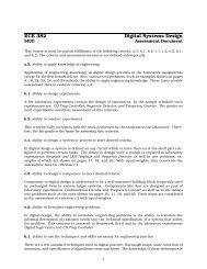
Two radiographs ofdifferent views of anormal elbow. Theseviews show the cleanl<strong>in</strong>es and lack ofsecondary change whichcharacterise a normaljo<strong>in</strong>t<strong>Elbow</strong> <strong>dysplasia</strong> <strong>in</strong> <strong>dogs</strong> - a new schemeexpla<strong>in</strong>edMatthew J. Pead, BVETMED, PHD, CERTSAO, MRCVSand Sue Guthrie, BA, BVETMED, PHD, MRCVS<strong>Elbow</strong> <strong>dysplasia</strong> has been identified as a significant problem <strong>in</strong> manybreeds. Importantly, the condition appears to be <strong>in</strong>creas<strong>in</strong>g worldwide.It beg<strong>in</strong>s <strong>in</strong> puppy hood, and can affect the dog for the rest of its life.S<strong>in</strong>ce the late 1960s, veter<strong>in</strong>ary surgeons rout<strong>in</strong>ely deal<strong>in</strong>g with lame <strong>dogs</strong>have been aware of an <strong>in</strong>creas<strong>in</strong>g number of problems which arise dur<strong>in</strong>gpuppyhood. Hip <strong>dysplasia</strong> was the first disease to be widely recognised, and ascheme to assess and control it is well established. More recently, elbow<strong>dysplasia</strong> (ED) has been identified as a significant problem <strong>in</strong> many breeds.Importantly the condition appears to be <strong>in</strong>creas<strong>in</strong>g worldwide and, althoughthe disease beg<strong>in</strong>s <strong>in</strong> puppyhood, it can affect the dog for the rest of its life.The pr<strong>in</strong>cipal cause of ED is the genetic make-up of the animal. Thus, ascheme which screens animals for elbow abnormalities and allows theanimals with the best elbows to be chosen for breed<strong>in</strong>g, can be successful <strong>in</strong>reduc<strong>in</strong>g the level of the problem <strong>in</strong> the can<strong>in</strong>e population.ELBOW DYSPLASIA THE DISEASE
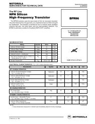
ED simply describes the abnormal development of the elbow (a dog's elbow iscircled below). The term <strong>in</strong>cludes a number of specific abnormalities, whichaffect different sites <strong>in</strong> the jo<strong>in</strong>t. They cause problems by affect<strong>in</strong>g the growthof the cartilage which forms the surface of the jo<strong>in</strong>t, or the structures around it.These abnormalities, called primary lesions, then start a secondaryosteoarthritic process. The most common primary lesions are:• Osteochondritis dissecans (OCD or OD)• Fragmented or ununited coronoid process (FCP)• Ununited anconeal process (UAP)Exam<strong>in</strong>ation of different radiograhic views allows detection ofabnormalitiesThe 'extended lateral 'position gives a view of thebones and jo<strong>in</strong>t surfaces ofthe elbow from the side. Inthis view, the position ofthe jo<strong>in</strong>t is similar to that <strong>in</strong>the stand<strong>in</strong>g dogThe flexed lateral view is aside on view of the elbow.This view allowsexam<strong>in</strong>ation of thesecondary changes <strong>in</strong> EDwhich occur <strong>in</strong> the shadedareas. Note how some ofthe shaded areas here areoverlaid by otherstructures, which makesthem difficult to exam<strong>in</strong>eThe 'crani-caudal' view istaken from the front to theback of the jo<strong>in</strong>t and givesa totally different picture.Evidence of primarydisease, which cannot beidentified on the lateralview, is seen on this view.It also enablesconfirmation of secondaryabnormalitiesThere are other, more rare, primary lesions which may occur <strong>in</strong> comb<strong>in</strong>ationwith these, or on their own. Primary lesions occur dur<strong>in</strong>g the growth of apuppy, and nearly always occur <strong>in</strong> both elbows to some degree.
The greater the degree of ED <strong>in</strong> the parents, the more likely they are topass the disease on to their offspr<strong>in</strong>g (as shown <strong>in</strong> this study ofSwedish Rottweilers)ParentsPer cent of offspr<strong>in</strong>g affectedNormal x Normal 31Normal x Mild ED 43Normal x Moderate ED 48EDxED 56Dogs <strong>in</strong> which elbow <strong>dysplasia</strong>causes lameness are only the 'tipof the iceberg'. These animals areobvious because of their lameness.However, there are many <strong>dogs</strong> withsubcl<strong>in</strong>ical disease that have an<strong>in</strong>creased risk of produc<strong>in</strong>goffspr<strong>in</strong>g with elbow <strong>dysplasia</strong>.These animals are not obvious andcan only be located by screen<strong>in</strong>gCONTROL OF ELBOW DYSPLASIAAs the genetic make-up of the dog is the overwhelm<strong>in</strong>g <strong>in</strong>fluence on thecause of ED, the disease can be controlled by m<strong>in</strong>imis<strong>in</strong>g the 'problem' genes<strong>in</strong> the population. This means select<strong>in</strong>g sires and dams with the best geneticmake-up. There is no laboratory genetic test available, like a DNA 'f<strong>in</strong>gerpr<strong>in</strong>t',to show which animals have the best genetic make-up for elbows. However,<strong>dogs</strong> can be screened by radiograph<strong>in</strong>g (X-ray<strong>in</strong>g) their elbows, and look<strong>in</strong>gfor the signs of ED.If sires and dams are only selected from animals with m<strong>in</strong>imal ED, most of thecl<strong>in</strong>ical and subcl<strong>in</strong>ical animals can be elim<strong>in</strong>ated from the breed<strong>in</strong>gprogramme, which prevents them pass<strong>in</strong>g their defective genetic make up onto the next generation. Success of such control depends on a high proportionof any breed participat<strong>in</strong>g, and mak<strong>in</strong>g the <strong>in</strong>formation public, so that low-riskanimals can be selected by anyone wish<strong>in</strong>g to breed. In other countries,screen<strong>in</strong>g schemes like this have been successful <strong>in</strong> significantly reduc<strong>in</strong>g theED problem. There is an <strong>in</strong>ternational standard for such schemesadm<strong>in</strong>istered by the International <strong>Elbow</strong> Work<strong>in</strong>g Group (IEWO), whichencourages a coord<strong>in</strong>ated approach to the problem.
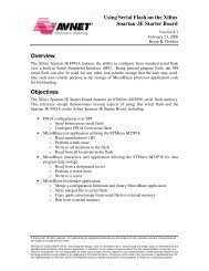
1. 2.2/3. Secondary changes - theradiograph on the left shows asmooth bump of osteoarthritic newbone (arrowed).This is mild (grade1) elbow <strong>dysplasia</strong>. The radiographbelow shows gross irregularosteoarthritis <strong>in</strong> the three arrowedareas. This is severe (grade 3)elbow <strong>dysplasia</strong>1. Primary abnormality - thiscranio-caudal radiograph showsan enlarged jo<strong>in</strong>t space (darkl<strong>in</strong>e) on one side. In this gap youcan see a large detachedfragment (white). This is anosteochondrosis OCD) lesion.When removed it was the size ofa little f<strong>in</strong>gernail!3.The breeds that have a higher <strong>in</strong>cidence of elbow displasia are:Basset Hound, Bernese Mounta<strong>in</strong> Dog,English Mastiff, German Shepherd Dog, Golden Retriever, Great Dane, Irish Wolfhound,Labrador Retriever, Newfoundland, RottweilerTHE NEW UK ELBOW DYSPLASIA SCHEMETo provide an opportunity for the control of ED <strong>in</strong> the UK, the <strong>British</strong>Veter<strong>in</strong>ary <strong>Association</strong> and the Kennel Club (BVA/KC) have <strong>in</strong>troduced ascreen<strong>in</strong>g scheme run along IEWO guidel<strong>in</strong>es. Previously the only elbowschemes runn<strong>in</strong>g <strong>in</strong> this country have been set up by <strong>in</strong>dividual groups suchas the Bernese Mounta<strong>in</strong> Dog Club of Great Brita<strong>in</strong> and the Guide Dogs forthe Bl<strong>in</strong>d <strong>Association</strong>. These have been successful <strong>in</strong> help<strong>in</strong>g to avoidbreed<strong>in</strong>g from animals with a high risk of produc<strong>in</strong>g ED <strong>in</strong> their offspr<strong>in</strong>g.The grad<strong>in</strong>g system is simpleGradeDescription0 Normal1 Mild ED2 Moderate ED or a primary lesion3 Severe ED
THE GRADING PROCEDUREInitially the scheme will require three radiographic views of each elbow, asdescribed on page 2. This ensures that all the areas of the jo<strong>in</strong>t whereabnormalities may occur can be exam<strong>in</strong>ed. Abnormalities are detected withgreater accuracy on exam<strong>in</strong>ation of three views, as compared to a s<strong>in</strong>gleview. The radiographs are exam<strong>in</strong>ed <strong>in</strong>dependently by two scrut<strong>in</strong>eers whowill look for primary lesions such as osteochondritis dissecans (OCD), and thesecondary osteoarthritis which occurs <strong>in</strong> ED. A grade far each elbow iscalculated from the presence of the primary lesions and the size and extent ofthe secondary lesions. The overall grade for an animal is simply the higher ofthe two <strong>in</strong>dividual grades. The grades for each elbow are not added togetheras they are for the two hips <strong>in</strong> the hip <strong>dysplasia</strong> scheme. Identification ofsubcl<strong>in</strong>ical disease and its grade <strong>in</strong> either elbow is the important factor isscreen<strong>in</strong>g so the grade of the worst elbow is always quoted as the overallgrade and will be published on the progeny's Kennel Club registrationdocuments, and <strong>in</strong> the Kennel Club Breed Records Supplement.The grad<strong>in</strong>g procedure and the records will be under cont<strong>in</strong>uous review,which may lead to periodic changes, and the publication of <strong>in</strong>formation forthose <strong>in</strong>volved <strong>in</strong> controll<strong>in</strong>g and treat<strong>in</strong>g ED. The scheme will be representedat IEWG meet<strong>in</strong>gs so that the UK keeps pace with, and participates <strong>in</strong>,<strong>in</strong>ternational developments <strong>in</strong> the management of ED.Gett<strong>in</strong>g a dog's elbows gradedOwners should contact their veter<strong>in</strong>ary surgeon and arrange an appo<strong>in</strong>tmentfor the dog to be radiographed (x-rays). The radiographs will usually be takenunder anaethesia or heavy sedation so the dog may have to be left at theveter<strong>in</strong>ary practice. <strong>Elbow</strong> radiographs can be taken at the same time asthose for the BVA/KC Hip Dysplasia Scheme. When tak<strong>in</strong>g the dog for itsradiographs owners should remember the follow<strong>in</strong>g.• The dog must be at least one year old, but there is no upper age limit.• The dog's KC registration certificate and any related transfer certificatesmust be available so that the appropriate details can be pr<strong>in</strong>ted on theradiographs.• The owner should sign the declaration (first part) of the certificate, to verifythe details are correct and grant permission for the use of the results.Once the radiographs are taken, the veter<strong>in</strong>ary surgeon fills out theappropriate section of the form and submits both the radiographs and the formto the BVA. The results and the radiographs are normally returned to theveter<strong>in</strong>ary surgeon with<strong>in</strong> three weeks with a certificate for the owner and acopy for the veter<strong>in</strong>ary surgeon. Once a grade has been given for a dog, theradiograph cannot be resubmitted; however, owners have the right to anappeal, which takes the form of a re-appraisal of the orig<strong>in</strong>al radiographs,made with<strong>in</strong> 45 days of the dispatch of the orig<strong>in</strong>al certificate. The wholeprocedure from <strong>in</strong>itial appo<strong>in</strong>tment to receiv<strong>in</strong>g the grades is handled throughthe veter<strong>in</strong>ary surgeon.
COSTSThe owner is liable for the veter<strong>in</strong>ary surgeon's fee for anaesthetis<strong>in</strong>g the dogand tak<strong>in</strong>g the radiographs, and the BVA's fee for the grad<strong>in</strong>g.ADVICE ON BREEDINGThe overall grade is used <strong>in</strong>ternationally as the basis far breed<strong>in</strong>g advice.Breeders are advised to select <strong>dogs</strong> with grades of ID or 1 <strong>in</strong> order to reducethe risk of ED <strong>in</strong> their offspr<strong>in</strong>g.As ED is a prevalent disease, especially <strong>in</strong> the breeds listed on page 3, suchadvice will only be effective if it is cont<strong>in</strong>ued over a number of generations.The most difficult part of accept<strong>in</strong>g such advice for many breeders is that anumber of <strong>dogs</strong> which have never been lame and exercise freely, also havehigh grades. This, however, is the subcl<strong>in</strong>ical population with the ability topass the problem on <strong>in</strong> the breed. To ga<strong>in</strong> long-term control of the diseasethese <strong>dogs</strong> ought not to be bred from.TREATMENT OF CLINICAL EDDogs which have cl<strong>in</strong>ical ED often become lame between six and 12 monthsof age. Initially the lameness may be difficult to ascribe to a particular jo<strong>in</strong>t.However, at this age a persistent forelimb lameness should be <strong>in</strong>vestigated bya veter<strong>in</strong>ary surgeon. There are other conditions lead<strong>in</strong>g to signs similar tothose of ED so the veter<strong>in</strong>ary surgeon needs to consider these as well.Diagnosis of ED is normally based on a forelimb lameness with pa<strong>in</strong> on flexion or extension of the elbow. The animal may have a short or stilted forelimbgait as both elbows are often affected. Confirmation of the diagnosis is madeby f<strong>in</strong>d<strong>in</strong>g primary or secondary lesions on radiographs of the elbow.Treatment methods vary depend<strong>in</strong>g an the nature and severity of the problem.Conservative treatment <strong>in</strong>volv<strong>in</strong>g weight restriction and control of exercise isalways important. Drugs may be used to relieve the pa<strong>in</strong> and <strong>in</strong>flammationassociated with the condition and to promote repair processes with<strong>in</strong> the jo<strong>in</strong>t.In some <strong>dogs</strong>, surgery is required to remove fragments of cartilage and bonefrom the jo<strong>in</strong>t to relieve pa<strong>in</strong>, but this is not always appropriate. In nearly allthe cases there are some secondary changes which lead to some problemswith the jo<strong>in</strong>t through out life, possibly restrict<strong>in</strong>g the dog's ability to exercise.However, most <strong>dogs</strong> will be comfortable with a fair level of exercise if treatedcarefully between the ages of six and 18 months.In April 1997, the BVA AWF, the AHT and the RSCPA held a jo<strong>in</strong>t symposiumon the subject of 'Hereditary diseases <strong>in</strong> <strong>dogs</strong>: what are they and what can bedone?' which <strong>in</strong>cluded a presentation on elbow <strong>dysplasia</strong>.
Copies of the proceed<strong>in</strong>gs, conta<strong>in</strong><strong>in</strong>g n<strong>in</strong>e papers <strong>in</strong> all, are available fromthe BVA AWF, 7 Mansfield Street, London W1G 9NQ, price £12.50 (<strong>in</strong>cludep&p).