Guidelines for the Echocardiographic Assessment of the Right Heart ...
Guidelines for the Echocardiographic Assessment of the Right Heart ...
Guidelines for the Echocardiographic Assessment of the Right Heart ...

You also want an ePaper? Increase the reach of your titles
YUMPU automatically turns print PDFs into web optimized ePapers that Google loves.
print & web 4C=FPO<br />
696 Rudski et al Journal <strong>of</strong> <strong>the</strong> American Society <strong>of</strong> Echocardiography<br />
July 2010<br />
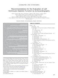
Figure 9 Examples <strong>of</strong> right ventricular fractional area change (FAC). Percentage FAC = 100 end-diastolic area (AreaED) endsystolic<br />
area (Area ES)/end-diastolic area. The endocardial border is traced in apical 4-chamber (A4C) views from <strong>the</strong> tricuspid annulus<br />
along <strong>the</strong> free wall to <strong>the</strong> apex, <strong>the</strong>n back to <strong>the</strong> annulus, along <strong>the</strong> interventricular septum at end-diastole (ED) and end-systole<br />
(ES). Trabeculation, tricuspid leaflets, and chords are included in <strong>the</strong> chamber. (Left) Normal subject, FAC 60%. (Middle) Moderately<br />
dilated right ventricle (RV), FAC 40%, and a markedly dilated left ventricle (LV). (<strong>Right</strong>) Dilated RV, FAC 20%, and <strong>the</strong> LV is <strong>for</strong>eshortened<br />
as a result <strong>of</strong> optimizing <strong>the</strong> view <strong>for</strong> <strong>the</strong> right ventricular chamber.<br />
Table 4 Systolic function<br />
Variable Studies n LRV (95% CI) Mean (95% CI) URV (95% CI)<br />
TAPSE (mm) (Figure 17) 46 2320 16 (15-18) 23 (22-24) 30 (29-31)<br />
Pulsed Doppler velocity at <strong>the</strong> annulus (cm/s) 43 2139 10 (9-11) 15 (14-15) 19 (18-20)<br />
Color Doppler velocities at <strong>the</strong> annulus (cm/s) 5 281 6 (5-7) 10 (9-10) 14 (12-15)<br />
Pulsed Doppler MPI (Figures 16 and 18) 17 686 0.15 (0.10-0.20) 0.28 (0.24-0.32) 0.40 (0.35-0.45)<br />
Tissue Doppler MPI (Figure 18) 8 590 0.24 (0.16-0.32) 0.39 (0.34-0.45) 0.55 (0.47-0.63)<br />
FAC (%) (Figure 8) 36 1276 35 (32-38) 49 (47-51) 63 (60-65)<br />
RV EF (%) (Figure 8) 12 596 44 (38-50) 58 (53-63) 71 (66-77)<br />
3D RV EF (%) 9 524 44 (39-49) 57 (53-61) 69 (65-74)<br />
IVA (m/s 2 ) 12 389 2.2 (1.4-3.0) 3.7 (3.0-4.4) 5.2 (4.4-5.9)<br />
CI, Confidence interval; EF, ejection fraction; FAC, fractional area change; IVA, isovolumic acceleration; LRV, lower reference value; MPI, myocardial<br />
per<strong>for</strong>mance index; RV,rightventricular;TAPSE, tricuspid annular plane systolic excursion; 3D, three-dimensional; URV, upper reference value.<br />
pulmonary annulus, is <strong>the</strong> most reproducible and should<br />
be generally used. For select cases such as suspected arrhythmogenic<br />
RV cardiomyopathy, <strong>the</strong> PLAX measure<br />
may be added. The upper reference limit <strong>for</strong> <strong>the</strong> PSAX<br />
distal RVOT diameter is 27 mm and <strong>for</strong> PLAX is 33<br />
mm (Table 2).<br />
FRACTIONAL AREA CHANGE AND VOLUMETRIC<br />
ASSESSMENT OF THE RIGHT VENTRICLE<br />
A. RV Area and FAC<br />
The percentage RV FAC, defined as (end-diastolic area end-systolic<br />
area)/end-diastolic area 100, is a measure <strong>of</strong> RV systolic function<br />
that has been shown to correlate with RV EF by magnetic resonance<br />
imaging (MRI). 25,35 RV FAC was found to be an independent<br />
predictor <strong>of</strong> heart failure, sudden death, stroke, and/or mortality in<br />
studies <strong>of</strong> patients after pulmonary embolism 36 and myocardial infarction.<br />
37,38 FAC is obtained by tracing <strong>the</strong> RV endocardium both<br />
in systole and diastole from <strong>the</strong> annulus, along <strong>the</strong> free wall to <strong>the</strong><br />
apex, and <strong>the</strong>n back to <strong>the</strong> annulus, along <strong>the</strong> interventricular<br />
septum. Care must be taken to trace <strong>the</strong> free wall beneath <strong>the</strong><br />
trabeculations (Figure 9).<br />
Recommendations: Two-dimensional Fractional Area<br />
Change is one <strong>of</strong> <strong>the</strong> recommended methods <strong>of</strong> quantitatively<br />
estimating RV function, with a lower reference value<br />
<strong>for</strong> normal RV systolic function <strong>of</strong> 35%.<br />
B. Two-Dimensional Volume and EF Estimation<br />
The complexity <strong>of</strong> estimating RV volume and function with 2D echocardiography<br />
has been well documented, and interested readers are<br />
referred to reviews <strong>for</strong> a more complete discussion. 29,39,40 In brief,<br />
<strong>the</strong> 2D echocardiographic methods <strong>of</strong> calculating RV volume can<br />
be divided into area-length methods, disk summation methods, and<br />
o<strong>the</strong>r methods.<br />
The area-length methods, initially adopted <strong>for</strong> biplane angiography,<br />
require an approximation <strong>of</strong> RV geometry, most commonly<br />
based on modified pyramidal or ellipsoidal models. 39,41,42 It<br />
underestimates MRI-derived RV volume and is inferior in comparison<br />
with 3D echocardiographic methods <strong>of</strong> RV volume estimation. 43<br />
The disk summation method has also been applied to determine<br />
a RV ‘‘body’’ volume, using predominantly <strong>the</strong> apical 4-chamber<br />
view. 44 RV volumes are <strong>the</strong>re<strong>for</strong>e underestimated because <strong>of</strong> <strong>the</strong> exclusion<br />
<strong>of</strong> <strong>the</strong> RVOT and technical limitations <strong>of</strong> <strong>the</strong> echocardiographic<br />
images.<br />
RV EF from 2D methods is calculated as (end-diastolic volume<br />
end-systolic volume)/end-diastolic volume. The lower reference limit