Guidelines for the Echocardiographic Assessment of the Right Heart ...
Guidelines for the Echocardiographic Assessment of the Right Heart ...
Guidelines for the Echocardiographic Assessment of the Right Heart ...

Create successful ePaper yourself
Turn your PDF publications into a flip-book with our unique Google optimized e-Paper software.
print & web 4C=FPO<br />
700 Rudski et al Journal <strong>of</strong> <strong>the</strong> American Society <strong>of</strong> Echocardiography<br />
July 2010<br />
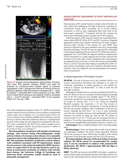
Figure 13 Doppler echocardiographic determination <strong>of</strong> pulmonary<br />
artery (PA) diastolic pressure (PADP) and mean PA<br />
pressure by continuous-wave Doppler signal <strong>of</strong> pulmonic<br />
regurgitation. Point 1 denotes <strong>the</strong> maximal PI velocity at <strong>the</strong> beginning<br />
<strong>of</strong> diastole. Mean PA pressure correlates with 4 (early<br />
PI velocity) 2 + estimated RAP, in this example 37 mm Hg + RAP.<br />
Point 2 marks <strong>the</strong> PI velocity at end-diastole. PADP is correlated<br />
with 4 (end PI velocity) 2 + estimated RAP. In this example,<br />
PADP is 21 mm Hg + RAP.<br />
basis <strong>of</strong> <strong>the</strong> fundamental equation <strong>of</strong> flow (F = DP/R), <strong>the</strong> abnormal<br />
exercise-induced increase in pressure can be ascribed to supranormal<br />
cardiac output (eg, in athletes) or to a normal increase in flow but<br />
a rise in resistance due to limited capability <strong>of</strong> <strong>the</strong> pulmonary vascular<br />
bed (eg, in chronic obstructive pulmonary disease or congenital heart<br />
disease). In this setting, <strong>the</strong> ratio <strong>of</strong> Dpressure (estimated by TR velocity)<br />
to flow (estimated by <strong>the</strong> RVOT time-velocity integral) may be<br />
helpful to distinguish whe<strong>the</strong>r <strong>the</strong> increased pressure is related to<br />
an increase in flow or in resistance. 96<br />
Recommendation: In patients with dyspnea <strong>of</strong> unknown<br />
etiology, with normal resting echocardiographic results<br />
and no evidence <strong>of</strong> coronary artery disease, it is reasonable<br />
to per<strong>for</strong>m stress echocardiography to assess stress-induced<br />
PH. This technique should be considered as well in subjects<br />
with conditions associated with PA hypertension. Supine<br />
bicycle exercise is <strong>the</strong> preferred method <strong>for</strong> SPAP measurement.<br />
An upper limit <strong>of</strong> 43 mm Hg should be used in patients<br />
at nonextreme workloads. In subjects with valvular<br />
heart disease, <strong>the</strong> American College <strong>of</strong> Cardiology and<br />
American <strong>Heart</strong> Association cut<strong>of</strong>fs should be used to<br />
help guide management.<br />
NONVOLUMETRIC ASSESSMENT OF RIGHT VENTRICULAR<br />
FUNCTION<br />
Determination <strong>of</strong> RV systolic function is similar to that <strong>of</strong> <strong>the</strong> left ventricle,<br />
albeit more challenging. The right ventricle has superficial circumferential<br />
muscle fibers responsible <strong>for</strong> its inward bellows<br />
movement, as well as inner longitudinal fibers that result in <strong>the</strong><br />
base-to-apex contraction. 20 Compared with <strong>the</strong> left ventricle, <strong>the</strong><br />
base-to-apex shortening assumes a greater role in RV emptying.<br />
Global assessment <strong>of</strong> RV function includes <strong>the</strong> myocardial per<strong>for</strong>mance<br />
index (MPI), RV dP/dt, RV EF, and FAC (see above). Regional<br />
approaches include tissue Doppler–derived and 2D strain, Dopplerderived<br />
systolic velocities <strong>of</strong> <strong>the</strong> annulus (S 0 ), and TAPSE. Each<br />
method is affected by <strong>the</strong> same limitations from <strong>the</strong> corresponding<br />
left-sided techniques. The RV EF may not represent RV contractility<br />
in <strong>the</strong> setting <strong>of</strong> significant TR, just as <strong>the</strong> LV EF is limited by mitral regurgitation.<br />
For S 0 and TAPSE, <strong>the</strong> regional velocities or displacement<br />
<strong>of</strong> <strong>the</strong> myocardium in a single segment may not truly represent <strong>the</strong><br />
function <strong>of</strong> <strong>the</strong> entire right ventricle. Regional strain measurements<br />
are plagued by <strong>the</strong> same issues, as well as that <strong>of</strong> poor reproducibility.<br />
There is a lack <strong>of</strong> outcomes data relating to quantification <strong>of</strong> RV systolic<br />
function. None<strong>the</strong>less, each <strong>of</strong> <strong>the</strong> methods is described below,<br />
reference values are displayed, and recommendations <strong>for</strong> measurements<br />
are proposed.<br />
A. Global <strong>Assessment</strong> <strong>of</strong> RV Systolic Function<br />
RV dP/dt. The rate <strong>of</strong> pressure rise in <strong>the</strong> ventricles (dP/dt) is an<br />
invasive measurement developed and validated as an index <strong>of</strong><br />
ventricular contractility or systolic function. It was initially described<br />
by Gleason and Braunwald 97 in 1962 in both <strong>the</strong> left<br />
and right ventricles.<br />
Although less studied and more sparsely used than <strong>for</strong> <strong>the</strong> left ventricle,<br />
RV dP/dt can also be accurately estimated from <strong>the</strong> ascending<br />
limb <strong>of</strong> <strong>the</strong> TR continuous-wave Doppler signal. 98,99 RV dP/dt is<br />
commonly calculated by measuring <strong>the</strong> time required <strong>for</strong> <strong>the</strong> TR jet<br />
to increase in velocity from 1 to 2 m/s. Using <strong>the</strong> simplified<br />
Bernoulli equation, this represents a 12 mm Hg increase in<br />
pressure. The dP/dt is <strong>the</strong>re<strong>for</strong>e calculated as 12 mm Hg divided by<br />
this time (in seconds), yielding a value in millimeters <strong>of</strong> mercury per<br />
second. Although <strong>the</strong> time from 1 to 2 m/s is most commonly<br />
used, <strong>the</strong> best correlation between echocardiographic and invasive<br />
measures was found by using <strong>the</strong> time <strong>for</strong> <strong>the</strong> TR velocity to<br />
increase from 0.5 to 2 m/s. 99 In this case, <strong>the</strong> numerator <strong>for</strong> <strong>the</strong> calculation<br />
is 15 mm Hg, representing <strong>the</strong> pressure difference as calculated<br />
from <strong>the</strong> simplified Bernoulli equation between 2 and 0.5 m/s.<br />
Advantages: This is a simple technique with a sound physiologic<br />
basis.<br />
Disadvantages: There are limited data in both normal subjects<br />
and pathologic conditions. RV dp/dt is load dependent. RVdp/dt<br />
will be less accurate in severe TR because <strong>of</strong> neglect <strong>of</strong> <strong>the</strong> inertial<br />
component <strong>of</strong> <strong>the</strong> Bernoulli equation and <strong>the</strong> rise in RA pressure.<br />
Recommendations: Because <strong>of</strong> <strong>the</strong> lack <strong>of</strong> data in normal<br />
subjects, RV dP/dt cannot be recommended <strong>for</strong> routine<br />
uses. It can be considered in subjects with suspected RV<br />
dysfunction. RV dP/dt < approximately 400 mm Hg/s is<br />
likely abnormal.<br />
RIMP. The MPI, also known as <strong>the</strong> RIMP or Tei index, is a global estimate<br />
<strong>of</strong> both systolic and diastolic function <strong>of</strong> <strong>the</strong> right ventricle. It is<br />
based on <strong>the</strong> relationship between ejection and nonejection work <strong>of</strong>