Guidelines for the Echocardiographic Assessment of the Right Heart ...
Guidelines for the Echocardiographic Assessment of the Right Heart ...
Guidelines for the Echocardiographic Assessment of the Right Heart ...

Create successful ePaper yourself
Turn your PDF publications into a flip-book with our unique Google optimized e-Paper software.
706 Rudski et al Journal <strong>of</strong> <strong>the</strong> American Society <strong>of</strong> Echocardiography<br />
July 2010<br />
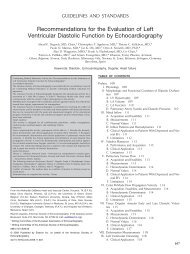
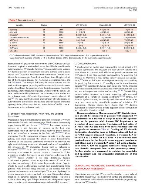
Table 6 Diastolic function<br />
Variable Studies n LRV (95% CI) Mean (95% CI) URV (95% CI)<br />
E (cm/s) 55 2866 35 (33-37) 54 (52-56) 73 (71-75)<br />
A (cm/s) 55 3096 21 (19-24) 40 (38-41) 58 (55-60)<br />
E/A ratio* 56 2994 0.8 (0.7-0.9) 1.4 (1.4-1.5) 2.1 (2.0-2.2)<br />
Deceleration time (ms) 25 1284 120 (105-134) 174 (163-186) 229 (214-243)<br />
IVRT (ms) 23 1241 23 (16-30) 48 (43-53) 73 (66-80)<br />
E 0 (cm/s) 40 1688 8 (7-9) 14 (13-14) 20 (19-21)<br />
A 0 (cm/s) 37 1575 7 (6-8) 13 (12-14) 20 (19-21)<br />
E 0 /A 0 ratio 29 1053 0.5 (0.4-0.6) 1.2 (1.1-1.3) 1.9 (1.7-2.0)<br />
E/E 0 ratio 3 359 2 (1-2) 4 (4-4) 6 (5-7)<br />
CI, Confidence interval; IVRT, Isovolumic relaxation time; LRV, lower reference value; URV, upper reference value.<br />
*Age-dependent: average E/A ratio = 1.6 in <strong>the</strong> third decade <strong>of</strong> life, decreasing by 0.1 <strong>for</strong> every subsequent decade.<br />
Estimation <strong>of</strong> RA pressure by measurement <strong>of</strong> IVC diameter and collapse<br />
with inspiration as described above should be factored into <strong>the</strong><br />
determination <strong>of</strong> RV diastolic function. The parameters used to assess<br />
RV diastolic function are essentially <strong>the</strong> same as those used to assess<br />
<strong>the</strong> left side. Those that have been most validated are Doppler velocities<br />
<strong>of</strong> <strong>the</strong> transtricuspid flow (E, A, and E/A), tissue Doppler velocities<br />
<strong>of</strong> <strong>the</strong> tricuspid annulus (E 0 ,A 0 ,E 0 /A 0 ), deceleration time, and<br />
IVRT (Table 6). The tricuspid E/E 0 ratio, RA area or volume, and diastolic<br />
strain rate appear promising and have attracted interest in recent<br />
studies. In addition, <strong>the</strong> presence <strong>of</strong> late diastolic antegrade flow in <strong>the</strong><br />
pulmonary artery (measured by pulsed Doppler with <strong>the</strong> sample volume<br />
positioned midway between <strong>the</strong> pulmonary valve leaflets and<br />
<strong>the</strong> pulmonary artery bifurcation) is a sign <strong>of</strong> restrictive diastolic filling.<br />
148 This sign, mainly described after tetralogy <strong>of</strong> Fallot repair, occurs<br />
when <strong>the</strong> elevated RV end-diastolic pressure causes premature<br />
opening <strong>of</strong> <strong>the</strong> pulmonary valve and transmission <strong>of</strong> <strong>the</strong> RA contraction<br />
A wave in <strong>the</strong> pulmonary artery.<br />
C. Effects <strong>of</strong> Age, Respiration, <strong>Heart</strong> Rate, and Loading<br />
Conditions<br />
Most studies have shown that <strong>the</strong>re is a modest correlation (r z 0.30)<br />
between <strong>the</strong> E/A ratio and increasing age. The E/A ratio decreases by<br />
approximately 0.1 per decade. 117,141,149,150 Inspiration causes an<br />
increase in E and <strong>the</strong>re<strong>for</strong>e an increase in <strong>the</strong> E/A ratio.<br />
Tachycardia causes an increase in E but a relatively greater increase<br />
in A and <strong>the</strong>re<strong>for</strong>e a decrease in <strong>the</strong> E/A ratio. 141,150,151 When<br />
comparing Doppler parameters between patients or in <strong>the</strong> same<br />
patient, one must consider <strong>the</strong> effect <strong>of</strong> <strong>the</strong>se hemodynamic<br />
variables on <strong>the</strong> observed parameters. Because <strong>of</strong> its thin wall, <strong>the</strong><br />
right ventricle is very sensitive to afterload (wall stress), especially in<br />
<strong>the</strong> presence <strong>of</strong> RV myocardial disease such as ischemia or<br />
infarction. 144,152,153 It is also sensitive to changes in preload,<br />
whereby a reduction in preload causes a decrease in E but<br />
a relatively smaller decrease in A and <strong>the</strong>re<strong>for</strong>e a decrease in E/<br />
A. 154-156 Tissue Doppler is less load dependent, because<br />
a reduction in preload causes an equal decrease in E 0 and A 0 and<br />
<strong>the</strong>re<strong>for</strong>e an unchanged E 0 /A 0 ratio. Importantly, tissue Doppler<br />
should be used to differentiate normal from pseudonormal filling<br />
patterns with elevated filling pressures as are hepatic vein flow<br />
patterns and vena cava size and collapse. 15 Last, <strong>the</strong> physiologic response<br />
to exercise is to increase both early rapid filling and atrial contribution.<br />
In ischemic patients, <strong>the</strong> pathophysiologic response is<br />
a failure <strong>of</strong> <strong>the</strong> early rapid filling to increase and a heightened dependency<br />
on atrial contribution and consequent elevated RA pressure. 157<br />
D. Clinical Relevance<br />
A small number <strong>of</strong> studies have evaluated <strong>the</strong> clinical impact <strong>of</strong> RV<br />
diastolic dysfunction. The tricuspid E/E0 ratio and RA volume have<br />
been shown to correlate well with hemodynamic parameters. An<br />
E/E0 ratio $ 4 had high sensitivity and specificity <strong>for</strong> predicting RA<br />
pressure $ 10 mm Hg in non–cardiac surgery intensive care unit patients,<br />
158 while an E/E0 ratio > 8 had good sensitivity and specificity<br />
<strong>for</strong> predicting RA pressure $ 10 mm Hg in cardiac transplantation<br />
patients. 159 In patients with chronic heart failure and PH, <strong>the</strong> presence<br />
<strong>of</strong> RV diastolic dysfunction was associated with worse functional class<br />
and was an independent predictor <strong>of</strong> mortality. 100,160 Diastolic filling<br />
patterns reflect response to <strong>the</strong>rapy, improving with successful<br />
treatment <strong>of</strong> a variety <strong>of</strong> cardiac conditions. 161-164 Finally, RV<br />
diastolic dysfunction may be clinically useful because it serves as an<br />
early and more easily quantifiable marker <strong>of</strong> subclinical RV<br />
dysfunction. Multiple studies have shown that RV diastolic<br />
dysfunction is usually present be<strong>for</strong>e apparent systolic dysfunction<br />
and be<strong>for</strong>e RV dilatation or RVH.<br />
Recommendations: Measurement <strong>of</strong> RV diastolic function<br />
should be considered in patients with suspected RV<br />
impairment as a marker <strong>of</strong> early or subtle RV dysfunction,<br />
or in patients with known RV impairment as<br />
a marker <strong>of</strong> poor prognosis. Transtricuspid E/A ratio, E/<br />
E0 ratio, and RA size have been most validated and are<br />
<strong>the</strong> preferred measures(Table 6). Grading <strong>of</strong> RV diastolic<br />
dysfunction should be done as follows: tricuspid E/A ratio<br />
< 0.8 suggests impaired relaxation, a tricuspid E/A ratio<br />
<strong>of</strong> 0.8 to 2.1 with an E/E0 ratio > 6 or diastolic flow<br />
predominance in <strong>the</strong> hepatic veins suggests pseudonormal<br />
filling, and a tricuspid E/A ratio > 2.1 with a deceleration<br />
time < 120 ms suggests restrictive filling (as does<br />
late diastolic antegrade flow in <strong>the</strong> pulmonary artery).<br />
Fur<strong>the</strong>r studies are warranted to validate <strong>the</strong> sensitivity<br />
and specificity and <strong>the</strong> prognostic implications <strong>of</strong> this<br />
classification.<br />
CLINICAL AND PROGNOSTIC SIGNIFICANCE OF RIGHT<br />
VENTRICULAR ASSESSMENT<br />
Quantitative assessment <strong>of</strong> RV size and function has proven to be <strong>of</strong><br />
important clinical value in a number <strong>of</strong> cardiac and pulmonary diseases.<br />
Numerous publications have demonstrated <strong>the</strong> prognostic significance<br />
<strong>of</strong> RV function.