Delta Vision Elite Microscope
Delta Vision Elite Microscope
Delta Vision Elite Microscope

You also want an ePaper? Increase the reach of your titles
YUMPU automatically turns print PDFs into web optimized ePapers that Google loves.
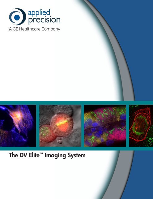
The DV <strong>Elite</strong> Imaging System
The <strong>Delta</strong><strong>Vision</strong> DifferenceDedicated to pushing the limits of imaging and microscopy, in 1994 Applied Precisionintroduced a novel concept — the first fully integrated, turn-key deconvolution imagingsystem. Our commitment to innovation, quality, performance and upgradeabilitymarked the beginning of the <strong>Delta</strong><strong>Vision</strong> Difference.We are committed to supporting our customers through upgrade solutions andservice contract options to ensure every <strong>Delta</strong><strong>Vision</strong> system remains an indispensibleimaging platform for life science research both now and in the future.The DV <strong>Elite</strong> Imaging SystemThe DV <strong>Elite</strong> is the newest edition to the <strong>Delta</strong><strong>Vision</strong> family of advanced microscopyimaging systems from Applied Precision. The DV <strong>Elite</strong> is designed for maximumflexibility and can handle most applications including time-lapse live cell imaging,TIRF, FRET, Photokinetics and DIC.• Maximum flexibility for all your imaging needs• Revolutionary live cell imaging performance• Outstanding focus stability for long term imaging experiments• Fully integrated system for seamless operation and upgradeabilityDV <strong>Elite</strong> shown with Advanced Isolation Table
The TruLight Illumination SystemThe illumination system is the heart of every microscope andcritical to system performance. Applied Precision recognizesthis and has developed the TruLight Illumination System,a radical new design for the <strong>Delta</strong><strong>Vision</strong> fluorescenceillumination path that consists of four key elements:Excitation —Concentration —Automation —Application —Powerful solid state illumination andultrafast wavelength switchingOptimized transmission increases light tosample by 5 timesSeamless switching between viewing andimaging modes for superior image qualityExpanded options, including Multi-line TIRFand quantitative photokineticsTruLight delivers outstanding signal to noise performanceand 5 times more light to the sample, enabling detection ofsmall, dim objects such as organelles and microbial particles.TruLight supports a vast range of options to enhance systemperformance, including UltimateFocus and Multi-line TIRF.Standard IlluminationDV <strong>Elite</strong> with Trulight Illumination
<strong>Delta</strong><strong>Vision</strong> ImagingIlluminationDrosophila ovary - Image courtesy In SituHybridization Course, Cold Spring Harbor LaboratoryInsightSSI Solid State IlluminationThe InsightSSI illumination module incorporates novel light source technologies foroptimal performance.• Extremely stable and long lasting illumination• Electronic control provides instant on/off operation• Microsecond switching between wavelengthsUltimateFocus UltimateFocus automatically maintains the sample z-position regardless of mechanicalor thermal changes that can impact your experiment.• Exclusive, patent-pending design• Real-time compensation of stage drift• Focus control within 25 nm8 micron thick fixed cryosection of mouse small intestine- Image courtesy Paul Appleton, Wellcome TrustBiocentre, DundeeNow with Focus Assist! The Focus Assist feedback loop of UltimateFocus determinesthe distance between the objective and the coverslip. It guides the user to bring theobjective into the area of focus without using the eyepieces or a camera.Illumination UniformityEvery <strong>Delta</strong><strong>Vision</strong> system utilizes our proprietary photosensor correction system.• Continuously measures the excitation light output to ensure data integrity• Provides automatic image correction for intensity fluctuations as required forquantitative imagingImaging of 100 micron slices of autumn maple leaf- Image courtesy Kyla Teplitz and Katie Buchanan,Applied PrecisionDifferential Interference Contrast (DIC)DIC light microscopy produces finely detailed high contrast images to rapidly visualizethe state of the cell. DIC combined with epifluorescence provides more informationwithin context of the sample.• Quickly identifies healthy cells within a large population• Establishes focal plane to minimize photodamage to cells• Monitors cell viability during time-lapse experimentsMitochondria trafficking in arabidopsis root hairs- Image courtesy Naohiro Kato, 3D Microscopy ofLiving Cells
Laser ModuleX4 Laser ModuleThe X4 Laser Module combines up to four lasers thatare active simultaneously for increased performance.The increased laser line selection allows greaterflexibility in the use of fluorescent dyes and proteins.• More powerful lasers for shorter exposure timesincrease the accuracy of photokinetic measurements• Specialized optics generate a near diffractionlimitedspot size to target a measurable region ofinterest• Rapid laser switching enables combinations ofmultiple lasers to photoactivate and photobleach thesample within the same experimentMulti-Line TIRFTotal Internal Reflection Fluorescence (TIRF) microscopyis a specialized technique used to image sampleswithin 100-200 nm of the coverslip surface. Becauselight travels different distances, the challenge of TIRFis to ensure that all laser lines penetrate the sampleto the same depth. Applied Precision has solved thislimitation by automating several key components tobring the lasers to the same focal plane.• Performs chromatic correction of each wavelength tothe same penetration depth at the coverslip• Increases imaging options using four lasers in ournew X4 Laser Module• Flexible experiment design enables the combinationof photokinetic applications (PA-GFP) with TIRFimagingPhotoactivation/Photokinetics/FRAP/FLIPPhotoactivable and photoconvertible proteins give theuser the ability to take a more active role in expandingthe type of data collected. The sub-diffraction limitedspot size of the X4 Laser Module enables exquisitecontrol over the target area for quantitative results.• Photoactivation to initiate fluorescence of a dye orprotein to act as a marker within a larger population• Photokinetics to precisely calculate the rate anddirection of movement of the fluorophore of interest• Fluorescence Recovery after Photobleaching (FRAP)to photobleach a region of interest and measure therecovery of fluorescence in the bleached area• Förster/Fluorescence Resonance Energy Transfer(FRET) to characterize the interactions of proteins thatare known to associate with each other15Epifluorescent (green) and TIRF (red) images of zyxin:GFP localized to focal adhesions inHeLa cells
Stage and EnvironmentApplied Precision’s patented Flexure StageFlexure StageApplied Precision’s patented Flexure Stages and exclusive NanoMotionIII Precision Control motors ensure precise stage movement andstability.• Industry leading accuracy and repeatability over its full range ofmotion• Superior repeatability for multiple point sample during time-lapseexperimentsOptional Microtiter StageMicrotiter StageThe Applied Precision Microtiter Stage is designed to accommodate96- and 384-well microtiter plates and increases the flexibility of any<strong>Delta</strong><strong>Vision</strong> system.• User-friendly software collects and reviews multiple fields of view inevery well• Preset imaging patterns easily and efficiently tackle large imagingassays• Streamlines labor-intensive tasks such as quantitating signaltransduction, measuring drug efficacy, or optimizing antibodytitrationsEnvironmental ControlPrecision environmental control is critical for long term imagingexperiments. Stringent heat and CO 2controls:• Duplicate incubator conditions• Decrease phototoxic stress on cells• Minimize thermal shifts for superior image qualityOptional Environmental Control Chamber available in clear (shown) andopaqueStage SpecificationsConventional Imaging ModeAbsolute Accuracy < 10.0 um per 25 mm (X,Y)< 0.6 um per 13 um (Z)Repeatability < ± 0.2 um (X,Y)< ± 0.1 um (Z)Step Resolution 20 nm (X,Y)5 nm (Z)Maximum Travel 25 mm (X) x 50 mm (Y)1 mm (Z)Automation Control 3D multisite visiting within a sampleMicrotiter Plate ModeAbsolute Accuracy < 10.0 um per 25 mm (X,Y)< 0.6 um per 13 um (Z)Repeatability < ± 0.2 um (X,Y)< ± 0.1 um (Z)Step Resolution 20 nm (X,Y)5 nm (Z)Maximum Travel 106 mm (X) x 70 mm (Y)1 mm (Z)Automation Control 3D with multisite visiting within well(s)
<strong>Delta</strong>vision InnovationDeconvolutionDeconvolution improves image resolution and contrastwithout sacrificing data integrity. The Applied Precisionexclusive deconvolution algorithm results in true quantitativedata.• Increased resolution in x, y and z axes• Deconvolve images on-the-fly to visualize data faster• Designed to be the best deconvolution system availablePoint-VisitingThe Applied Precision exclusive NanoMotion III technologyenables the user to visit a series of points within a singleor time-lapse experiment. This technology ensures that thestage will return to the exact location in x, y and z.• Provides unmatched point-to-point accuracy and precisionmotion control• Programs and stores hundreds of points to maximize datacollection within an experiment• Streams collected data to image processing tasks toimprove workflowCell TrackingCell tracking algorithms automatically reposition the stageto follow cells as they move.• Accurately tracks cells during long time-lapse experiments• Defines one cell of interest to track within a largepopulation• Uses collected data to calculate rates and direction ofmotionOptical Axis Integration (OAI)OAI is an approach for rapidly acquiring data in the zaxis based on user defined ranges. It quickly captures andvisualizes data that otherwise may be missed.• Images moving diffraction-limited objects that mayotherwise be lost between frames• Lowers total light exposure, increasing cell viability• Rapidly captures the total number of objects within a cell(e.g., centrosomes, viral particles)BSC1 cells - Image courtesy of AQLM, Marine Biological Laboratory,Woods Hole, MAGFP tagged plasmid RK2 in Vibrio cholerae - Image courtesy of T. Ho, Z.Zhong, S. Aung, and J. PoglianoPorcine aortic endothelial cells - Image courtesy Ronen Zaidel-Bar,Weizmann Institute of Science, Rehovot, Israel
Applied Precision, Inc1040 12th Ave NWIssaquah, WA 98027Tel: 425.557.1000www.appliedprecision.com© 2011 Applied Precision, Inc. All Rights Reserved. Rev A 080211Applied Precision and <strong>Delta</strong><strong>Vision</strong> are registered trademarks and<strong>Delta</strong><strong>Vision</strong> <strong>Elite</strong> and Monet are trademarks of Applied Precision, Inc.