Lymphangioma circumscriptum of the penis: a case report
Lymphangioma circumscriptum of the penis: a case report
Lymphangioma circumscriptum of the penis: a case report

You also want an ePaper? Increase the reach of your titles
YUMPU automatically turns print PDFs into web optimized ePapers that Google loves.
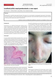
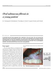
C a s e r e p o r tPenile lymphangioma <strong>circumscriptum</strong><strong>Lymphangioma</strong> <strong>circumscriptum</strong> <strong>of</strong> <strong>the</strong><strong>penis</strong>: a <strong>case</strong> <strong>report</strong>I. KokcamS U M M A R YWe <strong>report</strong> a <strong>case</strong> <strong>of</strong> lymphangioma <strong>circumscriptum</strong> <strong>of</strong> <strong>the</strong> <strong>penis</strong> in a 19-year-old male. The lesions developedduring puberty and resembled molluscum contagiosum and genital herpes. The <strong>case</strong> is presentedbecause <strong>of</strong> its rarity and to increase diagnostic awareness and treatment with non-surgical intervention.IntroductionK E YWORDSlymphangioma<strong>circumscriptum</strong>,<strong>penis</strong>,molluscumcontagiosum<strong>Lymphangioma</strong> <strong>circumscriptum</strong> (LC) is a developmentalanomaly <strong>of</strong> <strong>the</strong> lymphatic vascular system. LCrarely involves <strong>the</strong> <strong>penis</strong>; however, when such lesionsoccur on <strong>the</strong> <strong>penis</strong>, <strong>the</strong>y may be misdiagnosed as vesicularor tumoral diseases in <strong>the</strong> genital area (1, 2).Recognition and treatment <strong>of</strong> genital lesions <strong>of</strong> LC isimportant in order to alleviate <strong>the</strong> anxiety associatedwith having an STD. Additionally, <strong>the</strong>se lesions mustbe treated properly because <strong>the</strong>y may act as a portal <strong>of</strong>entry for infections, be cosmetically embarrassing, and,if large, cause functional impairment (3, 4).Case <strong>report</strong>A 19-year-old man applied to our outpatient clinicfor evaluation <strong>of</strong> multiple translucent and hemorrhagicvesicles on his <strong>penis</strong>. Over <strong>the</strong> previous three years,<strong>the</strong>se lesions occasionally bled and oozed serosanguinousfluid. Physical examination confirmed that <strong>the</strong>patient had multiple translucent and hemorrhagicvesicles on <strong>the</strong> shaft <strong>of</strong> <strong>the</strong> <strong>penis</strong> and <strong>the</strong> glans. Thesurface <strong>of</strong> <strong>the</strong> lesions was smooth and some were umbilicated,resembling molluscum contagiosum (Figure1). There was no associated lymphedema on <strong>the</strong> <strong>penis</strong>.Abdominal, pelvic, and penile ultrasonographic examinationswere within normal limits, as were also hematologicaland biochemical parameters. Histological examinationshowed numerous dilated lymphatic vesselsand mononuclear inflammatory cells throughout <strong>the</strong>papillary and reticular dermis (Figure 2).The patient refused surgical intervention; <strong>the</strong>reforewe recommended that <strong>the</strong> patient avoid mechanicaltrauma and cleanse <strong>the</strong> area daily with antiseptic agents.We also recommended that <strong>the</strong> patient apply silver sulfadiazinecream topically to any ruptured lesions. Over<strong>the</strong> next two years <strong>of</strong> follow-up, <strong>the</strong> patient did notdevelop new lesions. Instead, <strong>the</strong> overall number <strong>of</strong><strong>the</strong> lesions declined markedly and no o<strong>the</strong>r complicationswere observed.Acta Dermatoven APA Vol 16, 2007, No 2 81
Penile lymphangioma <strong>circumscriptum</strong>C a s e r e p o r tFigure 1. A few umbilicated and hemorrhagic vesiclesresembling molluscum contagiosum are seenaround <strong>the</strong> external urethral meatus and glans <strong>penis</strong>.DiscussionThe <strong>penis</strong> is a highly uncommon site for LC, with atotal <strong>of</strong> only 10 <strong>case</strong>s <strong>of</strong> penile LC previously <strong>report</strong>ed (1,3, 5–7). These <strong>report</strong>s suggest that LC <strong>of</strong> <strong>the</strong> <strong>penis</strong> may beei<strong>the</strong>r congenital or acquired. Acquired <strong>case</strong>s appear todevelop most frequently after infections <strong>of</strong> lymphaticchannels, irradiation, or surgery (1, 2, 7). However, this<strong>case</strong> <strong>of</strong> penile LC appeared spontaneously in adulthood.The diagnosis <strong>of</strong> penile LC is easy when <strong>the</strong> clinicianis aware <strong>of</strong> <strong>the</strong> disease; however, a biopsy <strong>of</strong> suchlesions may be necessary to confirm <strong>the</strong> diagnosis andto formulate appropriate treatment. Histologically, penileLC is characterized by greatly dilated lymph spacesin <strong>the</strong> upper dermis and <strong>the</strong> subcutis. The lumen is filledwith lymphatic fluid, but <strong>of</strong>ten contains red blood cells,R E F E R E N C E SFigure 2. Histopathological examination <strong>of</strong>cutaneous biopsy showing dilated lymphaticchannels in <strong>the</strong> upper dermis (H&E stain, X 200).lymphocytes, macrophages, and neutrophils (1). Thehistological findings <strong>of</strong> <strong>the</strong> <strong>case</strong> <strong>report</strong>ed here wereconsistent with <strong>the</strong>se features.There is currently no proven medical care for LC. Themain goal <strong>of</strong> treatment is typically cosmetic; however, treatmentcan also be applied to limit complications. In thisstudy we pursued a “watch and wait” policy, which is advisableif <strong>the</strong> condition is asymptomatic (8). The only radicalcure is to remove <strong>the</strong> superficial component as well as<strong>the</strong> deeper lymphatic cisterns. This is achieved throughsurgical destruction or laser ablation <strong>of</strong> lesions. Electrocautery,cryo<strong>the</strong>rapy, sclerosants, incision and drainage,and carbon dioxide laser can also be used to reduce <strong>the</strong>risk <strong>of</strong> infection and to reduce lymphorrhea (1, 8).In conclusion, penile lesions associated with LC maypose a diagnostic challenge, <strong>the</strong> risks <strong>of</strong> which are misdiagnosisand mistreatment. Physicians must <strong>the</strong>refore keepLC in mind when vesicular lesions <strong>of</strong> <strong>the</strong> <strong>penis</strong> exist.A U T H O R ' SA D D R E S S1. Gupta S, Radotra BD, Javaheri SM, Kumar B. <strong>Lymphangioma</strong> <strong>circumscriptum</strong> <strong>of</strong> <strong>the</strong> <strong>penis</strong> mimickingvenereal lesions. J Eur Acad Dermatol Venereol. 2003;17(5):598–600.2. Tsur H, Urson S, Schewach-Millet M. <strong>Lymphangioma</strong> <strong>circumscriptum</strong> <strong>of</strong> <strong>the</strong> glans <strong>penis</strong>. Cutis.1981;28:642–3.3. Maloudijan M, Stutz N, Hoerster S, et al. <strong>Lymphangioma</strong> <strong>circumscriptum</strong> <strong>of</strong> <strong>the</strong> <strong>penis</strong>. Eur J Dermatol.2006;16(4):451–2.4. Harwood CA, Mortimer PS. Acquired vulval lymphangiomata mimicking genital warts. Br J Dermatol.1993;129:334–6.5. Swanson DL. Genital lymphangioma with recurrent cellulitis in men. Int J Dermatol. 2006;45(7):800–4.6. Ferro F, Spagnoli A, Villa M, Papendieck CM. A salvage surgical solution for recurrent lymphangioma<strong>of</strong> <strong>the</strong> prepuce. Br J Plast Surg. 2005;58(1):97–9.7. Penko M, Bartenjev I, Zgavec B, Potocnik M. <strong>Lymphangioma</strong> <strong>circumscriptum</strong>. Acta DermatovenerolAlp Pannonica Adriat. 1996;5(2):51–6.8. Lebwohl MG, Heymann WR, Berth-Jones J, Coulson I, editors. Treatment <strong>of</strong> Skin Disease. 2nd ed.London: Mosby; 2006.Ibrahim Kokcam MD, Department <strong>of</strong> Dermatology, Faculty <strong>of</strong> Medicine,Firat University, Firat Medical Center, 23119 Elazig, Turkey,E-mail: ikokcam@firat.edu.tr82 Acta Dermatoven APA Vol 16, 2007, No 2