Localized actinic nasal porokeratosis: a case report
Localized actinic nasal porokeratosis: a case report
Localized actinic nasal porokeratosis: a case report

You also want an ePaper? Increase the reach of your titles
YUMPU automatically turns print PDFs into web optimized ePapers that Google loves.
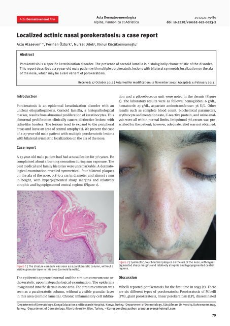
Acta Dermatovenerol APAActa DermatovenerologicaAlpina, Pannonica et Adriatica2012;21:79-80doi: 10.2478/v10162-012-0023-2<strong>Localized</strong> <strong>actinic</strong> <strong>nasal</strong> <strong>porokeratosis</strong>: a <strong>case</strong> <strong>report</strong>Arzu Ataseven 1 ✉ , Perihan Öztürk 2 , Nursel Dilek 3 , Ilknur Küçükosmanoğlu 1AbstractPorokeratisis is a specific keratinization disorder. The presence of cornoid lamella is histologically characteristic of the disorder.This <strong>report</strong> describes a 23-year-old male patient with multiple porokeratotic lesions with bilateral symmetric localization on the alaof the nose, which may be a rare variant of <strong>porokeratosis</strong>.Received: 17 October 2012 | Returned for modification: 17 November 2012 | Accepted: 11 February 2013IntroductionPorokeratosis is an epidermal keratinization disorder with anunclear etiopathogenesis. Cornoid lamella, a histopathologicalmarker, results from abnormal proliferation of keratinocytes. Thisabnormal proliferation clinically causes distinctive lesions withridge-like borders. The lesions tend to expand to the peripheralareas and leave an area of central atrophy (1). We present the <strong>case</strong>of a 23-year-old male patient with multiple porokeratotic lesionswith bilateral symmetric localization on the ala of the nose.The epidermis appeared normal and the stratum corneum was orthokeratoticupon histopathological examination. The epidermisinvaginated into the dermis in one area. The stratum corneum wasseen as a parakeratotic column, without a visible granular layerin this area (cornoid lamella). Chronic inflammatory cell infiltrationand a pilosebaceous unit were noted in the dermis (Figure2). The laboratory results were as follows: hemoglobin: 6 g/dL,hematocrit: 23 g/dL, aspartate aminotransferase: 36 U/L. Otherresults such as complete blood count, biochemical parameters,erythrocyte sedimentation rate, C-reactive protein, and urine analysiswere all within normal limits. Imiquimod 5% cream was prescribedfor the patient; however, adequate relief was not obtained.Case <strong>report</strong>A 23-year-old male patient had had a <strong>nasal</strong> lesion for 3½ years. Hecomplained about a burning sensation during sun exposure. Thepast medical and family histories were unremarkable. A dermatologicalexamination revealed symmetrical, four bilateral plaqueson the ala of the nose, 0.8 to 2 cm in diameter and almost 1 mmin height, with hyperpigmented sharp margins and relativelyatrophic and hypopigmented central regions (Figure 1).Figure 1 | The stratum corneum was seen as a parakeratotic column, without avisible granular layer in this area (cornoid lamella).Figure 2 | Symmetric, four bilateral plaques on the ala of the nose, with hyperpigmentedsharp margins and relatively atrophic and hypopigmented centralregions.DiscussionMibelli <strong>report</strong>ed <strong>porokeratosis</strong> for the first time in 1893 (2). Thereare six different types of <strong>porokeratosis</strong>: Porokeratosis of Mibelli(PM), giant <strong>porokeratosis</strong>, linear <strong>porokeratosis</strong> (LP), disseminated1Department of Dermatology, Konya Education and Research Hospital, Konya, Turkey. 2 Department of Dermatology, Sütçü İmam University, Kahramanmaraş,Turkey. 3 Department of Dermatology, Rize University, Rize, Turkey. ✉ Corresponding author: arzuataseven@hotmail.com79
A. Ataseven et al.superficial <strong>actinic</strong> <strong>porokeratosis</strong> (DSAP), palmoplantar <strong>porokeratosis</strong>(PPP), and punctate <strong>porokeratosis</strong> (PP) (3). DSAP isthe most common type (4). Porokeratosis of Mibelli most oftenaffects the limbs, particularly the hands and feet, the neck andshoulders, the face, and the genitals (5). Giant <strong>porokeratosis</strong>is a very rare form of <strong>porokeratosis</strong> and it is a morphologicalvariant of PM (3). Linear <strong>porokeratosis</strong> can further be classifiedas localized, zosteriform, systematized, or generalized.The distribution of all of these variants follows Blaschko’slines, which may be explained by cutaneous mosaicism (6).The distribution of typical lesions is symmetrical and usuallyaffects sun-exposed areas. The lesions generally spare the face,palms, soles, and mucosal surfaces (7). PPP initially occurs inthe palmoplantar areas with subsequent involvement of theother areas of the body, including sites with sun exposure toultraviolet radiation (8). Punctate <strong>porokeratosis</strong> is a rare varietyof <strong>porokeratosis</strong>. It is characterized by seed-like punctatekeratoses and/or pits on the palms and soles (9).The disease tends to mostly affect the extremities. Less commonly,the body and the face can be affected. Isolated face involvementsuch as that seen in our patient is rarely <strong>report</strong>ed.The type of <strong>porokeratosis</strong> of these <strong>case</strong>s were PM and DSAP.In one study, seven out of 197 <strong>case</strong>s of PM had primary faciallesions (10). Gutierrez et al. encountered only six <strong>case</strong>s of facial<strong>porokeratosis</strong> during 15 years of experience. These <strong>case</strong>sconsisted of one male and five females, and the patients’ agerange was 16 to 30 years (11). Even though the age of our <strong>case</strong>,23 years, was consistent with this <strong>report</strong>, the patient was male,whereas <strong>porokeratosis</strong> mostly affects females. Another patientwith bilateral lesions on the ala of the nose similar to our <strong>case</strong>was <strong>report</strong>ed by Ghorpade (4).Porokeratosis can occur with a genetic predisposition and itcan also be secondary to risk factors such as sun exposure, immunesuppression, and ultraviolet exposure. The incidence ofActa Dermatovenerol APA | 2012;21:79-80Bowen’s disease and squamous cell and spinous cell carcinomasis 6.8 to 11% in this disease, and so follow-up is essential.The risk is greater if the patient has linear-type <strong>porokeratosis</strong>,or a large or long-standing lesion (11).The etiology and pathogenesis of <strong>porokeratosis</strong> are obscurebut certainly complex and multifactorial. It has been suggestedthat the lesions of <strong>porokeratosis</strong> result from the peripheral expansionof an abnormal, mutant clone of epidermal keratinocytes(which would be inherited) located at the base of the parakeratoticcolumn (12).The following disorders should be considered in the differentialdiagnosis of <strong>porokeratosis</strong>: elastosis perforans for PM,<strong>actinic</strong> keratosis and stucco keratosis for DSAP, lichen sclerosuset atrophicus, lichen planus, acrokeratosis verruciformis,Bowen’s disease, and squamous cell carcinoma for single lesions(13). Discoid lupus erythematosus-like <strong>case</strong> can also beexpressed with similar clinical presentations (14).The histopathological presence of a cornoid lamella is oftenassociated with a diagnosis of <strong>porokeratosis</strong>. However, this featureis not pathognomonic for <strong>porokeratosis</strong> and can be foundin a number of other dermatological conditions, which includeseborrheic keratosis, verruca vulgaris, <strong>actinic</strong> keratosis, squamouscell carcinoma in situ, basal-cell carcinoma, milia, andscars. Notably, none of these entities has an inflammatory etiology(15).Keratolytic agents, topical 5-flourorasil, topical and oral retinoidagents, topical imiquimod, cryotherapy, photodynamictherapy, carbon dioxide laser application, dermabrasion, andexcision are used for treatment (16). We preferred to prescribetopical imiquimod our patient for three months.In conclusion, our <strong>case</strong> was a rare <strong>case</strong> of PM, with only symmetricalbilateral involvement of the ala of the nose. The <strong>case</strong>may be a rare variant of <strong>porokeratosis</strong> and the differential diagnosisof annular lesions should be borne in mind.References1. Lembo S, Panariello L, Nugnes L, Lembo C, Alaya F. Porokeratosis: two faces,one family. Case Rep Dermatol. 2009;1:52-5.2. Mibelli V. Contributo allo studio della ipercheratosi dei canali sudoriferi (porokeratosi).G It Mal Vener Pele. 1893;28:313-55.3. Bozdağ KE, Bicakci H, Ermete M. Giant <strong>porokeratosis</strong>. Int J Dermatol. 2004;43:518-20.4. Ghorpade A. <strong>Localized</strong> <strong>actinic</strong> <strong>nasal</strong> <strong>porokeratosis</strong>. Clin Exp Dermatol. 2010;35:211-2.5. Odeyinde S, Belcher H. Isolated single digit Porokeratosis of Mibelli: an unusual<strong>case</strong>. Dermatol Online J. 2012;18:13.6. Kudligi C, Bhagwat PV, Giriyan SS, Eshwarrao MS. Unilateral systematized linear<strong>porokeratosis</strong>: a <strong>report</strong> of a rare <strong>case</strong>. Indian J Dermatol. 2011;56:452-3.7. Lee HR, Han TY, Son SJ, Lee JH. Squamous cell carcinoma developing withinlesions of disseminated superficial <strong>actinic</strong> <strong>porokeratosis</strong>. Indian J Dermatol.2011;56:452-3.8. Irisawa R, Yamazaki M, Yamamoto T, Tsuboi R. A <strong>case</strong> of <strong>porokeratosis</strong> plantarispalmaris et disseminata and literature review. Dermatol Online J. 2012;18:5.9. Ghorpade A, Ramanan C. Punctuate <strong>porokeratosis</strong>. Indian J Dermatol VenereolLeprol. 1995;61:123-4.10. Wang NS, Gruson LM, Kamino H. Fasial follicular <strong>porokeratosis</strong>: a <strong>case</strong> <strong>report</strong>.Am J Dermatopathol. 2010;32:720-2.11. Gutierrez EL, Galarza C, Ramos W, Tello M, De Paz PC, Bobbio L, et al. Facial<strong>porokeratosis</strong>: a series of six patients. Australas J Dermatol. 2010;51:191-4.12. Kanitakis J, Euvrard S, Faure M, Claudy A. Porokeratosis and immunosuppression.Eur J Dermatol. 1998;8:459-65.13. Ikizoglu G. Genodermatozlar. In: Tüzün Y, Gürer MA, Serdaroglu S, Oguz O, AksungurVL (eds). Dermatoloji, 3rd ed. Istanbul: Nobel Tıp Kitabevi; 2008;1609-736.14. Nabai H, Mehregan AH. Porokeratosis of Mibelli. A <strong>report</strong> of two unusual <strong>case</strong>s.Dermatologica. 1979;159:325-31.15. Tran H, Bossenbroek NM, Rosenman K, Meehan SA, Sanchez M, Prystowsky S.Steroid-responsive facial eruption with cornoid lamella-a possible new entity.Dermatol Online J. 2008;14:9.16. Lee Y, Choi EH. Exclusive facial <strong>porokeratosis</strong>: histopathologically showing follicularcornoid lamella. J Dermatol. 2011;38:1072-5.80