Oral submucous fibrosis in a young patient - ResearchGate
Oral submucous fibrosis in a young patient - ResearchGate
Oral submucous fibrosis in a young patient - ResearchGate

Create successful ePaper yourself
Turn your PDF publications into a flip-book with our unique Google optimized e-Paper software.
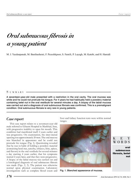
<strong>Oral</strong> <strong>submucous</strong> <strong>fibrosis</strong>: a case reportCase report<strong>Oral</strong> <strong>submucous</strong> <strong>fibrosis</strong> <strong>in</strong>a <strong>young</strong> <strong>patient</strong>M. J. Yazdanpanah, M. Banihashemi, F. Pezeshkpoor, S. Famili, P. Layegh, M. Katebi, and H. HamidiS UMMARYA seventeen-year-old male presented with a restriction <strong>in</strong> the oral cavity. The oral mucosa waswhite and he could not protrude his tongue. For 4 years he had habitually held a powdery materialconta<strong>in</strong><strong>in</strong>g betel nut <strong>in</strong> the oral vestibule for several m<strong>in</strong>utes a day. A biopsy of the labial mucosawas carried out and a diagnosis of oral <strong>submucous</strong> <strong>fibrosis</strong> was confirmed. This is a premalignantcondition. <strong>Oral</strong> <strong>submucous</strong> <strong>fibrosis</strong> is very rare <strong>in</strong> <strong>young</strong> <strong>patient</strong>s.Case reportThis case report relates to a seventeen-year-oldmale referred to Ghaem Hospital <strong>in</strong> Mashhad, Iran,with progressive <strong>in</strong>ability to open his mouth. Thiscondition had manifested itself 2 years earlier andwas progressive. On exam<strong>in</strong>ation, the <strong>in</strong>ter-<strong>in</strong>cisalopen<strong>in</strong>g was approximately 20 mm. The oral mucosawas blanched <strong>in</strong> appearance and he could notprotrude his tongue (Fig. 1). Question<strong>in</strong>g revealedthat he was <strong>in</strong> habit of hold<strong>in</strong>g a powdery material(conta<strong>in</strong><strong>in</strong>g betel nut, catechu, tobacco, lime, spices,and flavors) <strong>in</strong> the oral vestibule for several m<strong>in</strong>utesa day start<strong>in</strong>g 4 years earlier, that his symptomsstarted 2 years later, and that they were progressive.A biopsy of the labial mucosa was carried out anda histological diagnosis of oral <strong>submucous</strong> <strong>fibrosis</strong>was made (Figs. 2, 3). The <strong>patient</strong> was otherwisenormal upon physical exam<strong>in</strong>ation. Laboratory<strong>in</strong>vestigations such as complete blood count andliver and kidney function tests were with<strong>in</strong> normalranges.Fig. 1. Blanched appearance of mucosa.K E YWORDSoral<strong>submucous</strong><strong>fibrosis</strong>, betelnut176Acta Dermatoven APA Vol 18, 2009, No 4
Case report<strong>Oral</strong> <strong>submucous</strong> <strong>fibrosis</strong>: a case reportFig. 2. Microscopic view of histological sectionof OSMF lesions; chronic <strong>in</strong>flammatoryreaction and deep hypovascular <strong>fibrosis</strong> undersquamous epithelium with focal atrophy tohyperplasia, H&E sta<strong>in</strong><strong>in</strong>g, ×100.Fig. 3. Microscopic view of OSMF lesion; A:Hypovascular hyal<strong>in</strong>ized collagenous <strong>fibrosis</strong> <strong>in</strong>superior to <strong>in</strong>ferior dermis, B: Note entrapmentof striated muscle fascicles by deep and dense<strong>fibrosis</strong>, H&E sta<strong>in</strong><strong>in</strong>g, ×400.Discussion<strong>Oral</strong> <strong>submucous</strong> <strong>fibrosis</strong> (OSMF) is a chronicdisease with chronic <strong>in</strong>flammation and <strong>fibrosis</strong> of<strong>submucous</strong> tissues, caus<strong>in</strong>g restriction of the mouthopen<strong>in</strong>g (1). Areca nut chew<strong>in</strong>g has a significantrole <strong>in</strong> its etiology (2). Cases have been reportedworldwide wherever Asians migrate, but it occursmost commonly <strong>in</strong> India and southeast Asia (3, 4).The majority of OSMF cases belong to the 20- to40-year-old age group and there is a male-to-femaleratio of 1.8 to 2:1 (5–7). Most OSMF cases occur <strong>in</strong>lower socioeconomic groups (8).In the pathogenesis of OSMF, it is suggested thata multifactorial mechanism is at work, <strong>in</strong>clud<strong>in</strong>gareca nut chew<strong>in</strong>g, the <strong>in</strong>gestion of spicy redpepper, nutritional deficiency <strong>in</strong>clud<strong>in</strong>g vitam<strong>in</strong>sand trace elements, hypersensitivity to variousdietary constituents, and genetic and immunologicalsusceptibility (9–13).Histopathology f<strong>in</strong>d<strong>in</strong>gs are the ma<strong>in</strong>stay ofdiagnosis at present. The pr<strong>in</strong>cipal features of OSMFare less vascularized collagenous submucosa with arange of atrophy <strong>in</strong> the neighbor<strong>in</strong>g striated musclefibers, mild to moderate chronic <strong>in</strong>flammation,and epithelial changes consist<strong>in</strong>g of atrophy and avariable degree of dysplasia (14–17).The important pathological feature of OSMFis submucosal accumulation of collagen lead<strong>in</strong>g toepithelial atrophy (18, 19). It has been discoveredthat exposure of buccal mucosal fibroblasts toalkaloids may cause aggregation of collagen (20).<strong>Oral</strong> <strong>submucous</strong> <strong>fibrosis</strong> is a chronic conditionof the oral mucosa and oropharynx with thepotential for malignant transformation. Squamouscell carc<strong>in</strong>oma may occur <strong>in</strong> 7.6% of cases (9).An <strong>in</strong>terest<strong>in</strong>g po<strong>in</strong>t <strong>in</strong> our <strong>patient</strong> is hisrelatively <strong>young</strong> age. Proper preventive measuressuch as public education must be taken to reducethis serious disease.R EFERENCES1. WHO. Meet<strong>in</strong>g report. Control of oral cancer <strong>in</strong> develop<strong>in</strong>g countries. WHO Bull. 1984;62:617.2. Babu S, Bhat RV, Kumar PU, et al. A comparative cl<strong>in</strong>ico-pathological study of oral <strong>submucous</strong> <strong>fibrosis</strong> <strong>in</strong>habitual chewers of panmasala and betel quid. Cl<strong>in</strong> Toxicol. 1996;34:317–22.3. Tang JG, Jian XF, Gao ML, et al. Epidemiological survey of oral <strong>submucous</strong> <strong>fibrosis</strong> <strong>in</strong> Xiangtan city, HunanProv<strong>in</strong>ce, Ch<strong>in</strong>a. Community Dent <strong>Oral</strong> Epidemiol. 1997;25:177–80.4. Shah B, Lewis MA, Bedi R. <strong>Oral</strong> <strong>submucous</strong> <strong>fibrosis</strong> <strong>in</strong> an 11-year-old Bangladeshi girl liv<strong>in</strong>g <strong>in</strong> the UnitedK<strong>in</strong>gdom. Br Dent J. 2001;191:130–2.Acta Dermatoven APA Vol 18, 2009, No 4 177
<strong>Oral</strong> <strong>submucous</strong> <strong>fibrosis</strong>: a case reportCase report5. Sirsat SM, Khanolkar VR. Submucous <strong>fibrosis</strong> of the palate <strong>in</strong> diet-preconditioned Wistar rats: Induction bylocal pa<strong>in</strong>t<strong>in</strong>g of capsaic<strong>in</strong>–an optical and electron microscopic study. Arch Pathol. 1960;70:171–9.6. Shah N, Sharma PP. Role of chew<strong>in</strong>g and smok<strong>in</strong>g habits <strong>in</strong> the etiology of oral <strong>submucous</strong> <strong>fibrosis</strong>: A casecontrol study. J <strong>Oral</strong> Pathol Med. 1998;27:475–9.7. Wahi PN, Kapoor VL, Luthra UK, et al. Submucous <strong>fibrosis</strong> of the oral cavity: 2. Studies on epidemiology. BullWorld Health Organ. 1966;35:793–9.8. Ramanathan K. OSMF–An alternative hypothesis as to its causes. Med J Malaysia. 1981;36:243–5.9. Aziz SR. <strong>Oral</strong> <strong>submucous</strong> <strong>fibrosis</strong>: an unusual disease. J N J Dent Assoc. 1997;68:17–19.10. Liao PH, Lee TL, Yang LC, et al. Adenomatous polyposis coli gene mutation and decreased wild-type p53prote<strong>in</strong> expression <strong>in</strong> oral <strong>submucous</strong> <strong>fibrosis</strong>: a prelim<strong>in</strong>ary <strong>in</strong>vestigation. <strong>Oral</strong> Surg <strong>Oral</strong> Med <strong>Oral</strong> Pathol <strong>Oral</strong>Radiol Endod. 2001;92:202–7.11. van Wyk CW, Grobler-Rabie AF, Martell RW, et al. HLA-antigens <strong>in</strong> oral <strong>submucous</strong> <strong>fibrosis</strong>. J <strong>Oral</strong> PatholMed. 1994;23:23–7.12. Paul RR, Chatterjee J, Das AK, et al. Altered elemental profile as <strong>in</strong>dicator of homeostatic imbalance <strong>in</strong>pathogenesis of oral <strong>submucous</strong> <strong>fibrosis</strong>. Biol Trace Elem Res. 2002;87:45–56.13. Paul RR, Chatterjee J, Das AK, et al. Z<strong>in</strong>c and iron as bio<strong>in</strong>dicators of precancerous nature of oral <strong>submucous</strong><strong>fibrosis</strong>. Biol Trace Elem Res. 1996;54:213–30.14. Rosai J. Rosai and Ackerman’s surgical pathology. 9th ed., vol. 1. Ed<strong>in</strong>burgh: Mosbey; 2004.15. Neville BW, Damm DD, Allen CM, et al. <strong>Oral</strong> & maxillofacial pathology. 2nd ed. Philadelphia: W. B.Saunders Company; 2002.16. Canniff JP, Harvey W, Harris M. <strong>Oral</strong> <strong>submucous</strong> <strong>fibrosis</strong>: its pathogenesis and management. Br Dent J.1986;160:429–34.17. P<strong>in</strong>dborg JJ, Sirsat SM. <strong>Oral</strong> <strong>submucous</strong> <strong>fibrosis</strong>. <strong>Oral</strong> Surg <strong>Oral</strong> Med <strong>Oral</strong> Pathol. 1966;22:764–79.18. Hardie J. <strong>Oral</strong> <strong>submucous</strong> <strong>fibrosis</strong>. A review with case reports. J Can Dent Assoc. 1987;53:389–93.19. Huang IY, Shieh TY. Collagen content and types <strong>in</strong> oral <strong>submucous</strong> <strong>fibrosis</strong>. Gaoxiong Yi Xue Ke Xue ZaZhi. 1989;5:162–71.20. Harvey W, Scutt A, Meghji S, et al. Stimulation of human buccal mucosa fibroblasts <strong>in</strong> vitro by betel-nutalkaloids. Arch <strong>Oral</strong> Biol. 1986;31:45–9.A U T O R S 'ADDRESSESMohammad Javad Yazdanpanah, MD, Associate Professor of Dermatology,Department of Dermatology, Ghaem Hospital, Mashhad University ofMedical Sciences, Mashhad , Iran, correspond<strong>in</strong>g author, Tel.: +98 5118012861, +98 511 8409612, E-mail: yazdanpanahmj@mums.ac.irMahnaz Banihashemi, MD, Associate Professor of Dermatology, sameaddressFakhrozaman Pezeshkpoor, MD, Associate Professor of Dermatology, sameaddressSoroor Famili, MD, Associate Professor of Dermatology, same addressPouran Layegh, MD, Associate Professor of Dermatology, same addressMehrdad Katebi, MD, Assistant Professor of Pathology, same addressHamid Hamidi, MD, Resident of Dermatology, same address178Acta Dermatoven APA Vol 18, 2009, No 4