From Protein Structure to Function with Bioinformatics.pdf
From Protein Structure to Function with Bioinformatics.pdf
From Protein Structure to Function with Bioinformatics.pdf

Create successful ePaper yourself
Turn your PDF publications into a flip-book with our unique Google optimized e-Paper software.
Daniel John RigdenEdi<strong>to</strong>r<strong>From</strong> <strong>Protein</strong> <strong>Structure</strong><strong>to</strong> <strong>Function</strong> <strong>with</strong><strong>Bioinformatics</strong>
Prefaceviiof multiple methods <strong>with</strong>in integrated servers. This is more convenient for the userand also allows for determination of consensus predictions. Chapter 10 describesthe resources and operation of ProFunc and ProKnow which work in this way.Chapter 11 discusses published work in which structure-based methods wereapplied <strong>to</strong> predict functions for Structural Genomics outputs. This results in a valuablepicture of which methods typically prove most informative. The chapter concludes<strong>with</strong> a discussion of recent moves <strong>to</strong>wards community annotation as a way <strong>to</strong> tacklethe bottleneck in annotation of Structural Genomics results. Chapter 12 coversapplications of structure-based methods <strong>to</strong> model structures, derived by both comparativeand ab initio techniques. As well as a broad range of examples, publishedwork assessing the accuracy of model structures in function-relevant aspects andthe applicability of various methods <strong>to</strong> model structures is discussed.This book is designed <strong>to</strong> provide an up-<strong>to</strong>-date impression of the state-of-the-artin protein structure prediction and structure-based function prediction. Each methodschapter contains links <strong>to</strong> available servers and other resources which the readermay wish <strong>to</strong> apply <strong>to</strong> his or her protein. At the end of each chapter, authors pick outfuture directions and challenges in their respective areas. I hope the reader gains anaccurate impression of the impressive pace of research in these areas. Even whilethis book was being finalised, significant progress in a longstanding problem area– refinement of comparative models – was reported (Jagielska et al. 2008).Nevertheless, it seems that protein structures continually present new challenges <strong>to</strong>be met. Just as we may feel that the community is getting <strong>to</strong> grips <strong>with</strong> domainswapping, circular permutation, fibril formation and intrinsically disordered proteins,<strong>to</strong> name but a few previously unexpected phenomena, we are presented <strong>with</strong>metamorphic proteins (Murzin 2008) which may carry profound implications forour understanding of protein fold space. Will bioinformatics methods ever be able<strong>to</strong> predict which proteins can morph from one fold <strong>to</strong> another? That remains uncertain,but clearly the bioinformatics of protein structure-function will remain anexciting research area for many years <strong>to</strong> come.ReferencesJagielska A, Wroblewska L, Skolnick J (2008) <strong>Protein</strong> model refinement using an optimized physicsbasedall-a<strong>to</strong>m force field. Proc Natl Acad Sci USA 105:8268–8273Lesk AM (1997) CASP2: report on ab initio predictions. <strong>Protein</strong>s Suppl 1:151–166Murzin AG (2008) Metamorphic proteins. Science 320:1725–1726
ContentsSection I Generating and Inferring <strong>Structure</strong>s1 Ab Initio <strong>Protein</strong> <strong>Structure</strong> Prediction ................................................... 3Jooyoung Lee, Sitao Wu, and Yang Zhang1.1 Introduction ....................................................................................... 31.2 Energy <strong>Function</strong>s .............................................................................. 51.2.1 Physics-Based Energy <strong>Function</strong>s .......................................... 51.2.2 Knowledge-Based Energy <strong>Function</strong> Combined<strong>with</strong> Fragments ..................................................................... 91.3 Conformational Search Methods ...................................................... 131.3.1 Monte Carlo Simulations ...................................................... 141.3.2 Molecular Dynamics ............................................................. 151.3.3 Genetic Algorithm ................................................................ 151.3.4 Mathematical Optimization .................................................. 161.4 Model Selection ................................................................................ 161.4.1 Physics-Based Energy <strong>Function</strong> ........................................... 171.4.2 Knowledge-Based Energy <strong>Function</strong> ..................................... 171.4.3 Sequence-<strong>Structure</strong> Compatibility <strong>Function</strong> ......................... 181.4.4 Clustering of Decoy <strong>Structure</strong>s ............................................. 191.5 Remarks and Discussions ................................................................. 192 Fold Recognition ...................................................................................... 27Lawrence A. Kelley2.1 Introduction ....................................................................................... 272.1.1 The Importance of Blind Trials:The CASP Competition ........................................................ 282.1.2 Ab Initio <strong>Structure</strong> Prediction VersusHomology Modelling ............................................................ 282.1.3 The Limits of Fold Space ...................................................... 302.1.4 A Note on Terminology: ‘Threading’and ‘Fold Recognition’ ......................................................... 312.2 Threading .......................................................................................... 31ix
xContents2.2.1 Knowledge-Based Potentials ................................................ 322.2.2 Finding an Alignment ........................................................... 342.2.3 Heuristics for Alignment ...................................................... 352.3 Remote Homology Detection Without Threading ............................ 382.3.1 Using Predicted Structural Features ..................................... 392.3.2 Sequence Profiles and Hidden Markov Models .................... 412.3.3 Fold Classification and Support Vec<strong>to</strong>r Machines ................ 432.3.4 Consensus Approaches ......................................................... 452.3.5 Traversing the Homology Network ...................................... 452.4 Alignment Accuracy, Model Quality and StatisticalSignificance ....................................................................................... 472.4.1 Algorithms for Alignment Generationand Assessment ..................................................................... 472.4.2 Estimation of Statistical Significance ................................... 482.5 Tools for Fold Recognition on the Web ............................................ 492.6 The Future ......................................................................................... 503 Comparative <strong>Protein</strong> <strong>Structure</strong> Modelling ............................................ 57András Fiser3.1 Introduction ....................................................................................... 573.1.1 <strong>Structure</strong> Determines <strong>Function</strong> ............................................. 573.1.2 Sequences, <strong>Structure</strong>s, Structural Genomics ........................ 583.1.3 Approaches <strong>to</strong> <strong>Protein</strong> <strong>Structure</strong> Prediction .......................... 583.2 Steps in Comparative <strong>Protein</strong> <strong>Structure</strong> Modelling .......................... 603.2.1 Searching for <strong>Structure</strong>s Related<strong>to</strong> the Target Sequence .......................................................... 623.2.2 Selecting Templates .............................................................. 643.2.3 Sequence <strong>to</strong> <strong>Structure</strong> Alignment ......................................... 653.2.4 Model Building ..................................................................... 673.2.5 Model Evaluation .................................................................. 763.3 Performance of Comparative Modelling ........................................... 773.3.1 Accuracy of Methods ............................................................ 773.3.2 Errors in Comparative Models .............................................. 783.4 Applications of Comparative Modelling ........................................... 803.4.1 Modelling of Individual <strong>Protein</strong>s .......................................... 803.4.2 Comparative Modelling and the <strong>Protein</strong><strong>Structure</strong> Initiative ................................................................ 803.5 Summary ........................................................................................... 814 Membrane <strong>Protein</strong> <strong>Structure</strong> Prediction ............................................... 91Timothy Nugent and David T. Jones4.1 Introduction ....................................................................................... 914.2 Structural Classes .............................................................................. 92
Contentsxi4.2.1 Alpha-Helical Bundles ......................................................... 924.2.2 Beta-Barrels .......................................................................... 924.3 Membrane <strong>Protein</strong>s Are Difficult <strong>to</strong> Crystallise ............................... 944.4 Databases .......................................................................................... 944.5 Multiple Sequence Alignments ......................................................... 964.6 Transmembrane <strong>Protein</strong> Topology Prediction .................................. 984.6.1 Alpha-Helical <strong>Protein</strong>s .......................................................... 984.6.2 Beta-Barrel <strong>Protein</strong>s .............................................................. 1024.6.3 Whole Genome Analysis ...................................................... 1024.6.4 Data Sets, Homology, Accuracyand Cross-Validation ............................................................. 1034.7 3D <strong>Structure</strong> Prediction ..................................................................... 1054.8 Future Developments ........................................................................ 1075 <strong>Bioinformatics</strong> Approaches <strong>to</strong> the <strong>Structure</strong>and <strong>Function</strong> of Intrinsically Disordered <strong>Protein</strong>s ............................... 113Peter Tompa5.1 The Concept of <strong>Protein</strong> Disorder ...................................................... 1135.2 Sequence Features of IDPs ............................................................... 1155.2.1 The Unusual Amino AcidComposition of IDPs ............................................................ 1155.2.2 Sequence Patterns of IDPs .................................................... 1155.2.3 Low Sequence Complexity and Disorder ............................. 1165.3 Prediction of Disorder ....................................................................... 1165.3.1 Prediction of Low-Complexity Regions ............................... 1165.3.2 Charge-Hydropathy Plot ....................................................... 1175.3.3 Propensity-Based Predic<strong>to</strong>rs ................................................. 1175.3.4 Predic<strong>to</strong>rs Based on the Lackof Secondary <strong>Structure</strong> .......................................................... 1185.3.5 Machine Learning Algorithms .............................................. 1195.3.6 Prediction Based on Contact Potentials ................................ 1205.3.7 A Reduced Alphabet Suffices<strong>to</strong> Predict Disorder ................................................................ 1215.3.8 Comparison of Disorder Prediction Methods ....................... 1225.4 <strong>Function</strong>al Classification of IDPs ..................................................... 1225.4.1 Gene On<strong>to</strong>logy-Based <strong>Function</strong>alClassification of IDPs ........................................................... 1225.4.2 Classification of IDPs Basedon Their Mechanism of Action ............................................. 1235.4.3 <strong>Function</strong>-Related Structural Elements in IDPs ..................... 1265.5 Prediction of the <strong>Function</strong> of IDPs ................................................... 1285.5.1 Correlation of Disorder Pattern and <strong>Function</strong> ....................... 1285.5.2 Predicting Short Recognition Motifs in IDRs ....................... 1285.5.3 Prediction of MoRFs ............................................................. 129
xiiContents5.5.4 Combination of Information on Sequenceand Disorder: Phosphorylation Sitesand CaM Binding Motifs ...................................................... 1315.5.5 Flavours of Disorder ............................................................. 1315.6 Limitations of IDP <strong>Function</strong> Prediction ............................................ 1325.6.1 Rapid Evolution of IDPs ....................................................... 1325.6.2 Sequence Independence of <strong>Function</strong>and Fuzziness ........................................................................ 1335.6.3 Good News: Conservation and Disorder .............................. 1345.7 Conclusions ....................................................................................... 135Section II <strong>From</strong> <strong>Structure</strong>s <strong>to</strong> <strong>Function</strong>s6 <strong>Function</strong> Diversity Within Folds and Superfamilies ............................. 143Benoit H. Dessailly and Christine A. Orengo6.1 Defining <strong>Function</strong> ............................................................................. 1436.2 <strong>From</strong> Fold <strong>to</strong> <strong>Function</strong> ...................................................................... 1456.2.1 Definition of a Fold ............................................................... 1456.2.2 Prediction of <strong>Function</strong> Using FoldRelationships ......................................................................... 1486.3 <strong>Function</strong> Diversity Between Homologous <strong>Protein</strong>s .......................... 1516.3.1 Definitions ............................................................................. 1516.3.2 Evolution of <strong>Protein</strong> Superfamilies ....................................... 1526.3.3 <strong>Function</strong> Divergence During <strong>Protein</strong> Evolution ................... 1546.4 Conclusion ........................................................................................ 1627 Predicting <strong>Protein</strong> <strong>Function</strong> from Surface Properties .......................... 167Nicholas J. Burgoyne and Richard M. Jackson7.1 Surface Descriptions ......................................................................... 1677.1.1 The van der Waals Surface .................................................... 1677.1.2 Molecular Surface (Solvent Excluded Surface) .................... 1687.1.3 The Solvent Accessible Surface ............................................ 1687.2 Surface Properties ............................................................................. 1697.2.1 Hydrophobicity ..................................................................... 1697.2.2 Electrostatics Properties ........................................................ 1707.2.3 Surface Conservation ............................................................ 1717.3 <strong>Function</strong> Predictions Using Surface Properties ................................ 1717.3.1 Hydrophobic Surface ............................................................ 1727.3.2 Electrostatic Surface ............................................................. 1727.3.3 Surface Conservation ............................................................ 1737.3.4 Combining Surface Propertiesfor <strong>Function</strong> Prediction ......................................................... 1747.4 <strong>Protein</strong>-Ligand Interactions .............................................................. 1747.4.1 Properties of <strong>Protein</strong>-Ligand Interactions ............................. 174
Contentsxiii7.4.2 Predicting Binding Site Locations ........................................ 1757.4.3 Predictions of Druggability ................................................... 1787.4.4 Annotation of Ligand Binding Sites ..................................... 1787.5 <strong>Protein</strong>-<strong>Protein</strong> Interfaces ................................................................. 1807.5.1 Properties of <strong>Protein</strong>-<strong>Protein</strong> Interfaces ................................ 1807.5.2 Hot-Spot Regions in <strong>Protein</strong> Interfaces ................................ 1817.5.3 Predictions of Interface Location .......................................... 1827.6 Summary ........................................................................................... 1848 3D Motifs ................................................................................................... 187Elaine C. Meng, Benjamin J. Polacco,and Patricia C. Babbitt8.1 Background and Significance ........................................................... 1888.1.1 What Is <strong>Function</strong>? ................................................................. 1898.1.2 Three-Dimensional Motifs: Definitionand Scope .............................................................................. 1908.2 Overview of Methods ........................................................................ 1908.2.1 Motif Discovery .................................................................... 1908.2.2 Motif Description and Matching ........................................... 1918.2.3 Interpretation of Results ........................................................ 1938.3 Specific Methods ............................................................................... 1968.3.1 User-Defined Motifs ............................................................. 1978.3.2 Motif Discovery .................................................................... 2018.4 Related Methods ............................................................................... 2088.4.1 Hybrid (Point-Surface) Descriptions .................................... 2088.4.2 Single-Point-Centred Descriptions ....................................... 2088.5 Docking for <strong>Function</strong>al Annotation .................................................. 2108.6 Discussion ......................................................................................... 2128.7 Conclusions ....................................................................................... 2129 <strong>Protein</strong> Dynamics: <strong>From</strong> <strong>Structure</strong> <strong>to</strong> <strong>Function</strong> ................................... 217Marcus B. Kubitzki, Bert L. de Groot,and Daniel Seeliger9.1 Molecular Dynamics Simulations ..................................................... 2179.1.1 Principles and Approximations ............................................. 2189.1.2 Applications .......................................................................... 2209.1.3 Limitations – Enhanced SamplingAlgorithms ............................................................................ 2269.2 Principal Component Analysis ......................................................... 2309.3 Collective Coordinate Sampling Algorithms .................................... 2339.3.1 Essential Dynamics ............................................................... 2339.3.2 TEE-REX .............................................................................. 2349.4 Methods for <strong>Function</strong>al Mode Prediction ......................................... 2379.4.1 Normal Mode Analysis ......................................................... 237
xivContents9.4.2 Elastic Network Models .................................................. 2389.4.3 CONCOORD .................................................................. 2399.5 Summary and Outlook .................................................................. 24210 Integrated Servers for <strong>Structure</strong>-Informed<strong>Function</strong> Prediction ............................................................................... 251Roman A. Laskowski10.1 Introduction ................................................................................... 25110.1.1 The Problem of Predicting <strong>Function</strong>from <strong>Structure</strong> ................................................................. 25210.1.2 <strong>Structure</strong>-<strong>Function</strong> PredictionMethods .......................................................................... 25310.2 ProKnow ....................................................................................... 25410.2.1 Fold Matching ................................................................. 25410.2.2 3D Motifs ........................................................................ 25610.2.3 Sequence Homology ....................................................... 25710.2.4 Sequence Motifs ............................................................. 25710.2.5 <strong>Protein</strong> Interactions ......................................................... 25810.2.6 Combining the Predictions .............................................. 25810.2.7 Prediction Success .......................................................... 25810.3 ProFunc ......................................................................................... 25910.3.1 ProFunc’s <strong>Structure</strong>-Based Methods .............................. 25910.3.2 Assessment of the Structural Methods ............................ 26710.4 Conclusion .................................................................................... 26911 Case Studies: <strong>Function</strong> Predictions of StructuralGenomics Results ................................................................................... 273James D. Watson and Janet M. Thorn<strong>to</strong>n11.1 Introduction ................................................................................... 27311.2 Large Scale <strong>Function</strong> PredictionCase Studies .................................................................................. 27511.3 Some Specific Examples ............................................................... 28111.4 Community Annotation ................................................................ 28711.5 Conclusions ................................................................................... 28812 Prediction of <strong>Protein</strong> <strong>Function</strong> fromTheoretical Models ................................................................................. 293Iwona A. Cymerman, Daniel J. Rigden,and Janusz M. Bujnicki12.1 Background ................................................................................... 29312.2 <strong>Protein</strong> Models as a Community Resource ................................... 29512.2.1 Model Quality ................................................................. 29612.2.2 Databases of Models ....................................................... 297
Contentsxv12.3 Accuracy and Added Value of Model-DerivedProperties ...................................................................................... 29812.3.1 Implementation ............................................................... 30012.4 Practical Application ..................................................................... 30212.4.1 Plasticity of Catalytic Site Residues ............................... 30212.4.2 Mutation Mapping .......................................................... 30412.4.3 <strong>Protein</strong> Complexes .......................................................... 30512.4.4 <strong>Function</strong> Predictions fromTemplate-Free Models .................................................... 30612.4.5 Prediction of Ligand Specificity ..................................... 30912.4.6 <strong>Structure</strong> Modelling of AlternativelySpliced Isoforms ............................................................. 31012.4.7 <strong>From</strong> Broad <strong>Function</strong> <strong>to</strong> Molecular Details .................... 31212.5 What Next? ................................................................................... 314Index ................................................................................................................. 319
Chapter 1Ab Initio <strong>Protein</strong> <strong>Structure</strong> PredictionJooyoung Lee, Sitao Wu, and Yang ZhangAbstract Predicting protein 3D structures from the amino acid sequence stillremains as an unsolved problem after five decades of efforts. If the target proteinhas a homologue already solved, the task is relatively easy and high-resolutionmodels can be built by copying the framework of the solved structure. However,such a modelling procedure does not help answer the question of how and why aprotein adopts its specific structure. If structure homologues (occasionally analogues)do not exist, or exist but cannot be identified, models have <strong>to</strong> be constructedfrom scratch. This procedure, called ab initio modelling, is essential for a completesolution <strong>to</strong> the protein structure prediction problem; it can also help us understandthe physicochemical principle of how proteins fold in nature. Currently, the accuracyof ab initio modelling is low and the success is limited <strong>to</strong> small proteins (
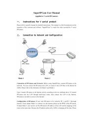
4 J. Lee et al.about 5.3 million protein sequences were deposited in the UniProtKB database(Bairoch et al. 2005) (http://www.ebi.ac.uk/swissprot). However, the correspondingnumber of protein structures in the <strong>Protein</strong> Data Bank (PDB) (Berman et al. 2000)(http://www.rcsb.org/pdb) is only about 44,000, less than 1% of the proteinsequences. The gap is rapidly widening as indicated in Fig. 1.1. Thus, developingefficient computer-based algorithm <strong>to</strong> predicting 3D structures from sequences isprobably the only avenue <strong>to</strong> fill up the gap.Depending on whether similar proteins have been experimentally solved, proteinstructure prediction methods can be grouped in<strong>to</strong> two categories. First, if proteinsof a similar structure are identified from the PDB library, the target model can beconstructed by copying the framework of the solved proteins (templates). The procedureis called “template-based modelling (TBM)” (Karplus et al. 1998; Jones1999; Shi et al. 2001; Ginalski et al. 2003b; Skolnick et al. 2004; Jaroszewski et al.2005; Soding 2005; Zhou and Zhou 2005; Cheng and Baldi 2006; Pieper et al.2006; Wu and Zhang 2008), which will be discussed in the subsequent chapters.Although high-resolution models can be often generated by TBM, the procedurecannot help us understand the physicochemical principle of protein folding.If protein templates are not available, we have <strong>to</strong> build the 3D models fromscratch. This procedure has been called by several names, e.g. ab initio modelling(Klepeis et al. 2005; Liwo et al. 2005; Wu et al. 2007), de novo modelling (Bradleyet al. 2005), physics-based modelling (Oldziej et al. 2005), or free modelling (Jauchet al. 2007). In this chapter, the term ab initio modelling is uniformly used <strong>to</strong> avoidconfusion. Unlike the template-based modelling, successful ab initio modellingprocedure could help answer the basic questions on how and why a protein adoptsthe specific structure out of many possibilities.Fig. 1.1 The number of available protein sequences (left ordinate) and the solved protein structures(right ordinate) are shown for the last 12 years. The ratio of sequence/structure is rapidlyincreasing. Data are taken from UniProtKB (Bairoch et al. 2005) and PDB (Berman et al. 2000)databases
1 Ab Initio <strong>Protein</strong> <strong>Structure</strong> Prediction 5Typically, ab initio modelling conducts a conformational search under the guidanceof a designed energy function. This procedure usually generates a number ofpossible conformations (structure decoys), and final models are selected from them.Therefore, a successful ab initio modelling depends on three fac<strong>to</strong>rs: (1) an accurateenergy function <strong>with</strong> which the native structure of a protein corresponds <strong>to</strong> themost thermodynamically stable state, compared <strong>to</strong> all possible decoy structures;(2) an efficient search method which can quickly identify the low-energy statesthrough conformational search; (3) selection of native-like models from a pool ofdecoy structures.This chapter gives a review on the current state of the art in ab initio proteinstructure prediction. This review is neither complete <strong>to</strong> include all available ab initiomethods nor in depth <strong>to</strong> provide all backgrounds/motivations behind them. For acomparative study of various ab initio modelling methods, readers are recommended<strong>to</strong> read a recent review by Helles (Helles 2008). The rest of the chapter isorganized as follows. Three major issues of ab initio modelling, i.e. energy function,conformational search engine and model selection scheme, will be describedin detail. New and promising ideas <strong>to</strong> improve the efficiency and effectiveness ofthe prediction are discussed. Finally, current progresses and challenges of ab initiomodelling are summarized.1.2 Energy <strong>Function</strong>sIn this section, we will discuss energy functions used for ab initio modelling. Itshould be noted that in many cases energy functions and the search procedures areintricately coupled <strong>to</strong> each other, and as soon as they are decoupled, the modellingprocedure often loses its power/validity. We classify the energy in<strong>to</strong> two groups:(a) physics-based energy functions and (b) knowledge-based energy functions,depending on the use of statistics from the existing protein 3D structures. A fewpromising methods from each group are selected <strong>to</strong> discuss according <strong>to</strong> theiruniqueness and modelling accuracy. A list of ab initio modelling methods is providedin Table 1.1 along <strong>with</strong> their properties about energy functions, conformationalsearch algorithms, model selection methods and typical running times.1.2.1 Physics-Based Energy <strong>Function</strong>sIn a strictly-defined physics-based ab initio method, interactions between a<strong>to</strong>msshould be based on quantum mechanics and the coulomb potential <strong>with</strong> only a fewfundamental parameters such as the electron charge and the Planck constant; all a<strong>to</strong>msshould be described by their a<strong>to</strong>m types where only the number of electrons is relevant(Hagler et al. 1974; Weiner et al. 1984). However, there have not been serious attempts<strong>to</strong> start from quantum mechanics <strong>to</strong> predict structures of (even small) proteins, simply
6 J. Lee et al.Table 1.1 A list of ab initio modelling algorithms reviewed in this chapter is shown along <strong>with</strong>their energy functions, conformational search methods, model selection schemes and typicalCPU time per targetAlgorithm & serveraddress Force-field type Search method Model selectionAMBER/CHARMM/OPLS (Brooks et al.1983; Weiner et al.1984; Jorgensen andTirado-Rives 1988;Duan and Kollman1998; Zagrovic et al.2002)UNRES (Liwo et al.1999, 2005; Oldziejet al. 2005)ASTRO-FOLD (Klepeisand Floudas 2003;Klepeis et al. 2005)ROSETTA (Simons et al.1997; Das et al. 2007)http://www.robetta.orgTASSER/Chunk-TASSER(Zhang and Skolnick2004a; Zhou andSkolnick 2007) http://cssb.biology.gatech.edu/skolnick/webservice/MetaTASSERI-TASSER (Wu et al.2007; Zhang 2007)http://zhang.bioin-formatics.ku.edu/I-TASSERPhysics-basedPhysics-basedMoleculardynamics(MD)Conformationalspace annealing(CSA)Lowest energyClustering/freeenergyTime costper CPUYearsHoursPhysics-based αBB/CSA/MD Lowest energy MonthsMonte Carlo(MC)MCMCPhysics- andknowledgebasedKnowledgebasedClustering/freeenergyClustering/freeenergyKnowledgebasedClustering/freeenergyMonthsHoursHoursbecause the computational resources required for such calculations are far beyondwhat is available now. Without quantum mechanical treatments, a practical startingpoint for ab initio protein modelling is <strong>to</strong> use a compromised force field <strong>with</strong> a largenumber of selected a<strong>to</strong>m types; in each a<strong>to</strong>m type, the chemical and physical propertiesof the a<strong>to</strong>ms are enough alike <strong>with</strong> the parameters calculated from crystal packing orquantum mechanical theory (Hagler et al. 1974; Weiner et al. 1984). Well-knownexamples of such all-a<strong>to</strong>m physics-based force fields include AMBER (Weiner et al.1984; Cornell et al. 1995; Duan and Kollman 1998), CHARMM (Brooks et al. 1983;Neria et al. 1996; MacKerell Jr. et al. 1998), OPLS (Jorgensen and Tirado-Rives 1988;Jorgensen et al. 1996), and GROMOS96 (van Gunsteren et al. 1996). These potentialscontain terms associated <strong>with</strong> bond lengths, angles, <strong>to</strong>rsion angles, van der Waals, andelectrostatics interactions. The major difference between them lies in the selection ofa<strong>to</strong>m types and the interaction parameters.
1 Ab Initio <strong>Protein</strong> <strong>Structure</strong> Prediction 7For the study of protein folding, these classical force fields were often coupled<strong>with</strong> molecular dynamics (MD) simulations. However, the results, from the viewpoin<strong>to</strong>f protein structure prediction, were not quite successful. (See Chapter 10for the use of MD in elucidation of protein function from known structures). Thefirst miles<strong>to</strong>ne in such MD-based ab initio protein folding is probably the 1997work of Duan and Kollman who simulated the villin headpiece (a 36-mer) inexplicit solvent for six months on parallel supercomputers. Although the authorsdid not fold the protein <strong>with</strong> high resolution, the best of their final model was<strong>with</strong>in 4.5 Å <strong>to</strong> the native state (Duan and Kollman 1998). With Folding@Home,a worldwide-distributed computer system, this small protein was recently foldedby Pande and coworkers (Zagrovic et al. 2002) <strong>to</strong> 1.7 Å <strong>with</strong> a <strong>to</strong>tal simulationtime of 300 ms or approximately 1,000 CPU years. Despite these remarkableefforts, the all-a<strong>to</strong>m physics-based MD simulation is far from being routinely usedfor structure prediction of typical-size proteins (~100–300 residues), not <strong>to</strong> mentionthe fact that the validity/accuracy has not yet been systematically tested evenfor a number of small proteins.Another protein structure niche where physics-based MD simulation can contributeis structure refinement. Starting from low-resolution protein models, the goal is <strong>to</strong>draw them closer <strong>to</strong> the native by refining the local side chain and peptide-backbonepacking. When the starting models are not very far away from the native, the intendedconformational change is relatively small and the simulation time would be much lessthan that required in ab initio folding. One of the early MD-based protein structurerefinements was for the GCN4 leucine zipper (33-residue dimer) (Nilges and Brunger1991; Vieth et al. 1994), where a low-resolution coiled-coil dimer structure (2–3 Å)was first assembled by Monte Carlo (MC) simulation before the subsequent MDrefinement. With the help of helical dihedral-angle restraints, Skolnick and coworkers(Vieth et al. 1994) were able <strong>to</strong> generate a refined structure of GCN4 <strong>with</strong> below 1 Åbackbone root-mean-square deviation (RMSD) using CHARMM (Brooks et al.1983) and the TIP3P water model (Jorgensen et al. 1983).Later, using AMBER 5.0 (Case et al. 1997) and TIP3P water model (Jorgensenet al. 1983), Lee et al. (2001) attempted <strong>to</strong> refine 360 low-resolution models generatedby ROSETTA (Simons et al. 1997) for 12 small proteins (
8 J. Lee et al.2000; Sorin and Pande 2005), OPLS-AA (Kaminski et al. 2001), GROMOS96(van Gunsteren et al. 1996), and ENCAD (Levitt et al. 1995) ) <strong>to</strong> refine 75 proteinsby in vacuo energy minimization. They found that a knowledge-baseda<strong>to</strong>mic contact potential outperforms the MM potentials by moving almost all testproteins closer <strong>to</strong> their native states, while the MM potentials, except forAMBER99, essentially drove decoys further away from their native structures.The vacuum simulation <strong>with</strong>out solvation may be partly the reason for the failureof the MM potentials. This observation demonstrates the possibility of combiningknowledge-based potentials <strong>with</strong> physics-based force fields for more successfulprotein structure refinement.While the physics-based potential driven by MD simulations was not particularlysuccessful in structure prediction, fast search methods (such as Monte Carlosimulations and genetic algorithms) based on physics-based potentials have shown<strong>to</strong> be promising in both structure prediction and structure refinement. One exampleis the ongoing project by Scheraga and coworkers (Liwo et al. 1999, 2005; Oldziejet al. 2005) who have been developing a physics-based protein structure predictionmethod solely based on the thermodynamic hypothesis. The method combines thecoarse grained potential of UNRES <strong>with</strong> the global optimization algorithm calledconformational space annealing (Oldziej et al. 2005). In UNRES, each residue isdescribed by two interacting off-lattice united a<strong>to</strong>ms, C αand the side chain centre.This effectively reduces the number of a<strong>to</strong>ms by 10, enabling one <strong>to</strong> handlepolypeptide chains of larger than 100 residues. The resulting prediction time forsmall proteins can be then reduced <strong>to</strong> 2–10 h. The UNRES energy function (Liwoet al. 1993) consists of pair wise interactions between all interacting parties andadditional terms such as local energy and correlation energy. The low energyUNRES models are then converted in<strong>to</strong> all-a<strong>to</strong>m representations based on ECEPP/3(Nemethy et al. 1992). Although many of the parameters of the energy function arecalculated by quantum-mechanical methods, some of them are derived from thedistributions and correlation functions calculated from the PDB library. For thisreason, one might question the authenticity of the true ab initio nature of theirapproach. Nevertheless, this method is probably the most faithful ab initio methodavailable (in terms of the application of a thorough global optimization <strong>to</strong> a physicsbasedenergy function) and it has been systematically applied <strong>to</strong> many CASP targetssince 1998. The most notable prediction success by this approach is for T061from CASP3, for which a model of 4.2 Å RMSD <strong>to</strong> the native for a 95-residue α-helical protein was generated <strong>with</strong> an accuracy gap from the rest of models by others.It is shown, for the first time in a clear-cut fashion that the ab initio method canprovide better models for the targets where the template-based methods fail. InCASP6, a structure genomics target of TM0487 (T0230, 102 residues) was folded<strong>to</strong> 7.3 Å by this approach. However, it seems that the scarcity and the best-but-stilllowaccuracy of such models by a pure ab initio modelling failed <strong>to</strong> draw muchattention from the protein science community, where accurate protein models arein great demand.Another example of the physics-based modelling approaches is the multistagehierarchical algorithm ASTRO-FOLD, proposed by Floudas and coworkers
1 Ab Initio <strong>Protein</strong> <strong>Structure</strong> Prediction 9(Klepeis and Floudas 2003; Klepeis et al. 2005). First, secondary structure elements(α-helices and β-strands) are predicted by calculating a free energy function ofoverlapping oligopeptides (typically pentapeptides) and all possible contactsbetween two hydrophobic residues. The free energy terms used include entropic,cavity formation, polarization, and ionization contributions for each oligopeptide.After transforming the calculated secondary structure propensity in<strong>to</strong> the upper andlower bounds of backbone dihedral angles and the distant restraints between C αa<strong>to</strong>ms, the final tertiary structure of the full length protein is modeled by globallyminimizing the ECEPP/3 all-a<strong>to</strong>m force field. This approach was successfullyapplied <strong>to</strong> an α-helical protein of 102 residues in a double-blind fashion (but not inan open community-wide way for relative performance comparison <strong>to</strong> other methods).The C αRMSD of the predicted model was 4.94 Å away from the experimentalstructure. The global optimization method used in this approach is a combinationof α branch and bound (αBB), conformational space annealing, and MD simulations(Klepeis and Floudas 2003; Klepeis et al. 2005). The relative performance ofthis method for a number of proteins is yet <strong>to</strong> be seen in the future.Taylor and coworkers (2008) recently proposed a novel approach which constructsprotein structural models by enumerating possible <strong>to</strong>pologies in a coarsegrainedform, given the secondary structure assignments and the physicalconnection constraints of the secondary structure elements. The <strong>to</strong>p scoring conformations,based on the structural compactness and element exposure, are thenselected for further refinement (Jonassen et al. 2006). The authors successfully folda set of five αβ sandwich proteins <strong>with</strong> length up <strong>to</strong> 150 residues <strong>with</strong> the firstmodel <strong>with</strong>in 4–6 Å RMSD of the native structure. Again, although appealing inmethodology, the performance of the approach in the open blind experiments andon the proteins of various fold-types is yet <strong>to</strong> be seen.In the recent development of ROSETTA (Bradley et al. 2005; Das et al. 2007),a physics-based a<strong>to</strong>mic potential is used in the second stage of Monte Carlo structurerefinement following the low-resolution fragment assembly (Simons et al.1997), which we will discuss in the next section.1.2.2 Knowledge-Based Energy <strong>Function</strong> Combined<strong>with</strong> FragmentsKnowledge-based potential refers <strong>to</strong> the empirical energy terms derived from thestatistics of the solved structures in deposited PDB, which can be divided in<strong>to</strong> twotypes as described by Skolnick (2006). The first one covers generic and sequenceindependentterms such as the hydrogen bonding and the local backbone stiffnessof a polypeptide chain (Zhang et al. 2003). The second contains amino-acid or protein-sequencedependent terms, e.g. pair wise residue contact potential (Skolnick etal. 1997), distance dependent a<strong>to</strong>mic contact potential (Samudrala and Moult 1998;Lu and Skolnick 2001; Zhou and Zhou 2002; Shen and Sali 2006), and secondarystructure propensities (Zhang et al. 2003, 2006; Zhang and Skolnick 2005a).
10 J. Lee et al.Although most knowledge-based force fields contain secondary structure propensitypropensities, it may be that local protein structures are rather difficult <strong>to</strong>reproduce in the reduced modelling. That is, in nature a variety of protein sequencesprefer either helical or extended structures depending on the subtle differences intheir local and global sequence environment, yet we have not yet found force fieldsthat can reproduce this subtlety properly. One way <strong>to</strong> circumvent this problem is <strong>to</strong>use secondary structure fragments, obtained from sequence or profile alignments,directly in<strong>to</strong> 3D model assembly. Another advantage of this approach is that the useof excised secondary structure fragment can significantly reduce the entropy of theconformational search.Here, we introduce two prediction methods utilizing knowledge-based energyfunctions, which are proved <strong>to</strong> be the most successful in ab initio protein structureprediction (Simons et al. 1997; Zhang and Skolnick 2004a).One of the best-known ideas for ab initio modelling is probably the one pioneeredby Bowie and Eisenberg, who generated protein models by assemblingsmall fragments (mainly 9-mers) taken from the PDB library (Bowie and Eisenberg1994). Based on a similar idea, Baker and coworkers developed ROSETTA (Simonset al. 1997), which was extremely successful for the free modelling (FM) targets inCASP experiments and made the fragment assembly approach popular in the field.In the recent developments of ROSETTA (Bradley et al. 2005; Das et al. 2007), theauthors first generated models in a reduced form <strong>with</strong> conformations specified <strong>with</strong>heavy backbone and C βa<strong>to</strong>ms. In the second phase, a set of selected low-resolutionmodels were subject <strong>to</strong> all-a<strong>to</strong>m refinement procedure using an all-a<strong>to</strong>m physicsbasedenergy function, which includes van der Waals interactions, pair wise solvationfree energy, and an orientation-dependent hydrogen-bonding potential. Theflowchart of the two-phase modelling is shown in Fig. 1.2 and details on the energyfunctions can be found in references (Bradley et al. 2005; Das et al. 2007). For theconformational search, multiple rounds of Monte Carlo minimization (Li andScheraga 1987) are carried out. The most notable example for this two-step pro<strong>to</strong>colis the blind prediction of an ab initio target (T0281 from CASP6, 70 residues),whose C αRMSD from its crystal structure is 1.6 Å (Bradley et al. 2005). In CASP7,a very extensive sampling was carried out using the distributed computing networkof Rosetta@home allowing about 500,000 CPU hours for each target domain.There was one target, T0283, which was a template-based modelling (TBM) targetbut was modeled by the ROSETTA ab initio pro<strong>to</strong>col. It generated a model ofRMSD = 1.8 Å over 92 residues out of the 112 residues (Fig. 1.3, left panel).Despite the significant success, the computational cost of the procedure is ratherexpensive for routine use.Partially because of the notable success of the ROSETTA algorithm, as well asthe limited availability of its energy functions <strong>to</strong> others, several groups initiateddevelopments of their own energy functions following the idea of ROSETTA.Derivatives of ROSETTA include Simfold (Fujitsuka et al. 2006) and Profesy (Leeet al. 2004); their energy terms include van der Waals interactions, backbone dihedralangle potentials, hydrophobic interactions, backbone hydrogen-bonding potential,rotamer potential, pair wise contact energies, beta-strand pairing, and a term
1 Ab Initio <strong>Protein</strong> <strong>Structure</strong> Prediction 11Fig. 1.2 Flowchart of the ROSETTA pro<strong>to</strong>colcontrolling the protein radius of gyration. However, their prediction seems <strong>to</strong> beonly partially successful in comparison <strong>to</strong> ROSETTA.Another successful free modelling approach, TASSER by Zhang and Skolnick(2004a), constructs 3D models based on a purely knowledge-based approach. Thetarget sequence is first threaded through a set of representative protein structures <strong>to</strong>search for possible folds. Contiguous fragments (>5 residues) are then excised fromthe threaded aligned regions and used <strong>to</strong> reassemble full-length models, while unalignedregions are built by ab initio modelling (Zhang et al. 2003). The proteinconformation in TASSER is represented by a trace of C αa<strong>to</strong>ms and side chain centresof mass, and the reassembly process is conducted by parallel Monte Carlo simulations(Zhang et al. 2002). The energy terms of TASSER include information aboutpredicted secondary structure propensities, backbone hydrogen bonds, a variety ofshort- and long-range correlations and hydrophobic energy based on the structural
12 J. Lee et al.Fig. 1.3 Two examples of successful free modelling from CASP7 are shown. T0283 (left panel)is a TBM target (from Bacillus halodurans) of 112 residues; the model was generated by all-a<strong>to</strong>mROSETTA (a hybrid knowledge- and physics-based approach) (Das et al. 2007) based on freemodelling, which gives a TM-score 0.74 (Zhang and Skolnick 2004b) and a RMSD 1.8 Å over thefirst 92 residues (13.8 Å overall RMSD is due <strong>to</strong> the wrong orientation of the C-terminal helix).T0382 (right panel) is a FM/TBM target (from Rhodopseudomonas palustris CGA009) of 123residues; the model was generated by I-TASSER (a purely knowledge-based approach) (Zhang2007) <strong>with</strong> a TM-score 0.66 and a RMSD 3.6 Å. Blue and red represent the model and the crystalstructures, respectivelystatistics from the PDB library. Weights of knowledge-based energy terms are optimizedusing a large-scale structure decoy set (Zhang et al. 2003) which coordinatesthe complicated correlations between various interaction terms.There are several new developments of TASSER. One is Chunk-TASSER (Zhouand Skolnick 2007) in Skolnick’s group, which first splits the target sequences in<strong>to</strong>subunits (or “chunks”), each containing three consecutive regular secondary structureelements (helix and strand). These chunks are then folded separately. Finally,the spatial restraints are extracted from the chunk models and used for the subsequentTASSER simulations.Another development is I-TASSER by Wu et al. (2007), which refines TASSERcluster centroids by iterative Monte Carlo simulations. The spatial restraints areextracted from the first round TASSER models and the template structures searchedby TM-align (Zhang and Skolnick 2005b) from the PDB library, which are exploitedin the second round simulations. The purpose is <strong>to</strong> remove the steric clashes fromthe first round models and refine the <strong>to</strong>pology. The flowchart of I-TASSER isshown in Fig. 1.4. Although the procedure uses structural fragments and spatialrestraints from threading templates, it often constructs models of correct <strong>to</strong>pologyeven when <strong>to</strong>pologies of constituting templates are incorrect. In CASP7, out of 19FM and FM/TBM targets, I-TASSER built models <strong>with</strong> correct <strong>to</strong>pology (~3–5 Å)for seven cases <strong>with</strong> sequences up <strong>to</strong> 155 residues long. Figure 1.3 (right panel)shows the example of T0382 (123 residues) where all initial templates were of
1 Ab Initio <strong>Protein</strong> <strong>Structure</strong> Prediction 13Fig. 1.4 Flowchart of I-TASSER protein structure modellingwrong <strong>to</strong>pology (>9 Å) but the final model is 3.6 Å away from the X-ray structure.Recently, Helles carried out a comparative study on 18 ab initio prediction algorithmsand concluded that I-TASSER is about the best method in term of the modellingaccuracy and CPU cost per target (Helles 2008).1.3 Conformational Search MethodsSuccessful ab initio modelling of protein structures depends on the availability ofa powerful conformation search method which can efficiently find the globalminimum energy structure for a given energy function <strong>with</strong> complicated energylandscape. His<strong>to</strong>rically, Monte Carlo and molecular dynamics are two popularsimulation methods <strong>to</strong> explore the conformational space of macromolecules suchas proteins. For complicated systems like proteins, canonical MD/MC methodsusually require a huge amount of computational resources for a complete explorationof the conformational space. The record for direct application of MD <strong>to</strong>obtain the protein native structure is not so impressive. One explanation for thefailure could be that the simulation time required <strong>to</strong> fold a small protein takes aslong as milliseconds, 10 12 times longer than the usual incremental time step offem<strong>to</strong>seconds (10 −15 s). The technical difficulty of MC simulations mainly comesfrom that the energy landscape of protein conformational space is typically quite
14 J. Lee et al.rugged containing many energy barriers, which may easily trap the MC simulationprocedures.In this section we discuss recent development in conformational search methods<strong>to</strong> overcome these problems. We intend <strong>to</strong> illustrate the key ideas of conformationalsearch methods used in various ab initio and related protein modelling procedures.Readers are recommended <strong>to</strong> read appropriate references for details. Unlike variousenergy functions used in ab initio modelling, the search methods should be, inprinciple, transferable between protein modelling methods, as well as otherproblems in science and technology. Currently, there exist no single omni-powerfulsearch method that outperforms the others for all cases, and the investigation andsystematic benchmarking on the performance of various search methods has yet <strong>to</strong>be carried out.1.3.1 Monte Carlo SimulationsSimulated annealing (SA) (Kirkpatrick et al. 1983) is probably the most popularconformational search method. SA is general in that it is easy and straightforward<strong>to</strong> apply <strong>to</strong> any kind of optimization problem. In SA, one typically performsMetropolis MC algorithm <strong>to</strong> generate a series of conformational states followingthe canonical Boltzmann energy distribution for a given temperature. SA initiallyexecutes high temperature MC simulation, followed by a series of simulations subject<strong>to</strong> a temperature-lowering schedule, hence the name simulated annealing. Asmuch as SA is simple, its conformational search efficiency is not so impressivecompared <strong>to</strong> other more sophisticated methods discussed below.When the energy landscape of the system under investigation is rugged (due <strong>to</strong>numerous energy barriers), MC simulations are prone <strong>to</strong> get stuck in meta-stablestates that will dis<strong>to</strong>rt the distribution of sampled states by breaking the ergodicityof sampling. To avoid this malfunction, many simulation techniques have beendeveloped, and one of the most successful approaches is based on the generalizedensemble approach in contrast <strong>to</strong> the usual canonical ensemble. This kind ofmethod was initially called by different names including multi-canonical ensemble(Berg and Neuhaus 1992) and entropic ensemble (Lee 1993). The underlying ideais <strong>to</strong> expedite the transition between states separated by energy barriers by modifyingthe transition probability so that the final energy distribution of samplingbecomes more or less flat rather than bell-shaped. A popular method similar in thisspirit is the replica exchange MC method (REM) (Kihara et al. 2001) where a se<strong>to</strong>f many canonical MC simulations <strong>with</strong> temperatures distributed in a selectedrange are simultaneously carried out. <strong>From</strong> time <strong>to</strong> time one attempts <strong>to</strong> exchangestructures (or equivalently temperatures) from neighboring simulations <strong>to</strong> samplestates in a wide range of energy spectrum as the means <strong>to</strong> overcome energy barriers.Parallel hyperbolic sampling (PHS) (Zhang et al. 2002) further extends the REMby dynamically deforming energy using an inverse hyperbolic sine function <strong>to</strong>lower the energy barrier.
1 Ab Initio <strong>Protein</strong> <strong>Structure</strong> Prediction 15Monte Carlo <strong>with</strong> minimization (MCM), originally developed by Li andScheraga (Li and Scheraga 1987), was successfully applied <strong>to</strong> the conformationalsearch of ROSETTA’s high-resolution energy function. In MCM, one performs MCmoves between local energy minima after local energy minimization of each perturbedprotein structure. For a given local energy minimum structure A, a trialstructure B is generated by random perturbation of A and is subsequently subject<strong>to</strong> local energy minimization. The usual Metropolis algorithm is used <strong>to</strong> determinethe acceptance of B over A by calculating the energy difference between the two.1.3.2 Molecular DynamicsMD simulation (discussed in detail in Chapter 10) solves New<strong>to</strong>n’s equations ofmotion at each step of a<strong>to</strong>m movement, which is probably the most faithful methoddepicting a<strong>to</strong>mistically what is occurring in proteins. The method is therefore mos<strong>to</strong>ftenused for the study of protein folding pathways (Duan and Kollman 1998). Thelong simulation time is one of the major issues of this method, since the incrementaltime scale is usually in the order of fem<strong>to</strong>seconds (10 −15 s) while the fastestfolding time of a small protein (less than 100 residues) is in the millisecond rangein nature. Currently no serious all-a<strong>to</strong>m MD simulations are attempted for proteinstructure prediction starting from either an extended or a random initial structure.When a low resolution model is available, MD simulations are often carried out forstructure refinement since the conformational changes are assumed <strong>to</strong> be small.One notable approach is the recent work of Scheraga and his coworkers, who haveimplemented <strong>to</strong>rsion space MD simulation <strong>with</strong> the coarse-grained energy functionUNRES (see the discussion above).1.3.3 Genetic AlgorithmConformational space annealing (CSA) (Lee et al. 1998) is one of the most successfulgenetic algorithms. By utilizing a local energy minimizer as in MCM and theconcept of annealing in conformational space, it searches the whole conformationalspace of local minima in its early stages and then narrows the search <strong>to</strong> smallerregions <strong>with</strong> low energy as the distance cu<strong>to</strong>ff is reduced. Here the distance cu<strong>to</strong>ffis defined as the similarity between two conformations, and it controls the diversityof the conformational population. The distance cu<strong>to</strong>ff plays the role of temperaturein the usual SA, and initially its value is set <strong>to</strong> a large number in order <strong>to</strong> force conformationaldiversity. The value is gradually reduced as the search progresses. CSAhas been successfully applied <strong>to</strong> various global optimization problems includingprotein structure prediction separately combined <strong>with</strong> ab initio modelling inUNRES (Oldziej et al. 2005) and ASTRO-FOLD (Klepeis and Floudas 2003;Klepeis et al. 2005), and <strong>with</strong> fragment assembly in Profesy (Lee et al. 2004).
16 J. Lee et al.1.3.4 Mathematical OptimizationThe search approach by Floudas and coworkers, α branch and bound (αBB)(Klepeis and Floudas 2003; Klepeis et al. 2005), is unique in the sense that themethod is mathematically rigorous, while all the others discussed here are s<strong>to</strong>chasticand heuristic methods. The search space is successively cut in<strong>to</strong> two halveswhile the lower and upper bounds of the global minimum (LB and UB) for eachbranched phase space are estimated. The estimate for the UB is simply the bestcurrently obtained local minimum energy, and the estimate for the LB comes fromthe modified energy function augmented by a quadratic term of the dissecting variables<strong>with</strong> the coefficient α (hence the name αBB). With a sufficiently large valueof α, the modified energy contains only one energy minimum, whose value servesas the lower bound. While performing successive dissection of the phase spaceaccompanied by estimates of LB and UB for each dissected phase space, phasespaces <strong>with</strong> LB higher than the global UB can be eliminated from the search. Theprocedure continues until one identifies the global minimum by locating a dissectedphase space where LB becomes identical <strong>to</strong> the global UB. Once the solution isfound, the result is mathematically rigorous, but large proteins <strong>with</strong> many degreesof freedom are yet <strong>to</strong> be addressed by this method.1.4 Model SelectionAb initio modelling methods typically generate lots of decoy structures during thesimulation. How <strong>to</strong> select appropriate models structurally close <strong>to</strong> the native stateis an important issue. The selection of protein models has been emerged as a newfield called Model Quality Assessment Programs (MQAP) (Fischer 2006). In general,modelling selection approaches can be classified in<strong>to</strong> two types, i.e. the energybased and the free-energy based. In the energy based methods, one designs a varietyof specific potentials and identifies the lowest-energy state as the final prediction.In the free-energy based approaches, the free-energy of a given conformation R canbe written as−bE( R)F( R) =− kT ln Z( R)=−kTln e dW,BB∫(1)where Z(R) is the restricted partition function which is proportional <strong>to</strong> the numberof occurrences of the structures in the neighborhood of R during the simulation.This can be estimated by the clustering procedure at a given RMSD cu<strong>to</strong>ff (Zhangand Skolnick 2004c).For the energy-based model selection methods, we will discuss three energy/scoring functions: (1) physics-based energy function; (2) knowledge-based energyfunction; (3) scoring function describing the compatibility between the targetsequence and model structures. In MQAP, there is another popular method which
1 Ab Initio <strong>Protein</strong> <strong>Structure</strong> Prediction 17takes the consensus conformation from the predictions generated by different algorithms(Wallner and Elofsson 2007), which has also called meta-server approaches(Ginalski et al. 2003a; Wu and Zhang 2007). The essence of this method is similar<strong>to</strong> the clustering approach since both assume the most frequently occurring state asthe near-native ones. This approach has been mainly used for selecting modelsgenerated by threading-servers (Ginalski et al. 2003; Wallner and Elofsson 2007;Wu and Zhang 2007).1.4.1 Physics-Based Energy <strong>Function</strong>For the development of all-a<strong>to</strong>m physics-based energy functions, Lazaridis andKarplus (1999a) exploited CHARMM19 (Neria et al. 1996) and EEF1 (Lazaridisand Karplus 1999b) solvation potential <strong>to</strong> discriminate the native structure fromdecoys that are generated by threading on other protein structures. They found theenergy of the native state is lower than those of decoys in most cases. Later, Petreyand Honig (2000) used CHARMM and a continuum treatment of the solvent,Brooks and coworkers (Dominy and Brooks 2002; Feig and Brooks 2002) usedCHARMM plus GB solvation, Felts et al. (2002) used OPLS plus GB, Lee andDuan (2004) used AMBER plus GB, and (Hsieh and Luo 2004) used AMBER plusPoisson-Boltzmann solvation potential on a number of structure decoy sets (includingthe Park-Levitt decoy set (Park and Levitt 1996), Baker decoy set (Tsai et al.2003), Skolnick decoy set (Kihara et al. 2001; Skolnick et al. 2003), and CASPdecoys set (Moult et al. 2001) ). All these authors obtained similar results, i.e. thenative structures have lower energy than decoys in their potentials. The claimedsuccess of model discrimination of the physics-based potentials seems contradictedby other less successful physics-based structure prediction results. Recently,Wroblewska and Skolnick (2007) showed that the AMBER plus GB potential canonly discriminate the native structure from roughly minimized TASSER decoys(Zhang and Skolnick 2004a). After a 2-ns MD simulation on the decoys, none ofthe native structures were lower in energy than the lowest energy decoy, and theenergy-RMSD correlation was close <strong>to</strong> zero. This result partially explains the discrepancybetween the widely-reported decoy discrimination ability of physicsbasedpotentials and the less successful folding/refinement results.1.4.2 Knowledge-Based Energy <strong>Function</strong>Sippl developed a pair wise residue-distance based potential (Sippl 1990) using thestatistics of known PDB structures in 1990 (its newest version is PROSA II (Sippl1993; Wiederstein and Sippl 2007) ). Since then, a variety of knowledge-basedpotentials have been proposed, which include a<strong>to</strong>mic interaction potential, solvationpotential, hydrogen bond potential, <strong>to</strong>rsion angle potential, etc. In coarse-grained
18 J. Lee et al.potentials, each residue is represented either by a single a<strong>to</strong>m or by a few a<strong>to</strong>ms,e.g., C α-based potentials (Melo et al. 2002), C β-based potentials (Hendlich et al.1990), side chain centre-based potentials (Bryant and Lawrence 1993; Kocheret al. 1994; Thomas and Dill 1996; Skolnick et al. 1997; Zhang and Kim 2000;Zhang et al. 2004), side chain and C α-based potentials (Berrera et al. 2003). One ofthe most widely-used knowledge-based potentials is a residue-specific, all-a<strong>to</strong>m,distance-dependent potential, which was first formulated by Samudrala and Moult(RAPDF) (Samudrala and Moult 1998); it counts the distances between 167 aminoacid specific pseudo-a<strong>to</strong>ms. Following this, several a<strong>to</strong>mic potentials <strong>with</strong> variousreference states have been proposed, including those by Lu and Skolnick (KBP)(Lu and Skolnick 2001), Zhou and Zhou (DFIRE) (Zhou and Zhou 2002), Wanget al. (self-RAPDF) (Wang et al. 2004), Tost<strong>to</strong> (vic<strong>to</strong>r/FRST) (Tosat<strong>to</strong> 2005), andShen and Sali (DOPE) (Shen and Sali 2006). All these potentials claimed thatnative structures can be distinguished from decoy structures in their tests. However,the task of selecting the near native models out of many decoys remains as a challengefor these potentials (Skolnick 2006); this is actually more important thannative structure recognition because in reality there are no native structures availablefrom computer simulations. Based on the CAFASP4-MQAP experiment in2004 (Fischer 2006), the best-performing energy functions are Vic<strong>to</strong>r/FRST(Tosat<strong>to</strong> 2005) which incorporates an all-a<strong>to</strong>m pair wise interaction potential, solvationpotential and hydrogen bond potential, and MODCHECK (Pettitt et al.2005) which includes C βa<strong>to</strong>m interaction potential and solvation potential. <strong>From</strong>CASP7-MQAP in 2006, Pcons developed by Elofsson group based on structureconsensus performed best (Wallner and Elofsson 2007).1.4.3 Sequence-<strong>Structure</strong> Compatibility <strong>Function</strong>In the third type of MQAPs, best models are selected not purely based on energyfunctions. They are selected based on the compatibility of target sequences <strong>to</strong> modelstructures. The earliest and still successful example is that by Luthy et al. (1992),who used threading scores <strong>to</strong> evaluate structures. Colovos and Yeates (1993) laterused a quadratic error function <strong>to</strong> describe the non-covalently bonded interactionsamong CC, CN, CO, NN, NO and OO, where near-native structures have fewererrors than other decoys. Verify3D (Eisenberg et al. 1997) improves the method ofLuthy et al. (1992) by considering local threading scores in a 21-residue window.Jones developed GenThreader (Jones 1999) and used neural networks <strong>to</strong> classifynative and non-native structures. The inputs of GenThreader include pairwise contactenergy, solvation energy, alignment score, alignment length, and sequence andstructure lengths. Similarly, based on neural networks, Wallner and Ellofsson builtProQ (Wallner and Elofsson 2003) for quality prediction of decoy structures. Theinputs of ProQ include contacts, solvent accessible area, protein shape, secondarystructure, structural alignment score between decoys and templates, and the fractionof protein regions <strong>to</strong> be modeled from templates. Recently, McGuffin developed a
1 Ab Initio <strong>Protein</strong> <strong>Structure</strong> Prediction 19consensus MQAP (McGuffin 2007) called ModFold that includes ProQ (Wallnerand Elofsson 2003), MODCHECK (Pettitt et al. 2005) and ModSSEA. The authorshowed that ModFold outperforms its component MQAP programs.1.4.4 Clustering of Decoy <strong>Structure</strong>sFor the purpose of identifying the lowest free-energy state, structure clusteringtechniques were adopted by many ab initio modelling approaches. In the work byShortle et al. (1998), for all 12 cases tested, the cluster-centre conformation of thelargest cluster was closer <strong>to</strong> native structures than the majority of decoys. Clustercentrestructures were ranked as the <strong>to</strong>p 1–5% closest <strong>to</strong> their native structures.Zhang and Skolnick developed an iterative structure clustering method, calledSPICKER (Zhang and Skolnick 2004c). Based on the 1,489 representative benchmarkproteins each <strong>with</strong> up <strong>to</strong> 280,000 structure decoys, the best of the <strong>to</strong>p fivemodels was ranked as <strong>to</strong>p 1.4% among all decoys. For 78% of the 1,489 proteins,the RMSD difference between the best of the <strong>to</strong>p five models and the most nativelikedecoy structure was less than 1 Å.In ROSETTA ab initio modelling (Bradley et al. 2005), structure decoys areclustered <strong>to</strong> select low-resolution models and these models are further refined byall-a<strong>to</strong>m simulations <strong>to</strong> obtain final models. In the case of TASSER/I-TASSER(Zhang and Skolnick 2004a; Wu et al. 2007), thousands of decoy models from MCsimulations are clustered by SPICKER (Zhang and Skolnick 2004c) <strong>to</strong> generatecluster centroids as final models. In the approach by Scheraga and coworkers(Oldziej et al. 2005), decoys are clustered and the lowest-energy structures amongthe clustered structures are selected.1.5 Remarks and DiscussionsSuccessful ab initio modelling from amino acid sequence alone is considered asthe “Holy Grail” of protein structure prediction (Zhang 2008), since this will markan eventual and complete solution <strong>to</strong> the problem. Except for the generation of 3Dstructures, ab initio modelling can also help us understand the underlying principleson how proteins fold in nature; this could not be done by the template-basedmodelling approaches which build 3D models by copying the framework of othersolved structures.An ideal approach <strong>to</strong> ab initio modelling would be <strong>to</strong> treat a<strong>to</strong>ms in a protein asinteracting particles according <strong>to</strong> an accurate physics-based potential, and fold the proteinby solving New<strong>to</strong>n’s equations of motion in each step of movements. A number ofmolecular dynamics simulations were carried out along this line of approach byexploiting the classic CHARMM and AMBER force fields. Although the MD basedsimulation is extremely important for the study of protein folding, the success in the
20 J. Lee et al.viewpoint of structure prediction is quite limited. One reason is the prohibitive computingdemand for a normal size protein. On the other hand, knowledge-based (or hybridknowledge- and physics-based) approaches appear <strong>to</strong> be progressing rapidly, producingmany examples of successful low-<strong>to</strong>-medium accuracy models often <strong>with</strong> correct <strong>to</strong>pologyfor proteins of up <strong>to</strong> 100 residues. Although very rare, successful higher resolutionmodels (
1 Ab Initio <strong>Protein</strong> <strong>Structure</strong> Prediction 21Berg BA, Neuhaus T (1992) Multicanonical ensemble: a new approach <strong>to</strong> simulate first-orderphase transitions. Phys Rev Lett 68(1):9–12Berman HM, Westbrook J, Feng Z, et al. (2000) The protein data bank. Nucleic Acids Res28(1):235–242Berrera M, Molinari H, Fogolari F (2003) Amino acid empirical contact energy definitions forfold recognition in the space of contact maps. BMC <strong>Bioinformatics</strong> 4:8Bowie JU, Eisenberg D (1994) An evolutionary approach <strong>to</strong> folding small alpha-helical proteinsthat uses sequence information and an empirical guiding fitness function. Proc Natl Acad SciUSA 91(10):4436–4440Bradley P, Misura KM, Baker D (2005) Toward high-resolution de novo structure prediction forsmall proteins. Science 309(5742):1868–1871Brooks BR, Bruccoleri RE, Olafson BD, et al. (1983) CHARMM: a program for macromolecularenergy, minimization, and dynamics calculations. J Comput Chem 4(2):187–217Bryant SH, Lawrence CE (1993) An empirical energy function for threading protein sequencethrough the folding motif. <strong>Protein</strong>s 16(1):92–112Case DA, Pearlman DA, Caldwell JA, et al. (1997) AMBER 5.0, University of California,San Francisco, CA.Chen J, Brooks CL (2007) Can molecular dynamics simulations provide high-resolution refinemen<strong>to</strong>f protein structure? <strong>Protein</strong>s 67(4):922–930Cheng J, Baldi P (2006) A machine learning information retrieval approach <strong>to</strong> protein fold recognition.<strong>Bioinformatics</strong> 22(12):1456–1463Colovos C, Yeates TO (1993) Verification of protein structures: patterns of nonbonded a<strong>to</strong>micinteractions. <strong>Protein</strong> Sci 2(9):1511–1519Cornell WD, Cieplak P, Bayly CI, et al. (1995) A second generation force field for the simulationof proteins, nucleic acids, and organic molecules. J Am Chem Soc 117:5179–5197Das R, Qian B, Raman S, et al. (2007) <strong>Structure</strong> prediction for CASP7 targets using extensive alla<strong>to</strong>mrefinement <strong>with</strong> Rosetta@home. <strong>Protein</strong>s 69(S8):118–128Dominy BN, Brooks CL (2002) Identifying native-like protein structures using physics-basedpotentials. J Comput Chem 23(1):147–160Duan Y, Kollman PA (1998) Pathways <strong>to</strong> a protein folding intermediate observed in a 1-microsecondsimulation in aqueous solution. Science 282(5389):740–744Eisenberg D, Luthy R, Bowie JU (1997) VERIFY3D: assessment of protein models <strong>with</strong> threedimensionalprofiles. Method Enzymol 277:396–404Fan H, Mark AE (2004) Refinement of homology-based protein structures by molecular dynamicssimulation techniques. <strong>Protein</strong> Sci 13(1):211–220Feig M, Brooks CL (2002) Evaluating CASP4 predictions <strong>with</strong> physical energy functions.<strong>Protein</strong>s 49(2):232–245Felts AK, Gallicchio E, Wallqvist A, et al. (2002) Distinguishing native conformations of proteinsfrom decoys <strong>with</strong> an effective free energy estima<strong>to</strong>r based on the OPLS all-a<strong>to</strong>m force fieldand the Surface Generalized Born solvent model. <strong>Protein</strong>s 48(2):404–422Fischer D (2006) Servers for protein structure prediction. Curr Opin Struct Biol 16(2):178–182Fujitsuka Y, Chikenji G, Takada S (2006) SimFold energy function for de novo protein structureprediction: consensus <strong>with</strong> Rosetta. <strong>Protein</strong>s 62(2):381–398Ginalski K, Elofsson A, Fischer D, et al. (2003a) 3D-Jury: a simple approach <strong>to</strong> improve proteinstructure predictions. <strong>Bioinformatics</strong> 19(8):1015–1018Ginalski K, Pas J, Wyrwicz LS, et al. (2003b) ORFeus: detection of distant homology usingsequence profiles and predicted secondary structure. Nucleic Acids Res 31(13):3804–3807Hagler A, Euler E, Lifson S (1974) Energy functions for peptides and proteins I. Derivation of a consistentforce field including the hydrogen bond from amide crystals. J Am Chem Soc 96:5319–5327Helles G (2008) A comparative study of the reported performance of ab initio protein structureprediction algorithms. J R Soc Interface 5(21):387–396Hendlich M, Lackner P, Weitckus S, et al. (1990) Identification of native protein folds amongst alarge number of incorrect models. The calculation of low energy conformations from potentialsof mean force. J Mol Biol 216(1):167–180
22 J. Lee et al.Hsieh MJ, Luo R (2004) Physical scoring function based on AMBER force field and Poisson-Boltzmann implicit solvent for protein structure prediction. <strong>Protein</strong>s 56(3):475–486Im W, Lee MS, Brooks CL (2003) Generalized born model <strong>with</strong> a simple smoothing function. JComput Chem 24(14):1691–1702Jaroszewski L, Rychlewski L, Li Z, et al. (2005) FFAS03: a server for profile–profile sequencealignments. Nucleic Acids Res 33(Web Server issue):W284–288Jauch R, Yeo HC, Kolatkar PR, et al. (2007) Assessment of CASP7 structure predictions for templatefree targets. <strong>Protein</strong>s 69(Suppl 8):57–67Jonassen I, Klose D, Taylor WR (2006) <strong>Protein</strong> model refinement using structural fragment tessellation.Comput Biol Chem 30(5):360–366Jones DT (1999) GenTHREADER: an efficient and reliable protein fold recognition method forgenomic sequences. J Mol Biol 287(4):797–815Jorgensen WL, Tirado-Rives J (1988) The OPLS potential functions for proteins. Energy minimizationsfor crystals of cyclic peptides and crambin. J Am Chem Soc (110):1657–1666Jorgensen WL, Chandrasekhar J, Madura JD, et al. (1983) Comparison of simple potential functionsfor simulating liquid water. J Chem Phys 79:926–935Jorgensen WL, Maxwell DS, Tirado-Rives J (1996) Development and testing of the OPLS All-A<strong>to</strong>m Force Field on conformational energetics and properties of organic liquids. J Am ChemSoc 118:11225–11236Kaminski GA, Friesner RA, Tirado-Rives J, et al. (2001) Evaluation and Reparametrization of theOPLS-AA Force Field for proteins via comparison <strong>with</strong> accurate quantum chemical calculationson peptides. J Phys Chem B 105:6474–6487Karplus K, Barrett C, Hughey R (1998) Hidden Markov models for detecting remote proteinhomologies. <strong>Bioinformatics</strong> 14:846–856Kihara D, Lu H, Kolinski A, et al. (2001) TOUCHSTONE: an ab initio protein structure predictionmethod that uses threading-based tertiary restraints. Proc Natl Acad Sci USA98(18):10125–10130Kirkpatrick S, Gelatt CD, Vecchi MP (1983) Optimization by simulated annealing. Science220(4598):671–680Klepeis JL, Floudas CA (2003) ASTRO-FOLD: a combina<strong>to</strong>rial and global optimization frameworkfor Ab initio prediction of three-dimensional structures of proteins from the amino acidsequence. Biophys J 85(4):2119–2146Klepeis JL, Wei Y, Hecht MH, et al. (2005) Ab initio prediction of the three-dimensional structureof a de novo designed protein: a double-blind case study. <strong>Protein</strong>s 58(3):560–570Kocher JP, Rooman MJ, Wodak SJ (1994) Fac<strong>to</strong>rs influencing the ability of knowledge-basedpotentials <strong>to</strong> identify native sequence-structure matches. J Mol Biol 235(5):1598–1613Lazaridis T, Karplus M (1999a) Discrimination of the native from misfolded protein models <strong>with</strong>an energy function including implicit solvation. J Mol Biol 288(3):477–487Lazaridis T, Karplus M (1999b) Effective energy function for proteins in solution. <strong>Protein</strong>s35(2):133–152Lee J (1993) New Monte Carlo algorithm: entropic sampling. Phys Rev Lett 71(2):211–214Lee J, Scheraga HA, Rackovsky S (1998) Conformational analysis of the 20-residue membraneboundportion of melittin by conformational space annealing. Biopolymers 46(2):103–116Lee J, Kim SY, Joo K, et al. (2004) Prediction of protein tertiary structure using PROFESY, anovel method based on fragment assembly and conformational space annealing. <strong>Protein</strong>s56(4):704–714Lee MC, Duan Y (2004) Distinguish protein decoys by using a scoring function based on a newAMBER force field, short molecular dynamics simulations, and the generalized born solventmodel. <strong>Protein</strong>s 55(3):620–634Lee MR, Tsai J, Baker D, et al. (2001) Molecular dynamics in the endgame of protein structureprediction. J Mol Biol 313(2):417–430Levitt M, Hirshberg M, Sharon R, et al. (1995) Potential-energy function and parameters for simulationsof the molecular-dynamics of proteins and nucleic-acids in solution. Comput PhysCommun 91(1–3):215–231
1 Ab Initio <strong>Protein</strong> <strong>Structure</strong> Prediction 23Li Z, Scheraga HA (1987) Monte Carlo-minimization approach <strong>to</strong> the multiple-minima problemin protein folding. Proc Natl Acad Sci USA 84(19):6611–6615Lindahl E, Hess B, van der Spoel D (2001) GROMACS 3.0: a package for molecular simulationand trajec<strong>to</strong>ry analysis. J Mol Model 7:306–317Liwo A, Pincus MR, Wawak RJ, et al. (1993) Calculation of protein backbone geometry fromalpha-carbon coordinates based on peptide-group dipole alignment. <strong>Protein</strong> Sci2(10):1697–1714Liwo A, Lee J, Ripoll DR, et al. (1999) <strong>Protein</strong> structure prediction by global optimization of apotential energy function. Proc Natl Acad Sci USA 96(10):5482–5485Liwo A, Khalili M, Scheraga HA (2005) Ab initio simulations of protein-folding pathways bymolecular dynamics <strong>with</strong> the united-residue model of polypeptide chains. Proc Natl Acad SciUSA 102(7):2362–2367Lu H, Skolnick J (2001) A distance-dependent a<strong>to</strong>mic knowledge-based potential for improvedprotein structure selection. <strong>Protein</strong>s 44(3):223–232Luthy R, Bowie JU, Eisenberg D (1992) Assessment of protein models <strong>with</strong> three-dimensionalprofiles. Nature 356(6364):83–85MacKerell Jr. AD, Bashford D, Bellott M, et al. (1998) All-a<strong>to</strong>m empirical potential for molecularmodeling and dynamics studies of proteins. J Phys Chem B 102 (18):3586–3616McGuffin LJ (2007) Benchmarking consensus model quality assessment for protein fold recognition.BMC <strong>Bioinformatics</strong> 8:345Melo F, Sanchez R, Sali A (2002) Statistical potentials for fold assessment. <strong>Protein</strong> Sci11(2):430–448Moult J, Fidelis K, Zemla A, et al. (2001) Critical assessment of methods of protein structure prediction(CASP): round IV. <strong>Protein</strong>s(Suppl 5):2–7Nemethy G, Gibson KD, Palmer KA, et al. (1992) Energy parameters in polypeptides. 10.Improved geometric parameters and nonbonded interactions for use in the ECEPP/3 algorithm,<strong>with</strong> application <strong>to</strong> proline-containing peptides. J Phys Chem B 96: 6472–6484Neria E, Fischer S, Karplus M (1996) Simulation of activation free energies in molecular systems.J Chem Phys 105(5):1902–1921Nilges M, Brunger AT (1991) Au<strong>to</strong>mated modeling of coiled coils: application <strong>to</strong> the GCN4dimerization region. <strong>Protein</strong> Eng 4(6):649–659Oldziej S, Czaplewski C, Liwo A, et al. (2005) Physics-based protein-structure prediction using ahierarchical pro<strong>to</strong>col based on the UNRES force field: assessment in two blind tests. Proc NatlAcad Sci USA 102(21):7547–7552Park B, Levitt M (1996) Energy functions that discriminate X-ray and near native folds from wellconstructeddecoys. J Mol Biol 258(2):367–392Petrey D, Honig B (2000) Free energy determinants of tertiary structure and the evaluation ofprotein models. <strong>Protein</strong> Sci 9(11):2181–2191Pettitt CS, McGuffin LJ, Jones DT (2005) Improving sequence-based fold recognition by using3D model quality assessment. <strong>Bioinformatics</strong> 21(17):3509–3515Pieper U, Eswar N, Davis FP, et al. (2006) MODBASE: a database of annotated comparative proteinstructure models and associated resources. Nucleic Acids Res 34(Database issue):D291–295Samudrala R, Moult J (1998) An all-a<strong>to</strong>m distance-dependent conditional probability discrimina<strong>to</strong>ryfunction for protein structure prediction. J Mol Biol 275(5):895–916Shen MY, Sali A (2006) Statistical potential for assessment and prediction of protein structures.<strong>Protein</strong> Sci 15(11):2507–2524Shi J, Blundell TL, Mizuguchi K (2001) FUGUE: sequence-structure homology recognition usingenvironment-specific substitution tables and structure-dependent gap penalties. J Mol Biol310(1):243–257Shortle D, Simons KT, Baker D (1998) Clustering of low-energy conformations near the nativestructures of small proteins. Proc Natl Acad Sci USA 95(19):11158–11162Simons KT, Kooperberg C, Huang E, et al. (1997) Assembly of protein tertiary structures fromfragments <strong>with</strong> similar local sequences using simulated annealing and Bayesian scoring functions.J Mol Biol 268(1):209–225
24 J. Lee et al.Sippl MJ (1990) Calculation of conformational ensembles from potentials of mean force. Anapproach <strong>to</strong> the knowledge-based prediction of local structures in globular proteins. J Mol Biol213(4):859–883Sippl MJ (1993) Recognition of errors in three-dimensional structures of proteins. <strong>Protein</strong>s17(4):355–362Skolnick J (2006) In quest of an empirical potential for protein structure prediction. Curr OpinStruct Biol 16(2):166–171Skolnick J, Jaroszewski L, Kolinski A, et al. (1997) Derivation and testing of pair potentials forprotein folding. When is the quasichemical approximation correct? <strong>Protein</strong> Science6:676–688Skolnick J, Zhang Y, Arakaki AK, et al. (2003) TOUCHSTONE: a unified approach <strong>to</strong> proteinstructure prediction. <strong>Protein</strong>s 53(Suppl 6):469–479Skolnick J, Kihara D, Zhang Y (2004) Development and large scale benchmark testing of thePROSPECTOR 3.0 threading algorithm. <strong>Protein</strong> 56:502–518Soding J (2005) <strong>Protein</strong> homology detection by HMM-HMM comparison. <strong>Bioinformatics</strong>21(7):951–960Sorin EJ, Pande VS (2005) Exploring the helix-coil transition via all-a<strong>to</strong>m equilibrium ensemblesimulations. Biophys J 88(4):2472–2493Summa CM, Levitt M (2007) Near-native structure refinement using in vacuo energy minimization.Proc Natl Acad Sci USA 104(9):3177–3182Taylor WR, Bartlett GJ, Chelliah V, et al. (2008) Prediction of protein structure from ideal forms.<strong>Protein</strong>s 70(4):1610–1619Thomas PD, Dill KA (1996) Statistical potentials extracted from protein structures: how accurateare they? J Mol Biol 257(2):457–469Tosat<strong>to</strong> SC (2005) The vic<strong>to</strong>r/FRST function for model quality estimation. J Comput Biol12(10):1316–1327Tsai J, Bonneau R, Morozov AV, et al. (2003) An improved protein decoy set for testing energyfunctions for protein structure prediction. <strong>Protein</strong>s 53(1):76–87van Gunsteren WF, Billeter SR, Eising AA, et al. (1996) Biomolecular simulation: theGROMOS96 manual and user guide. VDF Hochschulverlag AG an der ETH, Zurich.Vieth M, Kolinski A, Brooks CL, et al. (1994) Prediction of the folding pathways and structure ofthe GCN4 leucine zipper. J Mol Biol 237(4):361–367Wallner B, Elofsson A (2003) Can correct protein models be identified? <strong>Protein</strong> Sci12(5):1073–1086Wallner B, Elofsson A (2007) Prediction of global and local model quality in CASP7 using Pconsand ProQ. <strong>Protein</strong>s 69(S8):184–193Wang JM, Cieplak P, Kollman PA (2000) How well does a restrained electrostatic potential(RESP) model perform in calculating conformational energies of organic and biological molecules?JComput Chem 21(12):1049–1074Wang K, Fain B, Levit M, et al. (2004) Improved protein structure selection using decoy-dependentdiscrimina<strong>to</strong>ry functions. BMC Struct Biol 4(8)Weiner SJ, Kollman PA, Case DA, et al. (1984) A new force field for molecular mechanical simulationof nucleic acids and proteins. J Am Chem Soc 106: 765–784Wiederstein M, Sippl MJ (2007) ProSA-web: interactive web service for the recognition of errorsin three-dimensional structures of proteins. Nucleic Acids Res 35(Web Server issue):W407–410Wroblewska L, Skolnick J (2007) Can a physics-based, all-a<strong>to</strong>m potential find a protein’s nativestructure among misfolded structures? I. Large scale AMBER benchmarking. J Comput Chem28(12):2059–2066Wu S, Zhang Y (2007) LOMETS: a local meta-threading-server for protein structure prediction.Nucleic Acids Res 35(10):3375–3382Wu S, Zhang Y (2008) MUSTER: improving protein sequence profile-profile alignments by usingmultiple sources of structure information. <strong>Protein</strong>s 72(2):547–556
1 Ab Initio <strong>Protein</strong> <strong>Structure</strong> Prediction 25Wu S, Skolnick J, Zhang Y (2007) Ab initio modeling of small proteins by iterative TASSERsimulations. BMC Biol 5:17Zagrovic B, Snow CD, Shirts MR, et al. (2002) Simulation of folding of a small alpha-helicalprotein in a<strong>to</strong>mistic detail using worldwide-distributed computing. J Mol Biol323(5):927–937Zhang C, Kim SH (2000) Environment-dependent residue contact energies for proteins. Proc NatlAcad Sci USA 97(6):2550–2555Zhang C, Liu S, Zhou H, et al. (2004) An accurate, residue-level, pair potential of mean force forfolding and binding based on the distance-scaled, ideal-gas reference state. <strong>Protein</strong> Sci13(2):400–411Zhang Y (2007) Template-based modeling and free modeling by I-TASSER in CASP7. <strong>Protein</strong>s69(Suppl 8):108–117Zhang Y (2008) Progress and challenges in protein structure prediction. Curr Opin Struct Biol18(3):342–348Zhang Y, Skolnick J (2004a) Au<strong>to</strong>mated structure prediction of weakly homologous proteins on agenomic scale. Proc Natl Acad Sci U S A 101:7594–7599Zhang Y, Skolnick J (2004b) Scoring function for au<strong>to</strong>mated assessment of protein structure templatequality. <strong>Protein</strong>s 57:702–710Zhang Y, Skolnick J (2004c) SPICKER: a clustering approach <strong>to</strong> identify near-native proteinfolds. J Comput Chem 25(6):865–871Zhang Y, Skolnick J (2005a) The protein structure prediction problem could be solved using thecurrent PDB library. Proc Natl Acad Sci USA 102:1029–1034Zhang Y, Skolnick J (2005b) TM-align: a protein structure alignment algorithm based on the TMscore.Nucleic Acids Res 33(7):2302–2309Zhang Y, Kihara D, Skolnick J (2002) Local energy landscape flattening: parallel hyperbolicMonte Carlo sampling of protein folding. <strong>Protein</strong>s 48(2):192–201Zhang Y, Kolinski A, Skolnick J (2003) TOUCHSTONE II: a new approach <strong>to</strong> ab initio proteinstructure prediction. Biophys J 85(2):1145–1164Zhang Y, Hubner I, Arakaki A, et al. (2006) On the origin and completeness of highly likely singledomain protein structures. Proc Natl Acad Sci USA 103:2605–2610Zhou H, Skolnick J (2007) Ab initio protein structure prediction using chunk-TASSER. BiophysJ 93(5):1510–1518Zhou H, Zhou Y (2002) Distance-scaled, finite ideal-gas reference state improves structurederivedpotentials of mean force for structure selection and stability prediction. <strong>Protein</strong> Sci11(11):2714–2726Zhou H, Zhou Y (2005) Fold recognition by combining sequence profiles derived from evolutionand from depth-dependent structural alignment of fragments. <strong>Protein</strong>s 58(2):321–328
Chapter 2Fold RecognitionLawrence A. KelleyAbstract Fold recognition is concerned <strong>with</strong> the prediction of protein threedimensionalstructure from amino sequence by the detection of extremely remotehomologous or analogous relationships <strong>to</strong> known structures. As such it lies midwaybetween ab initio protein folding and close homology modelling. This chaptersurveys both the his<strong>to</strong>ry of the field and the current state-of-the art, from threadingand sequence profile matching <strong>to</strong> modern meta-server consensus approaches andhomology network analysis.2.1 IntroductionThe amino acid sequence of a protein determines its structure, which in turn determinesits biological function and mechanism of action. <strong>Protein</strong> folding is the bridgebetween the instructions for living things and the living thing itself. This key paradigmin biochemistry accounts for nearly one in four Nobel Prizes in Chemistrysince 1956 (Seringhaus and Gerstein 2007). In 2005 Science named the proteinfolding problem one of the 125 biggest unsolved problems in science (ScienceEdi<strong>to</strong>rial 2005).At the time of writing, over 5.8 million unique protein sequences have beenfound in the hundreds of genomes that have been sequenced. This number has beenexponentially growing for two decades now, and is set <strong>to</strong> grow even faster. The newmeta-genomics projects involving shotgun-sequencing random samples of seawateraround the globe every 200 miles are finding 1.3 million new genes and as many as50,000 new species in each barrel of seawater. Single sequencing machines cannow sequence 100 million base pairs in 24 h and this speed is set <strong>to</strong> increase andthe price set <strong>to</strong> drop.Meanwhile, despite the progress of the high-throughput structural genomicsinitiatives and the large arrays of NMR and crystallography robots working 24 ha day <strong>to</strong> determine protein structure, only 50,000 protein structures have so farbeen solved.L.A. KelleyStructural <strong>Bioinformatics</strong> Group, Department of Biological Sciences,Imperial College London, SW7 2AY, UKe-mail: l.a.kelley@ic.ac.ukD.J. Rigden (ed.) <strong>From</strong> <strong>Protein</strong> <strong>Structure</strong> <strong>to</strong> <strong>Function</strong> <strong>with</strong> <strong>Bioinformatics</strong>, 27© Springer Science + Business Media B.V. 2009
28 L.A. Kelley2.1.1 The Importance of Blind Trials: The CASP CompetitionOver the past 30 years a bewildering variety of techniques have been developed <strong>to</strong>attack the problem of protein structure prediction in general and fold recognition inparticular. As in any scientific endeavour, it is critical that any new technique isfully tested “experimentally”. It is for this reason that the Critical Assessment of<strong>Structure</strong> Prediction or “CASP” meeting was devised (http://predictioncenter.llnl.gov/; Moult et al. 2007). The purpose of the CASP meeting or competition (heldevery two years) is <strong>to</strong> mimic the real-world situation of being presented <strong>with</strong> anamino acid sequence for which we do not know the structure. However, there is acritical difference – the organisers of the meeting do know the structure. Theseproteins have had their structures newly solved by experimentalists, but this datahas not yet been released <strong>to</strong> the scientific community. As a result, the assessors ofthe CASP meeting are in the rather rare situation of knowing the 3-dimensionalstructures of a set of proteins unknown <strong>to</strong> the predic<strong>to</strong>rs.CASP acts as a true blind experimental assessment of the viability of techniquesfor structure prediction in the real world. Therefore, the CASP competition hasbeen my guide in deciding what methodologies <strong>to</strong> describe in this chapter. This isnot <strong>to</strong> say that other methodologies may not indeed be powerful predic<strong>to</strong>rs, whichfor whatever reason did not perform well at CASP. There are literally hundreds ofdifferent techniques that have been developed over the years, and <strong>to</strong> avoid burdeningthe reader, I have chosen <strong>to</strong> use the results of CASP as a filter. For a review ofthe most recent CASP7 meeting see the CASP7 supplement (Moult et al. 2007).2.1.2 Ab Initio <strong>Structure</strong> Prediction Versus Homology ModellingIf we are <strong>to</strong> have any hope of structurally characterising any significant fraction ofthe proteins in nature, barring the discovery of some revolutionary experimentaltechnique, then we will require a method <strong>to</strong> predict structure from sequence computationally.After Anfinsen showed in 1961 that ribonuclease could be refoldedafter denaturation while preserving enzyme activity, we have been beguiled by theidea that all the information required by a protein <strong>to</strong> adopt its final conformation isencoded in its sequence. As a result ‘pure’ methods using only the sequence itselfas input and the laws of physics (or approximations <strong>to</strong> them) have been pursued fordecades and are showing some progress. These are covered in Chapter 1 of thisbook. However, in general, these methods are either computationally intractable ordemonstrate poor performance on everything but the smallest proteins (
2 Fold Recognition 29It has long been clear that similar protein sequences fold <strong>to</strong> similar structures.Thus, given a novel protein sequence whose structure we require, henceforthknown as the ‘query’, we simply have <strong>to</strong> check if any other similar sequence <strong>with</strong>a known structure has already been solved. If the sequences are highly similar thenthis detection process is quite straightforward using simple alignment techniques.Using a simple measure of the similarity of amino acid types, such as the BLOSUMscoring matrix, coupled <strong>with</strong> a dynamic programming algorithm such as Smith-Waterman, one can rapidly and optimally (according <strong>to</strong> the scoring function) aligntwo sequences.Given an alignment between a sequence and a known structure, henceforthknown as the ‘template’, one can then build a crude model by simply copying thecorresponding three-dimensional coordinates of the template and re-labelling theamino acids in accordance <strong>with</strong> the equivalent residues from the alignment (Fig. 2.1).The model can be further refined using a slew of techniques described in the homologyFig. 2.1 Car<strong>to</strong>on representation of simple model building by query-template alignment. Thesequence of the known structure (‘Known sequence’) is shown aligned <strong>to</strong> the query sequence.Dashes represent insertions and deletions. Red letters indicate residue substitutions. Residue typeare coloured according <strong>to</strong> biophysical properties. Thin wavy lines connect equivalent positions inthe query and template
30 L.A. Kelleymodelling chapter of this book. The advantages of this approach are clear; it iscomputationally quick, and the accuracy of the resulting model will be very highgiven a high sequence similarity between query and template. This immediatelypoints <strong>to</strong> the method’s limitations. If no similar sequence has yet had its structuresolved, we can make no progress at all.So, we have two lines of attack in the search for a solution <strong>to</strong> the protein structureprediction problem. One approach, based on general physics principles, aims at providinga well-unders<strong>to</strong>od, universal technique <strong>to</strong> predict structure from sequence, <strong>with</strong>the added benefit of enabling protein design, a study of dynamics and much more.However, it is extremely difficult and will probably remain computationally intractablefor years <strong>to</strong> come. At the other extreme, we have a straightforward but highly limitedheuristic technique, homology modelling, which can give high accuracy models, bu<strong>to</strong>nly in a very limited number of cases. It is against this backdrop that the term ‘foldrecognition’ was coined, <strong>to</strong> act as a bridge between these two extremes.2.1.3 The Limits of Fold SpaceSeveral key observations about the nature of proteins are in order. Of the approximately50,000 experimentally determined protein structures in the protein data bank(Berman et al. 2000), the Structural Classification of <strong>Protein</strong>s (SCOP; Murzin et al.1995) has grouped these structures in<strong>to</strong> just 1,100 unique structural folds (unique<strong>to</strong>pologies), and ∼1,800 superfamilies (evolutionarily related protein families). Asmore and more structures are solved experimentally, the number of new folds discoveredincreases very slowly. And the rate of new fold discovery appears <strong>to</strong> be declining(Fig. 2.2). These findings have led <strong>to</strong> the broad acceptance of the view that there area finite and relatively small number of folds found in nature (Marsden et al.2006). There are hundreds if not thousands of examples in the structure databasedemonstrating that highly similar structures may have radically different sequences.So although it is true that highly similar sequences adopt highly similar structures, so<strong>to</strong>o do highly dissimilar sequences sometimes adopt similar structures.Thus, it appears that any sequence we choose from the database of sequencedgenomes has a high probability of adopting a structure we have already seen. Thebig question is how <strong>to</strong> work out which of the 50,000 structures is the right templateand how <strong>to</strong> align our sequence <strong>to</strong> that structure. Fold recognition is concerned<strong>with</strong> the search for scoring functions that can reliably detect the compatibilityof a sequence <strong>with</strong> a known structure and align them accurately when simplesequence similarity cannot be seen.Despite the size of sequence space, i.e. the space of all possible proteinsequences, the space of protein structures appears considerably smaller. Whetherthis is related <strong>to</strong> thermodynamics, the kinetics of folding or <strong>to</strong> evolutionaryselection is difficult <strong>to</strong> say and beyond the scope of this chapter. Nevertheless itis a highly fortui<strong>to</strong>us fact that has been of great benefit in the field of proteinstructure prediction.
2 Fold Recognition 31Fig. 2.2 Graph showing the number of experimentally-determined protein structures included inthe SCOP (Murzin et al. 1995) database <strong>to</strong>gether <strong>with</strong> the number of folds (as defined by SCOP)as a function of year. It can be seen that the number of structures added <strong>to</strong> SCOP is increasingrapidly, whereas the number of new folds has remained largely static since about 20042.1.4 A Note on Terminology: ‘Threading’and ‘Fold Recognition’There is often some confusion regarding the terms ‘threading’ and ‘fold recognition’.Some use the terms interchangeably, while others see threading as any techniquethat uses structural information in addition <strong>to</strong> sequence information. In thischapter I am adopting yet another position! I see fold recognition as the overarchinglabel used <strong>to</strong> describe any technique that can detect remote or tenuous connectionsbetween a sequence and a known structure. When I use the term threading(described in the next section) I am solely referring <strong>to</strong> techniques that explicitly try<strong>to</strong> model the pairwise interactions between amino acids in a three-dimensionalstructure. This does not include the far simpler algorithms that combine 1D stringsof predicted structural features <strong>to</strong>gether <strong>with</strong> sequence information (which aredescribed in 3.2).2.2 ThreadingWhat are we <strong>to</strong> make of the observation that many highly dissimilar proteinsequences adopt highly similar three-dimensional structures? We have a largebody of evidence that suggests that the naturally occurring (native) state of a proteinlies in a broad and deep energy well. The protein folds <strong>to</strong> its (usually, but not
32 L.A. Kelleyalways unique) structure driven by the energetically favourable interactionsbetween the amino acids <strong>with</strong>in the structure, and between the amino acids and thesurrounding solvent.If one were able <strong>to</strong> understand what spatial and solvent interactions stabilise agiven structure, then one could both detect compatible sequences given a structureand design sequences that fit that structure. This is the concept of threading. Givena sequence whose structure we wish <strong>to</strong> predict, one aligns or ‘drapes’ this sequenceover each of the known structures in our database. In each case one calculates ascore <strong>to</strong> represent how favourable our sequence is <strong>with</strong> each structure. A structure<strong>with</strong> a highly favourable score will be our prediction. But what are these favourableinteractions and how do we calculate their magnitude? Fortunately, thanks <strong>to</strong> thediligent work of many experimentalists around the world, we have a database ofnative protein structures; a database of favourable interactions.By careful statistical analysis of the distribution of the different amino acid typesthroughout known protein structures, powerful sequence-structure relationships can beinferred, and used <strong>to</strong> tackle prediction problems. These empirically-derived or ‘knowledge-based’force fields are widely used across the entire spectrum of protein structureprediction techniques and their key role in ab initio modelling means many of thedetails may be found in that chapter. Nevertheless, a brief summary will be useful.2.2.1 Knowledge-Based PotentialsTo empirically derive rules relating protein sequence <strong>to</strong> three-dimensional structurerequires (1) a large number of examples of sequences and their correspondingstructures and (2) a structural feature of proteins one wishes <strong>to</strong> analyse. A simpleillustration of the technique is the generation of a solvation potential. Any globularprotein in its folded native state has some residues buried in the (largely hydrophobic)interior and some residues (largely hydrophilic) on the surface exposed <strong>to</strong> the surroundingsolvent. It is straightforward <strong>to</strong> calculate <strong>to</strong> what extent a given residue R isexposed or buried in a protein of known structure. One method, albeit crude, issimply <strong>to</strong> measure how many other residues are <strong>with</strong>in a certain distance of theresidue R (more sophisticated methods are usually used; Richmond 1984; Kabschand Sander 1983). So it is possible <strong>to</strong> compile a list of every residue in every knownprotein structure <strong>to</strong>gether <strong>with</strong> its associated level of solvent accessibility (in termsof neighbours). With these data it is now possible <strong>to</strong> use a variety of statisticaltechniques <strong>to</strong> attempt <strong>to</strong> discover any relationship between amino acid type and itspropensity <strong>to</strong> be on the interior or exterior of the protein. One of the common methodsused is based on statistical mechanics or Bayesian statistics (for a comparison <strong>with</strong>other methods see Xia and Levitt 2000). First proposed by Tanaka and Scheraga(1976) and later refined by Sippl (1990) and Myazawa and Jernigan (1996), thesemethods all rely on Boltzmann statistics.First one assumes that protein structures in the database constitute a kind ofensemble and that the levels of solvent exposure of a residue type <strong>with</strong>in proteins
2 Fold Recognition 33distribute themselves according <strong>to</strong> a Boltzmann distribution. Second, one can calculatethe potential of mean force responsible for the observed statistics via theBoltzmann equation. The ‘energy’ associated <strong>with</strong> a given property p is:nE(p) = -log ⎡ ⎢⎣⎢n(p) ⎤⎥(p) ⎦⎥where n obs(p) is the observed value of p and n exp(p) is the ‘expected’ value of p in areference state that assumes there are no specific interactions or preferences.Implementing this usually means discretizing distances and producing a look-uptable of force-field values rather than the continuous differentiable functions usedin molecular mechanics (<strong>with</strong> some recent exceptions). <strong>From</strong> a threading perspective,this look-up table permits one <strong>to</strong> assign an ‘energy’ <strong>to</strong> a given threaded structure.Each amino acid in the model will have some degree of exposure/burial.Depending on the amino acid type in question, one can reference the look-up tablefor a value for having, say, a valine that is 30% exposed. One can assign an energy<strong>to</strong> the entire model by simply summing these ‘energies’ across all residues in themodel. (Note that summing can be used due <strong>to</strong> the log term in the equation).A more interesting and powerful energy function can be derived if one considersinteracting pairs of amino acids. One may count the frequency <strong>with</strong> which oneamino acid type is found in close proximity <strong>to</strong> another, e.g. how often do weobserve a leucine residue 4 Å from a valine residue? As above, one gathers suchstatistics for every different pairing of the 20 amino acid types. One then calculatesthe expected frequency based on the number of observations of each of the aminoacid types at any distance separation.In fact the typical pair-potentials in widespread use are usually rather moreelaborate. For those more mathematically inclined readers I present below the fulltreatment of a widely used pair potential. Others may feel free <strong>to</strong> skip thissection.Contacts can be classified in<strong>to</strong> distance bins up <strong>to</strong> some ceiling (say 30 Å).Distance bins can then be further subdivided in<strong>to</strong> sequence separation bins for closerange (say three <strong>to</strong> nine residues apart) and long range (>9 residues apart). In addition,even <strong>with</strong> 50,000 protein structures in the database, data sparsity can be aproblem <strong>with</strong> this many subdivisions and so an observation weighting scheme isintroduced (the M ijkσ term) which essentially only counts an observation if it hasbeen seen 1/σ times. The residue energy E kijfor pair ij separated by k residues indistance bin l is then calculated as:obsexp⎡tjEk=RTln [1 +Mtjks] -RTln ⎢1+M⎣tjksijf (l) ⎤kxx ⎥fk(l) ⎦where M ijkis the number of occurrences for pair ij separated by k residues, σ is theijobservation weight (often set <strong>to</strong> 1/50), fk(l) is the relative frequency of pair ij separatedby k residues in distance class l:
34 L.A. Kelleyijf (l)=kf(i, j,k,l)Mijkxxand fk(l) is the relative frequency of all pairs separated by k residues in distanceclass l:fxxk(l)=R∑ ∑ j∑ ∑RtR Ri ∑ jf(i, j,k,l)NKf(i, j,k,l)Here R is the number of residue types and N is the number of sequence separationclasses. The pair-potential for a given protein is the sum of the energies for all residue-residuecontacts <strong>with</strong>in the given separation parameters.There are many sources of variation in the detail of how such potentials are calculated.For example, a force-field may simply be based on the distances betweenalpha carbons of the backbone which may suffice for relatively crude recognitionof the gross <strong>to</strong>pology of a structure. One could add more a<strong>to</strong>m-based interactionsites, possibly <strong>to</strong> better account for hydrogen-bonding. The framework of theBoltzmann relation is not limited <strong>to</strong> distances. One may add in angular dependence,or the packing angle between beta-strands. A force-field may have different contributionsfrom residues separated by different distances along the sequence: i.e. onemay use different functions for residues close in sequence (i,i + 3) and those furtherapart (i,i + n; n > 10) as mentioned above.Clearly the power of a threading approach is essentially encapsulated in thepower of the energy function. As a result much past and current research focuseson the development of ever more elaborate, and hopefully more powerful, empiricalpotentials.2.2.2 Finding an AlignmentGiven a potential function that can assign a score <strong>to</strong> a given protein model structure,one is faced <strong>with</strong> a difficult task: finding the alignment of a sequence on<strong>to</strong> a structurethat minimises (or maximises) that potential function. If one were <strong>to</strong> ignore the factthat insertions and deletions in sequence occur in evolution, then one couldimplement a ‘gapless threading’ approach. This involves simply sliding the sequencethrough the structure, sampling every gapless alignment and assigning it a score.This has the advantage of being computationally fast, yet suffers severely from disallowinggaps. An insertion or deletion of just one residue would cause a frameshiftthat would prevent the detection of an otherwise excellent alignment. So permittinggaps is crucial <strong>to</strong> take in<strong>to</strong> account the nature of evolutionary variation.However, it is the allowance of these same gaps that turns a trivial problem in<strong>to</strong>an NP-hard problem for which no fast (polynomial time) solution is possible.Exhaustive enumeration of all possible gapped alignments of a sequence and
2 Fold Recognition 35Table 2.1 (a) BLOSUM scoring matrix (Seq)A R N D C Q F P S T W Y VA 4 −1 −2 −2 0 −1 . . −2 −1 1 0 −3 −2 0R −1 5 0 −2 −3 1 . . −3 −2 −1 −1 −3 −2 −3N −2 0 6 1 −3 0 . . −3 −2 1 0 −4 −2 −3D −2 −2 1 6 −3 0 . . −3 −1 0 −1 −4 −3 −3C 0 −3 −3 −3 9 −3 . . −2 −3 −1 −1 −2 −2 −1Q −1 1 0 0 −3 5 . . −3 −1 0 −1 −2 −1 −2. . . . . . . . . . . . . . . .. . . . . . . . . . . . . . . .F −2 −3 −3 −3 −2 −3 . . 6 −4 −2 −2 1 3 −1P −1 −2 −2 −1 −3 −1 . . −4 7 −1 −1 −4 −3 −2S 1 −1 1 0 −1 0 . . −2 −1 4 1 −3 −2 −2T 0 −1 0 −1 −1 −1 . . −2 −1 1 5 −2 −2 0W −3 −3 −4 −4 −2 −2 . . 1 −4 −3 −2 11 2 −3Y −2 −2 −2 −3 −2 −1 . . 3 −3 −2 −2 2 7 −1V 0 −3 −3 −3 −1 −2 . . −1 −2 −2 0 −3 −1 4(b) Simple secondary structure scoring matrix (SS)Predicted/known α-helix β-strand coilα-helix +1 −1 −1β-strand −1 +1 −1Coil −1 −1 +1(c) Simple solvent exposure scoring matrix (Solv)Predicted/known Buried ExposedBuried +1 −1Exposed −1 +1structure is clearly not feasible, especially when searching a database containingthousands of structures. For ordinary sequence alignment, where there are no pairwiseterms between two residues in the same protein, this alignment problem canbe solved by the recursive process of dynamic programming. However, when pairwiseterms such as knowledge-based potentials are introduced, dynamic programmingcannot be used. In conventional dynamic programming the score for aligninga residue in the sequence of interest <strong>with</strong> the candidate template structure is givenby a simple look-up table (e.g. BLOSUM or a profile/position specific scoringmatrix (PSSM); see below and Table 2.1). However, in threading, the score foraligning a residue in the sequence <strong>to</strong> a residue in the structure is determined by howall the other residues <strong>with</strong> which this site may interact have already been aligned.2.2.3 Heuristics for AlignmentBecause the threading problem is formally intractable (NP-hard), many heuristicshave been developed <strong>to</strong> attempt <strong>to</strong> sensibly sample likely alignments in the searchfor a pseudo-optimal solution in a reasonable computational time. One approach is
36 L.A. Kelley<strong>to</strong> limit the positions and sizes of gaps <strong>to</strong> likely regions of the template structure,e.g. between conserved secondary structure elements (Madej et al. 1995). Anotherapproach is the ‘frozen approximation’ (Fig. 2.3, left) (Westhead et al. 1995). Wenoted above that the chief problem in calculating the score for a particular residuealigned <strong>to</strong> a structural position, is that we already need <strong>to</strong> know how all the otherresidues are aligned, i.e. the environment of the residue. Since we don’t know this,the frozen approximation assumes the environment is that of the template sequenceitself. This is an elegantly simple solution that will clearly be a good approximationwhen the sequence of interest and the template sequence are similar. However, itwill fail when the sequences are highly divergent – which happens <strong>to</strong> be exactly thekind of case we need <strong>to</strong> solve.A more subtle variant of the frozen approximation was developed by (Skolnickand Kihara 2000) called the ‘defrosted approximation’ (Fig. 2.3, right). Here theenvironment of a residue is not taken from the template structure, but from an initial trialalignment of the query sequence <strong>with</strong> the structure using conventional profile-basedalignment techniques. This means at least the environment is based on the correctsequence, although the reliability of the resulting energy calculation will be largelyFig. 2.3 Car<strong>to</strong>on representation of threading. Left: The frozen approximation – here residue Mfrom the query sequence is tentatively aligned <strong>to</strong> V in the template structure. An empirical potentialis then evaluated for having an M residue surrounded by the environment G, L and F – takendirectly from the native template structure. Right: The defrosted approximation. A trial alignmenthas first been generated using, e.g. Profile alignment techniques. The environment around M isnow taken <strong>to</strong> be the environment resulting from the trial alignment. Faded sections indicate originaltemplate residues
2 Fold Recognition 37dependent on the quality of the initial alignment, which again was the problem inthe first place. To avoid some of this dependence on the initial alignment theprocess is iterated several times, realigning the sequence <strong>with</strong> a contribution fromthe threading score on the previous iteration.A quite complex approach was developed by (Jones et al. 1992) called ‘doubledynamic programming’ used in the program THREADER. This approach provedquite successful in the early days of CASP. A full description of this technique isbeyond the scope of this chapter. However in summary, the idea is <strong>to</strong> align a singleposition in the query sequence <strong>with</strong> a single position in the template structure. Thenone uses a conventional alignment algorithm <strong>to</strong> align the remainder of the sequence<strong>to</strong> optimise the potential <strong>with</strong> respect <strong>to</strong> this one fixed position. The optimal alignmentfound is then added <strong>to</strong> a scoring matrix. This process is repeated for everypossible (or at least a large reasonable subset) pair of residues in the sequence andstructure, each time accumulating the optimal alignments in the secondary scoringmatrix. Finally, the secondary scoring matrix is used <strong>to</strong> generate a final alignment,attempting <strong>to</strong> pass through as much of the accumulated alignments as possible. Itis this dual-level alignment that accounts for the name double-dynamic programming.It is essentially a method <strong>to</strong> break down the threading problem in<strong>to</strong> a largenumber of simple problems whose solutions are combined <strong>to</strong> produce a singleanswer – a theme repeated in many of the methods described below.The Gibbs Sampling Algorithm was applied by Bryant (1996) <strong>to</strong> the problem ofthreading. The technique begins <strong>with</strong> a random alignment. At each step it randomlychooses a core secondary structure element C, generates all possible alternativealignments for it, calculates each new alignment score S and chooses a new alignment<strong>with</strong> probability proportional <strong>to</strong> exp(−S/kT), where k is the Boltzmann constantand T is a notional ‘temperature’ of the system. At every iteration a differentrandomly chosen core element is the target for alignment. A simulated annealingpro<strong>to</strong>col is used whereby the ‘temperature’ of the system is slowly reduced overtime. Thus, starting <strong>with</strong> an initial high temperature means poorly scoring alignmentsare accepted almost as frequently as good scoring alignments. This is suitableduring the beginning of the simulation as it is highly unlikely <strong>to</strong> have an overallgood alignment by chance. However, as the temperature drops, it becomes progressivelyless likely that poorly scoring alignments are accepted and the system gradually‘settles’ on a globally low energy alignment. Simulated annealing is a widelyused approach <strong>to</strong> many optimisation problems in bioinformatics and elsewhere. Themethod does not guarantee a global optimum alignment, but is very fast and givesgood performance.The divide and conquer threading algorithm (Xu et al. 1998) repeatedlydivides the structure model in<strong>to</strong> sub-models, solves the alignment problem forsub-models, and combines the sub-solutions <strong>to</strong> find a globally optimal alignment.Similarly the branch and bound search algorithm (Lathrop and Smith 1996)repeatedly divides the threading search space in<strong>to</strong> smaller subsets and alwayschooses the most promising subset <strong>to</strong> split next. Eventually the most promisingsubset contains only one alignment, which is a global optimum. Finding the globaloptimum is extremely time-consuming, so a so-called ‘anytime’ version
38 L.A. Kelley(Lathrop 1999) of the program was developed that returns a good approximationquickly, but can return ever better results the longer it is permitted <strong>to</strong> run, eventuallyreturning a global optimum alignment.Yet another closely related approach is protein threading by linear programming(Xu et al. 2003). Linear programming is a general technique <strong>to</strong> solve complexproblems given a set of ‘constraints’. In the case of threading, these constraintsoften involve the idea that one section of a query sequence aligned <strong>to</strong> a structureimplies that downstream parts of the sequence must also be aligned <strong>to</strong> downstreamparts of the structure (and the equivalent constraint on upstream parts). These typesof constraint, being logical rather than continuous variables can be cast as an integerprogramming problem. Such problems are often solved by ‘relaxing’ the integerproblem <strong>to</strong> a continuous linear programming problem followed by the use of abranch-and-bound method.This overview of some of the techniques that have been applied <strong>to</strong> threadingillustrates how a diverse set of <strong>to</strong>ols developed in physics, mathematics and computerscience have all been focused on this one difficult problem over the last 15–20years, yet no single method has demonstrated dominance in the field. Despite havingvery powerful techniques <strong>to</strong> optimally align a sequence <strong>to</strong> a structure given anenergy function, it appears that it is the energy function itself where most of theweakness lies in terms of practical performance.2.3 Remote Homology Detection Without ThreadingThe threading approach was originally devised <strong>to</strong> tackle the issue of detecting thecompatibility of a sequence <strong>with</strong> a known structure. The finite number of folds innature indicated that, given a decent energy function and alignment algorithm, suchapproaches would succeed where sequence-based approaches would fail. Thesequence-based approaches require there <strong>to</strong> be some detectable sequence homologybetween a sequence of interest and a known structure, whereas the threading techniquesin theory required none.The early days of searching a database of sequences for potential homologueswas dominated by BLAST and other similar approaches. They were based on theidea of using a generic scoring function such as the BLOSUM or PAM matriceswhich provide a probability of a mutational transition between one amino acid typeand another based on a set of confidently aligned blocks of similar proteinsequences. These were simple 20 × 20 lookup tables that gave a score for a matchbetween any pair of amino acids in an alignment. Thus, in general, good scoreswould be awarded for aligning a hydrophobic residue <strong>to</strong> another hydrophobic residue(leucine aligned <strong>to</strong> valine for example) and poor scores were awarded formatching dissimilar residues (glutamate and tryp<strong>to</strong>phan for example). Combiningthis scoring function <strong>with</strong> a standard dynamic programming algorithm permittedmodest performance in detecting homologous relationships. If one were <strong>to</strong> searcha database of sequences <strong>with</strong> known structures, and subsequently build a model
2 Fold Recognition 39based on the returned alignment then one would have one of the simplest proteinstructure prediction techniques (Fig. 2.4a).The obvious shortcoming of this approach is the limited ability of the simple20 × 20 scoring functions <strong>to</strong> detect anything but close (>30% sequence identity)homology. Given that we know sequences can diverge well below this threshold ofsequence identity whilst maintaining highly similar structures, it was clear thatthere would be many homologous relationships we were missing <strong>with</strong> thisapproach, and which, if detectable, would permit a substantial increase in our ability<strong>to</strong> predict structure.2.3.1 Using Predicted Structural FeaturesOne of the earliest attempts at pushing homology recognition beyond simplesequence matching was developed by Bowie et al. (1991). The idea is based on thefact that certain structural features of a protein sequence can be predicted in theabsence of an explicit template. Most notably, the secondary structure, i.e. the locationsof alpha-helices and beta-strands, can now be predicted <strong>with</strong> an accuracyapproaching 80% using programs such as PSIPRED (Jones 1999a). Given thatstructure is more conserved than sequence, a pair of remotely homologous proteinswill contain similar patterns of secondary structure elements even in the absence ofany obvious sequence similarity. In addition, the solvent exposure of a residue canbe predicted <strong>with</strong> relatively high accuracy (e.g. Kim and Park 2003), as can thepresence of tight beta-hairpin turns (e.g. Kumar et al. 2005).These predicted structural features provide us <strong>with</strong> further information that canbe used <strong>to</strong>gether <strong>with</strong> the sequence matching. When aligning two amino acidsfrom the query and template one can calculate a compatibility score based on amutation matrix such as BLOSUM plus terms involving secondary structurematching and solvent exposure:S = Seq + SS + Solvij ij ij ijWhere S ijis the overall score for matching residue i in the query sequence <strong>with</strong> residuej in the template sequence, Seq ijis the score from the BLOSUM matrix formatching i and j, SS ijis the score for matching the predicted secondary structuretype at residue i <strong>with</strong> the known secondary structure at residue j, and Solv ijis thescore for matching the burial state predicted for residue i <strong>with</strong> the known burialstate at residue j. Simple versions of such scoring functions are depicted in Table2.1b and c, where identical states (helix matched <strong>to</strong> helix for example) receive ascore of +1 and all other combinations receive −1. Often the functions will be moreelaborate and be based on empirical observation of the frequency <strong>with</strong> whichthe different states tend <strong>to</strong> be aligned in known homologues. This is analogous <strong>to</strong>the progression from a simple identity-based sequence matching matrix <strong>to</strong>wards themore sensitive BLOSUM-style matrix.
40 L.A. KelleyFig. 2.4 Schematic representation of the progress of sequence-based fold recognition techniquesover time. The leftmost part of each figure represents the query sequence. The grey box <strong>to</strong> the righ<strong>to</strong>f each figure indicates a database of templates of known structure. The arrows indicate a comparisonbetween the query and a particular template. (a) Simple comparison of an amino acidsequence against a database of sequences. (b) Including predicted (query) and known (template)secondary structure information in the matching. Wavy lines indicate alpha-helices and lozengesindicate beta-strands. (c) Here the query is represented by a profile of multiple sequences, a PSSMor a Hidden Markov Model (the coloured grid). Each row of the grid represents a different homologoussequence, each column represents a different sequence position. (d) The inverse of(c) where now a query sequence is searched against a library of profiles. (e) Profile-profile comparison(still <strong>with</strong> a simple 3-state string representing secondary structure. (f) as (e) but now <strong>with</strong>
2 Fold Recognition 41The idea of combining sequence and secondary structure when searching a databaseis schematically represented in Fig. 2.4b. This general idea demonstrated significantlysuperior performance over standard sequence searching and was ordersof magnitude faster <strong>to</strong> compute than most threading algorithms – a feature which isparticularly important when searching large databases of templates.In the early days of the international CASP competition, threading approacheswere generally the <strong>to</strong>p performers <strong>with</strong> the hybrid sequence-structure approachesjust described following closely behind. However, two fac<strong>to</strong>rs were <strong>to</strong> push threadingoff the <strong>to</strong>p spot in the structure prediction game: (1) the explosion in the size ofthe sequence database and (2) the development of PSI-BLAST.2.3.2 Sequence Profiles and Hidden Markov ModelsAs the sequence databases were rapidly growing in size due <strong>to</strong> worldwide efforts atgenome sequencing, technological developments geared <strong>to</strong>wards using this informationefficiently were underway. A simple approach by Park et al. (1997) illustratedhow two homologous sequences, which have diverged beyond the pointwhere their homology can be recognised by a simple direct comparison, can berelated through a third sequence that is suitably intermediate between the two.Known as ‘intermediate sequence search’, this ‘hopping’ through sequence spacewas clearly going <strong>to</strong> be powerful, and a more refined approach was developed inPSI-BLAST (Altschul et al. 1997). Instead of using a fixed 20 × 20 scoring matrixfor every protein, and for every position in a protein, one could construct an n × 20scoring matrix, or profile that captures the specific mutational propensities of eachposition in a specific protein sequence. For this reason such a profile is often calleda position specific scoring matrix or PSSM.After an initial standard BLAST scan <strong>to</strong> collect relatively close homologues, the(pseudo) multiple sequence alignment of these homologues <strong>to</strong> the query sequencepermits one <strong>to</strong> calculate statistics based on the observed mutations at each positionin the query sequence. These statistics form the basis of a new scoring matrix whichcan be used for a subsequent round of searching. This process of collecting homologues,building a new scoring function and searching again <strong>with</strong> this new scoringfunction can be iterated many (usually between 5 and 10) times and is calledPosition Specific Iterated BLAST (PSI-BLAST). Coupling this powerful iterativeapproach <strong>with</strong> the growing sequence database permitted a substantial improvementin the detection of extremely remote homology, and this was reflected in theFig. 2.4 (continued) a profile used for secondary structure, i.e. Each position in each sequencehas a probability for each of the three types of secondary structure. Note, in this case one wouldprobably use predicted secondary structure for the templates even though one knows the truesecondary structure. This has been shown <strong>to</strong> perform well (e.g. Bennett-Lovsey et al. 2008)
42 L.A. Kelleyperformance at CASP4 of several groups who outperformed previously successfulthreading groups using PSI-BLAST or some variant.The key <strong>to</strong> the success of the PSI-BLAST approach lies in a realisation thatevery position in a protein sequence will be under different evolutionary pressures.For example, a glycine in one position may be highly conserved as it isrequired for a particularly tight turn of the protein chain <strong>to</strong> maintain its <strong>to</strong>pology.Any mutation in this glycine may be lethal as the protein would fail <strong>to</strong> foldcorrectly. A different glycine elsewhere in the sequence may be in a highly variableloop region under minimal selection pressure. Thus when aligning a querysequence against this structure, the first glycine must be present, but the secondone may vary. It is this position-specific mutational propensity that permits farmore sensitive remote homology detection.A typical use for PSI-BLAST-generated profiles is where the profile for a querysequence is scanned against a database of sequences from the PDB, or conversely,a query sequence is scanned against a library of template profiles. The profilesthemselves need not come from PSI-BLAST. Profile Hidden Markov Models(HMMs) are built from multiple sequence alignments but include more informationthan a standard profile, including the positions of common insertions anddeletions, and transition probabilities <strong>to</strong> and from match states <strong>to</strong> each position.Once again this was often coupled <strong>to</strong> the use of predicted structural features suchas secondary structure. The alternative sequence-profile and profile-sequenceapproaches are shown schematically in Figs. 2.4c and d.Improved profiles and HMMs can be built by using structure alignments ofremote homologues and by adding sequences of unknown structure that can be easilyaligned <strong>with</strong> each structure (Kelley et al. 2000; Tang et al. 2003). However, usingstructure alignments <strong>to</strong> build better profiles has often resulted in modest improvementsin remote homologue detection or alignment accuracy. This is probably due<strong>to</strong> the non-uniqueness of sequence alignments generated from structure alignments,especially in the vicinity of large insertions or deletions or significant structuralchanges. These may result in misalignments between sets of sequences related <strong>to</strong>each structure. The solution of Zhou and Zhou (2005) was <strong>to</strong> generate fragments ofproteins and use these <strong>to</strong> build profiles in their successful SP3 method.Hidden Markov Models have been used extensively by a variety of groups <strong>to</strong> goodeffect in recent years. As mentioned above, one of their key advantages over the relativelysimpler profiles generated by PSI-BLAST is the presence of extra informationregarding gaps and neighbouring residues. However, for both profiles and HMMs,the multiple sequence alignment from which they are derived is of key importance.The sequences used and the quality of the alignment are probably more important <strong>to</strong>the power of the profile than subtleties regarding the statistical handling of the alignmentin generating the profile. As a result many groups have found it useful <strong>to</strong> gatherhomologous sequences using PSI-BLAST, but use a more powerful multiple sequencealignment program <strong>to</strong> generate a more accurate multiple alignment.As a generalisation of sequence-profile alignments or sequence-HMM comparisons,profile-profile and HMM-HMM alignments have recently demonstrated significantlysuperior performance. Thus instead of using profiles (or HMMs) for only the
2 Fold Recognition 43query sequence or template sequence, they are used for both and compared <strong>to</strong> oneanother (Fig. 2.4e). Each position in a sequence can be considered as a vec<strong>to</strong>r ofprobabilities. In the case of simple profiles, one has a 20 dimensional probabilityvec<strong>to</strong>r (1 dimension per amino acid type). A position in the query sequence is similar<strong>to</strong> a position in a template structure if they are under similar evolutionary pressures,which would be reflected in them having similar probability vec<strong>to</strong>rs. Manydifferent techniques have recently been devised <strong>to</strong> compare such vec<strong>to</strong>rs (the simplestbeing a dot product), almost all of which surpass the simpler sequence-profilescoring approaches (e.g. Rychlewski et al. 2000; Ohlsen et al. 2004; Soeding 2005;Bennett-Lovsey et al. 2008).In the light of the success of profile-profile methods, many groups generalisedthe process <strong>to</strong> include secondary structure profiles, where instead of a simple3-state prediction of alpha helix, beta strand or coil, a probability was calculatedfor each state and treated as a vec<strong>to</strong>r <strong>with</strong> some evidence of improved performance(Tang et al. 2003; Bennett-Lovsey et al. 2008). This is shown schematicallyin Fig. 2.4f.However, as the power of profiles grew due <strong>to</strong> improved sequence databases,more careful profile construction and more intelligent profile-profile matchingalgorithms, the value of the additional predicted structural information seemed<strong>to</strong> diminish relative <strong>to</strong> its initially critical role in the early techniques of Bowieet al. (1991). The most successful techniques for predicting secondary structuretend <strong>to</strong> rely on a machine learning algorithm such as artificial neural networksor support vec<strong>to</strong>r machines, trained on windows of sequence profiles that havebeen generated by PSI-BLAST. The reason for only marginal gains from usingthis information probably stems from a lack of novel or ‘orthogonal’ data. Thesource data used <strong>to</strong> make the secondary structure prediction is often the sameprofile used in the sequence matching. Thus it may be speculated that much ofthe information in the secondary structure prediction is probably alreadyencoded in the profile from which it was derived.2.3.3 Fold Classification and Support Vec<strong>to</strong>r MachinesFold recognition is a classification problem. It can be cast as a series of questionsregarding whether a given sequence adopts one or other of a variety of folds. Assuch, it is a problem amenable <strong>to</strong> the techniques developed in the machine learningfield. Given known features of the query sequence, s, such as its amino acid composition,its sequence relatives, secondary structure prediction and so on, we wish<strong>to</strong> determine the most likely fold that s belongs <strong>to</strong> out of some set of folds F. Suchclassifiers can be broadly classified as generative or discriminative. A typicalgenerative classifier is a Naïve Bayes classifier. The idea here is <strong>to</strong> determine therelative importance of each feature (the parameters of the model) in predicting thefold by examining the frequencies <strong>with</strong> which each feature is associated <strong>with</strong> eachclass in some training set.
44 L.A. KelleyAs an example, the Naive Bayes classifier selects the most likely classificationF nbgiven the attribute values s 1, s 2, … s n. This results in:In general P(s i| f j) is estimated as:∏F = argmax P(f ) P(s | f )nb f j ∈ F j iP(s | f ) = n+mp ci jn+mWhere:n = The number of training examples for which f = f jn c= Number of examples for which f = f jand s = s ip = A priori estimate for P(s i| f j)m = The equivalent sample size (a weighting term for the prior)There are clear similarities between this sort of approach and the methods describedearlier for the generation of empirical energy functions.In contrast <strong>to</strong> generative classifiers where probabilities are determined from thetraining examples, discriminative classifiers attempt <strong>to</strong> directly maximise predictiveaccuracy on a training set. Neural networks and support vec<strong>to</strong>r machines (SVMs)are discriminative classifiers which have been used extensively in computationalbiology (e.g. Busuttil et al. 2004; Garg et al. 2005; Nguyen and Rajapakse 2003;Bradford and Westhead 2005).SVMs determine a decision boundary, or hyperplane, that can separate the inputdata in<strong>to</strong> one of two classes (e.g. fold A or not fold A) based on the value of thefeature vec<strong>to</strong>r s. In most difficult problems, the data is not separable using a linearfunction of the input features. SVMs cope <strong>with</strong> non-linearity by using a kernelfunction k(s t, s j) which measures the similarity of pairs of input examples s i, s j.During training, every example, positive and negative, is compared <strong>to</strong> every otherusing the kernel function, producing an n × n matrix of similarity values given n trainingexamples. The trick is that the kernel function, which can often be quite simple andfast <strong>to</strong> compute, takes the data in<strong>to</strong> a higher dimensional feature space where itcan now be linearly separated. The decision boundary determined in this way iscomposed usually of only a handful of the training examples that lie on the decisionboundary itself, and these are known as the support vec<strong>to</strong>rs as they ‘support’ theboundary like struts can support a building.SVMs have been used for remote homology detection, including SVM-Fisher(Jaakkola et al. 2000), SVM-k-spectrum (Leslie et al. 2002), SVM-pairwise (Liao andNoble 2003), SVM I-sites (Hou et al. 2003) and SVM-mismatch (Leslie et al. 2004).All of these techniques are in a sense ‘pure’ recognition techniques where nofinal alignment is produced <strong>to</strong> permit modelling. Instead they merely assign asequence <strong>to</strong> a class <strong>with</strong> some probability. This can be useful in some cases, bu<strong>to</strong>ften one desires a three-dimensional model of the query sequence and thus someadditional system is required for the (non-trivial) alignment stage.j
2 Fold Recognition 452.3.4 Consensus ApproachesRecent CASP experiments have demonstrated the dominance of consensus methodsthat combine the results of a number of fold recognition servers in<strong>to</strong> a singleprediction. These ‘meta-servers’ clearly outperform many of the individual methodsthey are built from such as those described above: sequence-profile alignment,HMMs, profile-profile alignment and threading.Some of the most popular techniques for combining multiple predictions inmeta-servers include Pcons (Wallner and Elofsson 2005), 3D-Shotgun (Fischer2003) and 3D-Jury (Ginalski et al. 2003). The simplest of these, yet still very powerful,is the 3D-Jury method. This involves comparing the three-dimensional modelsbuilt by the individual servers by structurally aligning them. It then re-ranks themodels based on their structural similarity <strong>to</strong> all the others in the pool. Thus if severalrelatively independent fold prediction systems have chosen similar templatesand generated similar alignments, then these will be ranked more highly than moreunusual models. The Pcons method combines this 3D-Jury methodology <strong>with</strong> aneural network trained <strong>to</strong> discriminate between models <strong>with</strong> and <strong>with</strong>out proteinlikefeatures (similar <strong>to</strong> an empirical energy function used in threading). Finally3D-Shotgun calculates a 3D-Jury score for each residue in each model, and assemblesa new model out of the most common or ‘popular’ pieces. This can lead <strong>to</strong>severe fragmentation in the model and, although some work has been geared<strong>to</strong>wards repairing such flaws, the problem remains.An extensive investigation of the source of the power of meta-servers was performedby (Bennett-Lovsey et al. 2008) which concluded that a large part of thisimprovement is not improved recall of remote homologues per se, but instead animprovement in precision, i.e. the elimination of false positives. This occursbecause when one combines many diverse structure prediction systems, the likelihoodthat they all make the same mistake is far smaller than the likelihood that theyagree. Any peculiarity in a sequence that may cause one or two prediction methods<strong>to</strong> fail is unlikely <strong>to</strong> have the same effect on the majority of methods. Combiningclassifiers or predictive algorithms in ensembles <strong>to</strong> improve performance is anestablished research area shared between statistical pattern recognition and machinelearning (Jain et al. 2000; Kuncheva and Whitaker 2003). Unfortunately, even afterseveral decades of research, the theoretical groundwork of ensemble theory doesnot yet provide us <strong>with</strong> a recipe for creating optimal ensembles. As a result, thework on meta-servers is generally founded on trial and error development.2.3.5 Traversing the Homology NetworkWe have seen <strong>with</strong> PSI-BLAST and intermediate sequence searching how combininga set of homologous relationships can lead <strong>to</strong> a rich and powerful search technique.Recent work has begun <strong>to</strong> explore this network of homologous relationships
46 L.A. Kelleyin ever greater detail. Profile-based approaches attempt <strong>to</strong> generate a single statisticalrepresentation for a set of related proteins – a kind of ‘average’ representative.However, this discards much of the information present in the network ofrelationships. A simple approach <strong>to</strong> retrieve some of this information was used byBateman and Finn (2007). Their method compares the output of two profilesearches and asks whether there are more sequences found in common betweenthe two outputs than expected by chance. For highly related query profiles, therewill be a large number of sequences in common. For unrelated queries, the outputswill only share sequence regions in common due <strong>to</strong> chance. This approach is analogous<strong>to</strong> investigating the first order structure of the homology network, i.e. comparingthe neighbours of one sequence <strong>to</strong> the neighbours of another. This simpleapproach was found <strong>to</strong> be highly effective at homology detection (there is noalignment generated by this method) significantly surpassing state-of-the-art profile-profilecomparison methods.Wes<strong>to</strong>n et al. (2004) used more of the global structure of the homology network<strong>with</strong> their Rankprop algorithm. The critical innovation that led <strong>to</strong> the successof the Google search engine is its ability <strong>to</strong> exploit global structure byinferring it from the local hyperlink structure of the Web. Google’s Pagerankalgorithm models the behaviour of a random web surfer who clicks on successivelinks at random and also periodically jumps <strong>to</strong> a random page. The webpages are ranked according <strong>to</strong> the probability distribution of the resulting randomwalk. The Rankprop algorithm begins <strong>with</strong> a precomputed protein similaritynetwork defined on the entire protein database. Analogous <strong>to</strong> a diffusion process,a query protein is added <strong>to</strong> the network and link information from the query <strong>to</strong>its direct sequence neighbours is propagated through the network <strong>to</strong> the neighboursof the neighbours, and so on. After propagation, database proteins areranked according <strong>to</strong> the amount of link information they received from thequery. This approach is shown <strong>to</strong> outperform standard sequence-profile searchingand is comparable <strong>to</strong> profile-profile searching, despite using PSI-BLAST <strong>to</strong>generate the initial similarity network.Finally, Heger et al. (2008) have developed an algorithm called Maxflow whichis capable of traversing large homology networks at the level of individual residues.It searches across the network of pairwise alignments for consistently alignedpairs of residues. This method stands out from the others because of its focus onalignment generation which is critical for protein modelling.All of these new network-centric approaches are exciting developments inhomology detection. One serious drawback is the enormous computational burdenrequired <strong>to</strong> generate all versus all similarity networks. It seems clear thattheir performance would increase if it were possible <strong>to</strong> create truly completenetworks from the current databases of ∼6 million sequences. However, reduceddatabases containing sequences <strong>with</strong>
2 Fold Recognition 472.4 Alignment Accuracy, Model Qualityand Statistical SignificanceFold recognition can be divided in<strong>to</strong> two problems (1) detection of an appropriatetemplate and (2) alignment <strong>to</strong> that template. Clearly any useful method fortemplate-based protein structure prediction must at minimum be able <strong>to</strong> detectappropriate templates. However, regardless of the quality of template detection,the quality of the alignment completely determines the quality of the resultingmodel. Errors in this alignment, despite using a good template, will still lead <strong>to</strong>poor models.Until this point, <strong>with</strong> the exception of some of the SVM classifiers discussed inSection 3.3, we have assumed that a system capable of accurate detection of templateswill also generate an accurate alignment. Although often a reasonableassumption, there will be many cases where this does not hold. Firstly, most of thesystems described essentially rank templates by some alignment score. In otherwords, there will always be a ‘<strong>to</strong>p scoring model’. Simply because one alignmentis better scoring than all the others does not imply the alignment is error-free.Secondly, most of these methods are attempting <strong>to</strong> detect extremely remote signalsof homology. As such, they may simply detect certain conserved motifs, or patchesof similarity, interrupted by long stretches of sequence where similarity is undetectableand thus the alignment is essentially ‘noise’. This in turn leads <strong>to</strong> large errorsin the resulting three-dimensional model.For these reasons many groups have investigated techniques <strong>to</strong> improve alignmentaccuracy and predict the quality of the resulting model. There are three waysin which one can tackle the problem of alignment accuracy: (1) improve the algorithmfor alignment generation directly (2) generate many alignments and developa system <strong>to</strong> pick the best one and (3) build 3D models from many alignments andassess the resulting models.2.4.1 Algorithms for Alignment Generation and AssessmentWe have already seen how using evolutionary information in the form of profiles orHMMs and predicted secondary structure improves homologue detection and thisis usually accompanied by a similar improvement in alignment accuracy (Elofsson2002). Zachariah et al. (2005) demonstrated that a more elaborate model for gapinitiation and extension during alignment by dynamic programming did notimprove homologue detection, but did increase alignment accuracy significantly.A successful approach in recent CASPs has been that of Venclovas andMargelevicius (2005). In this procedure, a set of sequences that bridge sequencespace between the query sequence and template(s) are used <strong>to</strong> initiate additionalPSI-BLAST searches against the nonredundant sequence database. Query–templatesequence alignments are then extracted from search results and their
48 L.A. Kelleyconsistency is analyzed. For regions where one dominant alignment variant is produced,the alignment is considered reliable, while the regions where the consistencyof query–template alignment is lacking are deemed unreliable. Thus alignmentaccuracy is increased by searching for a consensus of alignments – similar in spirit<strong>to</strong> the idea of 3D-Jury where consensus in structure space is sought. Prasad et al.(2004) take a similar approach using five different methods for alignment generationand searching for a consensus between them.Tress et al. (2003) looked at the distribution of residue-residue profile scoresalong the length of an alignment. They found that accurate regions of alignmentcould be reliably discriminated based on contiguous stretches of high scoringresidues.As mentioned earlier, a dynamic programming or HMM approach guarantees an‘optimal’ alignment given a scoring function. However, because the scoring functionsare not perfect, there may be many similar alignments <strong>to</strong> the ‘optimal’ one<strong>with</strong> slightly poorer scores which may in fact be more accurate from a structuralpoint of view. Similarly, alignment algorithms require some parameterisation of thelikelihood of insertions and deletions and these parameters will not be optimal forall proteins. For these reasons Jaroszewski et al. (2002) performed a systematicinvestigation of the ‘sub-optimal’ alignments near the ‘optimal’ one by varyingalignment parameters and weakening the strongest path through the dynamic programmingmatrix. In doing so they discovered that alignments far more accuratethan the ‘optimal’ one according <strong>to</strong> the scoring function may be found by a modestsearch of alignments ‘near’ the optimal one. This left open the question of how onecould reliably pick such improved alignments out of the large pool of alternatives.Chivian and Baker (2006) tackled this problem by building models based oneach alignment and assessing the models using a combination of structural clustering(e.g. 3D-Jury) and their finely tuned 3D protein energy function. Similarly,Wallner and Elofsson (2006) trained a neural network on the residue environmentsand profile-profile scores from a set of protein models <strong>to</strong> generate a predic<strong>to</strong>r ofmodel quality. Finally, McGuffin (2008) has used several programs for assessingmodel quality <strong>to</strong>gether <strong>with</strong> structural clustering techniques such as 3D-Jury asinput <strong>to</strong> a neural network predic<strong>to</strong>r.2.4.2 Estimation of Statistical SignificanceFor the techniques described in this chapter <strong>to</strong> be of practical use <strong>to</strong> the generalbioscience community requires reliable estimates of error. If a molecular biologistis confronted <strong>with</strong> a prediction <strong>with</strong>out indication of the likelihood of the prediction’saccuracy, the prediction is next <strong>to</strong> useless. In a sequence search, or a searchof a fold library, or a set of threading models, the common result is a list of scores.We know that after comparing a sequence <strong>with</strong> a library of potential models, thatthe vast majority of these models must be incorrect. Thus the majority of thesequence-structure scores can be treated as background noise. One may then use
2 Fold Recognition 49statistical measures <strong>to</strong> calculate whether a given score is significantly above thisnoise and <strong>to</strong> what degree.Currently there is no generic analytical description of the shape of the distributionof threading or fold recognition scores across different models and sequences,though it is well unders<strong>to</strong>od that the distribution of optimal scores is not normal.For gapped local alignment of two sequences, or a sequence and a sequence profile,the distribution of optimal alignment scores can be approximated by anextreme value distribution. Systems such as BLAST, PSI-BLAST, Hidden MarkovModels and many sequence-profile and profile-profile methods fit their outputscore distributions <strong>to</strong> an extreme value distribution from which it is then possible<strong>to</strong> calculate a p-value or e-value.Some profile-based methods approximate the distribution of scores by a normaldistribution and calculate Z-scores. The Z-scores are calculated <strong>with</strong> the mean andstandard deviation of the scores of a query sequence <strong>with</strong> the library of all structuralmodels. Similarly, many threading approaches use the optimal raw score asthe primary measure of structure and sequence compatibility and estimate the statisticalsignificance of the score assuming a normal distribution of the sequencescores threaded <strong>to</strong> a library of available models. The Gibbs-sampling threadingapproach (Bryant 1996) estimates the significance of the optimal score by comparison<strong>to</strong> the distribution of scores generated by threading a shuffled querysequence <strong>to</strong> the same structural model. The distribution of shuffled scores isassumed <strong>to</strong> be normal. More recently, many fold recognition systems forego anyexplicit statistical calculation and instead rely on machine learning approachessuch as neural networks and support vec<strong>to</strong>r machines, trained on a benchmark se<strong>to</strong>f known relationships, <strong>to</strong> predict an estimate of the accuracy.However, frequently the most cutting edge structure prediction systems attempting<strong>to</strong> probe extremely remote homologous relationships are highly empirical andin general do not have robust statistical measures of likely error. It is important forthe reader <strong>to</strong> realise that protein structure prediction is a very inexact science andas a result caution must be used when interpreting results. The most valuable <strong>to</strong>olin interpreting the results of structure prediction is invariably biological knowledgeof the gene or system under study.2.5 Tools for Fold Recognition on the WebA large number of fold recognition systems are freely available on the web for academicuse a sample of which are listed in Table 2.2. In the most recent CASP7 competition,I-TASSER, HHpred, Robetta and Pcons all performed strongly. Pcons, Bioinfo, andGenesilico are all meta- or consensus servers that gather results from standalone serversand process the models returned using structural clustering or machine learning techniques,generally outperforming any individual server. The Robetta server from DavidBaker’s lab is not limited <strong>to</strong> fold recognition but can handle the entire spectrum of proteinstructure prediction from comparative modelling <strong>to</strong> ab initio. The more recently
50 L.A. KelleyTable 2.2 Popular web servers for remote homology/fold recognition. ‘Consensus’ indicates theserver collates results from multiple independent servers <strong>to</strong> form a final prediction, whereas ‘single’indicates a server uses only its own local methods. The Model building/confidence measurecolumn indicates whether a server provides as output 3D coordinates of a potential model(‘Model’) and a score indicating the confidence in the model (Z-score, P-value, E-value, etc.). The‘FR/ab initio’ column indicates whether the server can produce results based only on remotehomology/fold recognition (‘FR’) or can additionally build models in the absence of a template(‘ab initio’)Consensus/singleModel building/confidence measure?Server name Web addressI-TASSER http://zhang.bioinfor-Single Model +matics.ku.edu/I-confidenceTASSER/Phyre http://www.imperial. Single Model +FRac.uk/phyre/confidenceSAM-T06 http://www.soe.ucsc.edu/ Single Model +FRcompbio/SAM_T06/confidenceT06-query.htmlHHpred http://<strong>to</strong>olkit.tuebingen. Single Confidence FRmpg.de/hhpredGenThreader http://bioinf.cs.ucl.ac.uk/ Single P-value FRpsipred/psiform.htmlPCONS http://pcons.net/ Consensus Model + Pcons score FRBioinfo http://meta.bioinfo.pl Consensus Model + E-value FRFFAS http://ffas.ljcrf.edu Single FFAS score FRRobettaSP 4http://robetta.bakerlab.org/http://sparks.informatics.iupui.edu/SP4/FR/ab initioFR + ab initioSingle Model +FR + ab initioconfidenceSingle Model + Z-score FRdeveloped I-TASSER server, although developed <strong>with</strong> fold recognition in mind, alsodemonstrated some promise in ab initio modelling at CASP7.The turnaround time for a job on most of these servers is usually less than anhour, <strong>with</strong> the important caveat that this will be heavily dependent on the numberof jobs in the queue at any one time. The ease of use and interpretation of resultsfrom these systems varies widely and their suitability for a given user will be heavilydependent on their level of experience. In addition, it is always wise <strong>to</strong> use severalservers when tackling a prediction problem <strong>to</strong> protect against spurious results.2.6 The FutureNone of the most successful methods in recent CASP competitions have solely utilisedthreading. Many methods use no threading at all, although some use a knowledgebasedpotential <strong>to</strong> post-screen potential models or in conjunction <strong>with</strong> profile
2 Fold Recognition 51methods (e.g. Jones 1999b; Zhang 2007). The initial dominance of threadingapproaches and their subsequent fall from favour poses interesting questions. Along-running debate in structural biology has concerned homology and analogy.Clearly many diverse sequences can adopt similar folds. Many researchers haveattributed this <strong>to</strong> the process of divergent evolution of a common ancestral sequenceunder the selection pressure for a particular structure. However, some researchershave suggested that in cases where one observes extreme differences in sequencesadopting the same fold, that these may be cases of convergent evolution – i.e. separateevolution <strong>to</strong>wards the same fold in the absence of a common ances<strong>to</strong>r. This isakin <strong>to</strong> the convergent evolution of the wing of a bat and that of a bird.There are clear examples of convergent evolution in proteins where similar localstructures have evolved multiple times. Probably the best-known example is that ofthe Ser/His/Asp catalytic triad (Dodson and Wlodawer 1998) which is found in atleast five different protein folds that cannot easily be considered <strong>to</strong> be homologous.Evidence such as this in favour of convergent evolution suggests that threadingapproaches may succeed where sequence-profile-based approaches would fail. Andyet the use of threading appears <strong>to</strong> be steadily diminishing; instead being supplantedby sequence/profile methods.There may be several reasons for this. Firstly, it remains an open questionwhether whole protein folds, rather than localized substructures, have arisen multipletimes during evolution. Nature may have stumbled upon simple folds such asfour helical bundles multiple times, but for more complicated structures this is difficult<strong>to</strong> demonstrate definitively. Several folds, such as TIM-barrels and beta trefoilswhich were previously thought <strong>to</strong> be examples of convergence are increasinglybeing revealed as homologues due <strong>to</strong> enhanced sensitivity in sequence comparisontechniques (Copley and Bork 2000; Ponting and Russell 2000).Secondly, even if true analogues exist it may be that sequence-based methodscan detect them because of common biophysical preferences required for a givenfold, which in turn will be reflected in a well constructed profile from many distanthomologous sequences. Thirdly, the widely accepted measure of success in proteinstructure prediction is CASP. An unfortunate side-effect of the pre-eminence of theCASP competition is that methods that can detect analogous protein fold relationshipswill tend <strong>to</strong> be obscured by those methods that accurately align proteinsequences <strong>to</strong> closer homologues in the ever-growing protein structure database.If a homologous relationship can be detected in the structure databases, it willinvariably provide a superior model than an analogous relationship. That is, as thesequence and structure databases grow, the requirement for analogy detectiondecreases as (a) ones ability <strong>to</strong> search further in sequence space increases, and(b) there are more and closer structural templates <strong>to</strong> choose from.This leads <strong>to</strong>wards the conclusion that simply solving the structures of a smallnumber of well-chosen proteins (Marsden et al. 2007) would enable the relativelyaccurate modelling of a large proportion of genomic sequences. <strong>From</strong> the perspectiveof designing novel folds or ab initio fold prediction this is unsatisfac<strong>to</strong>ry. Butfor an effective technology for assigning structure <strong>to</strong> genomes this is fine, as longas we have a sufficient number of well-chosen structures.
52 L.A. KelleyIt is unclear <strong>to</strong> what extent improvements in structure prediction have been theresult of increases in database size versus algorithm improvement. Although thesequence database has been growing in size exponentially, its information contenthas not. The vast majority of sequences added <strong>to</strong> the sequence database each yearare highly similar <strong>to</strong> those already contained <strong>with</strong>in the database. Recent work(Chubb, Kelley and Sternberg, manuscript in preparation) illustrates that despite agrowing sequence database, homology detection <strong>with</strong> standard <strong>to</strong>ols (such as Psi-BLAST) is plateauing. It is thus hard <strong>to</strong> imagine further significant increases inhomology detection based on mining the sequence database alone. It is also unclearhow much the recent improvements in ab initio structure prediction may be due <strong>to</strong>the increased structural database containing ever better fragments of structure foruse in fragment assembly methods (Zhang and Skolnick 2005).The database of sequences and structure will continue <strong>to</strong> increase. Even if algorithmicdevelopment on structure prediction were <strong>to</strong> cease <strong>to</strong>day, structure prediction accuracywould continue <strong>to</strong> improve. Ignoring protein design issues, structure prediction isan exercise in practical reduction of time and cost in determining protein structure.The desire <strong>to</strong> ‘solve’ the protein folding problem is alive and well as one of theholy grails of molecular biology. Yet even in the absence of such a ‘solution’ itseems likely that we will have usefully accurate models of most, if not all proteinsfound in nature <strong>with</strong>in a reasonable time. Whether that time is 5, 10 or 50 years willbe down <strong>to</strong> the ingenuity and diligent work of both experimentalists solving structuresand genomes, and modellers mining the information therein for all it is worth.This is no longer a question of ‘if’ but ‘when’.<strong>Protein</strong> design however remains a very different and daunting task, founded ona deep understanding of protein folding. To understand protein folding is <strong>to</strong> understandhow the ‘software’ of DNA becomes the ‘hardware’ of functional proteins.It is <strong>to</strong> understand, at a fundamental level, the nature of living things. However,there may be no elegant solution <strong>to</strong> the protein folding problem. Nature does notnecessarily find an elegant solution; simply one that works. Reluctantly, we mayhave <strong>to</strong> be satisfied <strong>with</strong> a complex predictive framework. Nevertheless the hope fora simple, computationally tractable and hither<strong>to</strong> undiscovered explanation for proteinfolding remains strong.Acknowledgements I would like <strong>to</strong> thank Dr. Benjamin Jefferys for his extensive help <strong>with</strong> theillustrations in this chapter.ReferencesAltschul SF, Madden TL, Schäffer AA, et al. (1997) Gapped BLAST and PSI-BLAST: a newgeneration of protein database search programs. Nucleic Acids Res. 25:3389–3402Bateman A and Finn RD (2007) SCOOP: a simple method for identification of novel proteinsuperfamily relationships. <strong>Bioinformatics</strong> 23:809–814Bennett-Lovsey RM, Herbert AD, Sternberg MJ, et al. (2008) Exploring the extremes of sequence/structure space <strong>with</strong> ensemble fold recognition in the program Phyre. <strong>Protein</strong>s. 70:611–625
2 Fold Recognition 53Berman HM, Westbrook J, Feng Z, et al. (2000) The protein data bank. Nucleic Acids Res28:235–242Bowie JU, Lüthy R, Eisenberg D (1991) A method <strong>to</strong> identify protein sequences that fold in<strong>to</strong> aknown three-dimensional structure. Science 253:164–170Bradford JR, Westhead DR (2005) Improved prediction of protein-protein binding sites using asupport vec<strong>to</strong>r machines approach. <strong>Bioinformatics</strong> 21:1487–1494Bryant SH (1996) Evaluation of threading specificity and accuracy. <strong>Protein</strong>s 26(2): 172–185Busuttil S, Abela J, and Pace GJ (2004) Support vec<strong>to</strong>r machines <strong>with</strong> profile-based kernels forremote protein homology detection. Genome Inform Ser Workshop Genome Inform15:191–200Chivian D, Baker D (2006) Homology modeling using parametric alignment ensemble generation<strong>with</strong> consensus and energy-based model selection. Nucleic Acids Res 34:e112Copley RR, Bork P (2000) Homology among (beta/alpha)(8) barrels: implications for the evolutionof metabolic pathways. J Mol Biol 303:627–641Dodson G, Wlodawer A (1998) Catalytic triads and their relatives. Trends Biochem Sci23:347–352Elofsson A (2002) A study on protein sequence alignment quality. <strong>Protein</strong>s 46:330–339Fisher D (2003) 3D-SHOTGUN: a novel, cooperative, fold-recognition meta-predic<strong>to</strong>r. <strong>Protein</strong>s51:434–441Garg A, Bhasin M, Raghava GP (2005) Support vec<strong>to</strong>r machine-based method for subcellularlocalization of human proteins using amino acid compositions, their order and similaritysearch. J Biol Chem 280:14427–14432Ginalski K, Elofsson A, Fischer D, et al. (2003) 3D-Jury: a simple approach <strong>to</strong> improve proteinstructure predictions. <strong>Bioinformatics</strong> 19:1015–1018Heger A, Mallick S, Wil<strong>to</strong>n C, et al. (2008) The global trace graph, a novel paradigm for searchingprotein sequence databases. <strong>Bioinformatics</strong> 23:2361–2367Hou Y, Hsu W, Lee ML, et al. (2003) Efficient remote homology detection using local structure.<strong>Bioinformatics</strong> 19:2294–2301.Jaakkola T, Diekhans M, Haussler D (2000) A discriminative framework for detecting remoteprotein homologies. J Comput Biol 7:95–114Jain AK, Duin RPW, Mao JC (2000) Statistical pattern recognition: A review. IEEE Trans PatternAnal 22:4–37Jaroszewski L, Li W, Godzik A (2002) In search for more accurate alignments in the twilight zone.Prot Sci 11:1702–1713Jones DT (1999a) <strong>Protein</strong> secondary structure prediction based on position-specific scoring matrices.J Mol Biol 292:195–202.Jones DT (1999b) GenTHREADER: an efficient and reliable protein fold recognition method forgenomic sequences. J Mol Biol 287:797–815Jones DT, Taylor WR, Thorn<strong>to</strong>n JM (1992) A new approach <strong>to</strong> protein fold recognition. Nature358:86–89Kabsch W, Sander C (1983) Dictionary of protein secondary structure: pattern recognition ofhydrogen-bonded and geometrical features. Biopolymers 22:2577–2637Kelley LA, MacCallum RM, Sternberg MJ (2000) Enhanced genome annotation using structuralprofiles in the program 3D-PSSM. J Mol Biol 299:499–520Kim H, Park H (2003) Prediction of protein relative solvent accessibility <strong>with</strong> support vec<strong>to</strong>rmachines and long-range interaction 3D local descrip<strong>to</strong>r. <strong>Protein</strong>s 54:557–562Kumar M, Bhasin M, Natt NK, et al. (2005) BhairPred: prediction of beta-hairpins in a proteinfrom multiple alignment information using ANN and SVM techniques. Nucleic Acids Res33(Web Server issue):154–159Kuncheva LI, Whitaker CJ (2003) Measures of diversity in classifier ensembles and their relationship<strong>with</strong> the ensemble accuracy. Mach Learn 51:181–207Lathrop RH (1999) An anytime local-<strong>to</strong>-global optimization algorithm for protein threading intheta (m2n2) space. J Comput Biol 6(3–4):405–418
54 L.A. KelleyLathrop RH, Smith TF (1996) Global optimum protein threading <strong>with</strong> gapped alignment andempirical pair potentials. J Mol Biol 255:641–665Leslie C, Eskin E, Noble WS (2002) The spectrum kernel: a string kernel for SVM protein classification.Pac Symp Biocomput 564–575Leslie CS, Eskin E, Cohen A, et al. (2004) Mismatch string kernels for discriminative proteinclassification. <strong>Bioinformatics</strong> 20:467–476Liao L, Noble WS (2003) Combining pairwise sequence similarity and support vec<strong>to</strong>r machinesfor detecting remote protein evolutionary and structural relationships. J Comput Biol10:857–868Madej T, Gilbrat J-F, Bryant SH (1995) Threading a database of protein cores. <strong>Protein</strong>s23:356–369Marsden RL, Lee D, Maibaum M, et al. (2006) Comprehensive genome analysis of 203 genomesprovides structural genomics <strong>with</strong> new insights in<strong>to</strong> protein family space. Nucleic Acids Res34:1066–1080McGuffin LJ (2008) The ModFOLD server for the quality assessment of protein structural models.<strong>Bioinformatics</strong> 24:586–587Miyazawa S, Jernigan RL (1996) Residue-residue potentials <strong>with</strong> a favorable contact pair termand an unfavorable high packing density term, for simulation and threading. J Mol Biol256(3):623–644Moult J, Fidelis K, Kryshtafovych A, et al. (2007) Critical assessment of methods of proteinstructure prediction - Round VII. <strong>Protein</strong>s 69 S8:3–9Murzin AG, Brenner SE, Hubbard T, et al. (1995) SCOP: a structural classification of proteinsdatabase for the investigation of sequences and structures. J Mol Biol 247:536–540Nguyen MN, Rajapakse JC (2003) Multi-class support vec<strong>to</strong>r machines for protein secondarystructure prediction. Genome Inform Ser Workshop Genome Inform 14:218–227Ohlson T, Wallner B, Elofsson A (2004) Profile-profile methods provide improved fold-recognition:a study of different profile-profile alignment methods. <strong>Protein</strong>s 57:188–197Park J, Teichmann SA, Hubbard T, et al. (1997) Intermediate sequences increase the detection ofhomology between sequences. J Mol Biol 273:349–354Pearson WR (1998) Empirical statistical estimates for sequence similarity searches. J Mol Biol276:71–84Ponting CP, Russell RB (2000) Identification of distant homologues of fibroblast growth fac<strong>to</strong>rssuggests a common ances<strong>to</strong>r for all beta-trefoil proteins. J Mol Biol 302:1041–1047Prasad JC, Vajda S, Camacho CJ (2004) Consensus alignment server for reliable comparativemodeling <strong>with</strong> distant templates. Nucleic Acids Res 32:W50–W54Richmond TJ (1984) Solvent accessible surface area and excluded volume in proteins. Analyticalequations for overlapping spheres and implications for the hydrophobic effect. J Mol Biol178:63–89Rychlewski L, Jaroszewski L, Li W, Godzik A (2000) Comparison of sequence profiles. Strategiesfor structural predictions using sequence information. <strong>Protein</strong> Sci 9:232–241Science Edi<strong>to</strong>rial (2005) So much more <strong>to</strong> know. Science 309:78–102Seringhaus M, Gerstein M (2007) Chemistry Nobel rich in structure. Science 315:40–41Sippl MJ (1990) Calculation of conformational ensembles from potentials of mean force. Anapproach <strong>to</strong> the knowledge-based prediction of local structures in globular proteins. J Mol Biol213:859–883Skolnick J, Kihara D (2000) Defrosting the frozen approximation: PROSPECTOR - a newapproach <strong>to</strong> threading. <strong>Protein</strong>s 42:319–331Soeding J (2005) <strong>Protein</strong> homology detection by HMM-HMM comparison. <strong>Bioinformatics</strong>21:951–960Tanaka S, Scheraga HA (1976) Medium- and long-range interaction parameters between aminoacids for predicting three-dimensional structures of proteins. Macromolecules 9:945–950Tang CL, Xie L, Koh IY, et al. (2003) On the role of structural information in remote homologydetection and sequence alignment: new methods using hybrid sequence profiles. J Mol Biol334:1043–1062
2 Fold Recognition 55Tress ML, Jones D, Valencia A (2003) Predicting reliable regions in protein alignments fromsequence profiles. J Mol Biol 330:705–718Venclovas C, Margelevicius M (2005) Comparative modeling in CASP6 using consensusapproach <strong>to</strong> template selection, sequence-structure alignment, and structure assessment.<strong>Protein</strong>s(Suppl 7):99–105Wallner B, Elofsson A (2005) Pcons5: combining consensus, structural evaluation and fold recognitionscores. <strong>Bioinformatics</strong> 21:4248–4254Wallner B, Elofsson A (2006) Dentification of correct regions in protein models using structural,alignment, and consensus information. Prot Sci 15:900–913Westhead DR, Collura VP, Eldridge MD, et al. (1995) <strong>Protein</strong> fold recognition by threading:comparison of algorithms and analysis of results. <strong>Protein</strong> Eng 8:1197–1204Wes<strong>to</strong>n J, Elisseeff A, Zhou D, et al. (2004) <strong>Protein</strong> ranking: from local <strong>to</strong> global structure in theprotein similarity network. PNAS 101:6559–6563Xia Y, Levitt M (2000) Extracting knowledge-based energy functions from protein structures byerror rate minimization. Comparison of methods using lattice model. J Chem Phys113:9318–9330Xu J, Li M, Kim D, et al. (2003) RAPTOR: optimal protein threading by linear programming.J Bioinform Comput Biol 1:95–117Xu Y, Xu D, Uberbacher EC (1998) An efficient computational method for globally optimalthreading. J Comput Biol 5:597–614Zachariah MA, Crooks GE, Holbrook SR, Brenner SE (2005) A generalized affine gap modelsignificantly improves protein sequence alignment accuracy. <strong>Protein</strong>s 58:329–338Zhang Y (2007) Template-based modeling and free modeling by I-TASSER in CASP7.<strong>Protein</strong>s(Suppl 8):108–117Zhang Y, Skolnick J (2005) The protein structure prediction problem could be solved using thecurrent PDB library. Proc Natl Acad Sci USA 102:1029–1034Zhou H, Zhou Y (2005) Fold recognition by combining sequence profiles derived from evolutionand from depth-dependent structural alignment of fragments. <strong>Protein</strong>s 58:321–328
Chapter 3Comparative <strong>Protein</strong> <strong>Structure</strong> ModellingAndrás FiserAbstract A prerequisite <strong>to</strong> understand cell functioning on the system level is theknowledge of three-dimensional protein structures that mediate biochemical interactions.The explosion in the number of available gene sequences set the stage forthe next step in genome scale projects, <strong>to</strong> obtain three dimensional structures foreach protein. To achieve this ambitious goal, the costly and slow structure determinationexperiments are boosted <strong>with</strong> theoretical approaches. The current state andrecent advances in structure modelling approaches are reviewed here, <strong>with</strong> specialemphasis on comparative structure modelling techniques.3.1 Introduction3.1.1 <strong>Structure</strong> Determines <strong>Function</strong><strong>Function</strong>al characterization of proteins is one of the most frequent problems in biology.While sequences provide valuable information, their high plasticity makes it frequentlyimpossible <strong>to</strong> identify functionally relevant residues (Todd et al. 2002). For example incase of enzymes, a similar function can be assumed between two proteins if theirsequence identity is above 40%, but if the sequence identity drops in between 30–40%only the first three Enzyme Commission (EC) numbers can be predicted reliably, andonly at 90% accuracy level. Below 30% sequence identity, structural information is necessary<strong>to</strong> essential for functional annotation. Meanwhile it is estimated that 75% ofhomologous enzymes share less than 30% identical positions (Todd et al. 2001). Anotherquantitative study on sequence and function divergence was based on the Gene On<strong>to</strong>logyclassification of function in 6,828 protein families (Sangar et al. 2007). It was confirmedthat among homologous proteins, the proportion of divergent functions decreases dramaticallyif a threshold of sequence identity is 50% or higher. However, even for proteinsA. FiserDepartment of Biochemistry, Albert Einstein College of Medicine, 1300 MorrisPark Ave, Bronx 10461, NY, USAe-mail: andras@fiserlab.org, http://www.fiserlab.orgD.J. Rigden (ed.) <strong>From</strong> <strong>Protein</strong> <strong>Structure</strong> <strong>to</strong> <strong>Function</strong> <strong>with</strong> <strong>Bioinformatics</strong>, 57© Springer Science + Business Media B.V. 2009
58 A. Fiser<strong>with</strong> more than 50% sequence identity, transfer of annotation between homologues leads<strong>to</strong> an erroneous attribution <strong>with</strong> a <strong>to</strong>tally dissimilar function in 6% of cases.<strong>Function</strong>al characterization of a protein is often facilitated by its three-dimensional(3D) structure. The insight that one may gain from a 3D model ranges fromsuch low level functional descriptions as confirming the fold (G. Wu et al. 2000)and inferring a general functional role, <strong>to</strong> such high resolution descriptions asunderstanding ligand specificities and designing inhibi<strong>to</strong>rs in the context of structurebased drug discovery (Becker et al. 2006; Evers et al. 2003).3.1.2 Sequences, <strong>Structure</strong>s, Structural GenomicsGenome scale sequencing projects have already produced around six millionunique sequences <strong>to</strong> date (Apweiler et al. 2004; C. H. Wu et al. 2006), a numberthat would double if metagenomic data from Craig Venter’s Global Ocean Surveywas added <strong>to</strong> public databases (Rusch et al. 2007; Venter et al. 2004; Yooseph et al.2007). Meanwhile only ∼50,000 of these proteins have their three-dimensionalstructures solved experimentally using X-ray crystallography or Nuclear MagneticResonance (NMR) spectroscopy (Berman et al. 2007). Because of the inherentlytime consuming and complicated nature of structure determination techniques, thefraction of experimentally known 3D models is expected <strong>to</strong> further shrink from thecurrent level of less than 1%. Computational approaches need <strong>to</strong> be employed <strong>to</strong>bridge the gap between the number of known sequences and that of 3D models.Structural genomics projects were launched worldwide around the year 2000.One key aim is the experimental solution of the three dimensional structures of acarefully selected few thousand target sequences of structurally uncharacterizedproteins. These newly solved structures could be used as templates for computationalmodelling of about 100 times more proteins whose sequences are related(Burley et al. 1999). These worldwide structural genomics efforts are becoming thedominant source of experimentally solved protein structures. In recent years 75%of new folds deposited <strong>to</strong> the PDB emerged from these efforts (Burley et al. 2008).Meanwhile these efforts further underline the importance of theoretical approaches<strong>to</strong> structure modelling, since more than 99% of all three dimensional models willbe obtained computationally (Manjasetty et al. 2007).3.1.3 Approaches <strong>to</strong> <strong>Protein</strong> <strong>Structure</strong> PredictionThe study of principles that dictate the three-dimensional structure of natural proteinscan be approached either through the laws of physics or the theory of evolution.Each of these approaches provides foundation for a class of protein structureprediction methods (Fiser et al. 2002).The first approach, ab initio or template-free modelling methods, discussed inChapter 1, predicts the structure from sequence alone (Bonneau and Baker 2001;
3 Comparative <strong>Protein</strong> <strong>Structure</strong> Modelling 59Pillardy et al. 2001). The ab initio methods assume that the native structure corresponds<strong>to</strong> the global free energy minimum accessible during the lifespan of theprotein, and attempt <strong>to</strong> find this minimum by an exploration of many conceivableprotein conformations (Dill and Chan 1997; Sali et al. 1994).The second class of methods, called template-based modelling, includes boththose threading techniques that return a full three dimensional description for thetarget (J. Xu et al. 2007) – see also Chapter 2 – and comparative modelling (Fiser2004). This class relies on detectable similarity spanning most of the modelledsequence and at least one known structure. Comparative modelling refers <strong>to</strong> thosetemplate-based modelling cases when not only the fold is determined from a possibleset of available templates, but a full a<strong>to</strong>m model is built (Marti-Renom et al.2000). When the structure of at least one protein in the family has been determinedby experimentation, the other members of the family can be modelled based ontheir alignment <strong>to</strong> the known structure. Comparative modelling approach <strong>to</strong> proteinstructure prediction is possible because a small change in the protein sequence usuallyresults in a small change in its 3D structure (Chothia and Lesk 1986). It is alsofacilitated by the fact that 3D structure of proteins from the same family is moreconserved than their amino-acid sequences (Lesk and Chothia 1980). Therefore, ifsimilarity between two proteins is detectable at the sequence level, structural similaritycan usually be assumed. The increasing applicability of comparative or template-basedmodelling is due <strong>to</strong> the observation that the number of different foldsthat proteins adopt is rather limited (Andreeva et al. 2008; Chothia et al. 2003;Greene et al. 2007).Both of these approaches <strong>to</strong> structure prediction have their advantages and limitations.In principle, ab initio approach can be applied <strong>to</strong> model any sequence.However, due <strong>to</strong> the complexity and our limited understanding of the protein foldingproblem, ab initio methods usually result in relatively low resolution models.Despite significant progress in ab initio protein structure prediction (R. Das et al.2007), it remains applicable <strong>to</strong> a limited number of sequences of approximately 100residues. Benchmarks indicate that ab initio techniques still cannot get the overallfold correct for the majority of targets (Jauch et al. 2007). Our increasing understandingabout the accuracy and performance of currently available forcefields andsampling techniques should be acknowledged as being due, in substantial part, <strong>to</strong>the stunning improvement in computational capacity. To further exploit thisresource several “largest ever” studies <strong>to</strong>ok off recently that expected <strong>to</strong> providefurther critical insights in<strong>to</strong> the folding process. These involve among others theRosetta@home (http://boinc.bakerlab.org/rosetta/), Folding@home (http://folding.stanford.edu/) and the IBM supported Blue Gene projects. In the Rosetta@homeand Folding@home projects the process of protein folding or modelling is studiedby running simulations on voluntarily contributing private computers, connectingup <strong>to</strong> a million CPUs worldwide. IBM aims at a similar scientific target by buildingBlue Gene, a computer farm of processors <strong>with</strong> an estimated 596 teraflops peakperformance. Currently various flavours of Blue Gene computers occupy a <strong>to</strong>tal offour of the <strong>to</strong>p ten positions in the TOP500 supercomputer list announced inNovember 2007 (http://www.research.ibm.com/bluegene/).
60 A. FiserIn contrast <strong>to</strong> ab initio techniques comparative protein structure modellingusually provides models that are comparable <strong>to</strong> low resolution X-ray crystallographyor medium resolution NMR solution structures. However, its applicabilityis limited <strong>to</strong> those sequences that can be confidently mapped <strong>to</strong> known structures.Currently, the probability of finding related proteins of known structurefor a sequence picked randomly from a genome ranges approximately from 30%<strong>to</strong> 80%, depending on the genome. Approximately 70% of all known sequenceshave at least one domain that is detectably related <strong>to</strong> at least one protein ofknown structure (Pieper et al. 2006). This fraction is more than an order of magnitudelarger than the number of experimentally determined protein structuresdeposited in the <strong>Protein</strong> Data Bank (Berman et al. 2007). The applicability ofcomparative modelling is steadily increasing because the increasing number ofexperimentally determined novel structures. This trend is accentuated by the<strong>Protein</strong> <strong>Structure</strong> Initiative (PSI) project that aims <strong>to</strong> determine at least onestructure for each protein families (Burley et al. 2008; Vitkup et al. 2001). Aftera five year period of feasibility tests and technology build-up (PSI-1, years2000–2005), these structural genomics efforts are now in “production phase”(PSI-2, years 2005–2010) and it is conceivable that their aim will be substantiallyachieved in less than 10 years, making comparative modelling applicable<strong>to</strong> most protein sequences.As we will see, in practice, template based modelling always includes informationthat is independent from the template, in form of various force restraints fromgeneral statistical observations or molecular mechanical force fields. As a consequenceof improving forcefields and search algorithms the most successfulapproaches are more and more often explore template independent conformationalspace (R. Das et al. 2007; Y. Zhang 2007). Similarly, the most successful ab initioapproaches, in fact, are using fragments of known structures <strong>to</strong> build up models(Bystroff and Baker 1998; Zhou et al. 2007). While it makes sense <strong>to</strong> discuss thetwo fundamental principles behind the techniques employed in structure modellingseparately, the current trends are pointing <strong>to</strong> approaches that extensively combineboth. While truly ab initio approaches can shed light on the dynamics of the actualfolding process, in practice, effective structure modelling almost always involves acertain flavour of template-based modelling.3.2 Steps in Comparative <strong>Protein</strong> <strong>Structure</strong> ModellingComparative or homology (template-based) protein structure modelling builds athree-dimensional model for a protein of unknown structure (the target) based onone or more related proteins of known structure (the templates) (Blundell et al.1987; Fiser 2004; Ginalski 2006; Greer 1981; Marti-Renom et al. 2000; Petrey andHonig 2005). The necessary conditions for getting a useful model are (i) detectablesimilarity between the target sequence and the sequence of the template structureand (ii) availability of a correct alignment between them.
3 Comparative <strong>Protein</strong> <strong>Structure</strong> Modelling 61All current comparative modelling methods consist of five sequential steps. Thefirst step is <strong>to</strong> search for proteins <strong>with</strong> known 3D structures that are related <strong>to</strong> thetarget sequence. The second step is <strong>to</strong> pick those structures that will be used as templates.The third step is <strong>to</strong> align their sequences <strong>with</strong> the target sequence. The fourthstep is <strong>to</strong> build the model for the target sequence given its alignment <strong>with</strong> the templatestructures. The last step is <strong>to</strong> evaluate the model, using a variety of criteria.There are several computer programs and web servers that au<strong>to</strong>mate the comparativemodelling process (Table 3.1). While the web servers are convenient anduseful (Battey et al. 2007; Fernandez-Fuentes et al. 2007a; Rai et al. 2006; Y. Zhang2007), the best results are still obtained by non-au<strong>to</strong>mated, expert use of the variousmodelling <strong>to</strong>ols (Kopp et al. 2007). Complex decisions for selecting the structurallyTable 3.1 Names and URLs of some online <strong>to</strong>ols useful for various aspects of comparativemodellingFold recognition by database searchesBLAST/PSI-BLAST www.ncbi.nlm.nih.gov/BLAST/FastA/SSEARCHwww.ebi.ac.uk/fasta33FFAS03ffas.ljcrf.edu/ffas-cgi/cgi/ffas.plFold recognition by threadingPHYRE/3D-PSSMwww.sbg.bio.ic.ac.uk/~3dpssmFUGUEwww-cryst.bioc.cam.ac.uk/~fugueLOOPPcbsuapps.tc.cornell.edu/MUSTERzhang.bioinformatics.ku.edu/MUSTERSAM-T06www.soe.ucsc.edu/research/compbio/SAM_T06/T06-query.htmlProspectcompbio.ornl.gov/structure/prospectPSIPREDbioinf.cs.ucl.ac.uk/psipred/psiform.htmlUCLA-DOEwww.doe-mbi.ucla.edu/Services/FOLD123D123d.ncifcrf.govSequence alignment <strong>to</strong>olsSmith-Watermanjaligner.sourceforge.net/ClustalWwww.ebi.ac.uk/clustalw/MUSCLEwww.drive5.com/lobster/T-COFFEEtcoffee.vital-it.chPROMALSprodata.swmed.edu/promals/promals.phpPROBCONSprobcons.stanford.eduComparative modelling, loop and side chain modellingMMMwww.fiserlab.org/servers/MMMM4Twww.fiserlab.org/servers/M4TMODELLERwww.salilab.org/modeller/modeller.htmlMODWEBmodbase.compbio.ucsf.edu/ModWeb20-html/modweb.htmlI-TASSERzhang.bioinformatics.ku.edu/I-TASSER/HHPRED<strong>to</strong>olkit.tuebingen.mpg.de/hhpred3D-JIGSAWwww.bmm.icnet.uk/servers/3djigsaw/CPH-MODELSwww.cbs.dtu.dk/services/CPHmodels/COMPOSERwww-cryst.bioc.cam.ac.uk(continued)
62 A. FiserTable 3.1 (continued)SWISS-MODELFAMSWHATIFPUDGE3D-JURYRAPPERESYPRED3DCONSENSUSPCONSLoop modellingARCHPREDMODLOOPWLOOPSide chain modellingSCWRLIRECSModel evaluationPROCHECKProsa-webWHATCHECKVERIFY3DANOLEAAQUAPROQswissmodel.expasy.org/workspacewww.pharm.kitasa<strong>to</strong>-u.ac.jp/famswww.cmbi.kun.nl/whatif/wiki.c2b2.columbia.edu/honiglab_public/index.php/Softwaremeta.bioinfo.plmordred.bioc.cam.ac.uk/~rapperwww.fundp.ac.be/sciences/biologie/urbm/bioinfo/esypred/structure.bu.edu/cgi-bin/consensus/consensus.cgipcons.netfiserlab.org/servers/archpredsalilab.org/modloopbioserv.rpbs.jussieu.fr/cgi-bin/WLoopdunbrack.fccc.edu/SCWRL3.phpirecs.bioinf.mpi-inf.mpg.de/index.phpwww.biochem.ucl.ac.uk/~roman/procheck/procheck.htmlprosa.services.came.sbg.ac.at/prosa.phpswift.cmbi.ru.nl/gv/whatchecknihserver.mbi.ucla.edu/Verify_3Dprotein.bio.puc.cl/cardex/servers/anolea/urchin.bmrb.wisc.edu/~jurgen/Aqua/server/www.sbc.su.se/~bjornw/ProQ/ProQ.cgiand biologically most relevant templates, optimally combining multiple templateinformation, refining alignments in non trivial cases, selecting segments for loopmodelling, including cofac<strong>to</strong>rs and ligands in the model or specifying externalrestraints require an expert knowledge that is difficult <strong>to</strong> fully au<strong>to</strong>mate (Fiser andSali 2003a) although more and more efforts on au<strong>to</strong>mation point <strong>to</strong> this direction(Contreras-Moreira et al. 2003; Fernandez-Fuentes et al. 2007b).3.2.1 Searching for <strong>Structure</strong>s Related <strong>to</strong> the Target SequenceComparative modelling usually starts by searching the <strong>Protein</strong> Data Bank (PDB)(Berman et al. 2007) of known protein structures using the target sequence as thequery. This search is generally done by comparing the target sequence <strong>with</strong> thesequence of each of the structures in the database.There are two main classes of protein comparison methods that are useful in foldidentification. The first class compares the sequences of the target <strong>with</strong> each of thedatabase templates independently. This can be done by using pairwise sequencesequencecomparison (Apos<strong>to</strong>lico and Giancarlo 1998). The performance of thesemethods in sequence searching (Pearson 2000; Sauder et al. 2000) and fold assignments(Brenner et al. 1998) has been evaluated exhaustively. The most popular
3 Comparative <strong>Protein</strong> <strong>Structure</strong> Modelling 63programs in the class include FASTA (Pearson 2000) and BLAST (Schaffer et al.2001). To improve the sensitivity of the sequence based searches evolutionaryinformation can be incorporated in form of multiple sequence alignment (Altschulet al. 1997; Henikoff et al. 2000; Krogh et al. 1994; Marti-Renom et al. 2004;Rychlewski et al. 2000). These approaches begin by finding all sequences in asequence database that are clearly related <strong>to</strong> the target and easily aligned <strong>with</strong> it.The multiple alignment of these sequences is the target sequence profile, whichimplicitly carries additional information about the location and pattern of evolutionaryconserved positions of the protein. The most well known program in thisclass is PSI-BLAST(Altschul et al. 1997), which implements a heuristic searchalgorithm for short motifs. A further step <strong>to</strong> increase the sensitivity of this approachis <strong>to</strong> pre-calculate sequence profiles for all the known structures and then use pairwisedynamic programming algorithm <strong>to</strong> compare the two profiles. This has beenimplemented, among other programs, in COACH (Edgar and Sjolander 2004) andin FFAS03 (Jaroszewski et al. 1998, 2005). The construction of profile-basedHidden Markov Models (HMM) is another sensitive way <strong>to</strong> locate universally conservedmotifs among sequences (Karplus et al. 1998). A substantial improvementin HMM approaches was achieved by incorporating information about predictedsecondary structural elements (Karchin et al. 2003; Karplus et al. 2005). Anotherdevelopment in this group of methods is the phylogenetic tree-driven HMM, whichselects a different subset of sequences for profile HMM analysis at each node in theevolutionary tree (Edgar and Sjolander 2003). Locating sequence intermediates thatare homologous <strong>to</strong> both sequences may also enhance the template searches (Johnand Sali 2004; Sauder et al. 2000). These more sensitive fold identification techniquesare especially useful for finding significant structural relationships whensequence identity between the target and the template drops below 25%. Moreaccurate sequence profiles and structural alignments can be constructed <strong>with</strong> consistency-basedapproaches such as T-Coffee (Moretti et al. 2007) PROMAL (andPROMAL3D for structures) (Pei and Grishin 2007; Pei et al. 2008), ProbCons (Doet al. 2005) etc. For recent reviews of multiple sequence alignments see (Edgar andBatzoglou 2006; Notredame 2007).The second class of methods relies on pairwise comparison of a proteinsequence and a protein structure; the target sequence is matched against alibrary of 3D profiles or threaded through a library of 3D folds. These methodsare also called fold assignment, threading or 3D template matching (Bowieet al. 1991; Finkelstein and Reva 1991; Jaroszewski et al. 1998; Jones 1999; Shiet al. 2001; Sippl 1995). These methods, discussed in detail in Chapter 2, areespecially useful when sequence profiles are not possible <strong>to</strong> construct becausethere are not enough known sequences that are clearly related <strong>to</strong> the target orpotential templates.Template search methods “outperform” the needs of comparative modelling inthe sense that they are able <strong>to</strong> locate sequences that are so remotely related as <strong>to</strong>render construction of a reliable comparative model impossible. The reason for thisis that sequence relationships are often established on short conserved segments,while a successful comparative modelling exercise requires an overall correct alignmentfor the entire modelled part of the protein. This is an important distinction
64 A. Fiserbetween fold recognition and comparative modelling: while both are templatebased and deliver a 3D description of the target as a result, fold recognition aims atidentifying the general 3D shape of the target sequence or at least the class ofshapes where it belongs <strong>to</strong>, while comparative modelling aims at generating an alla<strong>to</strong>m model for the entire target sequence.3.2.2 Selecting TemplatesOnce a list of potential templates is obtained using searching methods, it is necessary<strong>to</strong> select one or more templates that are appropriate for the particular modellingproblem. Several fac<strong>to</strong>rs need <strong>to</strong> be taken in<strong>to</strong> account when selecting a template.3.2.2.1 Considerations in Template SelectionThe simplest template selection rule is <strong>to</strong> select the structure <strong>with</strong> the highestsequence similarity <strong>to</strong> the modelled sequence. The family of proteins that includesthe target and the templates can frequently be organized in<strong>to</strong> sub-families. Theconstruction of a multiple alignment and a phylogenetic tree (Felsenstein 1981) canhelp in selecting the template from the subfamily that is closest <strong>to</strong> the targetsequence. The similarity between the “environment” of the template and the environmentin which the target needs <strong>to</strong> be modelled should also be considered. Theterm “environment” is used here in a broad sense, including everything that is notthe protein itself (e.g., solvent, pH, ligands, quaternary interactions). If possible, atemplate bound <strong>to</strong> the same or similar ligands as the modelled sequence shouldgenerally be used. The quality of the experimentally determined structure is anotherimportant fac<strong>to</strong>r in template selection. Resolution and R-fac<strong>to</strong>r of a crystal structureand the number of restraints per residue for an NMR structure are indicative of theiraccuracy. For instance, if two templates have comparable sequence similarity <strong>to</strong> thetarget, the one determined at the highest resolution should generally be used. Thecriteria for selecting templates also depend on the purpose of a comparative model.For example, if a protein-ligand model is <strong>to</strong> be constructed, the choice of the templatethat contains a similar ligand is probably more important than the resolutionof the template.3.2.2.2 Advantage of Using Multiple TemplatesIt is not necessary <strong>to</strong> select only one template. In fact, the optimal use of several templatesincreases the model accuracy (Fernandez-Fuentes et al. 2007a, b; Sanchez andSali 1997; Venclovas and Margelevicius 2005); however, not all modelling programsare designed <strong>to</strong> accept more than one template. The benefit of combining multipletemplate structures can be twofold. First, multiple template structures may be aligned<strong>with</strong> different domains of the target, <strong>with</strong> little overlap between them, in which case,
3 Comparative <strong>Protein</strong> <strong>Structure</strong> Modelling 65the modelling procedure can construct a homology-based model of the whole targetsequence. Second, the template structures may be aligned <strong>with</strong> the same part of thetarget and build the model on the locally best template.An elaborate way <strong>to</strong> select suitable templates is <strong>to</strong> generate and evaluate models foreach candidate template structure and/or their combinations. The optimized all-a<strong>to</strong>mmodels can then be evaluated by an energy or scoring function, such as the Z-score ofPROSA (Sippl 1995) or VERIFY3D (Eisenberg et al. 1997). These scoring methods areoften sufficiently accurate <strong>to</strong> allow selection of the most accurate of the generated models(Wu et al. 2000). This trial-and-error approach can be viewed as limited threading (i.e.,the target sequence is threaded through similar template structures). However theseapproaches are good only at selecting various templates on a global level.A recently developed method M4T (Multiple Mapping Method <strong>with</strong> MultipleTemplates) selects and combines multiple template structures through an iterativeclustering approach that takes in<strong>to</strong> account the “unique” contribution of each template,their sequence similarity among themselves and <strong>to</strong> the target sequence, andtheir experimental resolution (Fernandez-Fuentes et al. 2007a, b). The resulting modelssystematically outperformed models that were based on the single best template.Another important observation from this study was that below 40% sequenceidentity, models built using multiple templates are more accurate than those builtusing a single template only and this trend is accentuated as one moves in<strong>to</strong> moreremote target-template pair cases. Meanwhile the advantage of using multiple templatesgradually disappears above 40% target-template sequence identity cases (Fig.3.1). This suggests that in this range the average differences between the template andtarget structures are smaller than the average differences among alternative templatestructures that are all highly similar <strong>to</strong> the target (Fernandez-Fuentes et al. 2007b).3.2.3 Sequence <strong>to</strong> <strong>Structure</strong> AlignmentTo build a model, all comparative modelling programs depend on a list of assumedstructural equivalences between the target and template residues. This list is definedby the alignment of the target and template sequences. Many template search methodswill produce such an alignment and these sometimes can directly be used as theinput for modelling. Often, however, especially in the difficult cases, this initialalignment is not the optimal target-template alignment e.g., at less than 30%sequence identity (where sequence identity is defined as the number of identicalpositions in the alignment normalized by the length of the target sequence). Searchmethods tend <strong>to</strong> be tuned for detection of remote relationships, which is often realizedbased on a local motif and not for a full length, optimal alignment. Therefore,once the templates are selected, an alignment method should be used <strong>to</strong> align them<strong>with</strong> the target sequence. The alignment is relatively simple <strong>to</strong> obtain when the targettemplatesequence identity is above 40%. If the target-template sequence identity islower than 40%, the alignment accuracy becomes the most important fac<strong>to</strong>r affectingthe quality of the resulting model. A misalignment by only one residue position willresult in an error of approximately 4 Å in the model.
66 A. FiserFig. 3.1 Comparing accuracy (y-axis) of models built for the same set of 765 protein targetsequences using either one template (best E-value hit only; blue bars), or multiple templates(green bars). The percentage of sequence identity (x-axis) is calculated between the hit <strong>with</strong> thehighest E-value and the query sequence. Error bars indicate standard errors of the mean3.2.3.1 Taking Advantage of Structural Information in AlignmentsAlignments in comparative modelling represent a unique class, because on one sideof the alignment there is always a 3D structure, the template. Therefore alignmentscan be improved by including structural information from the template. For example,gaps should be avoided in secondary structure elements, in buried regions, orbetween two residues that are far in space. Some alignment methods take such criteriain<strong>to</strong> account (Blake and Cohen 2001; Jennings et al. 2001; Shi et al. 2001).When multiple template structures are available, a good strategy is <strong>to</strong> superposethem <strong>with</strong> each other first, <strong>to</strong> obtain a multiple structure-based alignment highlightingstructurally conserved residues (Al Lazikani et al. 2001; Petrey et al. 2003; Reddyet al. 2001). In the next step, the target sequence is aligned <strong>with</strong> this multiple structurebasedalignment. The benefits of using of multiple structures and multiple sequencesderive from the evolutionary and structural information about the templates as wellas evolutionary information about the target sequence, and often produces a betteralignment for modelling than the pairwise sequence alignment methods (Jaroszewskiet al. 2000; Sauder et al. 2000).Multiple Mapping Method (MMM) directly relies on information from the 3Dstructure (Rai and Fiser 2006; Rai et al. 2006). MMM minimizes alignment errors byselecting and optimally splicing differently aligned fragments from a set of alternative
3 Comparative <strong>Protein</strong> <strong>Structure</strong> Modelling 67input alignments. This selection is guided by a scoring function that determines thepreference of each alternatively aligned fragment of the target sequence in the structuralenvironment of the template. The scoring function has four terms, which areused <strong>to</strong> assess the compatibility of alternative variable segments in the protein environment:(a) environment specific substitution matrices from FUGUE (Shi et al.2001); (b) residue substitution matrix, BLOSUM (S. Henikoff and Henikoff 1992) (c)A 3D-1D substitution matrix, H3P2, that scores the matches of predicted secondarystructure of the target sequence <strong>to</strong> the observed secondary structures and accessibilitytypes of the template residues (Luthy et al. 1991); (d) a statistically derived residueresiduecontact energy term (Rykunov and Fiser 2007). MMM essentially performs alimited and inverse threading of short fragments: in this exercise the actual questionis not the identification of a right fold, but identification of the correct alignment mapping,among many alternatives, for sequence segments that are threaded on the samefold. These local mappings are evaluated in the context of the rest of the model, wherealignments provide a consistent solution and framework for the evaluation.3.2.4 Model BuildingWhen discussing the model building step <strong>with</strong>in comparative protein structuremodelling it is useful <strong>to</strong> distinguish two parts: template dependent and templateindependent modelling. This distinction is necessary because certain parts of thetarget must be built <strong>with</strong>out the aid of any template. These parts correspond <strong>to</strong> gaps inthe template sequence <strong>with</strong>in the target-template alignment. Modelling of these regionsis commonly referred <strong>to</strong> as loop modelling problem. It is evident, that these loopsare responsible for the most characteristic differences between the template andtarget, and therefore are chiefly responsible for structural and consequentlyfunctional differences. In contrast <strong>to</strong> these loops, the rest of the target, and inparticular the conserved core of the fold of the target, is built using informationfrom the template structure. First, we will review a few major approaches of thislatter part, the template dependent modelling. This is also the logical first step duringthe building of a model, since the template dependent modelling step provides astructure for most of the target protein, which then serves as a starting structuralframework for any subsequent loop modelling exercise.3.2.4.1 Template Dependent ModellingModelling by Assembly of Rigid BodiesThe first and still widely used approach in comparative modelling is <strong>to</strong> assemblea model from a framework of small number of rigid bodies obtained from thealigned template protein structures (Blundell et al. 1987; Browne et al. 1969;Greer 1990). The approach is based on the natural dissection of the protein structure
68 A. Fiserin<strong>to</strong> conserved core regions, variable loops that connect them, and side chains thatdecorate the backbone (Topham et al. 1993). A widely used program in this classis COMPOSER (Sutcliffe et al. 1987). The accuracy of a model can be somewhatincreased when more than one template structure is used <strong>to</strong> construct the frameworkand when the templates are averaged in<strong>to</strong> the framework using weights corresponding<strong>to</strong> their sequence similarities <strong>to</strong> the target sequence (Srinivasan andBlundell 1993).Modelling by Segment Matching or Coordinate ReconstructionThe basis of modelling by coordinate reconstruction is the finding that most hexapeptidesegments of protein structure can be clustered in<strong>to</strong> only 100 structurallydifferent classes (Unger et al. 1989). Thus, comparative models can be constructedby using a subset of a<strong>to</strong>mic positions from template structures as “guiding” positions,and by identifying and assembling short, all-a<strong>to</strong>m segments that fit theseguiding positions. The guiding positions usually correspond <strong>to</strong> the Cα a<strong>to</strong>ms of thesegments that are conserved in the alignment between the template structure andthe target sequence. The all-a<strong>to</strong>m segments that fit the guiding positions can beobtained either by scanning all the known protein structures, including those thatare not related <strong>to</strong> the sequence being modelled (Claessens et al. 1989; Holm andSander 1991), or by a conformational search restrained by an energy function(Bruccoleri and Karplus 1990; van Gelder et al. 1994). For example, a generalmethod for modelling by segment matching (SEGMOD) (Levitt 1992) is guided bythe positions of some a<strong>to</strong>ms (usually Cα a<strong>to</strong>ms) <strong>to</strong> find the matching segments in arepresentative database of all known protein structures. This method can constructboth main chain and side chain a<strong>to</strong>ms, and can also model gaps. Even some sidechain modelling methods (Chinea et al. 1995) and the class of loop constructionmethods based on finding suitable fragments in the database of known structures(T. A. Jones and Thirup 1986) can be seen as segment matching or coordinatereconstruction methods.Modelling by Satisfaction of Spatial RestraintsThe methods in this class begin by generating many constraints or restraints on thestructure of the target sequence, using its alignment <strong>to</strong> related protein structures asa guide. The procedure is conceptually similar <strong>to</strong> that used in determination of proteinstructures from NMR-derived restraints. The restraints are generally obtainedby assuming that the corresponding distances between aligned residues in the templateand the target structures are similar. These homology-derived restraints areusually supplemented by stereochemical restraints on bond lengths, bond angles,dihedral angles, and non-bonded a<strong>to</strong>m-a<strong>to</strong>m contacts that are obtained from amolecular mechanics force field (Brooks et al. 1983). The model is then derived byminimizing the violations of all the restraints. This can be achieved either by distancegeometry or real-space optimization. For example, an elegant distance geom-
3 Comparative <strong>Protein</strong> <strong>Structure</strong> Modelling 69etry approach constructs all-a<strong>to</strong>m models from lower and upper bounds on distancesand dihedral angles (Havel and Snow 1991). Although further efforts were made <strong>to</strong>apply distance geometry for comparative modelling, e.g. (Aszodi and Taylor 1996),more successful but also more conservative, real space modelling approaches dominatethe field, perhaps because evolution also proved <strong>to</strong> be surprisingly conservativein preserving structural features in various proteins (Kihara and Skolnick 2003).Comparative modelling by satisfaction of spatial restraints is implemented inthe computer program MODELLER (Fiser and Sali 2003a; Sali and Blundell1993), currently the most popular protein modelling program. In the first step ofmodel building, distance and dihedral angle restraints on the target sequence arederived from its alignment <strong>with</strong> template 3D structures. The form of theserestraints was obtained from a statistical analysis of the relationships betweensimilar protein structures. By scanning the database of alignments, tables quantifyingvarious correlations were obtained, such as the correlations between twoequivalent Cα-Cα distances, or between equivalent main chain dihedral anglesfrom two related proteins (Sali and Blundell 1993). These relationships areexpressed as conditional probability density functions (<strong>pdf</strong>’s) and can be useddirectly as spatial restraints. For example, probabilities for different values of themain chain dihedral angles are calculated from the type of residue considered,from the main chain conformation of an equivalent template residue, and fromsequence similarity between the two proteins. An important feature of the methodis that the forms of spatial restraints were obtained empirically, from a database ofprotein structure alignments, <strong>with</strong>out any user imposed subjective assumption.Finally, the model is obtained by optimizing the objective function in Cartesianspace. The optimization is carried out by the use of the variable target functionmethod (Braun and Go 1985) employing methods of conjugate gradients andmolecular dynamics <strong>with</strong> simulated annealing (Clore et al. 1986).A similar comprehensive package is NEST that can build a homology modelbased on single sequence-template alignment or from multiple templates. It can alsoconsider different structures for different parts of the target (Petrey et al. 2003).Combining Alignments, Combining <strong>Structure</strong>sIt is frequently difficult <strong>to</strong> select the best templates or calculate a good alignment.One way of improving a comparative model in such cases is <strong>to</strong> proceed <strong>with</strong> an iterationof template selection, alignment, and model building, guided by model assessment.This iteration can be repeated until no improvement in the model is detected(Fiser et al. 2003; Guenther et al. 1997). More recently these anecdotal and manualapproaches were au<strong>to</strong>mated (Petrey et al. 2003). For instance, an au<strong>to</strong>mated methodwas introduced that optimizes both the alignment and the model implied by it (Johnand Sali 2003). This task is achieved by a genetic algorithm pro<strong>to</strong>col that starts <strong>with</strong>a set of initial alignments and then iterates through re-alignment, model building andmodel assessment <strong>to</strong> optimize a model assessment score. During this iterative processnew alignments are constructed by application of a number of opera<strong>to</strong>rs, such asalignment mutations and cross-overs; comparative models corresponding <strong>to</strong> these
70 A. Fiseralignments are built and assessed by a variety of criteria, partly depending on ana<strong>to</strong>mic statistical potential. In another approach, a genetic algorithm was applied <strong>to</strong>au<strong>to</strong>matically combine templates and alignments. A relatively simple structuredependent scoring function was used <strong>to</strong> evaluate the sampled combinations. Despitesome limitations, the procedure is shown <strong>to</strong> be robust <strong>to</strong> alignment errors, whilesimplifying the task of selecting templates (Contreras-Moreira et al. 2003).Other attempts <strong>to</strong> optimize target-template alignments include the Robettaserver, where alignments are generated by dynamic programming using a scoringfunction that combines information on many protein features, including a novelmeasure of how obligate a sequence region is <strong>to</strong> the protein fold. By systematicallyvarying the weights on the different features that contribute <strong>to</strong> the alignment score,very large ensembles of diverse alignments are generated. A variety of approaches<strong>to</strong> select the best models from the ensemble, including consensus of the alignments,a hydrophobic burial measure, low- and high-resolution energy functions, and combinationsof these evaluation methods were explored (Chivian and Baker 2006).Those meta-server approaches that do not simply score and rank alternativemodels obtained from a variety of methods but further combine them could also beperceived as approaches that explore the alignment and conformational space for agiven target sequence (Kolinski and Bujnicki 2005).Another alternative for combined servers is provided by M4T. The M4T programau<strong>to</strong>matically identifies the best templates and explores and optimallysplices alternative alignments according <strong>to</strong> its internal scoring function thatfocuses on the features of the structural environment of each template (Fernandez-Fuentes et al. 2007b).Meta-ServersRecently Meta-server approaches have been developed <strong>to</strong> take advantage of thevariety of other existing programs. Meta-servers collect models from alternativemethods and either use them for inputs <strong>to</strong> make new models or look for consensussolutions <strong>with</strong>in them. For instance FAMS-ACE (Terashi et al. 2007) takes inputsfrom other servers as starting points for refinement and remodelling after whichVerify3D (Eisenberg et al. 1997) is used <strong>to</strong> select the most accurate solution. Otherconsensus approaches include PCONS, a neural network approach that identifies aconsensus model by combining information on reliability scores and structuralsimilarity of models obtained from other techniques (Wallner et al. 2007). 3D-JURY operates along the same idea, its selection is mainly based on the consensusof model structure similarity (Ginalski et al. 2003).3.2.4.2 Template Independent Modelling: Modelling Loops, InsertionsIn comparative modelling, target sequences often have inserted residues relative <strong>to</strong> thetemplate structures or have regions that are structurally different from the correspond-
3 Comparative <strong>Protein</strong> <strong>Structure</strong> Modelling 71ing regions in the templates. Thus, no structural information about these inserted segmentscan be extracted from the template structures. These regions frequentlycorrespond <strong>to</strong> surface loops. Loops often play an important role in defining the functionalspecificity of a given protein framework, forming the functional sites such asantibody complementary determining regions (Rudolph et al. 2006), ligand bindingsites (for ATP (Saraste et al. 1990), calcium (Grabarek 2006), and NAD(P) (Lesk1995), for example), DNA binding sites (Tainer et al. 1995) or enzyme active sites(e.g. Ser-Thr kinases (Johnson et al. 1998) or Asp proteases (Wlodawer et al. 1989) ).The accuracy of loop modelling is a major fac<strong>to</strong>r determining the usefulness of comparativemodels in applications such as ligand docking or functional annotation (Fig.3.2). Loop modelling can be seen as a mini protein folding problem because the correctconformation of a given segment of a polypeptide chain has <strong>to</strong> be calculatedmainly from the sequence of the segment itself. However, loops are generally <strong>to</strong>oshort <strong>to</strong> provide sufficient information about their local fold – unless a very substantialpart of the fragments match sequentially and a known conformation – and on theother hand, the environment of each loop is uniquely defined by the solvent and theprotein that cradles it. In a few rare cases it was shown that even identical decapeptidesin different proteins do not always have the same conformation (Fernandez-Fuentes and Fiser 2006; Mezei 1998).There are two main classes of loop modelling methods: (i) the database searchapproaches, where a segment that fits on the anchor core regions is found in a databaseof all known protein structures (Chothia and Lesk 1987; Jones and Thirup1986) and (ii) the conformational search approaches (Bruccoleri and Karplus 1987;Moult and James 1986; Shenkin et al. 1987). There are also methods that combinethese two approaches (de Bakker et al. 2003; Deane and Blundell 2001; vanVlijmen and Karplus 1997).Fragment Based Approach <strong>to</strong> Loop ModellingThe database or fragment search approach <strong>to</strong> loop modelling is accurate and efficientwhen a database of specific loops is created <strong>to</strong> address the modelling of thesame class of loops, such as β-hairpins (Sibanda et al. 1989), or loops on a specificFig. 3.2 Examples of loops (rendered in yellow) that are responsible for functional specificity<strong>with</strong>in protein superfamilies. <strong>From</strong> left <strong>to</strong> right: Flavodoxin, Immunoglobulin, Neuraminidasefrom, respectively, the α + β barrel, Ig and antiparallel β-barrel protein fold families
72 A. Fiserfold, such as the hypervariable regions in the immunoglobulin fold (Chothia et al.1989). Earlier it was predicted that it is unlikely that structure databanks will everreach a point when fragment based approaches become efficient <strong>to</strong> model loops(Fidelis et al. 1994), which resulted in a boost in the development of conformationalsearch approaches from around 2000. However, many details of the fold universehas been explored during the last decade due <strong>to</strong> the large number of new foldssolved experimentally, which had a profound effect on the extent of known structuralfragments. Recent analyses showed that loop fragments are not only well representedin current structure databanks but shorter segments are possibly completelyexplored already (Du et al. 2003). It was reported that sequence segments up <strong>to</strong> tenresidues had a related (i.e. at least 50% identical segment) in PDB <strong>with</strong> a knownconformation, and despite the six fold increase in sequence databank size and thedoubling of PDB since 2002 there was not a single unique loop conformationentered in the PDB or sequence segment observed that shares less than 50%sequence identity <strong>to</strong> a PDB fragment, which indicates that newly sequenced proteinskeep recycling the same set of already known short structural segments. Allsequence segments up <strong>to</strong> 10–12 residues have at least one corresponding structuralsegment that shares at least 50% identity thus ensuring structural similarity, excepta very few notable exceptions mentioned above (Fernandez-Fuentes and Fiser2006). Consequently more recent efforts have tried <strong>to</strong> classify loop conformationsin<strong>to</strong> more general categories, thus extending the applicability of the database searchapproach for more cases (Fernandez-Fuentes et al. 2006a; Michalsky et al. 2003).A recent work described the advantage of using HMM sequence profiles in classifyingand predicting loops (Espadaler et al. 2004). An another recently publishedloop prediction approach first predicts conformation for a query loop sequence andthen structurally aligns the predicted structural fragments <strong>to</strong> a set of non-redundantloop structural templates. These sequence-template loop alignments are then quantitativelyevaluated <strong>with</strong> an artificial neural network model trained on a set of predictions<strong>with</strong> known outcomes (Peng and Yang 2007).ArchPred, perhaps the most accurate database loop modelling approach, isbriefly described here (Fernandez-Fuentes et al. 2006a, b). ArchPred exploits ahierarchical and multidimensional database that has been set up <strong>to</strong> classify about300,000 loop fragments and loop flanking secondary structures. Besides the lengthof the loops and types of bracing secondary structures the database is organizedalong four internal coordinates, a distance and three types of angles characterizingthe geometry of stem regions (Oliva et al. 1997). Candidate fragments are selectedfrom this library by matching the length, the types of bracing secondary structuresof the query and satisfying the geometrical restraints of the stems and subsequentlyinserted in the query protein framework where their fit is assessed by the root meansquared deviation (RMSD) of stem regions and by the number of rigid body clashes<strong>with</strong> the environment. In the final step, remaining candidate loops are ranked by aZ-score that combines information on sequence similarity and fit of predicted andobserved φ/ψ main chain dihedral angle propensities. Confidence Z-score cut-offswere determined for each loop length that identify those predicted fragments tha<strong>to</strong>utperform a competitive ab initio method. A web server implements the method,
3 Comparative <strong>Protein</strong> <strong>Structure</strong> Modelling 73regularly updates the fragment library and performs predictions. Predicted segmentsare returned or, optionally, these can be completed <strong>with</strong> side chain reconstructionand subsequently annealed in the environment of the query protein byconjugate gradient minimization.In summary, the recent reports about the more favourable coverage of loop conformationsin the PDB suggest that database approaches are now limited by theirability <strong>to</strong> recognize suitable fragments, and not by the lack of these segments(i.e. sampling), as earlier thought.Ab Initio Modelling of LoopsTo overcome the limitations of the database search methods, conformationalsearch methods were developed. There are many such methods, exploiting differentprotein representations, objective function terms, and optimization or enumerationalgorithms. The search strategies include the minimum perturbationmethod (Fine et al. 1986), molecular dynamics simulations (Bruccoleri andKarplus 1987), genetic algorithms (Ring and Cohen 1993), Monte Carlo andsimulated annealing (Abagyan and Totrov 1994; Collura et al. 1993), multiplecopysimultaneous search (Zheng et al. 1993), self-consistent field optimization(Koehl and Delarue 1995), and an enumeration based on the graph theory(Samudrala and Moult 1998). Loop prediction by optimization is applicable <strong>to</strong>both simultaneous modelling of several loops and <strong>to</strong> those loops interacting <strong>with</strong>ligands, neither of which is straightforward for the database search approaches,where fragments are collected from unrelated structures <strong>with</strong> differentenvironments.The MODLOOP module in MODELLER implements the optimization-basedapproach (Fiser et al. 2000; Fiser and Sali 2003b). Loop optimization inMODLOOP relies on conjugate gradients and molecular dynamics <strong>with</strong> simulatedannealing. The pseudo energy function is a sum of many terms, includingsome terms from the CHARMM-22 molecular mechanics force field (Brookset al. 1983) and spatial restraints based on distributions of distances (Melo andFeytmans 1997; Sippl 1990) and dihedral angles in known protein structures. Tosimulate comparative modelling problems, the loop modelling procedure wasoptimized and evaluated on a large number of loops of known structure both innative and in only approximately correct environments. The performance of theapproach later was further improved by using CHARMM molecular mechanicforcefield <strong>with</strong> Generalized Born (GB) solvation potential <strong>to</strong> rank final conformations(Fiser et al. 2002). Incorporation of solvation terms in the scoring functionwas a central theme in several other subsequent studies (Das and Meirovitch2003; de Bakker et al. 2003; DePris<strong>to</strong> et al. 2003; Forrest and Woolf 2003).Improved loop prediction accuracy resulted from the incorporation of an entropylike term <strong>to</strong> the scoring function, the “colony energy”, derived from geometricalcomparisons and clustering of sampled loop conformations (Fogolari andTosat<strong>to</strong> 2005; Xiang et al. 2002). The continuous improvement of scoring
74 A. Fiserfunctions delivers improving loop modelling methods. Two recent loop modellingprocedures have been introduced that are utilizing the effective statisticalpair potential that is encoded in DFIRE (So<strong>to</strong> et al. 2008; Zhang et al. 2004).Another method is developed <strong>to</strong> predict very long loops using the ROSETTAapproach, essentially performing a mini folding exercise for the loop segments.(Rohl et al. 2004). In the Prime program large numbers of loops are generated byusing a dihedral angle-based building procedure followed by iterative cycles ofclustering, side chain optimization, and complete energy minimization of selectedloop structures using a full a<strong>to</strong>m molecular mechanic force field (OPLS) <strong>with</strong>implicit solvation model (Jacobson et al. 2004).3.2.4.3 Refining ModelsComparative models are constructed <strong>with</strong> the best possible set of restraints available,which is a usually a combination of various template structure dependent distanceand angle restraints combined <strong>with</strong> molecular mechanic force field terms andrestraints imposed by a variety of statistical potential functions. Because of thelarge number of available restraints the problem is overdefined. The model buildingstep is relatively straightforward and primarily focuses on resolving the conflictingrestraints. In case of MODELLER this is achieved by a combination of conjugategradient minimization and molecular dynamics simulation, and concludes a modeltypically just <strong>with</strong>in a few minutes. Because of the dominance of template dependentrestraints it is often difficult <strong>to</strong> generate a model that is more similar on thebackbone accuracy level <strong>to</strong> the target protein than <strong>to</strong> the actual template (if oneassumes no alignment errors). It is a difficult task <strong>to</strong> further refine models becauseof the fact that the most accurate restraints and forcefield terms were already usedin model building. It essentially poses the same task as an ab initio modelling problem,since any novel refinement should take place in a template independent style.Various studies and a recent survey suggested that most refinements decrease theaccuracy of models (Summa and Levitt 2007). There was only one molecularmechanic energy function that was able <strong>to</strong> improve the initial model but by a smallmargin only and a slightly better performance was achieved by using a statisticalpotentials.Other promising refinement approaches try <strong>to</strong> intelligently restrict the conformationalsearch space around the high quality initial model. This can be achieved bysimply defining a certain maximum deviation that is allowed for the backbonemovements during sampling (Kolinski et al. 2001). A more recent promisingapproach identifies Evolutionary and Vibrational Armonics subspace, a reducedsampling subspace that consists of a combination of evolutionarily favoured directions,defined by the principal components of the structural variation <strong>with</strong>in ahomologous family, plus <strong>to</strong>pologically favoured directions, derived from the lowfrequency normal modes of the vibrational dynamics, up <strong>to</strong> 50 dimensions. Thissubspace is accurate enough so that the cores of most proteins can be represented<strong>with</strong>in 1 Å accuracy, and reduced enough so that effective optimization approaches,
3 Comparative <strong>Protein</strong> <strong>Structure</strong> Modelling 75such as the Replica Exchange Monte Carlo simulation can be applied (Han et al.2008; Qian et al. 2004).3.2.4.4 Modelling of <strong>Protein</strong>s and Complexes <strong>with</strong> Additional,Experimental RestraintsSome comparative modelling techniques are able <strong>to</strong> incorporate constraints orrestraints derived from a number of different sources other than the homologous templatestructure. For example, restraints could be provided by rules for secondarystructure packing (Cohen et al. 1989), analyses of hydrophobicity (Aszodi and Taylor1994) and correlated mutations (Taylor and Hatrick 1994), empirical potentials ofmean force (Sippl 1995), nuclear magnetic resonance experiments (Sutcliffe et al.1992), or from experiments on chemical cross-linking, spin and pho<strong>to</strong>affinity labelling(Orr et al. 1998), hydrogen/deuterium exchange coupled <strong>with</strong> mass spectrometry(Xiao et al. 2006), hydroxyl radical footprinting (Kiselar et al. 2003), fluorescencespectroscopy, image reconstruction in electron microscopy (Topf et al. 2008), sitedirectedmutagenesis (Boissel et al. 1993) etc. In this way, a comparative model,especially in the difficult cases, could be improved by making it consistent <strong>with</strong> availableexperimental data and <strong>with</strong> more general knowledge about protein structure.In the past, comparative modelling relied mostly on template information and statistically-derivedrestraints from known protein structures and sequences. But it isexpected that <strong>with</strong> the advances of large scale genetic and proteomics techniquesmore and more experimentally derived restraints will be available for au<strong>to</strong>maticincorporation in the modelling process. In addition <strong>to</strong> delivering more accurate models,this should particularly facilitate the modelling of protein complexes andassemblies.A systematic approach <strong>to</strong> tackle the modelling of large protein complexes <strong>with</strong>the aid of experimental restraints was developed for the modelling of the nuclearpore complex, the largest known protein complex in the cell that consist of 456proteins (Alber et al. 2008). The approach integrated a wealth of experimentalinformation. For instance, quantitative immunoblotting determined the s<strong>to</strong>ichiometry,while hydrodynamics experiments provided insight about the approximateshape and excluded volume of each nucleoporins; immuno–EM helped incoarse localization of nucleoporins; affinity purification determined the compositionof complexes; cryo-EM and bioinformatics analysis uncovered locations oftransmembrane segments and overlay experiments gave information on directbinary interactions. All these data inputs were integrated in a hierarchical processthat combined comparative modelling, threading, rigid and flexible dockingtechniques. The ultimate goal of the data integration is <strong>to</strong> convert all availableexperimental information in<strong>to</strong> spatial restraints that can guide the generalizedmodelling procedure. The procedure is flexible <strong>to</strong> combine entities of variousrepresentations and resolutions (for instance a<strong>to</strong>ms, a<strong>to</strong>mistic models of proteins,symmetry units or whole assemblies) and optimization procedures (Alber et al.2007a, b, 2008). This and similar efforts will leverage benefits simultaneously
76 A. Fiserfrom efforts of genome sequencing, functional genomics, proteomics systemsbiology and structural biology.3.2.5 Model EvaluationAfter a model is built, it is important <strong>to</strong> check it for possible errors. The quality ofa model can be approximately predicted from the sequence similarity between thetarget and the template. Sequence identity above 30% is a relatively good predic<strong>to</strong>rof the expected accuracy of a model. If the target-template sequence identity fallsbelow 30%, the sequence identity becomes significantly less reliable as a measureof the expected accuracy of a single model. It is in such cases that model evaluationmethods are most informative.Two types of evaluation can be carried out. “Internal” evaluation of self-consistencychecks whether or not a model satisfies the restraints used <strong>to</strong> calculate it, includingrestraints that originate from the template structure or obtained from statisticalobservations. “External” evaluation relies on information that was not used in thecalculation of the model.Assessment of the stereochemistry of a model (e.g., bonds, bond angles, dihedralangles, and non-bonded a<strong>to</strong>m-a<strong>to</strong>m distances) <strong>with</strong> programs such asPROCHECK (Laskowski et al. 1993) and WHATCHECK (Hooft et al. 1996) is anexample of internal evaluation. Although errors in stereochemistry are rare and lessinformative than errors detected by methods for external evaluation, a cluster ofstereochemical errors may indicate that the corresponding region also containsother larger errors (e.g. alignment errors).As a minimum, external evaluations test whether or not a correct templatewas used. Luckily a wrong template can be detected easily <strong>with</strong> the currentlyavailable scoring functions. A more challenging task for the scoring functionsis the prediction of unreliable regions in the model. One way <strong>to</strong> approach thisproblem is <strong>to</strong> calculate a “pseudo energy” profile of a model, such as that producedby PROSA (Sippl 1993) or Verify3D (Eisenberg et al. 1997). The profilereports the energy for each position in the model (Fig. 3.3). Peaks in the profilefrequently correspond <strong>to</strong> errors in the model. There are several pitfalls in theuse of energy profiles for local error detection. For example, a region can beidentified as unreliable only because it interacts <strong>with</strong> an incorrectly modelledregion (Fiser et al. 2000). Other recent approaches usually combine a variety ofinputs <strong>to</strong> assess the models, either as a whole (Eramian et al. 2006) or locally(Fasnacht et al. 2007). In benchmarks the best quality assessor techniques usea simple consensus approach where reliability of a model is assessed by theagreement among alternative models that are sometimes obtained from a varietyof methods (Wallner and Elofsson 2005a, 2007). Model assessment is animportant but difficult area, due <strong>to</strong> a circular argument: scoring function termsof an effective model assessment approach should be used in the first place <strong>to</strong>produce accurate models.
3 Comparative <strong>Protein</strong> <strong>Structure</strong> Modelling 77Fig. 3.3 Residue energy, using a pairwise statistical potential, is plotted as a function of sequentialresidue positions for two alternative models of the same protein. Negative (blue colour) andpositive (red colour) energies indicate energetically favourable and unfavourable residue environments,respectively. The energy profiles correspond <strong>to</strong> the models shown on the right <strong>with</strong> theinaccurate model placed above the more accurate model. Corresponding parts in the models andenergy profiles use the same colour coding scheme while a colourless trace represents the actualexperimental structure3.3 Performance of Comparative Modelling3.3.1 Accuracy of MethodsAn informative way <strong>to</strong> test protein structure modelling methods, including comparativemodelling, is provided by the biennial meetings on Critical Assessment ofTechniques for <strong>Protein</strong> <strong>Structure</strong> Prediction (CASP) (Moult 2005). <strong>Protein</strong> modellersare challenged <strong>to</strong> model sequences <strong>with</strong> unknown 3D structure and <strong>to</strong> submittheir models <strong>to</strong> the organizers before the meeting. At the same time, the 3D structuresof the prediction targets are being determined by X-ray crystallography orNMR methods. They only become available after the models are calculated andsubmitted. Thus, a bona fide evaluation of protein structure modelling methods ispossible, although in these exercises it is not trivial <strong>to</strong> separate the contributionsfrom programs and human expert knowledge.Alternatively a large scale, continuous, and au<strong>to</strong>mated prediction benchmarkingexperiment is implemented in the program EVA – EValuation of Au<strong>to</strong>matic proteinstructure prediction (Eyrich et al. 2001). Every week EVA submits pre-releasedPDB sequences <strong>to</strong> participating modelling servers, collects the results and providesdetailed statistics on secondary structure prediction, fold recognition, comparativemodelling, and prediction on 3D contacts. The LiveBench program has implementedits evaluations in a similar spirit (Bujnicki et al. 2001).
78 A. FiserA rigorous statistical evaluation (Marti-Renom et al. 2002) of a blind predictionexperiment illustrated that the accuracies of the various model-building methods,using segment matching, rigid body assembly, satisfaction of spatial restraints orany combinations of these are relatively similar when used optimally (Dal<strong>to</strong>n andJackson 2007; Wallner and Elofsson 2005b). This also reflects on the fact that suchmajor fac<strong>to</strong>rs as template selection and alignment accuracy have a large impact onthe overall model accuracy, and that the core of protein structures is highly conserved.<strong>From</strong> a practical point of view models should be evaluated by their usefulnessregarding the functional insight they provide. A unique functional role must be connected<strong>with</strong> unique structural features, which is more often found in variable loopregions than in the conserved core. However, functional site descriptions are no<strong>to</strong>nly manually defined but, in an increasing fraction of cases, are missing or incomplete.This particularly applies <strong>to</strong> the outputs from Structural Genomics projects, whichoften focus specifically and deliberately on proteins of unknown function.Therefore, while large-scale benchmarking of modelling methods through the evaluationof the accuracy of functional annotations based on the resulting models isdesirable, it is not yet straightforward <strong>to</strong> carry out in practice (Chakravarty andSanchez 2004; Chakravarty et al. 2005).3.3.2 Errors in Comparative ModelsThe overall accuracy of comparative models spans a wide range. At the low end ofthe spectrum are the low resolution models whose only essentially correct featureis their fold. At the high end of the spectrum are the models <strong>with</strong> an accuracy comparable<strong>to</strong> medium resolution crystallographic structures (Baker and Sali 2001).Even low resolution models are often useful <strong>to</strong> address biological questions,because function can many times be predicted from only coarse structural featuresof a model, as later chapters of this book illustrate.The errors in comparative models can be divided in<strong>to</strong> five categories: (1) Errorsin side chain packing. (2) Dis<strong>to</strong>rtions or shifts of a region that is aligned correctly<strong>with</strong> the template structures. (3) Dis<strong>to</strong>rtions or shifts of a region that does not havean equivalent segment in any of the template structures. (4) Dis<strong>to</strong>rtions or shifts ofa region that is aligned incorrectly <strong>with</strong> the template structures. (5) A misfoldedstructure resulting from using an incorrect template. Significant methodologicalimprovements are needed <strong>to</strong> address each of these errors.Errors 3–5 are relatively infrequent when sequences <strong>with</strong> more than 40% identity<strong>to</strong> the templates are modelled. For example, in such a case, approximately 90%of the main chain a<strong>to</strong>ms are likely <strong>to</strong> be modelled <strong>with</strong> an RMS error of about 1 Å(Sanchez and Sali 1998). In this range of sequence similarity, the alignment ismostly straightforward <strong>to</strong> construct, there are not many gaps, and the structuraldifferences between the proteins are usually limited <strong>to</strong> loops and side chains.When sequence identity is between 30% and 40%, the structural differencesbecome larger, and the gaps in the alignment are more frequent and longer, misalignmentsand insertions in the target sequence become the major problems. As
3 Comparative <strong>Protein</strong> <strong>Structure</strong> Modelling 79a result, the main chain RMS error rises <strong>to</strong> about 1.5 Å for about 80% of residues.The rest of the residues are modelled <strong>with</strong> large errors because the methods generallyfail <strong>to</strong> model structural dis<strong>to</strong>rtions and rigid body shifts, and are unable <strong>to</strong>recover from misalignments. When sequence identity drops below 30%, the mainproblem becomes the identification of related templates and their alignment <strong>with</strong>the sequence <strong>to</strong> be modelled. In general, it can be expected that about 20% of residueswill be misaligned, and consequently incorrectly modelled <strong>with</strong> an errorlarger than 3 Å, at this level of sequence similarity. These misalignments are aserious impediment for comparative modelling because it appears that most structurallyrelated protein pairs share less than 30% sequence identity (Rost 1999).To put the errors in comparative models in<strong>to</strong> perspective, we list the differencesamong structures of the same protein that have been determined experimentally. A1 Å accuracy of main chain a<strong>to</strong>m positions corresponds <strong>to</strong> X-ray structures defined ata low-resolution of about 2.5 Å and <strong>with</strong> an R-fac<strong>to</strong>r of about 25% (Ohlendorf 1994),as well as <strong>to</strong> medium-resolution NMR structures determined from 10 inter-pro<strong>to</strong>ndistance restraints per residue (Fig. 3.4.). Similarly, differences between the highlyrefined X-ray and NMR structures of the same protein also tend <strong>to</strong> be about 1 ÅFig. 3.4 Illustrating accuracies of structural models obtained from various experimental andcomputational sources for the same, Der P 2 allergen protein. (A) Superposition of ten alternativeNMR solution structures (PDB code 1A9V) for Der P 2; average RMSD = 0.97 Å. (B)Superposition of X-ray crystallographic structures of two isoforms of Der P 2 protein (2F08(2.20 Å resolution) and 1KTJ (2.15 Å resolution) sharing 87% sequence identity). RMSD =1.33 Å. (C) Superposition of NMR and X-ray solutions of Der P 2 protein (1A9V and 1KTJ).RMSD = 2.2 Å. (D) Superposition of the comparative model built for 1NEP protein using 1KTJas a template and the X-ray solution structure of 1NEP. 1NEP and 1KTJ share 28% sequenceidentity representing a typical difficult comparative modelling scenario. RMSD = 1.66 Å. AllRMSD values refer <strong>to</strong> Cα superpositions
80 A. Fiser(Clore et al. 1993). Changes in the environment (e.g., oligomeric state, crystal packing,solvent, ligands) can also have a significant effect on the structure (Faber andMatthews 1990). Overall, comparative modelling based on templates <strong>with</strong> more than40% identity is almost as good as medium resolution experimental structures, simplybecause the proteins at this level of similarity are likely <strong>to</strong> be as similar <strong>to</strong> each otheras are the structures for the same protein determined by different experimental techniquesunder different conditions. However, the caveat in comparative protein modellingis that some regions, mainly loops and side chains, may have larger errors.The performance of comparative modelling may sometimes appear overstated,because what is usually discussed in the literature are the mean values of backbonedeviations. However, individual errors in certain residues essential for the proteinfunction, even in the context of an overall backbone RMSD of less than 1 Å, canstill be large enough <strong>to</strong> prevent reliable conclusions <strong>to</strong> be drawn regarding mechanism,protein function or drug design.3.4 Applications of Comparative Modelling3.4.1 Modelling of Individual <strong>Protein</strong>sComparative modelling is often an efficient way <strong>to</strong> obtain useful information aboutthe proteins of interest. For example, comparative models can be helpful in designinggenetic experiments, such as designing mutants <strong>to</strong> test hypotheses about the functionof a protein (Vernal et al. 2002; G. Wu et al. 1999), identifying active and bindingsites (Sheng et al. 1996). Models are useful for studying protein-protein and proteinligandinteractions, designing inhibi<strong>to</strong>rs, e.g. searching, designing and improving ligandsfor a given binding site (Ring et al. 1993), modelling substrate specificity (L. Z.Xu et al. 1996), predicting antigenic epi<strong>to</strong>pes (Sali et al. 1993), simulating proteinproteindocking (Vakser 1995). Models can reveal physico-chemical features that arenot possible <strong>to</strong> guess from sequence information only, for instance, inferring functionfrom calculated electrostatic potential around the protein (Sali et al. 1993) and ingeneral, rationalizing known experimental observations (Fiser et al. 2003). Modelsare also very useful <strong>to</strong> enhance structure solutions by facilitating molecular replacementin X-ray structure determination (Schwarzenbacher et al. 2008), refining modelsbased on NMR constraints (Barrien<strong>to</strong>s et al. 2001), confirming a remote structuralrelationship (Guenther et al. 1997; G. Wu et al. 1999).3.4.2 Comparative Modelling and the <strong>Protein</strong> <strong>Structure</strong> InitiativeThe full impact of the genome projects will only be realized once we assign andunderstand the functions of the new encoded proteins. This understanding will be
3 Comparative <strong>Protein</strong> <strong>Structure</strong> Modelling 81facilitated by structural information for all or almost all proteins. Much of the structuralinformation will be provided by structural genomics (Burley et al. 2008;Chance et al. 2002), a large-scale determination of protein structures by X-raycrystallography and nuclear magnetic resonance spectroscopy, combined efficiently<strong>with</strong> accurate, au<strong>to</strong>mated and large-scale comparative protein structure modellingtechniques. Given the performance of the current modelling techniques, it seemsreasonable <strong>to</strong> require models based on at least 30% sequence identity (Vitkup et al.2001), corresponding <strong>to</strong> one experimentally determined structure per sequencefamily, rather than fold family.To enable large-scale comparative modelling needed for structural genomics,the steps of comparative modelling are being assembled in<strong>to</strong> a completelyau<strong>to</strong>mated pipelines such as SWISS-MODEL reposi<strong>to</strong>ry or MODBASE(Kopp and Schwede 2006; Pieper et al. 2006) each of which contains morethan a million models. Statistics of these databases show that domains inapproximately 70% of the known protein sequences can be modelled. This isdue substantially of the almost 2,000 structures that were deposited by thestructural genomics centres, which focus on new folds or novel structure.These depositions contributed 73% of all novel structural features in the PDBin the last 7 years (Burley et al. 2008).While the current number of at least partially modelled proteins may lookimpressive, usually only one domain per protein is modelled. On average, in contrast,proteins have two or three domains. For example, the average length of a yeas<strong>to</strong>pen reading frame (ORF) is 472 amino acids, while the average size of domainsin CATH, a database of structural domains, is 175 amino acids. The average modelsize in MODBASE, a database of comparative models, is only slightly longer at 192residues. Furthermore, in two thirds of the modelling cases the template shares lessthan 30% sequence identity <strong>to</strong> the closest template.3.5 SummaryComparative modelling has already proven <strong>to</strong> be a useful <strong>to</strong>ol in many biological applicationsand its importance among structure prediction methods is expected <strong>to</strong> be furtheraccentuated because of the many experimental structures emerging from <strong>Protein</strong><strong>Structure</strong> Initiative projects and the continuous improvements in methodologies.The average sequence identity between structurally related proteins in general is justaround 8–9%, and most of them share less than 15% identity (Rost 1997). Comparativemodelling is largely restricted <strong>to</strong> that subset of sequences that share a recognizablesequence similarity <strong>to</strong> a protein <strong>with</strong> a known structure; therefore it is safe <strong>to</strong> assumethat this approach is still only scratching the surface of possibilities in terms of recognizingand utilizing useful structural information. Fold recognition methods discussed inChapter 2 will have an important role in extending the possibilities for comparativemodelling <strong>to</strong>wards ever remote homologues and even structural analogues.
82 A. FiserImproved and new methods <strong>to</strong> refine comparative models by adding accurateloops and side chains, refining internal packing of secondary structural elements,setting up scoring functions that can measure model quality, optimally combiningfragments from known folds and detecting errors in the 3D models are criticalissues. Even a small improvement in these techniques will have a large impactbecause most of the protein structural relationships are <strong>to</strong>o remote <strong>to</strong> utilize themin comparative modelling. On the other hand, while improvements in these <strong>to</strong>picsmay not have a significant impact on the overall accuracy of already existing proteinmodels, their importance in achieving functionally more reliable 3D models i.e.models that can confidently be used for functional annotation, can not be emphasizedenough.The above advances in comparative protein structure modelling techniques arenecessary prerequisites <strong>to</strong> develop new “structural proteomics” modelling methods<strong>with</strong> the aim of combining the basic building blocks of fold models in<strong>to</strong> physiologicallymore relevant quaternary structures and assemblies. This will create possibilitiesfor modelling interactions among the many known protein structures.Acknowledgments This review is partially based on our previous publication (Fiser 2004).ReferencesAbagyan R, Totrov M (1994) Biased probability Monte Carlo conformational searches and electrostaticcalculations for peptides and proteins. J Mol Biol 235:983–1002Alber F, Dokudovskaya S, Veenhoff LM, et al. (2007a) Determining the architectures of macromolecularassemblies. Nature 450:683–694Alber F, Dokudovskaya S, Veenhoff LM, et al. (2007b) The molecular architecture of the nuclearpore complex. Nature 450:695–701Alber F, Forster F, Korkin D, et al. (2008) Integrating diverse data for structure determination ofmacromolecular assemblies. Annu Rev Biochem 77:443–477Al Lazikani B, Sheinerman FB, Honig B (2001) Combining multiple structure and sequencealignments <strong>to</strong> improve sequence detection and alignment: application <strong>to</strong> the SH2 domains ofJanus kinases. Proc Natl Acad Sci USA 98:14796Altschul SF, Madden TL, Schaffer AA, et al. (1997) Gapped BLAST and PSI-BLAST: a newgeneration of protein database search programs. Nucleic Acids Res 25:3389–3402Andreeva A, Howorth D, Chandonia JM, et al. (2008) Data growth and its impact on the SCOPdatabase: new developments. Nucleic Acids Res 36:D419–425Apos<strong>to</strong>lico A, Giancarlo R (1998) Sequence alignment in molecular biology. J Comput Biol5:173–196Apweiler R, Bairoch A, Wu CH (2004) <strong>Protein</strong> sequence databases. Curr Opin Chem Biol8:76–80Aszodi A, Taylor WR (1994) Secondary structure formation in model polypeptide chains. <strong>Protein</strong>Eng 7:633–644Aszodi A, Taylor WR (1996) Homology modelling by distance geometry. Fold Des 1:325–334Baker D, Sali A (2001) <strong>Protein</strong> structure prediction and structural genomics. Science294:93–96Barrien<strong>to</strong>s LG, Campos-Olivas R, Louis JM, et al. (2001) 1H, 13C, 15N resonance assignmentsand fold verification of a circular permuted variant of the potent HIV-inactivating proteincyanovirin-N. J Biomol NMR 19:289–290
3 Comparative <strong>Protein</strong> <strong>Structure</strong> Modelling 83Battey JN, Kopp J, Bordoli L, et al. (2007) Au<strong>to</strong>mated server predictions in CASP7. <strong>Protein</strong>s69(Suppl 8):68–82Becker OM, Dhanoa DS, Marantz Y, et al. (2006) An integrated in silico 3D model-driven discoveryof a novel, potent, and selective amidosulfonamide 5-HT1A agonist (PRX-00023) for thetreatment of anxiety and depression. J Med Chem 49:3116–3135Berman H, Henrick K, Nakamura H, et al. (2007) The worldwide <strong>Protein</strong> Data Bank (wwPDB):ensuring a single, uniform archive of PDB data. Nucleic Acids Res 35:D301–303Blake JD, Cohen FE (2001) Pairwise sequence alignment below the twilight zone. J Mol Biol307:721–735Blundell TL, Sibanda BL, Sternberg MJ, et al. (1987) Knowledge-based prediction of proteinstructures and the design of novel molecules. Nature 326:347–352Boissel JP, Lee WR, Presnell SR, et al. (1993) Erythropoietin structure-function relationships.Mutant proteins that test a model of tertiary structure. J Biol Chem 268:15983–15993Bonneau R, Baker D (2001) Ab initio protein structure prediction: progress and prospects. AnnuRev Biophys Biomol Struct 30:173–189Bowie JU, Luthy R, Eisenberg D (1991) A method <strong>to</strong> identify protein sequences that fold in<strong>to</strong> aknown three- dimensional structure. Science 253:164–170Braun W, Go N (1985) Calculation of protein conformations by pro<strong>to</strong>n-pro<strong>to</strong>n distance constraints.A new efficient algorithm. J Mol Biol 186:611–626Brenner SE, Chothia C, Hubbard TJ (1998) Assessing sequence comparison methods <strong>with</strong> reliablestructurally identified distant evolutionary relationships. Proc Natl Acad Sci USA 95:6073–6078Brooks CL, III, Bruccoleri RE, Olafson BD, et al. (1983) CHARMM:A program for macromolecularenergy minimization and dynamics calculations. J Comp Chem 4:187–217Browne WJ, North ACT, Phillips DC, et al. (1969) A possible three-dimensional structure ofbovine lactalbumin based on that of hen’s egg-white lysosyme. J Mol Biol 42:65–86Bruccoleri RE, Karplus M (1987) Prediction of the folding of short polypeptide segments by uniformconformational sampling. Biopolymers 26:137–168Bruccoleri RE, Karplus M (1990) Conformational sampling using high-temperature moleculardynamics. Biopolymers 29:1847–1862Bujnicki JM, Elofsson A, Fischer D, et al. (2001) LiveBench-1: continuous benchmarking of proteinstructure prediction servers. <strong>Protein</strong> Sci 10:352–362Burley SK, Almo SC, Bonanno JB, et al. (1999) Structural genomics: beyond the human genomeproject. Nat Genet 23:151–157Burley SK, Joachimiak A, Montelione GT, et al. (2008) Contributions <strong>to</strong> the NIH-NIGMS proteinstructure initiative from the PSI production centers. <strong>Structure</strong> 16:5–11Bystroff C, Baker D (1998) Prediction of local structure in proteins using a library of sequencestructuremotifs. J Mol Biol 281:565–577Chakravarty S, Sanchez R (2004) Systematic analysis of added-value in simple comparative modelsof protein structure. <strong>Structure</strong> 12:1461–1470Chakravarty S, Wang L, Sanchez R (2005) Accuracy of structure-derived properties in simplecomparative models of protein structures. Nucleic Acids Res 33:244–259Chance MR, Bresnick AR, Burley SK, et al. (2002) Structural genomics: a pipeline for providingstructures for the biologist. <strong>Protein</strong> Sci 11:723–738Chinea G, Padron G, Hooft RW, et al. (1995) The use of position-specific rotamers in model buildingby homology. <strong>Protein</strong>s 23:415–421Chivian D, Baker D (2006) Homology modeling using parametric alignment ensemble generation<strong>with</strong> consensus and energy-based model selection. Nucleic Acids Res 34:e112Chothia C, Lesk AM (1986) The relation between the divergence of sequence and structure inproteins. EMBO J 5:823–826Chothia C, Lesk AM (1987) Canonical structures for the hypervariable regions of immunoglobulins.J Mol Biol 196:901–917Chothia C, Lesk AM, Tramontano A, et al. (1989) Conformations of immunoglobulin hypervariableregions. Nature 342:877–883Chothia C, Gough J, Vogel C, et al. (2003) Evolution of the protein reper<strong>to</strong>ire. Science300:1701–1703
84 A. FiserClaessens M, Van Cutsem E, Lasters I, et al. (1989) Modelling the polypeptide backbone <strong>with</strong>‘spare parts’ from known protein structures. <strong>Protein</strong> Eng 2:335–345Clore GM, Brunger AT, Karplus M, et al. (1986) Application of molecular dynamics <strong>with</strong> interpro<strong>to</strong>ndistance restraints <strong>to</strong> three-dimensional protein structure determination. A model studyof crambin. J Mol Biol 191:523–551Clore GM, Robien MA, Gronenborn AM (1993) Exploring the limits of precision and accuracyof protein structures determined by nuclear magnetic resonance spectroscopy. J Mol Biol231:82–102Cohen FE, Kuntz ID: Tertiary structure prediction, in Prediction of protein structure and theprinciples of protein conformations. Edited by Fasman GD. New York, Plenum, 1989,pp. 647–705Collura V, Higo J, Garnier J (1993) Modeling of protein loops by simulated annealing. <strong>Protein</strong> Sci2:1502–1510Contreras-Moreira B, Fitzjohn PW, Offman M, et al. (2003) Novel use of a genetic algorithm forprotein structure prediction: searching template and sequence alignment space. <strong>Protein</strong>s53(Suppl 6):424–429Dal<strong>to</strong>n JA, Jackson RM (2007) An evaluation of au<strong>to</strong>mated homology modelling methods at lowtarget template sequence similarity. <strong>Bioinformatics</strong> 23:1901–1908Das B, Meirovitch H (2003) Solvation parameters for predicting the structure of surface loops inproteins: transferability and entropic effects. <strong>Protein</strong>s 51:470–483Das R, Qian B, Raman S, et al. (2007) <strong>Structure</strong> prediction for CASP7 targets using extensive alla<strong>to</strong>mrefinement <strong>with</strong> Rosetta@home. <strong>Protein</strong>s 69(Suppl 8):118–128de Bakker PI, DePris<strong>to</strong> MA, Burke DF, et al. (2003) Ab initio construction of polypeptide fragments:accuracy of loop decoy discrimination by an all-a<strong>to</strong>m statistical potential and theAMBER force field <strong>with</strong> the Generalized Born solvation model. <strong>Protein</strong>s 51:21–40Deane CM, Blundell TL (2001) CODA: a combined algorithm for predicting the structurally variableregions of protein models. <strong>Protein</strong> Sci 10:599–612DePris<strong>to</strong> MA, de Bakker PI, Lovell SC, et al. (2003) Ab initio construction of polypeptide fragments:efficient generation of accurate, representative ensembles. <strong>Protein</strong>s 51:41–55Dill KA, Chan HS (1997) <strong>From</strong> Levinthal <strong>to</strong> pathways <strong>to</strong> funnels. Nat Struct Biol 4:10–19Do CB, Mahabhashyam MS, Brudno M, et al. (2005) ProbCons: Probabilistic consistency-basedmultiple sequence alignment. Genome Res 15:330–340Du P, Andrec M, Levy RM (2003) Have we seen all structures corresponding <strong>to</strong> short proteinfragments in the <strong>Protein</strong> Data Bank? An update. <strong>Protein</strong> Eng 16:407–414Edgar RC, Batzoglou S (2006) Multiple sequence alignment. Curr Opin Struct Biol16:368–373Edgar RC, Sjolander K (2003) SATCHMO: sequence alignment and tree construction using hiddenMarkov models. <strong>Bioinformatics</strong> 19:1404–1411Edgar RC, Sjolander K (2004) COACH: profile-profile alignment of protein families using hiddenMarkov models. <strong>Bioinformatics</strong> 20:1309–1318Eisenberg D, Luthy R, Bowie JU (1997) VERIFY3D: assessment of protein models <strong>with</strong> threedimensionalprofiles. Method Enzymol 277:396–404Eramian D, Shen MY, Devos D, et al. (2006) A composite score for predicting errors in proteinstructure models. <strong>Protein</strong> Sci 15:1653–1666Espadaler J, Fernandez-Fuentes N, Hermoso A, et al. (2004) ArchDB: au<strong>to</strong>mated protein loopclassification as a <strong>to</strong>ol for structural genomics. Nucleic Acids Res 32:D185–188Evers A, Gohlke H, Klebe G (2003) Ligand-supported homology modelling of protein bindingsitesusing knowledge-based potentials. J Mol Biol 334:327–345Eyrich VA, Marti-Renom MA, Przybylski D, et al. (2001) EVA: continuous au<strong>to</strong>matic evaluationof protein structure prediction servers. <strong>Bioinformatics</strong> 17:1242–1243Faber HR, Matthews BW (1990) A mutant T4 lysozyme displays five different crystal conformations.Nature 348:263–266Fasnacht M, Zhu J, Honig B (2007) Local quality assessment in homology models using statisticalpotentials and support vec<strong>to</strong>r machines. <strong>Protein</strong> Sci 16:1557–1568
3 Comparative <strong>Protein</strong> <strong>Structure</strong> Modelling 85Felsenstein J (1981) Evolutionary trees from DNA sequences: a maximum likelihood approach.J Mol Evol 17:368–376Fernandez-Fuentes N, Fiser A (2006) Saturating representation of loop conformational fragmentsin structure databanks. BMC Struct Biol 6:15Fernandez-Fuentes N, Oliva B, Fiser A (2006a) A supersecondary structure library and searchalgorithm for modeling loops in protein structures. Nucleic Acids Res 34:2085–2097Fernandez-Fuentes N, Zhai J, Fiser A (2006b) ArchPRED: a template based loop structure predictionserver. Nucleic Acids Res 34:W173–176Fernandez-Fuentes N, Madrid-Aliste CJ, Rai BK, et al. (2007a) M4T: a comparative protein structuremodeling server. Nucleic Acids Res 35:W363–368Fernandez-Fuentes N, Rai BK, Madrid-Aliste CJ, et al. (2007b) Comparative protein structuremodeling by combining multiple templates and optimizing sequence-<strong>to</strong>-structure alignments.<strong>Bioinformatics</strong> 23:2558–2565Fidelis K, Stern PS, Bacon D, et al. (1994) Comparison of systematic search and database methodsfor constructing segments of protein structure. <strong>Protein</strong> Eng 7:953–960Fine RM, Wang H, Shenkin PS, et al. (1986) Predicting antibody hypervariable loop conformations.II: minimization and molecular dynamics studies of MCPC603 from many randomlygenerated loop conformations. <strong>Protein</strong>s 1:342–362Finkelstein AV, Reva BA (1991) A search for the most stable folds of protein chains. Nature351:497–499Fiser A (2004) <strong>Protein</strong> structure modeling in the proteomics era. Expert Rev Proteomics1:97–110Fiser A, Sali A (2003a) Modeller: generation and refinement of homology-based protein structuremodels. Method Enzymol 374:461–491Fiser A, Sali A (2003b) ModLoop: au<strong>to</strong>mated modeling of loops in protein structures.<strong>Bioinformatics</strong> 19:2500–2501Fiser A, Do RK, Sali A (2000) Modeling of loops in protein structures. <strong>Protein</strong> Sci9:1753–1773Fiser A, Feig M, Brooks CL, III, et al. (2002) Evolution and physics in comparative protein structuremodeling. Acc Chem Res 35:413–421Fiser A, Filipe SR, Tomasz A (2003) Cell wall branches, penicillin resistance and the secrets ofthe MurM protein. Trends Microbiol 11:547–553Fogolari F, Tosat<strong>to</strong> SC (2005) Application of MM/PBSA colony free energy <strong>to</strong> loop decoy discrimination:<strong>to</strong>ward correlation between energy and root mean square deviation. <strong>Protein</strong> Sci14:889–901Forrest LR, Woolf TB (2003) Discrimination of native loop conformations in membrane proteins:decoy library design and evaluation of effective energy scoring functions. <strong>Protein</strong>s 52:492–509Ginalski K (2006) Comparative modeling for protein structure prediction. Curr Opin Struct Biol16:172–177Ginalski K, Elofsson A, Fischer D, et al. (2003) 3D-Jury: a simple approach <strong>to</strong> improve proteinstructure predictions. <strong>Bioinformatics</strong> 19:1015–1018Grabarek Z (2006) Structural basis for diversity of the EF-hand calcium-binding proteins. J MolBiol 359:509–525Greene LH, Lewis TE, Addou S, et al. (2007) The CATH domain structure database: new pro<strong>to</strong>colsand classification levels give a more comprehensive resource for exploring evolution.Nucleic Acids Res 35:D291–297Greer J (1981) Comparative model-building of the mammalian serine proteases. J Mol Biol153:1027–1042Greer J (1990) Comparative modeling methods: application <strong>to</strong> the family of the mammalian serineproteases. <strong>Protein</strong>s 7:317–334Guenther B, Onrust R, Sali A, et al. (1997) Crystal structure of the alpha-subunit of the clamploadercomplex of E. coli DNA polymerase III. Cell 91:335–345Han R, Leo-Macias A, Zerbino D, et al. (2008) An efficient conformational sampling method forhomology modeling. <strong>Protein</strong>s 71:175–188
86 A. FiserHavel TF, Snow ME (1991) A new method for building protein conformations from sequencealignments <strong>with</strong> homologues of known structure. J Mol Biol 217:1–7Henikoff JG, Pietrokovski S, McCallum CM, et al. (2000) Blocks-based methods for detectingprotein homology. Electrophoresis 21:1700–1706Henikoff S, Henikoff JG (1992) Amino acid substitution matrices from protein blocks. Proc NatlAcad Sci USA 89:10915–10919.Holm L, Sander C (1991) Database algorithm for generating protein backbone and side-chain coordinatesfrom a C alpha trace application <strong>to</strong> model building and detection of co-ordinateerrors. J Mol Biol 218:183–194Hooft RW, Vriend G, Sander C, et al. (1996) Errors in protein structures. Nature 381:272Jacobson MP, Pincus DL, Rapp CS, et al. (2004) A hierarchical approach <strong>to</strong> all-a<strong>to</strong>m protein loopprediction. <strong>Protein</strong>s 55:351–367Jaroszewski L, Rychlewski L, Zhang B, et al. (1998) Fold prediction by a hierarchy of sequence,threading, and modeling methods. <strong>Protein</strong> Sci 7:1431–1440Jaroszewski L, Rychlewski L, Godzik A (2000) Improving the quality of twilight-zone alignments.<strong>Protein</strong> Sci 9:1487–1496Jaroszewski L, Rychlewski L, Li Z, et al. (2005) FFAS03: a server for profile–profile sequencealignments. Nucleic Acids Res 33:W284–288Jauch R, Yeo HC, Kolatkar PR, et al. (2007) Assessment of CASP7 structure predictions for templatefree targets. <strong>Protein</strong>s 69(Suppl 8):57–67Jennings AJ, Edge CM, Sternberg MJ (2001) An approach <strong>to</strong> improving multiple alignments ofprotein sequences using predicted secondary structure. <strong>Protein</strong> Eng 14:227–231John B, Sali A (2003) Comparative protein structure modeling by iterative alignment, modelbuilding and model assessment. Nucleic Acids Res 31:3982–3992John B, Sali A (2004) Detection of homologous proteins by an intermediate sequence search.<strong>Protein</strong> Sci 13:54–62Johnson LN, Lowe ED, Noble ME, et al. (1998) The Eleventh Datta Lecture. The structural basisfor substrate recognition and control by protein kinases. FEBS Lett 430:1–11Jones DT (1999) GenTHREADER: an efficient and reliable protein fold recognition method forgenomic sequences. J Mol Biol 287:797–815Jones TA, Thirup S (1986) Using known substructures in protein model building and crystallography.EMBO J 5:819–822Karchin R, Cline M, Mandel-Gutfreund Y, et al. (2003) Hidden Markov models that use predictedlocal structure for fold recognition: alphabets of backbone geometry. <strong>Protein</strong>s51:504–514Karplus K, Barrett C, Hughey R (1998) Hidden Markov models for detecting remote proteinhomologies. <strong>Bioinformatics</strong> 14:846–856Karplus K, Katzman S, Shackleford G, et al. (2005) SAM-T04: what is new in protein-structureprediction for CASP6. <strong>Protein</strong>s 61(Suppl 7):135–142Kihara D, Skolnick J (2003) The PDB is a covering set of small protein structures. J Mol Biol334:793–802Kiselar JG, Janmey PA, Almo SC, et al. (2003) Structural analysis of gelsolin using synchrotronprotein footprinting. Mol Cell Proteomics 2:1120–1132Koehl P, Delarue M (1995) A self consistent mean field approach <strong>to</strong> simultaneous gap closure andside-chain positioning in homology modelling. Nat Struct Biol 2:163–170Kolinski A, Bujnicki JM (2005) Generalized protein structure prediction based on combinationof fold-recognition <strong>with</strong> de novo folding and evaluation of models. <strong>Protein</strong>s 61(Suppl7):84–90Kolinski A, Betancourt MR, Kihara D, et al. (2001) Generalized comparative modeling(GENECOMP): a combination of sequence comparison, threading, and lattice modeling forprotein structure prediction and refinement. <strong>Protein</strong>s 44:133–149Kopp J, Schwede T (2006) The SWISS-MODEL Reposi<strong>to</strong>ry: new features and functionalities.Nucleic Acids Res 34:D315–318
3 Comparative <strong>Protein</strong> <strong>Structure</strong> Modelling 87Kopp J, Bordoli L, Battey JN, et al. (2007) Assessment of CASP7 predictions for template-basedmodeling targets. <strong>Protein</strong>s 69(Suppl 8):38–56Krogh A, Brown M, Mian IS, et al. (1994) Hidden Markov models in computational biology.Applications <strong>to</strong> protein modeling. J Mol Biol 235:1501–1531Laskowski RA, Moss DS, Thorn<strong>to</strong>n JM (1993) Main-chain bond lengths and bond angles in proteinstructures. J Mol Biol 231:1049–1067Lesk AM (1995) NAD-binding domains of dehydrogenases. Curr Opin Struct Biol 5:775–783Lesk AM, Chothia C (1980) How different amino acid sequences determine similar protein structures:the structure and evolutionary dynamics of the globins. J Mol Biol 136:225–270Levitt M (1992) Accurate modeling of protein conformation by au<strong>to</strong>matic segment matching.J Mol Biol 226:507–533Luthy R, McLachlan AD, Eisenberg D (1991) Secondary structure-based profiles: use of structureconservingscoring tables in searching protein sequence databases for structural similarities.<strong>Protein</strong>s 10:229–239.Manjasetty BA, Shi W, Zhan C, et al. (2007) A high-throughput approach <strong>to</strong> protein structureanalysis. Genet Eng (NY) 28:105–128Marti-Renom MA, Stuart AC, Fiser A, et al. (2000) Comparative protein structure modeling ofgenes and genomes. Annu Rev Biophys Biomol Struct 29:291–325Marti-Renom MA, Madhusudhan MS, Fiser A, et al. (2002) Reliability of assessment of proteinstructure prediction methods. <strong>Structure</strong>(Camb) 10:435–440Marti-Renom MA, Madhusudhan MS, Sali A (2004) Alignment of protein sequences by theirprofiles. <strong>Protein</strong> Sci 13:1071–1087Melo F, Feytmans E (1997) Novel knowledge-based mean force potential at a<strong>to</strong>mic level. J MolBiol 267:207–222Mezei M (1998) Chameleon sequences in the PDB. <strong>Protein</strong> Eng 11:411–414Michalsky E, Goede A, Preissner R (2003) Loops In <strong>Protein</strong>s (LIP)–a comprehensive loop databasefor homology modelling. <strong>Protein</strong> Eng 16:979–985Moretti S, Armougom F, Wallace IM, et al. (2007) The M-Coffee web server: a meta-method forcomputing multiple sequence alignments by combining alternative alignment methods.Nucleic Acids Res 35:W645–648Moult J (2005) A decade of CASP: progress, bottlenecks and prognosis in protein structure prediction.Curr Opin Struct Biol 15:285–289Moult J, James MN (1986) An algorithm for determining the conformation of polypeptide segmentsin proteins by systematic search. <strong>Protein</strong>s 1:146–163Notredame C (2007) Recent evolutions of multiple sequence alignment algorithms. PLoS ComputBiol 3:e123Ohlendorf DH (1994) Accuracy of refined protein structures. Comparison of four independentlyrefined models of human interleukin 1 beta. Acta Crystallogr D Biol CrystallogrD50:808–812Oliva B, Bates PA, Querol E, et al. (1997) An au<strong>to</strong>mated classification of the structure of proteinloops. J Mol Biol 266:814–830Orr GA, Rao S, Swindell CS, et al. (1998) Pho<strong>to</strong>affinity labeling approach <strong>to</strong> map the Taxol-bindingsite on the microtubule. Method Enzymol 298:238–252Pearson WR (2000) Flexible sequence similarity searching <strong>with</strong> the FASTA3 program package.Method Mol Biol 132:185–219Pei J, Grishin NV (2007) PROMALS: <strong>to</strong>wards accurate multiple sequence alignments of distantlyrelated proteins. <strong>Bioinformatics</strong> 23:802–808Pei J, Kim BH, Grishin NV (2008) PROMALS3D: a <strong>to</strong>ol for multiple protein sequence and structurealignments. Nucleic Acids Res 36:2295–2300Peng HP, Yang AS (2007) Modeling protein loops <strong>with</strong> knowledge-based prediction of sequencestructurealignment. <strong>Bioinformatics</strong> 23:2836–2842Petrey D, Honig B (2005) <strong>Protein</strong> structure prediction: inroads <strong>to</strong> biology. Mol Cell20:811–819
88 A. FiserPetrey D, Xiang Z, Tang CL, et al. (2003) Using multiple structure alignments, fast model building,and energetic analysis in fold recognition and homology modeling. <strong>Protein</strong>s 53(Suppl6):430–435Pieper U, Eswar N, Davis FP, et al. (2006) MODBASE: a database of annotated comparative proteinstructure models and associated resources. Nucleic Acids Res 34:D291–295Pillardy J, Czaplewski C, Liwo A, et al. (2001) Recent improvements in prediction of proteinstructure by global optimization of a potential energy function. Proc Natl Acad Sci USA98:2329–23233Qian B, Ortiz AR, Baker D (2004) Improvement of comparative model accuracy by free-energyoptimization along principal components of natural structural variation. Proc Natl Acad SciUSA 101:15346–15351Rai BK, Fiser A (2006) Multiple mapping method: a novel approach <strong>to</strong> the sequence-<strong>to</strong>-structurealignment problem in comparative protein structure modeling. <strong>Protein</strong>s 63:644–661Rai BK, Madrid-Aliste CJ, Fajardo JE, et al. (2006) MMM: a sequence-<strong>to</strong>-structure alignmentpro<strong>to</strong>col. <strong>Bioinformatics</strong> 22:2691–2692Reddy BV, Li WW, Shindyalov IN, et al. (2001) Conserved key amino acid positions (CKAAPs)derived from the analysis of common substructures in proteins. <strong>Protein</strong>s 42:148–163Ring CS, Cohen FE (1993) Modeling protein structures: construction and their applications.FASEB J 7:783–890Ring CS, Sun E, McKerrow JH, et al. (1993) <strong>Structure</strong>-based inhibi<strong>to</strong>r design by using proteinmodels for the development of antiparasitic agents. Proc Natl Acad Sci USA 90:3583–3587Rohl CA, Strauss CE, Chivian D, et al. (2004) Modeling structurally variable regions in homologousproteins <strong>with</strong> rosetta. <strong>Protein</strong>s 55:656–677.Rost B (1997) <strong>Protein</strong> structures sustain evolutionary drift. Fold Des 2:S19–S24Rost B (1999) Twilight zone of protein sequence alignments. <strong>Protein</strong> Eng 12:85–94Rudolph MG, Stanfield RL, Wilson IA (2006) How TCRs bind MHCs, peptides, and corecep<strong>to</strong>rs.Annu Rev Immunol 24:419–466Rusch DB, Halpern AL, Sut<strong>to</strong>n G, et al. (2007) The Sorcerer II Global Ocean Sampling expedition:northwest Atlantic through eastern tropical Pacific. PLoS Biol 5:e77Rychlewski L, Jaroszewski L, Li W, et al. (2000) Comparison of sequence profiles. Strategies forstructural predictions using sequence information. <strong>Protein</strong> Sci 9:232–241Rykunov D, Fiser A (2007) Effects of amino acid composition, finite size of proteins, and sparsestatistics on distance-dependent statistical pair potentials. <strong>Protein</strong>s 67:559–568Sali A, Blundell TL (1993) Comparative protein modelling by satisfaction of spatial restraints.J Mol Biol 234:779–815Sali A, Matsumo<strong>to</strong> R, McNeil HP, et al. (1993) Three-dimensional models of four mouse mast cellchymases. Identification of proteoglycan binding regions and protease-specific antigenicepi<strong>to</strong>pes. J Biol Chem 268:9023–9034Sali A, Shakhnovich E, Karplus M (1994) How does a protein fold? Nature 369:248–251Samudrala R, Moult J (1998) A graph-theoretic algorithm for comparative modeling of proteinstructure. J Mol Biol 279:287–302Sanchez R, Sali A (1997) Evaluation of comparative protein structure modeling by MODELLER-3. <strong>Protein</strong>s(Suppl 1):50–58Sanchez R, Sali A (1998) Large-scale protein structure modeling of the Saccharomyces cerevisiaegenome. Proc Natl Acad Sci USA 95:13597–13602Sangar V, Blankenberg DJ, Altman N, et al. (2007) Quantitative sequence-function relationshipsin proteins based on gene on<strong>to</strong>logy. BMC <strong>Bioinformatics</strong> 8:294Saraste M, Sibbald PR, Wittinghofer A (1990) The P-loop–a common motif in ATP- and GTPbindingproteins. Trends Biochem Sci 15:430–434Sauder JM, Arthur JW, Dunbrack RL, Jr. (2000) Large-scale comparison of protein sequencealignment algorithms <strong>with</strong> structure alignments. <strong>Protein</strong>s 40:6–22Schaffer AA, Aravind L, Madden TL, et al. (2001) Improving the accuracy of PSI-BLAST proteindatabase searches <strong>with</strong> composition-based statistics and other refinements. Nucleic Acids Res29:2994–3005
3 Comparative <strong>Protein</strong> <strong>Structure</strong> Modelling 89Schwarzenbacher R, Godzik A, Jaroszewski L (2008) The JCSG MR pipeline: optimized alignments,multiple models and parallel searches. Acta Crystallogr D Biol Crystallogr64:133–140Sheng Y, Sali A, Herzog H, et al. (1996) Site-directed mutagenesis of recombinant human beta 2-glycoprotein I identifies a cluster of lysine residues that are critical for phospholipid bindingand anti-cardiolipin antibody activity. J Immunol 157:3744–3751Shenkin PS, Yarmush DL, Fine RM, et al. (1987) Predicting antibody hypervariable loop conformation.I. Ensembles of random conformations for ringlike structures. Biopolymers26:2053–2085Shi J, Blundell TL, Mizuguchi K (2001) FUGUE: sequence-structure homology recognition usingenvironment-specific substitution tables and structure-dependent gap penalties. J Mol Biol310:243–257Sibanda BL, Blundell TL, Thorn<strong>to</strong>n JM (1989) Conformation of beta-hairpins in protein structures.A systematic classification <strong>with</strong> applications <strong>to</strong> modelling by homology, electron densityfitting and protein engineering. J Mol Biol 206:759–777Sippl MJ (1990) Calculation of conformational ensembles from potentials of mean force. Anapproach <strong>to</strong> the knowledge-based prediction of local structures in globular proteins. J Mol Biol213:859–883Sippl MJ (1993) Recognition of errors in three-dimensional structures of proteins. <strong>Protein</strong>s17:355–362Sippl MJ (1995) Knowledge-based potentials for proteins. Curr Opin Struct Biol 5:229–235So<strong>to</strong> CS, Fasnacht M, Zhu J, et al. (2008) Loop modeling: Sampling, filtering, and scoring.<strong>Protein</strong>s 70:834–843Srinivasan N, Blundell TL (1993) An evaluation of the performance of an au<strong>to</strong>mated procedurefor comparative modelling of protein tertiary structure. <strong>Protein</strong> Eng 6:501–512Summa CM, Levitt M (2007) Near-native structure refinement using in vacuo energy minimization.Proc Natl Acad Sci USA 104:3177–3182Sutcliffe MJ, Haneef I, Carney D, et al. (1987) Knowledge based modelling of homologous proteins,Part I: three-dimensional frameworks derived from the simultaneous superposition ofmultiple structures. <strong>Protein</strong> Eng 1:377–384Sutcliffe MJ, Dobson CM, Oswald RE (1992) Solution structure of neuronal bungaro<strong>to</strong>xin determinedby two-dimensional NMR spectroscopy: calculation of tertiary structure using systematichomologous model building, dynamical simulated annealing, and restrained moleculardynamics. Biochemistry 31:2962–2970Tainer JA, Thayer MM, Cunningham RP (1995) DNA repair proteins. Curr Opin Struct Biol5:20–26Taylor WR, Hatrick K (1994) Compensating changes in protein multiple sequence alignments.<strong>Protein</strong> Eng 7:341–348Terashi G, Takeda-Shitaka M, Kanou K, et al. (2007) Fams-ace: a combined method <strong>to</strong> select thebest model after remodeling all server models. <strong>Protein</strong>s 69(Suppl 8):98–107Todd AE, Orengo CA, Thorn<strong>to</strong>n JM (2001) Evolution of function in protein superfamilies, froma structural perspective. J Mol Biol 307:1113–1143Todd AE, Orengo CA, Thorn<strong>to</strong>n JM (2002) Plasticity of enzyme active sites. Trends Biochem Sci27:419–426Topf M, Lasker K, Webb B, et al. (2008) <strong>Protein</strong> structure fitting and refinement guided by cryo-EM density. <strong>Structure</strong> 16:295–307Topham CM, McLeod A, Eisenmenger F, et al. (1993) Fragment ranking in modelling of proteinstructure. Conformationally constrained environmental amino acid substitution tables. J MolBiol 229:194–220Unger R, Harel D, Wherland S, et al. (1989) A 3D building blocks approach <strong>to</strong> analyzing and predictingstructure of proteins. <strong>Protein</strong>s 5:355–373Vakser IA (1995) <strong>Protein</strong> docking for low-resolution structures. <strong>Protein</strong> Eng 8:371–377van Gelder CW, Leusen FJ, Leunissen JA, et al. (1994) A molecular dynamics approach for thegeneration of complete protein structures from limited coordinate data. <strong>Protein</strong>s 18:174–185
90 A. Fiservan Vlijmen HW, Karplus M (1997) PDB-based protein loop prediction: parameters for selectionand methods for optimization. J Mol Biol 267:975–1001Venclovas C, Margelevicius M (2005) Comparative modeling in CASP6 using consensusapproach <strong>to</strong> template selection, sequence-structure alignment, and structure assessment.<strong>Protein</strong>s 61:99–105.Venter JC, Reming<strong>to</strong>n K, Heidelberg JF, et al. (2004) Environmental genome shotgun sequencingof the Sargasso Sea. Science 304:66–74Vernal J, Fiser A, Sali A, et al. (2002) Probing the specificity of a trypanosomal aromatic alphahydroxyacid dehydrogenase by site-directed mutagenesis. Biochem Biophys Res Commun293:633–639Vitkup D, Melamud E, Moult J, et al. (2001) Completeness in structural genomics. Nat Struct Biol8:559–566Wallner B, Elofsson A (2005a) Pcons5: combining consensus, structural evaluation and fold recognitionscores. <strong>Bioinformatics</strong> 21:4248–4254Wallner B, Elofsson A (2005b) All are not equal: a benchmark of different homology modelingprograms. <strong>Protein</strong> Sci 14:1315–1327Wallner B, Elofsson A (2007) Prediction of global and local model quality in CASP7 using Pconsand ProQ. <strong>Protein</strong>s 69(Suppl 8):184–193Wallner B, Larsson P, Elofsson A (2007) Pcons.net: protein structure prediction meta server.Nucleic Acids Res 35:W369–374Wlodawer A, Miller M, Jaskolski M, et al. (1989) Conserved folding in retroviral proteases: crystalstructure of a synthetic HIV-1 protease. Science 245:616–621Wu CH, Apweiler R, Bairoch A, et al. (2006) The Universal <strong>Protein</strong> Resource (UniProt): anexpanding universe of protein information. Nucleic Acids Res 34:D187–191Wu G, Fiser A, ter Kuile B, et al. (1999) Convergent evolution of Trichomonas vaginalis lactatedehydrogenase from malate dehydrogenase. Proc Natl Acad Sci USA 96:6285–6290Wu G, McArthur AG, Fiser A, et al. (2000) Core his<strong>to</strong>nes of the ami<strong>to</strong>chondriate protist, Giardialamblia. Mol Biol Evol 17:1156–1163Xiang Z, So<strong>to</strong> CS, Honig B (2002) Evaluating conformational free energies: The colony energyand its application <strong>to</strong> the problem of loop prediction. Proc Natl Acad Sci USA99:7432–7437Xiao H, Verdier-Pinard P, Fernandez-Fuentes N, et al. (2006) Insights in<strong>to</strong> the mechanism ofmicrotubule stabilization by Taxol. Proc Natl Acad Sci USA 103:10166–10173Xu J, Jiao F, Yu L (2007) <strong>Protein</strong> structure prediction using threading. Methods Mol Biol413:91–122Xu LZ, Sanchez R, Sali A, et al. (1996) Ligand specificity of brain lipid-binding protein. J BiolChem 271:24711–24719Yooseph S, Sut<strong>to</strong>n G, Rusch DB, et al. (2007) The Sorcerer II Global Ocean Sampling expedition:expanding the universe of protein families. PLoS Biol 5:e16Zhang C, Liu S, Zhou Y (2004) Accurate and efficient loop selections by the DFIRE-based alla<strong>to</strong>mstatistical potential. <strong>Protein</strong> Sci 13:391–399Zhang Y (2007) Template-based modeling and free modeling by I-TASSER in CASP7. <strong>Protein</strong>s69(Suppl 8):108–117Zheng Q, Rosenfeld R, Vajda S, et al. (1993) Determining protein loop conformation using scaling-relaxationtechniques. <strong>Protein</strong> Sci 2:1242–1248Zhou H, Pandit SB, Lee SY, et al. (2007) Analysis of TASSER-based CASP7 protein structureprediction results. <strong>Protein</strong>s 69(Suppl 8):90–97
Chapter 4Membrane <strong>Protein</strong> <strong>Structure</strong> PredictionTimothy Nugent and David T. JonesAbstract Transmembrane (TM) proteins fulfil many crucial cellular functionsand make up a large fraction of any given proteome <strong>with</strong> estimates suggesting up<strong>to</strong> 30% of all human genes may encode alpha-helical TM proteins. However, relativelyfew high resolution TM protein structures are known, making it all the moreimportant <strong>to</strong> extract as much structural information as possible from amino acidsequences. In this chapter, we discuss current <strong>to</strong>pology and structure predictionmethods against a background of knowledge that has been gleaned from membraneprotein sequence and structures analysis. We attempt <strong>to</strong> highlight potential pitfallsand identify major issues yet <strong>to</strong> be resolved.4.1 IntroductionTransmembrane (TM) proteins are involved in a wide range of important biologicalprocesses such as cell signalling, transport of membrane-impermeable molecules,cell-cell communication, cell recognition and cell adhesion. Many are also primedrug targets, and it has been estimated that more than half of all drugs currently onthe market target membrane proteins (Klabunde and Hessler 2002). However, due<strong>to</strong> the experimental difficulties involved in obtaining high quality crystals, this classof protein is severely under-represented in structural databases, making up only 1%of known structures in the PDB (White 2004). Given the biological and pharmacologicalimportance of TM proteins, an understanding of their structure and <strong>to</strong>pology– the <strong>to</strong>tal number of TM helices, their boundaries and in/out orientation relative <strong>to</strong>the membrane – is essential for functional analysis and directing further experimentalwork. In the absence of structural data, bioinformatic strategies thus turn <strong>to</strong>sequence-based prediction methods.T. Nugent and D.T. Jones*<strong>Bioinformatics</strong> Group, Department of Computer Science, University College, London, WC1E 6BT, UK*Corresponding author: e-mail: d.jones@cs.ucl.ac.ukD.J. Rigden (ed.) <strong>From</strong> <strong>Protein</strong> <strong>Structure</strong> <strong>to</strong> <strong>Function</strong> <strong>with</strong> <strong>Bioinformatics</strong>, 91© Springer Science + Business Media B.V. 2009
92 T. Nugent and D.T. Jones4.2 Structural Classes4.2.1 Alpha-Helical BundlesMembrane proteins can be classified in<strong>to</strong> two basic types: alpha-helical and betabarrelproteins. Alpha-helical membrane proteins form the major category of TMproteins and are present in all type of biological membranes including bacterialouter membranes. They consist of one or more alpha helices, each of which containsa stretch of hydrophobic amino acids, is embedded in the membrane and islinked <strong>to</strong> any subsequent helices by extramembranous loop regions. It is thoughtsuch proteins may have up <strong>to</strong> 20 TM helices allowing a wide range of differing<strong>to</strong>pologies. Loop regions are known <strong>to</strong> contain substructures including re-entrantloops – short alpha helices that enter and exit the membrane on the same side – aswell as amphipathic helices that lie parallel <strong>to</strong> the membrane plane and globulardomains.Alpha-helical TM proteins can be further divided in<strong>to</strong> a number of subtypes.Type I proteins have a single TM alpha helix, <strong>with</strong> the amino terminus exposed <strong>to</strong>the exterior side of the membrane and the carboxy terminus exposed <strong>to</strong> the cy<strong>to</strong>plasmicside. These proteins are subdivided in<strong>to</strong> two types. Type Ia – which constitutemost eukaryotic membrane proteins – contain cleavable signal sequences,while type Ib do not. Type II membrane proteins are similar <strong>to</strong> type I in that theyspan the membrane only once but their orientation is reversed; they have theiramino terminus on the cy<strong>to</strong>plasmic side of the cell and the carboxy terminus on theexterior.Type III membrane proteins have multiple TM helices in a single polypeptidechain and are also subdivided in<strong>to</strong> types a and b: type IIIa have cleavable signalsequences while type IIIb have their amino termini exposed on the exterior surfaceof the membrane, but do not have cleavable signal sequences. Type III membraneproteins include the G-protein-coupled recep<strong>to</strong>rs (GPCR) family, members ofwhich consist of seven transmembrane helices (Fig. 4.1). GPCRs comprise a largeprotein family of recep<strong>to</strong>rs that sense molecules outside the cell, activate signaltransduction pathways and ultimately invoke cellular responses.Type IV membrane proteins have multiple domains which form an assembly thatspans the membrane multiple times. Domains may reside on a single polypeptidechain but are often composed of more than one. Examples include Pho<strong>to</strong>systemI which is comprised of nine unique chains (PDB code 1jb0).4.2.2 Beta-BarrelsBeta-barrel TM proteins have been found in outer membranes of Gram-negativebacteria, cell walls of Gram-positive bacteria, and the outer membranes of mi<strong>to</strong>chondriaand chloroplasts. They consist of a series of anti-parallel beta strands
4 Membrane <strong>Protein</strong> <strong>Structure</strong> Prediction 93Fig. 4.1 (a) Bacteriorhodopsin from Halobacterium salinarium, a seven transmembrane helixG-protein coupled recep<strong>to</strong>r (GPCR), flanked by lipid bilayer. It acts as a pro<strong>to</strong>n pump, usingcaptured light energy <strong>to</strong> move pro<strong>to</strong>ns across the membrane out of the cell. PDB code 1py6. OtherGPCRs include halorhodopsin, a light-driven chloride pump, PDB code 1e12. (b) Car<strong>to</strong>on representationof bacteriorhodopsin <strong>to</strong>pologyembedded in the membrane, each of which is hydrogen-bonded <strong>to</strong> the strandsimmediately before and after it in the primary sequence, connected by extramembranousloops. The beta strands contain alternating polar and hydrophobic aminoacids so that the hydrophobic residues are orientated <strong>to</strong>ward the exterior where theycontact the surrounding lipids, and hydrophilic residues are oriented <strong>to</strong>ward theinterior pore. All beta-barrel transmembrane proteins have simple up-and-down<strong>to</strong>pology, which may reflect their common evolutionary origin and similar foldingmechanism. Beta-barrel TM proteins commonly form porins, 16 or 18-strandedbeta-barrels, which assemble in<strong>to</strong> water-filled channels that allow the passive diffusionof nutrients and waste products across the outer membrane (Fig. 4.2). Larger,potentially <strong>to</strong>xic compounds are prevented from entering the cell by the restrictivesize of the channel. Porin-like barrel structures account for as many as 2–3% of thegenes in Gram-negative bacteria (Wimley 2003).
94 T. Nugent and D.T. JonesFig. 4.2 A canonical beta-barrel protein, the monomeric porin OmpG from Escherichia coli,viewed from the side. Porins are transmembrane proteins <strong>with</strong> hollow centres through which smallmolecules can diffuse. PDB code 2f1c4.3 Membrane <strong>Protein</strong>s Are Difficult <strong>to</strong> CrystalliseTM proteins, which have both hydrophobic and hydrophilic regions on their surfaces,are much more difficult <strong>to</strong> isolate than water-soluble proteins, as the nativemembrane surrounding the protein must be disrupted and replaced <strong>with</strong> detergentmolecules <strong>with</strong>out causing any denaturation. Despite considerable efforts, relativelyfew TM proteins have yielded crystals that diffract <strong>to</strong> high resolution. Whileit is thought that TM proteins comprise approximately 30% of a proteome, they aresignificantly under-represented in structural databases such as the <strong>Protein</strong> DataBank (Bernstein et al. 1977) where they comprise only about 1% of <strong>to</strong>tal depositedstructures (White 2004). Tables 4.1 and 4.2 summarise the alpha-helical and betabarrelcrystal structures currently available (Lomize et al. 2006b). However, <strong>with</strong>advanced technologies such as synchrotron X-ray microscopy becoming available,it is now possible <strong>to</strong> determine X-ray structures from ever-smaller protein crystals.Combined <strong>with</strong> novel crystallisation methods such as the use of antibodies <strong>to</strong> solubiliseproteins, and lipidic phases as crystallisation media, the rate at which TMprotein structures are elucidated should increase over the coming years.4.4 DatabasesA number of databases now exist that serve as reposi<strong>to</strong>ries for the sequences andstructures of TM proteins. OPM (Lomize et al. 2006b), PDB_TM (Tusnády et al.2005b), CGDB (Sansom et al. 2008), MPDB (Raman et al. 2006) and Stephen White’s
4 Membrane <strong>Protein</strong> <strong>Structure</strong> Prediction 95Table 4.1 Alpha-helical transmembrane protein superfamilies, taken from the OPM database(Lomize et al. 2006b)<strong>Function</strong>SuperfamilyLight absorption-driven transportersRhodopsin-like proteinsOxidoreduction-driven transportersPho<strong>to</strong>synthetic reaction centers and pho<strong>to</strong>systemsElectrochemical potential-driven transporters Light-harvesting complexesP-P-bond hydrolysis-driven transporters Transmembrane cy<strong>to</strong>chrome b likePorters (uniporters, symporters, antiporters) Cy<strong>to</strong>chrome c oxidasesChannels including ion channelsMulti-heme cy<strong>to</strong>chromesEnzymesPro<strong>to</strong>n or Sodium translocating F/V/A-typeATPases<strong>Protein</strong>s <strong>with</strong> alpha-helical transmembrane P-type ATPase (P-ATPase)anchorsVitamine B12 transporter-like ABCtransportersSingle-helix ATPase regula<strong>to</strong>rsLipid flippase-like ABC transportersMolybdate uptake ABC transporterGeneral secre<strong>to</strong>ry pathway (Sec)Mi<strong>to</strong>chondrial Carrier (MC)Major Facilita<strong>to</strong>r Superfamily (MFS)Resistance-nodulation-cell divisionDicarboxylate/amino acid:Cation Symporter(DAACS)Monovalent cation/pro<strong>to</strong>n antiporter (CPA)Neurotransmitter sodium symporterAmmonia transporter (Amt)Drug/Metabolite Transporter (DMT)Voltage-gated channel likeLarge conductance mechanosensitive ionchannel (MscL)Small conductance mechanosensitive ionchannel (MscS)CorA Metal Ion Transporters (MIT)Ligand-gated ion channel (LIC) of neurotransmitterrecep<strong>to</strong>rsChloride Channel (ClC)Outer membrane auxiliary proteinsEpithelial sodium channel (EnaC)Magnesium ion transporter-E (MgtE)Major Intrinsic <strong>Protein</strong> (MIP)Methane monooxygenaseRhomboid proteinsDisulfide bond oxidoreductase-B (DsbB)T cell recep<strong>to</strong>r transmembrane dimerizationdomainSteryl-sulfate sulfohydrolase(continued)
96 T. Nugent and D.T. JonesTable 4.1 (continued)<strong>Function</strong>SuperfamilyStanninGlycophorin AInovirus (filamen<strong>to</strong>us phage) major coatproteinPili subunitsPulmonary surfactant-associated proteindatabase (http://blanco.biomol.uci.edu/) all contain TM proteins of known structuredetermined using X-ray and electron diffraction, nuclear magnetic resonance andcryoelectron microscopy. OPM, PDBTM and CGDB additionally contain orientationpredictions of the protein relative <strong>to</strong> the membrane based on water-lipid transferenergy minimisation (Lomize et al. 2006a), hydrophobicity/structural feature analysis(Tusnády et al. 2005a) and coarse grained molecular dynamic simulations (Sansomet al. 2008). For <strong>to</strong>pological studies, OPM provides N-terminus localisation information,while TOPDB (Tusnády et al. 2008) and Mp<strong>to</strong>po (Jayasinghe et al. 2001) also includeTM proteins of unknown 3D structure whose <strong>to</strong>pologies have been experimentallyvalidated using low-resolution techniques such as gene fusion, antibody and mutagenesisstudies. A number of TM protein databases collect information on specific familiesincluding potassium channels (Li and Gallin 2004) and GPCRs (Horn et al. 2003), whileothers such as LGICdb (Donizelli et al. 2006) and TCDB (Saier et al. 2006), focuson particular structural or functional classes.The Möller dataset (Möller et al. 2000), although in need of modification basedon recent SWISS-PROT annotations (Boeckmann et al. 2003), provides a diversetraining and validation set that suffers less from the prokaryotic bias present in 3Dstructure derived sets. As <strong>with</strong> all bioinformatics databases, care should be taken <strong>to</strong>ensure that a given resource is frequently updated. The rate at which new sequencesand structures are deposited in Genbank and the PDB (and occasionally retractede.g. Pornillos et al. 2005) results in significant manual annotation for databaseadministra<strong>to</strong>rs, and much evidence suggests that this workload often exceeds theamount of time an administra<strong>to</strong>r is willing <strong>to</strong> commit.4.5 Multiple Sequence AlignmentsMultiple sequence alignments play an important role in TM protein structure prediction.Homologous sequences identified via database searches can be used <strong>to</strong>construct sequence profiles which can significantly enhance TM <strong>to</strong>pology predictionaccuracy (Käll et al. 2005; Jones 2007), while template structures can be usedfor homology modelling.Conventional pair wise alignment methods return possible matches based on ascoring function that relies on amino acid substitution matrices such as PAM
4 Membrane <strong>Protein</strong> <strong>Structure</strong> Prediction 97Table 4.2 Beta-barrel transmembrane protein superfamilies, taken from the OPM database(Lomize et al. 2006b)SourceSuperfamilyOuter membranes of Gram-negative bacteria OMPA-likeOligomeric beta-barrels of Gram-positive OMPT-likebacteriaAu<strong>to</strong>transporter (AT)Trimeric au<strong>to</strong>transporterOM phospholipaseNucleoside-specific channel-forming membraneporinFadL outer membrane protein (FadL)OmpG porinTrimeric porinsSugar porinsOmp85-TpsB transportersLigand-gated protein channelsOuter Membrane Fac<strong>to</strong>r (OMF)Leukocidin-like(Dayhoff et al. 1978) or BLOSUM (Henikoff and Henikoff 1992). Such matricesare derived from globular protein alignments, and as amino acid composition,hydrophobicity and conservation patterns differ between globular and TM proteins(Jones et al. 1994a), they are in principle unsuitable for TM protein alignment. Anumber of TM-specific substitution matrices have therefore been developed, whichtake in<strong>to</strong> account such differences. For example, the JJT TM matrix (Jones et al.1994b) was based on the observation that polar residues in TM proteins are highlyconserved, while hydrophobic residues are more interchangeable. Other matricessuch as SLIM (Müller et al. 2001), were reported <strong>to</strong> have the highest accuracy fordetecting remote homologues in a manually curated GPCR dataset, while PHAT(Ng et al. 2000) has been shown <strong>to</strong> outperform JJT, especially on database searching.However, <strong>to</strong> date, no independent study has accessed these TM-specific substitutionmatrices on a common dataset.Few novel methods have been developed <strong>to</strong> improve actual TM protein alignment.STAM (Shafrir and Guy 2004) implemented higher penalties for insertion/deletions inTM segments compared <strong>to</strong> loop regions, <strong>with</strong> combinations of different substitutionmatrices <strong>to</strong> produce alignments resulting in more accurate homology models.PRALINE (Pirovano et al. 2008), which integrates state-of-the-art sequence predictiontechniques <strong>with</strong> membrane-specific substitution matrices, was shown <strong>to</strong> outperformstandard multiple alignment techniques such as ClustalW (Higgins et al. 1994)and MUSCLE (Edgar 2004) when tested on the TM alignment benchmark set <strong>with</strong>inBaliBASE (Bahr et al. 2001). Recent adjustment <strong>to</strong> BLAST and PSI-BLAST (Altschuland Koonin 1998) <strong>to</strong> reflect the composition of the query sequence should theoreticallyimprove results for TM protein searches (Altschul et al. 2005), though again thishas not been assessed. An advanced alignment method T-Coffee (Notredame et al.
98 T. Nugent and D.T. Jones2000), despite using a single generic scoring matrix, performs well at high sequenceidentities when tested again a benchmark data set of homologous membrane proteinstructures, while HMAP (Tang et al. 2003) can improve alignment significantly usinga profile-profile based approach incorporating structural information.4.6 Transmembrane <strong>Protein</strong> Topology Prediction4.6.1 Alpha-Helical <strong>Protein</strong>sAs previously discussed, the severe under-representation of TM proteins in structuraldatabases makes their study extremely difficult. Given the biological andpharmacological importance of TM proteins, an understanding of their <strong>to</strong>pology –the <strong>to</strong>tal number of TM helices, their boundaries and in/out orientation relative <strong>to</strong>the membrane – is therefore an important target for theoretical prediction methods.A number of experimental methods, including glycosylation analysis, insertiontags, antibody studies and fusion protein constructs, allow the <strong>to</strong>pological locationof a region <strong>to</strong> be identified. However, such studies are time consuming, often conflicting(Mao et al. 2003; Kyttälä et al. 2004), and also risk upsetting the natural<strong>to</strong>pology by altering the protein sequence.In the absence of structural data, bioinformatic strategies thus turn <strong>to</strong> sequencebasedprediction methods. Long before the arrival of the first crystal structures,stretches of hydrophobic residues long enough <strong>to</strong> span the lipid bilayer were identifiedas TM spanning helices. Early prediction methods by Kyte and Doolittle (1982) andEngelman et al. (1986), and later by Wimley and White (1996), relied on experimentallydetermined hydropathy indices <strong>to</strong> create a hydropathy plot for a protein. Thisinvolved taking a sliding window of 19–21 residues and averaging the score <strong>with</strong> peaksin the plots (regions of high hydrophobicity) corresponding <strong>to</strong> TM helices (Fig. 4.3).With more sequences came the discovery that aromatic Trp and Tyr residues tend<strong>to</strong> cluster near the ends of the transmembrane segments (Wallin et al. 1997), possiblyacting as physical buffers <strong>to</strong> stabilise TM helices <strong>with</strong>in the lipid bilayer. Morerecent studies identified the appearance of sequence motifs, such as the GxxxGmotif (Senes et al. 2000), <strong>with</strong>in TM helices and also periodic patterns implicated inhelix-helix packing and 3D structure (Samatey et al. 1995). However, perhaps themost important realisation was that positively-charged residues tend <strong>to</strong> cluster oncy<strong>to</strong>plasmic loop – the ‘positive-inside’ rule of Gunner von Heijne (von Heijne1992). Combined <strong>with</strong> hydrophobicity-based prediction of TM helices, this led <strong>to</strong>early <strong>to</strong>pology prediction methods such as TopPred (Claros and von Heijne 1994).4.6.1.1 Machine Learning-Based ApproachesDespite their success, these early methods based on the physicochemical principleof a sliding window of hydrophobicity combined <strong>with</strong> the ‘positive-inside’ rule
4 Membrane <strong>Protein</strong> <strong>Structure</strong> Prediction 99Fig. 4.3 A Kyte-Doolittle hydropathy plot. The protein sequence is scanned <strong>with</strong> a sliding windowof size 19–21 residues. At each position, the mean hydrophobic index of the amino acids<strong>with</strong>in the window is calculated and that value plotted as the midpoint of the window. This plotrepresents a TM protein <strong>with</strong> 4 TM heliceshave since been replaced by machine learning approaches which prevail overhydrophobicity methods due <strong>to</strong> their probabilistic formulation. A selection ofmachine learning-based predic<strong>to</strong>rs can be found in Table 4.3.Hidden Markov models (HMMs) were first applied <strong>to</strong> TM <strong>to</strong>pology prediction inTMHMM (Krogh et al. 2001) and HMMTOP (Tusnády and Simon 1998), and haveproved highly successful. TMHMM implements a cyclic model <strong>with</strong> seven states fora TM helix, while HMMTOP uses HMMs <strong>to</strong> distinguish between five structuralstates [helix core, inside loop, outside loop, helix caps (C and N) and globulardomains]. These states are connected by transition probabilities before dynamicprogramming is used <strong>to</strong> match a sequence against a model <strong>with</strong> the most probable<strong>to</strong>pology. HMMTOP also allows constrained predictions <strong>to</strong> be made, where specificresidues can be fixed <strong>to</strong> a <strong>to</strong>pological location based on experimental data.Neural networks (NNs) are employed by methods including PHDhtm (Rostet al. 1996) and MEMSAT3 (Jones 2007). PHDhtm uses multiple sequence alignments<strong>to</strong> perform a consensus prediction of TM helices by combining two NNs.The first creates a ‘sequence-<strong>to</strong>-structure’ network which represents the structuralpropensity of the central residue in a window. A ‘structure-<strong>to</strong>-structure’ networkthen smoothes these propensities <strong>to</strong> predict TM helices, before the positive-insiderule is applied <strong>to</strong> produce an overall <strong>to</strong>pology. MEMSAT3 uses a neural networkand dynamic programming in order <strong>to</strong> predict not only TM helices, but also <strong>to</strong> scorethe <strong>to</strong>pology and <strong>to</strong> identify possible signal peptides. Additional evolutionary informationprovided by multiple sequence alignments led <strong>to</strong> prediction accuraciesincreasing <strong>to</strong> as much as 80% using one dataset (Jones 2007) .
100 T. Nugent and D.T. JonesTable 4.3 Machine learning-based alpha-helical TM <strong>to</strong>pology predic<strong>to</strong>rs. MSA: Topology predictionsmade using multiple sequence alignments. HGA: Suitable for whole genome analysisMethod URL Algorithm FeaturesMEMSAT3 http://bioinf.cs.ucl.ac.uk/psipred/ NN Signal peptide,MSA, HGAMINNOU http://minnou.cchmc.org/ NN MSAPHDhtm http://www.predictprotein.org/ NN Signal peptide,MSA, constrainedPhobius http://phobius.sbc.su.se/ HMM HGATMHMMhttp://www.cbs.dtu.dk/services/TMHMM/HMM Re-entrantregion, HGAPRODIV-TMHMM http://www.pdc.kth.se/~hakanv/ HMM Constrainedprodiv-tmhmm/HMMTOP http://www.enzim.hu/hmm<strong>to</strong>p/ HMM MSAENSEMBLE http://pongo.biocomp.unibo. NN + HMM Re-entrant regionit/pongo/OCTOPUS http://oc<strong>to</strong>pus.cbr.su.se/ NN + HMM ConsensusSVM<strong>to</strong>phttp://bio-cluster.iis.sinica.edu. SVM Consensustw/~bioapp/SVM<strong>to</strong>p/PONGOhttp://pongo.biocomp.unibo. Multipleit/pongo/BPROMPT http://www.jenner.ac.uk/bprompt/ MultipleMore recently, Support Vec<strong>to</strong>r Machines (SVMs) have been applied <strong>to</strong> TM protein<strong>to</strong>pology prediction (Yuan et al. 2004; Lo et al. 2008). While NNs and HMMsare capable of producing multiple outputs, SVMs are binary classifiers thereforemultiple SVMs must be employed <strong>to</strong> classify the numerous residue preferencesbefore being combined in<strong>to</strong> a probabilistic framework. Although multiclass rankingSVMs do exist, they are generally considered unreliable since in many cases nosingle mathematical function exists <strong>to</strong> separate all classes of data from one another.However, SVMs are capable of learning complex relationships among the aminoacids <strong>with</strong>in a given window <strong>with</strong> which they are trained, particularly when provided<strong>with</strong> evolutionary information, and are also more resilient <strong>to</strong> the problem ofover-training compared <strong>to</strong> other machine learning methods, although numerousadjustable parameters can result in optimisation becoming extremely timeconsuming.4.6.1.2 Consensus ApproachesA number of methods now combine multiple machine learning approaches.ENSEMBLE (Martelli et al. 2003) uses a NN and two HMMs, while OCTOPUS(Viklund and Elofsson 2008) uses two sets of four NNs and one HMM. Both groupsreport higher prediction accuracies compared <strong>with</strong> methods based on only a single
4 Membrane <strong>Protein</strong> <strong>Structure</strong> Prediction 101classification algorithm. BPROMPT (Taylor et al. 2003), which takes a consensusapproach, combines the outputs of five different predic<strong>to</strong>rs <strong>to</strong> produce an overall<strong>to</strong>pology using a Bayesian belief network, while Nilsson et al. (2002) used a simplemajority-vote approach <strong>to</strong> return the best <strong>to</strong>pology from their five predic<strong>to</strong>rs. ThePONGO server (Amico et al. 2006) returns the results of five high scoring methodsin a graphical format for direct comparison. In most cases, but particularly proteinswhose <strong>to</strong>pology is not straightforward, considering a number of predictions by differentmethods is highly advisable (Fig. 4.4).4.6.1.3 Signal Peptides and Re-Entrant HelicesOne problem faced by modern <strong>to</strong>pology predic<strong>to</strong>rs is the discrimination betweenTM helices and other features composed largely of hydrophobic residues. Theseinclude targeting motifs such as signal peptides and signal anchors, amphipathichelices, and re-entrant helices – membrane penetrating helices that enter and exitthe membrane on the same side, common in many ion channel families (Fig. 4.5).The high similarity between such features and the hydrophobic profile of a TMhelix frequently leads <strong>to</strong> crossover between the different types of predictions.Should these elements be predicted as TM helices, the ensuing <strong>to</strong>pology predictionis likely <strong>to</strong> be severely disrupted. Some prediction methods, such as SignalP(Bendtsen et al. 2004) and TargetP (Emanuelsson et al. 2007), are effective in identifyingsignal peptides, and may be used as a pre-filter prior <strong>to</strong> analysis using a TM<strong>to</strong>pology predic<strong>to</strong>r. Phobius (Käll et al. 2004) uses a HMM <strong>to</strong> successfully addressthe problem of signal peptides in TM protein <strong>to</strong>pology prediction, whilePolyPhobius (Käll et al. 2005) further increases accuracy by including homologyinformation. Other methods such as TOP-MOD (Viklund et al. 2006) andOCTOPUS have attempted <strong>to</strong> incorporate identification of re-entrant regions in<strong>to</strong> aTM <strong>to</strong>pology predic<strong>to</strong>r but there is significant room for improvement. The problem,particularly regarding re-entrant helices, is the lack of reliable data <strong>with</strong> which <strong>to</strong>train machine-learning based methods.Fig. 4.4 Using a number of methods <strong>to</strong> form a consensus <strong>to</strong>pology prediction
102 T. Nugent and D.T. JonesFig. 4.5 Potassium channel subunit from Strep<strong>to</strong>myces lividans showing a short re-entrant helix(in the centre at the <strong>to</strong>p). PDB code 1r3j4.6.2 Beta-Barrel <strong>Protein</strong>sThe relative abundance of alpha-helical TM proteins in both complete proteomes and3D databases, when compared <strong>to</strong> beta-barrel TM proteins has resulted in the latterclass being somewhat overshadowed in terms of efforts <strong>to</strong> predict structure and <strong>to</strong>pology.Perhaps another reason is the relative ease <strong>with</strong> which alpha-helical TM helicescan be predicted due <strong>to</strong> their enrichment of hydrophobic residues. The anti-parallelbeta-strands of beta-barrel TM proteins contain alternating polar and hydrophobicamino acids, allowing the hydrophobic residues <strong>to</strong> orientate <strong>to</strong>wards the membranewhile the polar residues are oriented <strong>to</strong>ward the solvent-exposed surface. Early methodsused <strong>to</strong> predict such beta-strands relied on sliding window-based hydrophobicityanalyses in order <strong>to</strong> capture the alternating patterns (Schirmer and Cowan 1993), whileother approaches included the construction of special empirical rules using amino acidpropensities and prior knowledge of the structural nature of the proteins (Gromiha andPonnuswamy 1993). As the number of structures of beta-barrel proteins known ata<strong>to</strong>mic resolution increased, machine learning based methods began <strong>to</strong> emerge trainedon these larger datasets. These include NNs (Jacoboni et al. 2001; Gromiha et al.2004), HMMs (Martelli et al. 2002; Liu et al. 2003; Bagos et al. 2004) and SVM predic<strong>to</strong>rs(Park et al. 2005), using single sequences and multiple sequence alignments. Aselection of machine learning-based beta-barrel predic<strong>to</strong>rs can be found in Table 4.4.4.6.3 Whole Genome AnalysisLarge-scale genomics and proteomics projects are frequently identifying novelproteins, many of which are of unknown localisation and function. While some ofthe methods outlined above can accurately predict TM <strong>to</strong>pology, fewer are suitable
4 Membrane <strong>Protein</strong> <strong>Structure</strong> Prediction 103Table 4.4 Machine learning-based beta-barrel TM <strong>to</strong>pology predic<strong>to</strong>rs. MSA: Topology predictionsmade using multiple sequence alignments. HGA: Suitable for whole genome analysisMethod URL Algorithm FeaturesB2TMRhttp://gpcr.biocomp. NNMSAunibo.it/predic<strong>to</strong>rs/TMBETA-NET http://psfs.cbrc.jp/ NNMSA, HGAtmbeta-net/HMM-B2TMR http://gpcr.biocomp. HMMMSAunibo.it/predic<strong>to</strong>rs/PROFtmbhttp://www.rostlab. HMMHGAorg/services/PROFtmb/PRED-TMBB http://biophysics.biol. HMMHGAuoa.gr/PRED-TMBB/TMBETA-SVM http://tmbeta-svm. SVMHGAcbrc.jp/TMB-Hunt2http://bmbpcu36.leeds.ac.uk/HMM + SVM HGAfor discriminating between globular and TM proteins. To do so requires the method<strong>to</strong> be specially trained for this process, and that the program is available as a standalonepackage as web-based predic<strong>to</strong>rs are unsuitable for such large-scale submissions.A number of methods which are suitable for whole genome analysis ofalpha-helical and beta-barrel TM proteins are shown in Tables 4.3 and 4.4. In general,error rates are minimised by prior filtering <strong>to</strong> remove signal and transit peptidesusing methods such as SignalP and TargetP, since many globular proteins <strong>with</strong> suchsignal sequences are frequently predicted as single spanning TM proteins. Currently,the best methods are capable of error rates of less than 1% for alpha-helical TM proteins(Jones 2007) and less than 6% for beta-barrel TM proteins (Park et al. 2005).The results of applying an alpha-helical TM protein discrimina<strong>to</strong>r <strong>to</strong> a number ofproteomes are shown in Fig. 4.6.4.6.4 Data Sets, Homology, Accuracy and Cross-ValidationA key element when constructing any prediction method is the use of a high qualitydata set for both training and validation purposes. Extracting a training set fromavailable databases requires a large amount of work and requires a number of criticaldecisions <strong>to</strong> be made. As an example in the case of TM proteins, searches of databasessuch as the PDB using the keyword ‘transmembrane’ will return both genomicallyencoded TM proteins as well as TM proteins that are not native, such as entry1BH1 – a bilayer disrupting peptide found in bee venom – and 1CII, a bacterial
104 T. Nugent and D.T. JonesFig. 4.6 Eleven proteomes were filtered using the MEMSAT3 TM/globular discrimina<strong>to</strong>r. Thosepredicted <strong>to</strong> be TM proteins were subject <strong>to</strong> full TM <strong>to</strong>pology prediction. X-axis: TM helix count.Y-axis: Number of proteinscolicin used <strong>to</strong> form pores in the outer membranes of competing bacteria.Furthermore, errors in databases are not infrequent and add an element of noise.While such noise is often well <strong>to</strong>lerated by machine learning methods, the problemis more significant in smaller data sets.Another issue that needs <strong>to</strong> be addressed is homology in the data, <strong>with</strong> most datasets being reduced at a level of 30–40% sequence identity. Since structural TMprotein data is at a premium, this level is perhaps slightly higher than that whichwould be applied <strong>to</strong> globular protein data sets. Although there is an increased riskof overfitting, this is necessary <strong>to</strong> ensure training sets are of sufficient size. Allmachine learning methods have multiple free parameters and thus have the potential<strong>to</strong> overfit. That is, rather than identifying a pattern in a sequence, an example maybe learned ‘by heart’, including any noise that the sequence may contain. A methodthat has been overfitted is typically able <strong>to</strong> reproduce its training examples accurately,but will perform poorly on examples that it has not seen before. It is importantthat, when assessing the accuracy of a prediction method, homology in bothtraining and test data sets is reduced in order <strong>to</strong> avoid overfitting.In all cases, it is important that stringent cross-validation is performed. Crossvalidationis the statistical practice of partitioning a data set in<strong>to</strong> subsets such thata single subset is validated on a model trained using the remaining subsets, and the
4 Membrane <strong>Protein</strong> <strong>Structure</strong> Prediction 105process is continued until all subsets have been validated. Two types are commonin TM <strong>to</strong>pology prediction. In K-fold cross-validation, the data set is partitionedin<strong>to</strong> K subsets. Of the K subsets, a single subset containing a number of sequencesis retained as validation data for testing the model, while the remaining K-1 subsetsare used as training data. This process is then repeated K times (folds), <strong>with</strong> eachof the K subsets being used exactly once as the validation data. The K results fromthe folds can then either be combined or averaged <strong>to</strong> produce a single estimation.A more stringent, although computationally more intensive form of cross-validationis leave-one-out cross-validation (LOOCV), also referred <strong>to</strong> as a jack knife test.Jack knifing involves testing a single sequence from the data set against the remainingsequences which make up the training set, then repeating the test such that everysequence is validated once. This is the same as a K-fold cross-validation <strong>with</strong> Kbeing equal <strong>to</strong> the number of sequences in the data set.While some studies have attempted <strong>to</strong> compare TM <strong>to</strong>pology prediction accuracybetween different methods (e.g. Melén et al. 2003), significant progress hasbeen made since then. Currently, the best TM <strong>to</strong>pology predic<strong>to</strong>rs claim <strong>to</strong> predictcorrect <strong>to</strong>pologies for 80–93% of proteins, though in the absence of independentcross-validation using a common test set it is difficult <strong>to</strong> accurately compare methods.Those which perform well when tested on a particular data set, e.g. one containingfew signal peptides, may perform poorly when tested on a data set whichcontains many signal peptides. Methods optimised on a data set containing manyweakly hydrophobic TM helices may tend <strong>to</strong> over predict TM helices in other datasets. Current gold-standard TM protein data sets <strong>with</strong> <strong>to</strong>pologies derived solelyfrom structural data contain no more than 150 sequences when homology reduced(Lomize et al. 2006b), but a lack of consensus amongst these combined <strong>with</strong> thescarcity of necessary cross-validation files means that differences in accuracybetween methods may thus be a result of differences in training and validation datasets rather than significant differences in performance.4.7 3D <strong>Structure</strong> PredictionAs <strong>with</strong> globular proteins, 3D structure prediction of TM proteins can be dealt <strong>with</strong>via two approaches, homology modelling and ab initio modelling, covered inChapters 1 and 3 of this book.Homology modelling, also known as comparative modelling, involves the use of arelated template structure in order <strong>to</strong> build a 3D model of a target protein. The methodis based on the observation that protein structure is conserved more highly than aminoacid sequence, hence even proteins that have diverged significantly in sequence butstill share detectable similarity (>30% sequence identity) may also share commonstructural properties, particularly the overall fold. Due <strong>to</strong> the difficulties involved inobtaining high-resolution crystal structures, particularly <strong>with</strong> regard <strong>to</strong> TM proteins,homology modelling can provide useful structural models for generating hypothesesabout a protein’s function and directing further experimental work. The process can be
106 T. Nugent and D.T. Jonessubdivided in<strong>to</strong> four steps: template selection, target-template alignment, model constructionand model assessment, all of which can be performed iteratively in order <strong>to</strong>improve the quality of the final model (Sánchez and Sali 1997; Martí-Renom et al.2000). A selection of homology modelling programs are shown in Table 4.5.Aside from SWISS-MODEL (Peitsch 1996) which has a 7TM/GPCR interface,none of the methods in Table 4.5 are specifically designed <strong>to</strong> deal <strong>with</strong> TM proteins.Care must therefore be taken <strong>to</strong> ensure that models do not contain polar side chainsthat protrude in<strong>to</strong> the hydrophobic membrane region. Specific side chain modelling<strong>to</strong>ols such as SCWRL (Canutescu et al. 2003) may suffer from this same problem,though the accuracy of extramembranous regions of the model is likely <strong>to</strong> increase.Despite the lack of TM protein specific modelling <strong>to</strong>ols, recent research has demonstratedthat bioinformatics <strong>to</strong>ols currently applied <strong>to</strong> soluble proteins, from profilematching <strong>to</strong> secondary structure prediction and homology modelling, perform atleast as well on TM proteins (Forrest et al. 2006). Indeed, an important applicationof TM protein modelling lies in the identification and validation of drug targets, aswell as the identification and optimisation of lead compounds. Homology modelbaseddrug design has been applied <strong>to</strong> a number of kinases including epidermalTable 4.5 A selection of commonly used homology modelling programs (Adapted from Wallnerand Elofsson 2005)Program Description URLModellerSegMod/ENCADSWISS-MODEL3D-JIGSAWNestBuilderJackalSCWRL3Modelling by satisfying spatialrestraints. Includes denovo loop modelling.Modelling by segment matchingcombined <strong>with</strong> moleculardynamics refinement.Web server modelling byrigid-body assembly.Web server modelling server<strong>with</strong> energy minimisationusing CHARMM.Multiple template-basedmodelling using an artificialevolution method.Self Consistent Mean-Fieldtheory (SCMF) (Koehland Delarue 1996)approach for loop andside chain modelling.Modelling using a selectionof different programs.Backbone-dependent rotamerlibrary-based side chainmodelling.http://www.salilab.org/modeller/http://csb.stanford.edu/levitt/segmod/http://swissmodel.expasy.org/http://bmm.cancerresearchuk.org/~3djigsaw/http://www.charmm.org/http://wiki.c2b2.columbia.edu/honiglab_public/On request: koehl@cs.ucdavis.eduhttp://wiki.c2b2.columbia.edu/honiglab_public/index.phphttp://dunbrack.fccc.edu/SCWRL3.php
4 Membrane <strong>Protein</strong> <strong>Structure</strong> Prediction 107growth fac<strong>to</strong>r-recep<strong>to</strong>r tyrosine kinase protein (Ghosh et al. 2001), Bru<strong>to</strong>n’s tyrosinekinase (Mahajan et al. 1999) and Janus kinase 3 (Sudbeck et al. 1999).Ab initio modelling, or de novo modelling, involves the construction of a 3Dmodel in the absence of any structural data relating <strong>to</strong> the target protein or ahomologue. Research has focused in three main areas: alternate lower-resolutionrepresentations of proteins, accurate energy functions, and efficient sampling methods.While most methods address globular proteins, some efforts have been directedat TM protein structure prediction.ROSETTA (Rohl et al. 2004) is an ab initio modelling server that uses potentialfunctions for computing the lowest energy structure for an amino acid sequence.Feedback from the prediction is used continually <strong>to</strong> improve potential functions andsearch algorithms. A modified version of the ROSETTA algorithm (Barth et al.2007) uses an energy function that describes membrane intraprotein interactions ata<strong>to</strong>mic level and membrane protein/lipid interactions implicitly, while treatinghydrogen bonds explicitly. Results suggest that the model captures the essentialphysical properties that govern the solvation and stability of membrane proteins,allowing the structures of small TM protein domain (
108 T. Nugent and D.T. Jonescomplexity of ab initio protein folding methods means that it is not feasible <strong>to</strong> use suchmethod for structures <strong>with</strong> more than about 150 amino acids. Several approaches mightbe used <strong>to</strong> overcome this limitation. The simplest improvement <strong>to</strong> implement for FILMwould be <strong>to</strong> construct a more restricted supersecondary structure fragment library, perhapsbased solely on membrane protein structures. This would greatly bias the fragmentsearch <strong>to</strong> conformations likely <strong>to</strong> form part of large transmembrane structures. A furtherimprovement could be achieved by using larger structure fragments than just supersecondarymotifs. Future challenges <strong>to</strong> enable ROSETTA <strong>to</strong> make predictions on largerdomains include enhanced conformational sampling strategies and more accurate treatmen<strong>to</strong>f electrostatics.ReferencesAltschul SF, Koonin EV (1998) Iterated profile searches <strong>with</strong> PSI-BLAST – a <strong>to</strong>ol for discoveryin protein databases. Trends Biochem Sci 23:444–447Altschul SF, Woot<strong>to</strong>n JC, Gertz EM, et al. (2005) <strong>Protein</strong> database searches using compositionallyadjusted substitution matrices. FEBS J 272:5099–5100Amico M, Finelli M, Rossi I, et al. (2006) PONGO: a web server for multiple predictions of allalphatransmembrane proteins. Nucleic Acids Res 34:169–172Bagos PG, Liakopoulos TD, Spyropoulos IC, et al. (2004) A Hidden Markov Model method,capable of predicting and discriminating beta-barrel outer membrane proteins. BMC<strong>Bioinformatics</strong> 5:29Bahr A, Thompson JD, Thierry JC, et al. (2001) BAliBASE (Benchmark Alignment dataBASE):enhancements for repeats, transmembrane sequences and circular permutations. Nucleic AcidsRes 29:323–326Barth P, Schonbrun J, Baker D (2007) Toward high-resolution prediction and design of transmembranehelical protein structures. Proc Natl Acad Sci USA 104:15682–15687Bendtsen JD, Nielsen H, von Heijne G, et al. (2004). Improved prediction of signal peptides:SignalP 3.0. J Mol Biol 340:783–795Bernstein FC, Koetzle TF, Williams GJB, et al. (1977) The <strong>Protein</strong> Data Bank: a computer-basedarchival file for macromolecular structures. J Mol Biol 112:535–542Boeckmann B, Bairoch A, Apweiler R, et al. (2003) The SWISS-PROT protein knowledgebaseand its supplement TrEMBL in 2003. Nucleic Acids Res 31:365–370Canutescu AA, Shelenkov AA, Dunbrack RL (2003) A graph-theory algorithm for rapid proteinside-chain prediction. <strong>Protein</strong> Sci 12:2001–2014Claros MG, von Heijne G (1994) TopPred II: an improved software for membrane protein structurepredictions. Comput Appl Biosci 10:685–686Dayhoff MO, Schwartz RM, Orcutt BC (1978) A model of evolutionary change in proteins. Atlas<strong>Protein</strong> Seq Struct 5:345–352Donizelli M, Djite MA, Le Novère N (2006) LGICdb: a manually curated sequence database afterthe genomes. Nucleic Acids Res 34:267–269Edgar RC (2004) MUSCLE: multiple sequence alignment <strong>with</strong> high accuracy and high throughput.Nucleic Acids Res 32:1792–1797Emanuelsson O, Brunak S, von Heijne G, et al. (2007) Locating proteins in the cell using TargetP,SignalP and related <strong>to</strong>ols. Nat Pro<strong>to</strong>c 2:953–971Engelman DM, Steitz TA, Goldman A (1986) Identifying nonpolar transbilayer helices in aminoacid sequences of membrane proteins. Annu Rev Biophys Biophys Chem 15:321–353Forrest LR, Tang CL, Honig B (2006) On the accuracy of homology modeling and sequence alignmentmethods applied <strong>to</strong> membrane proteins. Biophys J 91:508–517
4 Membrane <strong>Protein</strong> <strong>Structure</strong> Prediction 109Ghosh S, Liu XP, Zheng Y, et al. (2001) Rational design of potent and selective EGFR tyrosinekinase inhibi<strong>to</strong>rs as anticancer agents. Curr Cancer Drug Targets 1:129–140Gromiha MM, Ponnuswamy PK (1993) Prediction of transmembrane beta-strands from hydrophobiccharacteristics of proteins. Int J Pept <strong>Protein</strong> Res 42:420–431Gromiha MM, Ahmad S, Suwa M (2004) Neural network-based prediction of transmembranebeta-strand segments in outer membrane proteins. J Comput Chem 25:762–767Henikoff S, Henikoff JG (1992) Amino acid substitution matrices from protein blocks. Proc NatlAcad Sci USA 89:10915–10919Higgins D, Thompson J, Gibson T, et al. (1994) CLUSTAL W: improving the sensitivity of progressivemultiple sequence alignment through sequence weighting, position-specific gap penaltiesand weight matrix choice. Nucleic Acids Res 22:4673–4680Horn F, Bettler E, Oliveira L, et al. (2003) GPCRDB information system for G protein-coupledrecep<strong>to</strong>rs. Nucleic Acids Res 31:294–297Jacoboni I, Martelli PL, Fariselli P, et al. (2001) Prediction of the transmembrane regions ofbeta-barrel membrane proteins <strong>with</strong> a neural network-based predic<strong>to</strong>r. <strong>Protein</strong> Sci10:779–787Jayasinghe S, Hris<strong>to</strong>va K, White SH (2001) MP<strong>to</strong>po: a database of membrane protein <strong>to</strong>pology.<strong>Protein</strong> Sci 10:455–458Jones DT (2001) Predicting novel protein folds by using FRAGFOLD. <strong>Protein</strong>s 5:127–132Jones DT (2007) Improving the accuracy of transmembrane protein <strong>to</strong>pology prediction usingevolutionary information. <strong>Bioinformatics</strong> 23:538–544Jones DT, Taylor WR, Thorn<strong>to</strong>n JM (1994a) A model recognition approach <strong>to</strong> the prediction ofall-helical membrane protein structure and <strong>to</strong>pology. Biochemsitry 33:3038–3049Jones DT, Taylor WR, Thorn<strong>to</strong>n JM (1994b) A mutation data matrix for transmembrane proteins.FEBS Lett 339:269–275Klabunde T, Hessler G (2002) Drug design strategies for targeting G-protein-coupled recep<strong>to</strong>rs.ChemBioChem 3:928–944Koehl P, Delarue M (1996) Mean-field minimization methods for biological macromolecules.Curr Opin Struct Biol 6:222–226Krogh A, Larsson B, von Heijne G, et al. (2001) Predicting transmembrane protein <strong>to</strong>pology <strong>with</strong>a hidden Markov model: application <strong>to</strong> complete genomes. J Mol Biol 305:567–580Kyte J, Doolittle RF (1982) A simple method for displaying the hydropathic character of a protein.J Mol Biol 157:105–132Kyttälä A, Ihrke G, Vesa J, et al. (2004) Two motifs target Batten disease protein CLN3 <strong>to</strong> lysosomesin transfected nonneuronal and neuronal cells. Mol Biol Cell 15:1313–1323Käll L, Krogh A, Sonnhammer E (2004) A combined transmembrane <strong>to</strong>pology and signal peptideprediction method. J Mol Biol 338:1027–1036Käll L, Krogh A, Sonnhammer E (2005) An HMM posterior decoder for sequence feature predictionthat includes homology information. <strong>Bioinformatics</strong> 21:251–257Li B, Gallin WJ (2004) VKCDB: voltage-gated potassium channel database. BMC <strong>Bioinformatics</strong> 5:3Liu Q, Zhu YS, Wang BH, et al. (2003) A HMM-based method <strong>to</strong> predict the transmembraneregions of beta-barrel membrane proteins. Comput Biol Chem 27:69–76Lo A, Chiu HS, Sung TY, et al. (2008) Enhanced membrane protein <strong>to</strong>pology prediction using ahierarchical classification method and a new scoring function. J Proteome Res 7:487–496Lomize AL, Pogozheva ID, Lomize MA, et al. (2006a) Positioning of proteins in membranes: acomputational approach. <strong>Protein</strong> Sci 15:1318–1333Lomize MA, Lomize AL, Pogozheva ID, et al. (2006b) OPM: orientations of proteins in membranesdatabase. <strong>Bioinformatics</strong> 22:623–625Mahajan S, Ghosh S, Sudbeck EA, et al. (1999) Rational design and synthesis of a novel antileukemicagent targeting Bru<strong>to</strong>n’s tyrosine kinase (BTK), LFM-A13. J Biol Chem274:9587–9599Mao Q, Foster BJ, Xia H, et al. (2003) Membrane <strong>to</strong>pology of CLN3, the protein underlyingBatten disease. FEBS Lett 541:40–46
110 T. Nugent and D.T. JonesMartelli PL, Fariselli P, Krogh A, et al. (2002) A sequence-profile-based HMM for predicting anddiscriminating beta barrel membrane proteins. <strong>Bioinformatics</strong> 18:46–53Martelli PL, Fariselli P, Casadio R (2003) An ENSEMBLE machine learning approach for theprediction of all-alpha membrane proteins. <strong>Bioinformatics</strong> 19:205–211Martí-Renom MA, Stuart AC, Fiser A, et al. (2000) Comparative protein structure modeling ofgenes and genomes. Annu Rev Biophys Biomol Struct 29:291–325Melén K, Krogh A, von Heijne G (2003) Reliability measures for membrane protein <strong>to</strong>pologyprediction algorithms. J Mol Biol 327:735–744Möller S, Kriventseva EV, Apweiler R (2000) A collection of well characterised integral membraneproteins. <strong>Bioinformatics</strong> 16:1159–1160Müller T, Rahmann S, Rehmsmeier M (2001) Non-symmetric score matrices and the detection ofhomologous transmembrane proteins. <strong>Bioinformatics</strong> 17:182–189Ng PC, Henikoff JG, Henikoff S (2000) PHAT: a transmembrane-specific substitution matrix.Predicted hydrophobic and transmembrane. <strong>Bioinformatics</strong> 16:760–676Nilsson J, Persson B, von Heijne G (2002) Prediction of partial membrane protein <strong>to</strong>pologiesusing a consensus approach. <strong>Protein</strong> Sci 11:2974–2980Notredame C, Higgins D, Heringa J (2000) T-Coffee: a novel method for fast and accurate multiplesequence alignment. J Mol Biol 302:205–217Park KJ, Gromiha MM, Hor<strong>to</strong>n P, et al. (2005) Discrimination of outer membrane proteins usingsupport vec<strong>to</strong>r machines. <strong>Bioinformatics</strong> 21:4223–4229Peitsch MC (1996) ProMod and Swiss-Model: internet-based <strong>to</strong>ols for au<strong>to</strong>mated comparativeprotein modelling. Biochem Soc Trans 24:274–279Pellegrini-Calace M, Carotti A, Jones DT (2003) Folding in lipid membranes (FILM): a novelmethod for the prediction of small membrane protein 3D structures. <strong>Protein</strong>s 50:537–545Pirovano W, Feenstra KA, Heringa J (2008). PRALINETM: a strategy for improved multiplealignment of transmembrane proteins. <strong>Bioinformatics</strong> 24:492–497Pornillos O, Chen Y, Chen AP, et al. (2005) X-ray structure of the EmrE multidrug transporter incomplex <strong>with</strong> a substrate. Science 310:1950–1953Raman P, Cherezov V, Caffrey M (2006) The membrane protein data bank. Cell Mol Life Sci63:36–51Rohl CA, Strauss CE, Misura KM, et al. (2004) <strong>Protein</strong> structure prediction using Rosetta. MethodEnzymol 383:66–93Rost B, Fariselli P, Casadio R (1996) Topology prediction for helical transmembrane proteins at86% accuracy. <strong>Protein</strong> Sci 4:521–533Saier MH, Tran CV, Barabote RD (2006) TCDB: the Transporter Classification Database formembrane transport protein analyses and information. Nucleic Acids Res 34:181–186.Samatey FA, Xu C, Popot JL (1995) On the distribution of amino acid residues in transmembranealpha-helix bundles. Proc Natl Acad Sci USA 92:4577–4581Sansom SP, Scott KA, Bond PJ (2008) Coarse-grained simulation: a high-throughput computationalapproach <strong>to</strong> membrane proteins. Biochem Soc Trans 36:27–32Schirmer T, Cowan SW (1993) Prediction of membrane-spanning beta-strands and its application<strong>to</strong> mal<strong>to</strong>porin. <strong>Protein</strong> Sci 2:1361–1363Senes A, Gerstein M, Engelman DM (2000) Statistical analysis of amino acid patterns in transmembranehelices: the GxxxG motif occurs frequently and in association <strong>with</strong> beta-branchedresidues at neighboring positions. J Mol Biol 296:921–936Shafrir Y, Guy HR (2004) STAM: simple transmembrane alignment method. <strong>Bioinformatics</strong>20:758–769Sánchez R, Sali A (1997) Advances in comparative protein-structure modelling. Curr Opin StructBiol 7:206–214Sudbeck EA, Liu XP, Narla RK, et al. (1999) <strong>Structure</strong>-based design of specific inhibi<strong>to</strong>rs of Januskinase 3 as apop<strong>to</strong>sis-inducing antileukemic agents. Clin Cancer Res 5:1569–1582Tang CL, Xie L, Koh IY, et al. (2003) On the role of structural information in remote homologydetection and sequence alignment: new methods using hybrid sequence profiles. J Mol Biol334:1043–1062
4 Membrane <strong>Protein</strong> <strong>Structure</strong> Prediction 111Taylor PD, Attwood TK, Flower DR (2003) BPROMPT: a consensus server for membrane proteinprediction. Nucleic Acids Res 31:3698–3700Tusnády GE, Simon I (1998) Principles governing amino acid composition of integral membraneproteins: application <strong>to</strong> <strong>to</strong>pology prediction. J Mol Biol 283:489–506Tusnády GE, Dosztányi Z, Simon I (2005a) TMDET: web server for detecting transmembraneregions of proteins by using their 3D coordinates. <strong>Bioinformatics</strong> 21:1276–1277Tusnády GE, Dosztányi Z, Simon I (2005b) PDB_TM: selection and membrane localization oftransmembrane proteins in the protein data bank. Nucleic Acids Res. 33:275–278Tusnády GE, Kalmár L, Simon I (2008) TOPDB: <strong>to</strong>pology data bank of transmembrane proteins.Nucleic Acids Res 36:234–239Viklund H, Elofsson A (2008) OCTOPUS: improving <strong>to</strong>pology prediction by two-track ANNbasedpreference scores and an extended <strong>to</strong>pological grammar. <strong>Bioinformatics</strong> 24:1662–1668Viklund H, Granseth E, Elofsson A (2006) Structural classification and prediction of reentrantregions in alpha-helical transmembrane proteins: application <strong>to</strong> complete genomes. J Mol Biol361:591–603Wallin E, Tsukihara T, Yoshikawa S, et al. (1997) Architecture of helix bundle membrane proteins:an analysis of cy<strong>to</strong>chrome c oxidase from bovine mi<strong>to</strong>chondria. <strong>Protein</strong> Sci 6:808–815Wallner B, Elofsson A (2005) Pcons5: combining consensus, structural evaluation and fold recognitionscores. <strong>Protein</strong> Sci 14:1315–1327White S (2004) The progress of membrane protein structure determination. <strong>Protein</strong> Sci13:1948–1949Wimley WC (2003) The versatile beta-barrel membrane protein. Curr Opin Struct Biol13:404–411Wimley WC, White SH (1996) Experimentally determined hydrophobicity scale for proteins atmembrane interfaces. Nat Struct Biol 3:842–848Yarov-Yarovoy V, Baker D, Catterall WA (2006) Voltage sensor conformations in the open andclosed states in ROSETTA structural models of K(+) channels. Proc Natl Acad Sci USA103:7292–7297Yuan Z, Mattick JS, Teasdale RD (2004) SVMtm: support vec<strong>to</strong>r machines <strong>to</strong> predict transmembranesegments. J Comput Chem 25:632–636von Heijne G (1992) Membrane protein structure prediction. Hydrophobicity analysis and thepositive-inside rule. J Mol Biol 225:487–494
Chapter 5<strong>Bioinformatics</strong> Approaches <strong>to</strong> the <strong>Structure</strong>and <strong>Function</strong> of IntrinsicallyDisordered <strong>Protein</strong>sPeter TompaAbstract Intrinsically disordered proteins (IDPs) exist and function <strong>with</strong>out welldefined structures, which demands the structure-function paradigm be reassessed.Evidence is mounting that they carry out important functions in signal transductionand regulation of transcription, primarily in eukaryotes. By a battery of biophysicaltechniques, the structural disorder of about 500 proteins has been demonstrated,and functional studies have provided the basis of classifying their functions in<strong>to</strong>various schemes. Indirect evidence suggests that the occurrence of disorder is widespread,and several thousand proteins <strong>with</strong> significant disorder exist in the humanproteome alone. To narrow the wide gap between known and anticipated IDPs, arange of bioinformatics algorithms have been developed, which can reliably predictthe disordered state from amino acid sequence. Attempts have also been made <strong>to</strong>predict IDP functions, although <strong>with</strong> much less success. Due <strong>to</strong> their fast evolution,and reliance on short motifs for function, sequence clues for recognizing IDPfunctions are rather limited. In this chapter we give a brief survey of the IDP field,<strong>with</strong> particular focus on their functions and bioinformatics approaches developedfor predicting their structure and function. Potential future directions of researchare also suggested and discussed.5.1 The Concept of <strong>Protein</strong> DisorderThe basic insight provided by the classical paradigm, which equated protein function<strong>with</strong> a stable 3D structure, had tremendous success in interpreting the function ofenzymes, recep<strong>to</strong>rs and structural proteins. Decades of structure determinationefforts and recent structural genomics programs have yielded 50,000 well-definedstructures deposited in the protein data bank (PDB, www.pdb.org), strongly reinforcingthe traditional view. The recent recognition that many proteins or regions ofP. TompaInstitute of Enzymology, Biological Research Center, Hungarian Academy of Sciences,1518 Budapest, Hungarye-mail: <strong>to</strong>mpa@enzim.huD.J. Rigden (ed.) <strong>From</strong> <strong>Protein</strong> <strong>Structure</strong> <strong>to</strong> <strong>Function</strong> <strong>with</strong> <strong>Bioinformatics</strong>, 113© Springer Science + Business Media B.V. 2009
114 P. Tompaproteins lack a well-defined three-dimensional structure under native, physiologicalconditions, however, challenged the universality of this paradigm (Tompa 2002,2005; Dyson and Wright 2005; Uversky et al. 2005). With the rapid accumulationof data in support of this emerging alternative view of proteins, the need for a reassessmentand extension of the structure-function paradigm became compelling(Wright and Dyson 1999).A range of biophysical techniques, primarily X-ray crystallography, NMR,SAXS and CD, have provided evidence that intrinsically disordered, or unstructured,proteins (IDPs/IUPs) or regions of proteins (IDRs) assume no well-definedconformations, but rather a fluctuating ensemble of alternative structural states(Tompa 2002, 2005; Dyson and Wright2005; Uversky et al. 2005). Superficially,they resemble the denatured states of globular proteins, whereas detailed structuralanalyses suggest that different IDPs may occupy conformational states anywherebetween the fully disordered (random coil) and compact (molten globule) states<strong>with</strong> characteristic distributions of transient secondary and tertiary contacts(Uversky et al. 2000; Uversky 2002). At variance <strong>with</strong> denatured globular proteins,IDP functions directly stem from the unfolded states, and are mostlyinvolved in regulating processes of signal transduction and gene transcription(Iakoucheva et al. 2002; Ward et al. 2004; Tompa et al. 2006). <strong>Function</strong>al classificationschemes of IDPs are actually based on whether their function directly stemsfrom disorder, or transient/permanent binding <strong>to</strong> partner molecules (Dunker et al.2002; Tompa 2002, 2005).Not only are IDPs able <strong>to</strong> function despite their lack of stable structures,structural disorder actually provides functional advantages in regula<strong>to</strong>ry functions,such as the separation of specificity from binding strength (Wright andDyson 1999), adaptability <strong>to</strong> various partners (Tompa et al. 2005), increased rateof interaction (Pontius 1993) and frequent involvement in post-translationalmodifications (Iakoucheva et al. 2004). These advantages enable IDPs <strong>to</strong> fit in<strong>to</strong>unique functional niches, and explain the advance of protein disorder in evolution,<strong>with</strong> a critical difference in frequency between eukaryotes and prokaryotes(Iakoucheva et al. 2002; Ward et al. 2004; Tompa et al. 2006). The advantagesalso explain a high level of disorder in functionally important regula<strong>to</strong>ry proteins,which also play central roles in disease, such as the prion protein (LopezGarcia et al. 2000), BRCA1 (Mark et al. 2005), tau protein (Schweers et al.1994), p53 (Bell et al. 2002), and α-synuclein (Weinreb et al. 1996). The currentmost complete collection of IDPs, the DisProt database (www.disprot.org), containsabout 500 disordered proteins, mostly observed serendipi<strong>to</strong>usly as such(Sickmeier et al. 2007). The application of predic<strong>to</strong>rs based on such collection ofproteins, however, suggests that, in the proteomes of metazoa, about 5–15%of proteins are fully disordered, and 30–50% of proteins contain at least onelong disordered region (Iakoucheva et al. 2002; Ward et al. 2004; Tompa et al.2006). To narrow this apparently wide gap in knowledge, a lot of effort is spen<strong>to</strong>n developing bioinformatics algorithms <strong>to</strong> predict disorder and function fromamino acid sequence. This review focuses on the principles and recent developmentsin this area of IDP research.
5 <strong>Structure</strong> and <strong>Function</strong> of Intrinsically Disordered <strong>Protein</strong>s 1155.2 Sequence Features of IDPsAlthough current disorder predic<strong>to</strong>rs are based on different principles, the underlyingand unifying feature is that IDPs have “unusual” amino acid composition andsequence that distinguishes them from ordered proteins.5.2.1 The Unusual Amino Acid Composition of IDPsIt has been observed first by Uversky (Uversky et al. 2000) and Dunker and colleagues(Dunker et al. 2001) that the frequency of amino acids in disordered proteinssignificantly differs from that of ordered proteins. The difference does not dependon the method used <strong>to</strong> establish the structural status of the protein, as they are alwaysdepleted in amino acids of low flexibility indexes (hydrophobic amino acids), andare enriched in amino acids of high flexibility indexes (polar and charged amino acids).The former group (Trp, Cys, Phe, Ile, Tyr, Val, and Leu) is termed order-promoting,whereas the latter (Ala, Arg, Gly, Gln, Ser, Pro, Glu, and Lys) are disorder-promoting(Dunker et al. 2001) amino acids. Similar trends have been found in other studies(Uversky et al. 2000; Tompa 2002), and it is now generally accepted that the twoprimary attributes determining disorder are a low overall level of hydrophobicity,which precludes the formation of a stable globular core, and a high net charge, whichfavours an extended structural state due <strong>to</strong> electrostatic repulsion.5.2.2 Sequence Patterns of IDPsAll these studies, and the subsequent success of simple prediction algorithms basedon amino acid propensities suggest that the primary determinant of disorder isamino acid composition. The superiority of predic<strong>to</strong>rs based on specific sequenceinformation, however, illustrates that the actual sequence of IDPs carries additionalinformation on disorder. Although the low number of experimentally verified disorderedsequences limits studies on such higher-order sequence features, this issuehas already been addressed in a couple of studies. By associating amino acids <strong>with</strong>properties such as hydrophobicity, polarity, size, aliphatic/aromatic nature, prolineand charge, simple but statistically overrepresented sequence patterns could beextracted from disordered segments (Lise and Jones 2005). Pro-rich patterns andcharged patterns by either positive or negative residues were found <strong>to</strong> dominate(e.g. Pos(itive)-Pos-X-Pos, Neg(ative)-Neg-Neg, as well as Glu-Glu-Glu, Lys-X-X-Lys-X-Lys and Pro-X-Pro-X-Pro). Thus, local sequence motifs are associated <strong>with</strong>disorder in proteins, a finding which can be rationalized due <strong>to</strong> the prominence ofrepeat expansion among mechanisms for evolution of IDPs, a procedure whichobviously generates such repetitive sequence motifs (Tompa 2003).
116 P. Tompa5.2.3 Low Sequence Complexity and DisorderAnother manifestation of the repetitive nature of IDPs is low sequence complexityof their polypeptide chains. Application of an entropy function (Shannon 1948) <strong>to</strong>amino acid sequences of proteins (Woot<strong>to</strong>n 1994a, b) has shown that globular proteinsappear mostly <strong>to</strong> be in a high-entropy (complexity) state, whereas in manyother proteins long regions apparently of low complexity can be observed. As muchas 25% of all amino acids in Swiss-Prot are in low-complexity regions, and 34% ofall proteins have at least one such segment (Woot<strong>to</strong>n 1994a, b). The exact relationshipof low complexity and disorder has been addressed in two studies. First, therelationship of alphabet size (number of amino acids) and complexity <strong>to</strong> the capacityof folding was studied (Romero et al. 1999). It was found that SwissProt proteinscover the entire possible range of alphabet size (1–20) and entropy range (K = 0.0–4.5),whereas globular domains only occupied a limited region (alphabet = 10–20,K = 3.0–4.2). Regions corresponding <strong>to</strong> lower values (down <strong>to</strong> alphabet size = 3and K = 1.5) mostly correspond <strong>to</strong> structured, fibrous proteins, such as coiled-coils,collagens and fibroins. It was concluded that a minimal alphabet size of 10 andentropy near 2.9 are necessary and sufficient <strong>to</strong> define a sequence that can fold in<strong>to</strong>a globular structure. By extending these studies <strong>to</strong> IDPs (Romero et al. 2001), it wasshown that the complexity distribution of disordered proteins is shifted <strong>to</strong> lowervalues, but significantly overlaps <strong>with</strong> that of ordered proteins. Overall, disorderedand low-complexity regions correlate and are abundant in proteomes, but lowcomplexityand disorder should not be treated as synonyms.5.3 Prediction of DisorderBased on the noted compositional bias, about 25 predic<strong>to</strong>rs of disorder have beendeveloped (see Table 5.1 (Ferron et al. 2006; Dosztanyi et al. 2007) ). The best predic<strong>to</strong>rsapproach the accuracy of the best secondary structure prediction algorithms,and the principles of comparing their performance have already been laid down.5.3.1 Prediction of Low-Complexity RegionsAs shown by the aforementioned studies, low sequence complexity differs from disorder,yet prediction of low complexity regions can be considered as a first reasonableapproach <strong>to</strong> assessing disorder, or at least the lack of globularity. The entropyfunction of Shannon (Shannon 1948), adapted <strong>to</strong> the case of protein sequences(Woot<strong>to</strong>n 1994a, b) forms the basis of the SEG program routinely used <strong>to</strong> identifysequentially biased fragments of low compositional complexity measures. This practicehas a definite value in delineating non-globular regions of proteins.
5 <strong>Structure</strong> and <strong>Function</strong> of Intrinsically Disordered <strong>Protein</strong>s 117Table 5.1 Disorder predic<strong>to</strong>rs. The table lists the most often used disorder predic<strong>to</strong>rs, their URLaddresses, and the principles they are based on. Further details on the predic<strong>to</strong>rs are found in thetext, and in references (Ferron et al. 2006; Dosztanyi et al. 2007)Predic<strong>to</strong>r URL PrinciplePONDR VSL2 http://www.ist.temple.edu/disprot/ SVM a <strong>with</strong> non-linear kernelpredic<strong>to</strong>rVSL2.phpDISOPRED2 http://bioinf.cs.ucl.ac.uk/disopred SVM, NN b for smoothingIUPred http://iupred.enzim.hu Estimated pairwise interaction energyDisEMBL http://dis.embl.de Neural networkGlobPlot http://globplot.embl.de Amino acid propensity, preference forordered secondary structureFoldUnfold http://skuld.protres.ru/~mlobanov/ Amino acid propensityogu/ogu.cgiFoldIndex http://bip.weizmann.ac.il/fldbin/ Amino acid propensityfindexNORSphttp://cubic.bioc.columbia.edu/ Secondary structure propensityPreLinkaSupport vec<strong>to</strong>r machinebneural networkservices/NORSphttp://genomics.eu.org/spip/PreLinkAmino acid propensity + hydrophobiccluster analysis5.3.2 Charge-Hydropathy PlotThe classical approach <strong>to</strong> assess the disordered status of a protein is based on theobservation of Uversky that a combination of low mean hydrophobicity and highnet charge distinguishes IDPs from ordered proteins. This principle can be appliedin a simple fashion, by plotting net charge vs. net hydrophobicity (Uversky et al.2000), in a plot termed either charge-hydropathy (CH) plot or Uversky plot. On theplot IDPs tend <strong>to</strong> be positioned in the high net charge – low net hydrophobicityregion, and are separated from globular proteins by a linear function of a formula< charge > = 2.743 < hydropathy > − 1.109 (Fig. 5.1), determined at high precisionin a later study (Oldfield et al. 2005a). A limitation of the CH plot is that it onlyenables a binary classification of proteins, <strong>with</strong>out providing information at aminoacid resolution. To deal <strong>with</strong> this situation, Sussman and colleagues have extendedthis principle (Prilusky et al. 2005) by applying a sliding window along a proteinsequence <strong>to</strong> calculate mean hydrophobicity and net charge and thereby predict thedisorder of the middle residue (FoldIndex, Fig. 5.2).5.3.3 Propensity-Based Predic<strong>to</strong>rsConceptually related <strong>to</strong> these approaches are other propensity-based predic<strong>to</strong>rs,which assess if a given disorder-related amino acid feature is enriched or depleted<strong>with</strong>in a pre-defined segment of the protein. GlobPlot (Linding et al. 2003a),
118 P. TompaFig. 5.1 Charge-hydropathy plot of protein disorder. Net charge vs. mean hydrophobicity hasbeen plotted for intrinsically disordered (full diamond) and ordered (empty circle) proteins. Thetwo are separated by a straight line < charge > = 2.743 < hydropathy > − 1.109, <strong>with</strong> arrows pointing<strong>to</strong> the lines delimiting the zone <strong>with</strong> a prediction accuracy of 95% for disordered proteins and97% of ordered proteins, at the expense of discarding 50% of all proteins (Reprinted <strong>with</strong>permission from Oldfield 2005a. Copyright 2005 American Chemical Society)for example, applies a measure based on a scale, expressing the propensity for agiven amino acid <strong>to</strong> be in a region of random coil vs. a regular secondary structure.DisEMBL (Linding et al. 2003b) uses three optional features in a similar fashion,“loops/coils” (in accord <strong>with</strong> the DSSP classification), “hot loops” (loops <strong>with</strong> highB-fac<strong>to</strong>rs in X-ray crystal structure), and “Remark 465” that describes the propensityof an amino acid <strong>to</strong> be missing from the PDB X-ray structures.A slightly different approach is applied for propensity-based prediction byPreLink (Coeytaux and Poupon 2005). PreLink combines the two properties ofdisordered regions (defined as linker regions connecting globular domains) thatthey have a biased amino acid composition and they usually contain no or smallhydrophobic clusters. To quantify these two properties, amino acids distributions inordered proteins and disordered regions have been calculated. In the querysequence, the distance <strong>to</strong> the nearest hydrophobic cluster is computed by an au<strong>to</strong>matedHydrophobic Cluster Analysis (HCA), which is based on a two-dimensional(2D) helical representation of protein sequences. Disorder score is based on bothamino acid composition and this distance.5.3.4 Predic<strong>to</strong>rs Based on the Lack of Secondary <strong>Structure</strong>A different, although not entirely unrelated approach relies on the propensity ofamino acids <strong>to</strong> form secondary structural elements (α-helix, β-strand and coil) <strong>with</strong>the underlying assumption that long regions (>70 consecutive amino acids) devoid
5 <strong>Structure</strong> and <strong>Function</strong> of Intrinsically Disordered <strong>Protein</strong>s 119Fig. 5.2 Foldindex plot of the disorder of p53. Disorder of the tumor suppressor p53 has beenpredicted by the FoldIndex algorithm (Prilusky et al. 2005). The plot is colour coded, <strong>with</strong> reddenoting predicted disorder and green denoting order, in agreement <strong>with</strong> biophysical data thatsuggest disorder <strong>with</strong>in the N-terminal trans-activa<strong>to</strong>r domain and the C-terminal tetramerizationand regula<strong>to</strong>ry domains (Bell et al. 2002; Dawson et al. 2003) (Reprinted from Prilusky et al.2005 by permission of Oxford University Press)of predicted regular secondary structure are structurally disordered (Liu and Rost2003). Whereas the predic<strong>to</strong>r, NORSp, performs comparably <strong>to</strong> other disorder predic<strong>to</strong>rs,it should be noted that there are well-ordered proteins composed entirely ofnon-repetitive local structural elements (termed loopy proteins (Liu et al. 2002) ),and IDPs, which contain transient local structural elements (Fuxreiter et al. 2004).The tendency of these latter <strong>to</strong> form structure is well-predictable, which sets a conceptuallimit <strong>to</strong> predictions based on the above principle.5.3.5 Machine Learning AlgorithmsArguably the most advanced approaches <strong>to</strong> disorder prediction are machine learning(ML) algorithms, i.e. predic<strong>to</strong>rs trained <strong>to</strong> distinguished sequences that encodeordered or disordered structures. Compared <strong>to</strong> the previous simpler approaches,these incorporate non-trivial amino acid features and hidden sequence properties,which probably explains their superior performance. At the same time, their correctprediction often does not rely on known principles, and thus they do not add <strong>to</strong> ourunderstanding of what defines disorder.The classical ML algorithm is PONDR (predic<strong>to</strong>r of natural disorderedregions), a neural network (NN) algorithm, which is based on local amino acidcomposition, flexibility and other sequence features (Romero et al. 1998). It has
120 P. Tompabeen developed in<strong>to</strong> several variants, enabling prediction of disorder <strong>with</strong>in theterminal regions of proteins (Li et al. 1999), prediction of regions likely <strong>to</strong> serveas recognition motifs (VL-XT (Iakoucheva et al. 2002) ) or a combined predictionof short and long regions of disorder (VSL2 (Peng et al. 2006) ). Becauseshort disordered regions are context dependent, i.e. their lack of structuredepends on their structural environment, whereas disorder of long regions standson its own, this combined approach results in one of the most powerful algorithmsof disorder prediction.A computationally different ML approach is the application of support vec<strong>to</strong>rmachines (SVMs), as exemplified by DISOPRED2 (Ward et al. 2004). This algorithmsearches for a hyperplane in a feature space that separates ordered and disorderedproteins. The hyperplane may either be linear or non-linear, and unbalancedclass frequencies of data from ordered (e.g. proteins in PDB) and disordered (e.g.proteins in DisProt (Sickmeier et al. 2007) ) proteins are taken in<strong>to</strong> consideration.Sequence profiles generated by PSI-BLAST are also incorporated as an input.5.3.6 Prediction Based on Contact PotentialsSome predic<strong>to</strong>rs operate on a completely different principle, based on the idea thatIDPs cannot fold because their amino acids cannot make sufficient inter-residueinteractions <strong>to</strong> overcome the unfavourable decrease in entropy accompanying folding.There are several predic<strong>to</strong>rs based on this principle, which apply simple statisticalprinciples (FoldUnfold (Galzitskaya et al. 2006) ), compare pairwise interactionpotentials (Ucon (Schlessinger et al. 2007) ), or estimate the <strong>to</strong>tal inter-residueinteraction energy of a chain (IUPred (Dosztanyi et al. 2005a, b) ). This latter isdescribed in some detail.To estimate the <strong>to</strong>tal pair wise interaction energy realized by a polypeptidechain, IUPred uses low-resolution force fields (statistical potentials) derivedfrom globular proteins. The underlying idea is that the contribution of a residuedepends not only on its type, but also on other amino acids, i.e. its potentialpartners, in the sequence. Because a probabilistic treatment of the potentialinteractions of all residues <strong>with</strong> all others is not tractable, the problem is simplifiedby a quadratic expression in the amino acid composition. The contributionof an amino acid is approximated by an energy predic<strong>to</strong>r matrix, which relatesthe energy contribution of amino acid i <strong>to</strong> that of amino acid j. The parametersof the matrix are determined by least squares fitting <strong>to</strong> actual globular proteins.By this approach, the average energy level of disordered proteins (−0.07 arbitraryunits) is significantly more unfavourable than that of globular proteins(−0.81 arbitrary units), which suggests that the approach is informative on thegross structural status of proteins (Fig. 5.3). When only a pre-defined localsequential neighbourhood is considered in the calculations, the approach providessequence-specific information on disorder, forming the basis of IUPred(Dosztanyi et al. 2005a, b).
5 <strong>Structure</strong> and <strong>Function</strong> of Intrinsically Disordered <strong>Protein</strong>s 121Fig. 5.3 Estimated pairwise interaction energies of globular proteins and IDPs. The <strong>to</strong>tal pairwiseinteraction energy of globular proteins (red +) and disordered proteins (blue ×) was estimated fromtheir amino acid composition and plotted as a function of their length. The negative directionrepresents increasing stabilization due <strong>to</strong> pairwise amino acid interactions. Averages in arbitraryenergy units (AEU) suggest more stabilization in the case of globular proteins (−0.81 AEU) thanIDPs (−0.07 AEU) (Reprinted from Dosztanyi et al. 2005b <strong>with</strong> permission from Elsevier)5.3.7 A Reduced Alphabet Suffices <strong>to</strong> Predict DisorderTo highlight key aspects of the underlying physical principles, it was shown that disorderedand ordered proteins can be discriminated by a reduced alphabet, i.e. by clusteringthe 20 amino acids in<strong>to</strong> a smaller number of groups (Weathers et al. 2004). It was foundthat an SVM that incorporates amino acid composition has a prediction accuracy of87 ± 2%, underscoring that composition is the major feature distinguishing disorder.Progressive significant reductions of parameter space, however, by clustering physically/chemicallysimilar amino acids, had little effect on accuracy, down <strong>to</strong> four vec<strong>to</strong>rsdescribing the 20 amino acids (84 ± 2%). In keeping <strong>with</strong> previous points, the compositionand relative weights of the vec<strong>to</strong>rs suggest that the primary determinants of disorderare simple general physicochemical features, rather than specific amino acids.
122 P. Tompa5.3.8 Comparison of Disorder Prediction MethodsAs already alluded <strong>to</strong>, different predic<strong>to</strong>rs perform at different levels, as demonstratedin the critical assessment of structure prediction algorithms experimentsCASP 6 (Jin and Dunbrack 2005) and CASP 7 (Bordoli et al. 2007). Of course, theperformance of disorder predic<strong>to</strong>rs depends on the criteria of evaluation, and alsoon the datasets used for evaluation. Basically, there are two fac<strong>to</strong>rs limiting directcomparison of predic<strong>to</strong>rs: (1) comparisons cannot be based on simple percent valuesof predicted disorder, because the number of positive hits can be easilyincreased at the expense of false positives, i.e. predicting disorder for orderedregions; and (2) the amount of data on order and disorder differ significantly, whichis difficult <strong>to</strong> handle when prediction accuracies are simply compared. Accordingly,the performance of methods is compared by a variety of measures, basically bydefining sensitivity and specificity, i.e. the ratio of correctly predicted disorder vs.the ratio of incorrectly predicted ordered regions. As a fair assessment, one mightstate that the predic<strong>to</strong>rs mentioned above perform at a level approaching the bestsecondary-structure prediction algorithms. To arrive at a dependable assessment ofdisorder, it is recommended that several predic<strong>to</strong>rs based on different principlesshould be used.5.4 <strong>Function</strong>al Classification of IDPsPredicting function of IDPs is even more challenging than predicting their structurefor several reasons. First, IDPs evolve very fast, and even though their structuralstate as such is often preserved, there is very little information on how much theirfunctions change. Another reason is that the functional classification of proteins/genes is usually done at the level of the whole gene, and it is often very obscure inwhat way and <strong>to</strong> what extent disorder of a segment (IDR) contributes <strong>to</strong> these. Inaddition, in many cases the functions of IDPs cannot be incorporated in<strong>to</strong> functionalclassification schemes developed for ordered proteins. The area of functionalclassification of IDPs witnesses immense activity, which has so far resulted in twofundamentally different approaches <strong>to</strong> classification. Key aspects of these arereviewed next.5.4.1 Gene On<strong>to</strong>logy-Based <strong>Function</strong>al Classification of IDPsIn several studies the prevalence of disorder in functional classes of proteins hasbeen addressed (Iakoucheva et al. 2002; Ward et al. 2004; Tompa et al. 2006; Xie et al.2007). These are usually based on the Gene On<strong>to</strong>logy (GO) scheme (Ashburneret al. 2000), and have addressed the prevalence of disorder in all three on<strong>to</strong>logies,
5 <strong>Structure</strong> and <strong>Function</strong> of Intrinsically Disordered <strong>Protein</strong>s 123namely molecular function (MF), biological process (BP) and cellular localization(CL). In practically complete agreement, different works suggest agree that the frequencyof disorder is sharply higher in eukaryotes than in prokaryotes, and thatdisorder is common in regula<strong>to</strong>ry and signalling functions. In terms of MF, the highestlevels of disorder appear in categories such as transcription regulation, proteinkinase, transcription fac<strong>to</strong>r, DNA binding, whereas it is lowest in oxidoreductase,catalytic, ligase, structural molecule categories. In terms of BP, categories such asdevelopment, protein phosphorylation, regulation of transcription, signal transductionhave the highest level of disorder, whereas in biosynthesis and energy pathways itappears infrequently. With respect <strong>to</strong> localization, it prevails in nuclear, cy<strong>to</strong>skeletaland chromosomal proteins, for example, <strong>with</strong> low levels in mi<strong>to</strong>chondrial, cy<strong>to</strong>plasmicand membrane proteins.When prediction is focused on long disordered regions, thought <strong>to</strong> be functionallysignificant (Xie et al. 2007), similar observations have been made. When SwissProtBP key-words were analyzed, significant positive (e.g., differentiation, transcription,transcription regulation), and negative (e.g., biosynthesis, transport, electron transport,glycolysis) correlations <strong>with</strong> disorder were found (Xie et al. 2007). When MFkeywords were analyzed, most positively correlated were ribonucleoprotein, ribosomalprotein, developmental protein, whereas negatively correlated were oxidoreductase,transferase, lyase, hydrolase. In terms of 710 functional SwissProtkey-words, 238 were in strongly positive, whereas 302 in strongly negative, correlation<strong>with</strong> disorder; 170 keywords were ambiguous.Taken <strong>to</strong>gether, all the pertinent studies agree that proteins of regula<strong>to</strong>ry functionsare positively correlated <strong>with</strong> disorder, whereas proteins <strong>with</strong> catalytic functions arenegatively associated.5.4.2 Classification of IDPs Based on Their Mechanismof ActionIn another system taking the molecular mechanisms of IDPs in<strong>to</strong> consideration,disordered proteins have been classified in<strong>to</strong> five (Tompa 2002) and later in<strong>to</strong> six(Tompa 2005) categories. Recent observations have suggested the addition of prionproteins as an additional category (Pierce et al. 2005). This classification scheme(Table 5.2) can accommodate all distinct modes of IDP/IDR actions described thusfar (Sickmeier et al. 2007).5.4.2.1 Entropic ChainsThe first functional category, unique <strong>to</strong> disordered proteins, is that of entropicchains, the function of which does not involve partner recognition, but directlyresults from disorder. Sub-categories <strong>with</strong>in this class are termed entropic springs,
124 P. TompaTable 5.2 Classification scheme of IDPs. Classification of IDPs encompassing seven functionalcategories based on their molecular modes of action. Two examples <strong>with</strong>in each category are given,specifying the binding partner (if applicable) and the actual cellular function of the protein<strong>Protein</strong> Partner <strong>Function</strong>Entropic chainsNup2p FG repeat region n.a. Gating in NPCK channel N-terminal region n.a. Timing of gate inactivationDisplay sitesCREB KID PKA Phosphorylation siteCyclin B N-terminal domain E3 ubiquitin ligase Ubiquitination siteChaperonesERD 10/14 (e.g.) Luciferase Prevention of aggregationhnRNP A1 (e.g.) DNA Strand re-annealingEffec<strong>to</strong>rsp27Kip1 CycA-Cdk2 Inhibition of cell-cycleSecurin Separase Inhibition of anaphaseAssemblersRNAP II CTD mRNA maturation fac<strong>to</strong>rs Regulation of mRNA maturationCREB p300/CBP Initiation of transcriptionScavengersCasein Calcium phosphate Stabilization of calciumphosphate in milkSalivary PRPs Tannin Neutralization of plant tanninsPrionsUre2pUtilization of urea undernitrogenSup35p NusA, mRNA Suppression of s<strong>to</strong>p codon,translation readthroughbristles/spacers, linkers, and clocks, and the underlying mechanisms can be bestdescribed as either influencing the localization of attached domains, or generatingforce against movements/structural changes (Dunker et al. 2002). The best characterizedexamples in this category are entropic gating in nuclear pore complex bydisordered regions of NUPs (Elbaum 2006), the entropic spacer/bristle function ofprojection domains of microtubule-associated proteins in the cy<strong>to</strong>skele<strong>to</strong>n(Mukhopadhyay and Hoh 2001), and the entropic spring action of the PEVK regionof titin, ensuring passive tension in resting muscle due <strong>to</strong> its elasticity (Trombitaset al. 1998).5.4.2.2 <strong>Function</strong> by Transient BindingIn the other six categories, IDPs function via molecular recognition, i.e. they bindother macromolecule(s) or small ligand(s) either transiently or permanently.Display sites are primarily targeted for post-translational modifications. For example,
5 <strong>Structure</strong> and <strong>Function</strong> of Intrinsically Disordered <strong>Protein</strong>s 125enzymatic modifications require flexible and structurally adaptable regions inproteins, as shown by limited proteolysis, which occurs in linker regions in globularproteins (Fontana et al. 1997). Phosphorylation (Iakoucheva et al. 2004), ubiquitination(Cox et al. 2002) and deacetylation (Khan and Lewis 2005) also preferentiallyoccur in locally disordered regions. The general correlation of disorder <strong>with</strong>such sites has been demonstrated by predicting disorder in proteins that containshort recognition elements (also known as linear motifs (Puntervoll et al. 2003) ).It was found that linear motifs preferentially reside in locally disordered sequentialenvironments <strong>with</strong>in the parent protein (Fuxreiter et al. 2007).Another category of IDPs functioning by transient binding is chaperones, assuggested in a statistical analysis on the level of disorder in protein- and RNAchaperones (Tompa and Csermely 2004). RNA chaperones have a very high proportionof disorder (40% of their residues fall in<strong>to</strong> long disordered regions), andprotein chaperones also tend <strong>to</strong> be among the most disordered proteins (15% oftheir residues are located <strong>with</strong>in long disordered regions). Because disorderedregions are often directly involved in chaperone function, an “entropy transfer”model of structural disorder in chaperone function could be formulated (Tompa andCsermely 2004). Implications of this model are verified by recent observations offully disordered chaperone proteins (Kovacs et al. 2008).5.4.2.3 <strong>Function</strong>s by Permanent BindingIn the other four categories IDPs/IDRs function by permanent partner binding.<strong>Protein</strong>s termed effec<strong>to</strong>rs bind and modify the activity of their partner, primarily anenzyme (Tompa 2002). Several IDPs characterized in great detail, such as p27Kip1,the inhibi<strong>to</strong>r of Cdks (Kriwacki et al. 1996; Lacy et al. 2004), securin, the inhibi<strong>to</strong>r ofseparase (Waizenegger et al. 2002) and calpastatin, the inhibi<strong>to</strong>r of calpain (Kiss et al.2008a, b), belong here. Interestingly, such effec<strong>to</strong>rs sometimes have the potential <strong>to</strong>both inhibit and activate their partners, as shown for p27Kip1 (Olashaw et al. 2004),or the C fragment of DHPR II–III loop (Haarmann et al. 2003). These and other observationshave led <strong>to</strong> the concept of the involvement of structural disorder in multiple,sometimes opposing, activities of proteins, i.e. moonlighting (Tompa et al. 2005).The next category of IDPs functioning by permanent partner binding is that ofassemblers, which either target the activity of attached domains, or assemble multiproteincomplexes (Tompa 2002). A high level of disorder in some scaffolding proteins,such as BRCA1 and Ste5 (Mark et al. 2005; Bhattacharyya et al. 2006), anincreased level of disorder in hub proteins of the interac<strong>to</strong>me (Dosztanyi et al.2006; Haynes et al. 2006; Patil and Nakamura 2006), and the correlation of theaverage level of disorder <strong>with</strong> the number of partners in multi-protein complexes(Hegyi et al. 2007) attest <strong>to</strong> the generality of this relation.In the third class <strong>with</strong>in this category, scavengers, there are disordered proteinswhich s<strong>to</strong>re and/or neutralize small ligand molecules. Milk nutrient casein(s), forexample, also function as calcium phosphate s<strong>to</strong>res in milk, enabling a high <strong>to</strong>talcalcium phosphate concentration (Holt et al. 1996).
126 P. TompaThe final functional category of IDPs is that of prions, not included in previousclassification schemes (Tompa 2002, 2005). Prions have been traditionally consideredas pathogens, mostly because of their causal association <strong>with</strong> “mad cow diseases”(Prusiner 1998). In a recent surge of papers, however, it has been shown thatthe au<strong>to</strong>catalytic conformational change underlying the prion phenomenon alsooccurs in the normal physiological functions of proteins of yeast (Tuite andKoloteva-Levin 2004), or even higher organisms, such as D. melanogaster (Si et al.2003a, b; Fowler et al. 2007). These prion proteins have disordered Q/N-rich priondomains (Pierce et al. 2005), primarily responsible for the au<strong>to</strong>catalytic conformationaltransition that has functional consequences on neighbouring domains.5.4.3 <strong>Function</strong>-Related Structural Elements in IDPsA special feature of the recognition functions of IDPs, that their transient structuralelements are involved in molecular recognition, is directly pertinent <strong>to</strong> predictingfunction from sequence. The presence of such elements, often discernible at thelevel of both sequence and structure, may be used in predicting function. Orderedproteins evolved a great variety of domains <strong>to</strong> contribute specialized recognitionfunctions (Pawson and Nash 2003; Seet et al. 2006), whereas their cognate partners,such as those of the SH3 domain (Hiroaki et al. 2001; Ferreon and Hilser 2004),14-3-3 domain (Bus<strong>to</strong> and Iglesias 2006) or PTB domain (Obenauer et al. 2003),tend <strong>to</strong> be short motifs <strong>with</strong>in flexible regions of proteins. There are several differentbut interconnected concepts involving such short motifs, emphasizing structuralor sequential aspects of their implication in function.5.4.3.1 Preformed Structural ElementsThe concept of preformed structural elements (PSEs) has arisen from the analysisof structures of IDPs in complex <strong>with</strong> their partners. The key question addressedwas if the local structure of an IDP in complex <strong>with</strong> its partner could bepredicted by secondary structure prediction algorithms (Fuxreiter et al. 2004).It was found that the accuracy of predicting these IDP secondary structuralelements is higher than that of predictions for their ordered partner proteins,which suggests that IDPs have a strong conformational preference for the conformationsattained in their bound states, i.e. they probably use elements forrecognition that are (partially) pre-formed in the solution state. This relation isstrongest for helices and is weakest for coils, and it has been corroborated bysolution NMR studies of several unbound IDPs, which sample a local structuresimilar <strong>to</strong> the bound state. Examples of such IDPs include the KID domain ofCREB (Radhakrishnan et al. 1998), the KID domain of Cdk inhibi<strong>to</strong>r p27Kip2(Kriwacki et al. 1996; Lacy et al. 2004), and the trans-activa<strong>to</strong>r domain of tumorsuppressor p53 (Lee et al. 2000).
5 <strong>Structure</strong> and <strong>Function</strong> of Intrinsically Disordered <strong>Protein</strong>s 1275.4.3.2 Molecular Recognition Elements/FeaturesThese previous observations are in direct connection <strong>with</strong> the predictability of recognitionelements <strong>with</strong>in IDPs, as demonstrated <strong>with</strong>in the concept of molecularrecognition elements/features (MoREs/MoRFs). In a series of studies, protein-proteincomplexes in PDB in which one partner is shorter than 30 amino acids, the otherlonger (Oldfield et al. 2005b), or one partner is between 10 and 70 amino acids inlength, and the other is a globular protein (Mohan et al. 2006; Vacic et al. 2007),have been analyzed. These analyses have yielded 372 examples of molecular recognitionfeatures (MoRFs), also termed molecular recognition elements (MoREs).MoRFs fall in<strong>to</strong> four basic categories, corresponding <strong>to</strong> their dominating secondarystructural element, such as α-MoRFs, β-MoRFs, ι-MoRFs, and mixed MoRFs. InMoRFs as a whole, 27% of residues are in the α-helical conformation, 12% are inβ-strands, and approximately 48% are in irregular conformations, <strong>with</strong> the remaining13% of residues actually missing from the a<strong>to</strong>mic coordinates. The close relationof the concept of MoRFs <strong>to</strong> PSEs and disorder is demonstrated by that the localstructural preferences of MoRFs are well predictable, exceeding those of globularproteins, and they correlate <strong>with</strong> disorder in the unbound state (Mohan et al. 2006).This analysis of MoRFs has led <strong>to</strong> the notion that MoRFs are recognizable bytheir distinguishing pattern of disorder. Usually, a downward spike on disorderscores, in particular <strong>with</strong> PONDR VL-XT (Iakoucheva et al. 2002), is a strongindication of the presence of a functionally important recognition element. Incombination <strong>with</strong> sequence motifs, and the functional analysis of proteins whichharbor MoRFs, analysis of these elements provide reasonable hints at thefunction of an IDP/IDR. It is of note that a significant enrichment of MoRFcontainingproteins has been observed in signalling functions (Mohan et al. 2006;Vacic et al. 2007).5.4.3.3 Short Linear Motifs in RecognitionThe analysis of sequences involved in protein-protein interactions has suggestedthat in certain proteins the element of recognition is a short motif of discernibleconservation, often denoted as a “consensus” sequence, such as those involved inmodification by kinases or binding by SH3 domains (Neduvaand Russell 2005).These motifs are evolutionarily variable, and are usually constructed as a few conservedspecificity determinant residues interspersed <strong>with</strong>in largely variable residues,<strong>with</strong> a typical length between 5 and 25 residues. They are often termed linearmotifs (LMs), also denoted as eukaryotic linear motifs (ELMs) and short linearmotifs (SLiMs). Analysis of LMs collected in the Eukaryotic Linear Motif (ELM)database (Puntervoll et al. 2003) by disorder predic<strong>to</strong>rs have shown that LMs andtheir about 20-residue long flanking segments tend <strong>to</strong> fall in<strong>to</strong> locally disorderedregions (Fuxreiter et al. 2007). Sequence-based prediction of LMs, combined <strong>with</strong>disorder prediction and additional functional information may be very useful inpredicting function of IDPs/IDRs.
128 P. Tompa5.5 Prediction of the <strong>Function</strong> of IDPsAs suggested by the foregoing considerations, reliable all-round prediction ofthe functions of IDPs is still a long way off, and we have only taken the firststeps <strong>to</strong>wards this goal. As discussed in the next section, there are severalapproaches that may shed some light on the function of an IDP not yet experimentallycharacterized. <strong>Function</strong>al correlation of the global pattern on disorder(Lobley et al. 2007), sequence-based prediction of short LMs by a variety ofalgorithms (Davey et al. 2006; Neduva and Russell 2006), prediction of MoRFsin IDPs/IDRs (Mohan et al. 2006; Vacic et al. 2007), and combination ofsequence information <strong>with</strong> disorder (Iakoucheva et al. 2004; Radivojac et al.2006) are reasonable approaches <strong>to</strong> assess the function of an unknown piece ofdisordered protein.5.5.1 Correlation of Disorder Pattern and <strong>Function</strong>Jones and colleagues have taken a direct approach <strong>to</strong> find association between theglobal pattern of disorder and the function of a protein (Lobley et al. 2007) describedby standard Gene On<strong>to</strong>logy (GO) categories. It was first found that both location- andlength-descrip<strong>to</strong>rs of disorder correlate <strong>with</strong> functional categories associated <strong>with</strong>signal transduction and transcription regulation. Both molecular function (MF) andbiological process (BP) annotations were used. The location descrip<strong>to</strong>rs displayedseveral trends associated <strong>with</strong> GO categories, such as an elevated level in the middleof the protein in transcription regula<strong>to</strong>r, DNA binding, and RNA pol II transcriptionfac<strong>to</strong>r functions, in the C-terminus in transcription fac<strong>to</strong>r activa<strong>to</strong>r, transcription fac<strong>to</strong>rrepressor, and transcription fac<strong>to</strong>r or in the N-terminus in potassium channelannotated proteins. Length descrip<strong>to</strong>rs showed even more significant associations<strong>with</strong> function than position descrip<strong>to</strong>rs. For example, disordered regions of more than500 continuous residues are over-represented in transcription-related categories,whereas shorter regions of the order of 50 residues or fewer are overrepresented inproteins performing metal ion binding, ion channel, and GTPase regula<strong>to</strong>ry functions.The observed associations could be used <strong>to</strong> improve prediction of protein function:an SVM predic<strong>to</strong>r applied <strong>to</strong> 26 GO categories, prediction of 11 BP categories and12 MF categories showed improvements resulting from the addition of disorder features.In all, disorder adds significantly <strong>to</strong> the prediction of protein function, <strong>with</strong>more significant improvements observed in BP than in MF classification.5.5.2 Predicting Short Recognition Motifs in IDRsA completely different but relevant approach is <strong>to</strong> predict short sequence motifsin IDPs/IDRs, which may then be directly related <strong>to</strong> certain functions, such aspost-translational modification or binding <strong>to</strong> cognate partners. As suggested
5 <strong>Structure</strong> and <strong>Function</strong> of Intrinsically Disordered <strong>Protein</strong>s 129above, function of IDPs is often is associated <strong>with</strong> short linear motifs (LMs,ELMs, SLiMs) involved in protein-protein interactions. Because the informationcontent of these short motifs is limited, specialized approaches had <strong>to</strong> be developed<strong>to</strong> recognize such regions in proteins. Two such approaches are mentioned here.One approach is DILIMOT (DIscovery of LInear MOTifs (Neduva and Russell2006) ), which exploits the fact that statistical reliability can be tremendouslyimproved if prediction is based on a set of sequences of a common functional feature(an interaction partner or localization), mediated by the short motif likely <strong>to</strong> bepresent in each of them. <strong>From</strong> the input sequences, regions unlikely <strong>to</strong> containinstances of linear motifs (globular domains, signal peptides, trans-membrane andcoiled-coil regions) are removed. Motifs are then uncovered in the remainingsequences by a pattern-matching algorithm, and ranked according <strong>to</strong> measures ofover-representation and conservation across homologues in related species.Performance is improved by comparing different species and randomization ofsequences. A previous application of the method <strong>to</strong> high-throughput interactiondatasets of yeast, fly, worm and human sequences resulted in the re-discovery ofmany previously known ELM instances, and also the recognition of novel motifs.For two new putative ELMs, direct binding experiments provided validation of thepredictions – a DxxDxxxD protein phosphatase 1 binding motif <strong>with</strong> a K dof 22 μMand a VxxxRxYS motif that binds Translin <strong>with</strong> a K dof 43 μM (Neduva andRussell 2005).Conceptually closely related <strong>to</strong> MILIMOT is the SliMDisc (Short LinearMotif Discovery) approach (Davey et al. 2006). It is founded on the recognitionthat evidence for the presence of such motifs is much strengthened when the samemotif occurs in several unrelated proteins, evolving by convergence. Finding suchmotifs is hampered by similarity in related proteins that arise by descent. To takethis recognition in<strong>to</strong> account, shared motifs are sought in proteins <strong>with</strong> little orno primary sequence similarity, from a group of proteins <strong>with</strong> a common attribute.This latter could be either a shared biological function, sub-cellular location or acommon interaction partner. Motifs discovered by a basic pattern recognitionalgorithm, such as TEIRESIAS, are up-weighted if present in apparently unrelatedsequences and down-weighted if apparently have arisen by common evolutionarydescent. Application of SLiMDisc <strong>to</strong> a benchmarking set of ELM proteins(Neduva and Russell 2005) has shown a significant improvement in performance.5.5.3 Prediction of MoRFsAs suggested above, MoREs/MoRFs are short functional motifs involved in partnerbinding, which are correlated reasonably well <strong>with</strong> local disorder of the protein(Mohan et al. 2006; Cheng et al. 2007; Vacic et al. 2007). Their recognition is thusof predictive value <strong>with</strong> respect <strong>to</strong> the function of the parent protein. It has beenshown that often such regions can be recognized as irregularities – usually a downwardspike – in disorder patterns obtained by predic<strong>to</strong>rs, especially by PONDR VL-XT
130 P. Tompa(see Fig. 5.4). Although this correlation has not been statistically tested, in severalcases local irregularity in disorder pattern and local functional region is clearlyestablished. Such a relation has been found in the case of proteinase inhibi<strong>to</strong>r IA 3and p21Cip1 (Vacic et al. 2007), or measles virus nucleoprotein (Bourhis et al.2005), among many other cases (Oldfield et al. 2005b; Uversky et al. 2005; Mohanet al. 2006; Vacic et al. 2007).Fig. 5.4 Prediction of MoRFs in p53. The disorder pattern of p53 was predicted by PONDRVL-XT suggesting the presence of short recognition molecular recognition elements (MoRFs) ofbinding functions. Irregularities (downward spikes) of disorder score match the position of regionsinvolved in Mdm2 binding (a), DNA binding (b) and tetramerization (c) as well as a region <strong>with</strong>inthe regula<strong>to</strong>ry domain that binds S100B(ββ) dimer (d). The binding partners are shown in blue(Reprinted <strong>with</strong> permission from Oldfield 2005b. Copyright 2005 American Chemical Society)
5 <strong>Structure</strong> and <strong>Function</strong> of Intrinsically Disordered <strong>Protein</strong>s 1315.5.4 Combination of Information on Sequence and Disorder:Phosphorylation Sites and CaM Binding MotifsThere are two examples when prediction of short recognition motifs has beenimproved by incorporating information on disorder, namely phosphorylation sitesand calmodulin binding sites (CaMBT) in proteins. Dunker and colleagues havereported (Iakoucheva et al. 2004), by comparing a collection of experimentallydetermined phosphorylation sites (at Ser, Thr or Tyr) <strong>to</strong> potential sites that are actuallynot phosphorylated, that the regions around phosphorylation sites are significantlyenriched in disorder-promoting amino acids, and depleted in order-promotingamino acids (Dunker et al. 2001). By combining the sets of positive examples andthe corresponding negative examples and considering local disorder, a predic<strong>to</strong>r ofphosphorylation sites could be constructed. DISPHOS (disorder-enhanced phosphorylationpredic<strong>to</strong>r) has an improved accuracy over other phosphorylation-sitepredic<strong>to</strong>r algorithms, such as NetPhos (Blom et al. 1999) and Scansite (Obenaueret al. 2003).The other thoroughly-studied example is the interaction between calmodulin(CaM) and its binding targets, which involves significant flexibility on the side ofboth partners. It is known that CaM usually wraps around a helical bindingpeptide/target (CaMBT) of about 20 amino acids in length (Ikuraand Ames 2006).In a comprehensive analysis it has been pointed out that CaM recognition requiresdisorder of the partner (Radivojac et al. 2006). For example, CaM-dependentenzymes are often stimulated by limited proteolytic digestion (e.g. calcineurin(Manalan and Klee 1983) or cyclic nucleotide phosphodiesterase (Tucker et al.1981) ), which suggests local disorder of the binding site. The inclusion of disorderwas used for developing a predic<strong>to</strong>r of CaMBTs <strong>with</strong> an improved performance(Radivojac et al. 2006).5.5.5 Flavours of DisorderAs a final word on the predictability of IDP function from sequence, it is pertinent<strong>to</strong> mention that in several studies it has been suggested IDPs can be clustered interms of their amino acid compositions, and these clusters show some correlation<strong>with</strong> function. Whereas the insight gained from these analyses is <strong>to</strong>o limited <strong>to</strong> beused for the prediction of function, it may be suggestive of important directions offuture research.The trans-activa<strong>to</strong>r domains of transcription fac<strong>to</strong>rs have a strong tendency <strong>to</strong> bedisordered (Sigler 1988; Minezaki et al. 2006), and they can be classified by theiramino acid composition. Traditionally, transcription fac<strong>to</strong>rs are distinguished onthe basis of the amino acid preferences of their trans-activa<strong>to</strong>r domains, such asacidic, Pro-rich and Gln-rich (Triezenberg 1995). Although the statistical foundationof these differences is practically non-existent, this categorization can be justi-
132 P. Tompafied by that function <strong>with</strong>in one category of transcription fac<strong>to</strong>rs is rather insensitive<strong>to</strong> amino acid changes as long as the above character of the domain is maintained(Hope et al. 1988). On the other hand, mutations that change this character impairtrans-activation function (Gill and Ptashne 1987). Thus, some features apparent atthe level of composition are closely related <strong>to</strong> function.Such possible general relations have been directly addressed by clustering IDPsin the composition space (Vucetic et al. 2003). The starting point of this analysiswas that disorder predic<strong>to</strong>rs trained on one group of proteins often perform poorlyon other groups, which indicates significant differences in sequence propertiesamong disordered proteins. Thus, Dunker and colleagues have clustered 145 IDPsby setting up competition among increasing numbers of predic<strong>to</strong>rs, <strong>with</strong> the criterionof prediction accuracy used <strong>to</strong> partition individual proteins. Three roughlyequally populated “flavours” of disorder, denoted as V, C, and S, could be identified,<strong>with</strong> slightly different amino acid compositions: flavour C has more His, Met,and Ala, flavour S has less His, and flavour V has more of the least flexible aminoacids (Cys, Phe, Ile, Tyr), than the other flavours. Flavours appear <strong>to</strong> have somediscernible functional associations. For example, 9 out of 10 E. coli ribosomal proteinsfall in<strong>to</strong> flavour V. On the other hand, IDPs binding <strong>to</strong> viral genomic RNA arepractically excluded from flavour V, along <strong>with</strong> DNA binding proteins. IDPsinvolved in protein-protein interactions are associated <strong>with</strong> flavours V andS. Not<strong>with</strong>standing the limitations of this analysis, it did clearly suggest that thetype of disorder manifested in composition is related <strong>to</strong> function, and, pending furtheranalysis, can be tentatively used for prediction purposes.5.6 Limitations of IDP <strong>Function</strong> PredictionAs should be clear from the foregoing discussions, prediction of the function ofIDPs from sequence is fraught <strong>with</strong> uncertainties. Analysis of the underlying difficultiesmay be approached from different directions which, nevertheless, areentirely intertwined: rapid evolution of IDPs and sequence independence of theirfunction. In the final section these will be discussed in some detail.5.6.1 Rapid Evolution of IDPsFast evolution of IDPs/IDRs has been directly demonstrated by comparing theamino acid replacement rate of disordered and globular regions in protein families,in which both regions are simultaneously present (Brown et al. 2002). In 26 suchfamilies, statistical measure of variability (average genetic distance) was calculatedby comparing each pair of sequences in multiple alignments. The results showedthat the disordered regions evolve much faster than the ordered region in 19 families,at about the same rate in 5 families, and significantly more slowly only in 2
5 <strong>Structure</strong> and <strong>Function</strong> of Intrinsically Disordered <strong>Protein</strong>s 133families. In a functional sense, no simple rules could be established, as the fasterevolvingdisordered regions include binding sites for proteins, DNA, and RNA, andmay also serve as flexible linkers. The situation of more slowly evolving disorderedregions is clearer, most of them being involved in DNA binding and engaging inextensive contacts <strong>with</strong> the partner. These contacts have probably limited acceptablechanges in the sequence (Brown et al. 2002).This issue was directly addressed in two other studies. Holt and Sawyer comparedthe replacement rate of the translated and non-translated regions of the geneof casein (Holt and Sawyer 1988). It was found that the region that actually encodesfor the amino acid sequence evolves faster, i.e. it is apparently subject <strong>to</strong> fewerevolutionary constraints than the non-translated region involved in interactions regulatingtranslation. In another study, Daughdrill and colleagues (2007) analyzed theevolution and function of the disordered linker region connecting two globulardomains in the 70 kDa subunit of replication protein A, RPA70 (Olson et al. 2005).Evolutionary rate studies showed large variability <strong>with</strong>in the linker, <strong>with</strong> many sitesevolving neutrally. Flexibility of the linker was studied by NMR spectroscopy.Direct measures of backbone flexibility, such as residual dipolar coupling and thetime of Brownian reorientation showed that the pattern of backbone flexibility isconserved despite large sequence variations. These results underscore that largesequence variation is compatible <strong>with</strong> preservation of function, a finding which ishighly detrimental <strong>to</strong> function prediction efforts.5.6.2 Sequence Independence of <strong>Function</strong> and FuzzinessIn keeping <strong>with</strong> these points, several recent mutagenesis studies have pointed <strong>to</strong> aunconventional relationship between sequence and function in IDPs. In these studies,sequences of functional regions were scrambled, but function was found <strong>to</strong> berather insensitive <strong>to</strong> randomization. The phenomenon is usually termed sequenceindependence (Ross et al. 2005; Tompa and Fuxreiter 2008). These observationsunderscore the limitations in our understanding of the relationship of sequence andfunction in IDPs.The classical observation comes from transcription fac<strong>to</strong>rs, where the acidictrans-activa<strong>to</strong>r domain (TAD) of Gcn4p could be replaced <strong>with</strong> random acidicsegments <strong>with</strong>out a major loss of biological activity (Hope et al. 1988). The possiblegenerality of this behaviour has led <strong>to</strong> the suggestion that the assembly ofthe transcriptional pre-initiation complex may not require the usual strict geometriccomplementarity demanded by specific protein-protein recognition(Sigler 1988). In a detailed follow-up study <strong>with</strong> the chimeric transcription fac<strong>to</strong>rEWS fusion protein (EFP), a similar behaviour was observed (Ng et al. 2007).In the highly repetitive TAD of EFP, individual repeats could be freely interchanged,their sequences randomized or even reversed, <strong>with</strong>out the loss of EFPfunction.
134 P. TompaSequence-independence was also found in two other systems, linker his<strong>to</strong>nesand prions. In the case of linker his<strong>to</strong>nes, the binding region of apop<strong>to</strong>tic nucleaseDNA fragmentation fac<strong>to</strong>r 40 (DFF40), was studied, and it was found that any segmen<strong>to</strong>f the C-terminal domain (CTD) of sufficient length could bind and activatethe enzyme, regardless of its primary sequence and location in intact CTDs (Hansenet al. 2006). In the case of yeast prions Ure2p and Sup35p, amyloid formation providesyet another rather general example for the sequence-independence of recognition(Ross et al. 2005). These physiological prions probably provide selectiveadvantages for host cells (Wickner et al. 1999). They contain Q/N-rich disorderedprion domains, which can be randomized <strong>with</strong>out the loss of the prion-like propertyof the protein (Ross et al. 2005).Partly based on these observations the concept of fuzziness has been developed(Tompa and Fuxreiter 2008). Fuzziness is a concept of disorder in the bound stateof IDPs, which have two, possibly interrelated, manifestations. In certain casesmolecular recognition and partner binding by an IDP occurs <strong>with</strong>out an inducedfolding, i.e. ordering of the IDP, as described for T-cell recep<strong>to</strong>r ζ-chains (Sigalovet al. 2004) and the product of the umuD gene (Simon et al. 2008). In other cases,binding occurs <strong>with</strong>out the acquisition of a single dominant structure, insteadinvolving multiple states, which may be considered as polymorphism in the boundstate. This has been observed in the case of T-cell fac<strong>to</strong>r 4 (Tcf4) binding <strong>to</strong> β-catenin(Graham et al. 2001) and nuclear localization signal (NLS) <strong>to</strong> α-importin(Fontes et al. 2000). Evidently, this phenomenon limits the predictability of IDPfunction, because it contradicts the strict correspondence between sequence andfunction of the protein.5.6.3 Good News: Conservation and DisorderAs a final note, we should also mention that disorder is not completely opposed <strong>to</strong>conservation, as certain disordered regions appear <strong>to</strong> be evolutionarily conserved(Chen et al. 2006a, b). In a comprehensive study of domain families, Dunker andcolleagues have shown that many regions of at least 20 consecutive amino acids hadsignificant conservation. Such regions, termed conserved disorder predictions(CDP), were found in almost 30% of the domain families. Most CDPs are short,only 9% of them exceeding 30 residues in length, and they usually cover less than15% of the respective domain. The longest CDP, however, is 171 amino acids(in dentin matrix protein). Perhaps even more significantly, 8.7% of CDPs covermore than half of their respective domains, and 16 CPDs cover the entire domain.The functions of the domains harboring CDPs coincide <strong>with</strong> the general functionalpreferences of IDPs, such as DNA/RNA binding, ribosome structure, protein binding(both signalling/regulation and complex formation). Although this informationhas not yet been used in predicting function, it may provide the foundation of futureimportant applications.
5 <strong>Structure</strong> and <strong>Function</strong> of Intrinsically Disordered <strong>Protein</strong>s 1355.7 ConclusionsIn general, the prediction of function is probably more difficult than prediction ofstructure, because similar structures may carry out completely different functions(see also Chapter 6). This is particularly true for IDPs, for which structure correspondsnot simply <strong>to</strong> the lack of a well-defined 3D fold, but <strong>to</strong> an ensemble ofinterconverting conformational states of various transient, but function-related,short- and long-range structural elements. A range of bioinformatics predic<strong>to</strong>rs reliablypredict the disordered state from amino acid sequence, and can also detectstructural elements probably related <strong>to</strong> function <strong>with</strong> reasonable accuracy. Attempts<strong>to</strong> predict the function of IDPs from sequence, however, lag far behind predictionof structure. In addition, success is limited by complicating fac<strong>to</strong>rs such as rapidevolution and frequent sequence-independence of function. Given the functionalimportance of many IDPs, one may anticipate significant activity in this area in thenear future.ReferencesAshburner M, Ball CA, Blake JA, et al. (2000) Gene on<strong>to</strong>logy: <strong>to</strong>ol for the unification of biology.The Gene On<strong>to</strong>logy Consortium. Nat Genet 25:25–29Bell S, Klein C, Muller L, et al. (2002) p53 contains large unstructured regions in its native state.J Mol Biol 322:917–927Bhattacharyya RP, Remenyi A, Good MC, et al. (2006) The Ste5 scaffold allosterically modulatessignaling output of the yeast mating pathway. Science 311:822–826Blom N, Gammel<strong>to</strong>ft S, Brunak S (1999) Sequence and structure-based prediction of eukaryoticprotein phosphorylation sites. J Mol Biol 294:1351–1362Bordoli L, Kiefer F, Schwede T (2007) Assessment of disorder predictions in CASP7. <strong>Protein</strong>s69(Suppl 8):129–136Bourhis JM, Receveur-Brechot V, Oglesbee M, et al. (2005) The intrinsically disordered C-terminaldomain of the measles virus nucleoprotein interacts <strong>with</strong> the C-terminal domain of thephosphoprotein via two distinct sites and remains predominantly unfolded. <strong>Protein</strong> Sci14:1975–1992Brown CJ, Takayama S, Campen AM, et al. (2002) Evolutionary rate heterogeneity in proteins<strong>with</strong> long disordered regions. J Mol Evol 55:104–110Bus<strong>to</strong>s DM, Iglesias AA (2006) Intrinsic disorder is a key characteristic in partners that bind 14-3-3 proteins. <strong>Protein</strong>s: Struct, Funct, <strong>Bioinformatics</strong> 63:35–42Chen JW, Romero P, Uversky VN, et al. (2006a) Conservation of intrinsic disorder in proteindomains and families: I. A database of conserved predicted disordered regions. J Proteome Res5:879–887Chen JW, Romero P, Uversky VN, et al. (2006b) Conservation of intrinsic disorder in proteindomains and families: II. functions of conserved disorder. J Proteome Res 5:888–898Cheng Y, Oldfield CJ, Meng J, et al. (2007) Mining alpha-Helix-Forming MolecularRecognition Features <strong>with</strong> Cross Species Sequence Alignments. Biochemistry46:13468–13477Coeytaux K, Poupon A (2005) Prediction of unfolded segments in a protein sequence based onamino acid composition. <strong>Bioinformatics</strong> 21:1891–1900
136 P. TompaCox CJ, Dutta K, Petri ET, et al. (2002) The regions of securin and cyclin B proteins recognizedby the ubiquitination machinery are natively unfolded. FEBS Lett 527:303–308Daughdrill GW, Narayanaswami P, Gilmore SH, et al. (2007) Dynamic behavior of an intrinsicallyunstructured linker domain is conserved in the face of negligible amino Acid sequence conservation.J Mol Evol 65:277–288Davey NE, Shields DC, Edwards RJ (2006) SLiMDisc: short, linear motif discovery, correctingfor common evolutionary descent. Nucleic Acids Res 34:3546–3554Dawson R, Muller L, Dehner A, et al. (2003) The N-terminal domain of p53 is natively unfolded.J Mol Biol 332:1131–1141Dosztanyi Z, Csizmok V, Tompa P, et al. (2005a) IUPred: web server for the prediction of intrinsicallyunstructured regions of proteins based on estimated energy content. <strong>Bioinformatics</strong>21:3433–3434Dosztanyi Z, Csizmok V, Tompa P, et al. (2005b) The pairwise energy content estimated fromamino acid composition discriminates between folded and intrinsically unstructured proteins.J Mol Biol 347:827–839Dosztanyi Z, Chen J, Dunker AK, et al. (2006) Disorder and sequence repeats in hub proteins andtheir implications for network evolution. J Proteome Res 5:2985–2995Dosztanyi Z, Sandor M, Tompa P, et al. (2007) Prediction of protein disorder at the domain level.Curr <strong>Protein</strong> Pept Sci 8:161–171Dunker AK, Lawson JD, Brown CJ, et al. (2001) Intrinsically disordered protein. J Mol GraphModel 19:26–59Dunker AK, Brown CJ, Lawson JD, et al. (2002) Intrinsic disorder and protein function.Biochemistry 41:6573–6582Dyson HJ, Wright PE (2005) Intrinsically unstructured proteins and their functions. Nat Rev MolCell Biol 6:197–208Elbaum M (2006) Materials science. Polymers in the pore. Science 314:766–767Ferreon JC, Hilser VJ (2004) Thermodynamics of binding <strong>to</strong> SH3 domains: the energetic impac<strong>to</strong>f polyproline II (P(II) ) helix formation. Biochemistry 43:7787–7797Ferron F, Longhi S, Canard B, et al. (2006) A practical overview of protein disorder predictionmethods. <strong>Protein</strong>s: Struct, Funct, <strong>Bioinformatics</strong> 65:1–14Fontana A, Polverino de Laure<strong>to</strong> P, De Filippis V, et al. (1997) Probing the partly folded states ofproteins by limited proteolysis. Fold Des 2:R17–26Fontes MR, Teh T, Kobe B (2000) Structural basis of recognition of monopartite and bipartitenuclear localization sequences by mammalian importin-alpha. J Mol Biol 297:1183–1194Fowler DM, Koulov AV, Balch WE, et al. (2007) <strong>Function</strong>al amyloid–from bacteria <strong>to</strong> humans.Trends Biochem Sci 32:217–224Fuxreiter M, Simon I, Friedrich P, et al. (2004) Preformed structural elements feature in partnerrecognition by intrinsically unstructured proteins. J Mol Biol 338:1015–1026Fuxreiter M, Tompa P, Simon I (2007) Structural disorder imparts plasticity on linear motifs.<strong>Bioinformatics</strong> 23:950–956Galzitskaya OV, Garbuzynskiy SO, Lobanov MY (2006) FoldUnfold: web server for the predictionof disordered regions in protein chain. <strong>Bioinformatics</strong> 22:2948–2949Gill G, Ptashne M (1987) Mutants of GAL4 protein altered in an activation function. Cell51:121–126.Graham TA, Ferkey DM, Mao F, et al. (2001) Tcf4 can specifically recognize beta-catenin usingalternative conformations. Nat Struct Biol 8:1048–1052Haarmann CS, Green D, Casarot<strong>to</strong> MG, et al. (2003) The random-coil ‘C’ fragment of the dihydropyridinerecep<strong>to</strong>r II-III loop can activate or inhibit native skeletal ryanodine recep<strong>to</strong>rs.Biochem J 372:305–316Hansen JC, Lu X, Ross ED, et al. (2006) Intrinsic protein disorder, amino acid composition, andhis<strong>to</strong>ne terminal domains. J Biol Chem 281:1853–1856Haynes C, Oldfield CJ, Ji F, et al. (2006) Intrinsic disorder is a common feature of hub proteinsfrom four eukaryotic interac<strong>to</strong>mes. PLoS Comput Biol 2:e100
5 <strong>Structure</strong> and <strong>Function</strong> of Intrinsically Disordered <strong>Protein</strong>s 137Hegyi H, Schad E, Tompa P (2007) Structural disorder promotes assembly of protein complexes.BMC Struct Biol 7:65Hiroaki H, Ago T, I<strong>to</strong> T, et al. (2001) Solution structure of the PX domain, a target of the SH3domain. Nat Struct Biol 8:526–530.Holt C, Sawyer L (1988) Primary and predicted secondary structures of the caseins in relation <strong>to</strong>their biological functions. <strong>Protein</strong> Eng 2:251–259.Holt C, Wahlgren NM, Drakenberg T (1996) Ability of a beta-casein phosphopeptide <strong>to</strong> modulatethe precipitation of calcium phosphate by forming amorphous dicalcium phosphate nanoclusters.Biochem J 314:1035–1039.Hope IA, Mahadevan S, Struhl K (1988) Structural and functional characterization of the shortacidic transcriptional activation region of yeast GCN4 protein. Nature 333:635–640.Iakoucheva L, Brown C, Lawson J, et al. (2002) Intrinsic Disorder in Cell-signaling and Cancerassociated<strong>Protein</strong>s. J Mol Biol 323:573–584Iakoucheva LM, Radivojac P, Brown CJ, et al. (2004) The importance of intrinsic disorder forprotein phosphorylation. Nucleic Acids Res 32:1037–1049Ikura M, Ames JB (2006) Genetic polymorphism and protein conformational plasticity in the calmodulinsuperfamily: two ways <strong>to</strong> promote multifunctionality. Proc Natl Acad Sci USA103:1159–1164Jin Y, Dunbrack RL, Jr. (2005) Assessment of disorder predictions in CASP6. <strong>Protein</strong>s 61(Suppl 7):167–175Khan AN, Lewis PN (2005) Unstructured conformations are a substrate requirement for the Sir2family of NAD-dependent protein deacetylases. J Biol Chem 280:36073–36078Kiss R, Bozoky Z, Kovacs D, et al. (2008a) Calcium-induced tripartite binding of intrinsicallydisordered calpastatin <strong>to</strong> its cognate enzyme, calpain. FEBS Lett 582:2149–2154Kiss R, Kovacs D, Tompa P, et al. (2008b) Local structural preferences of calpastatin, the intrinsicallyunstructured protein inhibi<strong>to</strong>r of calpain. Biochemistry 47:6936–6945Kovacs D, Kalmar E, Torok Z, et al. (2008) Chaperone activity of ERD10 and ERD14, two disorderedstress-related plant proteins. Plant Physiol 147:381–390Kriwacki RW, Hengst L, Tennant L, et al. (1996) Structural studies of p21Waf1/Cip1/Sdi1 in thefree and Cdk2-bound state: conformational disorder mediates binding diversity. Proc NatlAcad Sci USA 93:11504–11509Lacy ER, Filippov I, Lewis WS, et al. (2004) p27 binds cyclin-CDK complexes through a sequentialmechanism involving binding-induced protein folding. Nat Struct Mol Biol 11:358–364Lee H, Mok KH, Muhandiram R, et al. (2000) Local structural elements in the mostly unstructuredtranscriptional activation domain of human p53. J Biol Chem 275:29426–29432Li X, Romero P, Rani M, et al. (1999) Predicting protein disorder for N-, C-, and internal regions.Genome Inform Ser Workshop Genome Inform 10:30–40Linding R, Russell RB, Neduva V, et al. (2003a) GlobPlot: Exploring protein sequences for globularityand disorder. Nucleic Acids Res 31:3701–3708Linding R, Jensen LJ, Diella F, et al. (2003b) <strong>Protein</strong> disorder prediction: implications for structuralproteomics. <strong>Structure</strong> 11:1453–1459Lise S, Jones DT (2005) Sequence patterns associated <strong>with</strong> disordered regions in proteins.<strong>Protein</strong>s 58:144–150Liu J, Rost B (2003) NORSp: Predictions of long regions <strong>with</strong>out regular secondary structure.Nucleic Acids Res 31:3833–3835Liu J, Tan H, Rost B (2002) Loopy proteins appear conserved in evolution. J Mol Biol322:53–64Lobley A, Swindells MB, Orengo CA, et al. (2007) Inferring function using patterns of nativedisorder in proteins. PLoS Comput Biol 3:e162Lopez Garcia F, Zahn R, Riek R, et al. (2000) NMR structure of the bovine prion protein. ProcNatl Acad Sci USA 97:8334–8339Manalan AS, Klee CB (1983) Activation of calcineurin by limited proteolysis. Proc Natl Acad SciUSA 80:4291–4295
138 P. TompaMark WY, Liao JC, Lu Y, et al. (2005) Characterization of segments from the central region ofBRCA1: an intrinsically disordered scaffold for multiple protein-protein and protein-DNAinteractions? J Mol Biol 345:275–287Minezaki Y, Homma K, Kinjo AR, et al. (2006) Human transcription fac<strong>to</strong>rs contain a high fractionof intrinsically disordered regions essential for transcriptional regulation. J Mol Biol359:1137–1149Mohan A, Oldfield CJ, Radivojac P, et al. (2006) Analysis of molecular recognition features(MoRFs). J Mol Biol 362:1043–1059Mukhopadhyay R, Hoh JH (2001) AFM force measurements on microtubule-associated proteins:the projection domain exerts a long-range repulsive force. FEBS Lett 505:374–378.Neduva V, Russell RB (2005) Linear motifs: evolutionary interaction switches. FEBS Lett579:3342–3345Neduva V, Russell RB (2006) DILIMOT: discovery of linear motifs in proteins. Nucleic Acids Res34:W350–355Neduva V, Linding R, Su-Angrand I, et al. (2005) Systematic discovery of new recognition peptidesmediating protein interaction networks. PLoS Biol 3:e405Ng KP, Potikyan G, Savene RO, et al. (2007) Multiple aromatic side chains <strong>with</strong>in a disorderedstructure are critical for transcription and transforming activity of EWS family oncoproteins.Proc Natl Acad Sci USA 104:479–484Obenauer JC, Cantley LC, Yaffe MB (2003) Scansite 2.0: Proteome-wide prediction of cell signalinginteractions using short sequence motifs. Nucleic Acids Res 31:3635–3641Olashaw N, Bagui TK, Pledger WJ (2004) Cell cycle control: a complex issue. Cell Cycle3:263–264Oldfield CJ, Cheng Y, Cortese MS, et al. (2005a) Comparing and combining predic<strong>to</strong>rs of mostlydisordered proteins. Biochemistry 44:1989–2000Oldfield CJ, Cheng Y, Cortese MS, et al. (2005b) Coupled folding and binding <strong>with</strong> alpha-helixformingmolecular recognition elements. Biochemistry 44:12454–12470Olson KE, Narayanaswami P, Vise PD, et al. (2005) Secondary structure and dynamics of anintrinsically unstructured linker domain. J Biomol Struct Dyn 23:113–124Patil A, Nakamura H (2006) Disordered domains and high surface charge confer hubs <strong>with</strong> theability <strong>to</strong> interact <strong>with</strong> multiple proteins in interaction networks. FEBS Lett580:2041–2045Pawson T, Nash P (2003) Assembly of cell regula<strong>to</strong>ry systems through protein interactiondomains. Science 300:445–452Peng K, Radivojac P, Vucetic S, et al. (2006) Length-dependent prediction of protein intrinsic disorder.BMC <strong>Bioinformatics</strong> 7:208Pierce MM, Baxa U, Steven AC, et al. (2005) Is the prion domain of soluble Ure2p unstructured?Biochemistry 44:321–328Pontius BW (1993) Close encounters: why unstructured, polymeric domains can increase rates ofspecific macromolecular association. Trends Biochem Sci 18:181–186.Prilusky J, Felder CE, Zeev-Ben-Mordehai T, et al. (2005) FoldIndex: a simple <strong>to</strong>ol <strong>to</strong> predictwhether a given protein sequence is intrinsically unfolded. <strong>Bioinformatics</strong> 21:3435–3438Prusiner SB (1998) Prions. Proc Natl Acad Sci USA 95:13363–13383Puntervoll P, Linding R, Gemund C, et al. (2003) ELM server: a new resource for investigatingshort functional sites in modular eukaryotic proteins. Nucleic Acids Res 31:3625–3630Radhakrishnan I, Perez-Alvarado GC, Dyson HJ, et al. (1998) Conformational preferences in theSer133-phosphorylated and non-phosphorylated forms of the kinase inducible transactivationdomain of CREB. FEBS Lett 430:317–322Radivojac P, Vucetic S, O’Connor TR, et al. (2006) Calmodulin signaling: analysis and predictionof a disorder-dependent molecular recognition. <strong>Protein</strong>s 63:398–410Romero P, Obradovic Z, Kissinger CR, et al. (1998) Thousands of proteins likely <strong>to</strong> have longdisordered regions. Pac Symp Biocomputing 3:437–448Romero P, Obradovic Z, Dunker AK (1999) Folding minimal sequences: the lower bound forsequence complexity of globular proteins. FEBS Lett 462:363–367
5 <strong>Structure</strong> and <strong>Function</strong> of Intrinsically Disordered <strong>Protein</strong>s 139Romero P, Obradovic Z, Li X, et al. (2001) Sequence complexity of disordered protein. <strong>Protein</strong>s42:38–48Ross ED, Edskes HK, Terry MJ, et al. (2005) Primary sequence independence for prion formation.Proc Natl Acad Sci USA 102:12825–12830Schlessinger A, Punta M, Rost B (2007) Natively unstructured regions in proteins identified fromcontact predictions. <strong>Bioinformatics</strong> 23:2376–2384Schweers O, Schonbrunn-Hanebeck E, Marx A, et al. (1994) Structural studies of tau protein andAlzheimer paired helical filaments show no evidence for beta-structure. J Biol Chem269:24290–24297.Seet BT, Dikic I, Zhou MM, et al. (2006) Reading protein modifications <strong>with</strong> interaction domains.Nat Rev Mol Cell Biol 7:473–483Shannon CE (1948) A mathematical theory of communication. Bell Syst Tech J 27:379–423,623–656Si K, Giustet<strong>to</strong> M, Etkin A, et al. (2003a) A neuronal isoform of CPEB regulates local proteinsynthesis and stabilizes synapse-specific long-term facilitation in aplysia. Cell 115:893–904Si K, Lindquist S, Kandel ER (2003b) A neuronal isoform of the aplysia CPEB has prion-likeproperties. Cell 115:879–891Sickmeier M, Hamil<strong>to</strong>n JA, LeGall T, et al. (2007) DisProt: the Database of Disordered <strong>Protein</strong>s.Nucleic Acids Res 35:D786–793Sigalov A, Aivazian D, Stern L (2004) Homooligomerization of the cy<strong>to</strong>plasmic domain of the Tcell recep<strong>to</strong>r zeta chain and of other proteins containing the immunorecep<strong>to</strong>r tyrosine-basedactivation motif. Biochemistry 43:2049–2061Sigler PB (1988) Transcriptional activation. Acid blobs and negative noodles. Nature333:210–212Simon SM, Sousa FJ, Mohana-Borges R, et al. (2008) Regulation of Escherichia coli SOS mutagenesisby dimeric intrinsically disordered umuD gene products. Proc Natl Acad Sci USA105:1152–1157Tompa P (2002) Intrinsically unstructured proteins. Trends Biochem Sci 27:527–533Tompa P (2003) Intrinsically unstructured proteins evolve by repeat expansion. Bioessays25:847–855Tompa P (2005) The interplay between structure and function in intrinsically unstructured proteins.FEBS Lett 579:3346–3354Tompa P, Csermely P (2004) The role of structural disorder in the function of RNA and proteinchaperones. FASEB J. 18:1169–1175Tompa P, Fuxreiter M (2008) Fuzzy complexes: polymorphism and structural disorder in proteinproteininteractions. Trends Biochem Sci 33:2–8Tompa P, Szasz C, Buday L (2005) Structural disorder throws new light on moonlighting. TrendsBiochem Sci 30:484–489Tompa P, Dosztanyi Z, Simon I (2006) Prevalent structural disorder in E. coli and S. cerevisiaeproteomes. J Proteome Res 5:1996–2000Triezenberg SJ (1995) <strong>Structure</strong> and function of transcriptional activation domains. Curr OpinGenet Dev 5:190–196Trombitas K, Greaser M, Labeit S, et al. (1998) Titin extensibility in situ: entropic elasticity ofpermanently folded and permanently unfolded molecular segments. J Cell Biol 140:853–859.Tucker MM, Robinson JB, Jr., Stellwagen E (1981) The effect of proteolysis on the calmodulinactivation of cyclic nucleotide phosphodiesterase. J Biol Chem 256:9051–9058Tuite MF, Koloteva-Levin N (2004) Propagating prions in fungi and mammals. Mol Cell14:541–552Uversky VN (2002) Natively unfolded proteins: a point where biology waits for physics. <strong>Protein</strong>Sci 11:739–756Uversky VN, Gillespie JR, Fink AL (2000) Why are “natively unfolded” proteins unstructuredunder physiologic conditions? <strong>Protein</strong>s 41:415–427Uversky VN, Oldfield CJ, Dunker AK (2005) Showing your ID: intrinsic disorder as an ID forrecognition, regulation and cell signaling. J Mol Recognit 18:343–384
140 P. TompaVacic V, Oldfield CJ, Mohan A, et al. (2007) Characterization of molecular recognition features,MoRFs, and their binding partners. J Proteome Res 6:2351–2366Vucetic S, Brown CJ, Dunker AK, et al. (2003) Flavors of protein disorder. <strong>Protein</strong>s 52:573–584Waizenegger I, Gimenez-Abian JF, Wernic D, et al. (2002) Regulation of human separase bysecurin binding and au<strong>to</strong>cleavage. Curr Biol 12:1368–1378Ward JJ, Sodhi JS, McGuffin LJ, et al. (2004) Prediction and functional analysis of native disorderin proteins from the three kingdoms of life. J Mol Biol 337:635–645Weathers EA, Paulaitis ME, Woolf TB, et al. (2004) Reduced amino acid alphabet is sufficient <strong>to</strong>accurately recognize intrinsically disordered protein. FEBS Lett 576:348–352Weinreb PH, Zhen W, Poon AW, et al. (1996) NACP, a protein implicated in Alzheimer’s diseaseand learning, is natively unfolded. Biochemistry 35:13709–13715Wickner RB, Edskes HK, Maddelein ML, et al. (1999) Prions of yeast and fungi. <strong>Protein</strong>s asgenetic material. J Biol Chem 274:555–558Woot<strong>to</strong>n JC (1994a) Non-globular domains in protein sequences: au<strong>to</strong>mated segmentation usingcomplexity measures. Comput Chem 18:269–285Woot<strong>to</strong>n JC (1994b) Sequences <strong>with</strong> “unusual” amino acid compositions. Curr Opin Struct Biol4:413–421Wright PE, Dyson HJ (1999) Intrinsically unstructured proteins: re-assessing the protein structure-functionparadigm. J Mol Biol 293:321–331Xie H, Vucetic S, Iakoucheva LM, et al. (2007) <strong>Function</strong>al anthology of intrinsic disorder. 1.Biological processes and functions of proteins <strong>with</strong> long disordered regions. J Proteome Res6:1882–1898
Chapter 6<strong>Function</strong> Diversity Within Foldsand SuperfamiliesBenoit H. Dessailly and Christine A. OrengoAbstract With the advent of structural genomics initiatives, increasing numbersof three-dimensional structures are available for proteins of unknown function.However, the extent <strong>to</strong> which structural information helps understanding function isstill a matter of debate. Here, the value of detecting structural relationships at differentlevels (typically, fold and superfamily) for transferring functional annotationsbetween proteins is reviewed. First, function diversity of proteins sharing the samefold is investigated, and it is shown that although the identification of a fold can insome cases provide clues on functional properties, the diversity of functions <strong>with</strong>ina fold can be such that this information is very limited for some particularly diversefolds (e.g. super-folds). Next, since structural data can help detecting homologyin the absence of sequence similarity, function diversity between proteins fromthe same superfamily (homologous proteins) is analysed. The evolutionary causesand the mechanisms that have generated the observed functional diversity betweenrelated proteins are discussed, and helpful <strong>to</strong>ols for the correlated analysis of structure,function and evolution are reviewed.6.1 Defining <strong>Function</strong>Before discussing how the detection of fold or superfamily relationships can helpdetermining the function of a protein, it is necessary <strong>to</strong> define clearly the meaningof the term function in this chapter and, in particular, <strong>to</strong> delineate the aspects offunction that can be inferred best using structural information.<strong>Function</strong> is a relatively vague concept that covers many different aspects of the activityof a protein. Furthermore, the aspects covered by that single word vary <strong>with</strong> the differentfields of protein science. For example, a physiologist may describe the functionof a protein in terms of its impact on the global phenotype (e.g. “inducer of cell death”),B.H. Bessailly and C.A. Orengo*Department of Structural and Molecular Biology, University College London,London WC1E 6BT, UK*Corresponding author: e-mail: orengo@biochemistry.ucl.ac.ukD.J. Rigden (ed.) <strong>From</strong> <strong>Protein</strong> <strong>Structure</strong> <strong>to</strong> <strong>Function</strong> <strong>with</strong> <strong>Bioinformatics</strong>, 143© Springer Science + Business Media B.V. 2009
144 B.H. Dessailly and C.A. Orengowhereas a biochemist would generally define the function of the protein he studies onthe basis of its molecular interactions or catalytic activity (e.g. “Recep<strong>to</strong>r-interactingserine/threonine-protein kinase”). Because of these different usages of the word, it isvery difficult <strong>to</strong> provide a universal and widely accepted definition of function.However, it is not essential <strong>to</strong> come up <strong>with</strong> such a definition. The GeneOn<strong>to</strong>logy (GO) consortium have proposed a framework <strong>with</strong> which they have beenable <strong>to</strong> define or, most importantly, categorise the functions of proteins in a widelyaccepted way (The Gene On<strong>to</strong>logy Consortium 2000). In GO, three differentaspects of function are considered and defined separately. According <strong>to</strong> GO, thecellular component describes the biological structures <strong>to</strong> which the protein belongs(e.g. nucleus or ribosome); the biological process corresponds <strong>to</strong> the processes orpathways in which the protein is involved (e.g. metabolism, signal transduction orcell differentiation); the molecular function of a protein is the ensemble of activitiesit can undertake (e.g. binding, catalysis or transport).Three-dimensional structures of proteins mostly shed light on catalytic mechanismsand potential interactions <strong>with</strong> other molecules, both aspects which are coveredby the molecular function category. Consequently, it is essentially molecularfunction that is considered when dealing <strong>with</strong> structure-function relationship as isthe case in this chapter.Several databases and annotation systems are available for the description of themolecular function of proteins (see Table 6.1), and are very helpful for studyingstructure-function relationships, notably on an au<strong>to</strong>mated basis. Probably the oldestsystem for describing the molecular function of proteins is the Enzyme Commissionnumbering scheme (EC) in which enzymatic reactions are hierarchically classifiedusing a four-digit system, where each level describes increasingly detailed aspectsof the reaction, from the general type of catalytic activity (oxidoreductase, hydrolase,etc.) <strong>to</strong> the specific molecule that acts as substrate of the reaction (NomenclatureCommittee of the IUBMB 1992). In order <strong>to</strong> address long-standing limitations of theEC classification, two new databases have recently been set up <strong>to</strong> classify enzymesand their reactions: EzCatDB (“Enzyme catalytic-mechanism Database”) (Nagano2005) and MACiE (“Mechanism, Annotation and Classification in Enzymes”)(Holliday et al. 2007). Both of these databases focus on the description and classificationof enzymatic reaction mechanisms rather than the reactions themselves, sinceit has been argued that a reaction-based classification like the EC system is not necessarilyappropriate as a classification of the corresponding enzymes (O’Boyle et al.2007). Complementary <strong>to</strong> these databases, the Catalytic Site Atlas provides detailedinformation on the specific amino acid residues that directly participate in catalyticmechanisms for enzymes of known structure (Porter et al. 2004). Several databasesprovide further description of all protein residues involved in binding biologicallyimportant molecules such as substrates and cofac<strong>to</strong>rs (Lopez et al. 2007; Dessaillyet al. 2008). Other widely-used annotation systems for protein function includeKEGG, which was initially aimed at describing metabolic pathways and biologicalreaction networks, and has now extended in<strong>to</strong> a more widely-scoped classificationsystem of biological functions (Kanehisa et al. 2008); and FUNCAT (the <strong>Function</strong>alCatalogue), which classifies protein functions in<strong>to</strong> a unique hierarchical tree
6 <strong>Function</strong> Diversity Within Folds and Superfamilies 145Table 6.1 URLs and short descriptions of databases and <strong>to</strong>ols of interest mentioned in thetextName URL DescriptionCATH http://cathwww.biochem.ucl.ac.uk/ Structural classification of proteinsSCOPhttp://scop.mrc-lmb.cam.ac.uk/ Structural classification of proteinsscop/SFLD http://sfld.rbvi.ucsf.edu/ <strong>Function</strong>al classification of enzymesuperfamiliesPROCOGNATE http://www.ebi.ac.uk/thorn<strong>to</strong>n-srv/databases/procognate/index.Mapping of domains <strong>to</strong> their cognateligandshtmlGene On<strong>to</strong>logy http://www.geneon<strong>to</strong>logy.org Controlled vocabulary of proteinfunctionsEChttp://www.chem.qmul.ac.uk/ Classification of enzymatic reactionsiubmb/enzyme/EzCatDB http://mbs.cbrc.jp/EzCatDB/ Database of enzyme catalytic mechanismsMACiEhttp://www.ebi.ac.uk/thorn<strong>to</strong>n-srv/databases/MACiE/Database of enzyme reaction mechanismsKEGG http://www.genome.jp/kegg/ Integrated representation of genes,gene products and pathwaysFUNCAT http://mips.gsf.de/projects/funcat Annotation scheme of protein functionsDALIhttp://ekhidna.biocenter.helsinki. <strong>Structure</strong> alignmentfi/dali_serverFATCAT http://fatcat.burnham.org/ Flexible structure alignmentSSM http://www.ebi.ac.uk/msd-srv/ssm/ <strong>Structure</strong> alignment by secondarystructure matchingCATHEDRALhttp://www.cathdb.info/cgi-bin/CathedralServer.plAlgorithm <strong>to</strong> detect known folds inprotein structures(Ruepp et al. 2004). KEGG and FUNCAT have traditionally been more focused onthe biological processes in which proteins are involved rather than their molecularactivities, but both of these databases can nevertheless provide very useful cluesregarding what is referred <strong>to</strong> as molecular function in GO.6.2 <strong>From</strong> Fold <strong>to</strong> <strong>Function</strong>6.2.1 Definition of a Fold6.2.1.1 General UnderstandingThe fold adopted by a protein is generally unders<strong>to</strong>od as the global arrangemen<strong>to</strong>f its main elements of secondary structures, both in terms of their relative orientationsand of their <strong>to</strong>pological connections. A major difficulty directly arises from
146 B.H. Dessailly and C.A. Orengothis general statement since there are no objective rules <strong>to</strong> decide which are themain elements of secondary structure <strong>to</strong> be considered for defining the fold(Grishin 2001).One objective of this chapter is <strong>to</strong> describe how knowledge of relationshipsbetween proteins, such as sharing the same fold, helps in transferring functionalannotations from well-characterised proteins <strong>to</strong> proteins of unknown function. Aswill be discussed further in Section 6.3, the main assumption made in the processof transferring annotations between proteins is that evolutionarily related (i.e.homologous) proteins generally tend <strong>to</strong> share functional properties. But proteinsadopting the same fold are not necessarily homologous. It has been argued thatproteins can attain a given fold independently by convergent evolution, becauseonly a limited number of folds are physically acceptable (Russell et al. 1997). Forexample, it is not clear whether all superfamilies of proteins that adopt the TIM-like(β/α) 8barrel fold are evolutionarily related, as definitive evidence in that sense hasnot been found (Nagano et al. 2002).6.2.1.2 Practical DefinitionsSeveral databases have been set-up <strong>to</strong> classify protein structures in<strong>to</strong> a comprehensiveframework of structural relationships. The practical definition of a foldused in the most-widely used of these databases is given below. As will emergefrom the following definitions, the concept of fold is generally applied <strong>to</strong>domains rather than full-length proteins, but the definition of a domain can varybetween databases.CATH – The CATH database is a hierarchical classification of protein domainstructures (Orengo et al. 1997; Greene et al. 2007). The highest level of classificationassigns protein domains <strong>to</strong> three different classes based on their global contentin secondary structures. Within CATH classes, protein domains are classified in<strong>to</strong>different architectures that describe the orientation of secondary structures <strong>with</strong>outconsidering their connectivity. Domains in a given architecture are further sub-classifiedin<strong>to</strong> different <strong>to</strong>pologies, depending on how secondary structures are connected<strong>to</strong> one another. It is this <strong>to</strong>pology level that fits most closely <strong>to</strong> the generalnotion of a fold described above. In practise, assignment of domains <strong>to</strong> the <strong>to</strong>pologiesin CATH is performed au<strong>to</strong>matically <strong>with</strong> the structural alignment programSSAP (Orengo and Taylor 1996) and empirically derived cut-offs.SCOP – Like CATH, the Structural Classification of <strong>Protein</strong>s (SCOP) is ahierarchical classification of protein domain structures (Murzin et al. 1995;Andreeva et al. 2008), but the levels of classification differ between the two databases.As in CATH, the highest level of classification in SCOP is the structuralclass, but SCOP defines four different classes whereas CATH defines three. Thenext level of classification is the fold; two protein domains are assigned <strong>to</strong> thesame fold if they share the same major elements of secondary structure arrangedin a similar orientation, and <strong>with</strong> the same <strong>to</strong>pological connections. This definitioncorresponds well <strong>to</strong> the definition of the <strong>to</strong>pology level in CATH but, in practise,
6 <strong>Function</strong> Diversity Within Folds and Superfamilies 147assignments of individual domains can differ between the two databases becauseof the degree of subjectivity in each definition (i.e. which secondary structure elementsare <strong>to</strong> be considered major), and of the pro<strong>to</strong>cols used <strong>to</strong> assign the domains(au<strong>to</strong>mated in CATH, mostly manual in SCOP).FSSP – A purely objective definition of folds has been offered by FSSP(families of structurally similar proteins) (Holm and Sander 1996b). In FSSP,pair wise structural alignments were performed for a set of representative andnon-redundant PDB structures using the structural alignment program DALI(Holm and Sander 1993). Hierarchical clustering was applied using the scoresobtained from these pair wise structural alignments thus generating a so-calledfold tree, from which fold families were au<strong>to</strong>matically defined by cutting the treeat different levels of similarity.6.2.1.3 Paradigm Shift<strong>Structure</strong> is generally better conserved than sequence in evolution, and many proteinsdisplay common structural characteristics. As more and more three-dimensional proteinstructures were being solved in the mid-1990s, structural classification systemsbecame necessary in order <strong>to</strong> make some sense out of the increasing amount of data.This lead <strong>to</strong> the development of the above-mentioned hierarchical classifications ofprotein structures. The realisation that global structural motifs, such as the (β/α) 8barrelsor the 4-helix bundles, were observed in proteins that were unrelated in sequence,lead <strong>to</strong> the notion of fold that we have just described. Until recently, folds have beenunders<strong>to</strong>od as recurrent global structural motifs that incidentally act as practical divisionsof the protein structure space. Implicit in that view is the idea that fold space isdiscrete, in the sense that (a) each protein has a unique fold, which it will share <strong>with</strong>other related proteins, and which will distinguish it from most other unrelated proteins(though accounting for the existence of analogous folds, see Section 6.2.2.1); and (b)that each fold is structurally different and constitutes an isolated and non-overlappingstructural group from the others (Kolodny et al. 2006).But as more and more structural data becomes available, notably via structuralgenomics initiatives, the perception of the fold is changing in favour of a view of foldspace that is continuous rather than discrete (Harrison et al. 2002). It is now becomingwidely recognised that homologous proteins can actually adopt different folds (Grishin2001; Kolodny et al. 2006), and that some proteins can adopt multiple, changeable foldingmotifs depending on time and conditions (Andreeva and Murzin 2006). This hasconsequences on the usability of the fold for function prediction; the main argument forusing fold similarities when inferring function is that proteins sharing the same fold mayoften display remote homologies that would not be detectable otherwise, and thathomologous proteins should in turn tend <strong>to</strong> perform related functions (Moult andMelamud 2000). It necessarily follows from the finding that the relationship betweenfold and homology is not clear, that the relationship between fold and function is likely <strong>to</strong>be fuzzy as well. However, recent results obtained using the ensemble of currently availablestructural data in CATH suggest that the majority of folds are structurally coherent
148 B.H. Dessailly and C.A. Orengoand significantly distinct from other folds (Cuff et al. manuscript in preparation); andindeed, as will be shown presently, fold similarities can provide some clues on functionsimilarities between proteins (Martin et al. 1998).6.2.2 Prediction of <strong>Function</strong> Using Fold RelationshipsThis section focuses on functional properties that can be inferred using features thatdo not imply homology, i.e. functional properties that tend <strong>to</strong> arise by convergentevolution; issues regarding functional inference based on homology relationshipsare addressed in Section 6.3 of this chapter.In general, the determination of a protein structure and its fold will allow aresearcher <strong>to</strong> run a plethora of structure-based function prediction methods thatwould not be available if the structure wasn’t known. Some of these methods relyon the principle that knowing the structure allows one <strong>to</strong> detect global homologiesthat are not apparent at the sequence level (Lee et al. 2007). But other approachesare only making use of purely structural properties that are expected <strong>to</strong> be relevantfor a protein <strong>to</strong> perform its molecular function, <strong>with</strong> no evolutionary consideration.Many of these methods are covered by several other chapters in this book (seeChapters 7, 8, 10 and 11). Here, only situations that directly relate <strong>to</strong> knowledgeof the fold are discussed.6.2.2.1 Folds <strong>with</strong> a Single <strong>Function</strong>A newly solved protein structure can be used <strong>to</strong> search for fold similarities <strong>with</strong>previously known structures, via structure comparison programs that generallyassess the significance of detected structural similarities using specific scoringschemes. Several of these programs are publicly available and have been recentlybenchmarked using a large dataset of known structure similarities built from CATH(Kolodny et al. 2005; Redfern et al. 2007). Such programs include DALI (Holm andSander 1996a), FATCAT (Ye and Godzik 2004), SSM (Krissinel and Henrick2004), CE (Shindyalov and Bourne 1998) and CATHEDRAL (Redfern et al. 2007).If the new structure is from a protein of unknown function, the next step if foldsimilarity has been detected is <strong>to</strong> evaluate whether functional annotations can betransferred from structurally similar proteins.Some folds are adopted only by homologous proteins whereas other folds mayhave arisen partly by convergent evolution. These folds are coined homologousand analogous folds, respectively (Moult and Melamud 2000). Similarly, somefolds appear homogeneous in terms of functions whereas others are adopted byproteins <strong>with</strong> widely divergent functions. It is generally assumed that homologousfolds are more functionally homogeneous than analogous folds (Moult andMelamud 2000). Obviously, if a fold is associated <strong>to</strong> a unique function X, the recognitionof that fold in a protein of unknown function would directly allow <strong>to</strong>
6 <strong>Function</strong> Diversity Within Folds and Superfamilies 149annotate that protein <strong>with</strong> function X. But in practise, the situation is more complexbecause a functionally diverse fold can misleadingly appear <strong>to</strong> be related <strong>to</strong>only one function due <strong>to</strong> sampling bias.In any case, there are documented cases where fold identification has helpedpredicting the function of a protein (Moult and Melamud 2000). For example, thethree-dimensional structure of the ycaC gene product from Escherichia colirevealed a fold similar <strong>to</strong> that adopted by a family of amidohydrolases, and furtherinvestigation indicated that this protein had a similar catalytic apparatus as otherproteins sharing that fold (Colovos et al. 1998; Moult and Melamud 2000).Increasing numbers of successful examples of function prediction via fold identificationare being documented in the context of structural genomics that globally aimat solving large numbers of protein structures (Adams et al. 2007). In most cases,however, successful function prediction does not result from fold identificationonly, but rather from a combination of fold relationship <strong>with</strong> other evidence such assequence motif recognition or functional site similarities.6.2.2.2 SupersitesGenerally, three-dimensional structures are very helpful for identifying proteinfunctional sites, i.e. the subsets of residues that are crucial for the molecular functionof the protein. <strong>Function</strong>al sites mostly consist of binding sites (sets of proteinresidues that interact <strong>with</strong> ligands) (Dessailly et al. 2008) or catalytic sites (sets ofresidues that directly participate in an enzymatic reaction) (Porter et al. 2004).One reason why structures are useful for detecting functional site(s) is that thelatter tend <strong>to</strong> occupy well-conserved <strong>to</strong>pological locations in the structure.Furthermore, even when no definitive evidence supports homology between proteinsthat share a given fold, functional sites still tend <strong>to</strong> locate in similar regions of thethree-dimensional structures. Such functional sites are called supersites and havebeen shown <strong>to</strong> occur in a large number of analogous folds (or superfolds, see Section6.2.3.1), that is folds shared by non-homologous proteins (Russell et al. 1998).Figure 6.1 describes a very well-known example of supersite: the catalytic site ofproteins adopting the (β/α) 8barrel fold, in which the catalytic residues invariablyoccur at the C-terminal ends of the β-strands in the central parallel β-sheet, althoughthe particular β-strands <strong>to</strong> which they belong may vary (Nagano et al. 2002).6.2.2.3 SuperfoldsFolds that are adopted by proteins from many different superfamilies, and thatgenerally display remarkable functional diversity, have been called “superfolds”(Orengo et al. 1994). Striking examples of such superfolds comprising proteins<strong>with</strong> many different functions include the TIM-like (β/α) 8barrel fold which isadopted by proteins from more than 25 diverse superfamilies (Nagano et al.2002); and the Rossmann-fold, which is adopted by proteins from 114 CATH
150 B.H. Dessailly and C.A. OrengoFig. 6.1 Supersites in the (β/α) 8TIM-like barrel fold. Car<strong>to</strong>on illustrations of four proteins adoptingthe (β/α) 8barrel fold, which have been classified in different CATH (and SCOP) superfamilies:(a) E. coli Dihydropteroate Synthase (CATH domain ID: 1aj0A00), (b) P. furiosus Tryp<strong>to</strong>phanSynthase alpha-subunit (CATH domain ID: 1geqB00), (c) C. thermocellum Endo-1,4-beta-xylanaseZ (CATH domain ID: 1xyzA00), and (d) H. sapiens Aldehyde Reductase (CATH domain ID:2alrA00). The four structures have been superposed using CORA (Orengo 1999). They are shownin<strong>to</strong> a similar orientation, and common elements between the four structures are coloured in red.The positions of the catalytic residues in these four proteins (as defined in the Catalytic Site Atlas)are coloured green. In spite of major structural differences and the absence of evidence for homologybetween these proteins, the catalytic sites always locate around the C-terminal end of the coreβ-strands. Figures of three-dimensional structures were drawn using Molscript (Kraulis 1991) andrendered using Raster3D (Merritt and Bacon 1997)superfamilies (CATH v3.1), several of which are functionally diverse. Eventhough they represent a very small fraction of known folds, these superfolds seem<strong>to</strong> account for a disproportionate fraction of proteins in known genomes (Leeet al. 2005). Superfolds also cause a major problem for function prediction usingfold recognition since proteins sharing such a fold do not necessarily share thesame function. The existence of such folds and their considerable coverage of theprotein world has prompted caution regarding the usefulness of detecting foldrelationships for function prediction.
6 <strong>Function</strong> Diversity Within Folds and Superfamilies 1516.3 <strong>Function</strong> Diversity Between Homologous <strong>Protein</strong>sIn general, detecting homology (superfamily relationship) is much more helpful forfunction prediction than structural similarity alone (fold relationship). In this section,the relation between function diversity and structural homology is examinedand it is shown that even when homology is identified, many obstacles remain whenattempting <strong>to</strong> transfer functional annotations from one protein <strong>to</strong> another.6.3.1 DefinitionsBefore explaining how function diverges <strong>with</strong>in superfamilies, it is necessary <strong>to</strong>define clearly what a superfamily is, and how it is used in practise. The term family,which is used throughout this section, is also introduced.6.3.1.1 General UnderstandingA superfamily is an ensemble of proteins that are thought <strong>to</strong> be evolutionarilyrelated. Superfamily relationships can be determined by sequence similarities,which are detected using either traditional sequence alignment methods or moresensitive HMM searches (Reid et al. 2007). In the absence of sequence similarity,remote homologies can also be uncovered from structure and/or functionsimilarities. But contrary <strong>to</strong> the situation <strong>with</strong> sequence similarity, there is nowidely accepted approach <strong>to</strong> assess whether a structural or functional similarityis statistically significant. Because of that, the cut-offs used <strong>to</strong> define superfamilyrelationships can be arbitrary and somewhat subjective. Today, severaldatabases such as CATH and SCOP have come up <strong>with</strong> standard and widelyaccepteddefinitions of what superfamilies are (see Section 6.3.1.2). But in all ofthese, some degree of subjectivity in the assignment of proteins <strong>to</strong> superfamiliesremain, as hinted by the facts that they still rely on manual validation for thisspecific process, and that incompatible assignments are made in the differentdatabases for a number of domains (Greene et al. 2007; Andreeva et al. 2008).It is worth noting that both CATH and SCOP now pre-classify new protein structuresusing au<strong>to</strong>mated pro<strong>to</strong>cols, but final assignment <strong>to</strong> superfamilies still ultimatelyinvolves manual processing.The concept of a family is vaguer. Nowadays, a family is commonly unders<strong>to</strong>odas a sub-classification of homologous proteins according <strong>to</strong> some criteria. Forexample, a sequence family at a particular level of sequence similarity groups<strong>to</strong>gether all proteins that share at least that level of sequence similarity; a functionalfamily groups <strong>to</strong>gether homologues that have the same function; an orthologousfamily groups <strong>to</strong>gether orthologues; etc. Depending on the focus of the databases,the definition of a family will vary.
152 B.H. Dessailly and C.A. Orengo6.3.1.2 Practical DefinitionsOnly databases that consider structural data are described here.CATH and Gene3D – In the CATH classification, domains in a given <strong>to</strong>pology (seeSection 2.1.2) are further classified in the same Homologous superfamily (H-level) ifthey are believed <strong>to</strong> have a common ances<strong>to</strong>r. Two domains are considered homologousif they satisfy at least two of the following criteria: (a) structural similarity, assessedusing empirically-derived cut-offs; (b) sequence similarity, assessed using standardsequence comparison methods and HMM sequence searches; and (c) functional similarity,identified using manual analysis. Gene3D expands this classification <strong>to</strong> proteinsof unknown structure, by scanning sequences against a library of CATH profile-HMM’s, thus matching parts of these sequences <strong>to</strong> CATH homologous superfamilies(Yeats et al. 2008). CATH superfamilies are further divided in<strong>to</strong> sequence families thatare defined at different cut-offs of sequence identity. A cut-off of 35% sequence identityis used <strong>to</strong> define non-redundant groups of proteins (s35 families).SCOP and Superfamily – For SCOP superfamilies, homologies are determinedby sequence similarity or by manual comparison of structural and functional features(Andreeva et al. 2008). This manual assignment provides the community <strong>with</strong>a curated expert classification of domain structures, but suffers from the concomitantdrawback that any manual process is inevitably prone <strong>to</strong> subjective decisions.Domains are classified in<strong>to</strong> the same SCOP family if they are “clearly evolutionarilyrelated”. In practise, this definition generally means that protein domains aregrouped in<strong>to</strong> the same family if they share pair wise residue identities of more than30%. However, some domains are classified in<strong>to</strong> the same SCOP families in theabsence of high sequence identities if similar structures and functions providedefinitive evidence of common ancestry. This has the advantage of allowing forsome flexibility in the assignment of homology relationships but also gives moreroom for subjectivity in the process. The Superfamily database expands SCOP <strong>to</strong>proteins of unknown structure by annotating sequences <strong>with</strong> SCOP descriptions atthe family and superfamily level (Wilson et al. 2007). As <strong>with</strong> Gene3D, Superfamilyuses SCOP-based HMM profiles <strong>to</strong> assign matches in sequences.SFLD – The <strong>Structure</strong>-<strong>Function</strong> Linkage Database has been developed morerecently <strong>with</strong> the specific aim of studying the structure-function relationships amongsthomologous enzymes. It currently covers a relatively small set of superfamilies, ascompared <strong>with</strong> CATH and SCOP, but provides a detailed description of the evolutionof function <strong>with</strong>in these superfamilies. The SFLD imposes that enzymes <strong>with</strong>in superfamiliesshould not only be homologous but must share a mechanistic attribute in thecatalytic reaction using conserved structural elements (Pegg et al. 2006). SFLD familiesconsist of enzymes that perform the same overall reaction in a given superfamily.6.3.2 Evolution of <strong>Protein</strong> SuperfamiliesUltimately, the criterion <strong>to</strong> group proteins <strong>to</strong>gether in superfamilies is that the genesencoding them descend from a common ances<strong>to</strong>r gene. The processes by which anances<strong>to</strong>r gene gives rise <strong>to</strong> two (or more) copies of itself are commonly referred <strong>to</strong>under the term duplication.
6 <strong>Function</strong> Diversity Within Folds and Superfamilies 153By definition, a duplication event gives rise <strong>to</strong> homologous genes. But furtherdistinctions can be made. Genes that descend from a common ances<strong>to</strong>r gene viaduplication <strong>with</strong>in a given genome and in the absence of an accompanying speciationprocess are known as paralogues. Genes that descend from a common ances<strong>to</strong>rgene via duplication of the genome itself during speciation are known as orthologues.It is generally assumed that orthologous genes tend <strong>to</strong> preserve the functionof the ances<strong>to</strong>r gene, due <strong>to</strong> a strong selective pressure <strong>to</strong> ensure that the ancestralfunction is still performed in both descendant species (Tatusov et al. 1997). Basedon this assumption, some authors even define orthologues as homologues that havethe same function in different species. Several databases have been set up <strong>to</strong> defineorthologous genes from different sets of organisms (Dolinski and Botstein 2007).On the contrary, the presence of multiple copies of a given gene <strong>with</strong>in a genome,i.e. paralogues, could arguably often result in one of the copies being under strongselective pressure <strong>to</strong> maintain the original function thus allowing more freedom fordivergence for the other copies. The process by which one copy of a duplicatedgene conserves the function of the ances<strong>to</strong>r gene, whereas the other copies evolvealternative functions is known as neofunctionalisation. In the absence of selectivepressure on these additional copies, a frequent outcome of evolutionary divergenceis the loss of some of them in<strong>to</strong> pseudo-genes, which are gene relics no longerexpressed (Harrison and Gerstein 2002). This evolutionary process is callednonfunctionalisation. Subfunctionalisation is a third evolutionary process whichrefers <strong>to</strong> cases where multiple functions of an ancestral gene are divided betweenthe paralogues. In any case, paralogues are often considered <strong>to</strong> be more functionallydiverse than orthologues because of their larger freedom <strong>to</strong> diverge.In whatever order they occurred, the subsequent events of duplication in<strong>to</strong> orthologuesor paralogues that <strong>to</strong>ok place during biological his<strong>to</strong>ry have resulted in thecurrent protein superfamilies. Not all superfamilies seem <strong>to</strong> have been equally successfulin this process, as some of them are known <strong>to</strong> account for disproportionatelylarge numbers of genes in fully sequenced genomes (Marsden et al. 2006). To date,reasons for evolutionary success disparity of the different superfamilies are not clear,and arguments relating <strong>to</strong> structural and functional properties, or evolutionary dynamicshave been proposed (Goldstein 2008). It can be expected that older superfamilies,having had more time <strong>to</strong> diverge and explore different functions, should generally bemore extended in present time. For example, the HUP superfamily (CATH code3.40.50.620), which on account of phylogenetic considerations is believed <strong>to</strong> traceback <strong>to</strong> the RNA world, displays a very wide array of seemingly unrelated functions(Aravind et al. 2002); whereas several recent superfamilies that are observed exclusivelyin eukaryotic species are often restricted <strong>to</strong> very specific sets of functions.However, the age doesn’t seem <strong>to</strong> be the main fac<strong>to</strong>r explaining the varying sizes ofsuperfamilies. In a recent analysis of evolutionary dynamics of gene families thatcontain genes <strong>with</strong> essential functions (termed E-families) and gene families that donot contain such genes (termed N-families), it was proposed that paralogues in E-families are more likely <strong>to</strong> evolve new functions than those in N-families thus suggestingthat the function of ancestral genes in a family is a key determinant of itsevolutionary success (Shakhnovich and Koonin 2006). As will be shown in the nextsection, other arguments <strong>to</strong> explain the variable success of protein superfamilies mayderive from the mechanisms that have been proposed <strong>to</strong> explain function evolution.
154 B.H. Dessailly and C.A. Orengo6.3.3 <strong>Function</strong> Divergence During <strong>Protein</strong> EvolutionThe traditional approach for annotating a protein of unknown function is <strong>to</strong> look forhomologies between that protein and other well-characterised proteins, and <strong>to</strong> transferthe functional annotations from the latter <strong>to</strong> the former, assuming that proteins thatdescend from a common ances<strong>to</strong>r should share some degree of common functionality(Whiss<strong>to</strong>ck and Lesk 2003). But it is now a well-established fact that this approach iserror-prone and that its incautious application results in unmanageable propagation oferroneous annotations in databases (Devos and Valencia 2001).The major source of errors in this process is that the assumption following whichhomologous proteins have similar functions is inaccurate (Devos and Valencia2000). There are now numerous documented cases of related proteins <strong>with</strong> verydifferent functions, including the long-known example of hen egg-white lysozymeand mammalian α-lactalbumin that share more than 35% sequence identity andhave very similar structures. Yet, it is reasonable <strong>to</strong> assume that the larger the evolutionarydistance between two homologous proteins, the lower the probability ofthese proteins sharing the same function. Several studies have attempted <strong>to</strong> determinesequence identity cut-offs that would safely guarantee conservation of functionbetween pairs of homologues, but results are somewhat discordant and theissue is still under debate (Todd et al. 2001; Rost 2002; Tian and Skolnick 2003;Sangar et al. 2007). One likely explanation for the difficulty <strong>to</strong> derive universalsequence identity cut-offs for reliable transfer of function annotations betweenhomologues is the above-mentioned fact that different superfamilies have very differentpatterns of sequence and function divergence. Accordingly, many recentstudies focus on the analysis of sequence-structure-function relationships in specificsuperfamilies or subsets thereof, and may reveal highly valuable insights as <strong>to</strong>how the variations in sequence and structure correlate <strong>with</strong> variations in function.In the following section, we describe function variation <strong>with</strong>in superfamilies inmore detail, <strong>with</strong> particular emphasis on the mechanisms thought <strong>to</strong> bring aboutthis variation.6.3.3.1 <strong>Function</strong> Diversity at the Superfamily LevelThe sequences of proteins classified in the same superfamily have sometimesdiverged beyond levels that can be detected by standard sequence alignment methods.Even though three-dimensional structures are generally accepted <strong>to</strong> be farmore conserved than sequences during evolution, major differences can still beobserved between the structures of remote homologues. Such structural differences canarise from insertions/deletions (indels) of large elements of secondary structures oreven several of these. A recent study of indels amongst homologous structuresshowed that it is not uncommon for successive insertions of secondary structures <strong>to</strong>occur in the same location of the fold of a protein during evolution, thus giving rise<strong>to</strong> so-called nested indels (Jiang and Blouin 2007). Another analysis of insertions
6 <strong>Function</strong> Diversity Within Folds and Superfamilies 155<strong>with</strong>in CATH superfamilies showed that not only do inserted secondary structurestend <strong>to</strong> co-locate in the fold but that the resulting embellishments often occur close<strong>to</strong> functionally important regions such as enzyme catalytic sites or protein-proteininterfaces (Reeves et al. 2006); this observation indicates a correlation betweenstructural and functional changes.Insertions of new elements of secondary structure near the active site will mostlikely change the function, but more subtle changes such as residue substitutions ofimportant catalytic residues will also result in functional differences, even thoughthese are more likely <strong>to</strong> distinguish relatively close homologues than remote homologuesat the superfamily-level discussed here. Changes in domain context can alsoresult in drastic changes in the role of proteins, so that even if some aspect ofmolecular function is conserved, it can hardly be said of the proteins that their functionis the same (Hegyi and Gerstein 2001; Todd et al. 2001). This is the case of thePBP-like domains of eukaryotic and prokaryotic glutamate recep<strong>to</strong>rs, which bindthe same ligand in a similar <strong>to</strong>pological location, but widely differ in their functionat the cellular level (see Fig. 6.2).The long-term evolutionary processes via which function can diverge betweenhomologues are numerous and difficult <strong>to</strong> summarise. Nevertheless, in a recentattempt <strong>to</strong> understand and categorise such processes, Bash<strong>to</strong>n and Chothia havedescribed and illustrated a subset of these <strong>to</strong> understand how the function of homologousdomains can change depending on whether they are found in the context of single-domainproteins or combined <strong>with</strong> other domains in multi-domain proteins(Bash<strong>to</strong>n and Chothia 2007). Examples of the processes identified include caseswhere the domain function is modified by its combination <strong>with</strong> other domains thatmodify its substrate specificity, or cases where the fusion of domains results in multifunctionalproteins in which each domain is responsible for a particular function.The above-mentioned occurrence of structural changes in the vicinity of functionalregions points at the resulting functional diversity that is <strong>to</strong> be expected between superfamilymembers. And indeed, results from several studies indicate that remote homologues<strong>with</strong>in superfamilies often perform very different functions (Todd et al. 2001).Most of these studies are focused on the evolution of function <strong>with</strong>in particular superfamiliesthat generally show exceptional functional diversification, and prominentexamples of which include haloacid dehalogenases (Burroughs et al. 2006), shortchaindehydrogenases/reductases (Favia et al. 2008), enolases (Gerlt and Babbitt2001), HUP domains (Aravind et al. 2002) or “Two dinucleotide binding domains”flavoproteins (tDBDF’s) (Ojha et al. 2007). The study of these different groups of proteinshas revealed a large variety of processes by which function diverges between relatives,and these processes will now be considered separately <strong>with</strong> examples.Mechanistically Diverse SuperfamiliesA subset of much studied superfamilies constitute the core of the data in the<strong>Structure</strong>-<strong>Function</strong> Linkage Database (Pegg et al. 2006) and in spite of their functionaldiversity, and respecting the criteria of inclusion in SFLD (see Section
156 B.H. Dessailly and C.A. OrengoFig. 6.2 Multi-domain architectures of (a) periplasmic glutamate-binding protein from Gramnegativebacteria and (b) subunit NR2 of glutamate [NMDA] recep<strong>to</strong>r from rat. Individualdomains are represented as rectangles. N- and C-termini are represented <strong>with</strong> capital letters“N” and “C”, respectively. The ligand L-glutamate is represented as a brown sphere. The cellularmembrane in (b) is displayed as a double dotted line. The domains between which L-glutamate binds are coloured green. These domains are homologous <strong>to</strong> one another, both<strong>with</strong>in and between the two proteins (CATH superfamily 3.40.190.10). These two proteinshave very different functions, as suggested by their very different multi-domain architectures:(a) bacterial periplasmic glutamate-binding protein consists only of the two domains involvedin binding glutamate and freely transports the latter across the periplasm (Takahashi et al.2004). (b) Glutamate [NMDA] recep<strong>to</strong>r (subunit NR2) is part of a transmembrane channel thatplays a major role in excita<strong>to</strong>ry neurotransmission; it consists of five globular domains and itsbinding <strong>to</strong> L-glutamate participates in opening the channel for cation influx (Furukawa et al.2005). Even though the pair of green domains in these two proteins are homologous and sharethe ability <strong>to</strong> bind L-glutamate in a similar location of their structure, they undoubtedly havevery different functions6.3.1.2), all members of these superfamilies share a common mechanistic attributein the diverse reactions they catalyse.The SFLD is in fact specifically aimed at describing these mechanisticallydiverse enzyme superfamilies and provides a classification of evolutionarily relatedenzymes notably based on similarities in their functional mechanisms. For example,the SFLD superfamily of haloacid dehalogenases groups <strong>to</strong>gether enzymes thatcan process a vast variety of substrates, but always act via the formation of a covalentenzyme-substrate intermediate through a conserved aspartate (Glasner et al.2006), that in turn facilitates cleavage of C-Cl, P-C or P-O bonds. The haloaciddehalogenase superfamily contains 1,285 unique sequences classified in 20 differentfamilies, each of which catalyses a unique reaction (e.g. histidinol phosphatases– EC number 3.1.3.15; or trehalose phosphatases – EC number 3.1.3.12). Some
6 <strong>Function</strong> Diversity Within Folds and Superfamilies 157families are grouped <strong>to</strong>gether in<strong>to</strong> sub-groups that constitute a convenient intermediatelevel whose definition varies between superfamilies.Currently, the SFLD only covers six superfamilies. But the conservation of partsof the reaction chemistry <strong>with</strong>in superfamilies appears very common, beingobserved in 22 out of the 31 enzyme superfamilies that were studied by Todd et al.(2001). In contrast, substrate specificity was not conserved in 20 of these superfamilies(see below).The occurrence of a common mechanistic step in mechanistically diverse superfamiliessuggests that enzymes in these superfamilies have maintained aspects oftheir catalytic mechanism in the course of their evolutionary diversification. Suchsituations hint at an evolutionary scenario in which enzymes evolve new functions,via duplication and recruitment, by maintaining partial reaction mechanisms (ratherthan partial substrate specificity, see below), thus resulting in the mechanisticallydiverse superfamilies observed nowadays (Gerlt and Babbitt 2001).Specificity Diverse SuperfamiliesAn alternative scenario for the divergent evolution of enzymatic functions <strong>with</strong>insuperfamilies is one in which an ancestral enzyme <strong>with</strong> broad specificity duplicatesand the descendant copies specialise in binding more specific substrates. In such ascenario, substrate specificity is the dominant fac<strong>to</strong>r for function evolution in thesuperfamily. In their extensive analysis of enzyme superfamilies, Todd et al. (2001)showed that in most cases, reaction mechanisms were more conserved than substratespecificities between homologous enzymes. Out of 28 superfamilies thatwere involved in substrate binding, ten displayed no conservation of the substratewhatsoever, and another ten had very varied substrates <strong>with</strong> only a small commonchemical moiety such as a peptide bond (Todd et al. 2001).The expectation that substrate specificity might be conserved between homologousenzymes in a superfamily derives from Horowitz’s proposal on the backwardevolution of metabolic pathways (Horowitz 1945). This hypothesis suggests thatwhen the substrate of an enzyme becomes depleted, an organism possessing a newenzyme that is able <strong>to</strong> produce that substrate from a precursor compound which isavailable will have a selective advantage over others, and the new enzyme will befixed by evolution thus giving rise <strong>to</strong> an initial 2-step metabolic pathway. A similarevolutionary process can then take place for the other steps of the extant pathway.According <strong>to</strong> this scenario, pathway evolution goes backward as compared <strong>with</strong> thedirection of the metabolic flow (Rison and Thorn<strong>to</strong>n 2002). Because the originalenzyme has the ability <strong>to</strong> bind a substrate molecule that is the same as the product ofthe new enzyme, it has been suggested that this common property may be used as abasis for the evolution of the latter enzyme. Following this idea, all enzymes <strong>with</strong>in ametabolic pathway would be homologous, and the enzyme catalysing the final stepof the pathway would be the most ancient. In addition, the evolution of these enzymeswould have been driven by their substrate selectivity, and this would result in a tendencyof extant superfamilies <strong>to</strong> display commonalities in substrate specificity.
158 B.H. Dessailly and C.A. OrengoPossible examples of backward evolution have been collected, including that of thetryp<strong>to</strong>phan biosynthesis pathway in which several enzymes that catalyse sequentialsteps are clearly homologous (Todd et al. 2001; Gerlt and Babbitt 2001).However, results from several studies suggest that this hypothetical process hasactually played a marginal role in the evolution of metabolism, which instead,would have resulted mostly from a chemistry-driven recruitment of enzymesbetween pathways (Rison and Thorn<strong>to</strong>n 2002). Indeed, superfamilies in which thesubstrate selectivity is conserved seem rare in comparison <strong>with</strong> those cases wherethe catalytic mechanism is conserved. Interestingly, the TIM-barrel phosphoenolpyruvate-bindingenzymes superfamily, which was the only superfamily <strong>with</strong>absolutely conserved substrate specificity in the analysis of Todd et al. (2001),proved <strong>to</strong> be amongst the superfamilies <strong>with</strong> most diverse cognate ligands in a morerecent study (Bash<strong>to</strong>n et al. 2006), suggesting that the data used in the previousanalysis may have been misleading due <strong>to</strong> its scarcity.PROCOGNATE (Bash<strong>to</strong>n et al. 2008) is a very useful <strong>to</strong>ol for the analysisof ligand diversity bound by the different enzymes <strong>with</strong>in a superfamily. ThePROCOGNATE database maps enzymes <strong>to</strong> their cognate ligands, i.e. the ligandsthat the enzymes bind in vivo. Indeed, ligand data from PDB structures pose aproblem: frequently, non-specific ligands bind <strong>to</strong> the enzymes in their active sitethus mimicking the real ligand that binds in vivo (Dessailly et al. 2008). Thesecontaminants make it difficult <strong>to</strong> au<strong>to</strong>matically study ligand diversity in proteinsof known structures as it is not obvious how <strong>to</strong> distinguish them from the biologicalligands. PROCOGNATE is organised around the superfamilies (CATH, SCOPor PFAM) <strong>to</strong> which the enzymes belong. It is therefore useful for determining liganddiversity for any given superfamily of interest. For example, searchingPROCOGNATE for the mechanistically diverse haloacid dehalogenase superfamily(CATH code 3.40.50.1000) returns a list of 57 cognate PDB ligands and 17cognate KEGG compounds that bind <strong>to</strong> enzymes in that superfamily. The ancientand diverse HUP-domain superfamily (CATH code 3.40.50.620) is associated <strong>with</strong>92 PDB ligands and 29 KEGG ligands in PROCOGNATE. These 29 KEGG ligandsare shown in Fig. 6.3 and illustrate the diversity of molecules that can bebound by evolutionarily related proteins.<strong>Function</strong>al Changes Due <strong>to</strong> Changes in the Environmental Context<strong>Function</strong>al changes between duplicated copies of a protein can also arise not so muchfrom changes <strong>with</strong>in the protein itself, but rather from changes in the environmentalconditions in which the different copies are active. For example the recruitment of aprotein in new locations of an organism may theoretically result in its encounter <strong>with</strong>small molecules that were not present in the original environment of the ances<strong>to</strong>r protein,and the recruited protein may display unexpected ability <strong>to</strong> bind these newly availableligands. Likewise, the molecular function of a protein may change if other proteinsin its environment undergo mutations which result in the possibility for new interactionsor, on the contrary, in some protein-protein interactions becoming no longer possible.
6 <strong>Function</strong> Diversity Within Folds and Superfamilies 159Fig. 6.3 KEGG cognate ligands identified in PROCOGNATE as binding HUP domains (CATHsuperfamily 3.40.50.620). Three major categories of ligands are distinguished for clarity: (a)adenine-containing ligands and derivatives thereof, (b) amino-acids and derivatives thereof, and(c) diverse ligands that cannot be classified in either of the above two categories. Many moremolecules (92) are found <strong>to</strong> bind HUP domains in the PDB but are not shown here. This figureshows that evolutionarily related domains are able <strong>to</strong> bind <strong>to</strong> a diverse range of molecules
160 B.H. Dessailly and C.A. OrengoA known example of functional changes between homologous enzymes that isrelated <strong>to</strong> changes in the environment is described in the literature for the “Twodinucleotide binding domains” flavoproteins, where diversification of functionacross the superfamily has resulted from the conscription of different proteinpartners acting as electron accep<strong>to</strong>rs, via a conserved mode of protein-proteininteractions (Ojha et al. 2007).Enzyme – Non-enzymeA source of functional diversity in superfamilies that is not often discussed in theliterature is that arising from the loss/gain of catalytic capability between homologues.Indeed, the analysis of non-enzymatic proteins is not as straightforwardas that of enzymes, for which several annotation systems and analysis <strong>to</strong>ols arenow well-established (e.g. EC, KEGG and CSA; see Section 6.1) (Table 6.1).Non-enzymatic proteins are nevertheless frequently found in so-called enzymaticfamilies. The processes by which a protein loses catalytic capabilities are fairlystraightforward as the mere loss of a single crucial catalytic residue by substitutionwill generally lead <strong>to</strong> a loss of the enzymatic activity (Todd et al. 2002). The superfamilyof HUP domains (CATH code 3.40.50.620) consists mostly of enzymes, butcontains a few isolated examples of proteins <strong>with</strong> no known catalytic activity. Forexample, subunits of electron transferring flavoproteins constitute a separate functionalfamily and display significant sequence, structure, and function alterationsfrom other members of the superfamily (Aravind et al. 2002). An example at anotherlevel <strong>with</strong>in that superfamily is that of the cryp<strong>to</strong>chrome DASH, a non-enzyme thatshows striking similarities <strong>with</strong> evolutionarily related DNA repair pho<strong>to</strong>lyases interms of DNA binding and redox-dependent function, but also major differencesnotably in the active site (Brudler et al. 2003). There are also examples of superfamiliesthat are largely dominated by non-enzymes, such as the Periplasmic-Binding-<strong>Protein</strong> like domains (CATH code 3.40.190.10) in which many distinctfunctional families are identified on the basis of the molecules <strong>to</strong> which they bind,or of their role in the context of the cell, e.g. transporters or surface recep<strong>to</strong>rs.Extreme Examples of <strong>Function</strong>ally Diverse Superfamilies<strong>From</strong> the above discussion on mechanistically diverse and specificity diversesuperfamilies, it appears that most superfamilies maintain some degree of functionalcommonality between members in spite of their divergence. This is <strong>to</strong> beexpected since superfamilies consist of evolutionarily related proteins by definition,and the rules of parsimony make it reasonable <strong>to</strong> assume that homologousproteins may retain at least some aspect of their function in the course of evolution.However, examples of superfamilies also exist in which such commonalitieshave not been uncovered yet. In the previously mentioned analysis of large anddiverse superfamilies by Todd et al., one superfamily – the Hexapeptide Repeat<strong>Protein</strong>s – displayed neither commonalities in catalytic mechanism nor in substrateselectivity (Todd et al. 2001). Another example of superfamily for which anyfunctional similarity fails <strong>to</strong> emerge between members is that of the HUP-domains.
6 <strong>Function</strong> Diversity Within Folds and Superfamilies 161Figure 6.4 summarises the functional diversity in that superfamily, <strong>to</strong>gether <strong>with</strong>representative structures for the main functional groups. Yet, due <strong>to</strong> the difficulty<strong>to</strong> apprehend function, it may well be that even <strong>with</strong>in these extremely diversesuperfamilies, functional commonalities that are not apparent at this stage willcome <strong>to</strong> light as more data is collected and studied.6.3.3.2 <strong>Function</strong> Diversity Between Close HomologuesThe above sections described the amount of functional diversity that is <strong>to</strong> beexpected <strong>with</strong>in protein superfamilies, <strong>with</strong> particular emphasis on remote homologues.But functional diversity is also observed between closer homologues (e.g.Fig. 6.4 Diversity of structures and functions in the HUP-domains superfamily (CATH code3.40.50.620). HUP-domains adopt a Rossmann-like fold and have been shown <strong>to</strong> be very ancient(Aravind et al. 2002). Together, they form a very large superfamily <strong>with</strong> many different functions.In this figure, representative structures of the major functional groups in this superfamily aredisplayed in car<strong>to</strong>ons. These structures were multiply aligned <strong>with</strong> CORA (Orengo 1999) and themultiple alignment was used <strong>to</strong> derive the common core of the domain. Residues that constitutethe core are coloured red in each structure. The CATH domains that were used as representativesof each functional groups are: (a) 1dnpA01 for DNA repair pho<strong>to</strong>lyases, (b) 1ej2A00 for nucleotidyltransferases,(c) 1gpmA02 for N-type ATP pyrophosphatases, (d) 1n3lA01 for class I aminoacyltRNA synthetases and (e) 1o97D01 for electron transfer flavoproteins
162 B.H. Dessailly and C.A. Orengohen egg-white lysozyme and mammalian α-lactalbumin; see Section 6.3.3), andsometimes even between exactly identical proteins seen in diverse contexts.Typically, such proteins adopt entirely new functions as a consequence of theirrecruitment in a novel environment. A well-known example of such a protein is tha<strong>to</strong>f duck eye lens crystallins, which are identical in sequence <strong>to</strong> liver enolase andlactate dehydrogenase (Piatigorsky et al. 1994; Whiss<strong>to</strong>ck and Lesk 2003). Severalsuch cases have been documented and are collectively referred <strong>to</strong> as “moonlightingproteins” (Jeffery 2003). Furthermore, increasing evidence indicate that enzymescarry in them the potential for functional changes, in that they are generally able <strong>to</strong>catalyse promiscuous reactions in addition <strong>to</strong> the main, generally highly specific,reaction they are responsible for (Khersonsky et al. 2006). These extreme cases offunction diversity between proteins displaying no or very low differences insequence and structure are mentioned here in order <strong>to</strong> convey further the notion thatthe relationship between sequence, structure and function diversity is definitely ahighly complex one, and that simple and reliable rules <strong>to</strong> predict function fromsequence and structure are difficult <strong>to</strong> derive.6.4 ConclusionIn this chapter, the relationship between function and structural similarity has beenexplored. It was first shown that proteins sharing the same fold do not necessarilyshare the same function, but that knowledge of the structure and fold is often helpfulfor function annotation. The definition of a fold was discussed, <strong>with</strong> particularemphasis on the recent conceptual shift <strong>to</strong>wards a continuous rather than discreteview of fold space.<strong>Protein</strong>s sharing the same fold are not necessarily homologous. On the contrary,superfamilies are defined as groups of evolutionarily related proteins. Buteven <strong>with</strong>in superfamilies, proteins are likely <strong>to</strong> perform different functions.Diverse processes <strong>to</strong> explain the evolution of superfamilies, and of proteinfunction <strong>with</strong>in them have been considered in the literature, and these processesare commented upon here. It is shown that even though evolutionarily relatedproteins do not necessarily share the same function, common elements of functionalityare generally likely <strong>to</strong> remain between them. For example, mechanisticallydiverse superfamilies consist of enzymes that share a common mechanisticattribute in the enzymatic reactions they catalyse.The relationship between protein function, structure and homology is complex,and perfect prediction of one of these attributes from any of the others is still notyet possible <strong>with</strong>out errors. Nevertheless, identification of fold similarities orstructural homologies between proteins is clearly helpful in function prediction,and the increase in structure, sequence and function data from the various –omicsinitiatives promises <strong>to</strong> greatly improve our understanding of the relationshipsbetween these attributes.
6 <strong>Function</strong> Diversity Within Folds and Superfamilies 163ReferencesAdams MA, Suits MD, Zheng J, et al. (2007) Piecing <strong>to</strong>gether the structure-function puzzle:experiences in structure-based functional annotation of hypothetical proteins. Proteomics7:2920–2932Andreeva A, Murzin AG (2006) Evolution of protein fold in the presence of functional constraints.Curr Opin Struct Biol 16:399–408Andreeva A, Howorth D, Chandonia JM, et al. (2008) Data growth and its impact on the SCOPdatabase: new developments. Nucleic Acids Res 36:D419–D425Aravind L, Anantharaman V, Koonin EV (2002) Monophyly of class I aminoacyl tRNA synthetase,USPA, ETFP, pho<strong>to</strong>lyase, and PP-ATPase nucleotide-binding domains: implicationsfor protein evolution in the RNA. <strong>Protein</strong>s 48:1–14Bash<strong>to</strong>n M, Chothia C (2007) The generation of new protein functions by the combination ofdomains. <strong>Structure</strong> 15:85–99Bash<strong>to</strong>n M, Nobeli I, Thorn<strong>to</strong>n JM (2006) Cognate ligand domain mapping for enzymes. J MolBiol 364:836–852Bash<strong>to</strong>n M, Nobeli I, Thorn<strong>to</strong>n JM (2008) PROCOGNATE: a cognate ligand domain mapping forenzymes. Nucleic Acids Res 36:D618–D622Brudler R, Hi<strong>to</strong>mi K, Daiyasu H, et al. (2003) Identification of a new cryp<strong>to</strong>chrome class.<strong>Structure</strong>, function, and evolution. Mol Cell 11:59–67Burroughs AM, Allen KN, Dunaway-Mariano D, et al. (2006) Evolutionary genomics of the HADsuperfamily: understanding the structural adaptations and catalytic diversity in a superfamilyof phosphoesterases and allied enzymes. J Mol Biol 361:1003–1034Colovos C, Cascio D, Yeates TO (1998) The 1.8 A crystal structure of the ycaC gene product fromEscherichia coli reveals an octameric hydrolase of unknown specificity. <strong>Structure</strong>6:1329–1337Dessailly BH, Lensink MF, Orengo CA, et al. (2008) LigASite–a database of biologically relevantbinding sites in proteins <strong>with</strong> known apo-structures. Nucleic Acids Res 36:D667–D673Devos D, Valencia A (2000) Practical limits of function prediction. <strong>Protein</strong>s 41:98–107Devos D, Valencia A (2001) Intrinsic errors in genome annotation. Trends Genet 17:429–431Dolinski K, Botstein D (2007) Orthology and functional conservation in eukaryotes. Annu RevGenet 41:465–507Favia AD, Nobeli I, Glaser F, et al. (2008) Molecular docking for substrate identification: theshort-chain dehydrogenases/reductases. J Mol Biol 375:855–874Furukawa H, Singh SK, Mancusso R, et al. (2005) Subunit arrangement and function in NMDArecep<strong>to</strong>rs. Nature 438:185–192Gerlt JA, Babbitt PC (2001) Divergent evolution of enzymatic function: mechanisticallydiverse superfamilies and functionally distinct suprafamilies. Annu Rev Biochem70:209–246Glasner ME, Gerlt JA, Babbitt PC (2006) Evolution of enzyme superfamilies. Curr Opin ChemBiol 10:492–497Goldstein RA (2008) The structure of protein evolution and the evolution of protein structure.Curr Opin Struct Biol 18:170–177Greene LH, Lewis TE, Addou S, et al. (2007) The CATH domain structure database: new pro<strong>to</strong>colsand classification levels give a more comprehensive resource for exploring evolution.Nucleic Acids Res 35:D291–D297Grishin NV (2001) Fold change in evolution of protein structures. J Struct Biol 134:167–185Harrison A, Pearl F, Mott R, et al. (2002) Quantifying the similarities <strong>with</strong>in fold space. J MolBiol 323:909–926Harrison PM, Gerstein M (2002) Studying genomes through the aeons: protein families, pseudogenesand proteome evolution. J Mol Biol 318:1155–1174Hegyi H, Gerstein M (2001) Annotation transfer for genomics: measuring functional divergencein multi-domain proteins. Genome Res 11:1632–1640
164 B.H. Dessailly and C.A. OrengoHolliday GL, Almonacid DE, Bartlett GJ, et al. (2007) MACiE (Mechanism, Annotation andClassification in Enzymes): novel <strong>to</strong>ols for searching catalytic mechanisms. Nucleic Acids Res35:D515–D520Holm L, Sander C (1993) <strong>Protein</strong> structure comparison by alignment of distance matrices. J MolBiol 233:123–138Holm L, Sander C (1996a) Mapping the protein universe. Science 273:595–603Holm L, Sander C (1996b) The FSSP database: fold classification based on structure-structurealignment of proteins. Nucleic Acids Res 24:206–209Horowitz NH (1945) On the evolution of biochemical syntheses. Proc Natl Acad Sci USA31:153–157Jeffery CJ (2003) Moonlighting proteins: old proteins learning new tricks. Trends Genet19:415–417Jiang H, Blouin C (2007) Insertions and the emergence of novel protein structure: a structurebasedphylogenetic study of insertions. BMC <strong>Bioinformatics</strong> 8:444Kanehisa M, Araki M, Go<strong>to</strong> S, et al. (2008) KEGG for linking genomes <strong>to</strong> life and the environment.Nucleic Acids Res 36:D480–D484Khersonsky O, Roodveldt C, Tawfik DS (2006) Enzyme promiscuity: evolutionary and mechanisticaspects. Curr Opin Chem Biol 10:498–508Kolodny R, Koehl P, Levitt M (2005) Comprehensive evaluation of protein structure alignmentmethods: scoring by geometric measures. J Mol Biol 346:1173–1188Kolodny R, Petrey D, Honig B (2006) <strong>Protein</strong> structure comparison: implications for the nature of‘fold space’, and structure and function prediction. Curr Opin Struct Biol 16:393–398Kraulis PJ (1991) Molscript: a program <strong>to</strong> produce both detailed and schematic plots of proteinstructures. J Appl Cryst 24:946–950Krissinel E, Henrick K (2004) Secondary-structure matching (SSM), a new <strong>to</strong>ol for fast proteinstructure alignment in three dimensions. Acta Crystallogr D Biol Crystallogr 60:2256–2268Lee D, Grant A, Marsden RL, et al. (2005) Identification and distribution of protein families in120 completed genomes using Gene3D. <strong>Protein</strong>s 59:603–615Lee D, Redfern O, Orengo C (2007) Predicting protein function from sequence and structure. NatRev Mol Cell Biol 8:995–1005Lopez G, Valencia A, Tress M (2007) FireDB–a database of functionally important residues fromproteins of known structure. Nucleic Acids Res 35:D219–D223Marsden RL, Ranea JA, Sillero A, et al. (2006) Exploiting protein structure data <strong>to</strong> explore theevolution of protein function and biological complexity. Philos Trans R Soc Lond B Biol Sci361:425–440Martin AC, Orengo CA, Hutchinson EG, et al. (1998) <strong>Protein</strong> folds and functions. <strong>Structure</strong>6:875–884Merritt EA, Bacon DJ (1997) Raster3d version 2: pho<strong>to</strong>realistic molecular graphics. MethodEnzymol 277:505–524Moult J, Melamud E (2000) <strong>From</strong> fold <strong>to</strong> function. Curr Opin Struct Biol 10:384–389Murzin AG, Brenner SE, Hubbard T, et al. (1995) SCOP: a structural classification of proteinsdatabase for the investigation of sequences and structures. J Mol Biol 247:536–540Nagano N (2005) EzCatDB: the Enzyme Catalytic-mechanism Database. Nucleic Acids Res 33:D407–D412Nagano N, Orengo CA, Thorn<strong>to</strong>n JM (2002) One fold <strong>with</strong> many functions: the evolutionaryrelationships between TIM barrel families based on their sequences, structures and functions.J Mol Biol 321:741–765Nomenclature Committee of the International Union of Biochemistry and Molecular Biology andWebb EC (1992) Enzyme Nomenclature: Recommendations of the Nomenclature Committeeof the International Union of Biochemistry and Molecular Biology on the Nomenclature andClassification of Enzymes. Academic, San Diego, CAO’Boyle NM, Holliday GL, Almonacid DE, et al. (2007) Using reaction mechanism <strong>to</strong> measureenzyme similarity. J Mol Biol 368:1484–1499
6 <strong>Function</strong> Diversity Within Folds and Superfamilies 165Ojha S, Meng EC, Babbitt PC (2007) Evolution of function in the “Two Dinucleotide BindingDomains” flavoproteins. PLoS Comput Biol 3(7):e121Orengo CA (1999) CORA–<strong>to</strong>pological fingerprints for protein structural families. <strong>Protein</strong> Sci8:699–715Orengo CA, Taylor WR (1996) SSAP: sequential structure alignment program for protein structurecomparison. Method Enzymol 266:617–635Orengo CA, Jones DT, Thorn<strong>to</strong>n JM (1994) <strong>Protein</strong> superfamilies and domain superfolds. Nature372:631–634Orengo CA, Michie AD, Jones S, et al. (1997) CATH–a hierarchic classification of protein domainstructures. <strong>Structure</strong> 5:1093–1108Pegg SC, Brown SD, Ojha S, et al. (2006) Leveraging enzyme structure-function relationships forfunctional inference and experimental design: the structure-function linkage database.Biochemistry 45:2545–2555Piatigorsky J, Kan<strong>to</strong>row M, Gopal-Srivastava R, et al. (1994) Recruitment of enzymes and stressproteins as lens crystallins. EXS 71:241–250Porter CT, Bartlett GJ, Thorn<strong>to</strong>n JM (2004) The Catalytic Site Atlas: a resource of catalytic sitesand residues identified in enzymes using structural data. Nucleic Acids Res 32:D129–D133Redfern OC, Harrison A, Dallman T, et al. (2007) CATHEDRAL: a fast and effective algorithm<strong>to</strong> predict folds and domain boundaries from multidomain protein structures. PLoS ComputBiol 3(11):e232Reeves GA, Dallman TJ, Redfern OC, et al. (2006) Structural diversity of domain superfamiliesin the CATH database. J Mol Biol 360:725–741Reid AJ, Yeats C, Orengo CA (2007) Methods of remote homology detection can be combined <strong>to</strong>increase coverage by 10% in the midnight zone. <strong>Bioinformatics</strong> 23:2353–2360Rison SC, Thorn<strong>to</strong>n JM (2002) Pathway evolution, structurally speaking. Curr Opin Struct Biol12:374–382Rost B (2002) Enzyme function less conserved than anticipated. J Mol Biol 318:595–608Ruepp A, Zollner A, Maier D, et al. (2004) The FunCat, a functional annotation scheme for systematicclassification of proteins from whole genomes. Nucleic Acids Res 32:5539–5545Russell RB, Saqi MA, Sayle RA, et al. (1997) Recognition of analogous and homologous proteinfolds: analysis of sequence and structure conservation. J Mol Biol 269:423–439Russell RB, Sasieni PD, Sternberg MJ (1998) Supersites <strong>with</strong>in superfolds. Binding site similarityin the absence of homology. J Mol Biol 282:903–918Sangar V, Blankenberg DJ, Altman N, et al. (2007) Quantitative sequence-function relationshipsin proteins based on gene on<strong>to</strong>logy. BMC <strong>Bioinformatics</strong> 8:294Shakhnovich BE, Koonin EV (2006) Origins and impact of constraints in evolution of genefamilies. Genome Res 16:1529–1536Shindyalov IN, Bourne PE (1998) <strong>Protein</strong> structure alignment by incremental combina<strong>to</strong>rialextension (CE) of the optimal path. <strong>Protein</strong> Eng 11:739–747Takahashi H, Inagaki E, Kuroishi C, et al. (2004) <strong>Structure</strong> of the Thermus thermophilus putative periplasmicglutamate/glutamine-binding protein. Acta Crystallogr D Biol Crystallogr. 60:1846–1854Tatusov RL, Koonin EV, Lipman DJ (1997) A genomic perspective on protein families. Science278:631–637The Gene On<strong>to</strong>logy Consortium (2000) Gene on<strong>to</strong>logy: <strong>to</strong>ol for the unification of biology. NatGenet 25:25–29Tian W, Skolnick J (2003) How well is enzyme function conserved as a function of pairwisesequence identity? J Mol Biol 333:863–882Todd AE, Orengo CA, Thorn<strong>to</strong>n JM (2001) Evolution of function in protein superfamilies, froma structural perspective. J Mol Biol 307:1113–1143Todd AE, Orengo CA, Thorn<strong>to</strong>n JM (2002) Sequence and structural differences between enzymeand nonenzyme homologs. <strong>Structure</strong> 10:1435–1451Whiss<strong>to</strong>ck JC, Lesk AM (2003) Prediction of protein function from protein sequence and structure.Q Rev Biophys 36:307–340
166 B.H. Dessailly and C.A. OrengoWilson D, Madera M, Vogel C, et al. (2007) The SUPERFAMILY database in 2007: families andfunctions. Nucleic Acids Res 35:D308–D313Ye Y, Godzik A (2004) FATCAT: a web server for flexible structure comparison and structuresimilarity searching. Nucleic Acids Res 32:W582–W585Yeats C, Lees J, Reid A, et al. (2008) Gene3D: comprehensive structural and functional annotationof genomes. Nucleic Acids Res 36:D414–D418
Chapter 7Predicting <strong>Protein</strong> <strong>Function</strong> from SurfacePropertiesNicholas J. Burgoyne and Richard M. JacksonAbstract The situation of having a protein structure <strong>with</strong> no knowledge of anassociated function is a frequent outcome of current structural genomics initiatives.Understanding the properties of the protein surface is just one of several approachesthat may be of use in characterising protein function. The analysis of the surface ofthe protein can be useful in understanding both native biological as well as proteindruginteraction partners. In this chapter we introduce the concept of protein surface.This is followed by introductions <strong>to</strong> the commonly studied surface propertiesand their use in the assignment of generic function and more specific predictionsrelating <strong>to</strong> protein-ligand, protein-drug and protein-protein interactions.7.1 Surface DescriptionsBefore considering what the surface of a protein can tell us about its function andinteraction partners, it is worth defining the different representations of the surfaceand what they mean. <strong>From</strong> a structural biology perspective there can be several differentdefinitions for a protein’s surface. Each has different characteristics thatgovern when they are used. The most commonly encountered surface descriptionsare discussed below.7.1.1 The van der Waals SurfaceAlthough rarely used <strong>to</strong> define the surface per se, it is important <strong>to</strong> introduce thevan der Waals surface as it is the foundation of the following definitions. A<strong>to</strong>ms inthe molecule are represented by overlapping spheres, <strong>with</strong> the radius of each sphereN.J. Burgoyne and R.M. Jackson*Institute of Molecular and Cellular Biology, Faculty of Biological Science,University of Leeds, Leeds, LS2 9JT, UK*Corresponding author: e-mail: r.m.jackson@leeds.ac.ukD.J. Rigden (ed.) <strong>From</strong> <strong>Protein</strong> <strong>Structure</strong> <strong>to</strong> <strong>Function</strong> <strong>with</strong> <strong>Bioinformatics</strong>, 167© Springer Science + Business Media B.V. 2009
168 N.J. Burgoyne and R.M. Jacksonequal <strong>to</strong> the van der Waals’ radius of the representative a<strong>to</strong>m. The van der Waalssurface is the outermost surface of the a<strong>to</strong>ms. It is infrequently used <strong>to</strong> describe theprotein surface as the majority of the free space between a<strong>to</strong>ms in the protein issmaller than the solvent a<strong>to</strong>ms (see Fig. 7.1). However, it is the basis of a common3D graphical representation of the molecule in the form of the Corey-Pauling-Koltun (CPK) model, also called the space-filling molecular model.7.1.2 Molecular Surface (Solvent Excluded Surface)A more useful description is the molecular surface, also called the Connolly surface(Connolly 1984). This is the surface made by the contact point of a solvent molecule(commonly water) modelled as a sphere of radius 1.4 Å rolling over the van derWaal’s surface. The molecular surface is similar <strong>to</strong> the van der Waals contact surfacebut the spaces inaccessible <strong>to</strong> solvent are covered <strong>with</strong> a surface known as re-entrant(see Fig. 7.1). As a consequence the molecular surface represents a continuous functionalsurface of the molecule, the surface that is available <strong>to</strong> interact <strong>with</strong>.7.1.3 The Solvent Accessible SurfaceThe Solvent Accessible Surface (SAS) is the most commonly used surface representationin structural biology, and was first defined by Lee and Richards (1971).Like the molecular surface it is a functional surface of a protein <strong>with</strong> respect <strong>to</strong> theFig. 7.1 Surface definitions: van der Waals surface; molecular surface; solvent accessible surface
7 Predicting <strong>Protein</strong> <strong>Function</strong> from Surface Properties 169surrounding solvent, but unlike the previous definition, the solvent accessible surfaceis generated from the centre of the solvent probe as it rolls over the van derWaals surface. Because the SAS is defined using the centre of the solvent probe,the SAS of a given protein a<strong>to</strong>m is proportional <strong>to</strong> the number of solvent moleculesthat could simultaneously be in contact <strong>with</strong> it. This distinction is fundamentalwhen dividing the surface based on property. By definition, the reentrant regions ofa molecular surface are not associated <strong>to</strong> one a<strong>to</strong>m and have a property associated<strong>with</strong> two or more protein a<strong>to</strong>ms. Any portion of the SAS is associated <strong>with</strong> only asingle protein a<strong>to</strong>m (Fig. 7.1). This difference is important when assigning a property<strong>to</strong> regions of a surface, for example when defining the surface as polar/nonpolar.The molecular surface is less suitable for assigning properties <strong>to</strong> individuala<strong>to</strong>ms of the protein surface; hence SAS has been more widely adopted for characterisingphysical/biological properties of protein surfaces. These properties will bediscussed in the following sections.7.2 Surface PropertiesFollowing the definition of the protein surface, it is then important <strong>to</strong> define whatthe important surface properties are. These can be chemical, physical or biological.The most widely used properties are briefly introduced.7.2.1 HydrophobicityPolar and non-polar a<strong>to</strong>ms at the surface of a protein will interact <strong>with</strong> the surroundingsolvent in different ways. The hydrogen-bonds of polar water molecules arelargely satisfied by hydrogen-bond groups of polar a<strong>to</strong>ms at the surface. Non-polara<strong>to</strong>ms cannot form the same bonds. This difference in interaction forms the basis forthe hydrophobic effect in aqueous solvents (Chothia and Janin 1975). Non-polarsurface is unattractive for water molecules because water molecules placed next <strong>to</strong>it cannot form the same number of hydrogen-bonds as in bulk solution. As a consequencethe water molecules will arrange themselves in<strong>to</strong> a semi-stable network <strong>to</strong>optimise the hydrogen bonding between themselves. This ordering provides a drivingforce <strong>to</strong> minimise the contact <strong>with</strong> non-polar surface, whereby changes in thenon-polar surface area of solutes are related <strong>to</strong> the free energy of processes in solution<strong>with</strong> the unique properties of the molecular surface giving rise <strong>to</strong> differences<strong>with</strong> respect <strong>to</strong> the solvent accessible surface in modelling the hydrophobic effect(Jackson and Sternberg 1993). The free energy associated <strong>with</strong> this process is thegreatest stabilising force in determining the structure of globular proteins (whichtypically have a fairly hydrophobic core and polar surface). The effect is also significantas a driving force in the association of molecules. The hydrophobicity of a proteinsurface is thus a key property in function prediction.
170 N.J. Burgoyne and R.M. JacksonThe simplest measure of hydrophobicity is a simple sum of exposed polar andnon-polar surface area, but other more sophisticated measures exist. These equatethe hydrophobicity of individual a<strong>to</strong>m types <strong>to</strong> solvation energies derived fromexperimental sources or databases of representative protein structures. A commonapproach is <strong>to</strong> treat the energy of solvation as a function of the loss in SAS areaand the observed transfer energies of the a<strong>to</strong>ms involved. The transfer energy of agiven molecule is the free energy change derived from moving the moleculebetween two different environments. Where protein interactions are concerned themost applicable reference states come from moving each amino acid betweenwater and octanol (used as a proxy <strong>to</strong> the interior of a protein) (Fauchere andPliska 1983). However, values for the transfer between other different environmentsalso exist, which act as reference states for other physical processes:vapour-water (Wolfenden et al. 1981), cyclohexane-water (Radzicka et al. 1988).Alternatively, A<strong>to</strong>mic Solvation Potential (ASP) values are optimised by comparingthe energy difference between the structures of native proteins and deliberatelymis-folded decoy proteins (Wang et al. 1995). The derived values have been usedin protein folding and docking simulations.7.2.2 Electrostatics PropertiesThe electrostatic character of a molecular surface determines the specificity of manyof the interactions that are made at the surface of the protein. This character is influencedby a<strong>to</strong>ms that occur throughout the whole molecule not just by those that lie onthe surface. Charge complementarity is one of the drivers for a specific interaction.For example, the proteins that bind DNA and RNA typically have a number of positivelycharged residues that can associate <strong>with</strong> the negatively charged sugar-phosphatebackbone. The E. coli transcription fac<strong>to</strong>r MetJ also uses positively charged α-helixdipole moments <strong>to</strong> enhance binding <strong>to</strong> the DNA (Garvie and Phillips 2000). Theenzyme superoxide dismutase has a steep charge gradient across its surface whichenhances the rate of productive complex formation. Unlike the majority of enzymes,the rate-limiting step of making the superoxide radical ion safe is the speed of bindingrather than the chemical reaction that follows (Getzoff et al. 1983).The simplest measure of an electrostatic surface potential is given by Coulomb’slaw. This describes the potential at a surface point as the sum of the charges on surroundinga<strong>to</strong>ms in the protein, weighted by their distance and dielectric characterof the intervening medium. More sophisticated modelling at an a<strong>to</strong>mistic level<strong>with</strong>out the explicit inclusion of water molecules can be done using the Poisson-Boltzman equation (Fogolari et al. 2002). It models water implicitly, using anapproximation of the effects of solvent on the interactions of biomolecules in solutionsof different ionic strength. Several computer programs can be used <strong>to</strong> numericallysolve the equation for biomolecules e.g. DelPhi (Rocchia et al. 2002). Theresulting electrostatic potential can be displayed at the protein surface using anumber of molecular graphics programs.
7 Predicting <strong>Protein</strong> <strong>Function</strong> from Surface Properties 1717.2.3 Surface ConservationThe pseudo-random process of divergent evolution means that the majority of residuesin proteins that have a conserved function will be conserved because they are essential<strong>to</strong> either the structure, or function, of the protein. A residue conservation profile is generatedby analysing a multiple sequence alignment of a protein family, <strong>with</strong> a methodsuch as Scorecons (Valdar 2002), which is in turn mapped on<strong>to</strong> the protein a<strong>to</strong>ms.Displaying this information (whilst giving no physical or chemical information) givesan indication of conserved regions on the protein surface. This information can be effectivewhen identifying a conserved binding or active site in a given protein family.Only a small proportion of the protein surface is usually functional. Only a fewresidues are essential for the catalysis in an active site. Even for functions that occupy arelatively large surface area, like protein-protein interfaces, only a small number of theresidues contribute significantly <strong>to</strong> the stability (see Section 7.5.2). These few “functionalresidues” are often well conserved in many of the evolutionarily related proteins thathave a similar role. Thus function can be inferred by comparison of the conservation ofsurface residues at the surface. The interpretation of conservation is often difficult, unlessthe proteins are clearly orthologues because protein mutations can occur that completelychange the function resulting in mixtures of paralogues and orthologues in the proteinfamily which is being analysed. Whilst there are ways <strong>to</strong> exclude paralogues from themultiple alignments (O’Brien et al. 2005), care must be taken when comparing sequences<strong>with</strong> 30% sequence identity as only half of binding sites are correctly predicted (Devosand Valencia 2000), but greater sequence similarity does not necessarily imply similarfunction (Todd et al. 2002). The TIM-barrel family of proteins demonstrate that the samefold can host a variety of different functions, as discussed in detail in Chapter 6.One problem is determining which of the conserved residues are the most functionallyimportant. The evolutionary trace method (Lichtarge et al. 1996) uses aphylogenetic tree <strong>to</strong> approximate the evolutionary process used <strong>to</strong> obtain the rangeof sequences that show some similarity <strong>to</strong> a query sequence. The hierarchical cladesare taken <strong>to</strong> represent groups of sequences <strong>with</strong> conserved function, where the mostspecific groups at the tips of the branches have the most specific functions. Residuesat a given position in the alignment that are conserved in all of the sequences of oneclade, but not in less specific clades are termed “evolutionary trace” residues. Theseresidues are the defining residues of the clade and can be considered essential <strong>to</strong> thefunction of the group of sequences. The ConSurf server (Pupko et al. 2002) uses amodification of the basic evolutionary trace procedure <strong>to</strong> colour a protein structure<strong>to</strong> better highlight the location of conserved residues on a protein surface.7.3 <strong>Function</strong> Predictions Using Surface PropertiesOne of the main goals of the structural genomics initiatives is <strong>to</strong> populate new foldspace, particularly in order <strong>to</strong> determine structures for proteins of unknown functionor of medical importance. In the case of those <strong>with</strong> no assigned function the
172 N.J. Burgoyne and R.M. Jacksonchoice of targets is often determined by the absence of detectable homology <strong>to</strong>proteins <strong>with</strong> known function <strong>with</strong> the aim that novel domains will be discovered.The main structure-based functional assignment methods involve the comparisonbetween local sequence and/or structural motifs. This <strong>to</strong>pic is covered in detail inChapters 8 and 11. There are examples of allied approaches based on conservedareas of protein surface rather than protein structure in general. These examplesare covered below classified according <strong>to</strong> the surface properties described inSection 7.2.7.3.1 Hydrophobic SurfaceExtensive hydrophobic surface area is inherently unstable and is uncommon at theprotein-protein interface of non-obligate interactions, which must be independentlystable in water. Early protein interface prediction methods did involve the identificationof hydrophobic patches on the protein surface (Lijnzaad et al. 1996).However, these methods were usually applied <strong>to</strong> the prediction of obligate interfaces,such as protein oligomeric interactions. This problem is in some senses artificialbecause obligate interfaces are formed during protein folding, and theirprediction is inherently different <strong>to</strong> the prediction of non-obligate interfaces. Nonobligateinterfaces also have a slight increase in hydrophobic content, compared <strong>to</strong>protein surface in general, and this presence is exploited as a guide in predicting thestructure of protein-protein complexes from the independent monomers (Berchanskiet al. 2004).7.3.2 Electrostatic SurfaceEnzyme active sites often employ regions of high electrostatic potential. Thisinvolves a trade off between biological function and stability (Beadle and Shoichet2002). Thus electrostatic potential may sometimes be utilised for the prediction offunctional sites. These regions on the surface can be used <strong>to</strong> predict active sites(Elcock 2001), and the location of DNA/RNA binding sites on proteins (Tsuchiyaet al. 2005). For example, PatchFinderPlus displays the largest positive electrostaticpatch on a protein (Fig. 7.2) that often corresponds <strong>to</strong> the binding site in nucleicacid binding proteins (Stawiski et al. 2003).There are examples where the conserved function is a consequence of a conservedsurface rather than a conserved protein fold or even conserved residues. Thebest known example of this is the catalytic triad of the serine-proteases, where eventhe three triad active site residues (most commonly His, Asp and Ser) can vary.What does stay the same is the surface of the active site and the electrostatic natureof the catalytic cavity. As a consequence there is value in comparing the molecularsurfaces of unknown proteins <strong>to</strong> databases of surfaces defined in a similar manner.
7 Predicting <strong>Protein</strong> <strong>Function</strong> from Surface Properties 173Fig. 7.2 PatchFinderPlus prediction. Shows the crystal structure of restriction endonuclease BglIprotein (spacefill) bound <strong>to</strong> its DNA (car<strong>to</strong>on) recognition sequence (Newman et al. 1998). Thelargest positive electrostatic patch is shown in dark greyFor example, eF-Site (Kinoshita and Nakamura 2003) searches a database of surfacesdescribed in terms of their electrostatics and hydrophobic nature. A queryprotein can be searched against the most suitable database (antibody, active site,phosphate-binding site or a database derived from PROSITE definitions) and aselection of similar proteins are returned based only on their similarity <strong>to</strong> the surfaceof the query.7.3.3 Surface ConservationSurface conservation, as assessed by the ConSurf algorithm, has been instructive inpredicting the function of a number of proteins. One example involved an analysisof C-type lectin (CTLs) proteins that are presented on the outer surface of cells inorder <strong>to</strong> bind glycoproteins involved in immunity and endocy<strong>to</strong>sis (Ebner et al.2003). CTLs have a very similar structure <strong>to</strong> the C-type lectin like domains (CTLDs)but the later cannot bind sugar moieties. ConSurf analysis of all domain memberswas useful in defining the conserved surface residues that define the CTL bindingsites. This conservation pattern was notably absent from CTLD structures. This disparitywas utilised <strong>to</strong> classify functionally unassigned CTL/CTLD domains.
174 N.J. Burgoyne and R.M. Jackson7.3.4 Combining Surface Properties for <strong>Function</strong> Prediction<strong>Protein</strong> surface properties other than electrostatics and hydrophobicity, can alsoinfluence protein function. For example, several other properties have beenincluded in the surface-based function prediction procedure of HotPatch (Pettitet al. 2007). Here function can be assigned by comparison <strong>to</strong> databases of proteins<strong>with</strong> similar functions. Eleven properties (including four different electrostaticproperties, three measures of concavity and roughness, and four measuresof hydrophobicity and hydrophilicity), are used <strong>to</strong> describe fifteen different,hierarchically arranged, protein surface properties. These defined functionsinclude the generic function (the surface patch has a property) that breaks in<strong>to</strong> abinding function (comprising protein-protein, oligomeric, DNA-RNA, smallmoleculeand carbohydrate interfaces), an enzyme function (including protease,hydrolase, transferase, oxidoreductase and kinase) and small ion binding (anionsand cations) functions. Relationships between the properties of each of the functionalgroups are defined and patches from a query protein can be assigned basedon similarity. Patches that match none of the categories can be assigned as havingno known function.7.4 <strong>Protein</strong>-Ligand InteractionsPerhaps the most important functions directly relating <strong>to</strong> the surface of a protein arethe interactions made <strong>with</strong> other molecules. These are fundamental <strong>to</strong> all aspects oflife from metabolism <strong>to</strong> signalling and beyond. This section focuses on proteinsmallmolecule ligand interactions. Other forms of interaction are discussed laterand in Chapter 9. Small molecule ligands are important, not only as biologicalmolecules, but also as drug molecules which are used <strong>to</strong> control the aberrant functionof proteins in disease states.7.4.1 Properties of <strong>Protein</strong>-Ligand Interactions<strong>Protein</strong>-ligand interfaces constitute two major groups; enzyme active site regionsthat bind small molecules for the purpose of performing chemical transformation,and binding pockets that bind <strong>with</strong>out catalysis. Residues in the region of theactive site are under evolutionary pressure in order <strong>to</strong> conserve the catalytic activityas well as the ability <strong>to</strong> bind the required molecules specifically. <strong>Protein</strong> familiesthat transport or respond <strong>to</strong> small-molecules <strong>with</strong>out catalysis obviously lack theevolutionary pressure <strong>to</strong> maintain catalytic activity and the properties of proteinligandbinding-sites often reflect the diversity in the small-molecules that arebound by different family members. The difficulties and costs associated <strong>with</strong>
7 Predicting <strong>Protein</strong> <strong>Function</strong> from Surface Properties 175developing sufficiently selective drugs for this class of interactions attest <strong>to</strong> thisvariation.7.4.2 Predicting Binding Site LocationsOne thing that does seem <strong>to</strong> be common among protein-ligand binding sites is thepresence of enclosing pockets. When one molecule is smaller than another the easiestway <strong>to</strong> form extensive contact is by engulfing the ligand in a pocket. In addition,in enzymes this also has the added benefit of removing the substrate from solutionand therefore lowering the high solvent reorganisation energy associated <strong>with</strong> solutionreactions (Yadav et al. 1991). There are two major approaches <strong>to</strong> definingproperties on the surface of a protein, geometrically and energetically; these twogroups are described in the following sections.7.4.2.1 Geometrically Defined Ligand Binding SitesThe fundamental idea behind geometry based approaches is that small-moleculesfavour the largest cavity on the protein surface in which <strong>to</strong> bind (Laskowski et al.1996). There are many different approaches for defining these cavities (Laurie andJackson 2006), a few of which are covered here. The simplest methods firstenclose the protein structures in a 3D grid. In Pocket (Levitt and Banaszak 1992)probes are passed along each x, y and z grid line where cavities are defined byregions of free space enclosed by regions of protein interior. LIGSITE (Hendlichet al. 1997) makes this approach less specific <strong>to</strong> protein orientation by repeatingthe search along the cubic diagonals: this method has been re-implemented onlineas Pocket-Finder (Laurie and Jackson 2005). PASS (Brady and S<strong>to</strong>uten 2000)places probes at every position in the protein where they can lie adjacent <strong>to</strong>, butnot overlap <strong>with</strong>, three protein a<strong>to</strong>ms. These probes effectively cover the surfaceof the protein, and are filtered based on the number of protein a<strong>to</strong>ms <strong>with</strong>in a givendistance of the probes – those in cavities will lie closer <strong>to</strong> more protein a<strong>to</strong>ms thanthose that are not. Cycles of probe placing and filtering in this way eventually fillcavities <strong>with</strong> probes.SurfNet (Laskowski 1995) places spheres between pairs of a<strong>to</strong>ms in the proteinwhere the diameter of the sphere is reduced until no other protein a<strong>to</strong>ms are overlapped(this is not always possible, in which case the sphere is removed). Theretained spheres accumulate in the protein cavities. These cavities can be viewedonline for any PDB code using the “Clefts” option in PDBsum (Laskowski et al.2005). CASTp (Binkowski et al. 2003) defines the outer surface a<strong>to</strong>ms using aDelaunay representation. This is a geometric approach that assigns each a<strong>to</strong>m in themolecule the largest possible enclosing polyhedral space. The spaces of neighbouringa<strong>to</strong>ms lie against each other, but where there are no neighbours in a particulardirection the spaces represent surface a<strong>to</strong>ms. Connecting the a<strong>to</strong>m centres of these
176 N.J. Burgoyne and R.M. JacksonFig. 7.3 Schematic showing discrete flow theory. One Delaunay triangle acts as a sink for theflow. CASTp considers this a true pocketsurface spaces generates a triangulated surface. These triangular polyhedrons arepruned using the discrete flow theory (see Fig. 7.3). The polyhedrons that areenclosed by others are used <strong>to</strong> define the cavities.The general procedure adopted by all of these methods is <strong>to</strong> define a boundarybetween protein surface and pocket area. But an additional boundary, between thepocket area and open space, must also be defined. In some geometric definitionsthese boundaries are dependent on the structure of the protein and predicted sitevolumes show a tendency <strong>to</strong> increase <strong>with</strong> protein size. In energetically definedbinding sites (see below) sites have volumes roughly equivalent <strong>to</strong> ligand volumesirrespective of the overall size of the protein. This is more in keeping <strong>with</strong> the ideathat a ligand of a given size will occupy a similar sized binding pocket irrespectiveof protein size (Laurie and Jackson 2005).7.4.2.2 Energetically Defined Binding SitesRather than using a geometric approach, the energetic approach defines pockets asregions that are most likely <strong>to</strong> interact <strong>with</strong> other molecules (Laurie and Jackson2005). In Q-SiteFinder the proteins are first enclosed in grids where interactingprobes (modelled as a methyl (−CH 3) group) can be placed at all intersections thatdo not coincide <strong>with</strong> the protein a<strong>to</strong>ms. The score of the probes are calculated asthe van der Waals interaction potential energy of that group in each location. Theprobes <strong>with</strong> a high enough affinity are retained and clustered. The defined clustersare ranked such that the pocket <strong>with</strong> the most favourable interaction energy (usuallythe largest) is assumed <strong>to</strong> be the most likely ligand-binding site. It was found that
7 Predicting <strong>Protein</strong> <strong>Function</strong> from Surface Properties 17790% of the actual binding sites tested were represented among the <strong>to</strong>p three rankedpockets (see Fig. 7.4).7.4.2.3 Theoretical Microscopic Titration CurvesThe electrostatic properties of a protein surface influence the behaviour of ionisablegroups in the neighbourhood. In many cases, the ionisable side chains intimatelyinvolved in chemical catalysis have neighbourhoods that significantlyperturb their ionisation. In particular, they are often found <strong>to</strong> be able <strong>to</strong> maintaina particular partially pro<strong>to</strong>nated state over an abnormally large pH range. Theresult is pKa values and titration curves that differ in value and shape, respectively,compared <strong>to</strong> those of average residues of the same type. Since electrostaticproperties and ionization behaviour can be calculated computationally, this providesthe opportunity <strong>to</strong> predict catalytic site residues based on their anomaloustheoretical microscopic titration curves (e.g. An<strong>to</strong>siewicz et al. 1994; Elcock,2001). An application of this method, <strong>with</strong> THEMATICS (standing for theoreticalmicroscopic titration curves), successfully flagged catalytic residues in sevendifferent enzymes structures, <strong>with</strong> rather few false positives, and made no predictionsfor a non-catalytic protein included as a control (Ondrechen et al. 2001).The method has since been improved by the development of statistical measuresfor categorising theoretical titration curves (Ko et al. 2005), and the introductionFig. 7.4 Q-SiteFinder prediction. The active site molecular surface of acetylcholinesterase (PDBcode: 1EVE) where the binding location of the small molecule drug, aricept, is well defined bythe <strong>to</strong>p ranked pocket (transparent grey surface)
178 N.J. Burgoyne and R.M. Jacksonof support vec<strong>to</strong>r machines <strong>to</strong> improve the sensitivity of predictions (Tong et al.2008). Most recently, the performance of the method was shown <strong>to</strong> be littleaffected if apo structures rather than holo structures (the two having substantialconformational differences) were analysed (Murga et al. 2008). While the methodis powerful in its independence of availability or not of homologous sequences,it should be emphasised that it is only suitable for predictions of catalytic sites,not binding sites in general.7.4.3 Predictions of DruggabilityThe current range of proteins that are both relevant <strong>to</strong> disease and can be successfullytargeted by small molecule drugs is restricted <strong>to</strong> only a small number of proteinfamilies, and these can be extended by sequence similarity <strong>to</strong> only 5% of theproteins in the human proteome (Hopkins and Groom 2002). Finding other potentialdrug targets by experimental means such as high-throughput ligand screeningis a time-consuming process, and one of the main expenses in the drug discoveryprocess <strong>with</strong> 60% of drug discovery projects failing because the target is deemednot <strong>to</strong> be druggable (Brown and Superti-Furga 2003). Predicting whether a bindingsite can bind a typical drug-like molecule is one of the newest challenges in proteinstructural biology (Cheng et al. 2007).One recent approach has extended the geometry-based pocket detection algorithmsdescribed earlier <strong>with</strong> estimates of protein surface desolvation and valuesdescribing the curvature of the protein surface (Cheng et al. 2007). These are combined<strong>with</strong> estimates of typical protein-ligand interaction properties (such as thecorrelation between ligand molecular weight and buried protein surface) <strong>to</strong> createa single empirical-based parameter for drug binding ability. A similar potential hasbeen generated from the analysis of drug binding pockets by nuclear magneticresonance (Hajduk et al. 2005). Both these potentials agree that large, easily desolvated,pockets which include other sub-pockets are favoured for the binding ofdrug-like molecules.7.4.4 Annotation of Ligand Binding SitesPredicting protein binding sites and their small molecule druggability is an importantfirst step in the drug development process. Predicting which of the few ligandsin libraries approaching tens of thousands of compounds are likely <strong>to</strong> be selective,potent, and make effective drugs is the next important step. This is another problemwhere understanding the properties of protein surfaces can also help. Typicallythese problems are addressed initially by virtual ligand screening involving dockingor pharmacophore matching methods <strong>to</strong> a ligand library (Oledzki et al. 2006).However, energetic-based annotations of ligand binding sites can help visualise and
7 Predicting <strong>Protein</strong> <strong>Function</strong> from Surface Properties 179therefore direct computational approaches <strong>to</strong>wards the most energetically importantsurface points. Energetic approaches for doing this were pioneered by PeterGoodford and his widely used program GRID (Goodford 1985). In this case, as inother methods that followed, the interaction of the protein <strong>with</strong> a probe a<strong>to</strong>m at a3D grid point on the protein surface is modelled by the protein-probe interactionenergy i.e. the van der Waals, hydrogen-bonding and electrostatic contributions.This reflects the ability of the probe <strong>to</strong> interact favourably <strong>with</strong> the protein at thatpoint. Particularly favourable points of interaction can be retained for visualisation(Fig. 7.5), and may aid in the design of new ligands. Another similar approach is <strong>to</strong>use a knowledge-based potential. For example, SuperStar (Nissink et al. 2000), isderived from the statistical analysis of a large number of protein-ligand interactionsobserved in the structural database. Either application can help <strong>to</strong> annotate a potentialbinding site, defining regions where possible interactions between chemicalgroups are preferable.Fig. 7.5 Q-fit (Jackson, 2002) grid map, for the binding site of periplasmic binding protein (PDBcode: 2GBP), showing the most favourable grid interaction points for the hydroxyl oxygen probe(white). The bound ligand glucose (not present in the calculation) is shown for reference. It canbe seen that four of the five hydroxyl groups of glucose are picked out well by the most favourableprobe positions
180 N.J. Burgoyne and R.M. Jackson7.5 <strong>Protein</strong>-<strong>Protein</strong> InterfacesThe genomic era has made great leaps forward in the understanding of the quantityand expression of genes in the genome. But the products of these genes are far lesswell unders<strong>to</strong>od, especially when viewed as a whole. There exist experimentalmethods <strong>to</strong> determine networks of protein-protein interactions, and whole areas ofbioinformatics dedicated <strong>to</strong> their analysis. The subject of this section is howeverlimited <strong>to</strong> the analysis and prediction of individual protein interfaces using theirsurface properties. Specifically, we focus on those interactions that occur throughassociation in which the proteins involved are independently stable in solution.These are known as non-obligate protein-protein interactions and form transientcomplexes, as opposed <strong>to</strong> the proteins that can only exist in oligomeric states (obligateinteractions).7.5.1 Properties of <strong>Protein</strong>-<strong>Protein</strong> Interfaces<strong>From</strong> observing large numbers of interactions we can learn statistical trends thattypically define a protein-protein interface. It is worth noting that for all of theproperties mentioned here there will be examples of complexes which have valuesthat are far higher than the average, and others that are far lower. A protein interfacebetween two globular proteins is typically a flat circular patch of protein surface,where the size is usually directly proportional <strong>to</strong> the size of the proteins (Jones andThorn<strong>to</strong>n 1996). Small monomers, like superoxide dismutase, can have interfaceareas as small as 700 Å 2 while the much larger tetrameric catalase monomers eachhave 10,500 Å 2 of interface (Janin et al. 1988). An average size interface is 800 Å 2per monomer, which is about 10% of the surface of a typical globular protein (Janinand Chothia 1990; Jones and Thorn<strong>to</strong>n 1995).Typically 55% of the interface surface is non-polar, 25% is polar and the remaining20% is contributed by charged a<strong>to</strong>ms (Janin and Chothia 1990). This makes an averageprotein interface less hydrophobic than the protein interior but more so than the res<strong>to</strong>f the protein surface. In addition, the interface is usually less charged than the rest ofthe protein surface. To form stable complexes, the interacting proteins must satisfy theburied polar and charged groups that lie <strong>with</strong>in the interface. Indeed it is the complementaritybetween such groups that is thought <strong>to</strong> be the source of the specificity ininteractions (Chothia and Janin 1975). Complementarity is also high between hydrogenbonding groups, <strong>with</strong> 80% of such groups in either interface being satisfied by theinteracting interface (Xu et al. 1997). An average protein interface will have one satisfiedhydrogen-bonding group per 80 Å 2 of buried surface (Lo Conte et al. 1999). Acharged group in an interface will not normally be counter balanced by interacting <strong>with</strong>a group of opposite charge, but will instead be surrounded by other complementarypolar groups from the interacting molecule (Lo Conte et al. 1999).
7 Predicting <strong>Protein</strong> <strong>Function</strong> from Surface Properties 1817.5.2 Hot-Spot Regions in <strong>Protein</strong> InterfacesIt has been shown that certain interface residues, when mutated <strong>to</strong> alanine (in aprocess termed alanine-scanning mutagenesis), have a much greater affect on thestability of the complex than the other residues in the protein interface (Clacksonand Wells 1995). The most significant amino acids, termed “hot-spot” residues,tend <strong>to</strong> lie at the bot<strong>to</strong>m of small pockets on the protein surface (Bogan and Thorn1998). The rest of the pocket is lined by residues of intermediate importance. Mos<strong>to</strong>f the hot-spot residues thus defined are s<strong>to</strong>red in the database ASEdb (Thorn andBogan 2001). Analysis of these residues suggests that a good hot-spot residue islarge and aromatic (tryp<strong>to</strong>phan or tyrosine) or the positively charged arginine (butnot lysine). Hot-spot residues are less likely <strong>to</strong> be the smaller residues that are eitheramphiphilic (serine, theronine) or hydrophobic (valine and leucine) (Bogan andThorn 1998).Analysis of interface pockets suggests that the most significant differencebetween the pockets in the interface and those in the rest of the protein is that theinterfacial ones are more easily desolvated (Burgoyne and Jackson 2006). Thiswork used the pocket detection program Q-SiteFinder (Section 7.4.2.3, Fig. 7.6) <strong>to</strong>identify all surface pockets. These were then ranked according <strong>to</strong> a variety of surfaceproperties. The property of pocket desolvation most reliably ranked interface pocketsFig. 7.6 The surface of the DNAse I (white, PDB code: 1ATN) in complex <strong>with</strong> actin (notshown), <strong>with</strong> all predicted Q-SiteFinder pockets (grey/black). The pockets coloured in black correspond<strong>to</strong> those occupied by a<strong>to</strong>ms from actin
182 N.J. Burgoyne and R.M. Jacksonhighly among those in the rest of the protein. The ease of desolvation weights aromaticand aliphatic surface above polar and, most crucially, charged surface.Charged a<strong>to</strong>ms are the least easily desolvated. This suggests that hotspot residuesprobably function <strong>to</strong> increase the ease of desolvation on binding or <strong>to</strong> facilitate theloss of water from the pockets. It might appear that the inclusion of arginine as ahot spot residue is strange due <strong>to</strong> the fact it would be hard <strong>to</strong> desolvate. However,if a positively charged residue is required in the interface for molecular recognitionthen the charged guanidinium group of arginine is much easier <strong>to</strong> desolvate than theamine group of lysine. It has also been suggested that the hydrophobic environmentmay reduce the effective dielectric constant at the location of important hydrogenbonds therefore strengthening the interactions (Bogan and Thorn 1998). It is alsoworth noting that hot-spot residues can be amongst the most conserved residues inthe protein family (Ma et al. 2003), but this conservation is generally only retainedin multiple sequence alignments when the interacting proteins in the alignment areinterlogs (interlogs are interacting proteins whose homologous proteins from otherspecies also interact).7.5.3 Predictions of Interface LocationAs mentioned earlier, non-obligate protein interfaces have no single defining characteristic,so searching for a protein interface using a single property is usuallyunsuccessful. Combining multiple properties, none of which are independentlyindicative of the binding site, can make the prediction of protein interfaces fairlyaccurate. Several approaches make their predictions by picking the most interfacelikepatch that occurs in a set of overlapping circular patches that cover the entireprotein surface. Each patch is assessed according <strong>to</strong> a range of properties where thevalues of each patch are assessed against models of a typical protein interface.These models are defined from the assessment of the same properties over largenumbers of known interfaces.The simplest procedure, implemented as Sharp2 (Murakami and Jones 2006),uses a single formula <strong>to</strong> combine six assessed properties (Jones and Thorn<strong>to</strong>n1997). These include: (1) hydrophobicity, measured using values derived fromexperiment, (2) solvation, using similar values, (3) a measure of how likely eachresidue is <strong>to</strong> be in the interface, (4) the planarity of the patch, (5) the roughness ofthe patch, and (6) the SAS area of the patches. This procedure is successful forapproximately 65% of all complexes tested. By using various machine-learningapproaches (procedures that can identify relationships between the properties)the prediction level can be improved. PPI-Pred (Bradford and Westhead 2005)takes a very similar approach applying machine-learning <strong>to</strong> the same properties asSharp2 (Fig. 7.7). Other approaches use different parameters, for example residue:hydrophobicity, a<strong>to</strong>mic solvation energy, residue surface accessibility and conservationhave been successfully applied (Bordner and Abagyan 2005). Anothermethod, ProMate (Neuvirth et al. 2004), uses hydrophobicity, a<strong>to</strong>mic distribution,
Table 7.1 Online resources and <strong>to</strong>ols relevant <strong>to</strong> function prediction by analysis of proteinsurfacesMethodURL<strong>Function</strong> prediction using protein surfaceef-Sitehttp://ef-site.hgc.jpPatchFinderPlushttp://pfp.technion.ac.il/Scorconshttp://www.ebi.ac.uk/thorn<strong>to</strong>n-srv/databases/cgi-bin/valdar/scorecons_server.plConSurfhttp://consurf.tau.ac.il/hotpatchhttp://hotpatch.mbi.ucla.eduLigand binding site predictionPASShttp://www.ccl.net/cca/software/UNIX/pass/overview.shtmlCASTphttp://sts-fw.bioengr.uic.edu/castp/SurfNethttp://www.biochem.ucl.ac.uk/~roman/surfnet/surfnet.htmlPDBsumhttp://www.ebi.ac.uk/pdbsum/Pocket-Finderhttp://www.bioinformatics.leeds.ac.uk/pocketfinderLIGSITE cschttp://scoppi.biotec.tu-dresden.de/pocket/PocketPickerhttp://gecco.org.chemie.uni-frankfurt.de/pocketpicker/index.htmlQ-SiteFinderhttp://www.bioinformatics.leeds.ac.uk/qsitefinderTHEMATICShttp://pfweb.chem.neu.edu/thematics/submit.html<strong>Protein</strong>-protein interface predictionSharp 2http://www.bioinformatics.sussex.ac.uk/SHARP2/sharp2.htmlPPI-PREDhttp://www.bioinformatics.leeds.ac.uk/ppi_pred/InterProSurfhttp://curie.utmb.edu/Cons-PPISPhttp://pipe.scs.fsu.edu/ppisp.htmlProMatehttp://bioportal.weizmann.ac.il/promate/Fig. 7.7 Interface prediction <strong>with</strong> PPI-Pred. Showing the predicted binding sites on the surface offerredoxin reductase (PDB code: 1EWY, chain A). Label A indicates the position of the <strong>to</strong>p rankedbinding site patch, which corresponds closely <strong>with</strong> the true ferredoxin binding site, B and C are thesecond and third ranked patches respectively covering other areas on the protein surface
184 N.J. Burgoyne and R.M. Jacksonchemical character propensities, residue neighbour propensity, residue conservation,secondary structure, residue sequence separation, the length of loops and the locationof crystallographic water <strong>to</strong> similar effect. Several other related approaches arealso listed in Table 7.1. A comparison of the performance of these methods hasbeen made (Zhou and Qin 2007).7.6 SummaryThis chapter has detailed the current approaches which use surface properties in anumber of important applications including: protein function prediction; predictingbinding site location and druggability; annotation of ligand binding sites for structure-baseddrug design; and protein interface prediction. The various online <strong>to</strong>olsand methods have been introduced and the interested reader is referred <strong>to</strong> the publishedarticles for further details. We have attempted <strong>to</strong> give a broad overview of themethods and their practical applications; however, this is not a comprehensivereview of all methods in this important and emerging area.ReferencesAn<strong>to</strong>siewicz J, McCammon JA, Gilson MK (1994) Prediction of pH-dependent properties ofproteins. J Mol Biol 238:415–436.Beadle BM, Shoichet BK (2002) Structural bases of stability-function tradeoffs in enzymes. J MolBiol 321:285–296.Berchanski A, Shapira B, Eisenstein M (2004) Hydrophobic complementarity in protein-proteindocking. <strong>Protein</strong>s 56:130–142.Binkowski TA, Naghibzadeh S, Liang J (2003) CASTp: computed atlas of surface <strong>to</strong>pography ofproteins. Nucleic Acids Res 31:3352–3355.Bogan AA, Thorn KS (1998) Ana<strong>to</strong>my of hot spots in protein interfaces. J Mol Biol280:1–9.Bordner AJ, Abagyan R (2005) Statistical analysis and prediction of protein-protein interfaces.<strong>Protein</strong>s 60:353–366.Bradford JR, Westhead DR (2005) Improved prediction of protein-protein binding sites using asupport vec<strong>to</strong>r machines approach. <strong>Bioinformatics</strong> 21:1487–1494.Brady GP Jr, S<strong>to</strong>uten PF (2000) Fast prediction and visualization of protein binding pockets <strong>with</strong>PASS. J Comput Aided Mol Des 14:383–401.Brown D, Superti-Furga G (2003) Rediscovering the sweet spot in drug discovery. Drug DiscovToday 8:1067–1077.Burgoyne NJ, Jackson RM (2006) Predicting protein interaction sites: binding hot-spots in protein-proteinand protein-ligand interfaces. <strong>Bioinformatics</strong> 22:1335–1342.Cheng AC, Coleman RG, Smyth KT, et al. (2007) <strong>Structure</strong>-based maximal affinity model predictssmall-molecule druggability. Nat Biotechnol 25:71–75.Chothia C., Janin J (1975) Principles of protein-protein recognition. Nature 256: 705–708.Clackson T, Wells JA (1995) A hot spot of binding energy in a hormone-recep<strong>to</strong>r interface.Science 267:383–386.Connolly ML (1984) Analytical molecular surface calculation. J Appl Cryst 16:548–558.
7 Predicting <strong>Protein</strong> <strong>Function</strong> from Surface Properties 185Devos D, Valencia A (2000) Practical limits of function prediction. <strong>Protein</strong>s 41:98–107.Ebner S, Sharon N, Ben-Tal N (2003) Evolutionary analysis reveals collective propertiesand specificity in the C-type lectin and lectin-like domain superfamily. <strong>Protein</strong>s53:44–55.Elcock AH (2001) Prediction of functionally important residues based solely on the computedenergetics of protein structure. J Mol Biol 312:885–896.Fauchere JL, Pliska V (1983) Hydrophobic paramaters-pi of amino-acid side-chains from the partitioningof N-acetyl-amino-acid amides. Eur J Med Chem 18:369–375.Fogolari F, Brigo A, Molinari H (2002) The Poisson-Boltzmann equation for biomolecular electrostatics:a <strong>to</strong>ol for structural biology. J Mol Recognit 15:377–392.Garvie CW, Phillips SE (2000) Direct and indirect readout in mutant Met repressor-opera<strong>to</strong>r complexes.<strong>Structure</strong> 8:905–914.Getzoff ED, Tainer JA, Weiner PK, et al. (1983) Electrostatic recognition between superoxide andcopper, zinc superoxide dismutase. Nature 306:287–290.Goodford PJ (1985) A computational procedure for determining energetically favorable bindingsites on biologically important macromolecules. J Med Chem 28:849–857.Hajduk PJ, Huth JR, Fesik SW (2005) Druggability indices for protein targets derived from NMRbasedscreening data. J Med Chem 48:2518–2525.Hendlich M, Rippmann F, Barnickel G (1997) LIGSITE: au<strong>to</strong>matic and efficient detection ofpotential small molecule-binding sites in proteins. J Mol Graph Model 15:359–363.Hopkins AL, Groom CR (2002) The druggable genome. Nat Rev Drug Discov 1:727–730.Jackson RM (2002) Q-fit: a probabilistic method for docking molecular fragments by samplinglow energy conformational space. J Comput Aided Mol Des 16:43–57.Jackson RM, Sternberg MJE (1993) <strong>Protein</strong> surface area defined. Nature 366:638.Janin J, Chothia C (1990) The structure of protein-protein recognition sites. J Biol Chem265:16027–16030.Janin J, Miller S, Chothia C (1988) Surface, subunit interfaces and interior of oligomeric proteins.J Mol Biol 204:155–164.Jones S, Thorn<strong>to</strong>n JM (1995) <strong>Protein</strong>-protein interactions: a review of protein dimer structures.Prog Biophys Mol Biol 63:31–65.Jones S, Thorn<strong>to</strong>n JM (1996) Principles of protein-protein interactions. Proc Natl Acad Sci USA93:13–20.Jones S, Thorn<strong>to</strong>n JM (1997) Prediction of protein-protein interaction sites using patch analysis.J Mol Biol 272:133–143.Kinoshita K, Nakamura H (2003) Identification of protein biochemical functions by similaritysearch using the molecular surface database eF-site. <strong>Protein</strong> Sci 12:1589–1595.Ko J, Murga LF, André P, et al. (2005) Statistical criteria for the identification of protein activesites using Theoretical Microscopic Titration Curves. <strong>Protein</strong>s 59:183–95.Laskowski RA (1995) SURFNET: a program for visualizing molecular surfaces, cavities, andintermolecular interactions. J Mol Graph 13:323–330.Laskowski RA, Luscombe NM, Swindells MB, et al. (1996) <strong>Protein</strong> clefts in molecular recognitionand function. <strong>Protein</strong> Sci 5:2438–2452.Laskowski RA, Chistyakov VV, Thorn<strong>to</strong>n JM (2005) PDBsum more: new summaries and analysesof the known 3D structures of proteins and nucleic acids. Nucleic Acids Res 33:D266–268.Laurie AT, Jackson RM (2005) Q-SiteFinder: an energy-based method for the prediction of protein-ligandbinding sites. <strong>Bioinformatics</strong> 21:1908–1916.Laurie AT, Jackson RM (2006) Methods for the prediction of protein-ligand binding sites forstructure-based drug design and virtual ligand screening. Curr <strong>Protein</strong> Pept Sci 7:395–406.Lee B, Richards FM (1971) The interpretation of protein structures: estimation of static accessibility.J Mol Biol 55:379–400.Levitt DG, Banaszak LJ (1992) POCKET: a computer graphics method for identifying and displayingprotein cavities and their surrounding amino acids. J Mol Graph 10:229–234.Lichtarge O, Bourne HR, Cohen FE (1996) Evolutionarily conserved Galphabetagamma bindingsurfaces support a model of the G protein-recep<strong>to</strong>r complex. Proc Natl Acad Sci USA93:7507–7511.
186 N.J. Burgoyne and R.M. JacksonLijnzaad P, Berendsen HJ, Argos P (1996) Hydrophobic patches on the surfaces of protein structures.<strong>Protein</strong>s 25:389–397.Lo Conte L, Chothia C, Janin J (1999) The a<strong>to</strong>mic structure of protein-protein recognition sites.J Mol Biol 285:2177–2198.Ma B, Elkayam T, Wolfson H, et al. (2003) <strong>Protein</strong>-protein interactions: structurally conservedresidues distinguish between binding sites and exposed protein surfaces. Proc Natl Acad SciUSA 100:5772–5777.Murakami Y, Jones S (2006) SHARP2: protein-protein interaction predictions using patch analysis.<strong>Bioinformatics</strong> 22:1794–1795.Murga LF, Ondrechen MJ, Ringe D (2008) Prediction of interaction sites from apo 3D structureswhen the holo conformation is different. <strong>Protein</strong>s 72:980–992.Neuvirth H, Raz R, Schreiber G (2004) ProMate: a structure based prediction program <strong>to</strong> identifythe location of protein-protein binding sites. J Mol Biol 338:181–199.Newman M, Lunnen K, Wilson G, et al. (1998) Crystal structure of restriction endonuclease BglIbound <strong>to</strong> its interrupted DNA recognition sequence. EMBO J 17:5466–5476.Nissink JWM, Verdonk ML, Klebe G (2000) Simple knowledge-based descrip<strong>to</strong>rs <strong>to</strong> predict protein-ligandinteractions. methodology and validation. J Comput Aided Mol Des 14:787–803.O’Brien KP, Remm M, Sonnhammer EL (2005) Inparanoid: a comprehensive database of eukaryoticorthologs. Nucleic Acids Res 33:D476–480.Oledzki PR, Laurie AT, Jackson RM (2006) <strong>Protein</strong>–ligand docking and structure-based drugdesign. Edited by Westhead DR, Dunn MJ In, Encyclopedia of Genetics, Genomics,Proteomics and <strong>Bioinformatics</strong>, 1–17.Ondrechen MJ, Clif<strong>to</strong>n JG, Ringe D (2001) THEMATICS: a simple computational predic<strong>to</strong>r ofenzyme function from structure. Proc Natl Acad Sci USA 98:12473–12478.Pettit FK, Bare E, Tsai A, et al. (2007) HotPatch: a statistical approach <strong>to</strong> finding biologically relevantfeatures on protein surfaces. J Mol Biol 369:863–879.Pupko T, Bell RE, Mayrose I, et al. (2002) Rate4Site: an algorithmic <strong>to</strong>ol for the identification offunctional regions in proteins by surface mapping of evolutionary determinants <strong>with</strong>in theirhomologues. <strong>Bioinformatics</strong> 18(Suppl 1):S71–77.Radzicka A, Pedersen L, Wolfenden R (1988) Influences of solvent water on protein folding: free energiesof solvation of cis and trans peptides are nearly identical. Biochemistry 27:4538–4541.Rocchia W, Sridharan S, Nicholls A, et al. (2002) Rapid grid-based construction of the molecularsurface and the use of induced surface charge <strong>to</strong> calculate reaction field energies: applications<strong>to</strong> the molecular systems and geometric objects. J Comput Chem 23:128–137.Stawiski EW, Gregoret LM, Mandel-Gutfreund Y (2003) Annotating nucleic acid-binding functionbased on protein structure. J Mol Biol 326:1065–1079.Thorn KS, Bogan AA (2001) ASEdb: a database of alanine mutations and their effects on the freeenergy of binding in protein interactions. <strong>Bioinformatics</strong> 17:284–285.Todd AE, Orengo CA, Thorn<strong>to</strong>n JM (2002) Sequence and structural differences between enzymeand nonenzyme homologs. <strong>Structure</strong> 10:1435–1451.Tong W, Williams RJ, Wei Y, et al. (2008) Enhanced performance in prediction of protein activesites <strong>with</strong> THEMATICS and support vec<strong>to</strong>r machines. <strong>Protein</strong> Sci 17:333–341.Tsuchiya Y, Kinoshita K, Nakamura H (2005) PreDs: a server for predicting dsDNA-binding siteon protein molecular surfaces. <strong>Bioinformatics</strong> 21:1721–1723.Valdar WS (2002) Scoring residue conservation. <strong>Protein</strong>s 48:227–241.Wang Y, Zhang H, Li W, et al. (1995) Discriminating compact nonnative structures from the nativestructure of globular proteins. Proc Natl Acad Sci USA 92:709–713.Wolfenden R, Andersson L, Cullis PM, et al. (1981) Affinities of amino acid side chains for solventwater. Biochemistry 20:849–855.Xu D, Tsai CJ, Nussinov R (1997) Hydrogen bonds and salt bridges across protein-protein interfaces.<strong>Protein</strong> Eng 10:999–1012.Yadav A, Jackson RM, Holbrook JJ, et al. (1991) Role of solvent reorganization energies in thecatalytic activity of enzymes. J Am Chem Soc. 113:4800–4805.Zhou H-X, Qin S (2007) Interaction-site prediction for protein complexes: a critical assessment.<strong>Bioinformatics</strong> 23:2203–2209.
Chapter 83D MotifsElaine C. Meng, Benjamin J. Polacco, and Patricia C. BabbittAbstract Three-dimensional (3D) motifs are patterns of local structure associated<strong>with</strong> function, typically based on residues in binding or catalytic sites. <strong>Protein</strong>structures of unknown function can be annotated by comparing them <strong>to</strong> known3D motifs. Many methods have been developed for identifying 3D motifs and forsearching structures for their occurrence. Approaches vary in the type and amoun<strong>to</strong>f input evidence, how the motifs are described and matched, whether the resultsinclude a measure of statistical significance, and how the motifs relate <strong>to</strong> function.Compared <strong>to</strong> algorithm development, less progress has been made in providing publiclysearchable databases of 3D motifs that are both functionally specific and covera broad range of functions. A roadblock has been the difficulty of generating detailedstructure-function classifications; instead, au<strong>to</strong>mated, large-scale studies have reliedupon pre-existing classifications of either structure or function. Complementary <strong>to</strong>3D motif methods are approaches focused on molecular surface descriptions, globalstructure (fold) comparisons, predicting interactions <strong>with</strong> other macromolecules,and identifying physiological substrates by docking databases of small molecules.Abbreviations: 3D: three-dimensional, CSA: Catalytic Site Atlas, DRESPAT:Detection of REcurring Sidechain PATterns, EC: Enzyme Commission, FFF:Fuzzy <strong>Function</strong>al Form, GASPS: Genetic Algorithm Search for Patterns in<strong>Structure</strong>s, GO: Gene On<strong>to</strong>logy, PAR-3D: <strong>Protein</strong> Active site Residues using3-Dimensional structural motifs, PDB: <strong>Protein</strong> Data Bank, PINTS: Patterns inNon-homologous Tertiary <strong>Structure</strong>s, RMSD: root-mean-square deviation, S-BLEST:E.C. Meng, B.J. Polacco, and P.C. BabbittUniversity of California San Francisco (UCSF) Department of Pharmaceutical Chemistry,600 16th Street, San Francisco, CA 94158-2517P.C. Babbitt*UCSF Department of Biopharmaceutical Sciences, 1700 4th Street, San Francisco,CA 94158-2330*Corresponding author: e-mail: babbitt@cgl.ucsf.eduD.J. Rigden (ed.) <strong>From</strong> <strong>Protein</strong> <strong>Structure</strong> <strong>to</strong> <strong>Function</strong> <strong>with</strong> <strong>Bioinformatics</strong>, 187© Springer Science + Business Media B.V. 2009
188 E.C. Meng et al.<strong>Structure</strong>-Based Local Environment Search Tool, SCOP: Structural Classificationof <strong>Protein</strong>s, SOIPPA: Sequence Order-Independent Profile-Profile Alignment,SPASM: SPatial Arrangements of Sidechains and Mainchains, TESS: TEmplateSearch and Superposition8.1 Background and SignificanceThe genomic approach <strong>to</strong> biology has resulted not only in copious amounts of newsequence and structure data, but also the prospect of obtaining a complete “partslist” for many organisms. However, a parts list is of little use <strong>with</strong>out some understandingof what each part does. Even <strong>with</strong> entire genome sequences in hand, notall genes have been identified, and among identified genes, significant numbershave not been annotated <strong>with</strong> any function. The amount of sequence data far outweighsthe available structures, so <strong>to</strong> a large extent, the assignment of functions(functional annotation) has been performed by large-scale sequence searching andtransferring functional information <strong>to</strong> the query from any sequences found <strong>to</strong> besignificantly similar. Many sequence motifs have been identified in characterizedsets of proteins and connected <strong>to</strong> some aspect of structure or function. However, thereliability and functional specificity of transferred annotations diminish assequences become less similar (Devos and Valencia 2001; Rost 2002). <strong>Function</strong>alspecificity refers <strong>to</strong> the narrowness of an inference; for example, “leucyl aminopeptidase”is more specific than “peptidase.”<strong>Protein</strong> structures may reveal important similarities or possible evolutionary relationshipsthat are not evident from their sequences alone. <strong>Protein</strong>s may have divergedso far that their sequences cannot be aligned <strong>with</strong> confidence, yet retain similar overallstructures, or folds (Chothia and Lesk 1986; Rost 1997). The use of fold similarity forannotation transfer (discussed in Chapter 6), however, shares limitations <strong>with</strong> the useof sequence similarity: the reliability of annotation transfer between related proteins islower for more distant relationships, and proteins <strong>with</strong> very similar folds can performdifferent functions (Babbitt and Gerlt 1997; Todd et al. 2001). Therefore, fine-scaledetail and local structure must be considered <strong>to</strong> accurately describe and predict function.Three-dimensional motifs represent patterns of local structure. Over evolutionarytime, identical proteins can diverge by the accumulation of random neutral changesthat do not change function (neutral drift), but retain structural components critical <strong>to</strong>that function. Ideally, a 3D motif will describe exactly these function-critical structuralcomponents and serve as a sensitive and specific signature of the function. A shared3D motif may also reflect convergent evolution between different folds, capturingsimilar arrangements of side chains associated <strong>with</strong> a similar function. A well-knownexample is the serine protease Asp-His-Ser catalytic triad, used in a mechanisticallysimilar way by structurally dissimilar proteases (see below).Structural genomics initiatives seek <strong>to</strong> determine the structures of all proteins, inrecognition of the importance of such data for annotation and other applicationssuch as drug development. The magnitude of this task is reduced somewhat by
8 3D Motifs 189clustering similar sequences and targeting a representative from each cluster, allowingcomparative models <strong>to</strong> be generated for the remaining sequences. The <strong>to</strong>talnumber of structures in the <strong>Protein</strong> Data Bank (PDB) (Berman et al. 2000) and thenumber from structural genomics initiatives have continued <strong>to</strong> grow at an increasingrate over the past several years. Many of the structures are of unknown function.These trends suggest that 3D motif methods will become more prevalent and usefulas more structures are determined and modelled.8.1.1 What Is <strong>Function</strong>?<strong>Function</strong> can be described at many levels and from many perspectives. Objectiveclassifications of function are needed for training and testing any method of functionalannotation. The gene on<strong>to</strong>logy (GO) system (Ashburner et al. 2000) is a hierarchicalset of functional descrip<strong>to</strong>rs ranging from broad <strong>to</strong> specific in each of threecategories: biological process, cellular component, and molecular function. For thespecific molecular functions of enzymes, GO embeds the Enzyme Commission (EC)system (International Union of Biochemistry and Molecular Biology: NomenclatureCommittee and Webb 1992) which is also hierarchical: catalyzed reactions aredescribed <strong>with</strong> four integers, where the first number refers <strong>to</strong> a broad class of reactionsand the last number refers <strong>to</strong> a specific substrate. GO also includes molecularfunction terms for stable binding relationships (where binding is not functionallyassociated <strong>with</strong> membrane transport or catalytic activity).Because 3D motifs are based on a<strong>to</strong>mic coordinates, they relate most naturally <strong>to</strong>detailed molecular functions such as catalysis of a particular reaction or binding ofa particular ligand. However, GO and EC do not include any details on enzymaticmechanism or which parts of a structure are directly responsible for a function(Babbitt 2003). For example, two enzymes that catalyze the same overall reactionwill be assigned the same EC number even if their overall structures and catalyticmechanisms are very different. Conversely, enzymes that are clearly homologous,and that share mechanistic features (such as a common partial reaction), may catalyzedifferent overall reactions <strong>with</strong> EC numbers that differ in all four integers.Besides functional classifications, structural classifications are also frequentlyused for training and testing annotation methods. SCOP (Structural Classificationof <strong>Protein</strong>s) (Murzin et al. 1995) and CATH (Class, Architecture, Topology, andHomologous superfamily) (Orengo et al. 1997) are hierarchical classifications ofprotein domains, or compact units of structure (Richardson 1981) observed <strong>to</strong>have been “mixed and matched” in evolution (Chothia et al. 2003). In SCOP,domains are classified in<strong>to</strong> families, superfamilies, folds, and classes. Familyassignments are often evident from sequence data alone, representing “easy”cases for annotation. Most benchmarking <strong>with</strong> SCOP has focused on superfamilyassignments. Superfamily membership provides many clues <strong>to</strong> a protein’s function,but it is not functionally specific. A structure may perform any of severalfunctions known for other superfamily members, or perform a related function
190 E.C. Meng et al.not previously seen <strong>with</strong>in that superfamily. Like functional classifications,structural classifications do not provide direct information about the specificstructural features associated <strong>with</strong> a function.8.1.2 Three-Dimensional Motifs: Definition and ScopeThree-dimensional motifs are spatial patterns of points based on a few residues(generally under a dozen) associated <strong>with</strong> some protein function or classificationof interest. They are sometimes called active site templates, since the residuesmay contribute <strong>to</strong> a binding or catalytic pocket, or structural templates. Thepositions of one or a few a<strong>to</strong>ms per residue are used, and the points are labelled<strong>with</strong> additional information, such as a<strong>to</strong>m and residue type, used in matching.Three-dimensional motifs are local and discontinuous; the residues are clusteredin space but not necessarily in sequence, and sequence order is usually disregardedduring matching. They do not describe peptide chain conformations orwhole domains.8.2 Overview of Methods8.2.1 Motif DiscoveryThree-dimensional motifs are identified by mining structures for patterns of a<strong>to</strong>msor residues deemed functionally relevant. Discovery methods can be grouped in<strong>to</strong>a few general categories:1. Literature. Residues important for a particular function are gleaned from the literatureand other sources of experimental data. This “expert knowledge”approach, while capable of producing high-quality motifs for which the constituentresidues have been experimentally shown <strong>to</strong> be functionally important, istime-intensive and resistant <strong>to</strong> au<strong>to</strong>mation.2. Undirected mining. A sample of all structures is searched for statistically anomalouspatterns, <strong>with</strong>out presupposition of their functional roles.3. Individual structures. <strong>Structure</strong>s are considered one by one. Sets of residues neara bound ligand or named in PDB SITE records are simply taken as a motif.There is no attempt <strong>to</strong> group structures or <strong>to</strong> generate consensus patterns.4. Positive examples. Motifs are identified by comparisons among true positivestructures (proteins that perform the function of interest or that belong <strong>to</strong> thestructural classification of interest). Other structures are not considered.5. Positive and negative examples. Motifs are generated based on their ability <strong>to</strong>identify true positive structures and <strong>to</strong> exclude other structures.
8 3D Motifs 191To improve the signal for detecting functionally relevant motifs, residue conservationin sequence alignments and spatial clustering of the residues in a given motifare often considered as well.8.2.2 Motif Description and MatchingPoints in a 3D motif are either a<strong>to</strong>ms or pseudoa<strong>to</strong>ms derived directly from thea<strong>to</strong>m positions of a structure. A side chain centroid, for example, is simply a pseudoa<strong>to</strong>mat the average position of the a<strong>to</strong>ms in the side chain. Up <strong>to</strong> a few pointsare used per residue in the motif, and the points are labelled <strong>with</strong> additional informationsuch as a<strong>to</strong>m type, residue type, or physicochemical characteristics.When a structure is searched for matches <strong>to</strong> a 3D motif, qualitative rules governwhich points in the structure are allowed <strong>to</strong> pair <strong>with</strong> which points in the motif, andgeometric cu<strong>to</strong>ffs determine which sets of points are sufficiently spatially similar <strong>to</strong>be considered a match, or hit. Match stringency also depends on the numbers of residuesand points in the motif. There is a tradeoff between match stringency and <strong>to</strong>lerance:it may be desirable <strong>to</strong> allow for residue substitutions, conformational flexibility,and low structural resolution, but doing so will increase the number of biologicallymeaningless hits along <strong>with</strong> the hard-<strong>to</strong>-find meaningful ones. Including specifica<strong>to</strong>m positions in a 3D motif emphasizes localized interactions such as hydrogenbonding, whereas using centroids of functional groups or side chains is more accommodatingof flexibility and type substitutions (Fig. 8.1). Representing side chaingroups that are symmetrical, e.g., the aromatic ring in Phe, as a single point alsoprecludes having <strong>to</strong> compare them in multiple ways (Oldfield 2002).Searching can be computationally intensive, especially considering that thousandsof structures may be compared <strong>to</strong> thousands of motifs. Three-dimensionalmotif searching has relied on the development of efficient algorithms, often involvingone or more of the following:●Geometric hashing. Hashing is a somewhat broad term for reducing complexdata <strong>to</strong> a simpler form that can be compared more rapidly. Multiple values suchas distances, angles, and residue types can be collapsed <strong>with</strong> a function in<strong>to</strong>fewer numbers or even a single number. Other sets of values that reduce <strong>to</strong> thesame result correspond <strong>to</strong> potentially matching substructures. Geometric hashingencodes spatial relationships among points (Fischer et al. 1994), but otherkinds of information such as physicochemical descrip<strong>to</strong>rs can be included(Shulman-Peleg et al. 2004). Individual substructure matches that imply similartransformations (translations/rotations <strong>to</strong> superimpose the paired points) can becollated in<strong>to</strong> larger groups before the more computationally intensive steps oftransformation and scoring are performed (Pennec and Ayache 1998). Hashingor preprocessing the data takes time, but only needs <strong>to</strong> be done once per structureand can greatly speed up comparisons.
192 E.C. Meng et al.Fig. 8.1 Active site residues from members of the enolase superfamily, illustrating aspects ofmotif representation and specificity. The superimposed side chains of two basic and three acidicresidues are shown from each of the following: mandelate racemase (yellow, PDB 2mnr), enolase(salmon, PDB 4enl), and methylaspartate ammonia lyase (blue, PDB 1kcz). Balls indicate alphacarbonand side chain centroid locations. Single-letter codes near the alpha-carbons indicateresidue types: H for histidine, K for lysine, D for aspartic acid, and E for glutamic acid. While theacidic residues at the two lower left positions are highly conserved in type and conformation,variations in the sites include: (1) differing (albeit similar) residue types at the other three positions; (2)different side chain conformations, exemplified by the two lysines on the right; (3) different locationsin primary sequence, where the basic residue on the upper left is C-terminal <strong>to</strong> the others inenolase but N-terminal in the sequences of the other two proteins. Using side chain centroidsrather than the positions of functional a<strong>to</strong>ms generally imparts more <strong>to</strong>lerance <strong>to</strong> changes in conformationand residue type. Including side chain a<strong>to</strong>ms or centroids (not just backbone) decreases<strong>to</strong>lerance <strong>to</strong>ward side chain flexibility, but conversely, provides more specificity for a precisearrangement of a<strong>to</strong>ms in a functional site. Including a<strong>to</strong>ms from the backbone decreases <strong>to</strong>lerance<strong>to</strong>ward cases of functional residue migration, where an important side chain can emanate fromdifferent locations in the sequence (Todd et al. 2002). The image was created <strong>with</strong> UCSF Chimera(Pettersen et al. 2004) (http://www.cgl.ucsf.edu/chimera)●Methods based on graph theory. A graph consists of vertices (points) and edges(lines that connect pairs of vertices). A molecular structure or 3D motif can betreated as a labelled graph. For example, a<strong>to</strong>ms can be represented as verticeslabelled <strong>with</strong> residue type, and edges connecting each pair of vertices can belabelled <strong>with</strong> the corresponding intera<strong>to</strong>mic distances. Isomorphic-subgraph algorithmslook for the occurrence of a smaller graph and all of its edges <strong>with</strong>in alarger graph. Such an algorithm can be applied along <strong>with</strong> the graph labels <strong>to</strong> findsets of a<strong>to</strong>ms in structures that match the types and distances <strong>with</strong>in a 3D motif(Artymiuk et al. 1994; Spriggs et al. 2003). Tolerance values allow similar butnon-identical distances <strong>to</strong> match. Clique detection (Schmitt et al. 2002) isultimately similar, but the graph in this case describes the geometries of both
8 3D Motifs 193structures <strong>to</strong>gether. A vertex in the graph represents a pair of a<strong>to</strong>ms or pseudoa<strong>to</strong>ms,one from structure A and one from structure B (where “structure” could be a 3Dmotif). Only a<strong>to</strong>ms <strong>with</strong> matching types are allowed <strong>to</strong> pair. Two vertices are connectedby an edge if the distance between the two a<strong>to</strong>ms in A matches the distancebetween the two a<strong>to</strong>ms in B, <strong>with</strong>in the specified <strong>to</strong>lerance. A clique is a graph inwhich every vertex is connected <strong>to</strong> every other vertex. Thus, clique detection identifiesa set of a<strong>to</strong>ms from A <strong>with</strong> internal distances completely consistent <strong>with</strong>those among a paired set of a<strong>to</strong>ms from B.● Depth-first searching. <strong>Structure</strong>s are searched exhaustively for the presence of a3D motif. The search space is limited by restrictions on the a<strong>to</strong>m or residue typesallowed <strong>to</strong> match, and by geometric cu<strong>to</strong>ffs such as distance <strong>to</strong>lerances and anupper bound on the overall root-mean-square deviation (RMSD).● Match extension. Partial or seed matches <strong>to</strong> a 3D motif are initially identified,and then attempts are made <strong>to</strong> extend the match <strong>to</strong> the entire motif.These methods will all seek motifs in a given static structure. Recent data showthat combining a motif searching algorithm <strong>with</strong> Molecular Dynamics simulationsthat sample conformational space (see also Chapter 9) is a promising, albeit computationallyexpensive, avenue <strong>to</strong>wards improving the results of the search (Glazeret al. 2008).8.2.3 Interpretation of ResultsMany web servers are available for comparing query structures <strong>to</strong> databases of 3Dmotifs (Table 8.1). What can be inferred about the function of a query when itmatches a motif? Several issues must be considered when deciding whether amatch is meaningful, and if so, what it means.What function is implied depends on how the motif was generated. If the motifrepresents a set of residues experimentally determined <strong>to</strong> be important for a particularfunction, a match suggests the query structure may perform that function.Similarly, matches <strong>to</strong> a motif based on multiple structures that perform some function(positive examples) are suggestive of that function, especially if the motif wasdesigned <strong>to</strong> exclude negative examples. What function is implied, however, dependson the criteria for membership in the positive set. For example, if the structures inthe positive set all bind adenosine but catalyze a variety of reactions, a match wouldimply adenosine binding but not necessarily adenosine deaminase activity. If thepositive set consists of representatives from a SCOP superfamily, a match wouldsuggest that the query protein also belongs <strong>to</strong> that superfamily, but would not implyanything more specific about its function.Ideally, for motif discovery the set of positive examples should be as diverseas possible while retaining the common feature, and the negative examplesshould be as similar as possible <strong>to</strong> the positive examples while lacking that feature.In practice, the positive and negative sets may not be ideal, and part or all of
194 E.C. Meng et al.Table 8.1 Web servers for 3D motif searching and comparisonName and URL Server function 3D motif database aCatalytic Site Atlas (CSA)www.ebi.ac.uk/thorn<strong>to</strong>nsrv/Databases/cgi-bin/CSS/makeEbiHtml.cgi?file=form.htmlfunClustpdbfun.uniroma2.it/FunclustGASPSdbgaspsdb.rbvi.ucsf.eduPAR-3Dsunserver.cdfd.org.in:8080/protease/PAR_3DpdbFunpdbfun.uniroma2.itPDBSiteScanwww.mgs.bionet.nsc.ru/cgi-bin/mgs/fastprot/pdbsitescan.pl?stage=0PINTSwww.russell.embl.de/pintsWeekly results: www.russell.embl.de/pints-weeklyCompares query <strong>to</strong> motif database<strong>with</strong> JESS, estimatessignificanceIdentifies motifs shared bythree or more of up <strong>to</strong>20 input structures usingQuery3DCompares query <strong>to</strong> 3D motifsrepresenting SCOP andGO classifications usingRIGOR b , estimatessignificanceCompares query <strong>to</strong> motifsexpressed as distance andangle rangesCompares specified probeand target residue setsusing Query3DCompares query <strong>to</strong> all or asubset of motifs in thePDBSite databaseCompares query <strong>to</strong> motif database,user-defined motif<strong>to</strong> protein database, or twoproteins <strong>to</strong> each other;estimates significanceAlpha-carbon/beta-carbon andfunctional a<strong>to</strong>m motifsfor 147 well-characterizedenzyme families, also available for download:www.ebi.ac.uk/thorn<strong>to</strong>n-srv/databases/Cgi-bin/CSS/makeEbiHtml.cgi?File=downloadTemplateLibrary.html(No database)Alpha-carbon/side chain centroidmotifs: 4,385 motifsrepresenting 272 GOmolecular functions, 3,599motifs representing 186SCOP superfamilies and137 SCOP families, 4,581motifs representing 376groups in which proteinsshare both SCOP superfamilyand GO molecularfunctionAlpha-carbon/beta-carbonmotifs for six proteaseclasses and ten glycolyticpathway enzymes, alphacarbon/beta-carbon/sidechain pseudoa<strong>to</strong>m motifsfor metal-binding sites ofthree or four residues>12 million individual residuesof which sets can be specified<strong>with</strong> Boolean combinationsof descrip<strong>to</strong>rs36,273 backbone-a<strong>to</strong>m motifsbased on SITE annotationsof individual structures ortheir interfaces <strong>with</strong> otherproteins, RNA, and DNALigand-binding and SITEannotatedmotifs consistingof side chain points frompolar residues(continued)
8 3D Motifs 195Table 8.1 (continued)Name and URL Server function 3D motif database aProFuncwww.ebi.ac.uk/thorn<strong>to</strong>nsrv/databases/profuncProKnowProknow.mbi.ucla.eduProtemotprotemot.csbb.ntu.edu.twSuMosumo-pbil.ibcp.fr commercialsite: www.medit.fr/products-page2-medsumo.htmlMulti-search including motifsearch <strong>with</strong> JESS: wholequery vs. motif database,query fragments vs. wholechains; estimates significanceMulti-search including motifsearches <strong>with</strong> RIGOR,returns GO annotationsCompares query <strong>to</strong> all or asubset (enzymes only orspecific class) of ligandbindingmotifsCompares query structure,chain, or ligand-bindingsite <strong>to</strong> database of structuresor just their ligandbindingsites bActive site motifs from theCSA, 13,057 ligand-bindingand 1,200 DNA-bindingmotifs from individualstructures, 11,750 wholechains; residues are representedby side chain a<strong>to</strong>ms,smaller residues also byone or more backbonea<strong>to</strong>ms10,230 motifs <strong>with</strong> GOannotations from theirsource structures, 7,819 ifelectronic annotations areexcluded2,362 alpha-carbon bindingsite motifs, 1,051 fromenzymes34,210 ligand-binding sites inaddition <strong>to</strong> whole structuresdescribed as spatial patternsof functional groupsa<strong>From</strong> publication, web site, or communications <strong>with</strong> authors; may not be currentbNoncommercial use onlya derived 3D motif could still reflect common ancestry or coincidence rather thanshared function.Interpretation of matches <strong>to</strong> motifs generated by the “individual structures”method is rather subjective. Often, as in sequence-based inference, annotations aresimply transferred <strong>to</strong> the query from the source of the best-matching motif. Amatch <strong>to</strong> a binding site motif might be interpreted conservatively as a prediction ofbinding specificity, but other annotations like catalytic activity, family, and superfamilymight also be inferred (rightly or wrongly). It should be noted that mereproximity <strong>to</strong> a ligand does not guarantee that a residue is important for binding orcatalysis; substitutions may be well <strong>to</strong>lerated. Besides using residues near a ligand,another way <strong>to</strong> derive a motif from a single structure is <strong>to</strong> use annotations in thePDB file called SITE records. Although these annotations are intended <strong>to</strong> list residuesin binding sites, the meaning of matches <strong>to</strong> such motifs is also unclear, sincethere are no definitive criteria as <strong>to</strong> which residues should be listed. Furthermore,many PDB files of ligand-bound structures do not contain any SITE records.Another important consideration is match stringency, which depends on themotif size and representation and on the qualitative rules and numerical cu<strong>to</strong>ffs
196 E.C. Meng et al.used in matching. An alpha-carbon-only description is less specific than one thatincludes side chain a<strong>to</strong>ms. Three-dimensional motifs <strong>with</strong> fewer residues are lessspecific. Stringent match settings (only allowing residues of identical types <strong>to</strong>pair, a low RMSD cu<strong>to</strong>ff) can restrict results <strong>to</strong> closely related proteins even ifmeaningful matches <strong>to</strong> more distantly related proteins could be obtained <strong>with</strong>looser criteria.Most methods provide numerical scores <strong>to</strong> rank hits and indicate match quality.For example, RMSD values indicate the geometric fit between points in a 3D motifand the corresponding points in a structure. RMSD is an appropriate measure forranking matches <strong>to</strong> a given motif, but it is not useful for comparing among motifsof different sizes. Further, some motifs are more likely <strong>to</strong> be matched merelybecause they contain more common residues. To account for these issues and providea better ranking of hits, some methods calculate statistical significance orexpectation values (e.g., p-values or E-values). Some limitations must be kept inmind, however, as these values depend on any underlying assumptions of a statisticalmodel and on the data used <strong>to</strong> parameterize the model.Regardless of how a 3D motif was generated, it can be evaluated against asample of structures. When the sample includes validated positive and negativeexamples, the results can be expressed in terms of sensitivity, the ability <strong>to</strong> identifythe positive examples, and specificity, the ability <strong>to</strong> exclude the negative examples.When the sample includes just negative examples, the resulting RMSD distributioncan be used <strong>to</strong> estimate the statistical significance of matches <strong>to</strong> that motif.The usefulness of these derived quantities depends on an adequately large andrepresentative sample.A consensus approach may be helpful, where multiple hits <strong>to</strong> related motifs orsimilar results obtained <strong>with</strong> different programs or databases (Table 8.1) may converge<strong>to</strong> a common prediction.Finally, common sense must be applied. For example, there may be a significantmatch <strong>to</strong> an active site motif but no pocket for binding the substrate. Further, statisticalsignificance is not the same thing as biological significance – a biologicallysignificant motif may not score as statistically significant compared <strong>to</strong> a motif thathas no biological importance. Thus, it is prudent <strong>to</strong> inspect matches visually and <strong>to</strong>evaluate them using biologically relevant criteria before using them <strong>to</strong> infer functionor any other characteristic of a protein.8.3 Specific MethodsThe studies of 3D motifs can be described according <strong>to</strong> how they treat the problemof choosing motifs. First are those studies that focus on evaluating methods forfinding matches <strong>to</strong> any user-defined motif, and leave the problem of choosing a relevantmotif <strong>to</strong> the users of the method or later studies. Second are those studies thattreat motif discovery or generation of motif libraries as essential <strong>to</strong> their method, ifnot the primary goal of their method.
8 3D Motifs 1978.3.1 User-Defined MotifsMethods for matching user-defined 3D motifs are typically demonstrated on a fewmotifs known from the literature. The most commonly used example is the Ser-His-Aspcatalytic triad, first recognized in serine proteases (Blow et al. 1969; Wright et al.1969) and later in other hydrolases such as esterases and lipases. A good test systembecause it has been very well studied and there are many examples in the PDBdatabase of known structures, the catalytic triad has often been used <strong>to</strong> evaluate theperformance of methods for generating and evaluating 3D motifs. The catalytictriad occurs in different folds, and thus it encompasses cases of both divergent andconvergent evolution (Fig. 8.2).With a serine protease query, geometric hashing using minimal structural information,only alpha-carbon positions (and not their residue types or sequence order),was able <strong>to</strong> identify not only other serine proteases, but also similar substructuresFig. 8.2 Two serine proteases superimposed at their catalytic triads reveals the close similarity ofresidues in the active sites despite different overall folds. (a) Ribbon diagrams of trypsin (blue/light blue, PDB 1sgt) and proteinase K, a homologue of subtilisin, (red/salmon, PDB 2pkc) showthat the two proteins have different folds <strong>with</strong> no corresponding secondary structure elements, yettheir catalytic triads (displayed in stick representation) overlap. They are considered <strong>to</strong> have nocommon ances<strong>to</strong>r. (b) The side chains of the catalytic triads are shown enlarged <strong>to</strong> display thesimilar orientations of the catalytic triad residues (1sgt: Asp102, His57, Ser195; and 2pkc: Asp39,His69, Ser224). The similarity of the catalytic triad in these non-homologous structures demonstratesthe ability of 3D motifs <strong>to</strong> detect similar functions in a pair of proteins where homologybasedmethods will fail. The image was created <strong>with</strong> UCSF Chimera (Pettersen et al. 2004)(http://www.cgl.ucsf.edu/chimera)
198 E.C. Meng et al.<strong>with</strong>in subtilisins, which contain the catalytic triad in a different fold (Fischer et al.1994). Sequence-order independence is an important feature of many 3D motifmethods; however, in this work, these substructures were relatively large (>50 residues)and were identified from the entire structures rather than being predefined.In other early work in this field, the Thorn<strong>to</strong>n group classified catalytic-triadcontainingprotease and lipase structures in<strong>to</strong> four fold groups (Wallace et al. 1996).It was observed that oxygen a<strong>to</strong>ms from the serine and aspartic acid residuesoccupy nearly constant positions relative <strong>to</strong> the histidine ring across all four groups,whereas the rest of the side chain a<strong>to</strong>ms only superimposed well <strong>with</strong>in each group.An overall 3D motif or template containing just the histidine ring and two oxygenswas generated, along <strong>with</strong> group-specific templates containing the entire sidechains. To speed comparison, the TESS (TEmplate Search and Superposition) geometrichashing method was developed (Wallace et al. 1997). In this approach, oneresidue in a template provides a frame of reference, the surrounding a<strong>to</strong>ms arebinned in<strong>to</strong> 3D grid cells, and the information is hashed. <strong>Structure</strong>s <strong>to</strong> be searchedrequire similar pre-processing, where each residue of the same type as the templatereference residue (histidine in the catalytic triad example) is used <strong>to</strong> define a spatialpattern for hashing. Besides the need for pre-processing and file s<strong>to</strong>rage, TESSimposes some limitations on how motifs are defined and matched. The backtrackingconstraint solver JESS (not an acronym) was developed <strong>to</strong> address these issues<strong>with</strong>out sacrificing speed (Barker and Thorn<strong>to</strong>n 2003); it performs a depth-firstsearch on efficiently arranged descriptions of structures. The JESS paper alsodescribes obtaining E-values by comparing each 3D motif <strong>to</strong> a reference set ofstructures and modelling the resulting range of RMSD values as a mixture of normaldistributions (Barker and Thorn<strong>to</strong>n 2003).“Fuzzy functional forms” (FFF) consisting of just the alpha-carbons of importantresidues were shown <strong>to</strong> be useful for screening both experimentally determinedstructures and modeled structures of low <strong>to</strong> moderate resolution (Fetrow andSkolnick 1998; Di Gennaro et al. 2001). Glutaredoxins/thioredoxins were recognized<strong>with</strong> a motif of two cysteines and a proline, <strong>with</strong> additional restrictions thatthe proline must be in the cis conformation and the cysteines must be in a CxxCmotif near the N-terminus of a helix. T1 ribonucleases were recognized <strong>with</strong> a sixresiduemotif. In subsequent work, fuzzy functional forms for identifying broadfamilies were combined <strong>with</strong> sequence-based active site profiles for finer subclassification(Cammer et al. 2003). The FFF motif for the disulphide oxidoreductaseactive site found in many proteins is shown in Fig. 8.3.The program ASSAM uses subgraph isomorphism <strong>to</strong> find occurrences of a userdefinedpattern of residues (Artymiuk et al. 1994). Side chain functional groups areeach represented by two or three pseudoa<strong>to</strong>ms, and the distances among thesepoints in a motif are compared <strong>to</strong> the corresponding distances <strong>with</strong>in a structure.Residues can be labelled by type or chemical classification (for example, hydrophobic).The tradeoff between specificity and distance <strong>to</strong>lerance was demonstrated forthe catalytic triad, and further examples were presented and discussed. Enhancements<strong>to</strong> the original program include the ability <strong>to</strong> use backbone a<strong>to</strong>ms and <strong>to</strong> label residuesby secondary structure and degree of solvent exposure (Spriggs et al. 2003).
8 3D Motifs 199Fig. 8.3 The FFF motif for the disulphide oxidoreductase active site is found in many proteins.Illustrated are T4 glutaredoxin, 1aaz, chain A (left), human thioredoxin, 4trx (middle) and prolinedisulphide isomerase, 1dsb, chain A (right). The three key residues which define this FFF are twocysteines (red side chains) and a proline (cyan side chain). The active site structure of these proteinsis conserved, although the rest of the protein structures exhibit some differences. Using thesethree key residues, the active site signature for each protein was identified (fragments shown asblue ribbons in each protein). Global sequence alignment, produced using ClustalW, of these threeproteins shows the location of the key residues (red and cyan, underlined) and the active site signaturefragments (blue) <strong>with</strong>in the whole sequence. The alignment illustrates the lack of overallsequence similarity between the three proteins, even though the active site structure itself is highlyconservedThe program SPASM (SPatial Arrangements of Sidechains and Mainchains)represents each residue in a 3D motif by its alpha-carbon (CA) and/or side chaincentroid (SC) (Kleywegt 1999). The user specifies which residue types areallowed <strong>to</strong> match each residue in the motif, and hits are identified by a depthfirstexhaustive search. Intra-motif CA-CA and SC-SC distance <strong>to</strong>lerances areused <strong>to</strong> eliminate possibilities before the patterns are superimposed <strong>to</strong> calculateRMSD. Sequence order constraints are optional. Examples included using anactive-site pattern of three acidic residues <strong>to</strong> recognize a family of glucanases.SPASM executables and related files can be downloaded from the UppsalaSoftware Fac<strong>to</strong>ry (see Table 8.2).The Babbitt group used SPASM <strong>with</strong> not only family motifs, each associated<strong>with</strong> a single function (reaction catalyzed), but also superfamily motifs associated<strong>with</strong> a common mechanistic step in different overall reactions (Meng et al. 2004).
200 E.C. Meng et al.Table 8.2 Web sites for 3D motif software downloadName and URL Description DownloadsNes<strong>to</strong>r3Dstaffnet.kings<strong>to</strong>n.ac.uk/∼ku33185/Nes<strong>to</strong>r3D.htmlPAR-3Dsunserver.cdfd.org.in:8080/protease/PAR_3DUppsala Software Fac<strong>to</strong>ryalpha2.bmc.uu.se/usfNes<strong>to</strong>r3D generates a consensusmotif given inputstructures and specificationof how <strong>to</strong> superimposethem.PAR-3D compares a structure<strong>to</strong> distance and angleranges for predefinedmotifs.SPASM compares a userdefined3D motif <strong>to</strong> adatabase of structures,RIGOR compares a querystructure <strong>to</strong> a database of3D motifs.Nes<strong>to</strong>r3D Java jar filesrequiring Java 1.5 or later,tested only on WindowsPAR-3D Perl scripts andgeometric descriptions of6 protease motifs, 10 glycolyticpathway enzymemotifs, and 2 metal-bindingsite motifsSPASM and RIGOR executablesfor Unix-based platformsincluding Mac OSX; SPASM- and RIGORsearchabledatabases.RIGOR database contains73,164 motifs from individualstructures, 57,719labelled <strong>with</strong> residue typeand 15,445 unlabelled(“engineerable”)Motifs based on individual structures identified superfamily members <strong>with</strong> greatersensitivity and specificity than a consensus motif, suggesting that averaging coordinatesmay not be helpful when the structures are highly divergent.The Match Augmentation algorithm performs a prioritized search, starting <strong>with</strong>seed matches of the three highest-ranked residues in a 3D motif and incorporatingprogressively lower-ranked residues, <strong>to</strong> return any matches <strong>with</strong> RMSD values belowa cu<strong>to</strong>ff (Chen et al. 2005). Residues were represented by their alpha-carbons , labelled<strong>with</strong> residue type, and ranked by evolutionary importance inferred from sequencealignments (Kristensen et al. 2006), although a different ranking method could havebeen used. Prioritization decreases the search space, and performance is furtherenhanced by efficient distance comparisons. Augmentation (expanding a match) proceedsby a stack-based depth-first search. Finally, statistical significance is assessed<strong>with</strong> a nonparametric model based on the distribution of RMSD values when themotif is compared <strong>to</strong> a sample of chains from the PDB. Random samples of only 5%were found sufficient for this purpose (Chen et al. 2005). For five- <strong>to</strong> eight-pointmotifs representing isofunctional families (members of which catalyze the identicalreaction), averaged motif coordinates were shown <strong>to</strong> provide sensitivity similar <strong>to</strong> tha<strong>to</strong>f the most sensitive single-structure motifs and specificity similar <strong>to</strong> the averagespecificity of the single-structure motifs (Chen et al. 2007b).
8 3D Motifs 2018.3.2 Motif Discovery8.3.2.1 LiteraturePerhaps the most reliable but least au<strong>to</strong>matable approach <strong>to</strong> motif discovery is <strong>to</strong>mine the published literature for experimental evidence on which residues areimportant for the function of a protein. For 3D motifs, the focus is more on residuesthat provide a specific binding or catalytic capability rather than stabilizing thestructure, although it is not always possible <strong>to</strong> separate these aspects of function.The Catalytic Site Atlas (CSA) (Table 8.1) contains several hundred familiesof enzymes, each comprised of a structure <strong>with</strong> catalytic residue annotations fromthe literature and a set of related sequences (Porter et al. 2004). Representativestructural templates (3D motifs) based on side chain functional a<strong>to</strong>ms or onalpha- and beta-carbons are available for a subset of the families (Torrance et al.2005). These can be searched <strong>with</strong> a structure of interest or downloaded (Table8.1). Searching is performed <strong>with</strong> the program JESS (Barker and Thorn<strong>to</strong>n 2003);chemically similar residue types such as aspartate and glutamate are allowed <strong>to</strong>match. Statistical significance is evaluated <strong>with</strong> a formula that incorporates thenumber of residues in a motif, the number of points per residue, residue abundances,and parameters determined empirically by treating the RMSD distributionsas exponents of power functions (Stark et al. 2003). This formula estimatesbackground RMSD distributions a priori so it is not necessary <strong>to</strong> compare eachmotif <strong>to</strong> a random or reference set of structures.8.3.2.2 Undirected MiningUndirected mining refers <strong>to</strong> finding common patterns in an unbiased set of structures,where “unbiased” means not chosen based on any common feature or function.In practice, there are <strong>to</strong>o many possible combinations of amino acids instructures <strong>to</strong> consider them all, and the search space must be restricted.Russell performed all-by-all pairwise comparisons among a representative set ofstructures (Russell 1998). The search space was limited <strong>with</strong> distance constraintsand by disregarding nonpolar residues, disulphide-bonded cysteines, and residuesnot well conserved in sequence alignments. To detect cases of convergent evolution,matches between proteins of the same fold were ignored. The process identifiedseveral metal-binding sites and active site patterns, including the catalytic triad.The program TRILOGY also disregards residues that are not well conserved insequence alignments (Bradley et al. 2002). Patterns are required <strong>to</strong> occur in at leastthree different SCOP superfamilies. Triplets of potentially matching residues, includingconservative substitutions, are identified and merged in<strong>to</strong> larger patterns. However,TRILOGY is designed <strong>to</strong> identify sequence-structure patterns, not simply 3D motifs;residue patterns must be similar in sequence spacing and order as well as in 3D.
202 E.C. Meng et al.Oldfield analyzed a representative set of structures by excluding small nonpolarresidues, treating residues as single points, collating triplets of residues in<strong>to</strong>groups of like types, and for each group, sorting interresidue distances in<strong>to</strong> bins0.5 Å wide (Oldfield 2002). Highly populated bins in the resulting 3D his<strong>to</strong>gram(a triplet has three interresidue distances) represent frequent patterns of that triple<strong>to</strong>f types. Frequent patterns were coalesced as possible <strong>to</strong> include more than threeresidues. The process identified several known 3D motifs such as binding sites andthe catalytic triad. Programs for comparing structures <strong>to</strong> the motifs were alsodescribed (Oldfield 2002).Another study involved all-by-all pairwise comparisons of likely functional sitescomprised of residues lining cavities and which were either near a ligand or conservedin sequence alignments (Ausiello et al. 2007). While directed at sitesexpected <strong>to</strong> be important for function, this mining was still undirected in that thestructures were not grouped by any structural or functional criteria. To identifycases of convergent evolution, the authors focused on matches in which the pairedresidues were in different orders in the respective sequences. Known examples ofsuch permutations were identified, as well as a novel case. Matching sets of residueswere found <strong>with</strong> the Query3D program (Ausiello et al. 2005a), which performsan exhaustive depth-first search using two points per residue, alpha-carbonand side chain centroid. Query3D identifies matches of up <strong>to</strong> ten residue pairs,where paired residues are of similar types and the entire match superimposes <strong>with</strong>an RMSD value below a cu<strong>to</strong>ff.8.3.2.3 Individual <strong>Structure</strong>sSeveral databases of 3D motifs have been generated using only information fromeach source structure individually. For example, binding site motifs can be collectedby taking residues <strong>with</strong>in a cu<strong>to</strong>ff distance of ligands, nucleic acids, or evenother protein chains. Often, these studies focus on the search <strong>to</strong>ols rather than onthe databases, and some also present results of other types of searches, includingsearches <strong>with</strong> motifs taken from published literature.The PINTS (Patterns in Non-homologous Tertiary <strong>Structure</strong>s) server (Stark andRussell 2003) (Table 8.1) compares a query structure <strong>to</strong> a database of 3D motifs,either binding sites defined as residues <strong>with</strong>in 3 Å of a ligand, or SITE motifs basedon PDB SITE annotations. Alternatively, a user-defined motif can be compared <strong>to</strong>a database of proteins (such as representatives at different SCOP levels), or twospecified structures can be compared <strong>to</strong> each other. Similar <strong>to</strong> Russell’s earlierundirected mining work (Russell 1998), PINTS performs a depth-first search anddisregards nonpolar residues. The server uses side chain a<strong>to</strong>ms and allows certainsimilar residue types <strong>to</strong> match. Statistical significance is estimated <strong>with</strong> a methoddeveloped by the authors (Stark et al. 2003), described above for the CSA. Resultsof weekly comparisons of newly deposited PDB structures <strong>to</strong> the motif databasesare also available at the PINTS web site (Stark et al. 2004) (Table 8.1).
8 3D Motifs 203In addition <strong>to</strong> SITE motifs, the PDBSite database (Ivanisenko et al. 2005) (Table8.1) includes interaction sites <strong>with</strong> other proteins, RNA, and DNA. Residues <strong>with</strong>at least three a<strong>to</strong>ms <strong>with</strong>in 5 Å of the other chain are included in an interaction site.All or a subset of the sites in the database can be compared <strong>to</strong> a structure of interest<strong>with</strong> PDBSiteScan (Ivanisenko et al. 2004) (Table 8.1), which uses residue types,backbone a<strong>to</strong>m positions, and user-specified cu<strong>to</strong>ffs. A superposition of the querystructure and matching 3D motifs can be downloaded in PDB format.The program RIGOR is essentially the same as SPASM, except designed for theinverse process: comparing a structure <strong>to</strong> a database of 3D motifs instead of comparinga motif <strong>to</strong> a database of structures (Kleywegt 1999). RIGOR executables andthe associated database of motifs are available for download from the UppsalaSoftware Fac<strong>to</strong>ry (Table 8.2). The database includes sites around bound ligands,stretches of consecutive identical residues, and certain other residue clusters. Eachligand site is included twice, <strong>with</strong> and <strong>with</strong>out residues labelled by type. Matches<strong>to</strong> a motif unlabelled by residue type may suggest that the query structure could beengineered <strong>to</strong> contain such a site.3D-motif searches are central <strong>to</strong> function predictions made by two servers thatintegrate the results produced <strong>with</strong> other data. These servers are discussed in detailin Chapter 11 but are briefly mentioned here for completeness. The ProKnowserver (Pal and Eisenberg 2005) (Table 8.1) performs multiple sequence- and structure-basedsearches <strong>with</strong> a single query, including a RIGOR search of 3D motifsfrom individual structures. Each database searched by ProKnow includes GO annotations,and the end result is a list of possible GO annotations for the query and theirBayesian scores (estimated probabilities). However, many of the GO terms are verygeneral. The second integrated method, the ProFunc server (Laskowski et al.2005b) (Table 8.1) also performs multiple sequence- and structure-based searches.JESS (Barker and Thorn<strong>to</strong>n 2003) is used <strong>to</strong> search enzyme active site templatesfrom the CSA and triplets of ligand- or nucleic acid-binding residues from a nonredundantsubset of the PDB. To provide more thorough coverage of structure space,a “reverse template” search is also performed, where the query is broken up in<strong>to</strong> 3Dmotifs that are compared <strong>to</strong> a representative set of whole structures from the PDB(Laskowski et al. 2005a). JESS hits are further evaluated by expanding the comparison<strong>to</strong> a 10Å-radius sphere centred on the matched motif. Matches are ranked<strong>with</strong> a score that favours overlaid pairs of residues of similar types <strong>with</strong> similarsequence spacing and order. Thus, the motif search is made more specific but alsoless local and consequently less likely <strong>to</strong> identify examples of convergent evolution.Each motif is (or has been) scanned against a sample of structures from the PDB,and E-values are computed assuming an extreme value distribution of scores(Laskowski et al. 2005a).The SuMo server (Jambon et al. 2005) (Table 8.1) compares a query structure <strong>to</strong>either a “PDB full structures” database (all entries but <strong>with</strong> redundant chains <strong>with</strong>inan entry removed) or just the ligand-binding sites from that database. The query canbe an entire structure, a chain, or a ligand-binding site. SuMo represents structuresas graphs of triangles of chemical groups, which include various hydrogen bonddonors and accep<strong>to</strong>rs, aromatic rings, etc. (Jambon et al. 2003). When two structures
204 E.C. Meng et al.are compared, pairs of similar triangles are identified first, and then consistent setsof pairs, or patches. Patches are refined by pruning pairs of chemical groups thatsuperimpose relatively poorly or differ significantly in their degree of burial.The Protemot web server (Chang et al. 2006) (Table 8.1) compares a structure<strong>to</strong> a database of binding site motifs, defined as residues <strong>with</strong> at least one a<strong>to</strong>m<strong>with</strong>in 4.5 Å of a ligand. Redundant chains at 60% sequence identity and sites bindingbiologically uninteresting ligands were excluded from the database. It is possible<strong>to</strong> search all motifs or only those from enzymes or particular classes of enzymes.A query structure is filtered down <strong>to</strong> only the alpha-carbons near a cavity, labelledby residue type. This information is hashed and compared <strong>to</strong> hashes for databaseentries. The user specifies a similarity threshold for residue pairing. The 100 bestrough matches are then refined <strong>to</strong> include more residues, and only matches <strong>with</strong>similar cavity directionalities and RMSD values <strong>with</strong>in 1.5 Å are returned. A matchis shown graphically, but there is no list of which residues were paired, only thePDB codes of the hits.The pdbFun web server (Ausiello et al. 2005b) (Table 8.1) allows probe and targetsets of residues <strong>to</strong> be compared using Query3D (Ausiello et al. 2005a)(described in the previous section). Residues can be specified individually by hand,or predefined sets or their Boolean combinations can be used. One type of predefinedset is a binding site, the residues <strong>with</strong>in 3.5 Å of a ligand. Active sites asdefined in the CATRES database (Bartlett et al. 2002) from literature informationare also available. The probe set can only include residues from one chain, but thetarget set can contain up <strong>to</strong> the entire pdbFun database (∼50,000 chains).Specification of the probe and target residue sets is very flexible, but can be confusing.To enable fast searching, the RMSD cu<strong>to</strong>ff is very stringent and cannot beadjusted. However, Query3D can be obtained from the authors for local use (onUnix platforms). In this implementation, desired cu<strong>to</strong>ff settings can be defined bythe user (Ausiello et al. 2005a).8.3.2.4 Positive ExamplesLocal structural features shared among proteins that perform a given function orthat belong <strong>to</strong> a particular structural classification may be taken as a 3D motif. Thisapproach uses multiple positive examples <strong>to</strong> determine which a<strong>to</strong>ms or residues <strong>to</strong>include in a motif, although the motif coordinates may be taken from a single structurerather than averaged. Negative examples are not considered during motif generation,although they are often used in evaluation.Some ligand-centric studies used a rigid part of the ligand <strong>to</strong> superimpose the bindingsites. For example, adenine was superimposed <strong>to</strong> compare different adeninemononucleotide-binding sites. One study involved all-by-all comparisons of the 121adenine mononucleotide complexes then available, 38 after redundancy filtering(Kobayashi and Go 1997). For each pair of structures, the number of correspondinga<strong>to</strong>m pairs (based on element and proximity) near adenine and how well they superimposedwere evaluated. A high similarity was found between structures of different
8 3D Motifs 205folds; they shared a 3D motif of a<strong>to</strong>ms from a four-residue backbone segment andthree sequentially separated residues (Kobayashi and Go 1997). A similar approachwas used <strong>to</strong> generate consensus binding-site motifs (Nebel et al. 2007). By the timeof this study, many more structures had become available, and adenine mono-, di-,and tri-phosphate complexes were handled separately. Pairwise similarities wereevaluated as the fraction of a<strong>to</strong>ms near ligand that could be assigned <strong>to</strong> correspondingpairs. <strong>Structure</strong>s were clustered using these similarity values, and outliers wereremoved. Within each cluster, only a<strong>to</strong>ms common in all pairwise comparisonswere retained, and their positions were averaged <strong>to</strong> generate a 3D motif. Finally,highly similar motifs were merged. The resulting 13 motifs are derived from 3 <strong>to</strong>20 structures, contain 6 <strong>to</strong> 71 a<strong>to</strong>ms, and correspond in most cases <strong>to</strong> some knownclassification of structure or function. The motif coordinates are available as supplementaryinformation <strong>to</strong> the publication (Nebel et al. 2007).Consensus templates were developed for porphyrin-binding sites usingNes<strong>to</strong>r3D (Nebel 2006). Templates generated by this program can include a<strong>to</strong>ms,pseudoa<strong>to</strong>ms representing functional groups, and “solvent” (actually points on agrid <strong>to</strong> represent the cavity volume). Nes<strong>to</strong>r3D includes a graphical interface andis available for download (Table 8.2). Users must specify a list of PDB files andwhich a<strong>to</strong>ms <strong>to</strong> fit <strong>to</strong> superimpose the structures; several other parameters areoptionally adjustable.An all-by-all comparison of 3,737 phosphate environments from protein-nucleotidecomplexes allowed classification in<strong>to</strong> 476 tight clusters and ten broader groupings(Brakoulias and Jackson 2004). The clusters generally agreed <strong>with</strong> classificationsbased on global structure or function. An efficient clique detection method wasused <strong>to</strong> find corresponding sets of a<strong>to</strong>ms, so it was not necessary <strong>to</strong> use liganda<strong>to</strong>ms <strong>to</strong> superimpose the structures.SOIPPA (Sequence Order-Independent Profile-Profile Alignment) finds sharedpatterns of local structure in pairwise comparisons (Xie and Bourne 2008). A proteinstructure is reduced <strong>to</strong> its alpha-carbons, each associated <strong>with</strong> a geometricpotential value and a profile of allowed substitutions obtained from an au<strong>to</strong>maticallyconstructed sequence alignment. The geometric potential of an alpha-carbonis calculated from its distance <strong>to</strong> the surface and the arrangement of neighbouringalpha-carbons (Xie and Bourne 2007). A possible match between two structuresstarts <strong>with</strong> a pair of points <strong>with</strong> similar geometric potentials; neighbouring pairs canbe added if their distances and surface normal angles are consistent. Each alphacarbonpair is weighted by the similarity of their substitution profiles, and a maximumweightcommon subgraph is found. The alignment score after superposition is asum over pairs incorporating pair weight, coincidence in space, and angle betweensurface normals. Statistical significance is estimated <strong>with</strong> a nonparametric model ofthe distribution of scores when the pattern is compared <strong>to</strong> a representative set ofstructures. SOIPPA was used <strong>to</strong> compare diverse adenine-binding structures and <strong>to</strong>search a representative set of structures for matches <strong>to</strong> known functional sites; itwas better able <strong>to</strong> align binding sites and identify local similarities than were globalsequence or structure comparisons. This work focused more on identifying relationshipsthan on 3D motif discovery.
206 E.C. Meng et al.The Common Structural Cliques method was proposed for identifying functionallyrelevant a<strong>to</strong>ms in structures that share a function but are evolutionarily unrelatedor distantly related (Milik et al. 2003). Each protein is reduced <strong>to</strong> a graph thatincludes only representative a<strong>to</strong>ms from each side chain. Sets of four a<strong>to</strong>ms areextracted and compared <strong>to</strong> identify common structural cliques, or sets <strong>with</strong> equivalentintera<strong>to</strong>mic distances and a<strong>to</strong>m types in both structures. These are merged in<strong>to</strong>larger sets of corresponding a<strong>to</strong>ms. Notably, the resulting 3D motifs can includedifferent weights for different intera<strong>to</strong>mic distances, even weights of zero. Thisallows matching even when certain distances vary significantly due <strong>to</strong> flexibility.For example, a motif could include a<strong>to</strong>ms in domains on either side of a hinge. Lowor zero weights for interdomain distances allow a motif <strong>to</strong> recognize structures <strong>with</strong>different hinge conformations, while weights for intradomain distances can be kepthigh <strong>to</strong> specify precise geometric relationships <strong>with</strong>in each domain. A limitation isthat results from pairwise comparisons are not combined au<strong>to</strong>matically.DRESPAT (Detection of REcurring Sidechain PATterns) extracts a shared motiffrom a set of positive example structures (Wangikar et al. 2003). Each protein isreduced <strong>to</strong> a graph of one functional a<strong>to</strong>m per residue, excluding residues <strong>with</strong>nonpolar side chains and disulphide-bonded cysteines. Patterns of three or moreresidues are extracted and compared <strong>to</strong> patterns <strong>with</strong> the same residue types fromthe other structures, considering alpha- and beta-carbons in addition <strong>to</strong> the functionala<strong>to</strong>ms and discarding matches <strong>with</strong> distance deviations and/or RMSD valuesgreater than specified cu<strong>to</strong>ffs. Other adjustable parameters are the pattern size(default three <strong>to</strong> six residues) and how many of the input structures must containthe pattern. Based on pattern occurrence in randomly chosen sets of structures,empirical relationships were derived for calculating the statistical significance of adiscovered pattern from its size, the <strong>to</strong>tal number of input structures, and thenumber of input structures required <strong>to</strong> contain the pattern. Results were presentedfor nonredundant sets from 17 SCOP superfamilies. It was found that motifs of atleast four residues derived from sets of five or more structures typically corresponded<strong>to</strong> functional sites. Too many additional patterns were obtained from onlypairwise comparisons of evolutionarily related structures. DRESPAT is availablefrom the authors as C++ code (Wangikar et al. 2003).The funClust server (Ausiello et al. 2008) (Table 8.1) identifies 3D motifs sharedby multiple input structures. Up <strong>to</strong> 20 structures can be used. The structures are filteredby sequence identity and then compared all-by-all <strong>with</strong> Query3D (Ausiello etal. 2005a). Query3D uses two points per residue, alpha-carbon and side chain centroid.Besides the maximum sequence identity, users can specify whether cu<strong>to</strong>ffs inRMSD and side chain proximity should be low, medium, or high; whether buriedor hydrophobic residues should be excluded; and whether similar residue typesshould be allowed <strong>to</strong> pair in addition <strong>to</strong> identical ones. The server reports motifs ofthree or more residues found in three or more of the input structures.The PAR-3D (<strong>Protein</strong> Active site Residues using 3-Dimensional structuralmotifs) server (Goyal et al. 2007) (Table 8.1) compares an uploaded structure <strong>to</strong>motifs for six classes of proteases, ten glycolytic pathway enzymes, and metal-bindingsites of three or four residues (Goyal and Mande 2008). The motifs, each generated
8 3D Motifs 207from a training set of structures, are expressed as allowed ranges of intera<strong>to</strong>mic distancesand other geometric scalars rather than as 3D coordinates. Sensitivity andspecificity values for the motifs are available at the web site, and the program(Perl scripts and associated data files) can also be downloaded (Table 8.2).8.3.2.5 Positive and Negative ExamplesThe main difference between the “positive and negative examples” and “positiveexamples” approaches is that the former explicitly considers structures outside theclassification of interest during motif discovery. In other words, motif generationand evaluation of motif specificity are intertwined.Geometric sieving refines an existing motif or list of putatively important residuesbased on geometric uniqueness (Chen et al. 2007a). RMSD distributions forcandidate motifs (subsets of the input list) are obtained by comparing them <strong>to</strong> arepresentative sample of structures. Geometric sieving does not require segregationof the positive and negative examples; instead, it is assumed that the low-RMSD tail in a distribution represents true positives and the rest of the distributionrepresents false positives. For motifs of a given number of residues, the one <strong>with</strong>the highest median RMSD is taken as the most geometrically unique, as it providesthe best separation between the main part of the distribution and the low-RMSD tail. The main limitation is that the “right” residues must have beenincluded in the starting motif.Given positive and negative example structures, GASPS (Genetic AlgorithmSearch for Patterns in <strong>Structure</strong>s) finds patterns of residues that best separate thetwo groups (Polacco and Babbitt 2006). No prior residues list is required, andhow the positive/negative groups are defined is independent of the method. Theunderlying search <strong>to</strong>ol is SPASM (Kleywegt 1999), <strong>with</strong> residues represented byalpha-carbons and side chain centroids and only identical types allowed <strong>to</strong> match.To limit the search space, GASPS considers only the 100 most conserved residuesin a structure chain, based on an au<strong>to</strong>matically constructed sequence alignment.An initial candidate motif is constructed by picking one residue randomly andthen choosing four more, also randomly except in the vicinity of the first. Eachof 50 initial candidates is scored on how well it separates the positive and negativestructures in terms of best match RMSD values. In each round of the geneticalgorithm, the 16 highest-scoring motifs are used as the parents of 36 new motifs,and the <strong>to</strong>p-scoring motif after 50 rounds is declared the winner. Motifs areallowed <strong>to</strong> contain from three <strong>to</strong> ten residues. Sensitive and specific motifs wereobtained for diverse superfamilies (Babbitt and Gerlt 2000) and serine proteases.Most of the residues in the motifs were functionally important, but in some cases,residues <strong>with</strong> no known functional role were found <strong>to</strong> be equally predictive(Polacco and Babbitt 2006).The GASPSdb server (Polacco and Babbitt, in preparation) (Table 8.1) comparesa query structure <strong>to</strong> databases of 3D motifs previously generated by GASPSfor several protein classification schemes: SCOP superfamilies, SCOP families,
208 E.C. Meng et al.proteins sharing a GO molecular function classification, and proteins sharing bothSCOP superfamily and GO molecular function classifications. Redundancy-filteredsets of structures were used. A motif was generated from each member structureof a positive group, and members of the remaining groups were used as the negativeexamples. Motifs were only generated for groups <strong>with</strong> at least six structures afterredundancy filtering. The server uses RIGOR (Kleywegt 1999) for searching.Potential commercial users must first contact RIGOR’s author for licensing information(see the Uppsala Software Fac<strong>to</strong>ry web site, Table 8.2). Statistical significanceis estimated <strong>with</strong> the function developed by the authors of PINTS (Stark et al. 2003).8.4 Related MethodsHybrid point-surface and single-point-centred descriptions of local structure cannotbe strictly categorized as 3D motif approaches, but share many similarities.Methods primarily based on surface descriptions are covered in Chapter 7.8.4.1 Hybrid (Point-Surface) DescriptionsCavbase (Schmitt et al. 2002; Kuhn et al. 2006) and SiteEngine (Shulman-Peleg et al.2004) describe binding sites as collections of pseudoa<strong>to</strong>ms and their associatedsurface patches. The pseudoa<strong>to</strong>ms represent surface-exposed functional groups ofvarious types, such as hydrogen bond donor or accep<strong>to</strong>r. Comparisons involve findinggeometrically and physicochemically consistent sets of pseudoa<strong>to</strong>ms, superimposingstructures based on those matches, and then scoring based on surface patchoverlap and physicochemical similarity. Surface points typically far outnumber thepseudoa<strong>to</strong>ms, so scoring is relatively computationally demanding. The two methodsdiffer in details of the pseudoa<strong>to</strong>m types and scoring, and in how matching isperformed: Cavbase performs clique detection, while SiteEngine uses geometrichashing. Cavbase and an associated database of sites are available as part of thecommercial software package Relibase+ licensed by the Cambridge CrystallographicData Centre. The SiteEngine web server (Shulman-Peleg et al. 2005) (Table 8.3)performs pairwise comparisons but not database searches. A SiteEngine executablefor Linux can be downloaded for noncommercial use only (Table 8.3).8.4.2 Single-Point-Centred DescriptionsThe program FEATURE (Bagley and Altman 1995) describes local structure as aset of properties in concentric shells emanating from a single point. The propertiesinclude descrip<strong>to</strong>rs of a<strong>to</strong>ms, functional groups, residues, secondary structure, and
8 3D Motifs 209Table 8.3 Web servers for related approachesName and URL Server function DownloadsS-BLESTwww.sblest.orgSiteEnginebioinfo3d.cs.tau.ac.il/SiteEngineWebFEATUREfeature.stanford.edu/webfeatureCompares residue-centred patternsin a query <strong>to</strong> those ina redundancy-filtered subse<strong>to</strong>f the PDB, reports the bestmatchingchains and theirannotationsCompares the binding site ofa ligand-bound structure <strong>to</strong>the entire surface region ofanother structureCompares query <strong>to</strong> precalculatedpoint-centred patterns representinga few dozen functionalsitesS-BLEST can also be usedremotely as a web servicevia the visualizationprogram UCSFChimera (www.cgl.ucsf.edu/chimera). DownloadChimera plug-in forWindows, Linux, MacOSX from Life ScienceWeb: www.lifescienceweb.orgLinux executable for noncommercialuse onlyDownload FEATURE codefrom:simtk.org/home/featuresimple biophysical characteristics. The radial distributions of properties aroundpoints representing the function of interest are compared <strong>to</strong> those around controlpoints, and differences are evaluated for statistical significance. Because values aresummed over spherical shells, however, directional information is lost. TheWebFEATURE server (Liang et al. 2003) (Table 8.3) compares a structure <strong>with</strong> anyof several precalculated FEATURE patterns representing different types of sites.Over a hundred patterns are available, but the set is rather limited in function space;many of them are simply centred on different a<strong>to</strong>ms in a single type of site.Precomputed matches for a specified pattern, a specified PDB entry, or a set ofstructural genomics proteins are also available.The S-BLEST (<strong>Structure</strong>-Based Local Environment Search Tool) web server(Mooney et al. 2005; Peters et al. 2006) (Table 8.3) compares a FEATURE patterncentred on each residue in a query structure <strong>to</strong> a database of such patterns,one for every residue in a nonredundant subset of the PDB. The distribution ofsimilarities for a query residue yields Z-scores for individual database residues.The overall match score of a chain in the database is the average of its K bestresidue Z-scores, where K is a number specified by the user. Chains <strong>with</strong> scoresbetter than a specified cu<strong>to</strong>ff are listed, along <strong>with</strong> their GO, EC, and SCOPannotations and similar results from sequence-based comparisons. S-BLEST canalso be used remotely as a web service via the molecular graphics program UCSFChimera (Pettersen et al. 2004). The client software can be downloaded from LifeScience Web (see Table 8.3).
210 E.C. Meng et al.8.5 Docking for <strong>Function</strong>al AnnotationUltimately, the ligand specificity and catalytic capabilities of a protein depend onthe arrangement of a<strong>to</strong>ms in its binding or active site(s). While 3D motif approachesseek <strong>to</strong> associate spatial patterns of a<strong>to</strong>ms directly <strong>with</strong> functions, one can imagineusing the structure of a protein <strong>to</strong> select likely ligands, then using the resulting predictionsof binding specificity <strong>to</strong> help infer the protein’s molecular function. Incomputational docking, many small organic molecule structures are each positioned<strong>with</strong>in a protein’s binding site and scored by complementarity. Hundreds <strong>to</strong>thousands of poses may be evaluated for each compound. The <strong>to</strong>p-scoring moleculesfrom a database search are predicted as the most likely <strong>to</strong> bind the protein andthe best candidates for testing experimentally.This process of computational molecular recognition may sound straightforward,but in practice there are many challenges. One is the large number of metabolitesthat may need <strong>to</strong> be considered as potential ligands. Another is the difficultyof evaluating structural complementarity rapidly yet accurately enough <strong>to</strong> distinguishtrue ligands from similar structures that do not bind. For accurate ranking, itmay be necessary <strong>to</strong> allow conformational flexibility during docking, furtherincreasing the computational burden. Traditionally, database docking has beenapplied <strong>to</strong> the discovery of drug leads. The need for accurate rankings is evengreater in functional annotation than in lead discovery, because screening experimentallyfor an unknown catalytic activity is much more difficult than screening forbinding, and because the goal is <strong>to</strong> annotate thousands of proteins in an au<strong>to</strong>matedor semi-au<strong>to</strong>mated fashion. By contrast, lead discovery applications typically focuson one or a few well-characterized targets.Despite these challenges, this approach has recently begun <strong>to</strong> receive considerableattention (Macchiarulo et al. 2004; Paul et al. 2004; Kalyanaraman et al. 2005;Tyagi and Pleiss 2006; Favia et al. 2008). In two published examples of functionprediction by docking (Hermann et al. 2007; Song et al. 2007), the protein of interestwas first identified as a member of a functionally diverse superfamily ofenzymes. This allowed narrowing the search space of potential substrates. However,different approaches were used in each study <strong>to</strong> obtain accurate rankings of thedocked molecules.One study focused on a family of proteins that, based on sequence information,could be recognized as members of the enolase superfamily but not reliablyannotated <strong>with</strong> more detailed functions (Song et al. 2007). Members of theenolase superfamily all catalyze abstraction of a pro<strong>to</strong>n from a carbon adjacent <strong>to</strong>a carboxylate group, but their substrates and overall reactions vary significantly(Babbitt and Gerlt 2000). As no experimentally determined structures were availablefor the family, a homology model was constructed for one of the sequencesbased on the most similar known structure (35% sequence identity), an Ala-Gluepimerase. N-succinyl amino acid racemases also clustered near the unknownfamily in sequence similarity space, so dipeptides and N-succinyl amino acids wereincluded in the libraries for computational and experimental screening. Despite
8 3D Motifs 211its greater overall similarity <strong>to</strong> the epimerases than <strong>to</strong> the racemases, the proteinwas found in parallel experimental characterization and in silico docking studies <strong>to</strong>catalyze racemization of N-succinyl-Arg/Lys. Furthermore, although the homologymodel was based on an Ala-Glu epimerase structure, docking and rescoring <strong>with</strong> aphysics-based function and side chain flexibility reproduced the experimentalpreference for N-succinylated basic amino acids. Crystal structures of theprotein in complex <strong>with</strong> either substrate were obtained and showed remarkableagreement <strong>with</strong> the docked coordinates. If the side chains were kept rigid, however,docking was much less successful at identifying these substrates (Song et al.2007).In the other study, a structure determined as part of a structural genomicseffort could be identified as a member of the amidohydrolase superfamily basedon its fold and the presence of certain highly conserved active site residues(Hermann et al. 2007). However, the residues were not diagnostic of which (ifany) of the dozens of hydrolysis reactions known for the superfamily might becatalyzed by this particular enzyme. Only metabolites <strong>with</strong> hydrolysis-susceptiblesubstructures such as amide, ester, and phosphoester groups were chosenfor docking. Because enzymes evolved <strong>to</strong> catalyze reactions, not just <strong>to</strong> bindsubstrates, the authors reasoned that using transition-state-like versions of themolecules for docking should provide better predictions. Thus, transition-statelikestructures of each metabolite were generated: amides and esters were elaboratedin<strong>to</strong> tetrahedral forms, phosphoesters in<strong>to</strong> trigonal bipyramidal forms,and so on. Prior validation studies had shown that using such high-energy formsin docking improved the rankings of known substrates (Hermann et al. 2006).In the prediction work (Hermann et al. 2007), it was noted that many <strong>to</strong>p-scoringcompounds contained an adenine moiety in which the exocyclic nitrogenhad been elaborated in<strong>to</strong> a tetrahedral form, as would be generated duringdeamination. Experimentally, the enzyme was found <strong>to</strong> catalyze deamination ofthree of four adenine-containing metabolites tested, but not cy<strong>to</strong>sine derivatives,even though the most similar sequences were chlorohydrolases and cy<strong>to</strong>sinedeaminases. A crystal structure of the enzyme in complex <strong>with</strong> one of thedeamination products showed the same interactions as predicted by docking.Validated functional assignment of this enzyme allowed annotation of severaladditional sequences that also contained the characteristic active site residues.It should be noted that limiting the reaction space <strong>to</strong> hydrolysis reactions helpedrender the problem tractable, since generating transition-state-like formsgreatly increases the number of molecules <strong>to</strong> be docked. Scoring includedsteric, electrostatic, and desolvation contributions, but protein flexibility wasnot considered.Three-dimensional motif approaches and docking for functional annotationboth focus on local structure rather than global structure or sequence similarity.While more computationally demanding than 3D motif matching, docking hasthe potential <strong>to</strong> extrapolate <strong>to</strong> functions not associated <strong>with</strong> previouslycharacterized structures; a “new” substrate may be predicted <strong>to</strong> bind the protein ofinterest.
212 E.C. Meng et al.8.6 DiscussionA rationale for using 3D motifs over fold comparisons is that the structural featuresdirectly involved in a function should serve as the most precise and effective signatureof that function. It is more difficult, however, <strong>to</strong> determine which residues aretruly important for a function than <strong>to</strong> use the entire fold. Although mere proximity<strong>to</strong> a catalytic or binding site is strongly suggestive of a residue’s importance, it isan imperfect indica<strong>to</strong>r. As more structures are solved, however, methods thatextract motifs from diverse sets of positive examples are becoming more viable.That approach shifts the burden from the direct identification of residues <strong>to</strong> theselection of appropriate sets of structures <strong>to</strong> represent functions.What is the most natural classification of proteins from the perspective ofthese “fine structure” 3D motifs? In enzymes, individual residues or functionalgroups play different roles in the course of a reaction: substrate recognition,catalysis of a particular step in the reaction, stabilization of an intermediate, orsome combination of these. To complicate matters, as proteins evolve <strong>to</strong> performnew functions, they can make use of existing local structural features that contributethe same partial function <strong>to</strong> both the new and old functions (Babbitt and Gerlt2000; Bartlett et al. 2003). This explains in part why members of a homologousbut diverse group of enzymes often make use of the same configuration of a smallnumber of amino acids, despite catalyzing different overall reactions. Forenzymes, one can imagine a hierarchy of motifs, including patterns associated<strong>with</strong> sub-functions shared among all members of a divergent group and largerpatterns correlated <strong>with</strong> the overall reaction catalyzed by members of a moreclosely related set. A natural classification would then include a tier for homologousgroups known <strong>to</strong> share a sub-function, <strong>with</strong> specific structural features (a 3Dmotif) associated <strong>with</strong> that sub-function (Babbitt 2003). It may be possible <strong>to</strong>describe small-molecule binding functions in a similar way, <strong>with</strong> ligand substructuresserving in an analogous role <strong>to</strong> that served by partial reactions; furthermore,binding is rarely a complete description of function, and additional elements ofstructure may be associated <strong>with</strong> conformational changes or recognition of othermolecules upon ligand binding.The difficulty in generating such structure-based functional classificationsillustrates the need for obtaining more detailed information on structures andenzymatic mechanisms <strong>to</strong> identify which sub-functions are more conservedthrough evolution than others.8.7 ConclusionsThree-dimensional motifs are patterns of local structure usually associated <strong>with</strong>function, typically based on residues in binding or catalytic sites. Over evolutionarytime, proteins can diverge by the accumulation of random changes thatdo not change function, but retain structural components critical <strong>to</strong> that function.
8 3D Motifs 213Ideally, a 3D motif will describe exactly these function-critical structural componentsand serve as a sensitive and specific signature of the function. Manymethods have been developed for identifying 3D motifs and for searching structuresfor their occurrence. Compared <strong>to</strong> algorithm development, less progresshas been made in providing publicly searchable databases of 3D motifs that areboth functionally specific and cover a broad range of functions. The number ofunannotated structures can only continue <strong>to</strong> grow, especially if those generatedby comparative modelling (see Chapter 3) are included. Challenges include generatingdescriptions of function and detailed structure-function classificationsthat are machine-readable <strong>with</strong>out loss of accuracy or meaning, and developingsemi-au<strong>to</strong>mated and au<strong>to</strong>mated methods for incorporating the constant influx ofnew sequences and structures.Acknowledgements We acknowledge support from NIH GM60595 and NSF DBI-0234768.Molecular graphics were produced <strong>with</strong> the UCSF Chimera package from the Resource forBiocomputing, Visualization, and Informatics at the University of California, San Francisco (supportedby NIH P41 RR-01081). We thank Jacquelyn Fetrow and Stacy Knutson (Wake ForestUniversity) for providing Fig. 8.3 as an example of a result from their FFF/DASP/PASSS motifanalysis software.ReferencesArtymiuk PJ, Poirrette AR, Grindley HM, et al. (1994) A graph-theoretic approach <strong>to</strong> the identificationof three-dimensional patterns of amino acid side chains in protein structures. J Mol Biol243:327–344Ashburner M, Ball CA, Blake JA, et al. (2000) Gene on<strong>to</strong>logy: <strong>to</strong>ol for the unification of biology.The Gene On<strong>to</strong>logy Consortium. Nat Genet 25:25–29Ausiello G, Via A, Helmer-Citterich M (2005a) Query3d: a new method for high-throughputanalysis of functional residues in protein structures. BMC <strong>Bioinformatics</strong> 6(Suppl 4):S5Ausiello G, Zanzoni A, Peluso D, et al. (2005b) pdbFun: mass selection and fast comparison ofannotated PDB residues. Nucleic Acids Res 33:W133–137Ausiello G, Peluso D, Via A, et al. (2007) Local comparison of protein structures highlights casesof convergent evolution in analogous functional sites. BMC <strong>Bioinformatics</strong> 8(Suppl 1):S24Ausiello G, Gherardini PF, Marcatili P, et al. (2008) FunClust: a web server for the identificationof structural motifs in a set of non-homologous protein structures. BMC <strong>Bioinformatics</strong>9(Suppl 2):S2Babbitt PC (2003) Definitions of enzyme function for the structural genomics era. Curr OpinChem Biol 7:230–237Babbitt PC, Gerlt JA (1997) Understanding enzyme superfamilies. Chemistry as the fundamentaldeterminant in the evolution of new catalytic activities. J Biol Chem 272:30591–30594Babbitt PC, Gerlt JA (2000) New functions from old scaffolds: how nature reengineers enzymesfor new functions. Adv <strong>Protein</strong> Chem 55:1–28Bagley SC, Altman RB (1995) Characterizing the microenvironment surrounding protein sites.<strong>Protein</strong> Sci 4:622–635Barker JA, Thorn<strong>to</strong>n JM (2003) An algorithm for constraint-based structural template matching:application <strong>to</strong> 3D templates <strong>with</strong> statistical analysis. <strong>Bioinformatics</strong> 19:1644–1649Bartlett GJ, Porter CT, Borkakoti N, et al. (2002) Analysis of catalytic residues in enzyme activesites. J Mol Biol 324:105–121
214 E.C. Meng et al.Bartlett GJ, Borkakoti N, Thorn<strong>to</strong>n JM (2003) Catalysing new reactions during evolution: economyof residues and mechanism. J Mol Biol 331:829–860Berman HM, Westbrook J, Feng Z, et al. (2000) The <strong>Protein</strong> Data Bank. Nucleic Acids Res28:235–242Blow DM, Birk<strong>to</strong>ft JJ, Hartley BS (1969) Role of a buried acid group in the mechanism of actionof chymotrypsin. Nature 221:337–340Bradley P, Kim PS, Berger B (2002) TRILOGY: Discovery of sequence-structure patterns acrossdiverse proteins. Proc Natl Acad Sci USA 99:8500–8505Brakoulias A, Jackson RM (2004) Towards a structural classification of phosphate binding sitesin protein-nucleotide complexes: an au<strong>to</strong>mated all-against-all structural comparison usinggeometric matching. <strong>Protein</strong>s 56:250–260Cammer SA, Hoffman BT, Speir JA, et al. (2003) <strong>Structure</strong>-based active site profiles for genomeanalysis and functional family subclassification. J Mol Biol 334:387–401Chang DT, Weng YZ, Lin JH, et al. (2006) Protemot: prediction of protein binding sites <strong>with</strong>au<strong>to</strong>matically extracted geometrical templates. Nucleic Acids Res 34:W303–309Chen BY, Fofanov VY, Kristensen DM, et al. (2005) Algorithms for structural comparison andstatistical analysis of 3D protein motifs. Pac Symp Biocomput 10:334–345Chen BY, Fofanov VY, Bryant DH, et al. (2007a) The MASH pipeline for protein function predictionand an algorithm for the geometric refinement of 3D motifs. J Comput Biol 14:791–816Chen BY, Bryant DH, Cruess AE, et al. (2007b) Composite motifs integrating multiple proteinstructures increase sensitivity for function prediction. Comput Syst <strong>Bioinformatics</strong> Conf6:343–355Chothia C, Lesk AM (1986) The relation between the divergence of sequence and structure inproteins. EMBO J 5:823–826Chothia C, Gough J, Vogel C, et al. (2003) Evolution of the protein reper<strong>to</strong>ire. Science300:1701–1703Devos D, Valencia A (2001) Intrinsic errors in genome annotation. Trends Genet 17:429–431Di Gennaro JA, Siew N, Hoffman BT, et al. (2001) Enhanced functional annotation of proteinsequences via the use of structural descrip<strong>to</strong>rs. J Struct Biol 134:232–245Favia AD, Nobeli I, Glaser F, et al. (2008) Molecular docking for substrate identification: theshort-chain dehydrogenases/reductases. J Mol Biol 375:855–874Fetrow JS, Skolnick J (1998) Method for prediction of protein function from sequence using thesequence-<strong>to</strong>-structure-<strong>to</strong>-function paradigm <strong>with</strong> application <strong>to</strong> glutaredoxins/thioredoxins andT1 ribonucleases. J Mol Biol 281:949–968Fischer D, Wolfson H, Lin SL, et al. (1994) Three-dimensional, sequence order-independent structuralcomparison of a serine protease against the crystallographic database reveals active sitesimilarities: potential implications <strong>to</strong> evolution and <strong>to</strong> protein folding. <strong>Protein</strong> Sci 3:769–778Glazer DS, Radmer RJ, Altman RB (2008) Combining molecular dynamics and machine learning<strong>to</strong> improve protein function recognition. Pac Symp Biocomput 2008:332–343.Goyal K, Mande SC (2008) Exploiting 3D structural templates for detection of metal-binding sitesin protein structures. <strong>Protein</strong>s 70:1206–1218Goyal K, Mohanty D, Mande SC (2007) PAR-3D: a server <strong>to</strong> predict protein active site residues.Nucleic Acids Res 35:W503–505Hermann JC, Ghanem E, Li Y, et al. (2006) Predicting substrates by docking high-energy intermediates<strong>to</strong> enzyme structures. J Am Chem Soc 128:15882–15891Hermann JC, Marti-Arbona R, Fedorov AA, et al. (2007) <strong>Structure</strong>-based activity prediction foran enzyme of unknown function. Nature 448:775–779International Union of Biochemistry and Molecular Biology: Nomenclature Committee, Webb EC(1992) Enzyme nomenclature 1992: recommendations of the Nomenclature Committee of theInternational Union of Biochemistry and Molecular Biology on the nomenclature and classificationof enzymes. Academic, San Diego, CAIvanisenko VA, Pintus SS, Grigorovich DA, et al. (2004) PDBSiteScan: a program for searchingfor active, binding and posttranslational modification sites in the 3D structures of proteins.Nucleic Acids Res 32:W549–554
8 3D Motifs 215Ivanisenko VA, Pintus SS, Grigorovich DA, et al. (2005) PDBSite: a database of the 3D structureof protein functional sites. Nucleic Acids Res 33:D183–187Jambon M, Imberty A, Deleage G, et al. (2003) A new bioinformatic approach <strong>to</strong> detect common3D sites in protein structures. <strong>Protein</strong>s 52:137–145Jambon M, Andrieu O, Combet C, et al. (2005) The SuMo server: 3D search for protein functionalsites. <strong>Bioinformatics</strong> 21:3929–3930Kalyanaraman C, Bernacki K, Jacobson MP (2005) Virtual screening against highly chargedactive sites: identifying substrates of alpha-beta barrel enzymes. Biochemistry 44:2059–2071Kleywegt GJ (1999) Recognition of spatial motifs in protein structures. J Mol Biol285:1887–1897Kobayashi N, Go N (1997) A method <strong>to</strong> search for similar protein local structures at ligand bindingsites and its application <strong>to</strong> adenine recognition. Eur Biophys J 26:135–144Kristensen DM, Chen BY, Fofanov VY, et al. (2006) Recurrent use of evolutionary importance forfunctional annotation of proteins based on local structural similarity. <strong>Protein</strong> Sci15:1530–1536Kuhn D, Weskamp N, Schmitt S, et al. (2006) <strong>From</strong> the similarity analysis of protein cavities <strong>to</strong>the functional classification of protein families using cavbase. J Mol Biol 359:1023–1044Laskowski RA, Watson JD, Thorn<strong>to</strong>n JM (2005a) <strong>Protein</strong> function prediction using local 3D templates.J Mol Biol 351:614–626Laskowski RA, Watson JD, Thorn<strong>to</strong>n JM (2005b) ProFunc: a server for predicting protein functionfrom 3D structure. Nucleic Acids Res 33:W89–93Liang MP, Banatao DR, Klein TE, et al. (2003) WebFEATURE: an interactive web <strong>to</strong>ol for identifyingand visualizing functional sites on macromolecular structures. Nucleic Acids Res31:3324–3327Macchiarulo A, Nobeli I, Thorn<strong>to</strong>n JM (2004) Ligand selectivity and competition betweenenzymes in silico. Nat Biotechnol 22:1039–1045Meng EC, Polacco BJ, Babbitt PC (2004) Superfamily active site templates. <strong>Protein</strong>s55:962–976Milik M, Szalma S, Olszewski KA (2003) Common Structural Cliques: a <strong>to</strong>ol for protein structureand function analysis. <strong>Protein</strong> Eng 16:543–552Mooney SD, Liang MH, DeConde R, et al. (2005) Structural characterization of proteins usingresidue environments. <strong>Protein</strong>s 61:741–747Murzin AG, Brenner SE, Hubbard T, et al. (1995) SCOP: a structural classification of proteinsdatabase for the investigation of sequences and structures. J Mol Biol 247:536–540Nebel JC (2006) Generation of 3D templates of active sites of proteins <strong>with</strong> rigid prostheticgroups. <strong>Bioinformatics</strong> 22:1183–1189Nebel JC, Herzyk P, Gilbert DR (2007) Au<strong>to</strong>matic generation of 3D motifs for classification ofprotein binding sites. BMC <strong>Bioinformatics</strong> 8:321Oldfield TJ (2002) Data mining the protein data bank: residue interactions. <strong>Protein</strong>s 49:510–528Orengo CA, Michie AD, Jones S, et al. (1997) CATH–a hierarchic classification of protein domainstructures. <strong>Structure</strong> 5:1093–1108Pal D, Eisenberg D (2005) Inference of protein function from protein structure. <strong>Structure</strong>13:121–130Paul N, Kellenberger E, Bret G, et al. (2004) Recovering the true targets of specific ligands byvirtual screening of the protein data bank. <strong>Protein</strong>s 54:671–680Pennec X, Ayache N (1998) A geometric algorithm <strong>to</strong> find small but highly similar 3D substructuresin proteins. <strong>Bioinformatics</strong> 14:516–522Peters B, Moad C, Youn E, et al. (2006) Identification of similar regions of protein structures usingintegrated sequence and structure analysis <strong>to</strong>ols. BMC Struct Biol 6:4Pettersen EF, Goddard TD, Huang CC, et al. (2004) UCSF Chimera–a visualization system forexplora<strong>to</strong>ry research and analysis. J Comput Chem 25:1605–1612Polacco BJ, Babbitt PC (2006) Au<strong>to</strong>mated discovery of 3D motifs for protein function annotation.<strong>Bioinformatics</strong> 22:723–730
216 E.C. Meng et al.Porter CT, Bartlett GJ, Thorn<strong>to</strong>n JM (2004) The Catalytic Site Atlas: a resource of catalytic sitesand residues identified in enzymes using structural data. Nucleic Acids Res 32:D129–133Richardson JS (1981) The ana<strong>to</strong>my and taxonomy of protein structure. Adv <strong>Protein</strong> Chem34:167–339Rost B (1997) <strong>Protein</strong> structures sustain evolutionary drift. Fold Des 2:S19–24Rost B (2002) Enzyme function less conserved than anticipated. J Mol Biol 318:595–608Russell RB (1998) Detection of protein three-dimensional side chain patterns: new examples ofconvergent evolution. J Mol Biol 279:1211–1227Schmitt S, Kuhn D, Klebe G (2002) A new method <strong>to</strong> detect related function among proteinsindependent of sequence and fold homology. J Mol Biol 323:387–406Shulman-Peleg A, Nussinov R, Wolfson HJ (2004) Recognition of functional sites in proteinstructures. J Mol Biol 339:607–633Shulman-Peleg A, Nussinov R, Wolfson HJ (2005) SiteEngines: recognition and comparison ofbinding sites and protein-protein interfaces. Nucleic Acids Res 33:W337–341Song L, Kalyanaraman C, Fedorov AA, et al. (2007) Prediction and assignment of function for adivergent N-succinyl amino acid racemase. Nat Chem Biol 3:486–491Spriggs RV, Artymiuk PJ, Willett P (2003) Searching for patterns of amino acids in 3D proteinstructures. J Chem Inf Comput Sci 43:412–421Stark A, Russell RB (2003) Annotation in three dimensions. PINTS: Patterns in Non-homologousTertiary <strong>Structure</strong>s. Nucleic Acids Res 31:3341–3344Stark A, Sunyaev S, Russell RB (2003) A model for statistical significance of local similarities instructure. J Mol Biol 326:1307–1316Stark A, Shkuma<strong>to</strong>v A, Russell RB (2004) Finding functional sites in structural genomics proteins.<strong>Structure</strong> 12:1405–1412Todd AE, Orengo CA, Thorn<strong>to</strong>n JM (2001) Evolution of function in protein superfamilies, froma structural perspective. J Mol Biol 307:1113–1143Todd AE, Orengo CA, Thorn<strong>to</strong>n JM (2002) Plasticity of enzyme active sites. Trends Biochem Sci27:419–426Torrance JW, Bartlett GJ, Porter CT, et al. (2005) Using a library of structural templates <strong>to</strong> recognisecatalytic sites and explore their evolution in homologous families. J Mol Biol347:565–581Tyagi S, Pleiss J (2006) Biochemical profiling in silico–predicting substrate specificities of largeenzyme families. J Biotechnol 124:108–116Wallace AC, Laskowski RA, Thorn<strong>to</strong>n JM (1996) Derivation of 3D coordinate templates forsearching structural databases: application <strong>to</strong> Ser-His-Asp catalytic triads in the serine proteinasesand lipases. <strong>Protein</strong> Sci 5:1001–1013Wallace AC, Borkakoti N, Thorn<strong>to</strong>n JM (1997) TESS: a geometric hashing algorithm for deriving3D coordinate templates for searching structural databases. Application <strong>to</strong> enzyme active sites.<strong>Protein</strong> Sci 6:2308–2323Wangikar PP, Tendulkar AV, Ramya S, et al. (2003) <strong>Function</strong>al sites in protein families uncoveredvia an objective and au<strong>to</strong>mated graph theoretic approach. J Mol Biol 326:955–978Wright CS, Alden RA, Kraut J (1969) <strong>Structure</strong> of subtilisin BPN’ at 2.5 angstrom resolution.Nature 221:235–242Xie L, Bourne PE (2007) A robust and efficient algorithm for the shape description of proteinstructures and its application in predicting ligand binding sites. BMC <strong>Bioinformatics</strong>8(Suppl 4):S9Xie L, Bourne PE (2008) Detecting evolutionary relationships across existing fold space, usingsequence order-independent profile-profile alignments. Proc Natl Acad Sci USA105:5441–5446
Chapter 9<strong>Protein</strong> Dynamics: <strong>From</strong> <strong>Structure</strong> <strong>to</strong> <strong>Function</strong>Marcus B. Kubitzki, Bert L. de Groot, and Daniel SeeligerAbstract Understanding protein function requires detailed knowledge aboutprotein dynamics, i.e. the different conformational states the system can adopt.Despite substantial experimental progress, simulation techniques such as moleculardynamics (MD) currently provide the only routine means <strong>to</strong> obtain dynamicalinformation at an a<strong>to</strong>mic level on timescales of nano- <strong>to</strong> microseconds. Even <strong>with</strong>the current development of computational power, sampling techniques beyond MDare necessary <strong>to</strong> enhance conformational sampling of large proteins and assembliesthereof. The use of collective coordinates has proven <strong>to</strong> be a promising means inthis respect, either as a <strong>to</strong>ol for analysis or as part of new sampling algorithms.Starting from MD simulations, several enhanced sampling algorithms for biomolecularsimulations are reviewed in this chapter. Examples are given throughoutillustrating how consideration of the dynamic properties of a protein sheds ligh<strong>to</strong>n its function.9.1 Molecular Dynamics SimulationsOver the last decades, experimental techniques have made substantial progress inrevealing the three-dimensional structure of proteins, in particular X-ray crystallography,nuclear magnetic resonance (NMR) spectroscopy and cryo-electronmicroscopy. Going beyond the static picture of single protein structures has proven<strong>to</strong> be more challenging, although, a number of techniques such as NMR relaxation,fluorescence spectroscopy or time-resolved X-ray crystallography have emerged(Kempf and Loria 2003; Weiss 1999; Moffat 2003; Schotte et al. 2003), yieldinginformation about the inherent conformational flexibility of proteins. Despite thisenormous variety, experimental techniques having spatio-temporal resolution in theM.B. Kubitzki*, B.L. de Groot, and D. SeeligerComputational Biomolecular Dynamics Group, Max Planck Institute for Biophysical Chemistry,Am Fassberg 11, 37077, Goettingen, Germany*Corresponding author: e-mail: mkubitz@gwdg.deD.J. Rigden (ed.) <strong>From</strong> <strong>Protein</strong> <strong>Structure</strong> <strong>to</strong> <strong>Function</strong> <strong>with</strong> <strong>Bioinformatics</strong>, 217© Springer Science + Business Media B.V. 2009
218 M.B. Kubitzki et al.nano- <strong>to</strong> microsecond as well as the nanometre regime are not routinely available,and thus information on the conformational space accessible <strong>to</strong> proteins in vivo oftenremains obscure. In particular, details on the pathways between different knownconformations, frequently essential for protein function, are usually unknown. Here,computer simulation techniques provide an attractive possibility <strong>to</strong> obtain dynamicinformation on proteins at a<strong>to</strong>mic resolution in the nanosecond <strong>to</strong> microsecond timerange. Of all ways <strong>to</strong> simulate protein motions (Adcock and McCammon 2006),molecular dynamics (MD) techniques are among the most popular.Since the first report of MD simulations of a protein some 30 years ago(McCammon et al. 1977), MD has become an established <strong>to</strong>ol in the study of biomolecules.Like all computational branches of science, the MD field benefits fromthe ever increasing improvements in computational power. This progression alsoallowed for advancements in simulation methodology that have led <strong>to</strong> a largenumber of algorithms for such diverse problems as cellular transport, signal transduction,allostery, cellular recognition, ligand-docking, the simulation of a<strong>to</strong>micforce microscopy and enzymatic catalysis.9.1.1 Principles and ApproximationsDespite substantial algorithmic advances, the basic theory behind MD simulationsis fairly simple. For biomolecular systems having N particles, the numerical solutionof the time-dependent Schrödinger equationiħ ∂ Y(,) rt = HY(,)rt∂tfor the N-particle wave function ψ(r,t) of the system is prohibitive. Several approximationsare therefore required <strong>to</strong> allow the simulation of solvated biomolecules attimescales on the order of nanoseconds. The first of these relates <strong>to</strong> positions ofnuclei and electrons: due <strong>to</strong> the much lower mass and consequently much highervelocity of the electrons compared <strong>to</strong> the nuclei, electrons can often be assumed <strong>to</strong>instantaneously follow the motion of the nuclei. Thus, <strong>with</strong>in the Born-Oppenheimerapproximation, only the nuclear motion has <strong>to</strong> be considered, <strong>with</strong> the electronicdegrees of freedom influencing the dynamics of the nuclei in the form of a potentialenergy surface V(r).The second essential approximation used in MD is <strong>to</strong> describe nuclear motionclassically by New<strong>to</strong>n’s equations of motionm dr 2iidt=−∇ V( r, …, r ),2 i 1where m iand r iare the mass and the position of the i-th nucleus. With the nuclearmotion described classically, the Schrödinger equation for the electronic degreesof freedom has <strong>to</strong> be solved <strong>to</strong> obtain the potential energy V(r). However, due <strong>to</strong>N
9 <strong>Protein</strong> Dynamics: <strong>From</strong> <strong>Structure</strong> <strong>to</strong> <strong>Function</strong> 219the large number of electrons involved, a further simplification is necessary.A semi-empirical force field is introduced which approximates V(r) by a largenumber of functionally simple energy terms for bonded and non-bonded interactions.In its general formVr () = V + V + V + V + V + V=bonds angles dihedrals improper Coul LJ∑bonds∑1 l2 1ki( li− li, ) + ∑ kq 0i( qi −qi,0)22anglesV2 1 1j d ∑ kx (ijkl2x − x 0)n+ ( + cos( n − )) +dihedralsimproper12qq⎛i js ⎞ijsij+ ∑ + 4eij ⎜ ⎟ − ⎛ 6⎡⎞ ⎤∑ ⎢i, j;i≠j4pe0erriji j i j ⎝ rij⎠ ⎝ ⎜ ⎟⎥., ; ≠ ⎢ r⎣ij ⎠ ⎥⎦22The simple terms are often harmonic (e.g. V bonds,V angles,V improper) or motivated byphysical laws (e.g. Coulomb V Coul, Lennard-Jones V LJ). They are defined by theirfunctional form and a small number of parameters, e.g. an a<strong>to</strong>mic radius for van derWaals interactions. All parameters are determined using either ab initio quantumchemical calculations or comparisons of structural or thermodynamical data <strong>with</strong>suitable averages of small molecule MD ensembles. Between different force fields(Brooks et al. 1983; Weiner et al. 1986; Van Gunsteren and Berendsen 1987;Jorgensen et al. 1996) the number of energy terms, their functional form and theirindividual parameters can vary considerably.Given the above description of proteins as point masses (positions r i, velocities v i)moving in a classical potential under external forces F i, a standard MD simulationintegrates New<strong>to</strong>n’s equations of motion in discrete timesteps Δt on the fem<strong>to</strong>secondtimescale by some numerical scheme, e.g. the leap-frog algorithm (Hockneyet al. 1973):ΔtΔtFi() tvi( t+ ) = vi( t− ) + Δt2 2 miΔtri( t+ Δt) = ri( t) + vi( t+) Δ t.2Besides interactions <strong>with</strong> membranes and other macromolecules, water is the principalnatural environment for proteins. For a simulation of a model system thatmatches the in vivo system as close as possible, water molecules and ions in physiologicalconcentration are added <strong>to</strong> the system in order <strong>to</strong> solvate the protein.Having a simulation box filled <strong>with</strong> solvent and solute, artefacts due <strong>to</strong> the boundariesof the system may arise, such as evaporation, high pressure due <strong>to</strong> surface tensionand preferred orientations of solvent molecules on the surface. To avoid such artefacts,
220 M.B. Kubitzki et al.periodic boundary conditions are often applied. In this way, the simulation systemdoes not have any surface. This, however, may lead <strong>to</strong> new artefacts if the moleculesartificially interact <strong>with</strong> their periodic images due <strong>to</strong> e.g. long-range electrostaticinteractions. These periodicity artefacts are minimized by increasing the size of thesimulation box. Different choices of unit cells, e.g., cubic, dodecahedral or truncatedoctahedral allow an optimal fit <strong>to</strong> the shape of the protein, and, therefore,permit a suitable compromise between the number of solvent molecules whilesimultaneously keeping the crucial protein-protein distance high.As the solvent environment strongly affects the structure and dynamics of proteins,water must be described accurately. Besides the introduction of implicit solventmodels, where water molecules are represented as a continuous mediuminstead of individual “explicit” solvent molecules (Still et al. 1990; Gosh et al.1998; Jean-Charles et al. 1991; Luo et al. 2002), a variety of explicit solvent modelsare used these days (e.g. Jorgensen et al. 1983). These models differ in thenumber of particles used <strong>to</strong> represent a water molecule and the assigned staticpartial charges, reflecting the polarity and, effectively, in most force fields, polarization.Because these charges are kept constant during the simulation, explicitpolarization effects are thereby excluded. Nowadays, several polarizable watermodels (and force fields) exist, see Warshel et al. (2007) for a recent review.In solving New<strong>to</strong>n’s equations of motion, the <strong>to</strong>tal energy of the system is conserved,resulting in a microcanonical NVE ensemble having constant particlenumber N, volume V and energy E. However, real biological subsystems of the sizestudied in simulations constantly exchange energy <strong>with</strong> their surrounding.Furthermore, a constant pressure P of usually 1 bar is present. To account for thesefeatures, algorithms are introduced which couple the system <strong>to</strong> a temperature andpressure bath (Anderson 1980; Nose 1984; Berendsen et al. 1984), leading <strong>to</strong> acanonical NPT ensemble.9.1.2 ApplicationsMolecular Dynamics simulations have become a standard technique in protein scienceand are routinely applied <strong>to</strong> a wide range of problems. Conformational dynamicsof proteins, however, is still a demanding task for MD simulations since functionalconformational transitions often occur at timescales of microseconds <strong>to</strong> secondswhich are not routinely accessible <strong>with</strong> current algorithms and computer power.9.1.2.1 Nuclear Transport Recep<strong>to</strong>rsDespite their computational demands, MD simulations have been successfullyapplied <strong>to</strong> study functional modes of proteins. As an illustration, we will discuss insome detail a recent study (Zachariae and Grubmüller 2006) that revealed a strikinglyfast conformational transition of the exportin CAS (Cse1p in yeast) from the
9 <strong>Protein</strong> Dynamics: <strong>From</strong> <strong>Structure</strong> <strong>to</strong> <strong>Function</strong> 221open <strong>to</strong> the closed state. CAS/Cse1p is a nuclear transport recep<strong>to</strong>r consisting of960 amino acids that binds importin-α and RanGTP in the nucleus. The heterotrimericcomplex (Fig. 9.1) can cross nuclear pores and dissociates by catalyzedGTP hydrolysis in the cy<strong>to</strong>plasm and, thus, represents an important part of thenucleocy<strong>to</strong>plasmic transport cycle in cells.For the function of the importin-α/CAS system it is essential that, after dissociationof the complex in the cy<strong>to</strong>plasm, CAS/Cse1p undergoes a large conformationalchange that prevents reassociation of the complex. X-ray structures ofCse1p show that the cargo bound conformation adopts a superhelical structure<strong>with</strong> curls around the bound RanGTP (Fig. 9.2 left), whereas the cy<strong>to</strong>plasmicform exhibits a closed ring conformation that leads <strong>to</strong> occlusion of the RanGTPbinding site (Fig. 9.2 right). In order <strong>to</strong> understand the mechanism of this conformationalswitch, Zachariae and Grubmüller carried out MD simulations of Cse1pstarting from the cargo bound conformation. They found that, mainly driven byelectrostatic interactions, the structure of Cse1p spontaneously collapses andadopts a conformation close <strong>to</strong> the experimentally determined cy<strong>to</strong>plasmic form<strong>with</strong>in a relatively short timescale of 10 ns. Simulations of mutants <strong>with</strong> differentelectrostatic surface potentials did not reveal a significant conformational changebut remained in an open conformation which is in good agreement <strong>with</strong> experimentalfindings (Cook et al. 2005). This example shows that functionally relevantconformational changes that occur on short time scales can be studied by MDsimulations. However, in this particular case the simulation has – due <strong>to</strong> the removalof importin-α and RanGTP – not been started from an equilibrium conformationFig. 9.1 Heterotrimeric complex of Cse1p (blue), RanGTP (yellow) and importin-α (red). Cse1padopts a superhelical structure and binds RanGTP and importin−α. The complex can cross nuclearpores and dissociates by catalyzed GTP hydrolysis in the cy<strong>to</strong>plasm
222 M.B. Kubitzki et al.Fig. 9.2 Nucleoplasmic (left) and cy<strong>to</strong>plasmic (right) form of Cse1p. In the nucleoplasmic form,Cse1p is bound <strong>to</strong> RanGTP and importin-α (both not shown) and adopts a superhelical structure.After dissociation in the cy<strong>to</strong>plasm, Cse1p undergoes a large conformational change and forms aring conformation that occludes the RanGTP binding site and prevents reassociation of the complex.The structures are coloured in a spectrum from blue (N-terminus) <strong>to</strong> red (C-terminus)and thus, presumably, no significant energy barrier had <strong>to</strong> be overcome <strong>to</strong> reachthe closed conformation. When simulations are started from a free energy minimum,which is usually the case, the accessible time scales are often <strong>to</strong>o short <strong>to</strong>overcome higher energy barriers and, thus, <strong>to</strong> observe functionally relevant conformationaltransitions. This is known as the “sampling problem” and is a generalproblem for MD simulations.9.1.2.2 LysozymeMD simulations of bacteriophage T4-lysozyme (T4L), an enzyme which is sixtimes smaller than Cse1p, impressively illustrate this sampling problem for relativelylong MD trajec<strong>to</strong>ries. T4L has been extensively studied <strong>with</strong> X-ray crystallography(Faber and Matthews 1990; Kuroki et al. 1993) and, since it has beencrystallized in many different conformations, represents one of the rare caseswhere information about functionally relevant modes can be directly obtained ata<strong>to</strong>mic resolution from experimental data (Zhang et al. 1995; de Groot et al.1998). The domain character of this enzyme is very pronounced (Matthews andReming<strong>to</strong>n 1974) and from the differences between crystallographic structuresof various mutants of T4L it has been suggested that a hinge-bending mode ofT4L (Fig. 9.3) is an intrinsic property of the molecule (Dixon et al. 1992).Moreover, the domain fluctuations are predicted <strong>to</strong> be essential for the functionof the enzyme, allowing the substrate <strong>to</strong> enter and the products <strong>to</strong> leave theactive site in the open configuration, <strong>with</strong> the closed state presumably requiredfor catalysis.
9 <strong>Protein</strong> Dynamics: <strong>From</strong> <strong>Structure</strong> <strong>to</strong> <strong>Function</strong> 223Fig. 9.3 Hinge-bending motion in bacteriophage T4-lysozyme. Domain fluctuations (domainsare coloured differently) are essential for enzyme function, allowing the substrate <strong>to</strong> enter and theproducts <strong>to</strong> leave the active siteThe wealth of experimental data also provides the opportunity <strong>to</strong> assess the reliabilityand sampling performance of simulation methods. Two MD simulationshave been carried out using a closed (simulation 1) and an open conformation(simulation 2) as starting points, respectively. In order <strong>to</strong> assess the sampling efficiencya principal components analysis (PCA, see Section 9.2 below) has been carriedout on the ensemble of experimentally determined structures and the X-rayensemble and the two MD trajec<strong>to</strong>ries have been projected on<strong>to</strong> the first two eigenvec<strong>to</strong>rs.The first eigenvec<strong>to</strong>r represents the hinge-bending motion, whereas thesecond eigenvec<strong>to</strong>r represents a twist of the two domains of T4L. The projectionsare shown in Fig. 9.4. The X-ray ensemble is represented by dots, each dot representinga single conformation. Movement along the first eigenvec<strong>to</strong>r (x-axis)describes a collective motion from the closed <strong>to</strong> the open state. It can be seen thatneither of the individual the MD trajec<strong>to</strong>ries, represented by lines, fully samples theentire conformational space covered by the X-ray ensemble, although the simulationtimes (184 ns for simulation 1 and 117 ns for simulation 2) are one order ofmagnitude larger than in the previously discussed Cse1p simulation. <strong>From</strong> thephase space density one can assume that an energy barrier exists between the closedand the open state and neither simulation achieves a full transition, from the closed<strong>to</strong> the open state, or vice versa.
224 M.B. Kubitzki et al.Fig. 9.4 Principal components analysis of bacteriophage T4-lysozyme. The X-ray ensemble isrepresented by dots, MD trajec<strong>to</strong>ries by lines. A movement along the first eigenvec<strong>to</strong>r (x-axis)represents a collective motion from the open <strong>to</strong> the closed state. Neither simulation 1 – startedfrom a closed conformation – nor simulation 2 – started from an open conformation – show a fulltransition due <strong>to</strong> an energy barrier that separates the conformational states9.1.2.3 AquaporinsAquaporins present a prime example of how MD simulations have contributed <strong>to</strong> theunderstanding of protein function both in terms of dynamics and energetics.Aquaporins facilitate efficient and selective permeation of water across biologicalmembranes. Related aquaglyceroporins in addition also permeate small neutral soluteslike glycerol. Available high-resolution structures provided invaluable insightsin the molecular mechanisms acting in aquaporins (Fu et al. 2000; Murata et al.2000; de Groot et al. 2001; Sui et al. 2001). However, mostly static information isavailable from such structures and we can therefore not directly observe aquaporins“at work”. So far, there is no experimental method that offers sufficient spatial andtime resolution <strong>to</strong> moni<strong>to</strong>r permeation through aquaporins on a molecular level. MDsimulations therefore complement experiments by providing the progression of thebiomolecular system at a<strong>to</strong>mic resolution. As permeation is known <strong>to</strong> take place onthe nanosecond timescale, spontaneous permeation can be expected <strong>to</strong> take place inmulti-nanosecond simulations, allowing a direct observation of the functionaldynamics. Hence, such simulations have been termed “real-time simulations”(de Groot and Grubmüller 2001).Indeed, spontaneous permeation events were observed in MD simulations ofaquaporin-1 and the aquaglyceroporin GlpF. These simulations identified that the
9 <strong>Protein</strong> Dynamics: <strong>From</strong> <strong>Structure</strong> <strong>to</strong> <strong>Function</strong> 225efficiency of water permeation is accomplished by providing a hydrogen bond complementarityinside the channel comparable <strong>to</strong> bulk water, thereby establishing a lowpermeation barrier (de Groot and Grubmüller 2001; Tajkhorshid et al. 2002). Thesimulations furthermore identified that the selectivity in these channels is accomplishedby a two-stage filter. The first stage of the filter is located in the central par<strong>to</strong>f the channel at the conserved asparagine/proline/alanine (NPA) region; the secondstage is located on the extracellular face of the channel in the aromatic/arginine (ar/R)constriction region (Fig. 9.5). As water permeation takes place on the nanosecondtimescale, permeation coefficients can be directly computed from the simulations,and compared <strong>to</strong> experiment. Quantitative agreement was found between permeationcoefficients from experiment and simulation, thereby validating the simulations.A long standing question in aquaporin research has been the mechanism bywhich pro<strong>to</strong>ns are excluded from the aqueous pores. The MD simulations addressingwater permeation revealed a pronounced water dipole orientation patternacross the channel, <strong>with</strong> the NPA region as its symmetry centre (de Groot andFig. 9.5 (a) Water molecules are strongly aligned inside the aquaporin-1 channel, <strong>with</strong> theirdipoles pointing away from the central NPA region (de Groot and Grubmüller 2001). The waterdipoles (yellow arrows) rotate by approximately 180 degrees while permeating though the AQP1pore. The red and blue colours indicate local electrostatic potential, negative and positive, respectively.(b) Hydrogen bond energies per water molecule (solid black lines) in AQP1 (left) and GlpF(right). <strong>Protein</strong>-water hydrogen bonds (green) compensate for the loss of water-water hydrogenbonds (cyan). The main protein-water interaction sites are the ar/R region and the NPA site
226 M.B. Kubitzki et al.Grubmüller 2001). In the simulations, the water molecules were found <strong>to</strong> rotate by180 degrees on their path through the pore (Fig. 9.5a). In a series of simulationsaddressing the mechanism of pro<strong>to</strong>n exclusion it was found that the pronouncedwater orientation is due <strong>to</strong> an electric field in the channel centred at the NPAregion (de Groot et al. 2003; Chakrabarti et al. 2004; Ilan et al. 2004). Electrostaticeffects therefore form the structural basis of pro<strong>to</strong>n exclusion. A debate continuesabout the origin of the electrostatic barrier, where both direct electrostatic effectscaused by helix dipoles has been suggested (de Groot et al. 2003; Chakrabartiet al. 2004), as well as a specific desolvation effects (Burykin and Warshel 2003).The most recent results suggest that both effects contribute approximately equally(Chen et al. 2006).Recently, MD simulations allowed for the elucidation of the mechanism ofselectivity of neutral solutes in aquaporins and aquaglyceroporins. Aquaporinswere found <strong>to</strong> be permeated solely by small polar molecules like water, and <strong>to</strong> someextent also ammonia, whereas aquaglyceroporins are also permeated by apolarmolecules like CO 2and larger molecules like glycerol, but not urea (Hub and deGroot 2008). For aquaporins, an inverse relation was observed between permeabilityand solute hydrophobicity – solutes competing <strong>with</strong> permeating water moleculesfor hydrogen bonds <strong>with</strong> the channel determine the permeation barrier. A combinationof size exclusion and hydrophobicity therefore underlies the selectivity inaquaporins and aquaglyceroporins.9.1.3 Limitations – Enhanced Sampling AlgorithmsAlthough molecular dynamics simulations have become an integral part of structuralbiology and provided numerous invaluable insights in<strong>to</strong> biological processesat the a<strong>to</strong>mic level, limitations occur due <strong>to</strong> both methodological restrictions andlimited computer power. Methodological limitations arise from the classicaldescription of a<strong>to</strong>ms and the approximation of interactions by simple energy termsinstead of the Schrödinger equation. This means that chemical reactions (bondbreaking and formation) can not be described. Also polarization effects and pro<strong>to</strong>ntunnelling lie out of the scope of classical MD simulations.The second class of limitations arises from the computational demands of MDsimulations. Although bonds are usually treated as constraints thereby eliminatingthe highest frequency motions, the timestep length in MD simulations usually cannotbe chosen longer than 2 fs. Hence, a nanosecond simulation requires 500,000force calculations and integration steps. Given current algorithm techniques andcomputer power, timescales of 100 ns are accessible <strong>with</strong>in 3–4 weeks for a solvated200 amino acid protein.However, biologically relevant protein motions like large conformational transitions,folding and unfolding usually take place on the micro- <strong>to</strong> (milli)secondtimescale. Thus it becomes evident that, despite ever increasing computer power,which roughly grows by a fac<strong>to</strong>r of 100 per decade, MD simulations will not solvethe “sampling problem” anytime soon by just waiting for faster computers.
9 <strong>Protein</strong> Dynamics: <strong>From</strong> <strong>Structure</strong> <strong>to</strong> <strong>Function</strong> 227Therefore, alternative methods – partly based on MD – have been developed <strong>to</strong>specifically address the problem of conformational sampling and <strong>to</strong> predict functionallyrelevant protein motions.Reducing the number of particles is one approach. Since proteins are usuallystudied in solution most of the simulation system consists of water molecules.The development of implicit solvent models is therefore a promising means <strong>to</strong>reduce computational demands (Still et al. 1990; Gosh et al. 1998; Jean-Charleset al. 1991; Luo et al. 2002). Another possibility <strong>to</strong> reduce the number of particlesis the use of so-called coarse-grained models (Bond et al. 2007). In thesemodels, a<strong>to</strong>ms are grouped <strong>to</strong>gether, for instance typically four water moleculesare treated as one pseudo-particle (bead). These groupings have two effects.First, the number of particles is reduced and, second, the timestep, dependingon the fastest motions in the system, can be increased. However, coarse-grainingis not restricted <strong>to</strong> water molecules. Representations of several a<strong>to</strong>ms up <strong>to</strong>complete amino acids by a single bead are nowadays used. This allows for adrastic reduction of computational demands, thereby enabling the simulation oflarge macromolecular aggregates on micro- <strong>to</strong> millisecond timescales. Thisgain in efficiency, however, comes <strong>with</strong> an inherent reduction of accuracy compared<strong>to</strong> all-a<strong>to</strong>m descriptions of proteins, restricting current models <strong>to</strong> semiquantitativestatements. Essential for the success of coarse-grained simulationsare the parametrizations of force fields that are both accurate and transferable,i.e. force fields capable of describing the general dynamics of systems havingdifferent compositions and configurations. As the graining becomes coarser,this process becomes increasingly difficult, since more specific interactionsmust effectively be included in fewer parameters and functional forms. This hasled <strong>to</strong> a variety of models for proteins, lipids and water, representing differentcompromises between accuracy and transferability (see e.g. Marrink et al. 2004).Other MD based enhanced sampling methods, which retain the a<strong>to</strong>misticdescription, include replica exchange molecular dynamics (REMD) and essentialdynamics (ED) which are discussed in subsequent sections. Moreover, a number ofnon-MD based methods are discussed that aim <strong>to</strong>wards the prediction of functionalmodes of proteins.9.1.3.1 Replica ExchangeThe aim of most computer simulations of biomolecular systems is <strong>to</strong> calculatemacroscopic behaviour from microscopic interactions. Following equilibriumstatistical mechanics, any observable that can be connected <strong>to</strong> macroscopicexperiments is defined as an ensemble average over all possible realizations ofthe system. However, given current computer hardware, a fully converged samplingof all possible conformational states <strong>with</strong> the respective Boltzmann weightis only attainable for simple systems comprising a small number of amino acids(see e.g. Kubitzki and de Groot 2007). For proteins, consisting of hundreds <strong>to</strong>thousands of amino acids, conventional MD simulations often do not convergeand reliable estimates of experimental quantities can not be calculated.
228 M.B. Kubitzki et al.This inefficiency in sampling is a result of the ruggedness of the systems’ freeenergy landscape, a concept put forward by Frauenfelder (Frauenfelder et al. 1991;Frauenfelder and Leeson 1998). The global shape is supposed <strong>to</strong> be funnel-like, <strong>with</strong>the native state populating the global free energy minimum (Anfinsen 1973).Looking in more detail, the complex high-dimensional free energy landscape ischaracterized by a multitude of almost iso-energetic minima, separated from eachother by energy barriers of various heights. Each of these minima corresponds <strong>to</strong> oneparticular conformational substate, <strong>with</strong> neighboring minima corresponding <strong>to</strong> similarconformations. Within this picture, structural transitions are barrier crossings,<strong>with</strong> the transition rate depending on the height of the barrier. For MD simulationsat room temperature, only those barriers are easily overcome that are smaller than orcomparable <strong>to</strong> the thermal energy k BT and the observed structural changes are small,e.g. side chain rearrangements. Therefore the system will spend most of its time inlocally stable states (kinetic trapping) instead of exploring different conformationalstates. This wider exploration is of greater interest, due <strong>to</strong> its connection <strong>to</strong> biologicalfunction, but requires that the system be able <strong>to</strong> overcome large energy barriers.Unfortunately, since MD simulations are mostly restricted <strong>to</strong> the nanosecond timescale,functionally relevant conformational transitions are rarely observed.A plethora of enhanced sampling methods have been developed <strong>to</strong> tackle thismulti-minima problem (see e.g. Van Gunsteren and Berendsen 1990; Tai 2004;Adcock and McCammon 2006 and references therein). Among them, generalizedensemble algorithms have been widely used in recent years (for a review, see e.g.Mitsutake et al. 2001; Iba 2001). Generalized ensemble algorithms sample an artificialensemble that is either constructed from combinations or alterations of theoriginal ensemble. Algorithms of the second category (e.g. Berg and Neuhaus1991) basically modify the original bell-shaped potential energy distribution p(V)of the system by introducing a so-called multicanonical weight fac<strong>to</strong>r w(V), suchthat the resulting distribution is uniform, p(V)w(V) = const. This flat distributioncan then be sampled extensively by MD or Monte-Carlo techniques because potentialenergy barriers are no longer present. Due <strong>to</strong> the modifications introduced,estimates for canonical ensemble averages of physical quantities need <strong>to</strong> beobtained by reweighting techniques (Kumar et al. 1992; Chodera et al. 2007). Themain problem <strong>with</strong> these algorithms, however, is the non-trivial determination ofthe different multicanonical weight fac<strong>to</strong>rs by an iterative process involving shorttrial simulations. For complex systems this procedure can be very tedious andattempts have been made <strong>to</strong> accelerate convergence of the iterative process (Bergand Celik 1992; Kumar et al. 1996; Smith and Bruce 1996; Hansmann 1997;Bartels and Karplus 1998).The replica exchange (REX) algorithm, developed as an extension of simulatedtempering (Marinari and Parisi 1992), removes the problem of finding correctweight fac<strong>to</strong>rs. It belongs <strong>to</strong> the first category of algorithms where a generalizedensemble, built from several instances of the original ensemble, is sampled. Due <strong>to</strong>its simplicity and ease of implementation, it has been widely used in recent years.Most often, the standard temperature formulation of REX is employed (Sugita andOkamo<strong>to</strong> 1999), <strong>with</strong> the general Hamil<strong>to</strong>nian REX framework gaining increasing
9 <strong>Protein</strong> Dynamics: <strong>From</strong> <strong>Structure</strong> <strong>to</strong> <strong>Function</strong> 229attention (Fukunishi et al. 2002; Liu et al. 2005; Sugita et al. 2000; Affentrangeret al. 2006; Christen and van Gunsteren 2006; Lyman and Zuckerman 2006).In standard temperature REX MD (Sugita and Okamo<strong>to</strong> 1999), a generalizedensemble is constructed from M + 1 non-interacting copies, or “replicas”, of thesystem at a range of temperatures {T 0,…,T M} (T m≤ T m + 1; m = 0,…,M), e.g. by distributingthe simulation over M + 1 nodes of a parallel computer (Fig. 9.6 left).A state of this generalized ensemble is characterized by S = {…,s m,…}, where s mrepresents the state of replica m having temperature T m. The algorithm now consistsof two consecutive steps: (a), independent constant-temperature simulations of eachreplica, and (b), exchange of two replicas S = {…,s m,…,s n,…}→ S′= {…,s n′,…,s m,…} according <strong>to</strong> a Metropolis-like criterion. The exchange acceptance probabilityis thereby given byPS ( → S′ ) = min { 1,exp {( b −b )[ V −V]}}(9.1)m n m n<strong>with</strong> V mbeing the potential energy and β m−1= k BT m. Iterating steps a and b, the trajec<strong>to</strong>riesof the generalized ensemble perform a random walk in temperature space,which in turn induces a random walk in energy space. This facilitates an efficientand statistically correct conformational sampling of the energy landscape of thesystem, even in the presence of multiple local minima.The choice of temperatures is crucial for an optimal performance of the algorithm.Replica temperatures have <strong>to</strong> be chosen such that (a) the lowest temperatureis small enough <strong>to</strong> sufficiently sample low-energy states, (b) the highesttemperature is large enough <strong>to</strong> overcome energy barriers of the system of interest,and (c) the acceptance probability P(S→S¢) is sufficiently high, requiringadequate overlap of potential energy distributions for neighboring replicas. Forlarger systems simulated <strong>with</strong> explicit solvent the latter condition presents themain bottleneck. A simple estimate (Cheng et al. 2005; Fukunishi et al. 2002)shows that the potential energy difference ΔV~N dfΔT is dominated by the contributionfrom the solvent degrees of freedom N dfsol, constituting the largest frac-t 0 t 1 t 2 t 3 t 0 t 1 t 2 t 3T 2es(T 2 , T 0 )T 0 (T 0 , T 0 )T 1es(T 1 , T 0 )Fig. 9.6 Schematic comparison of standard temperature REX (left panel) and the TEE-REXalgorithm (right panel) for a three-replica simulation. Temperatures are sorted in increasing order,T i + 1> T i. Exchanges («) are attempted (…) <strong>with</strong> frequency ν ex. Unlike REX, only an essentialsubspace {es} (red boxes) containing a few collective modes is excited <strong>with</strong>in each TEE-REXreplica. Reference replica (T 0, T 0), containing an approximate Boltzmann ensemble, is used foranalysis
230 M.B. Kubitzki et al.tion of the <strong>to</strong>tal number of degrees of freedom N dfof the system. Obtaining areasonable acceptance probability therefore relies on keeping the temperaturegaps ΔT= T m+1−T msmall (typically only a few Kelvin) which drasticallyincreases computational demands for systems having more than a few thousandparticles. Despite this severe limitation, REX methods have become an established<strong>to</strong>ol for the study of peptide folding/unfolding (Zhou et al. 2001; Rao andCaflisch 2003; García and Onuchic 2003; Pitera and Swope 2003; Seibert et al.2005), structure prediction (Fukunishi et al. 2002; Kokubo and Okamo<strong>to</strong> 2004),phase transitions (Berg and Neuhaus 1991) and free energy calculations (Sugitaet al. 2000; Lou and Cukier 2006).Going beyond conventional MD, another class of enhanced sampling algorithmsis successfully applied <strong>to</strong> the task of elucidating protein function. These algorithmsmake use of the fact that fluctuations in proteins are generally correlated. Extractingsuch collective modes of motion and their application in new sampling algorithmswill be the focus of the following two sections.9.2 Principal Component AnalysisPrincipal component analysis (PCA) is a well-established technique <strong>to</strong> obtain alow-dimensional description of high-dimensional data. Its applications includedata compression, image processing, data visualization, explora<strong>to</strong>ry data analysis,pattern recognition and time series prediction (Duda et al. 2001). In the context ofbiomolecular simulations PCA has become an important <strong>to</strong>ol in the extraction andclassification of relevant information about large conformational changes from anensemble of protein structures, generated either experimentally or theoretically(García 1992; Gō et al. 1983; Amadei et al. 1993). Besides PCA, a number ofsimilar techniques are nowadays used, most notably normal mode analysis (NMA)(Brooks and Karplus 1983; Gō et al. 1983; Levitt et al. 1983), quasi-harmonicanalysis (Karplus and Kushick 1981; Levy et al. 1984a, b; Teeter and Case 1990)and singular-value decomposition (Romo et al. 1995; Bahar et al. 1997).PCA is based on the notion that by far the largest fraction of positional fluctuationsin proteins occurs along only a small subset of collective degrees of freedom.This was first realized from NMA of a small protein (Brooks and Karplus 1983; Gōet al. 1983; Levitt et al. 1983). In NMA (see Section 9.4.1), the potential energysurface is assumed <strong>to</strong> be harmonic and collective variables are obtained by diagonalizationof the Hessian 1 matrix in a local energy minimum. Quasi-harmonic analysis,PCA and singular-value decomposition of MD trajec<strong>to</strong>ries of proteins that donot assume harmonicity of the dynamics, have shown that indeed protein dynamics1Second derivative ( 2 V)/(x ix j) of the potential energy
9 <strong>Protein</strong> Dynamics: <strong>From</strong> <strong>Structure</strong> <strong>to</strong> <strong>Function</strong> 231is dominated by a limited number of collective coordinates, even though the majormodes are frequently found <strong>to</strong> be largely anharmonic. These methods identify thosecollective degrees of freedom that best approximate the <strong>to</strong>tal amount of fluctuation.The subset of largest-amplitude variables form a set of generalized internal coordinatesthat can be used <strong>to</strong> effectively describe the dynamics of a protein. Often, asmall subset of 5–10% of the <strong>to</strong>tal number of degrees of freedom yields a remarkablyaccurate approximation. As opposed <strong>to</strong> <strong>to</strong>rsion angles as internal coordinates,these collective internal coordinates are not known beforehand but must be definedeither using experimental structures or an ensemble of simulated structures. Oncean approximation of the collective degrees of freedom has been obtained, this informationcan be used for the analysis of simulations as well as in simulation pro<strong>to</strong>colsdesigned <strong>to</strong> enhance conformational sampling (Grubmüller 1995; Zhang et al.2003; He et al. 2003; Amadei et al. 1996).In essence, a principal component analysis is a multi-dimensional linear leastsquares fit procedure in configuration space. The structure ensemble of a molecule,having N particles, can be represented in 3N-dimensional configurationspace as a distribution of points <strong>with</strong> each configuration represented by a singlepoint. For this cloud, always one axis can be defined along which the maximalfluctuation takes place. As illustrated for a two-dimensional example (Fig. 9.7),if such a line fits the data well, all data points can be approximated by only theprojection on<strong>to</strong> that axis, allowing a reasonable approximation of the positioneven when neglecting the position in all directions orthogonal <strong>to</strong> it. If this axis ischosen as coordinate axis, the position of a point can be represented by a singlecoordinate. The procedure in the general 3N-dimensional case works similarly.Given the first axis that best describes the data, successive directions orthogonal<strong>to</strong> the previous set are chosen such as <strong>to</strong> fit the data second-best, third-best, andso on (the principal components). Together, these directions span a 3N-dimensionalspace. Mathematically, these directions are given by the eigenvec<strong>to</strong>rs μ iof thecovariance matrix of a<strong>to</strong>mic fluctuationsyy’x’xabFig. 9.7 Illustration of PCA in two dimensions. Two coordinates (x, y) are required <strong>to</strong> identify apoint in the ensemble in panel (a), whereas one coordinate x′ approximately identifies a point inpanel (b)
232 M.B. Kubitzki et al.( )( − )C = x() t − x x() t x T,<strong>with</strong> the angle brackets á×ñ representing an ensemble average. The eigenvalues λ icorrespond <strong>to</strong> the mean square positional fluctuation along the respective eigenvec<strong>to</strong>r,and therefore contain the contribution of each principal component <strong>to</strong> the <strong>to</strong>talfluctuation (Fig. 9.8).Applications of such a multidimensional fit procedure on protein configurationsfrom MD simulations of several proteins have proven that typically the first10 <strong>to</strong> 20 principal components are responsible for 90% of the fluctuations of aprotein (Kitao et al. 1991; García 1992; Amadei et al. 1993). These principal componentscorrespond <strong>to</strong> collective coordinates, containing contributions from everya<strong>to</strong>m of the protein. In a number of cases these principal modes were shown <strong>to</strong> beinvolved in the functional dynamics of the studied proteins (Amadei et al. 1993;Van Aalten et al. 1995a, b; de Groot et al. 1998). Hence, the subspace responsiblefor the majority of all fluctuations has been referred <strong>to</strong> as the essential subspace(Amadei et al. 1993).The fact that a small subset of the <strong>to</strong>tal number of degrees of freedom (essentialsubspace) dominates the molecular dynamics of proteins originates from the presenceof a large number of internal constraints and restrictions defined by the a<strong>to</strong>micinteractions present in a biomolecule. These interactions range from strong covalentbonds <strong>to</strong> weak non-bonded interactions, whereas the restrictions are given by thedense packing of a<strong>to</strong>ms in native-state structures.Overall, protein dynamics at physiological temperatures has been describedas diffusion among multiple minima (Kitao et al. 1998; Amadei et al. 1999;Kitao and Gō 1999). The dynamics on short timescales is dominated by fluctuations<strong>with</strong>in a local minimum, corresponding <strong>to</strong> eigenvec<strong>to</strong>rs having low eigenvalues.On longer timescales large fluctuations are dominated by a largelyanharmonic diffusion between multiple wells. These slow dynamical transitionsare usually represented by the largest-amplitude modes of a PCA. In contrast <strong>to</strong>normal mode analysis, PCA of a MD simulation trajec<strong>to</strong>ry does not rest on the0.141.0Eigenvalue [nm 2 ]a0.100.060.02Total Fluctuation0.00 5 10 15 20 25 0 5 10 15 20 25IndexbIndexFig. 9.8 Typical PCA eigenvalue spectrum (MD ensemble of guanylin backbone structures). Thefirst five eigenvec<strong>to</strong>rs (panel a) cover 80% of all observed fluctuations (panel b)0.80.60.40.2
9 <strong>Protein</strong> Dynamics: <strong>From</strong> <strong>Structure</strong> <strong>to</strong> <strong>Function</strong> 233assumption of a harmonic potential. In fact, PCA can be used <strong>to</strong> study the degreeof anharmonicity in the molecular dynamics of the simulated system. For proteins,it was shown that at physiological temperatures, especially the majormodes of collective fluctuation that are frequently functionally relevant, aredominated by anharmonic fluctuations (Amadei et al. 1993; Hayward et al. 1995).9.3 Collective Coordinate Sampling AlgorithmsAnalyzing MD simulations in terms of collective coordinates (obtained e.g. byPCA or NMA) reveals that only a small subset of the <strong>to</strong>tal number of degreesof freedom dominates the molecular dynamics of biomolecules. As proteinfunction could in many cases been linked <strong>to</strong> these essential subspace modes(e.g. Brooks and Karplus 1983; Gō et al. 1983; Levitt et al. 1983), the dynamics<strong>with</strong>in this low-dimensional space was termed “essential dynamics” (ED).This not only aids the analysis and interpretation of MD trajec<strong>to</strong>ries but alsoopens the way <strong>to</strong> enhanced sampling algorithms that search the essential subspacein either a systematic or explora<strong>to</strong>ry fashion (Grubmüller 1995; Amadeiet al. 1996).9.3.1 Essential DynamicsThe first attempts in this direction were aimed at a simulation scheme inwhich the equations of motion were solely integrated along a selection of primaryprincipal modes, thereby drastically reducing the number of degrees offreedom (Amadei et al. 1993). However, these attempts proved problematicbecause of non-trivial couplings between high- and low-amplitude modes,even though after diagonalization the modes are linearly independent (orthogonal).Therefore, instead, a series of techniques has prevailed that take in<strong>to</strong>account the full-dimensional simulation system and enhance the motion alonga selection of principal modes. The most common of these techniques areconformational flooding (Grubmüller 1995) and ED sampling (Amadei et al.1996; de Groot et al. 1996a, b). In conformational flooding, an additionalpotential energy term that stimulates the simulated system <strong>to</strong> explore newregions of phase space is introduced on a selection of principal modes,whereas in ED sampling a similar goal is achieved by geometrical constraintsalong a selection of principal modes. With these techniques a sampling efficiencyenhancement of up <strong>to</strong> an order of magnitude can be achieved, providedthat a reasonable approximation of the principal modes has been obtainedfrom a conventional simulation. However, due <strong>to</strong> the applied structural orenergetic bias on the system, the ensemble generated by ED sampling and
234 M.B. Kubitzki et al.conformational flooding is not canonical, restricting analysis <strong>to</strong> structuralquestions.9.3.2 TEE-REXEnhanced sampling methods such as ED (Amadei et al. 1996) achieve their samplingpower (Amadei et al. 1996; de Groot et al. 1996a, b) primarily from the factthat a small number of internal collective degrees of freedom dominate the configurationaldynamics of proteins. Yet, systems simulated <strong>with</strong> such methods are alwaysin a non-equilibrium state, rendering it difficult <strong>to</strong> extract thermodynamic, i.e. equilibriumproperties of the system from such simulations. On the other hand, generalizedensemble algorithms such as REX not only enhance sampling but yield correctstatistical ensembles necessary for the calculation of equilibrium properties whichcan be subjected <strong>to</strong> experimental verification. However, REX quickly becomescomputationally prohibitive for systems of more than a few thousand particles,limiting its current applicability <strong>to</strong> smaller peptides (Pitera and Swope 2003;Cecchini et al. 2004; Nguyen et al. 2005; Liu et al. 2005; Seibert et al. 2005). Thenewly developed Temperature Enhanced Essential dynamics Replica EXchange(TEE-REX) algorithm (Kubitzki and de Groot 2007) combines the favourable propertiesof REX <strong>with</strong> those resulting from a specific excitation of functionally relevantmodes, while at the same time avoiding the drawbacks of both approaches.Figure 9.6 shows a schematic comparison of standard temperature REX (left)and the TEE-REX algorithm (right). TEE-REX builds upon the REX framework,i.e. a number of replicas of the system are simulated independently in parallel <strong>with</strong>periodic exchange attempts between neighbouring replicas. In contrast <strong>to</strong> REX, ineach but the reference replica, only those degrees of freedom are thermally stimulatedthat contribute significantly <strong>to</strong> the <strong>to</strong>tal fluctuations of the system (essentialsubspace {es}). This way, several benefits are combined and drawbacks avoided. Incontrast <strong>to</strong> standard REX, the specific excitation of collective coordinates promotessampling along these often functionally relevant modes of motion, i.e. the advantagesof ED are used. To counterbalance the disadvantages associated <strong>with</strong> such aspecific excitation, i.e. the construction of biased ensembles, the scheme is embedded<strong>with</strong>in the REX pro<strong>to</strong>col. Thereby ensembles are obtained having approximateBoltzmann statistics and the enhanced sampling properties of REX are utilized. Theexchange probability (equation 9.1) between two replicas crucially depends on theexcited number of degree of freedom of the system. Since the stimulated number ofdegrees of freedom makes up only a minute fraction of the <strong>to</strong>tal number of degreesof freedom of the system, the bottleneck of low exchange probabilities in all-a<strong>to</strong>mREX simulations is bypassed. For given exchange probabilities, large temperaturedifferences ΔT can thus be used, such that only a few replicas are required.Figure 9.9 shows a two-dimensional projection of the free energy landscape ofdialanine, calculated <strong>with</strong> MD (panel A) and TEE-REX (panel B). The thermodynamicbehaviour of a system is completely known once a thermodynamic potential
9 <strong>Protein</strong> Dynamics: <strong>From</strong> <strong>Structure</strong> <strong>to</strong> <strong>Function</strong> 235ΔG MDΔG TEE−REX0−10−20−0.3a−0.10.1Eigenvec<strong>to</strong>r 1 [nm]0.30.50.30.1−0.1−0.3−0.5Eigenvec<strong>to</strong>r 2 [nm]00−10−10−20(kJ/mol)−20−0.3 −0.1b0.10.3Eigenvec<strong>to</strong>r 1 [nm]0.50.30.1−0.1−0.3−0.5Eigenvec<strong>to</strong>r 2 [nm]Fig. 9.9 Comparison of two-dimensional relative free energy surfaces (in units of kJ/mol) ofdialanine generated by MD (panel a) and TEE-REX (panel b). Deviations Δ G TEE-REX– G MDarecommensurate <strong>with</strong> the statistical errors of ∼ 0.1 k BTsuch as the relative Gibbs free energy ΔG is available. Comparing free energies thusenables us <strong>to</strong> decide <strong>to</strong> which degree ensembles created by different simulationmethods coincide. In doing so, ensemble convergence is an absolute necessity. Forthe dialanine test case, this requirement is met. A detailed analysis of the shape ofthe free energy surfaces generated by MD and TEE-REX shows that the maximumabsolute deviations of 1.5 kJ/mol @ 0.6 k BT from the ideal case ΔG TEE-REX− G MD= 0,commensurate <strong>with</strong> the maximum statistical errors of 0.15 k BT found for eachmethod. The small deviations found for the TEE-REX ensemble are presumablydue <strong>to</strong> the exchange of non-equilibrium structures in<strong>to</strong> the TEE-REX referenceensemble.The sampling efficiency of the TEE-REX algorithm compared <strong>to</strong> MD was evaluatedfor guanylin, a small 13 amino-acid peptide hormone (Currie et al. 1992).Trajec<strong>to</strong>ries generated <strong>with</strong> both methods-using the same computational effortwereprojected in<strong>to</strong> (φ,ψ)-space as well as different two-dimensional subspacesspanned by PCA modes calculated from an MD ensemble of guanylin structures.<strong>From</strong> these projections, the time evolution of sampled configuration space volumewas measured. Overall, the sampling performance of MD is quite limited compared<strong>to</strong> TEE-REX, the latter outperforming MD on average by a fac<strong>to</strong>r of 2.5, dependingon the subspace used for projecting.9.3.2.1 Applications: Finding Transition Pathways in Adenylate KinaseUnderstanding the functional basis for many protein functions (Gerstein et al. 1994;Berg et al. 2002; Karplus and Gao 2004; Xu et al. 1997) requires detailed knowledgeof transitions between functionally relevant conformations. Over the last yearsX-ray crystallography and NMR spectroscopy have provided mostly static picturesof different conformational states of proteins, leaving questions related <strong>to</strong> theunderlying transition pathway unanswered. For a<strong>to</strong>mistic MD simulations, elucidatingthe pathways and mechanisms of protein conformational dynamics poses a
236 M.B. Kubitzki et al.challenge due <strong>to</strong> the long timescales involved. In this respect, E. coli adenylatekinase (ADK) is a prime example. ADK is a monomeric enzyme that plays a keyrole in energy maintenance <strong>with</strong>in the cell, controlling cellular ATP levels by catalyzingthe reaction Mg 2+ :ATP + AMP«Mg 2+ :ADP + ADP. Structurally, the enzymeconsists of three domains (Fig. 9.10): the large central “CORE” domain (lightgrey), an AMP binding domain referred <strong>to</strong> as “AMPbd” (black), and a lid-shapedATP-binding domain termed “LID” (dark grey), which covers the phosphate groupsat the active centre (Müller et al. 1996). In an unligated structure of ADK the LIDand AMPbd adopt an open conformation, whereas they assume a closed conformationin a structure crystallized <strong>with</strong> the transition state inhibi<strong>to</strong>r Ap 5A (Müller andSchulz 1992). Here, the ligands are contained in a highly specific environmentrequired for catalysis. Recent 15 N nuclear magnetic resonance spin relaxation studies(Shapiro and Meirovitch 2006) have shown the existence of catalytic domainmotions in the flexible AMPbd and LID domains on the nanosecond time scale,while the relaxation in the CORE domain is on the picosecond time scale (Tugarinovet al. 2002; Shapiro et al. 2002). For ADK, several computational studies haveaddressed its conformational flexibility (Temiz et al. 2004; Maragakis andKarplus 2005; Lou and Cukier 2006; Whitford et al. 2007; Snow et al. 2007).However, due <strong>to</strong> the magnitude and timescales involved, spontaneous transitionsbetween the open and closed conformations have not been achieved until now byall-a<strong>to</strong>m MD simulations. Using TEE-REX, spontaneous transitions between theopen and closed structures of ADK are facilitated, and a fully a<strong>to</strong>mistic descriptionof the transition pathway and its underlying mechanics could be achieved (Kubitzkiand de Groot 2008). To this end, different essential subspaces {es} were constructedfrom short MD simulations of either conformation as well as from a combinedensemble holding structures from both the open and closed conformation. Inthe latter case, {es} modes were excited containing the difference X-ray mode connectingthe open and closed experimental structures.The observed transition pathway can be characterized by two phases. Startingfrom the closed conformation (Fig. 9.10 left), the LID remains essentially closedLIDAMPbdCORECLOSED phase 1 phase 2 OPENFig. 9.10 Closed (left) and open (right) crystal structures of E. coli adenylate kinase (ADK)<strong>to</strong>gether <strong>with</strong> intermediate structures characterizing the two phases of the closed-open transition.ADK has domains CORE (light grey), AMPbd (black) and LID (dark grey). The transition stateinhibi<strong>to</strong>r Ap 5A is removed in the closed crystal structure (left)
9 <strong>Protein</strong> Dynamics: <strong>From</strong> <strong>Structure</strong> <strong>to</strong> <strong>Function</strong> 237while the AMPbd, comprising helices α 2and α 3, assumes a half-open conformation.In doing so, α 2bends <strong>to</strong>wards helix α 4of the CORE by 15 degrees <strong>with</strong>respect <strong>to</strong> α 3. This opening of the AMP binding cleft could facilitate an efficientrelease of the formed product. For the second phase, a partially correlated openingof the LID domain <strong>to</strong>gether <strong>with</strong> the AMPbd is observed. Compared <strong>to</strong> coarsegrainedapproaches, all-a<strong>to</strong>m TEE-REX simulations allow detailed analyses ofinter-residue interactions. For ADK, a highly stable salt bridge between residuesAsp118 and Lys136 forms during phase one, connecting the LID and COREdomains. Estimating the <strong>to</strong>tal non-bonded interaction between LID and CORE, itwas found that this salt-bridge contributes substantially <strong>to</strong> the interaction of thetwo domains. Breaking this salt bridge via mutation, e.g. Asp118Ala, should thusdecrease the stability of the open state. <strong>From</strong> a comparison of fourteen <strong>Protein</strong>Data Bank (PDB) structures from yeast, maize, human and bacterial ADK, elevenstructures feature such a salt-bridge motif at the LID-CORE interface.Alternative transition pathways seem possible, however an analysis of all TEE-REXsimulations suggests a high free energy barrier obstructing the full opening of theAMPbd after the LID has opened. Together <strong>with</strong> the observed larger fluctuations in secondarystructure elements, indicating high internal strain energies, the enthalpic penaltyalong this route possibly renders it unfavourable as a transition pathway of ADK.9.4 Methods for <strong>Function</strong>al Mode PredictionAs discussed in the previous section, functional modes in proteins are usually those<strong>with</strong> the lowest frequencies. Apart from molecular dynamics based techniques,there are several alternative methods that focus on the prediction of these essentialdegrees of freedom based on a single input structure.9.4.1 Normal Mode AnalysisNormal mode analysis (NMA) is one of the major simulation techniques used <strong>to</strong>probe the large-scale, shape-changing motions in biological molecules (Gō et al.1983; Brooks and Karplus 1983; Levitt et al. 1983). These motions are often coupled<strong>to</strong> function and a consequence of binding other molecules like substrates,drugs or other proteins. In NMA studies it is implicitly assumed that the normalmodes <strong>with</strong> the largest fluctuation (lowest frequency modes) are the ones that arefunctionally relevant, because, like function they exist by evolutionary “design”rather than by chance.Normal mode analysis is a harmonic analysis. The underlying assumption is thatthe conformational energy surface can be approximated by a parabola, despite thefact that functional modes at physiological temperatures are highly anharmonic
238 M.B. Kubitzki et al.(Brooks and Karplus 1983; Austin et al. 1975). To perform a normal mode analysisone needs a set of coordinates, a force field describing the interactions betweenconstituent a<strong>to</strong>ms, and software <strong>to</strong> perform the required calculations. The performanceof a normal mode analysis in Cartesian coordinate space requires three maincalculation steps.1. Minimization of the conformational potential energy as a function of the a<strong>to</strong>miccoordinates.2. The calculation of the so-called “Hessian” matrixVH = ∂ 2∂x∂xwhich is the matrix of second derivatives of the potential energy <strong>with</strong> respect <strong>to</strong> themass-weighted a<strong>to</strong>mic coordinates.3. The diagonalization of the Hessian matrix. This final steps yields eigenvaluesand eigenvec<strong>to</strong>rs (the “normal modes”).Energy minimization can require quite a lot of CPU time. Furthermore, as theHessian matrix is a 3N × 3N matrix, where N is the number of a<strong>to</strong>ms, the last stepcan be computationally demanding.ij9.4.2 Elastic Network ModelsElastic or Gaussian network models (Tirion 1996) (ENM) are basically a simplificationof normal mode analysis. Usually, instead of an all a<strong>to</strong>m representation, onlyC αa<strong>to</strong>ms are taken in<strong>to</strong> account. This means a ten-fold reduction of the number ofparticles which decreases the computational effort dramatically. Moreover, as theinput coordinates are taken as representing the ground state, no energy minimizationis required.The potential energy is calculated according <strong>to</strong>γV = ∑ ( rij−rij)2 0rij< RCwhere γ denotes the spring constant and R Cthe cut-off distance. Regarding thedrastic assumptions inherent in normal mode analysis, these simplifications donot mean a severe loss of quality. This, <strong>to</strong>gether <strong>with</strong> the relatively low computationalcosts, explains the current popularity of elastic network models. ENM calculationsare also offered on web servers such as ElNemo (Suhre and Sanejouand2004a, b) (http://www.igs.cnrs-mrs.fr/elnemo/) and AD-ENM (Zheng andDoniach 2003; Zheng and Brooks 2005) (http://enm.lobos.nih.gov/).0 2
9 <strong>Protein</strong> Dynamics: <strong>From</strong> <strong>Structure</strong> <strong>to</strong> <strong>Function</strong> 2399.4.3 CONCOORDCONCOORD (de Groot et al. 1997) uses a geometry-based approach <strong>to</strong> predictprotein flexibility. The three-dimensional structure of a protein is determined byvarious interactions such as covalent bonds, hydrogen bonds and non-polar interactions.Most of these interactions remain intact during functionally relevantconformational changes. This notion lies at the heart of the CONCOORD simulationmethod: based on an input structure, alternative structures are generated thatshare the large majority of interactions found in the original configuration. To thisend, in the first step of a CONCOORD simulation (Fig. 9.11) interactions in asingle input structure are analyzed and turned in<strong>to</strong> geometrical constraints,mainly distance constraints <strong>with</strong> upper and lower bounds for a<strong>to</strong>mic distances butalso angle constraints and information about planar and chiral groups. This geometricaldescription of the structure can be compared <strong>to</strong> a construction plan of theprotein. In the second step, starting from random a<strong>to</strong>mic coordinates, the structureis iteratively rebuilt based on the predefined construction plan, commonlyseveral hundreds of times. As each run starts from random coordinates, themethod does not suffer from sampling problems like MD simulations and theresulting ensemble covers the whole conformational space that is available <strong>with</strong>inthe predefined constraints. However, the method does not provide informationabout the path between two conformational substates or about timescales andenergies (Fig. 9.12).9.4.3.1 ApplicationsCONCOORD and the newly developed extension tCONCOORD (t stands fortransition) (Seeliger et al. 2007) have been applied <strong>to</strong> diverse proteins. Adenylatekinase displays a distinct domain-closing motion upon binding <strong>to</strong> its substrate(ATP/AMP) or an inhibi<strong>to</strong>r (see Fig. 9.13 left) <strong>with</strong> a C α-RMSD of 7.6 Åbetween the ligand-bound and the ligand-free conformation. Two tCONCOORDsimulations were carried out using a closed conformation (PDB 1AKE) as input.In one simulation the ligand (Ap 5A) was removed. Fig. 9.13 (right) shows theresult of a principal components analysis (PCA) applied <strong>to</strong> the experimentalstructures. The first eigenvec<strong>to</strong>r (x-axis) corresponds <strong>to</strong> the domain-openingmotion indicated by the arrow in Fig. 9.13 (left). Every dot in the plot representsa single structure. Red dots represent the ensemble that has been generated usingthe closed conformation of adenylate kinase <strong>with</strong>out ligand as input. Green dotsrepresent the ensemble that has been generated using the ligand-bound structureas input. Whereas the simulation <strong>with</strong> inhibi<strong>to</strong>r basically samples closed conformationsaround the ligand-bound state, the ligand-free simulation samples both,open and closed conformations, thereby reaching the experimentally determinedopen conformations <strong>with</strong> RMSD’s of 2.4, 2.6, and 3.1 Å for 1DVR, 1AK2, and4AKE, respectively. In structure-based drug design, often the reverse problem,
240 M.B. Kubitzki et al.Fig. 9.11 Schematic representation of the CONCOORD method for generating structure ensemblesfrom a single input structure. In a first step (program dist) a single input structure is analyzedand turned in<strong>to</strong> a geometric description of the protein. In a second step (program disco) the structureis rebuilt based on the predefined constraints, starting from random coordinates
9 <strong>Protein</strong> Dynamics: <strong>From</strong> <strong>Structure</strong> <strong>to</strong> <strong>Function</strong> 241Fig. 9.12 Comparison of the sampling properties of Molecular Dynamics and CONCOORD onhypothetic energy landscapes. A MD-trajec<strong>to</strong>ry (left) “walks” on the energy landscape, therebyproviding information about timescales and paths between conformational substates. The (nondeterministic)CONCOORD-ensemble (right) “jumps” on the energy landscape, thereby offeringbetter sampling of the conformational spaceFig. 9.13 Left: Overlay of X-ray structures of adenylate kinase. Right: principal componentanalysis. Two tCONCOORD ensembles are projected on<strong>to</strong> the first two eigenvec<strong>to</strong>rs of a PCAcarried out on an ensemble of X-ray structures. The ensemble represented by red dots has beenstarted from a closed conformation (1AKE) <strong>with</strong> removed inhibi<strong>to</strong>r. The generated ensemblesamples both, closed and open conformations. The ensemble represented <strong>with</strong> green dots has alsobeen started from a closed conformation (1AKE) but <strong>with</strong> inhibi<strong>to</strong>r present. The generatedensemble only samples closed conformations around the ligand bound conformationpredicting ligand-bound structures from unbound conformations, needs <strong>to</strong> beaddressed. A tCONCOORD simulation starting <strong>with</strong> an open conformation(4AKE) as input produced structures that approach the closed conformations<strong>with</strong> RMSD’s of 2.5, 2.9, and 3.3 Å for 1DVR, 1AK2, and 1AKE, respectively.Thus, the functional domain-opening motion has been predicted in both cases,when using a closed, ligand-bound conformation as input and when using anopen, ligand-free conformation as input.Because of its computational efficiency, CONCOORD can be routinely applied<strong>to</strong> extract functionally relevant modes of flexibility for molecular systems that arebeyond the size limitations of other a<strong>to</strong>mistic simulation techniques like moleculardynamics simulations. An application <strong>to</strong> the chaperonin GroEL-GroES complex
242 M.B. Kubitzki et al.that contains more than 8,000 amino acids revealed a novel form of couplingbetween intra-ring and inter-ring cooperativity (de Groot et al. 1999). Each GroELring displays two main modes of collective motion: the main conformational transitionupon binding of the co-chaperonin GroES, and a secondary transition uponATP binding (Fig. 9.14 upper right panel). CONCOORD simulations of a singleGroEL ring did not show any coupling between these modes, whereas simulationsof the double ring system showed a strict correlation between the two modes,thereby providing an explanation for how nucleotide binding is coupled <strong>to</strong> GroESaffinity in the double ring, but not in a single ring.9.5 Summary and OutlookComputational methods gain growing recognition in structural biology and proteinresearch. <strong>Protein</strong> function is usually a dynamic process involving structural rearrangementsand conformational transitions between stable states. Since suchdynamic processes are difficult <strong>to</strong> study experimentally, in silico methods can significantlycontribute <strong>to</strong> the understanding of protein function at a<strong>to</strong>mic resolution.Fig. 9.14 Asymmetric GroEL-GroES complex (left), <strong>to</strong>gether <strong>with</strong> CONCOORD simulationresults (right). The GroEL-GroES complex consists of the co-chaperonin GroES (blue), the transringof GroEL, bound <strong>to</strong> GroES (red), and the cis-ring (green). A principal component analysisrevealed two main structural transitions per GroEL ring, upon nucleotide binding (vertical axis inthe right panels) and GroES binding (horizontal axis), respectively. In simulations of the doublering, but not in a single ring, these modes were found <strong>to</strong> be coupled, suggesting a couplingbetween intra-ring and inter-ring cooperativity
9 <strong>Protein</strong> Dynamics: <strong>From</strong> <strong>Structure</strong> <strong>to</strong> <strong>Function</strong> 243The most prominent method <strong>to</strong> study protein dynamics is molecular dynamics(MD), where a<strong>to</strong>ms are treated as classical particles and their interactions areapproximated by an empirical force field. New<strong>to</strong>n’s equations of motion are solvedat discrete time steps, leading <strong>to</strong> a trajec<strong>to</strong>ry that describes the dynamical behaviourof the system. Despite their growing popularity the scope of application for MDsimulations is limited by computational demands. Within the next 10 years theaccessible timescales for the simulation of average sized proteins will, in all likelihood,not exceed the low microsecond range for most biomolecular systems.However, since functionally relevant protein dynamics is usually represented bycollective, low-frequency motions taking place on the micro- <strong>to</strong> millisecond timescale,standard MD simulations are ill-suited <strong>to</strong> be routinely applied <strong>to</strong> study conformationaldynamics of large biomolecules.Different methodologies have been developed <strong>to</strong> alleviate this sampling problemthat standard MD suffers from. One approach is <strong>to</strong> reduce the number of particles,either by fusing groups of a<strong>to</strong>ms in<strong>to</strong> pseudo-a<strong>to</strong>ms (coarse-graining), or by replacingexplicit solvent molecules <strong>with</strong> an implicit solvent continuum model. In bothcases the number of particles is significantly reduced, facilitating much longer timescales than in all-a<strong>to</strong>m simulations using explicit solvent. However, the loss of“resolution” inherent <strong>to</strong> both methods may limit their accuracy and hence, theirapplicability. Other approaches retain the a<strong>to</strong>mistic description and pursue differentsampling strategies.Generalized ensemble algorithms such as Replica Exchange (REX) make use ofthe fact that conformational transitions occur more frequently at higher temperatures.In standard temperature REX, several copies (replicas) of the system aresimulated <strong>with</strong> MD at different temperatures, <strong>with</strong> frequent exchanges betweenreplicas. Thereby, low-temperature replicas utilize the enhanced barrier-crossingcapabilities of high-temperature replicas. Although dynamical information gets lostin this setup, each replica still represents a Boltzmann ensemble at its correspondingtemperature, providing valuable information about thermodynamics and thusthe stability of different conformational substates. Although often used in the contex<strong>to</strong>f protein folding, REX simulations at full a<strong>to</strong>mic resolution quickly becomecomputationally very demanding for systems comprising more than a few thousanda<strong>to</strong>ms.Whereas REX is an unbiased sampling method, several other methods existthat bias the system in order <strong>to</strong> enhance sampling predominantly along certaincollective degrees of freedom. <strong>Function</strong>ally relevant protein motions often correspond<strong>to</strong> those eigenvec<strong>to</strong>rs of the covariance matrix of a<strong>to</strong>mic fluctuations havingthe largest eigenvalues. If these eigenvec<strong>to</strong>rs are known from a principalcomponent analysis (PCA), either using experimental data or previous simulations,they can be used in simulation pro<strong>to</strong>cols like Conformational Flooding orEssential Dynamics (ED). However, in both methods the enhancement in samplingis paid for by losing the canonical properties of the resulting trajec<strong>to</strong>ry.The recently developed TEE-REX pro<strong>to</strong>col combines the favourable properties ofREX <strong>with</strong> those resulting from a specific excitation of functionally relevant modes (ase.g. in ED), while at the same time avoiding the aforementioned drawbacks of each
244 M.B. Kubitzki et al.method. In particular, approximate canonical integrity of the reference ensemble ismaintained and sampling along the main collective modes of motion is significantlyenhanced. The resulting reference ensemble can thus be used <strong>to</strong> calculate equilibriumproperties of the system which allows comparison <strong>with</strong> experimental data.Although significant progress has been made in the development of enhancedsampling methods, computational demands of MD based methods are still largeand simulations usually take several weeks <strong>to</strong> months of computation time on multiprocessorstate-of-the-art computer clusters. For many questions in structuralbiology it is already beneficial <strong>to</strong> have an idea about possible protein conformationsand functional modes <strong>with</strong>out the need <strong>to</strong> get detailed information about energeticsand timescales. In this respect, elastic network models offer a cheap way <strong>to</strong> get anestimate of possible functional protein motions. Although drastic assumptions aremade and no a<strong>to</strong>mistic picture is obtained the predicted collective motions are oftenin qualitatively good agreement <strong>with</strong> experimental results. Another computationalefficient way which retains the a<strong>to</strong>mistic description of protein structures is theCONCOORD method where a protein is described <strong>with</strong> geometrical constraints.Based on a construction plan derived from a single input structure, an ensemble ofstructures is generated which represents an exhaustive sampling of conformationalspace that is available <strong>with</strong>in the predefined constraints. However, no informationabout timescales or energies is obtained.Right now there is no single method that is routinely applicable <strong>to</strong> predict functionallyrelevant protein motions from a given three-dimensional structure.However, there are a large number of methods available, capturing different aspectsof the problem and contributing <strong>to</strong> our understanding of protein function. Thus,combinations of existing methods will presumably be the most straightforward wayof enhancing the predictive power of in silico methods.ReferencesAdcock SA, McCammon JA (2006) Molecular dynamics: survey of methods for simulating theactivity of proteins. Chem Rev 106:1589–1615Affentranger R, Tavernelli I, di Iorio E (2006) A novel Hamil<strong>to</strong>nian replica exchange MD pro<strong>to</strong>col<strong>to</strong> enhance protein conformational space sampling. J Chem Theory Comput 2:217–228Amadei A, Linssen ABM, Berendsen HJC (1993) Essential dynamics of proteins. <strong>Protein</strong>s17:412–425Amadei A, Linssen ABM, de Groot BL, et al. (1996) An efficient method for sampling the essentialsubspace of proteins. J Biom Str Dyn 13:615–626Amadei A, de Groot BL, Ceruso M-A, et al. (1999) A kinetic model for the internal motions ofproteins: diffusion between multiple harmonic wells. <strong>Protein</strong>s 35:283–292Anderson HC (1980) Molecular dynamics simulations at constant pressure and/or temperature.J Chem Phys 72:2384–2393Anfinsen CB (1973) Principles that govern the folding of protein chains. Science 181:223–230Austin RH, Beeson KW, Eisenstein L, et al. (1975) Dynamics of ligand binding <strong>to</strong> myoglobin.Biochemistry 14(24):5355–5373Bahar I, Erman B, Haliloglu T, et al. (1997) Efficient characterization of collective motions and interresiduecorrelations in proteins by low-resolution simulations. Biochemistry 36:13512–13523
9 <strong>Protein</strong> Dynamics: <strong>From</strong> <strong>Structure</strong> <strong>to</strong> <strong>Function</strong> 245Bartels C, Karplus M (1998) Probability distributions for complex systems: Adaptive umbrellasampling of the potential energy. J Phys Chem B 102:865–880Berendsen HJC, Postma JPM, di Nola A, et al. (1984) Molecular dynamics <strong>with</strong> coupling <strong>to</strong> anexternal bath. J Chem Phys 81:3684–3690Berg BA, Celik T (1992) New approach <strong>to</strong> spin-glass simulations. Phys Rev Lett 69:2292–2295Berg BA, Neuhaus T (1991) Multicanonical algorithms for first-order phase transitions. Phys Lett267:249–253Berg JM, Tymoczko JL, Stryer L (2002) Biochemistry, fifth edition. WH Freeman, New YorkBond PJ, Holyoake J, Ivetac A, et al. (2007) Coarse-grained molecular dynamics simulations ofmembrane proteins and peptides. J Struct Biol 157:593–605Brooks B, Karplus M (1983) Harmonic dynamics of proteins: normal modes and fluctuations inbovine pancreatic trypsin inhibi<strong>to</strong>r. Proc Natl Acad Sci USA 80:6571–6575Brooks BR, Bruccoleri RE, Olafson BD, et al. (1983) CHARMM: a program for macromolecularenergy minimization and dynamics calculations. J Comp Chem 4:187–217Burykin A, Warshel A (2003) What really prevents pro<strong>to</strong>n transport through aquaporin? Chargeself-energy versus pro<strong>to</strong>n wire proposals. Biophys J 85:3696–3706Cecchini M, Rao F, Seeber M, et al. (2004) Replica exchange molecular dynamics simulations ofamyloid peptide aggregation. J Chem Phys 121:10748–10756Chakrabarti N, Tajkhorshid E, Roux B, et al. (2004) Molecular basis of pro<strong>to</strong>n blockage inaquaporins. <strong>Structure</strong> 12:65–74Chen H, Wu Y, Voth GA (2006) Origins of pro<strong>to</strong>n transport behavior from selectivity domainmutations of the aquaporin-1 channel. Biophys J 90:L73–L75Cheng X, Cui G, Hornak V, et al. (2005) Modified replica exchange simulation for local structurerefinement. J Phys Chem B 109:8220–8230Chodera JD, Swope WC, Pitera JW, et al. (2007) Use of the weighted his<strong>to</strong>gram analysis methodfor the analysis of simulated and parallel tempering simulations. J Chem Theory Comput3:26–41Christen M, van Gunsteren WF (2006) Multigraining: an algorithm for simultaneous fine-grainedand coarse-grained simulation of molecular systems. J Chem Phys 124:154106Cook A, Fernandez E, Lindner D, et al. (2005) The structure of the nuclear export recep<strong>to</strong>r cse1in its cy<strong>to</strong>solic state reveals a closed conformation incompatible <strong>with</strong> cargo binding. Mol Cell18:355–357Currie MG, Fok KF, Ka<strong>to</strong> J, et al. (1992) Guanylin: an endogenous activa<strong>to</strong>r of intestinal guanylatecyclise. Proc Natl Acad Sci USA 89:947–951de Groot BL, Grubmüller H (2001) Water permeation across biological membranes: Mechanismand dynamics of aquaporin-1 and GlpF. Science 294:2353–2357de Groot BL, Amadei A, Scheek RM, et al. (1996a) An extended sampling of the configurationalspace of HPr from E coli. <strong>Protein</strong>s 26:314–322de Groot BL, Amadei A, van Aalten DMF, et al. (1996b) Towards an exhaustive sampling of theconfigurational spaces of the two forms of the peptide hormone guanylin. J Biomol Str Dyn13:741–751de Groot BL, van Aalten DMF, Scheek RM, et al. (1997) Prediction of protein conformationalfreedom from distance constraints. <strong>Protein</strong>s 29:240–251de Groot BL, Hayward S, van Aalten DMF, et al. (1998) Domain motions in bacteriophage T4lysozyme: a comparison between molecular dynamics and crystallographic data. <strong>Protein</strong>s31:116–127de Groot BL, Vriend G, Berendsen HJC (1999) Conformational changes in the chaperoninGroEL: new insights in<strong>to</strong> the allosteric mechanism. J Mol Biol 286:1241–1249de Groot BL, Engel A, Grubmüller H (2001) A refined structure of human Aquaporin-1. FEBSLett 504: 206–211de Groot BL, Friga<strong>to</strong> T, Helms V, et al. (2003) The mechanism of pro<strong>to</strong>n exclusion in theaquaporin-1 water channel. J Mol Biol 333:279–293Dixon MM, Nicholson H, Shewchuk L, et al. (1992) <strong>Structure</strong> of a hinge-bending bacteriophageT4 lysozyme mutant Ile3 → Pro. J Mol Biol 227:917–933
246 M.B. Kubitzki et al.Duda RO, Hart PE, S<strong>to</strong>rk DG (2001) Pattern Classification, second edition. Wiley, New YorkFaber HR, Matthews BW (1990) A mutant T4 lysozyme displays five different crystal conformations.Nature 348:263–266Frauenfelder H, Leeson DT (1998) The energy landscape in non-biological and biological molecules.Nat Struct Biol 5:757–759Frauenfelder H, Sligar SG, Wolynes PG (1991) The energy landscapes and motions of proteins.Science 254:1598–1603Fu D, Libson A, Miercke LJ, et al. (2000) <strong>Structure</strong> of a glycerol-conducting channel and the basisfor its selectivity. Science 290: 481–486Fukunishi H, Watanabe O, Takada S (2002) On the Hamil<strong>to</strong>nian replica exchange method forefficient sampling of biomolecular systems: application <strong>to</strong> protein structure prediction. J ChemPhys 116:9058–9067García AE (1992) Large-amplitude nonlinear motions in proteins. Phys Rev Lett68:2696–2699García AE, Onuchic JN (2003) Folding a protein in a computer: An a<strong>to</strong>mic description of thefolding/unfolding of protein A. Proc Natl Acad Sci USA 100:13898–13903G N, Noguti T, Nishikawa T (1983) Dynamics of a small globular protein in terms of low-frequencyvibrational modes. Proc Natl Acad Sci USA 80:3696–3700Gerstein M, Lesk AM, Chothia C (1994) Structural mechanisms for domain movements in proteins.Biochemistry 33:6739–6749Gosh A, Rapp CS, Friesner RA (1998) Generalized Born model based on a surface integral formulation.J Phys Chem B 102:10983–10990Grubmüller H (1995) Predicting slow structural transitions in macromolecular systems:Conformational flooding. Phys Rev E 52:2893–2906Hansmann UHE (1997) Effective way for determination of multicanonical weights. Phys Rev E56:6200–6203Hayward S, Kitao A, Gō N (1995) Harmonicity and anharmonicity in protein dynamics: a normalmode analysis and principal component analysis. <strong>Protein</strong>s 23:177–186He J, Zhang Z, Shi Y, et al. (2003) Efficiently explore the energy landscape of proteins in moleculardynamics simulations by amplifying collective motions. J Chem Phys 119:4005–4017Hockney RW, Goel SP, Eastwood JW (1973) 10000 particle molecular dynamics model <strong>with</strong> longrangeforces. Chem Phys Lett 21:589–591Hub JS, de Groot BL (2008) Mechanism of selectivity in aquaporins and aquaglyceroporins. ProcNatl Acad Sci USA 105: 1198–1203Iba Y (2001) Extended ensemble Monte Carlo. Int J Mod Phys C 12:623–656Ilan B, Tajkhorshid E, Schulten K, et al. (2004) The mechanism of pro<strong>to</strong>n exclusion in aquaporinchannels. <strong>Protein</strong>s 55:223–228Jean-Charles A, Nicholls A, Sharp K, et al. (1991) Electrostatic contributions <strong>to</strong> solvation energies:comparison of free energy perturbation and continuum calculations. J Am Chem Soc113:1454–1455Jorgensen WL, Chandrasekhar J, Madura JD, et al. (1983) Comparison of simple potential functionsfor simulating liquid water. J Chem Phys 79:926–935Jorgensen WL, Maxwell DS, Tirado-Rives J (1996) Development and testing of the OPLS alla<strong>to</strong>mforce field on conformational energetics and properties of organic liquids. J Am ChemSoc 118:11225–11236Karplus M, Gao YQ (2004) Biomolecular mo<strong>to</strong>rs: the F1-ATPase paradigm. Curr Opin Struct Biol14:250–259Karplus M, Kushick JN (1981) Method for estimating the configurational entropy of macromolecules.Macromolecules 14:325–332Kempf JG, Loria JP (2003) <strong>Protein</strong> dynamics from solution NMR theory and applications. CellBiochem Biophys 37:187–211Kitao A, Gō N (1999) Investigating protein dynamics in collective coordinate space. Curr OpinStruct Biol 9:143–281
9 <strong>Protein</strong> Dynamics: <strong>From</strong> <strong>Structure</strong> <strong>to</strong> <strong>Function</strong> 247Kitao A, Hirata F, Gō N (1991) The effects of solvent on the conformation and the collectivemotions of proteins - normal mode analysis and molecular-dynamics simulations of melittin inwater and vacuum. Chem Phys 158:447–472Kitao A, Hayward S, Gō N (1998) Energy landscape of a native protein: Jumping-among-minimamodel. <strong>Protein</strong>s 33:496–517Kokubo H, Okamo<strong>to</strong> Y (2004) Prediction of membrane protein structures by replica-exchangeMonte Carlo simulations: case of two helices. J Chem Phys 120:10837–10847Kubitzki MB, de Groot BL (2007) Molecular dynamics simulations using temperature-enhancedessential dynamics replica exchange. Biophys J 92:4262–4270Kubitzki MB, de Groot BL (2008) The a<strong>to</strong>mistic mechanism of conformational transition in adenylatekinase: a TEE-REX molecular dynamics study. <strong>Structure</strong> 16:1175–1182Kumar S, Bouzida D, Swendsen RH, et al. (1992) The weighted his<strong>to</strong>gram analysis method forfree-energy calculations on biomolecules. I. the method. J Comp Chem 13:1011–1021Kumar S, Payne PW, Vásquez M (1996) Method for free-energy calculations using iterative techniques.J Comput Chem 17:1269–1275Kuroki R, Weaver LH, Matthews BW (1993) A covalent enzyme-substrate intermediate <strong>with</strong> saccharidedis<strong>to</strong>rtion in a mutant T4 lysozyme. Science 262:2030–2033Levitt M, Sander C, Stern PS (1983) Normal-mode dynamics of a protein: Bovine pancreatictrypsin inhibi<strong>to</strong>r. Int J Quant Chem: Quant Biol Symp 10:181–199Levy RM, Karplus M, Kushick J, et al. (1984a) Evaluation of the configurational entropy forproteins: application <strong>to</strong> molecular dynamics of an α-helix. Macromolecules17:1370–1374Levy RM, Srinivasan AR, Olsen WK, et al. (1984b) Quasi-harmonic method for studying verylow frequency modes in proteins. Biopolymers 23:1099–1112Liu P, Kim B, Friesner RA, et al. (2005) Replica exchange <strong>with</strong> solute tempering: A methodfor sampling biological systems in explicit water. Proc Natl Acad Sci USA102:13749–13754Lou H, Cukier RI (2006) Molecular dynamics of apo-adenylate kinase: a distance replicaexchange method for the free energy of conformational fluctuations. J Phys Chem B110:24121–24137Luo R, David L, Gilson ML (2002) Accelerated Poisson-Boltzmann calculations for static anddynamic systems. J Comput Chem 23:1244–1253Lyman E, Zuckerman DM (2006) Ensemble-based convergence analysis of biomolecular trajec<strong>to</strong>ries.Biophys J 91:164–172Maragakis P, Karplus M (2005) Large amplitude conformational change in proteins explored <strong>with</strong>a plastic network model: adenylate kinase. J Mol Biol 352:807–822Marinari E, Parisi G (1992) Simulated tempering: a new Monte Carlo scheme. Europhys Lett19:451–458Marrink SJ, de Vries AH, Mark AE (2004) Coarse grained model for semiquantitative lipid simulations.J Phys Chem B 108:750–760Matthews BW, Reming<strong>to</strong>n SJ (1974) The three dimensional structure of the lysozyme from bacteriophageT4. Proc Natl Acad Sci USA 71:4178–4182McCammon JA, Gelin BR, Karplus M (1977) Dynamics of folded proteins. Nature267:585–590Mitsutake A, Sugita Y, Okamo<strong>to</strong> Y (2001) Generalized-ensemble algorithms for molecular simulationsof biopolymers. Biopolymers 60:96–123Moffat K (2003) The frontiers of time-resolved macromolecular crystallography: movies andchirped X-ray pulses. Faraday Discuss 122:65–77Murata K, Mitsuoka K, Walz T, et al. (2000) Structural determinants of water permeation throughAquaporin-1. Nature 407: 599–605Müller CW, Schulz GE (1992) <strong>Structure</strong> of the complex between adenylate kinase from Eschericiacoli and the inhibi<strong>to</strong>r Ap 5A refined at 19 Å resolution: a model for a catalytic transition state.J Mol Biol 224:159–177
248 M.B. Kubitzki et al.Müller CW, Schlauderer G, Reinstein J, et al. (1996) Adenylate kinase motions during catalysis:an energetic counterweight balancing substrate binding. <strong>Structure</strong> 4:147–156Nguyen PH, Mu Y, S<strong>to</strong>ck G (2005) <strong>Structure</strong> and energy landscape of a pho<strong>to</strong>switchable peptide:a replica exchange molecular dynamics study. <strong>Protein</strong>s 60:485–494Nose S (1984) A unified formulation of the constant temperature molecular dynamics method.J Chem Phys 81:511–519Pitera JW, Swope W (2003) Understanding folding and design: replica-exchange simulations of“Trp-cage” miniproteins. Proc Natl Acad Sci USA 100:7587–7592Rao F, Caflisch A (2003) Replica exchange molecular dynamics simulations of reversible folding.J Chem Phys 119:4035–4042Romo TD, Clarage JB, Sorensen DC, et al. (1995) Au<strong>to</strong>matic identification of discrete substatesin proteins: singular value decomposition analysis of time-averaged crystallographic refinements.<strong>Protein</strong>s 22:311–321Schotte F, Lim M, Jackson TA, et al. (2003) Watching a protein as it functions <strong>with</strong> 150 ps timeresolvedX-ray crystallography. Science 300:1944–1947Seeliger D, Haas J, de Groot BL (2007) Geometry-based sampling of conformational transitionsin proteins. <strong>Structure</strong> 15:1482–1492Seibert MM, Patriksson A, Hess B, et al. (2005) Reproducible polypeptide folding and structureprediction using molecular dynamics simulations. J Mol Biol 354:173–183Shapiro YE, Meirovitch E (2006) Activation energy of catalysis-related domain motion in E coliadenylate kinase. J Phys Chem B 110:11519–11524Shapiro YE, Kahana E, Tugarinov V, et al. (2002) Domain flexibility in ligand-free and inhibi<strong>to</strong>rbound Eschericia coli adenylate kinase based on a mode-coupling analysis of 15 N spin relaxation.Biochemistry 41:6271–6281Smith GR, Bruce AD (1996) Multicanonical Monte Carlo study of solid-solid phase coexistencein a model colloid. Phys Rev E 53:6530–6543Snow C, Qi G, Hayward S (2007) Essential dynamics sampling study of adenylate kinase: comparison<strong>to</strong> citrate synthase and implication for the hinge and shear mechanisms of domainmotion. <strong>Protein</strong>s 67:325–337Still WC, Tempczyk A, Hawley RC, et al. (1990) Semianalytical treatment of solvation formolecular mechanics and dynamics. J Am Chem Soc 112:6127–6129Sugita Y, Okamo<strong>to</strong> Y (1999) Replica-exchange molecular dynamics method for protein folding.Chem Phys Lett 314:141–151Sugita Y, Kitao A, Okamo<strong>to</strong> Y (2000) Multidimensional replica-exchange method for free-energycalculations. J Chem Phys 113:6042–6051Suhre K, Sanejouand YH (2004a) ElNemo: a normal mode web-server for protein movementanalysis and the generation of templates for molecular replacement. Nucl Acids Res32:610–614Suhre K, Sanejouand YH (2004b) On the potential of normal mode analysis for solving difficultmolecular replacement problems. Act Cryst D 60:796–799Sui H, Han B-G, Lee JK, et al. (2001) Structural basis of water-specific transport through theAQP1 water channel. Nature 414: 872–878Tai K (2004) Conformational sampling for the impatient. Biophys Chem 107:213–220Tajkhorshid E, Nollert P, Jensen MØ, et al. (2002) Control of the selectivity of the aquaporin waterchannel family by global orientational tuning. Science 296: 525–530Teeter MM, Case DA (1990) Harmonic and quasi harmonic descriptions of crambin. J Phys Chem94:8091–8097Temiz NA, Meirovitch E, Bahar I (2004) Eschericia coli adenylate kinase dynamics: comparisonof elastic network model modes <strong>with</strong> mode-coupling 15 N-NMR relaxation data. <strong>Protein</strong>s57:468–480Tirion MM (1996) Large amplitude elastic motions in proteins from a single-parameter a<strong>to</strong>micanalysis. Phys Rev Lett 77:186–195
9 <strong>Protein</strong> Dynamics: <strong>From</strong> <strong>Structure</strong> <strong>to</strong> <strong>Function</strong> 249Tugarinov V, Shapiro YE, Liang Z, et al. (2002) A novel view of domain flexibility in E coli adenylatekinase based on structural mode-coupling 15 N NMR spin relaxation. J Mol Biol315:155–170Van Aalten DMF, Amadei A, Vriend G, et al. (1995a) The essential dynamics of thermolysin -confirmation of hinge-bending motion and comparison of simulations in vacuum and water.Prot Eng 8:1129–1136Van Aalten DMF, Findlay JBC, Amadei A, et al. (1995b) Essential dynamics of the cellular retinolbinding protein - evidence for ligand induced conformational changes. Prot Eng8:1129–1136Van Gunsteren WF, Berendsen HJC (1987) Groningen Molecular Simulation (GROMOS) LibraryManual. Biomos, GroningenVan Gunsteren WF, Berendsen HJC (1990) Computer-simulation of molecular-dynamics – methodology,applications, and perspectives in chemistry. Angew Chem Int Edit Engl29:992–1023Warshel A, Ka<strong>to</strong> M, Pisliakov AV (2007) Polarizable force fields: his<strong>to</strong>ry test cases and prospects.J Chem Theory Comput 3:2034–2045Weiner SJ, Kollman PA, Nguyen DT, et al. (1986) An all a<strong>to</strong>m force field for simulations of proteinsand nucleic acids. J Comp Chem 7:230–252Weiss S (1999) Fluorescence spectroscopy of single biomolecules. Science 283:1676–1683Whitford PC, Miyashita O, Levy Y, et al. (2007) Conformational transitions of adenylate kinase:switching by cracking. J Mol Biol 366:1661–1671Xu Z, Horwich AL, Sigler PB (1997) The crystal structure of the asymmetric Gro-EL-GroES-(ADP) 7chaperonin complex. Nature 388:741–750Zachariae U, Grubmüller H (2006) A highly strained nuclear conformation of the exportin Cse1prevealed by molecular dynamics simulations. <strong>Structure</strong> 14:1469–1478Zhang X-J, Wozniak JA, Matthews BW (1995) <strong>Protein</strong> flexibility and adaptability seen in 25crystal forms of T4 lysozyme. J Mol Biol 250:527–552Zhang Z, Shi Y, Liu H (2003) Molecular dynamics simulations of peptides, and proteins <strong>with</strong>amplified collective motions. Biophys J 84:3583–3593Zheng W, Brooks BR (2005) Probing the local dynamics of nucleotide-binding pocket coupled <strong>to</strong>the global dynamics: myosin versus kinesin. Biophys J 89(1):167–178Zheng W, Doniach S (2003) A comparative study of mo<strong>to</strong>r-protein motions by using a simpleelastic-network model. Proc Natl Acad Sci USA 100(23):13253–13258Zhou R Berne BJ, Germain R (2001) The free energy landscape for β-hairpin folding in explicitwater. Proc Natl Acad Sci USA 98:14931–14936
Chapter 10Integrated Servers for <strong>Structure</strong>-Informed<strong>Function</strong> PredictionRoman A. LaskowskiAbstract No single method for predicting a protein’s function from its threedimensionalstructure is perfect; some methods work well in some cases, whereasother methods perform better in others. Consequently, it makes sense <strong>to</strong> applya number of different predictive methods <strong>to</strong> a given protein structure and obtaineither a consensus or the most likely prediction from them all. In this chapter wedescribe two web servers, ProKnow (http://proknow.mbi.ucla.edu) and ProFunc(http://www.ebi.ac.uk/profunc), that use a cocktail of methods for predicting functionfrom 3D structure.10.1 IntroductionPredicting the function of a newly solved protein structure is a bit like trying <strong>to</strong>solve a tantalizing detective mystery. The 3D structure undoubtedly holds the clues<strong>to</strong> what the protein does; but the problem is how <strong>to</strong> identify those clues, how <strong>to</strong>assess their reliability, how <strong>to</strong> spot and discard any red herrings, and how <strong>to</strong> piecethe remaining clues <strong>to</strong>gether <strong>to</strong> arrive at the final solution of the mystery.This problem has really only arisen fairly recently as a direct result of the variousStructural Genomics (SG) initiatives that were started up at the start of thisdecade. Before then, experimentalists would already know much about their proteinbefore embarking on determination of its 3D structure and would have selected itfor its biological interest. Much of the point of solving the structure was <strong>to</strong> gain aninsight in<strong>to</strong> how the protein achieved its biological function at the a<strong>to</strong>mic level. Themotivation of the SG groups, <strong>with</strong> their high-throughput structure determinationmethods, differed markedly. Now a protein would be solved if it belonged <strong>to</strong> a family<strong>with</strong> no structural representatives, or was expected <strong>to</strong> have a novel fold, or was relevant<strong>to</strong> some disease. Knowledge of its function no longer came in<strong>to</strong> it.R.A. LaskowskiEuropean <strong>Bioinformatics</strong> Institute, Wellcome Trust Genome Campus, Hinx<strong>to</strong>n, Cambridge,CB10 1SD, UKe-mail: roman@ebi.ac.ukD.J. Rigden (ed.) <strong>From</strong> <strong>Protein</strong> <strong>Structure</strong> <strong>to</strong> <strong>Function</strong> <strong>with</strong> <strong>Bioinformatics</strong>, 251© Springer Science + Business Media B.V. 2009
252 R.A. LaskowskiConsequently, many structures started <strong>to</strong> emerge of proteins of unknown function.Indeed, about a third of the SG structural models are of proteins of unknownor uncertain function. This rather limits their usefulness. No longer do the modelsexplain how the protein’s function is achieved as the protein’s function is not infact known.Surely, though, the structure, once known, should reveal all? After all, the his<strong>to</strong>ryof structural biology shows that 3D structure explains function. Virtuallyevery structure that had previously been solved had helped explain some biologicalor biochemical process. So, given the structure – hey pres<strong>to</strong> – out should popthe function.Sadly, few things in life – or in bioinformatics – are that simple. The structuremay explain a function, but only if you know the function already. Despite theavailability of the many, diverse methods discussed earlier in this book, it is surprisinglydifficult <strong>to</strong> determine the function from the structure alone.10.1.1 The Problem of Predicting <strong>Function</strong> from <strong>Structure</strong>Why is this so? Firstly, if one has a protein of unknown function it means that, no<strong>to</strong>nly is there no experimental information about its function, but also that the standardsequence methods for functional annotation have failed. These methods, particularlythe various profile methods such as the Hidden Markov Model (HMM)methods, have become quite sophisticated in recent years and can detect similarityof function at quite low levels of sequence identity. So if these methods have failedwe really are relying on the 3D structure alone.The structure can provide clues <strong>to</strong> function at various levels and in varyingdegrees of reliability as the preceding chapters have described. Chapter 6 showedhow, at the global level, a protein’s fold can very often give a clue <strong>to</strong> its functionas some folds are strongly associated <strong>with</strong> certain functions. So the first step inidentifying function from structure is invariably <strong>to</strong> find a protein of known function<strong>with</strong> a similar fold. There are a large number of fold comparison servers onthe web that will do this, and these have been compared in several reviews (Sierkand Pearson 2004; Novotny et al. 2004; Carugo 2006). However, you need <strong>to</strong> bearin mind that a similarity of fold does not necessarily imply a similarity of function.For example, the so-called superfolds (Orengo et al. 1994; see also Chapter 6),such as the TIM-barrel family, can support large numbers of different functions(Nagano et al. 1999; Anantharaman et al. 2003). And, if the protein has a novelfold – a successful outcome in the eyes of some SG consortia – there will be nofold match at all.More locally, the surface of the protein, particularly its clefts and pockets, canhold important clues <strong>to</strong> function (Chapter 7) as can specific local arrangements ofresidues, such as those involved in catalysis, DNA recognition, etc. (Chapter 8). Soyou may be able <strong>to</strong> identify, say, a possible ATP-binding site. This would be animportant clue <strong>to</strong> function, but not the full s<strong>to</strong>ry.
10 Integrated Servers for <strong>Structure</strong>-Informed <strong>Function</strong> Prediction 253Plus there are various spanners that gum up the works. Firstly, it is often difficult<strong>to</strong> solve the whole intact protein. In these cases the best one can get is a structuralmodel for part of the protein – say, just a single domain. On its own this domainmay say little about the protein’s function. Secondly, even if the whole protein issolved, it may be just one component of a multi-protein complex. Again, the structuregives only part of the s<strong>to</strong>ry. More dastardly still are the so-called moonlightingproteins which can actually have more than one function, depending on their context:cellular location, environment, and so on (Jeffery 1999). And some proteinscan alter their function according <strong>to</strong> which alternatively spliced variant is expressedat any given time (Stamm et al. 2005).Another problem <strong>with</strong> function prediction is the difficulty of assessing the successor failure of a given prediction method and, indeed, even defining what ismeant by function. <strong>Function</strong> can be described at many levels, ranging from biochemicalfunction through biological processes and pathways, all the way up <strong>to</strong>the organ or organism level (Shrager 2003). Consequently, a given protein may beannotated at several different levels of functional specificity: for example, ubiquitinlikedomain, signalling protein, predicted serine hydrolase, probable eukaryoticD-amino acid tRNA deacetylase, and so on. Thus it is difficult <strong>to</strong> judge theaccuracy of any such assignment, especially if the assignment is one of the morevague ones.A common strategy for assessing function prediction methods is <strong>to</strong> use the GeneOn<strong>to</strong>logy (GO) (The Gene On<strong>to</strong>logy Consortium 2000; Camon et al. 2004). This isan open source scheme for functional annotation of protein sequences. It is amachine-readable on<strong>to</strong>logy based on a controlled vocabulary of functional descrip<strong>to</strong>rsand many function prediction methods couch their results in terms of GOcodes. Although not strictly hierarchical, the GO functional descrip<strong>to</strong>rs range fromthe truly unspecific (e.g. enzyme) down <strong>to</strong> the highly precise (e.g. 1-pyrroline-4-hydroxy-2-carboxylate deaminase).10.1.2 <strong>Structure</strong>-<strong>Function</strong> Prediction MethodsAs the previous chapters show, there are very many different methods for predictingfunction from structure. Several reviews have described them and considered theirusefulness (Kim et al. 2003; Watson et al. 2005; Rigden 2006). None of the methodsis perfect and none can hope <strong>to</strong> be successful in all situations. For example,some methods are only suitable for enzymes – and so cannot help at all if the proteinin question is not an enzyme. Other methods rely strongly on some match –whether of the fold, or a motif, or a binding site, etc. – <strong>to</strong> a protein of knownstructure. So if no match can be found, or the match is merely <strong>to</strong> another hypotheticalprotein, such a method effectively returns a blank.Consequently, a sensible approach is <strong>to</strong> throw a large number of these predictivemethods at the protein structure and see what drops out. The two servers describedin this chapter do just that. They are ProKnow from UCLA at http://proknow.mbi.
254 R.A. Laskowskiucla.edu, and ProFunc from the European <strong>Bioinformatics</strong> Institute (EBI) at http://www.ebi.ac.uk/profunc. Both use sequence-based as well as structure-based predictionsand are largely au<strong>to</strong>mated: one uploads a PDB-format file and waitspatiently for the results.To illustrate the two methods we use a fairly recently solved 3D structure as anexample. It is the structure of a putative acetyltransferase from Vibrio choleraesolved in 2005 by the Midwest Center for Structural Genomics (MCSG). It wasreleased by the PDB as entry 2fck on 28 February 2006 (Cuff et al. 2007). Thefunction of this protein was only tentatively known at the time its structure wasbeing solved; its sequence had over 50% identity <strong>to</strong> a ribosomal-protein-serineacetyltransferase and contained several sequence motifs characteristic of acetyltransferaseactivity. Once its structure was known, these tentative functional assignmentswere greatly strengthened as it revealed strong structural similarities, bothglobal and local, <strong>to</strong> other – distantly related – acetyltransferases. The strongestsimilarities occurred at the putative binding site where coenzyme A (coA) is likely<strong>to</strong> bind. Some of these similarities will be illustrated below.10.2 ProKnowThe first of the two integrated servers described here is ProKnow (Pal andEisenberg 2005) at UCLA (http://proknow.mbi.ucla.edu). The current version,ProKnow 2.0, runs six principal prediction methods on any uploaded 3D structure(Fig. 10.1). In fact, the server can also accept just a protein sequence; in whichcase, one of the six methods is dropped. The six features examined include theprotein’s overall fold, various 3D structural motifs (omitted for sequence-onlysubmissions), sequence similarities, sequence motifs, and functional linkagesfrom the Database of Interacting <strong>Protein</strong>s (DIP) and the Prolinks Database. Eachmethod may provide one or more functional clues, <strong>with</strong> varying degrees of confidence.These clues are weighted using Bayes’ theorem and combined <strong>to</strong> give themost likely overall function, expressed as GO terms and measures of confidencefor each. A map showing the relationship between the <strong>to</strong>p GO predictions isreturned (Fig. 10.2), allowing the user <strong>to</strong> more confidently interpret the predictions.Also given are the detailed hits and their scores. The <strong>to</strong>p results for ourexample structure, 2fck, are shown in Fig. 10.3. Here, essentially only one hit ofsignificance was returned: N-acetyltransferase, which is very confidently predictedand agrees <strong>with</strong> the protein’s putative function.10.2.1 Fold MatchingThe first stage in ProKnow is the identification of other protein structures havingthe same, or most similar, fold <strong>to</strong> that of the query protein. This actually is a bit of
10 Integrated Servers for <strong>Structure</strong>-Informed <strong>Function</strong> Prediction 255Fig. 10.1 Schematic diagram of the sequence- and structure-based methods applied <strong>to</strong> any protein3D structure submitted <strong>to</strong> the ProKnow function prediction server. The sequence-based methodsare PSI-BLAST (Altschul et al. 1997) and PROSITE (Hulo et al. 2004). The structure-basedmethods are the Dali fold search (Holm and Sander 1998) and RIGOR structural motif search(Kleywegt 1999). The last two methods use DIP, the Database of Interacting <strong>Protein</strong>s (Xenarioset al. 2002) and the Prolinks Database (Bowers et al. 2004) <strong>to</strong> identify any interesting functionallinkages for each of the PSI-BLAST hits. The Gene On<strong>to</strong>logy (GO) functional annotations areobtained from all the results and combined using Bayesian weighting <strong>to</strong> arrive at a set of functionalprediction and associated reliability estimatesa cheat as ProKnow requires the user <strong>to</strong> first run the Dali fold-matching program(Holm and Sander 1998) before uploading the results, in FSSP format, <strong>to</strong> ProKnow.The matches obtained from Dali provide the first set of clues used by ProKnowabout the protein’s function.Curiously, if only the sequence is submitted <strong>to</strong> ProKnow, then ProKnow doesall the work itself: it identifies a fold compatible <strong>with</strong> the sequence and uses thatfor clues about function. To identify the most likely fold it uses the UCLA foldrecognitionserver. This has a several-step strategy. First it tries <strong>to</strong> match thesequence <strong>to</strong> PDB entries using BLAST. It then tries an iterative PSI-BLAST. Ifboth fail, it uses a prediction of the protein’s secondary structure, obtained fromthe PSIPRED server at University College London (Bryson et al. 2005). This predictionis fed in<strong>to</strong> the Sequence Derived Properties (SDP) program (Fischer andEisenberg 1997) which tries <strong>to</strong> find a suitable fold. Finally, if even this gives nothing,a method called Directional A<strong>to</strong>mic Solvation EnergY (DASEY) (Mallick etal. 2002) is applied.One thing that needs <strong>to</strong> be remembered when relying on the results of any foldrecognition,or threading, method is that these methods are something of a BlackArt, and require careful interpretation Occasionally, they can give approximatelythe right answer – usually for small, single-domain proteins where a <strong>to</strong>pologicallynear-correct model is obtained (Moult 2005); but, in general, accuracy varieswidely. If the sequence is a very long one the chances of success are even smaller
256 R.A. LaskowskiFig. 10.2 Gene On<strong>to</strong>logy map generated for PDB entry 2fck showing the hierarchy of functionalterms, from the general <strong>to</strong> the specific. Where ProKnow identifies more than one functional predictionthe map returned will show a network of possibilities, each linked <strong>to</strong> any others that aresimilar, <strong>with</strong> the connections colour-coded by the similarity of the termsas the protein almost certainly comprises several structural domains, the boundariesof which would ideally be manually identified. Each domain’s fold has then <strong>to</strong> berecognized. Even if these stages are successful, the 3D arrangement of the domainsmay be crucial for the protein’s function, and methods <strong>to</strong> predict domain packingare in their infancy (Wollacott et al. 2007; Berrondo et al. 2008).10.2.2 3D MotifsAfter the fold-matching stage, the protein’s 3D structure is scanned for any 3Dmotifs that are strongly associated <strong>with</strong> function. These come from the RIGORdatabase of au<strong>to</strong>matically generated structural motifs (Kleywegt 1999). Eachmotif consists of an ‘interesting’ arrangement of residues in a PDB structuralmodel. Three rules distinguish interesting arrangements from uninteresting ones:(a) the protein contains n sequential residues of the same type (e.g. four consecutivearginine residues), (b) a set of neighbouring residues are all hydrophobic, or
10 Integrated Servers for <strong>Structure</strong>-Informed <strong>Function</strong> Prediction 257Fig. 10.3 (a) The <strong>to</strong>p ProKnow functional predictions for PDB entry 2fck. The <strong>to</strong>p hit predicts,<strong>with</strong> high confidence, the protein <strong>to</strong> have N-acetyltransferase activity. (b) Master table of cluesused in each GO term prediction for 2fck. Clicking on any of the numbers in the table shows thedetails for the given clueall polar/charged, or a mixture of hydrophobic and polar/charged, and (c) residuesthat all make contact <strong>to</strong> a single hetero compound. ProKnow uses over 10,000RIGOR motifs; associated <strong>with</strong> each are the GO terms of the corresponding proteinchain.10.2.3 Sequence HomologyThe PSI-BLAST program (Altschul et al. 1997) is used for identifying proteins inthe UniProt/SWISS-PROT and UniProt/TrEMBL sequence databases that arehomologous <strong>to</strong> the target protein. Any matches that have GO annotations add theirfunctional clues <strong>to</strong> the pot.10.2.4 Sequence MotifsThe target protein’s sequence is then scanned for sequence motifs using thePROSITE database of functionally-associated motifs (Hulo et al. 2004). Again,each motif has a set of associated GO codes.
258 R.A. Laskowski10.2.5 <strong>Protein</strong> InteractionsThe final set of features extracted by ProKnow relate <strong>to</strong> protein-protein interactionstaken from the Database of Interacting <strong>Protein</strong>s (DIP) (Xenarios et al. 2002) andfunctional annotations from the Prolinks Database (Bowers et al. 2004). Anysequence matched by the PSI-BLAST search can return a functional linkage ifpresent in either DIP or Prolinks.10.2.6 Combining the PredictionsOnce all processes have completed, the functions (i.e. GO terms) associated <strong>with</strong> anyextracted features that reoccur are combined using Bayes’ Theorem weighting. Thisprovides an estimate of the significance of any predicted GO term. Only terms relating<strong>to</strong> molecular function and biological process are considered – i.e. terms relating <strong>to</strong> cellularcomponent are ignored. The significance of any predicted GO term is reflectedby three numbers. The first is the Bayesian weight which represents the probability,0.0–1.0, of the predicted GO term being correct. The second is the evidence rank andrelates <strong>to</strong> how reliable a particular GO assignment is deemed <strong>to</strong> be in the first place.GO assignments come from various sources: inferred by the cura<strong>to</strong>r, inferred fromdirect assay, inferred from sequence or structural similarity, and so on. These have arange of reliabilities, the most reliable being any that have direct experimental evidence<strong>to</strong> support them. The source of the annotation is recorded by the evidence codein the GO data. In ProKnow, each type of evidence code is assigned a rank <strong>to</strong> quantifyits reliability, and the ranks from several predictions are averaged <strong>to</strong> give the evidencerank. The third measure of significance is the clue count which is the number ofweights used <strong>to</strong> calculate the Bayesian weight and is related <strong>to</strong> how many of theProKnow sequence and structure methods contributed <strong>to</strong> a given GO prediction.10.2.7 Prediction SuccessFigure 10.3 shows some of the output on our example structure, 2fck. The Dali foldmatches were almost exclusively <strong>to</strong> acetyltransferases. The UniProt BLASTsearches found a number of strong sequence matches <strong>to</strong> acetyltransferases. TheRIGOR search threw in a few red herrings <strong>with</strong> matches <strong>to</strong> fibroblast growth fac<strong>to</strong>rs,a lipid-binding protein called lipovitellin, and an integrase. More red herrings werecontributed by the PROSITE hits, all <strong>to</strong> short motifs, two being phosphorylationsites and one a myris<strong>to</strong>ylation site (all are annotated on the PROSITE web site <strong>with</strong>the comment: “This entry can, in some cases, be ignored by a program (because itis <strong>to</strong>o unspecific)”). The DIP search returned nothing. Nevertheless, the overwhelminglystrongest prediction was the one that appears <strong>to</strong> be the correct one; namely, thatthe protein is an acetyltransferase. So in this case the overall prediction looks right.
10 Integrated Servers for <strong>Structure</strong>-Informed <strong>Function</strong> Prediction 259In general, ProKnow performs quite well. Its authors tested it on a nonredundantdata set of proteins of known function and found that around 70% of thefunctional annotations were correct (Pal and Eisenberg 2005). Less specific predictions(e.g. hydrolase) tended <strong>to</strong> be more accurate than more specific ones (e.g. leucylaminopeptidase). The prediction accuracy has been increased slightly by the recentinclusion of Prolinks, not present in the original version, and should improve moreas the coverage of Prolinks increases.10.3 ProFuncThe second integrated server described here is ProFunc (Laskowski et al. 2005b) at theEuropean <strong>Bioinformatics</strong> Institute (EBI), http://www.ebi.ac.uk/profunc, developed aspart of a collaboration <strong>with</strong> the Midwest Center for Structural Genomics (MCSG).ProFunc allows the user <strong>to</strong> either upload a protein structural model or <strong>to</strong> enter the PDBcode of a structure already in the <strong>Protein</strong> Data Bank. In the latter case, if ProFunc hasalready been run on that PDB entry, the results will be displayed immediately.When ProFunc runs it applies a number of sequence- and structure-based methods<strong>to</strong> the structure, as shown in Fig. 10.4. A processor farm is used, <strong>with</strong> differentmethods sent <strong>to</strong> different processors. Several of the compute intensive method arethemselves subdivided <strong>to</strong> run in parallel on multiple processors. Processing is usuallycomplete <strong>with</strong>in about an hour.The results of each method are then summarized, <strong>with</strong> further details availablefor each method. However, the results are not combined in any sophisticated way,as is done in ProKnow. Rather, there is a summary at the <strong>to</strong>p of the results pageshowing the most commonly occurring GO terms and protein names, but this ismeant only as a quick guide. The primary aim of the server is <strong>to</strong> present the resultsin an easily accessible format <strong>to</strong> enable researchers <strong>to</strong> interpret them, using theirown expertise and knowledge of the protein in question.Now, although ProFunc does apply a number of sequence-based methods, usingseveral well-known search techniques such as FASTA and InterProScan (Quevillonet al. 2005), we will only describe the structure-based methods here as most of themare unique <strong>to</strong> this server.10.3.1 ProFunc’s <strong>Structure</strong>-Based Methods10.3.1.1 Fold-MatchingThe first of the structure-based methods is a search against a representative subse<strong>to</strong>f the PDB for structures <strong>with</strong> the same, or similar, overall fold as the target. TheSSM (Secondary <strong>Structure</strong> Matching) program is used (Krissinel and Henrick2004). It performs a fast graph-matching procedure <strong>to</strong> compare the secondary struc-
260 R.A. LaskowskiFig. 10.4 Schematic diagram of the sequence- and structure-based methods used in ProFunc. Thesequence-scans in the left-hand column include searches against the protein sequences in the PDBand uniprot databases. The interproscan and Superfamily searches return any sequence motifs fromtheir databases that are present in the query protein’s sequence. For each uniprot BLAST hit thegene neighbours search locates the gene in its genome, if available, and identifies all neighbouringgenes. The first of the structure-based searches in the middle column uses SSM <strong>to</strong> identify structures<strong>with</strong> the most similar overall fold <strong>to</strong> that of the query protein. Surface clefts are computed andcan be visualized coloured by residue type or residue conservation. Two types of structural motifsare identified: helix-turn-helix (HTH) motifs, characteristic of many DNA-binding proteins, andnests, which are often found at functionally important locations. Finally, in the right-hand column,are the various template methods that find local 3D matches <strong>to</strong> known protein structuresture elements (SSEs) of the target structure against those of the structures in thedatabase. Any strong matches are superposed and an r.m.s.d. for equivalent Cαs iscalculated <strong>to</strong>gether <strong>with</strong> a z-score measure of significance and SSM’s own significancemeasure, called the Q-score. In ProFunc the <strong>to</strong>p ten hits, ordered by Q-score,are shown and any, or all, can be viewed superposed on the target structure usingthe molecular graphics viewer RasMol (Sayle and Milner-White 1995).The <strong>to</strong>p fold match <strong>to</strong> our example structure, 2fck, is shown in Fig. 10.5. Thematch is <strong>to</strong> PDB entry 1s7f, a RimL N(α)-acetyltransferase from Salmonella typhimurium(Vetting et al. 2005). This protein is responsible for converting the prokaryoticribosomal protein from L12 <strong>to</strong> L7 by acetylation of its N-terminalamino group. The protein forms a homodimer and the dimer interface creates a
10 Integrated Servers for <strong>Structure</strong>-Informed <strong>Function</strong> Prediction 261abcFig. 10.5 The closest fold <strong>to</strong> that of 2fck, found by the SSM fold-matching program, is PDBentry 1s7f, a RimL N(α)-acetyltransferase from Salmonella typhimurium. (a) Overall 3D structureof 2fck and (b) overall structure of 1s7f in the same orientation. (c) The two structure superposedand each shown as a Cα trace, 2fck in yellow and 1s7f in purple. The matched regions are shownusing a thicker representation of the trace in each structurelarge trough capable of binding the N-terminal helices of L12. The protein alsobinds coenzyme A (coA) some distance away from the substrate binding site.10.3.1.2 Helix-Turn-Helix (HTH) DNA-Binding MotifsThe second of the structure-based methods is a search for any helix-turn-helix(HTH) motifs which match a database of such motifs extracted from PDB structuresknown <strong>to</strong> be involved in DNA binding (Jones et al. 2003; Aravind et al. 2005).False positives are filtered out using parameters based on a combination of solvent
262 R.A. Laskowskiaccessibility and electrostatic potential (Shanahan et al. 2004). Any positive hits,therefore, may indicate that the target protein is a DNA binder, although, of course,this says little more about the protein’s function than that.Our example structure, 2fck, returned no HTH hits – as one would expect.10.3.1.3 NestsThe third method searches for any nest motifs in the structure. These are frequentlyassociated <strong>with</strong> functional sites. A nest is an anion or cation binding siteformed by three or more amino acids in the sequence whose main chain ψ-ϕ dihedralangles alternate between the right- and left-handed helical (α and γ) regionsof the Ramachandran plot (Watson and Milner-White 2002a, b). Again, a RasMolview shows the location of the nest in the context of the whole 3D structure.ProFunc assigns a score <strong>to</strong> each nest based on: the number of NH a<strong>to</strong>ms that areaccessible <strong>to</strong> solvent, the conservation scores of its constituent residues, andwhether the nest occurs in one of the larger clefts on the surface. Nests can beuseful when none of the other methods have much <strong>to</strong> say about the protein’s function.In these cases, nests can suggest locations on the 3D structure which may befunctionally important.The 2fck structure contains several nests, three of which score highly enough <strong>to</strong>indicate that they may be functionally significant. And indeed, the <strong>to</strong>p-scoring nestis in the protein’s likely substrate binding site (based on the similarity identifiedabove <strong>to</strong> the 1s7f structure), while nests two and three are found at the entrance <strong>to</strong>the coA binding site.10.3.1.4 Surface CleftsNext, all the clefts in the protein’s surface are computed, using the SURFNETprogram (Laskowski 1995). The clefts are ranked in order of size and can beviewed in RasMol. The viewing options allow for the cleft surfaces <strong>to</strong> be colouredby specific properties, such as cleft size, residue type or residue conservationscore. Cleft size is important as the largest cleft in a protein’s surface tends <strong>to</strong> bewhere the protein’s active site is located (Laskowski et al. 1996). Also importantis residue conservation, as clusters of highly conserved residues, particularly iflocated in a large pocket, are highly indicative of a functional site (Lichtarge andSowa 2002; Madabushi et al. 2002; Glaser et al. 2003). Like nest analysis, studyof the protein’s surface clefts is of most use when the other methods have failed orhave suggested only vague possibilities. And it can also suggest functionallyimportant parts of the structure.In our example structure, the largest cleft does indeed correspond <strong>to</strong> the protein’sputative binding site, matching the location of the bound coA in the related structuresidentified from the fold-match above and the template methods described next.
10 Integrated Servers for <strong>Structure</strong>-Informed <strong>Function</strong> Prediction 26310.3.1.5 Template MethodsThe final ProFunc methods involve four different types of residue template searches(Laskowski et al. 2005c). The templates are defined as specific 3D conformationsof, typically, three amino acid residues. The template searches are carried out by afast 3D search algorithm called JESS (Barker and Thorn<strong>to</strong>n 2003), performed inparallel on the processor farm.Enzyme TemplatesThe first group of templates are the enzyme active site templates which come fromthe manually compiled Catalytic Site Atlas (CSA) (Porter et al. 2004). Here eachtemplate consists of two <strong>to</strong> five residues that are identified in the literature as beingcatalytic or are highly-conserved residues in the neighbourhood of the catalyticresidues. A strong match (see below) <strong>to</strong> one of these templates can be highly suggestiveof the protein’s function.Ligand- and DNA-Binding TemplatesThe next two groups of templates are the ligand- and DNA-binding templates.These are au<strong>to</strong>matically generated once a week so as <strong>to</strong> be as up-<strong>to</strong>-date as thePDB. The ligand templates are generated by considering in turn every type of HetGroup (as defined in the PDB’s Het Group Dictionary) and retrieving a non-homologouslist of structures in the PDB that contain this Het Group. Residues interacting<strong>with</strong> the Het Group in each selected structure are marked. The templates, consistingof groups of three residues from each structure’s marked residues, are saved astemplates for that Het Group. The selection criteria governing which groups ofthree residues can form a template are as follows: each of the residues must be<strong>with</strong>in 5 Å of one of the others in the template, each template can have at most onehydrophobic residue (i.e. Ala, Phe, Ile, Leu, Met, Pro or Val) – this is <strong>to</strong> bias thetemplates <strong>to</strong>wards surface residues – and no two templates from the same structurecan have more than one residue in common. The order in which the potential templatesare considered is biased by their relative importance. Thus a template containingresidues that make several hydrogen bonds <strong>to</strong> the given Het Group are morehighly valued than those whose residues only interact <strong>with</strong> the Het Group via asmall number of non-bonded contacts.The DNA-binding templates are generated in exactly the same way except thatall DNA and RNA molecules are treated as though they were a single Het Group.As of May 2008, there were 584 CSA templates, 97,534 ligand-binding templatesand 3,390 DNA-binding templates in these template databases.Figure 10.6 shows a template match in 2fck <strong>to</strong> a coA ligand-binding template takenfrom PDB entry 1s7n, a RimL N(α)-acetyltransferase from Salmonella typhimurium.
264 R.A. LaskowskiFig. 10.6 A ligand-binding template match from ProFunc. (a) The three purple residues correspond<strong>to</strong> a ligand-binding template for a coenzyme A (coa) ligand, shown coloured by a<strong>to</strong>m type(carbon grey, nitrogen blue, oxygen red, sulphur yellow and phosphorus orange). The templateresidues are Asn138, Ser141 and Cys134 from PDB entry 1s7n, a RimL N(α)-acetyltransferasefrom Salmonella typhimurium. The three yellow residues correspond <strong>to</strong> the residues in the querystructure, PDB entry 2fck, that match the template residues. They are: Asn140, Ser143 andCys136, respectively. The RMSD of the 14 matched side chain a<strong>to</strong>ms is 1.18 Å. (b) As in a, but<strong>with</strong> additional matching residues lying <strong>with</strong>in 10 Å of the template’s centre shown. These areresidues of identical residue type which overlap when the query and template structures are superposedon the basis of the template match. The purple residues are from the template structure(1s7n) while the yellow ones are from the query structure (2fck)Reverse TemplatesThe fourth group of templates are intended <strong>to</strong> find any matches that the first three setsmay have missed. They are the reverse templates which are computed from the targetstructure itself. They are generated using much the same rules as for the ligand- andDNA-binding templates. The main differences are that, firstly, the whole protein structureis considered, rather than merely the residues in contact <strong>with</strong> ligand or DNA, andsecondly that the weighting of each of the templates is by residue conservation (asobtained from a multiple alignment of the sequences returned by a BLAST searchagainst the UniProt sequence database). The templates are selected so that, ideally,each residue in the protein is present in at least one template, although, if <strong>to</strong>o many aregenerated, their number is capped at twice the number of residues in the sequence.The <strong>to</strong>p reverse template hit <strong>to</strong> 2fck is shown in Fig. 10.7. The match is <strong>to</strong> PDBentry 1s7f, a RimL N(α)-acetyltransferase from S. typhimurium. This is the apoform of the 1s7n structure matched by the SSM fold-matching program and theligand-binding templates above.Template Searching and ScoringThe template searches can return hundreds, thousands or even tens of thousandsof matches, particularly in the case of the reverse templates. The problem, then, is<strong>to</strong> discard fortui<strong>to</strong>us matches and retain only significant matches, ranked in order
10 Integrated Servers for <strong>Structure</strong>-Informed <strong>Function</strong> Prediction 265of relevance. ProFunc does this by comparing the environment around the templateresidues in their parent structure <strong>with</strong> the environment around the residuesthat were matched. Residues <strong>with</strong>in 10 Å of the template’s geometrical centre inboth structures are paired off according <strong>to</strong> their degree of similarity and overlap.Where alternative pairings are possible an optimization procedure is applied <strong>to</strong>maximize the numbers of paired identical or similar residues in equivalent 3Dpositions. The number of paired residues gives a crude measure of the local similarityof the matched sites in the two proteins (Figs. 10.6b and 10.7b). However, thiscrude measure still lets through <strong>to</strong>o many false positives. Therefore the measurethat is actually used takes in<strong>to</strong> account the relative positions of the paired residuesin their respective amino acid sequences. If the paired residues appear inthe same order in both sequences then the likelihood of the sequences beinghomologues is high.To see why this is so, consider two sequences descended from a common ances<strong>to</strong>rprotein which have diverged so much that their relationship cannot be detectedby sequence methods. However, if both have retained the same function, then theregion that will have changed least is likely <strong>to</strong> be the active site. Any significantchange here will have altered the function. The net result will be that the highestlevel of similarity between the two proteins will be among the residues in the vicinityof the active site. These residues will be close in 3D, but may be scattered alongthe lengths of the two sequences. That is why the similarity can detected in 3D, butmay be virtually impossible <strong>to</strong> pick up from comparison of the sequences.Figure 10.7c provides an illustration of this. It shows a sequence alignmentbetween 2fck and its <strong>to</strong>p reverse template hit, 1s7f. The alignment has been drivenby the residues determined <strong>to</strong> be equivalent in the local matching proceduredescribed above. The residues are marked by the double dots between thesequences. (The single dots correspond <strong>to</strong> residues that have lost their 3D-equivalentpartners in the alignment). One can see that the paired residues, which lie in acompact region in 3D, are spread out across nearly the full length of bothsequences.More interestingly, while the whole alignment gives a sequence identity of24.7% between the two proteins, 16 of the 44 residues <strong>with</strong>in 10 Å of the templatecentre are identical, giving a local sequence identity of 36.4%. As this region corresponds<strong>to</strong> a significant part of the coA binding site in the 1s7f structure it providesstrong structural evidence that 2fck also binds coA. It also covers part of the putativesubstrate binding site, but not enough <strong>to</strong> suggest the substrates of both proteinsare the same nor, indeed, that they perform the same function.In addition <strong>to</strong> the local similarity score, various other statistics are quoted byProFunc. One of these is an estimated E-value associated <strong>with</strong> the score. For thereverse templates these are calculated from the distribution of all scores obtained ina given search using the same procedure that FASTA uses for computing its E-values(Pearson 1998). For the other template searches the E-values are calculated usingpre-computed parameters. The hits are ranked by E-value and categorized in<strong>to</strong> fourgroups: certain matches (E < 10 −6 ), probable matches (10 −6 < E < 0.01), possiblematches (0.01 < E < 0.1) and long shots (0.1 < E < 10.0).
266 R.A. LaskowskiGly97Gly99Tyr98Tyr100Leu116Leu114abcFig. 10.7 A reverse template match between 2fck and 1s7f, a RimL N(α)-acetyltransferase fromSalmonella typhimurium. (a) The yellow residues are the template residues from 2fck (Gly99,Tyr100 and Leu116) which match <strong>to</strong> the purple residues (Gly97, Tyr98 and Leu114, respectively)in 1s7f <strong>with</strong> an RMSD of 0.62 Å for 17 matched a<strong>to</strong>ms. (b) The equivalent residues of identicaltype <strong>with</strong>in 10 Å of the template centre. There are 16 residues in all (out of 44 <strong>with</strong>in 10 Å) givinga local similarity of 36.4%. A further 20 superposed residues (not shown) are of similar type (e.g.Ile for Val). (c) Sequence alignment obtained from the structural superposition. The <strong>to</strong>p row showsthe secondary structure of 2fck and the bot<strong>to</strong>m shows that of 1s7f; any helices are represented bythe jagged elements and β-strands by arrows. The three highlighted residues in the sequence alignmentcorrespond <strong>to</strong> the template residues. Double dots between the two sequences identify theresidues contained <strong>with</strong>in the 10 Å sphere centred on the template and hence show which residueswere used <strong>to</strong> drive the alignment. The boxed regions represent segments of the alignment wherethe sequence identity of the two sequences exceeds 35%. The long thin arrows at the bot<strong>to</strong>m showstructurally “fittable” regions; that is, segments from both proteins whose Cα a<strong>to</strong>ms can be structurallysuperposed <strong>with</strong> an RMSD of less than 3.0 ÅAlso quoted is the overall structural similarity of the structures and the longeststretch of the two sequences that superposes <strong>with</strong> an root-mean-square deviation(RMSD) of 3.0 Å on the Cα a<strong>to</strong>ms. This latter can be particularly revealing whenthere is a long overlap, suggesting a significant structural match, even for the longshot cases.
10 Integrated Servers for <strong>Structure</strong>-Informed <strong>Function</strong> Prediction 26710.3.1.6 PDBsum Structural AnalysesAlthough not strictly relevant <strong>to</strong> function prediction, a useful side effect of submittinga structure <strong>to</strong> ProFunc is that a set of PDBsum pages are also generated for it.PDBsum is a largely pic<strong>to</strong>rial protein structure atlas at http://www.ebi.ac.uk/pdbsumthat performs a number of structural analyses on the submitted protein and illustratesthe results using various schematic diagrams (Laskowski et al. 2005a). A couple ofexamples are given in Fig. 10.8.10.3.2 Assessment of the Structural MethodsHow good are the structural methods at predicting protein function? The authors ofProFunc tried <strong>to</strong> answer this question by applying ProFunc <strong>to</strong> 92 models of proteinsof known function solved by the MCSG (Watson et al. 2007). In each case theProFunc predictions were adjusted <strong>to</strong> remove information from structures releasedafter the deposition of the given MCSG structure <strong>to</strong> better reflect what could havebeen predicted at the time of the structure’s deposition. The analysis showed that70% would have had their functions correctly assigned had the ProFunc server beenavailable at the time, <strong>with</strong> the correct predictions for over three-quarters of thesecoming from more than a single method.The two most successful of the structure based methods were the SSM foldcomparison method and the reverse templates. Both had a success rate of 50–60%.In fact, in most cases the two methods had the same <strong>to</strong>p matches, although occasionallyone method found a correct match that the other did not. This may suggestthat, as the two methods gave such similar results, all you really need is aFig. 10.8 (continued)
268 R.A. LaskowskiFig. 10.8 Example analyses from the PDBsum pages generated when any structure is submitted<strong>to</strong> ProFunc. (a) A schematic diagram of the protein chain showing the protein’s secondary structureelements (α-helices and β-sheets) <strong>to</strong>gether various structural motifs such as β- and γ-turns,and β-hairpins. In this example residues interacting <strong>with</strong> bound ligands are indicated by the reddots above the single-letter amino acid code. In the 2fck structure the ligands are not particularly
10 Integrated Servers for <strong>Structure</strong>-Informed <strong>Function</strong> Prediction 269fold comparison method, such as SSM. However, far from being redundant, thereverse template method provides much more specific information about thesimilarities between any two structures. Furthermore, it identifies the regionssharing the highest similarity and consequently most likely <strong>to</strong> be the functionalsite. Also, it provides stronger confirma<strong>to</strong>ry evidence for a putative functionshowing, as it does, the key residues involved.Of course, the only true way of validating a prediction is <strong>to</strong> confirm it experimentally.This is difficult, time-consuming and expensive although some progresshas been made <strong>to</strong>wards development of high-throughput functional assays (Yakuninet al. 2004).10.4 ConclusionHere we have described ProKnow and ProFunc, two integrated servers that use acombination of structure- and sequence-based matching methods <strong>to</strong> try <strong>to</strong> predictthe function of a protein from an uploaded 3D structural model. In most cases, theyare able <strong>to</strong> offer some suggestions about possible function, although these maysometimes be rather vague (e.g. DNA-binding activity). In other cases, however, alltheir methods draw a blank and they fail completely. The most challenging casesare structures belonging <strong>to</strong> uncharacterized protein families possessing a novel fold.So, all one may be left <strong>with</strong> is the knowledge that the structure has an interestinglookingcleft in its surface, lined by highly conserved residues, but <strong>with</strong> no hint ofwhat might bind there. For cases such as these, new methods need <strong>to</strong> be developedand incorporated in<strong>to</strong> the servers. Of most utility would be methods that can predictwhat a given protein’s likely substrate is from an analysis of the structure alone.That is, the methods should not rely on a match <strong>to</strong> an existing structure as, for novelfolds, there is by definition no such match. At present, such methods are very computeintensiveand tend <strong>to</strong> commence <strong>with</strong> some idea of class of substrate at least(e.g. Hermann et al. 2006). So, for the time being, prediction of a protein’s functionwill continue <strong>to</strong> rely on clever sleuthing and deduction.Acknowledgement The author would like <strong>to</strong> thank Debnath Pal for help <strong>with</strong> ProKnow and forhis useful comments on this chapter.Fig. 10.8 (continued) interesting or functionally informative, deriving from elements of the crystallizationsolution, and comprise 12 nitrate ions and one molecule of glycerol. (b) Topologydiagram of the 2fck protein chain. The diagram illustrates how the β-strands, represented by thelarge pink arrows, join up, side-by-side, <strong>to</strong> form the domain’s central β-sheet. The diagram alsoshows the relative locations of the α-helices, here represented by the red cylinders. The small bluearrows indicate the directionality of the protein chain, from the N- <strong>to</strong> the C-terminus. The numbers<strong>with</strong>in the secondary structural elements correspond <strong>to</strong> the residue numbering given in the PDB file.The diagram is generated from the output of the Hera program (Hutchinson and Thorn<strong>to</strong>n 1990)
270 R.A. LaskowskiReferencesAltschul SF, Madden TL, Schaffer AA, et al. (1997) Gapped BLAST and PSI-BLAST: a newgeneration of protein database search programs. Nucleic Acids Res. 25:3389–3402.Anantharaman V, Aravind L, Koonin EV (2003) Emergence of diverse biochemical activitiesin evolutionarily conserved structural scaffolds of proteins Curr Opin Chem Biol7:12–20.Aravind L, Anantharaman V, Balaji S, et al. (2005) The many faces of the helix-turn-helix domain:transcription regulation and beyond. FEMS Microbiol Rev 29:231–262.Barker JA, Thorn<strong>to</strong>n JM (2003) An algorithm for constraint-based structural template matching:application <strong>to</strong> 3D templates <strong>with</strong> statistical analysis. <strong>Bioinformatics</strong> 19:1644–1649.Berrondo M, Ostermeier M, Gray JJ (2008) <strong>Structure</strong> prediction of domain insertion proteins fromstructures of individual domains. <strong>Structure</strong> 16:513–527.Bowers PM, Pellegrini M, Thompson MJ, et al. (2004) Prolinks: a database of protein functionallinkages derived from coevolution. Genome Biol 5:R35.Bryson K, McGuffin LJ, Marsden RL, et al. (2005) <strong>Protein</strong> structure prediction servers atUniversity College London. Nucleic Acids Res 33:W36–W38.Camon E, Magrane M, Barrell D, et al. (2004) The Gene On<strong>to</strong>logy Annotation (GOA) Database:sharing knowledge in Uniprot <strong>with</strong> Gene On<strong>to</strong>logy. Nucleic Acids Res 32:D262–D266.Carugo O (2006) Rapid methods for comparing protein structures and scanning structure databases.Curr. <strong>Bioinformatics</strong> 1:75–83.Cuff ME, Li H, Moy S, et al. (2007) Crystal structure of an acetyltransferase protein from Vibriocholerae strain N16961. <strong>Protein</strong>s 69:422–427.Fischer D, Eisenberg D (1997) Assigning folds <strong>to</strong> the proteins encoded by the genome ofMycoplasma genitalium. Proc. Natl Acad Sci USA 94:11929–11934.Glaser F, Pupko T, Paz I, et al. (2003) ConSurf: identification of functional regions in proteins bysurface-mapping of phylogenetic information. <strong>Bioinformatics</strong> 19:163–164.Hermann JC, Ghanem E, Li Y, et al. (2006) Predicting substrates by docking high-energy intermediates<strong>to</strong> enzyme structures. J. Am. Chem. Soc. 128:15882–15891.Holm L, Sander C (1998) Touring the fold space <strong>with</strong> DALI/FSSP. Nucleic Acids Res.26:316–319.Hulo N, Sigrist CJ, Le Saux V, et al. (2004) Recent improvements <strong>to</strong> the PROSITE database.Nucleic Acids Res. 32:D134–D137.Hutchinson EG, Thorn<strong>to</strong>n JM (1990) HERA: a program <strong>to</strong> draw schematic diagrams of proteinsecondary structures. <strong>Protein</strong>s 8:203–212.Jeffery CJ (1999) Moonlighting proteins. Trends Biochem. Sci. 24:8–11.Jones S, Barker JA, Nobeli I, et al. (2003) Using structural motif templates <strong>to</strong> identify proteins<strong>with</strong> DNA binding function. Nucleic Acids Res. 31:2811–2823.Kim SH, Shin DH, Choi IG, et al. (2003) <strong>Structure</strong>-based functional inference in structuralgenomics. J. Struct. Funct. Genomics 4:129–135.Kleywegt GJ (1999) Recognition of spatial motifs in protein structures. J. Mol. Biol.285:1887–1897.Krissinel E, Henrick K (2004) Secondary-structure matching (SSM), a new <strong>to</strong>ol for fast proteinstructure alignment in three dimensions. Acta Crystallogr. D60:2256–2268.Laskowski RA (1995) SURFNET: a program for visualizing molecular surfaces, cavities andintermolecular interactions. J. Mol. Graph. 13:323–330.Laskowski RA, Luscombe NM, Swindells MB, et al. (1996) <strong>Protein</strong> clefts in molecular recognitionand function. <strong>Protein</strong> Science 5:2438–2452.Laskowski RA, Chistyakov VV, Thorn<strong>to</strong>n JM (2005a) PDBsum more: new summaries and analysesof the known 3D structures of proteins and nucleic acids. Nucleic Acids Res. 33:D266–D268.Laskowski RA, Watson JD, Thorn<strong>to</strong>n JM (2005b) ProFunc: a server for predicting protein functionfrom 3D structure. Nucleic Acids Res. 33:W89–W93.
10 Integrated Servers for <strong>Structure</strong>-Informed <strong>Function</strong> Prediction 271Laskowski RA, Watson JD, Thorn<strong>to</strong>n JM (2005c) <strong>Protein</strong> function prediction using local 3D templates.J. Mol. Biol. 352:614–626.Lichtarge O, Sowa ME (2002) Evolutionary predictions of binding surfaces and interactions. Curr.Opin. Struct. Biol. 12:21–27.Madabushi S, Yao H, Marsh M, et al. (2002) Structural clusters of evolutionary trace residues arestatistically significant and common in proteins. J. Mol. Biol. 316:139–154.Mallick P, Weiss R, Eisenberg D (2002) The directional a<strong>to</strong>mic solvation energy: an a<strong>to</strong>m-basedpotential for the assignment of protein sequences <strong>to</strong> known folds. Proc. Natl. Acad. Sci. USA99:16041–16046.Moult J (2005) A decade of CASP: progress, bottlenecks and prognosis in protein structure prediction.Curr. Opin. Struct. Biol. 15:285–289.Nagano N, Hutchinson EG, Thorn<strong>to</strong>n JM (1999) Barrel structures in proteins: au<strong>to</strong>matic identificationand classification including a sequence analysis of TIM barrels. Prot. Sci.8:2072–2084.Novotny M, Madsen D, Kleywegt GJ (2004) Evaluation of protein fold comparison servers.<strong>Protein</strong>s 54:260–270.Orengo CA, Jones DT, Thorn<strong>to</strong>n JM (1994). <strong>Protein</strong> superfamilies and domain superfolds. Nature372:631–634.Pal D, Eisenberg D (2005) Inference of protein function from protein structure. <strong>Structure</strong>13:121–130.Pearson WR (1998) Empirical statistical estimates for sequence similarity searches. J. Mol. Biol.276:71–84.Porter CT, Bartlett GJ, Thorn<strong>to</strong>n JM (2004) The Catalytic Site Atlas: a resource of catalyticsites and residues identified in enzymes using structural data. Nucleic Acids Res. 32:D129–D133.Quevillon E, Silven<strong>to</strong>inen V, Pillai S, et al. (2005) InterProScan: protein domains identifier.Nucleic Acids Res. 33:W116–W120.Rigden DJ (2006) Understanding the cell in terms of structure and function: insights from structuralgenomics. Curr. Opin. Biotechnol. 17:457–464.Sayle RA, Milner-White EJ (1995) RASMOL: biomolecular graphics for all. Trends Biochem.Sci. 20:374–376.Shanahan HP, Garcia MA, Jones S, et al. (2004) Identifying DNA-binding proteins using structuralmotifs and the electrostatic potential. Nucleic Acids Res. 32:4732–41.Shrager J (2003) The fiction of function. <strong>Bioinformatics</strong> 19:1934–1936.Sierk ML, Pearson WR (2004) Sensitivity and selectivity in protein structure comparison. <strong>Protein</strong>Sci. 13:773–785.Stamm S, Ben-Ari S, Rafalska I, et al. (2005) <strong>Function</strong> of alternative splicing. Gene 344:1–20.The Gene On<strong>to</strong>logy Consortium (2000) Gene On<strong>to</strong>logy <strong>to</strong>ol for the unification of biology. Nat.Genet. 25:25–29.Watson JD, Milner-White EJ (2002a) A novel main-chain anion-binding site in proteins: the nest.A particular combination of phi,psi values in successive residues gives rise <strong>to</strong> anion-bindingsites that occur commonly and are found often at functionally important regions. J. Mol. Biol.315:171–182.Watson JD, Milner-White EJ (2002b) The conformations of polypeptide chains where the mainchainparts of successive residues are enantiomeric. Their occurrence in cation and anion-bindingregions of proteins. J. Mol. Biol. 315:183–191.Watson JD, Laskowski RA, Thorn<strong>to</strong>n JM (2005) Predicting protein function from sequence andstructural data. Curr. Opin. Struct. Biol. 15:275–284.Watson JD, Sanderson S, Ezersky A, et al. (2007) Towards fully au<strong>to</strong>mated structure-based functionprediction in structural genomics: a case study. J. Mol. Biol. 367:1511–1522.Wollacott AM, Zanghellini A, Murphy P, et al. (2007) Prediction of structures of multidomainproteins from structures of the individual domains. <strong>Protein</strong> Sci. 16:165–175.
272 R.A. LaskowskiXenarios I, Salwinski L, Duan XJ, et al. (2002) DIP, database of interacting proteins: aresearch <strong>to</strong>ol for studying cellular networks of protein interactions. Nucleic Acids Res.30:303–305.Yakunin AF, Yee AA, Savchenko A, et al. (2004) Structural proteomics: a <strong>to</strong>ol for genome annotation.Curr. Opin. Chem. Biol. 8:42–48.
Chapter 11Case Studies: <strong>Function</strong> Predictionsof Structural Genomics ResultsJames D. Watson and Janet M. Thorn<strong>to</strong>nAbstract The development of high-throughput protein structure determinationpipelines by the various Structural Genomics initiatives around the globe hasresulted in the deposition of several thousand protein structures in the <strong>Protein</strong> DataBank. However, due <strong>to</strong> the nature of the target selection process and the requirementfor rapid data release, a significant proportion of these structures have little orno functional information. In order <strong>to</strong> address this problem a vast array of computationalmethods have been developed <strong>to</strong> predict a protein’s function from its threedimensional structure. The approaches range from large scale fold comparison <strong>to</strong>highly specific residue templates, each <strong>with</strong> its own advantages and disadvantages.Here we look at the application of these methods in Structural Genomics andreview attempts <strong>to</strong> determine how successful function prediction from structure hasbeen, <strong>with</strong> specific examples illustrating some of the success s<strong>to</strong>ries.11.1 IntroductionGenome sequencing projects around the globe have already provided enormousamounts of data on the genes that are essential <strong>to</strong> a number of organisms, and thisinformation is expanding rapidly <strong>with</strong> the large-scale metagenomics projects currentlyunder way (Yooseph et al. 2007). By comparison, the amount of proteinstructure data available lags far behind. Structural genomics aims <strong>to</strong> bridge thisgap by solving, in a high-throughput manner, a large number of novel structuresthat can be used <strong>to</strong> model a larger number of sequences (Fox et al. 2008; Service2005). A consequence of this approach has been the deposition of large numbersof structures <strong>with</strong> little or no functional annotation. This is in direct contrast <strong>to</strong>traditional structural biology where the function of a protein is often known inadvance and the structure is often solved <strong>to</strong> identify the biochemical mechanismsand unique subtleties of its action.J.D. Watson and J.M. Thorn<strong>to</strong>n*European <strong>Bioinformatics</strong> Institute, Wellcome Trust Genome Campus, Hinx<strong>to</strong>n,Cambridgeshire, CB10 1SD, UK*Corresponding author: e-mail: thorn<strong>to</strong>n@ebi.ac.ukD.J. Rigden (ed.) <strong>From</strong> <strong>Protein</strong> <strong>Structure</strong> <strong>to</strong> <strong>Function</strong> <strong>with</strong> <strong>Bioinformatics</strong>, 273© Springer Science + Business Media B.V. 2009
274 J.D. Watson and J.M. Thorn<strong>to</strong>nThe experimental elucidation of function is a highly resource intensiveprocess so, faced <strong>with</strong> a large number of structures of unknown function, oneof the major goals of modern bioinformatics is the accurate and au<strong>to</strong>maticprediction of protein function. A variety of computational methods now existwhich aim <strong>to</strong> predict protein function, many of which have been discussed indetail in the previous chapters, but they effectively fall in<strong>to</strong> two major categories:those which are predominantly sequence-based and those which arestructure-based.Sequence analysis is usually the first step in predicting a protein’s functionas significant sequence similarity is still the most reliable way <strong>to</strong> infer function.A number of studies have investigated this and have shown that homologousproteins sharing over 40% sequence identity are likely <strong>to</strong> have conservedfunction (Todd et al. 2001). However, care must be taken when inferring functionas there are a number of exceptions where almost identical proteins havedifferent functions, as well as those where proteins <strong>with</strong> almost undetectablesequence similarity have evolved the same function (Whiss<strong>to</strong>ck and Lesk2003). The development of powerful and sensitive profile- and pattern-basedmethods has increased our ability <strong>to</strong> infer functional similarities through thedetection of increasingly distant sequence relationships. Other methods developed<strong>to</strong> help gain functional clues involve looking at residue conservation,phylogenetic profiles, gene location and large scale genome organisation.When the sequence provides few clues <strong>to</strong> function or there are no detectablehomologues in the databases, a protein’s structure can be used <strong>to</strong> gain furtherinsight. As elements of a protein’s structure are often conserved for functionalreasons, structure-based approaches can identify more distant relationshipsthan sequence. The methods which have been developed range from large scalefold (Krissinel and Henrick 2004; Holm and Sander 1995) and biologicalassembly comparisons (Krissinel and Henrick 2007) (see also Chapter 6),down through localised pockets and clefts (Laskowski 1995; Glaser et al.2006; Binkowski et al. 2004) (also discussed in Chapter 7), <strong>to</strong> highly specificthree-dimensional clusters of functional residues (Laskowski et al. 2005a;Stark and Russell 2003; Kristensen et al. 2008) (see Chapter 8).No one method is 100% successful and therefore a more prudent approach is<strong>to</strong> use as many methods as possible <strong>to</strong> try <strong>to</strong> gain functional clues: the moreindependent methods that agree on the same putative function, the more likely itis <strong>to</strong> be a correct prediction. As a result a number of servers have been developedthat utilise a range of methods <strong>to</strong> try <strong>to</strong> predict function. Some of these resources,such as the ProKnow server (Pal and Eisenberg 2005), try <strong>to</strong> make an overallconsensus prediction, whereas others like the ProFunc server (Laskowski et al.2005b) present the results of a variety of methods for the user <strong>to</strong> interpret <strong>with</strong>their expert insight (see Chapter 10). The question that arises, however, is howsuccessful have all of the attempts <strong>to</strong> predict function from structure actuallybeen? In this chapter we shall review the various attempts <strong>to</strong> answer this question,and the difficulties encountered, <strong>with</strong> reference <strong>to</strong> case studies from structuralgenomics projects.
11 <strong>Function</strong> Predictions of Structural Genomics Results 27511.2 Large Scale <strong>Function</strong> Prediction Case StudiesConsidering the fact that structural genomics initiatives have been producing vastnumbers of structures over a number of years now, there have been surprisingly fewstudies in<strong>to</strong> structure-based function prediction. Here we review various attempts<strong>to</strong> address the effectiveness of structure-based function prediction using structuralgenomics targets. A summary of these examples and the methods key <strong>to</strong> their successis provided in Table 11.1.An early review of 15 hypothetical proteins of known structure and their functionalassignment (Teichmann et al. 2001) provided some glimpses of the qualityof functional assignments that can be made from structure. The structures in conjunction<strong>with</strong> alignments of homologous sequences were used <strong>to</strong> find surface cavitiesand grooves in which conserved residues indicated an active site. Using acombination of any bound co-fac<strong>to</strong>rs in the structures and available experimentaldata for the protein in question or related sequences, assessments were made as <strong>to</strong>the depth of functional information that could be obtained. For the 15 proteinsexamined, detailed functional information was obtained for a quarter of them, somefunctional information could be obtained for half of them, and no functional informationcould be obtained for the remaining quarter.In 2003, Kim et al. published an analysis of eight structures, some solved at theBerkeley Center for Structural Genomics, others <strong>with</strong> collabora<strong>to</strong>rs, where the proteinstructure provided functional or evolutionary insights. The examples demonstratedin the paper were classed by the authors in<strong>to</strong> one of five categories:1. Remote homologues. Here function is inferred from structural similarity thatcould not be observed through sequence. In this example they use the case ofMJ0882 which was initially suggested <strong>to</strong> be a putative methyltransferase throughfold similarity and later experimentally verified (Huang et al. 2002).2. <strong>Protein</strong>s <strong>with</strong> unexpected bound ligands. Here function can be inferred bythe chance binding of a substrate or cofac<strong>to</strong>r. The first example, MJ0577from Methanococcus jannaschii, involved the identification of a bound ATPin the structure, thus suggesting an ATP hydrolysis function. More detailedanalysis of the ATP-binding pocket of MJ0577 indicated the presence ofsome common motifs found in nucleotide-binding proteins, but the sequentialarrangement of them differed from existing examples, so that motifbasedmethods were unable <strong>to</strong> detect them. Subsequent experimental assaysconfirmed an ATP hydrolysis function but only in the presence of cellextract, thus suggesting the protein is a molecular switch that requires one ormore partner proteins.The second example, TM841 from Thermo<strong>to</strong>ga maritima, is a member of thelarge DegV Pfam family and is a member of the COG1307 group which hasunknown function. Solution of the structure of TM841 showed the presence ofa bound palmitate molecule, thus demonstrating fatty acid binding. Comparisonof other members of the DegV and COG1307 families indicated a greater degreeof conservation in the region binding the head group of the palmitate and more
276 J.D. Watson and J.M. Thorn<strong>to</strong>nTable 11.1 Summary of functional predictions and their origins for large-scale analyses. The table shows a summary of the examples discussed in reviewswhich attempt <strong>to</strong> address the effectiveness of structure-based function prediction using structural genomics targets. For each of the proteins in the articlesdescribed in Section 11.3, a simple summary of the analysis performed is given along <strong>with</strong> crosses in the appropriate columns indicating which of the structure-basedapproaches was most informativeStudy <strong>Protein</strong> DescriptionKim et al. (2003) MJ0882 Putative methyltransferase through fold similarity– later experimentally verified.Watson et al.(2007)Adams et al.(2007)MJ0577 Bound ATP suggested an ATP hydrolysis function.TM841 Bound palmitate molecule demonstrated fattyacid binding.MJ0226 Novel fold but weak similarity <strong>to</strong> nucleotidebinding proteins and HAM1 protein.MJ0285 Multimeric assembly forms a hollow sphere<strong>with</strong> “windows”. Prompted question as <strong>to</strong>the nature of action.MPN625 Two conserved cysteine residues found <strong>to</strong> lie inthe cleft of a putative active site. This siteresembles those in the 2-cysteine peroxiredoxinfamily.BioH Novel carboxylesterase. Enzyme active-sitetemplate search identifies the catalytic triadIsdG Fold comparison and reverse templates methodsindicate a monooxygenase function.ChuS Three of the four conserved histidine residuesfound adjacent <strong>to</strong> or pointing in<strong>to</strong> one oftwo large clefts. Shows unusual haem coordination.Fold or assembly(see Chapter 6)XXXKey analyses providing functional cluesSurface/cleft (seeChapter 7)X XXXTemplate (seeChapter 8) Bound ligandXXX
11 <strong>Function</strong> Predictions of Structural Genomics Results 277YgiN Fold similarity <strong>to</strong> ActVA-Orf6, a monooxygenasefamily member. Identification of twoprior reports in the literature lent additionalsupportive evidence.YjjX Fold similar <strong>to</strong> nucleotide binding proteins.Closer inspection of the active sitesrevealed significant similarities that suggesteda novel ITPase/XTPase function.YhhW Fold similarity <strong>to</strong> family <strong>with</strong> broad functionalrange. Local similarities in molecular surfacesidentified a deep charged pocket next<strong>to</strong> a metal binding site. Shows significantsimilarity <strong>to</strong> quercetin 2,3-dioxygenase.z3393 Global structural and local molecular surfacecomparisons suggest gentisate 1,2-dioxygenaseactivity.XX XX XX X
278 J.D. Watson and J.M. Thorn<strong>to</strong>nvariation in the domain that binds the tail of the palmitate, indicating that differentmembers may bind different fatty acids <strong>with</strong> selectivity for tail length.3. “Twilight zone” proteins. Here neither sequence nor structure strongly suggestsfunction. In the example presented the structure of MJ0226 had a novel fold butshowed weak similarity <strong>to</strong> nucleotide binding proteins (Hwang et al. 1999).Experimental analysis identified the biochemical function as a novel nucleotidetriphosphatase. In conjunction <strong>with</strong> observed weak similarities <strong>to</strong> HAM1 protein(Noskov et al. 1996) the authors suggested a possible role in preventingmutations through removal of non-standard nucleotide triphosphates. This predictionwas later confirmed through a complementation experiment(Stepchenkova et al. 2005).4. New molecular function for known cellular function. Here the overall functionof the protein is known but the biochemical details of its mechanism of actionare revealed by the structure. In the first example quoted, MJ0285 from M. jannaschii,the protein is annotated as being a small heat shock protein inducedunder cellular stress. The structure shows that 24 copies of the protein form ahollow sphere <strong>with</strong> eight triangular “windows” and six square “windows” (Kimet al. 1998). Based on this information, the question was posed as <strong>to</strong> whetherpartially denatured proteins are trapped inside the sphere or attached <strong>to</strong> the outsidesurface. The results of biochemical experiments strongly suggest that thepartially denatured cellular proteins under stress are bound <strong>to</strong> the outer surfaceof the sphere, thus preventing them from aggregating and inactivating.The second example, MPN625, is a member of the OsmC domain family. Thefamily shows a wide range of sequences, but two highly conserved cysteine residuesare identified by multiple sequence alignment. The crystal structure ofMPN625 revealed that the two conserved cysteine residues lie in the cleft of aputative active site and that this site resembles those in the 2-cysteine peroxiredoxinfamily whose function is <strong>to</strong> inactivate reactive oxygen species (Schroderet al. 2000). Therefore the comparison of these active sites combined <strong>with</strong>known cellular function provided hints <strong>to</strong> the molecular function of this proteinfamily and an explanation for the differences in substrate specificity.5. <strong>Protein</strong>s where their function remains unknown. Here the two examples quoted,Aq1575 from Aquifex aeolicus and MPN314 from Mycoplasma pneumoniae,are both hypothetical proteins which are members of Pfam domains of unknownfunction. In both cases there is evidence from conserved residues <strong>to</strong> suggest aputative active site, but searches of all the motif and functional databases failed<strong>to</strong> provide any clues as <strong>to</strong> the molecular function of these proteins.Perhaps the largest analysis performed <strong>to</strong> date is that of Watson et al. (2007) whichexamined the effectiveness of the ProFunc server for structure-based function predictionusing structures solved by the Midwest Center for Structural Genomics(MCSG). In this study, all 319 structures solved by the MCSG during the first stageof the NIH/NIGMS <strong>Protein</strong> <strong>Structure</strong> Initiative (PSI-1) were classified in<strong>to</strong> those<strong>with</strong> known function, those <strong>with</strong> putative functions assigned and those of unknownfunction. Only those proteins of known function were examined, as the aim was <strong>to</strong>
11 <strong>Function</strong> Predictions of Structural Genomics Results 279determine how successful the structure-based methods in ProFunc were at predictingfunction. These 93 proteins of known function were submitted <strong>to</strong> the ProFuncserver and the <strong>to</strong>p scoring matches from each method retrieved and s<strong>to</strong>red. Theresults were then backdated <strong>to</strong> the deposition date for each structure <strong>to</strong> ensure thatwhat was being measured was how successful the server would have been had itbeen fully operational from the start. The resultant <strong>to</strong>p hit for each method was thenmanually compared <strong>with</strong> the known function for each protein and a judgementmade as <strong>to</strong> whether the prediction was correct.The results from the study indicated that, of the methods available as part of theProFunc server, the fold recognition and “reverse template” approaches were themost successful <strong>with</strong> approximately 60% of the known functions identified correctly.Detailed investigation revealed that both of these methods often identify thesame function by matching <strong>to</strong> the same protein but cases could be found where onemethod succeeded where the other failed. This is due <strong>to</strong> the fact that the fold matchingis looking at the global similarity in compared proteins whereas the “reversetemplate” approach is a very local comparison. One major drawback identified inthe study was its inability <strong>to</strong> address the question of how structure-based approachescompare against sequence-based approaches. However, this is a generic problemwhich has not been adequately addressed in the literature due <strong>to</strong> the immense difficultiesinvolved in accurately rolling back the sequence databases, as well as patternsand profiles derived from them, <strong>to</strong> a particular date.In addition <strong>to</strong> the general comparison of methods Watson et al. (2007) presentedsome specific examples of function predictions, sometimes verified. An example ofsuccessful structure-based function prediction was the BioH protein fromEscherichia coli (Sanishvili et al. 2003). This protein was known <strong>to</strong> be involved inbiotin synthesis but no biochemical function had been assigned <strong>to</strong> it. Analysis ofthe structure using ProFunc returned a highly significant match (<strong>with</strong> an RMSD of0.28 Å) <strong>to</strong> an enzyme active-site template for the Ser-His-Asp catalytic triad of thelipases. Fold comparison using DALI indicated structural similarity <strong>to</strong> a largenumber of proteins <strong>with</strong> a variety of enzymatic functions although the sequenceidentity of these hits was low, ranging from 15–25%. The closest matches includeda bromoperoxidase (EC 1.11.1.10), an aminopeptidase (EC 3.4.11.5), two epoxidehydrolases (EC 3.3.2.3), two haloalkane dehalogenases (EC 3.8.1.5), and a lyase(EC 4.2.1.39). Only through extensive manual analysis of these enzymes and a literaturereview would it have become obvious that each of these contain a Ser-His-Aspcatalytic triad in their active sites. The enzyme active-site template search identifiedthe presence of the catalytic triad instantly. Experimental characterisation of thisprotein revealed it <strong>to</strong> be a novel carboxylesterase acting on short acyl chainsubstrates (Sanishvili et al. 2003).A further example illustrating how functional clues can be derived from thestructure involves a hypothetical protein (IsdG) from Staphylococcus aureus.Sequence-based analysis by methods in ProFunc revealed a variety of functions,including antibiotic biosynthesis monooxygenase, cysteine peptidase, oxidoreductase,methyltransferase, epimerase, transportation, possible RNA binding, andothers. When the structure was examined using the MSDfold/SSM service, the <strong>to</strong>p
280 J.D. Watson and J.M. Thorn<strong>to</strong>nfold matches were all found <strong>to</strong> be hypothetical proteins <strong>with</strong> no functional annotation,looking further down the list of hits all except one of those remaining were<strong>to</strong> various monooxygenases. No significant matches were found <strong>to</strong> any of theenzyme, DNA or ligand templates but the reverse template scan provided a largenumber of matches. Once again the majority of the <strong>to</strong>p hits were <strong>to</strong> proteins ofunknown function but the first significant hit <strong>with</strong> an assigned function was <strong>to</strong> amonooxygenase from Strep<strong>to</strong>myces coelicolor (PDB entry 1lq9). The combinedresults from the fold comparison and reverse templates methods therefore indicateda monooxygenase function. Subsequent experimental analysis has characterisedthe protein as a haem-degrading enzyme <strong>with</strong> structural similarity <strong>to</strong>monooxygenases (Wu et al. 2005). This is a prime example where the structurecan lend additional supporting evidence <strong>to</strong> one out of many equally confidentsequence-based predictions.More recently a paper by Adams et al. (2007) discussed the structure-basedfunctional annotation of hypothetical proteins. The paper illustrated five case studieswhere a variety of methods used in combination <strong>with</strong> biochemical assays providedfunctional annotations. The first case involved a protein (ChuS) <strong>with</strong> a novelfold where initial operon studies combined <strong>with</strong> gene knockouts suggested that theprotein was involved in haem uptake and utilisation. As the structure solved was theapo-form of the protein and it was a novel fold, the initial structure-based analysesdid not suggest any particular function. However, further biochemical analysis suggesteda haem oxygenase function. A multiple sequence alignment of ChuS <strong>with</strong> itshomologues highlighted four conserved histidine residues which, when mappedon<strong>to</strong> the structure of ChuS, revealed that three of the four conserved histidine residuesare adjacent <strong>to</strong> or pointing in<strong>to</strong> one of two large clefts on opposite sides of thecentral core. This prompted further structural studies <strong>with</strong> haem co-crystallisationand mutagenesis of the conserved histidines. These additional studies indicated thatthe haem coordination is unlike any other haem degradation enzyme and that ChuSis the first haem oxygenase <strong>to</strong> be identified in E. coli.The second example illustrated the case of protein YgiN. The structure of thisprotein was scanned against the SCOP database using the MSDfold (SSM) serverand found <strong>to</strong> have fold similarity <strong>to</strong> ActVA-Orf6, a monooxygenase from S. coelicolor.Members of the ActVA-Orf6 mono-oxygenase family are involved in thesynthesis of large, polyketide compounds in the antibiotic biosynthetic pathwaysof Gram-positive bacteria. The ActVA-Orf6 acts as an enzyme late on in the processtailoring an antifungal compound, dihydrokalafungin, <strong>to</strong> confer its specificactivity (Sciara et al. 2003). As E. coli was not known <strong>to</strong> produce the same compound,the natural substrate for YgiN was expected <strong>to</strong> differ. Initial attempts <strong>to</strong>biochemically characterise the enzyme were fruitless until two prior reports in theliterature were identified as actually referring <strong>to</strong> the YgiN protein. With this additionalexperimental information and structural information on the various substratesused previously, the authors suggested that the YgiN protein may beinvolved in menadione metabolism. Further experimental work resulted in thestructure being co-crystallised <strong>with</strong> menadione, in its apo form and <strong>with</strong> flavinadenine dinucleotide (Adams and Jia 2006).
11 <strong>Function</strong> Predictions of Structural Genomics Results 281The third case involved a protein (YjjX) for which the function could not beinferred from genomic location or sequence motifs. In this example the structureshowed a similar fold <strong>to</strong> a number of nucleotide binding proteins (including theMJ0226 protein discussed previously in the paper by Kim et al. 2003). Closerinspection of the active sites of YjjX and these structural matches identified significantsimilarity, <strong>with</strong> a number of residues either conserved or substituted <strong>with</strong>functionally equivalent residues. Further biochemical analysis identified the YjjXprotein as a novel ITPase/XTPase that acts as a housekeeping enzyme in E. coliduring oxidative stress <strong>to</strong> prevent the accumulation and subsequent incorporationin<strong>to</strong> nucleic acids of non-canonical nucleotides.The fourth case differed from the others in that it involved annotation of a protein<strong>with</strong>in a superfamily. Here the YhhW protein (previously annotated as a memberof the cupin superfamily) was structurally determined and, as anticipated,showed a similar core fold <strong>to</strong> known cupins <strong>with</strong> sequence suggesting the closestrelationship <strong>with</strong> the pirins. The broad range of functions <strong>with</strong>in the cupin superfamilyprevented direct annotation from the global structural similarities so theauthors looked <strong>to</strong> local similarities in the molecular surfaces of the proteins. Theidentification of a deep charged pocket next <strong>to</strong> a metal binding site in YhhW andone of its homologues hPirin, suggested a possible active site. Inspection of thispocket showed significant similarity <strong>to</strong> that of quercetin 2,3-dioxygenase whichwas lent additional support through successful manual docking of quercetin <strong>to</strong> thepirin homologues. The quercetin 2,3-dioxygenase activity was confirmed by biochemicalassay and was the first enzymatic activity determined for any members ofthe pirin family. This example also illustrates the problems faced when dealing <strong>with</strong>large protein superfamilies. Often the global similarities such as the fold are insufficient<strong>to</strong> narrow down the function and more detailed local analysis is needed.The final example in this study is of another member of the cupin superfamily,the z3393 gene product from E. coli. Its sequence shows that it is more closelyrelated <strong>to</strong> the gentisate 1,2-dioxygenases than <strong>to</strong> other cupins. Global structural andlocal molecular surface comparisons lent support <strong>to</strong> the prediction of gentisate1,2-dioxygenase activity. The authors expect the structure of z3393 <strong>to</strong> aid futuremechanistic studies of the enzyme and <strong>to</strong> further understanding of how the gentisateoperon may be associated <strong>with</strong> pathogenic strains of E. coli.11.3 Some Specific ExamplesAlthough there are relatively few large-scale studies, there are a large number ofinteresting individual structures, published by the various structural genomics consortiaor in collaboration <strong>with</strong> them, where the structure has been vital for someelement of functional characterisation. One such example is that of Tm0936 fromThermo<strong>to</strong>ga maritima (deposited as PDB entry 2plm) which was solved by a non-SG lab using clones provided by the JCSG. This protein had been deposited as aprotein of unknown function but is known <strong>to</strong> belong <strong>to</strong> the amidohydrolase family
282 J.D. Watson and J.M. Thorn<strong>to</strong>nin Pfam (PF01979) which contains a number of deaminases and is part of a wideramidohydrolase superfamily clan. As an unknown function protein it was selectedfor analysis using ProFunc and the results indicated a probable match <strong>to</strong> one of the189 known enzyme active site templates used by the server.The enzyme match (<strong>with</strong> an e-value of 2.45 × 10 −4 ) is <strong>to</strong> the active site templatefor adenosine deaminase (EC 3.5.4.4) derived from PDB entry 1a4l, which is involvedin purine metabolism. The overall sequence identity between Tm0936 and this structure,based on a pairwise FASTA alignment, is only 24%, yet the structural similarity(calculated by ProFunc as the number of residue-pairs in the structural alignment thatlie in one or more fittable segments, as a percentage of the number of residues equivalencedby the alignment) is 95% indicating a high level of structural similarity. Withina 10 Å sphere of the template match the local sequence identity is 27.7% which indicatesthere is a higher level of similarity around the active site than throughout theentire protein. In addition <strong>to</strong> this strong match there are a number of reverse templatematches <strong>to</strong> other deaminases and amidohydrolases.The authors of the structure of Tm0936 had published a predicted function for thisprotein as an adenine deaminase (Hermann et al. 2007). The approach they usedinvolved structure-based targeted docking using metabolic intermediates which are ina high energy state, the principle being that docking initial substrates or products <strong>to</strong>enzymes may not be as effective as docking the intermediates that are stabilised by theenzyme. The resultant hit list of putative ligands was dominated by adenine analogues,appearing well-positioned <strong>to</strong> undergo C6-deamination. Four were tested as substrates,<strong>with</strong> three showing substantial catalytic rate constants. The structure of the complexbetween Tm0936 and the product (S-inosylhomocysteine) resulting from the deaminationof S-adenosylhomocysteine was determined. The enzyme template match showeda very close overlap between this bound ligand and the DCF ligand (deoxycoformycin,an inosine analogue) bound in the template structure of 1a4l (Fig. 11.1).Fold analysis using MSDfold picks up various amidohydrolases and guanine/cy<strong>to</strong>sine deaminases. The reason that MSDfold did not identify the adenosinedeaminase structure as the best hit is that the Tm0936 structure has some additionalembellishments outside the core match. This resulted in the structural similarityfalling below the default cu<strong>to</strong>ff of 70% of secondary structure elements required fora match (Fig. 11.2). This highlights the power of local comparisons and is an illustrativecase where function can be accurately identified by strong enzyme templatematches that could not have been identified by sequence methods alone.Another interesting case comes from the MCSG <strong>with</strong> the recent publication of thestructures of the open (R) and closed (T) states of prephenate dehydratase (PDT) (Tanet al. 2008), which have implications for our understanding of its allosteric regulationby L-phenylalanine and other amino acids. This enzyme (EC 4.2.1.51) converts prephenate<strong>to</strong> phenylpyruvate in l-phenylalanine biosynthesis. Its key role in the biosynthesisof l-Phe in organisms that utilize the shikimate pathway makes it an essentialenzyme in microorganisms. The fact that this enzyme is not found in humans meansit could serve as a possible drug target against microbial pathogens.The structures deposited in the PDB (2qmw and 2qmx) come from two differen<strong>to</strong>rganisms and represent the first crystal structures of PDT in relaxed (R)
11 <strong>Function</strong> Predictions of Structural Genomics Results 283Fig. 11.1 Enzyme active site template match illustrating the overlap between the bound S-inosylhomocysteine(SIB) ligand in Tm0936 (orange sticks; PDB entry 2plm) and the deoxycoformycin(DCF) ligand, an inosine analogue, bound <strong>to</strong> a template structure derived from an adenosinedeaminase (purple sticks; PDB entry 1a41). The bound zinc a<strong>to</strong>ms in both structures are displayedas overlapping spheres, the colouring following ligands bound <strong>to</strong> their respective proteins. Residuesfrom Tm0936 and adenosine deaminase are coloured blue and red, respectivelyFig. 11.2 Stereo view of the MSDfold superposition of the query structure (Tm0936, PDB entry2plm – coloured blue) <strong>with</strong> the structure used <strong>to</strong> generate the enzyme active site template (PDBentry 1a4l – coloured red). The additional secondary structure elements in the query protein canclearly be seen <strong>to</strong> the left of the image
284 J.D. Watson and J.M. Thorn<strong>to</strong>nand tense (T) states (from Staphylococcus aureus and Chlorobium tepidum,respectively). The two enzymes show low sequence identity (27.3%) but thesame overall architecture and domain organization: both enzymes are tetrameric(forming dimers of dimers – see Fig. 11.3 for images of the individual dimers)and are made up of a catalytic domain (PDT domain) and a regula<strong>to</strong>ry domain(ACT domain). Based on these two PDT structures the authors proposed that thePDT active site is located in the cleft between two PDT subdomains (Fig. 11.4).This structure-based prediction was supported by sequence analysis andmutagenesis data. The mapping of conserved residues from multiple sequencealignments on<strong>to</strong> the structure located the most highly conserved residues at thebot<strong>to</strong>m of the cleft between subdomains. Residues, shown <strong>to</strong> be critical for PDTactivity in E. coli by site-directed mutagenesis, were shown <strong>to</strong> be equivalent <strong>to</strong>residues associated <strong>with</strong> the cleft between the two PDT subdomains (Zhanget al. 2000). Additional mutagenesis data for Corynebacterium glutamicum PDTconfirmed equivalent residues <strong>to</strong> be involved in substrate binding and/or catalyticactivity (Hsu et al. 2004). All in all, the data suggest that the cleft and conservedresidues <strong>with</strong>in it form the active site of prephenate dehydratase, <strong>with</strong>T168 being the most likely key catalytic residue.The identification of the likely active site was followed by identification of theallosteric control site. The location of this l-Phe binding site in PDT is similar <strong>to</strong>the effec<strong>to</strong>r binding sites observed in several other ACT domain containingenzymes involved in binding amino acids or other small ligands. The presence ofbound l-Phe in the structure allowed the visualisation of how its interaction <strong>with</strong>the ACT domains in the Chlorobium tepidum structure. The examination of thebinding residues shows that most interactions are likely <strong>to</strong> be non-specific, whichexplains why other amino acids such as methionine can also bind inside the pocketand regulate catalytic activity (Liberles et al. 2005).Fig. 11.3 PDT structures deposited by the MCSG illustrating the overall similarities and structuraldifferences: (a) The R-state structure from Staphylococcus aureus (PDB entry 2qmw) (b)The T-state structure from Chlorobium tepidum (PDB entry 2qmx). The two enzymes share only27.3% sequence identity
11 <strong>Function</strong> Predictions of Structural Genomics Results 285Fig. 11.4 Monomer of PDT from Staphylococcus aureus coloured by subdomain. The proposedPDT active site is located in a cleft between two subdomains, as circled on the diagramComparison of the PDT structures from Staphylococcus aureus and Chlorobiumtepidum indicated how binding of the l-phenylalanine changed the ACT dimerconformation. How these changes are propagated <strong>to</strong> the active site blocking PDTenzymatic activity is discussed in detail by Tan et al. (2008). The authors suggestthat binding of l-Phe introduces a number of major conformational changes(local and global) in PDT that modify the relative orientation of domains in thefull protein, which leads <strong>to</strong> changes in accessibility of the active site. Specifically,the movement results in a split of the large opening <strong>to</strong> the PDT extended catalyticsite at the centre of the PDT dimer in<strong>to</strong> two smaller openings, resulting inreduced access <strong>to</strong> the catalytic site for prephenate and released phenylpyruvate.This example indicates how analysis of structures solved as part of a structuralgenomics initiative can have biological significance way beyond simple functionprediction.Yet another example from the MCSG involves the AF0491 protein from A. fulgidus(Savchenko et al. 2005), a homologue of the human Shwachman-Bodian-Diamond syndrome (SBDS) protein. SBDS is a rare au<strong>to</strong>somal recessive disordercaused by mutations in the SBDS gene on chromosome 7 and is characterized byabnormal pancreatic exocrine function, skeletal defects, and haema<strong>to</strong>logical dysfunction(Boocock et al. 2003). The structure of the AF0491 archaeal homologuewas determined and revealed a three domain protein (Fig 11.5).The C-terminal domain consists of a commonly occurring fold thus making itdifficult <strong>to</strong> infer function. However these domains are found <strong>to</strong> occur in manyRNA-binding and DNA-binding proteins. The central domain also adopts a commonfold, the winged helix-turn-helix (wHTH). The HTH domain (Aravind et al.
286 J.D. Watson and J.M. Thorn<strong>to</strong>nFig. 11.5 Monomer of AF0491 protein from A. fulgidus, a homologue of the human Shwachman-Bodian-Diamond syndrome (SBDS) protein. The three domains are coloured orange, green andpurple in order from the N-terminus <strong>to</strong> the C-terminus2005) is widely used in DNA binding and has also been identified in RNA-bindingproteins (Schade et al. 1999). In this case however, the surface of AF0491 does nothave the expected general basic character so is not expected <strong>to</strong> have a nucleic acidbinding function, rather it is suggested by the authors that part of this domain mayactually be involved in protein-protein interactions.The N-terminal domain is a novel fold and is also where most of the diseaselinkedmutations are identified in SBDS patients. A subsequent structural searchidentified this new fold in a yeast protein, YHR087W. The identification of thisyeast structural homologue allowed for additional experiments not possible <strong>with</strong>the human protein. Experimental investigation of the SBDS structural andsequence homologues (YHR087W and YLR022C respectively) indicate links <strong>to</strong>RNA metabolism. Strains lacking the YLR022C gene are not viable but TAPtaggedYLR022C co-purified <strong>with</strong> numerous ribosomal proteins and proteinsassociated <strong>with</strong> rRNA processing. Strains lacking YHR087W are viable, so astrain deleted for YHR087W was crossed <strong>with</strong> a miniarray of 383 other deletionstrains, each lacking a protein implicated in RNA metabolism. A number of thesecombinations showed substantial lethality and all the genetic interactions identifiedfor YHR087W support a role for the protein in RNA processing. Although thedata link the SBDS protein <strong>to</strong> ribosomal biogenesis, the specific role of SBDS inthe pathway remains <strong>to</strong> be determined, and the fundamental differences betweenbacterial ribosomal biogenesis (Lecompte et al. 2002) and that of Archaea andEukarya mean that any functional inferences must be treated <strong>with</strong> care. However,this is an example where the structural determination of a bacterial homologue ofa human protein has identified additional homologues in yeast useful for experimental-basedinference of function.
11 <strong>Function</strong> Predictions of Structural Genomics Results 28711.4 Community AnnotationThe large numbers of protein structures produced by structural genomics haveunfortunately not been associated <strong>with</strong> equally large numbers of related publications.In fact, the publication of structures is now seen as one of the key bottlenecksin the high-throughput structure determination pipelines (Rigden 2006). This doesnot reflect a lack of interest in publication, poor target selection or a lack of interestingstructures. Rather, it reflects the fact that many of the structural genomics initiativesare required as part of their funding <strong>to</strong> rapidly deposit and release their data <strong>to</strong>the public. This means that the often time consuming prediction or experimentaldetermination of function is performed after release of the structure, often in collaboration<strong>with</strong> other labora<strong>to</strong>ries which specialise in the particular protein beingstudied. One way <strong>to</strong> deal <strong>with</strong> this bottleneck has been <strong>to</strong> reduce the time taken <strong>to</strong>perform the experiments by developing high-throughput experimental screens forenzymatic activities (Kuznetsova et al. 2005). This has shown some success(Proudfoot et al. 2004) but its limitations <strong>to</strong> assaying a number of key enzyme reactionsmean it cannot yet be applied <strong>to</strong> more generic function determination. A newapproach <strong>to</strong> improve the annotation of proteins solved by structural genomics hasbeen <strong>to</strong> explore the possibility of community-wide annotation of these proteinsusing wikis (Giles 2007; Mons et al. 2008).One of the first such attempts has been the TOPSAN (The Open <strong>Protein</strong><strong>Structure</strong> Annotation Network) project initiated by the Joint Centre for StructuralGenomics (JCSG), which now includes entries for structures solved by the MidwestCentre for Structural Genomics (MCSG) and the New York SGX Research Centerfor Structural Genomics (NYSGXRC). The project takes the form of a wiki (foundat http://www.<strong>to</strong>psan.org/) that is viewable by anyone, although only registeredusers can annotate the pages. The idea behind TOPSAN is that collective annotationby a global community of experts, each specialising in their particular areas,can provide much more comprehensive knowledge about all the available proteinssolved than any one individual or small group ever could. The annotation pageswould therefore offer the wider community a combination of au<strong>to</strong>matically generatedand expert-curated annotations of protein structures. The initial pro<strong>to</strong>type versionis now being scaled up <strong>to</strong> include all structural genomics targets from the<strong>Protein</strong> <strong>Structure</strong> Initiative (PSI).A more large scale effort is that of the PDBWiki (http://pdbwiki.org/), whichwas created in August 2007 by the Structural Proteomics Group at the Max-Planck-Institute for Molecular Genetics. This time the focus of the project was every structuredeposited in the PDB, each page providing basic information on the protein along<strong>with</strong> a number of links <strong>to</strong> other databases and a variety of sequence and structureanalysis <strong>to</strong>ols.This has been taken a step further <strong>with</strong> a new PDB-based wiki calledProteopedia (http://www.proteopedia.org/). The primary aim of this service is <strong>to</strong>present structural and functional knowledge about macromolecules, in a mannerthat is readily accessible <strong>to</strong> students, scientists and the public. Every entry in the
288 J.D. Watson and J.M. Thorn<strong>to</strong>nPDB has its own page au<strong>to</strong>matically seeded using information retrieved from OCA(Prilusky 1996) and other sources. There are a number of key difference inProteopedia compared <strong>to</strong> PDBWiki, the greatest of which is its unique linking oftext <strong>with</strong> scenes in a Jmol viewer (rather than a static image of the protein,Proteopedia provides a fully interactive Jmol view of the entry). Using a sceneauthor system, anyone editing a page can easily create scenes <strong>to</strong> highlight keyregions of a protein or restrict the view <strong>to</strong> the region being discussed in the text.This makes the Proteopedia resource more than just a set of static pages <strong>to</strong> displayinformation, instead providing an extra layer of interaction that can be used <strong>to</strong>more easily demonstrate ideas and illustrate regions of interest. The site is alsounique in that there are no anonymous edits and the user’s full name is credited ascontributing <strong>to</strong> the page. This approach has one additional advantage: users canhave their own visible but non-editable areas in the system. This allows for thegeneration of <strong>to</strong>pic-based or example pages that remain stable and can thereforebe used as a teaching <strong>to</strong>ol as well as a community annotation <strong>to</strong>ol.11.5 ConclusionsThere have been a great number of structures deposited by the Structural Genomicsgroups which have contributed <strong>to</strong> our treasury of protein 3D structures. However,due <strong>to</strong> the rapid data release required by these projects a large proportion of structureshave little or no functional information. The ability <strong>to</strong> predict a protein’s functionfrom sequence and structure has been something of a Holy Grail forbioinformaticians and consequently a wide variety of methods have been developedover the years. Each of these methods has its own pros and cons and there arenumerous individual case studies highlighting how one particular method can providebiological insight where others failed. No single method at this time shows a100% success rate and therefore the continued development of novel structuralanalysis and function prediction <strong>to</strong>ols is essential <strong>to</strong> helping our understanding.There have been a few attempts <strong>to</strong> perform large-scale analyses of the ability <strong>to</strong>predict protein function from structure <strong>with</strong> some success but there are a few keyproblems that need <strong>to</strong> be addressed in this field. One of the greatest problems facedwhen developing function prediction methods from structure is that there is no real“gold standard” dataset <strong>to</strong> test on. As a result each method developer has had <strong>to</strong>pick their own data <strong>to</strong> test on, resulting in difficulties when comparing differentmethods <strong>to</strong> one another. The second major hurdle that needs <strong>to</strong> be addressed is theproblem of comparing structure-based approaches against sequence-based methods.Although not impossible in all cases, the ability <strong>to</strong> remove information fromthe sequence databases and the patterns and profiles constructed using them is amuch more difficult problem. This only applies <strong>to</strong> “after the event” types of analysis(like that performed by Watson et al. 2007). If, instead, all function predictionswere made on the day of the structure’s release, <strong>to</strong> be s<strong>to</strong>red and analysed at a future date,this problem would be overcome. This process has already been started using the
11 <strong>Function</strong> Predictions of Structural Genomics Results 289ProFunc server <strong>with</strong> results on all MCSG structures being s<strong>to</strong>red for future analysisand comparison. Neither of these issues is trivial and will require careful constructionif the datasets are <strong>to</strong> be used in future <strong>to</strong> assess and compare methods.In order <strong>to</strong> maximise the chances of detecting the correct function, there hasbeen a move <strong>to</strong>wards the development of combina<strong>to</strong>rial methods, which aim <strong>to</strong> utiliseas much information as possible. The integration of existing bioinformaticstechniques has been in direct response <strong>to</strong> the large numbers of proteins beingreleased <strong>with</strong> little or no functional annotation. One of the great challenges facingprotein function prediction is the incorporation of data and analyses from all areasof biological sciences, and as the amount of information available rises the recentmoves <strong>to</strong>wards community-wide annotation projects are likely <strong>to</strong> be pivotal in helpingprovide deeper biological insights.Acknowledgements The authors would like <strong>to</strong> thank Roman Laskowski and Vicky Schneider fortheir useful comments on this chapter.ReferencesAdams MA, Jia Z (2006) Modula<strong>to</strong>r of Drug Activity B from Escherichia coli: crystal structureof a prokaryotic homologue of DT-diaphorase. J Mol Biol 359:455–465Adams MA, Suits MD, Zheng J, et al. (2007) Piecing <strong>to</strong>gether the structure-function puzzle:experiences in structure-based functional annotation of hypothetical proteins. Proteomics7:2920–2932Aravind L, Anantharaman V, Balaji S, et al. (2005) The many faces of the helix-turn-helix domain:transcription regulation and beyond. FEMS Microbiol Rev 29:231–262Binkowski, TA, Freeman P, Liang J (2004) pvSOAR: detecting similar surface patterns of pocketand void surfaces of amino acid residues on proteins. Nucleic Acids Res 32: W555–W558Boocock GR, Morrison JA, Popovic M, et al. (2003) Mutations in SBDS are associated <strong>with</strong>Shwachman-Diamond syndrome. Nat Genet 33:97–101Fox BG, Goulding C, Malkowski MG, et al. (2008) Structural genomics: from genes <strong>to</strong> structures<strong>with</strong> valuable materials and many questions in between. Nat Methods 5:129–132Giles J (2007) Key biology databases go wiki. Nature 445: 691Glaser F, Morris RJ, Najmanovich RJ, et al. (2006) A method for localizing ligand binding pocketsin protein structures. <strong>Protein</strong>s 62:479–488Hermann JC, Marti-Arbona R, Fedorov AA, et al. (2007) <strong>Structure</strong>-based activity prediction foran enzyme of unknown function. Nature 448:775–779Holm L, Sander C (1995) Dali: a network <strong>to</strong>ol for protein structure comparison. Trends BiochemSci 20:478–480Hsu SK, Lin LL, Lo HH, et al. (2004) Mutational analysis of feedback inhibition and catalyticsites of prephenate dehydratase from Corynebacterium glutamicum. Arch Microbiol181:237–244Huang L, Hung LW, Odell M, et al. (2002) <strong>Structure</strong>-based experimental confirmation of biochemicalfunction <strong>to</strong> a methyltransferase, MJ0882, from hyperthermophile Methanococcusjannaschii. J Struct Funct Genomics 2:121–127Hwang KY, Chung JH, Kim S-H, et al. (1999) <strong>Structure</strong>-based identification of a novel NTPasefrom Methanococcus jannaschii. Nat Struct Biol 6:691–696Kim KK, Kim R, Kim S-H (1998) Crystal structure of a small heat shock protein. Nature394:595–599
290 J.D. Watson and J.M. Thorn<strong>to</strong>nKim SH, Shin DH, Choi IG, et al. (2003) <strong>Structure</strong>-based functional inference in structuralgenomics. J Struct Funct Genomics 4:129–135Krissinel E, Henrick K (2004) Secondary-structure matching (SSM), a new <strong>to</strong>ol for fast proteinstructure alignment in three dimensions. Acta Crystallogr D Biol Crystallogr 60(Pt 12 Pt 1):2256–2268Krissinel E, Henrick K (2007) Inference of macromolecular assemblies from crystalline state.J Mol Biol 372:774–797Kristensen, DM, Ward RM, Lisewski AM, et al. (2008) Prediction of enzyme function based on3D templates of evolutionarily important amino acids. BMC <strong>Bioinformatics</strong> 9:17Kuznetsova E, Proudfoot M, Sanders SA, et al. (2005) Enzyme genomics: application of generalenzymatic screens <strong>to</strong> discover new enzymes. FEMS Microbiol Rev 29:263–279Laskowski RA (1995) SURFNET: a program for visualizing molecular surfaces, cavities, andintermolecular interactions. J Mol Graph 13:323–328Laskowski RA, Watson JD, Thorn<strong>to</strong>n JM (2005a) <strong>Protein</strong> function prediction using local 3D templates.J Mol Biol 351:614–626Laskowski RA, Watson JD, Thorn<strong>to</strong>n JM (2005b) ProFunc: a server for predicting protein functionfrom 3D structure. Nucleic Acids Res 33:W89–W93Lecompte O, Ripp R, Thierry JC, et al. (2002) Comparative analysis of ribosomal proteins incomplete genomes: an example of reductive evolution at the domain scale. Nucleic Acids Res30:5382–5390Liberles JS, Thaoraolfsson M, Martainez A (2005) Allosteric mechanisms in ACT domain containingenzymes involved in amino acid metabolism. Amino Acids 28:1–12Mons B, Ashburner M, Chichester C, et al. (2008) Calling on a million minds for communityannotation in Wiki<strong>Protein</strong>s. Genome Biol 9:R89Noskov VN, Staak K, Shcherbakova PV, et al. (1996) HAM1, the gene controlling 6-N-hydroxylaminopurinesensitivity and mutagenesis in the yeast Saccharomyces cerevisiae. Yeast12:17–29Pal D, Eisenberg D (2005) Inference of protein function from protein structure. <strong>Structure</strong>13:121–130Prilusky J (1996) OCA, a browser-database for protein structure/function. URL http://bip.weizmann.ac.il/ocaand mirrors worldwideProudfoot M, Kuznetsova E, Brown G, et al. (2004) General enzymatic screens identify three newnucleotidases in E. coli: biochemical characterization of SurE, YfbR, and YjjG. J Biol Chem279:54687–54694Rigden DJ (2006) Understanding the cell in terms of structure and function: insights from structuralgenomics. Curr Opin Biotechnol 17:457–464Sanishvili R, Yakunin AF, Laskowski RA, et al. (2003) Integrating structure, bioinformatics, andenzymology <strong>to</strong> discover function: BioH, a new carboxylesterase from Escherichia coli. J BiolChem 278:26039–26045Savchenko A, Krogan N, Cort JR, et al. (2005) The Shwachman-Bodian-Diamond Syndromeprotein family is involved in RNA metabolism. J Biol Chem 280:19213–19220Schade M, Turner CJ, Lowenhaupt K, et al. (1999) <strong>Structure</strong>-function analysis of the Z-DNAbindingdomain Zalpha of dsRNA adenosine deaminase type I reveals similarity <strong>to</strong> the (alpha+ beta) family of helix-turn-helix proteins. EMBO J 18:470–479Schroder E, Littlechild JA, Lebedev AA, et al. (2000) Crystal structure of decameric 2-CysPeroxiredoxin from himan erythrocytes at 1.7 Å resolution. <strong>Structure</strong> 8:605–615Sciara G, Kendrew SG, Miele AE, et al. (2003) The structure of ActVA-Orf6, a novel type ofmonooxygenase involved in actinorhodin biosynthesis. EMBO J 22:205–215Service R (2005) Structural biology. Structural genomics, round 2. Science 307:1554–1558Stark A, Russell RB (2003) Annotation in three dimensions. PINTS: patterns in non-homologoustertiary structures. Nucleic Acids Res 31:3341–3344Stepchenkova EI, Kozmin SG, Alenin VV, et al. (2005) Genome-wide screening for genes whosedeletions confer sensitivity <strong>to</strong> mutagenic purine base analogs in yeast. BMC Genet 6:31
11 <strong>Function</strong> Predictions of Structural Genomics Results 291Tan K, Li H, Zhang R, et al. (2008) <strong>Structure</strong>s of open (R) and close (T) states of prephenatedehydratase (PDT) - implication of allosteric regulation by L-phenylalanine. J Struct Biol162:94–107Teichmann SA, Murzin AG, Chothia C (2001) Determination of protein function, evolution andinteractions by structural genomics. Curr Opin Struct Biol 11:354–363Todd A, Orengo C, Thorn<strong>to</strong>n JM (2001) Evolution of function in protein superfamilies, from astructural perspective. J Mol Biol 307:1113–1143Watson JD, Sanderson S, Ezersky A, et al. (2007) Towards fully au<strong>to</strong>mated structure-based functionprediction in structural genomics: a case study. J Mol Biol 367:1511–1522Whiss<strong>to</strong>ck JC, Lesk AM (2003) Prediction of protein function from protein sequence and structure.Q Rev Biophys 36:307–340Wu R, Skaar EP, Zhang R, et al. (2005) Staphylococcus aureus IsdG and IsdI, heme-degradingenzymes <strong>with</strong> structural similarity <strong>to</strong> monooxygenases. J Biol Chem 280:2840–2846Yooseph S, Sut<strong>to</strong>n G, Rusch DB, et al. (2007) The Sorcerer II Global Ocean Sampling expedition:expanding the universe of protein families. PLoS Biol 5:e16Zhang S, Wilson DB, Ganem B (2000) Probing the catalytic mechanism of prephenate dehydrataseby site-directed mutagenesis of the Escherichia coli P-protein dehydratase domain.Biochemistry 39:4722–4728
Chapter 12Prediction of <strong>Protein</strong> <strong>Function</strong>from Theoretical ModelsIwona A. Cymerman, Daniel J. Rigden, and Janusz M. BujnickiAbstract Comparative protein modelling is a mature technique and template-freemodelling is already a valuable supplement for small proteins of novel or unrecognisablefolds. The developments in the area of structure-based function inference,stimulated by Structural Genomics projects, have resulted in a battery of methodsthat can be applied <strong>to</strong> models of all origins. There are important limitations in modelaccuracy and, partly as a consequence, in the performance of function predictionalgorithms when applied <strong>to</strong> models. Nevertheless, this chapter shows how diverseaspects of protein function can be illuminated by modelling of different kinds,frequently facilitating the planning and interpretation of experimental results.Important challenges remain in establishing a constructive dialogue between modellersand experimental biologists <strong>to</strong> allow an expansion of the practical applicationsof proteins models. Databases of protein models, containing indica<strong>to</strong>rs ofpredicted structural quality, may have an important future role.12.1 BackgroundThe rapid progress in computer sciences and speed up of data exchange capabilitiesover the past decades have enormously influenced the direction and methodologyof biological research. These advances enabled the development of large-scale initiativeslike genome sequencing projects, microarray methodology and structuralgenomics. These in turn reversed the direction of individual investigations. Thus,instead of searching for the genes and proteins responsible for observed pheno-I.A. CymermanInternational Institute of Molecular and Cell Biology, Trojdena 4, 02-109 Warsaw, PolandD.J. RigdenSchool of Biological Sciences, University of Liverpool, Liverpool L69 7ZB, UKJ.M. Bujnicki*Institute of Molecular Biology and Biotechnology, Faculty of Biology, Adam MickiewiczUniversity, Umul<strong>to</strong>wska 89, 61-614 Poznań, Poland*Corresponding author: e-mail: iamb@genesilico.plD.J. Rigden (ed.) <strong>From</strong> <strong>Protein</strong> <strong>Structure</strong> <strong>to</strong> <strong>Function</strong> <strong>with</strong> <strong>Bioinformatics</strong>, 293© Springer Science + Business Media B.V. 2009
294 I.A. Cymerman et al.types, scientists often focus on seeking functions for the vast numbers of sequencescollected in the databases. Obviously, the change from the descriptive <strong>to</strong> the morepredictive applications requires the development of novel <strong>to</strong>ols.The most basic information of interest regarding a particular gene or proteinis its associated function. The most common approach <strong>to</strong> function predictionstarts from the observation that proteins <strong>with</strong> similar sequences frequently havesimilar functions. Particularly <strong>with</strong> the recent increase in the number of availablesequences, related sequences can be aligned and grouped in<strong>to</strong> families. If thefunction of one of the family members is known, the remaining sequences arehypothetically assigned <strong>with</strong> the same function “through inheritance”. This raisesthe question as <strong>to</strong> whether one needs a protein structure <strong>to</strong> predict protein functionalityor is sequence information sufficient. At first sight one could answer thatit depends on the sequence similarity between two compared protein sequences.It is widely accepted that sequence identity higher than about 30% is a strongindica<strong>to</strong>r that the proteins may share a very similar structure that can be predictedby comparative approaches (see Chapter 3), yielding generally accurate models.Below this threshold, however, assignment of protein structure requires moresophisticated approaches (such as those outlined in Chapters 1 and 2) and is nolonger as accurate. Since function depends on structure, one could envisage thata similar dependency holds also for transfer of functional annotations. This, however,is not necessarily the case, as significant functional variation can beobserved even for proteins <strong>with</strong> very similar sequences and structures. Forinstance, functional annotations based on Gene On<strong>to</strong>logy are conserved in only80% of protein pairs even for proteins sharing 90–100% identity; this value dropsbelow 50% for proteins <strong>with</strong> less than 30% sequence identity. Some aspects offunction appear <strong>to</strong> be more conserved than others, e.g. if the enzymatic functionis considered according <strong>to</strong> the EC system, all four EC numbers tend <strong>to</strong> be conservedin nearly 100% cases above 70% pairwise sequence identity. For sequences<strong>with</strong> pairwise identity below 30%, the conservation of EC numbers also dropsbelow 50% (Tress et al. 2008).Conservation of function is more complex than conservation of structure,because functional overlap (e.g. identical function in two copies of one gene aftera duplication) is subjected <strong>to</strong> evolutionary rate acceleration, which howeverdepends on pre-existing functional utility of the protein encoded by the ancestralsingle<strong>to</strong>n (Jordan et al. 2004). Thus, a duplication that gives rise <strong>to</strong> paralogousproteins <strong>with</strong> very similar sequences and structures either results in a loss, byinactivating mutation or deletion, of one the copies (thus reversal <strong>to</strong> the ancestralstate) or in functional divergence of one or both copies, as <strong>to</strong> reduce the overlap.On the other hand, orthologous sequences tend <strong>to</strong> retain identical function, oftendespite considerable sequence divergence. Pairwise sequence comparisons arehowever unable <strong>to</strong> distinguish between orthologous and paralogous sequences,thus arguing against its use for function annotation. There are a number of methodsthat carry out function prediction based on evolutionary analyses, and discriminationof paralogous vs orthologous sequences (e.g. FlowerPower(Krishnamurthy et al. 2007) ). However, these methods require the availability of
12 Prediction of <strong>Protein</strong> <strong>Function</strong> from Theoretical Models 295a large number of sequences <strong>with</strong> relatively homogenous rates of divergence,from which patterns of duplications can be inferred. Such methods also encounterproblems in cases where sequences do not retain their function despiteorthology.Analyses of function conservation can be greatly aided by consideration ofsequence not only as a linear string of amino acid residues, but also in a contex<strong>to</strong>f its three-dimensional structure. Since function is typically conferred by aminoacids that are close in space and not necessarily in sequence, functional considerationsmay be restricted <strong>to</strong> analyzing only a given functional site. Thus, functionalconservation typically requires the spatial conservation of importantamino acid residues and not necessarily the conservation of the overall sequenceidentity. This approach can be illustrated by a simple case: the loss of just onecatalytic residue has a small effect on global sequence similarity, but typicallycompletely eliminates one aspect of protein function (e.g. the protein may stillbind the substrate, but no longer catalyze the reaction that requires a functionalgroup of the missing residue). Thus, comparison of residues in functional sitesand analysis of more diffuse properties such as various features of protein surface(see Chapters 7 and 8.) are more appropriate for comparative functionalanalyses than considerations of linear sequences. However, such analyses obviouslyrequire knowledge of the protein structure. In this chapter we will discusshow computational modelling can contribute <strong>to</strong> providing these structures and,in particular, show how the models enhance our understanding of proteinfunction.12.2 <strong>Protein</strong> Models as a Community ResourceAs also mentioned in Chapter 3, one of the aims of the structural genomics approach,in particular the <strong>Protein</strong> <strong>Structure</strong> Initiative (PSI), is <strong>to</strong> experimentally obtain suchprotein structures that would maximize coverage of protein fold space. Over the past7 years PSI centres have determined nearly 3,000 protein structures, about 40% ofall the novel structures, <strong>with</strong> previously uncharacterized folds, deposited in the<strong>Protein</strong> Data Bank (PDB), a global reposi<strong>to</strong>ry for protein structures (Service 2008a).At the same time, in the field of protein structure prediction, much effort has beendevoted <strong>to</strong> the improvement of algorithms and <strong>to</strong>ols enabling the theoretical structuralmodels <strong>to</strong> be as close <strong>to</strong> the experimentally solved structures as possible. Thesuccessive rounds of Critical Assessment of <strong>Structure</strong> Prediction (Kryshtafovych etal. 2005) modelling benchmark (CASP) have shown that the accuracy of the predictedstructures improves continuously. This would be of little practical use of theresulting models were of poor quality but, in fact, for 80% of CASP’s targets on theaverage, models are built that are close enough <strong>to</strong> the true structures <strong>to</strong> add usefulinformation over the template (Kryshtafovych et al. 2007). (The added value ofmodels <strong>with</strong> respect <strong>to</strong> sequences and templates is discussed in more detail below).The growing number of structurally characterized new folds by structural genomics
296 I.A. Cymerman et al.and improvement of computational methods accuracy results in the possibility ofproducing an increasing number of proteins models. According <strong>to</strong> recent estimates,the novel structures solved by the PSI initiative allow the creation of about 40,000homology models that could not otherwise be made (Service 2008b). To allow fullexploitation of this opportunity, however, these models should be freely available <strong>to</strong>biologists, along <strong>with</strong> information about their reliability.12.2.1 Model QualityThe usefulness of the protein structure for function prediction is dictated by itsquality. Most of the experimentally solved structures represent the data <strong>with</strong> thesufficient resolution <strong>to</strong> infer their chemical mode of action but the same can notnecessarily be said of protein models. As for structure, the quality of the modeldetermines the functional information that can be deduced from it. For example,models of moderate quality are of limited use for applications such as computationaldocking of drug-like ligands. As mentioned earlier, the accuracy of homologymodels drops <strong>with</strong> the decrease of sequence similarity between the templateand the target (see also Chapter 3), so one could use a rule of the thumb that modelsbased on closely related templates are usually “good”, while those based on remotetemplates are usually “bad”. This observation, however, assumes that homologymodels are based on perfect alignments, and that no optimization is carried out <strong>to</strong>modify the starting conformation in the direction of the true structure. Besides, thedegree of relationship <strong>to</strong> a template cannot be exploited <strong>to</strong> assess models built bytemplate-free (ab initio or de novo) modelling methods (Chapter 1), because theydo not use the templates at all.Recently, a number of model quality assessment programs (MQAPs) have beendeveloped <strong>to</strong> predict the quality of individual theoretical models, <strong>with</strong>out knowledgeof the native structures. Some of these methods (e.g. PROQ (Wallner andElofsson 2003) ) specialize in discrimination between native, near-native and nonnativestructures, while others (e.g. PROQres (Wallner and Elofsson 2006),ModFold (McGuffin 2008), or MetaMQAP (Pawlowski et al. 2008)) focus on predictinghow far individual parts of the model may deviate from their counterpartsin the native structure. At present there is no universal score that can confidentlyassess global or local accuracy of structural models. However, analyses of proteinfunction based on a theoretical model should take in<strong>to</strong> account the predicted globalquality, if global features are considered (such as charge, surface shape etc.), andthe predicted local quality for consideration of active sites and other particularregions. Here, particularly useful are these methods that directly predict the deviationof different parts of a given model from the unknown native structure.Consideration of their predictions may at least help <strong>to</strong> avoid over-interpretation ofthe models. Obviously there is no point in analyzing the geometry of interactionsbetween potential catalytic side chains, if the backbone of the corresponding residuesis predicted <strong>to</strong> <strong>with</strong>in only 5 Å.
12 Prediction of <strong>Protein</strong> <strong>Function</strong> from Theoretical Models 29712.2.2 Databases of ModelsThe efficient exploitation of structural protein models requires their presentation <strong>to</strong> thescientific community in such a way that they can be used by individual researchers.The establishment of public reposi<strong>to</strong>ries should encourage the use of protein structuralmodels for guiding bench experiments.Nowadays two kinds of 3D protein structure model databases are widely accessible.Databases such as MODBASE (Pieper et al. 2006) and the SWISS-MODELReposi<strong>to</strong>ry (Kopp and Schwede 2004) (see Table 12.1) contain models generatedby fully au<strong>to</strong>mated procedures. Other databases as PMDB (<strong>Protein</strong> Model Database)(Castrignano et al. 2006) are designed <strong>to</strong> provide access <strong>to</strong> manually built models.Independent of how they were constructed, all databases allow for the web-interfacedretrieval of the model of interest as well as providing supporting informationincluding model reliability assessment and function annotation.To provide accurate models suitable for experimental applications the methodsimplemented by the SWISS-MODEL Reposi<strong>to</strong>ry and MODBASE are based on thecomparative modelling, which is currently the most reliable modelling technique forlarge-scale initiatives. Both databases indicate the level of target-<strong>to</strong>-templatesequence identity and the SWISS-MODEL Reposi<strong>to</strong>ry has the additional restrictionof including only models <strong>with</strong> a target-<strong>to</strong>-template sequence identity higher than40%. The target sequences are selected from the UniProt database (http://www.expasy.uniprot.org/) and both model reposi<strong>to</strong>ries are regularly updated <strong>to</strong> ensure thattheir content reflects the current state of sequence and structure databases, integratingnew or modified target sequences, and making use of new template structures. Bothmodel databases provide also the alignments on which the models were based, butthey exploit different methods <strong>to</strong> evaluate model quality. Models deposited in theSWISS-MODEL Reposi<strong>to</strong>ry are assessed by packing quality of the models by ANOLEA(Melo and Feytmans 1998) and calculation of force field energies by GROMOS96(van Gunsteren 1996) while MODBASE assign the models reliability scores derivedfrom statistical potentials (Melo et al. 2002). The model view mode implemented inthe SWISS-MODEL Reposi<strong>to</strong>ry provides a graphical representation of the InterProfunctional and domain level annotations in the target sequence. MODBASE on theother hand allows for the 3D visualization of putative ligand binding and SNPannotation sites.Table 12.1 Web-accessible databases of pre-computed modelsDatabaseURL http://SWISS-MODEL Reposi<strong>to</strong>ry (Kopp and swissmodel.expasy.org/reposi<strong>to</strong>ry/Schwede 2004)MODBASE (Pieper et al. 2006)salilab.org/modbasePMDB <strong>Protein</strong> Model Database (Castrignano www.caspur.it/PMDBet al. 2006)<strong>Protein</strong> Model Portalwww.proteinmodelportal.org/DBMODELING (Silveira et al. 2005)laboheme.df.ibilce.unesp.br/db_modeling/
298 I.A. Cymerman et al.The collections of models in MODBASE and the SWISS-MODEL Reposi<strong>to</strong>ry,<strong>to</strong>gether <strong>with</strong> model resources generated by PSI centres, are accessible via a singleinterface provided by the <strong>Protein</strong> Model Portal. The Portal is designed <strong>to</strong> simultaneouslyquery all integrated databases in the search of pre-computed models of thegiven amino acid sequence. The result page presents the list of models deposited inparticular databases and provides access <strong>to</strong> them.Models generated by human experts (usually difficult cases that require sophisticatedtechniques for model building and identification of the most reliable structureamong alternatives) are deposited <strong>with</strong>in the PMDB database. PMDB isdesigned <strong>to</strong> provide access <strong>to</strong> models published in the scientific literature, <strong>to</strong>gether<strong>with</strong> validating experimental data, but the majority of currently released models arepredictions submitted <strong>to</strong> the CASP experiment. PMDB allows individual predic<strong>to</strong>rs<strong>to</strong> submit their models along <strong>with</strong> related supporting evidence. Users can freelyretrieve models referring <strong>to</strong> the same target protein or <strong>to</strong> different regions of thesame protein. Available information for each target includes the protein name,sequence and length, organism and, whenever applicable, links <strong>to</strong> the SwissProtsequence database (http://www.ebi.ac.uk/swissprot/). After the structure of a targetis solved, the database entry is also linked <strong>to</strong> the experimental structure in the PDB(http://www.rcsb.org/pdb/home/home.do).As well as large-scale general reposi<strong>to</strong>ries there are also databases dedicated <strong>to</strong>particular organisms. One example is DBMODELING (Silveira et al. 2005) – adatabase aimed <strong>to</strong> serve as platform for drug design against infectious agents. Todate it contains comparative models of proteins encoded in genomes of only twopathogenic organisms, Mycobacterium tuberculosis and Xylella fastidiosa, thecausative agent of citrus variegated chlorosis.12.3 Accuracy and Added Value of Model-Derived PropertiesAlthough a perfect predic<strong>to</strong>r of overall model accuracy has yet <strong>to</strong> be developed, itis already possible <strong>to</strong> determine the average accuracy of particular structural modelderivedproperties (SDP). Most proteins rely on intermolecular recognition in order<strong>to</strong> perform their tasks, <strong>with</strong> ligands ranging from small molecules <strong>to</strong> multi-proteincomplexes. Therefore, surface properties in particular (see Chapters 7 and 8) suchas residue exposure state, accessible surface area, pockets and electrostatic potentialare directly related <strong>to</strong> the function.The accuracy of prediction of those model-derived properties was addressed inthe large-scale analyses of simple comparative models performed by Chakravartyand Sanchez (Chakravarty et al. 2005). It was shown that the overall accuracy of allanalyzed structural properties drops as a function of template-<strong>to</strong>-target sequencesimilarity, but that this decrease has different degrees of impact on the accuracy ofdifferent structural features (Table 12.2). For example, alignment errors show negligibleeffect on the correctness of the prediction of accessible surface area (ASA)while the correctness of the electrostatic potential prediction is already affected
12 Prediction of <strong>Protein</strong> <strong>Function</strong> from Theoretical Models 299Table 12.2 The accuracy and added value of structure-derived properties in single-templatebased comparative models (Chakravarty and Sanchez 2004; Chakravarty et al. 2005)Model-derived property Accuracy Added valueAllIncreases <strong>with</strong> template-<strong>to</strong>-targetidentityIncreases when template<strong>to</strong>-targetidentity dropsResidue exposure state Decreases <strong>with</strong> the protein size;affected by alignment errorsbelow 30% sequence identityNo added valueBuried residuesneighbourhoodExposed residuesneighbourhoodAccessible surface area (ASA)Surface pockets identificationSurface pockets compositionElectrostatic potential (EP)No clear dependence on proteinsize, higher than for exposedresidues; affected by alignmenterrors below 30%sequence identityNo clear dependence on proteinsize, lower than for buriedresidues; affected by alignmenterrors below 30%sequence identityError in <strong>to</strong>tal ASA increases<strong>with</strong> protein size, influenceof misalignment is verysmallPocket artefacts; increasednumber of surface pockets incomparison <strong>to</strong> the templateand target structure; alignmenterrors have no cleareffect on the number ofpocketsAffected by alignment errorsbelow 50% sequence identityNo added valueModerate added valueModerate added valueNegative added valueHigh added valueHigh added valuewhen the sequence identity drops below 50% (Table 12.2). It has <strong>to</strong> be stressed thatthe analysis was performed on the basis of the set of comparative single-templatemodels where no loop modelling was performed. Thus the results are representativeof the models produced by large-scale fully au<strong>to</strong>mated methods. More elaboratemodelling procedures consisting of the use of multiple templates and refinementmay provide improved models and may increase accuracy of the predictions ofparticular properties for a given sequence similarity.Knowing how reliable the different model-derived properties are, it is interesting<strong>to</strong> investigate what additional information (added value) they carry <strong>with</strong> reference<strong>to</strong> the template structures used for model building. Again, systematic analysis of themodel added value was performed on the large scale only for single-template models(Chakravarty and Sanchez 2004), but it provides valuable guidelines as <strong>to</strong> whichparticular model-derived properties can be informative (Table 12.2).In general, the greater the difference between target and template sequences, themore significant the added value becomes. This results from the fact that lower-simi-
300 I.A. Cymerman et al.larity cases contain less information in the template about the size and physicochemicalproperties of particular residues in the target. However, not all structure-derivedproperties provide additional information <strong>with</strong> respect <strong>to</strong> the template. For SDPs thatdepend mostly on position of residues, such as exposure state, neighbourhood of buriedresidues and number of surface pockets, models do not provide added-value. It isprobably caused by the fact that buried residues are more conserved than exposedresidues, comprising protein cores that are responsible for protein integrity.For other SDPs, such as neighbourhood of exposed residues and <strong>to</strong>tal accessiblesurface area (ASA), models show some added-value. This is very important as residuesaccessible <strong>to</strong> the solvent are responsible for interactions <strong>with</strong> other molecules,thus determining the biological function of the protein.Finally, for properties that strongly depend on the physicochemical characteristicsof the amino acids in the sequence, such as composition of pockets andelectrostatic potential, models show large added-value. The identification ofcharged regions is of large value as they may represent binding or active sites (seeChapter 7).To summarize: generally the studies performed by Chakravarty et al. demonstratethat, <strong>with</strong> the exception of the detection of pockets, most model-derivedstructural properties exhibit some level of added-value. The more a given propertydepends on the sequence of the protein the more useful a model will be in estimatingthe value of that property. Encouragingly, depending on the feature, 25–40%sequence identity between target and template was sufficient <strong>to</strong> produce a SDPestimate of comparable accuracy <strong>to</strong> that available from an NMR structure.12.3.1 ImplementationThe knowledge about the added-value of particular structural and surface propertiesof models raises the question whether they can be also useful for the functionprediction. In 1998 Fetrow and Skolnick proposed a multi-step procedure thatenables identification of protein functional sites in low-<strong>to</strong>-moderate resolutionmodels (Fetrow and Skolnick 1998). Based on geometry, residue identity, distancesbetween alpha carbons and conformation, the active site residues become athree dimensional descrip<strong>to</strong>r termed Fuzzy <strong>Function</strong>al Form (FFF). AfterwardsFFFs are used <strong>to</strong> screen the set of three dimensional models <strong>to</strong> identify <strong>with</strong>inthem those containing similar structural motifs. The usefulness of the method wasproved by the identification of the novel members of the disulphide glutaredoxin/thioredoxin protein family <strong>with</strong>in in the yeast (Fetrow and Skolnick 1998) and E.coli genomes (Fetrow et al. 1998), whose function could not be identified bysequence comparison methods. The great advantage of FFF and related approachesis that the method distinguishes protein pairs <strong>with</strong> similar active sites fromproteins pairs that may have similar folds, but not necessarily similar active sites.The FFF technology was further developed <strong>to</strong> the method called active siteprofiling (Cammer et al. 2003) and was successfully combined <strong>with</strong> experimental
12 Prediction of <strong>Protein</strong> <strong>Function</strong> from Theoretical Models 301procedures <strong>to</strong> determine new serine hydrolases in yeast (Baxter et al. 2004). Themain advantage of the method is that it does not rely on residue conservationacross an entire family and the key functional residues are specifically identifiedregardless of overall global sequence similarity <strong>to</strong> any other protein exhibiting thesame function. It could therefore be applicable <strong>to</strong> identification and annotation ofdifferent functional sites, including enzyme-active sites, regula<strong>to</strong>ry and cofac<strong>to</strong>rbindingsites.It is also worth mentioning a hybrid approach that combines protein surfaceanalysis <strong>with</strong> evolutionary methods that has been proposed by Pawlowski and coworkers(Pawlowski and Godzik 2001). By mapping different features (e.g. chargeand hydrophobicity) on<strong>to</strong> a spherical approximation of protein surface, they createdsurface maps for entire proteins. By this way, entire proteins can be compared <strong>to</strong>infer global functional similarities, e.g. according <strong>to</strong> simple numerical measures ofmap similarity between two or more proteins. It was shown that surface map comparisonallows a better function prediction than general sequence analysis methodsand can reproduce known examples of functional variation <strong>with</strong>in a divergent groupof proteins, including the detection of unexpected sets of common functional propertiesfor seemingly distant paralogs. The method, which is now available via a webserver (Sasin et al. 2007), was also shown <strong>to</strong> be robust enough <strong>to</strong> allow the use ofprotein models from comparative modelling instead of experimental structures.Other studies have addressed whether more specific function predictions can bemade as accurately for models as for experimental structures. For metal-bindingsites, the results of the MetSite method that combines sequence and structure informationwere encouraging (Sodhi et al. 2004). Although performance <strong>with</strong> modelledstructures was inferior <strong>to</strong> that <strong>with</strong> experimental structures, correct metal site predictionscould be made for around half of reliable mGenTHREADER-derivedmodels. Notably, these models are backbone-only so that performance would notbe at all affected by errors in side chain positioning. Similarly, a method for predictingDNA-binding ability using sequence information, structural asymmetry in distributionof some amino acids and dipole moments, has been benchmarked againstboth experimental structures and models (Szilagyi and Skolnick 2006). The methoduses Cα-only structures. Performance of this method vs that obtained forexperimental structures, was found <strong>to</strong> decrease only very slightly for models of up<strong>to</strong> 6 Å RMS deviation from native structure. Thus, it will be appropriate <strong>to</strong> use themethod on model structures of all kinds, including the template-free and fold recognition-derivedmodels for which lower accuracy would be expected.One of the important practical applications of protein models is for in silicoscreening against small compound databases in order <strong>to</strong> pick out likely inhibi<strong>to</strong>rsfor development in<strong>to</strong> drug leads (Jacobson and Sali 2004). Since the focus of thisbook is protein function, those applications are not discussed in this chapter.Nevertheless, small molecule docking, in an identical fashion as <strong>with</strong> the pharmaceuticalscenario, is now starting <strong>to</strong> be used for prediction of protein function. Inthis way, as discussed in Chapter 8, the best-fitting compounds from sets of candidatescan be hypothesised <strong>to</strong> represent true ligands (Hermann et al. 2007; Songet al. 2007). It is therefore relevant here <strong>to</strong> mention studies that benchmark the
302 I.A. Cymerman et al.performance of protein models, compared <strong>to</strong> experimental structures, in small moleculedocking. Two articles each show that protein models have value, but differ intheir comparisons <strong>with</strong> experimental structures. McGovern and Shoichet (2003)compared enrichment of known ligands vs decoys in docking results for holo, apoand model structures of nine enzymes. Templates used for model constructionshared 34–87% sequence identity <strong>with</strong> targets overall, and 45–100% identity in theregion of the binding site. In the best enrichment class were results for eight holostructure, two apo structures and three models, confirming the general superiorityof experimental structures. Nevertheless, modelled structures as a whole almostalways gave better than random selection of active compounds. There was a tendencyfor models built using more closely-related templates <strong>to</strong> perform better, butsmall conformational changes in the binding site could sometimes lead <strong>to</strong> poorperformance even in these cases. Later, Oshiro et al. (2004) compared the enrichmen<strong>to</strong>f known active compounds in docking results for compound databases ofexperimental structures and comparative models, several for each, of CDK2 andfac<strong>to</strong>r VIIa. The templates used for model construction shared 37–77% sequenceidentity in the vicinity of the binding site. Remarkably, where the local sequenceidentity of the model was higher than 50%, performance was similar <strong>to</strong> tha<strong>to</strong>btained <strong>with</strong> an experimental structure. Below 50% binding site identity, performancewas clearly degraded. Taken <strong>to</strong>gether, both papers encourage the use of modelsfor docking studies where the obviously preferable experimental structures areunavailable. It will be exciting <strong>to</strong> assess the performance of models in direct docking-for-functionpredictions such as those mentioned earlier.12.4 Practical ApplicationAfter the model is built, evaluated and perhaps deposited in the database, its role is<strong>to</strong> serve as a <strong>to</strong>ol for the better understanding of protein structure-function relations.Typically, this analysis will be done by the bioinformatician or collabora<strong>to</strong>rs, butdatabases of models potentially allow any researcher <strong>to</strong> take explore what modelstructures say about function. Earlier chapters of this book show the many anddiverse ways in which structures may be used <strong>to</strong> infer function. Examples are nowpresented <strong>to</strong> illustrate the application of many of these techniques <strong>to</strong> structuralinformation generated by template-free modelling, fold recognition or comparativemodelling (Chapters 1–3, respectively).12.4.1 Plasticity of Catalytic Site ResiduesDespite the efforts undertaken by the structural genomics initiatives <strong>to</strong> cover theprotein fold space by providing structural templates for all existing protein families,there are cases where the sequence similarity criterion is insufficient <strong>to</strong> assign
12 Prediction of <strong>Protein</strong> <strong>Function</strong> from Theoretical Models 303any defined functionality <strong>to</strong> the analyzed family. In many cases, however, the proteinstructure can be inferred <strong>with</strong> the aid of protein fold-recognition methods,alone or in combination <strong>with</strong> ab initio modelling (Kolinski and Bujnicki 2005) andthen used <strong>to</strong> pinpoint the potential active site, suggesting a possible function. Thiscan be exemplified by the published analysis (Feder and Bujnicki 2005) of familyof sequences grouped <strong>to</strong>gether in the Clusters of Orthologous Groups (COG) database(Tatusov et al. 2003) as COG4636 and annotated as “uncharacterized proteinconserved in Cyanobacteria”. The detailed analysis of sequence conservation<strong>with</strong>in COG4636 family combined <strong>with</strong> secondary structure prediction revealed apattern of α-helices and β-strands associated <strong>with</strong> conserved carboxylate residues,which has been previously identified in the PD-(D/E)XK superfamily of nucleases(Bujnicki 2003).This similarity suggested that members of COG4636 may belong<strong>to</strong> the PD-(D/E)XK superfamily (Figs. 12.1a, b). However, the multiple sequenceFig. 12.1 Spatial conservation of the PD-(D/E)XK active site. Only the structurally superimposedcommon cores are shown, terminal regions and insertions have been omitted for clarity of the presentation.The upper panels show bona fide PD-(D/E)XK nucleases – (a) Holliday junctionresolvase Hje (PDB code 1ob8) and (b) restriction endonuclease Ngo-MIV (PDB code 1fiu). Thelower panels present COG4636 structures – (c) a theoretical model (Feder and Bujnicki 2005) and(d) crystal structure of another member of the family (PDB code 1wdj). The side chains of thetypical and variant PD-(D/E)xK active site are coloured orange except the Lys residues which arecoloured blue. This figure was made using PyMOL (http://pymol.sourceforge.net), as were allfigures in this chapter
304 I.A. Cymerman et al.alignment revealed that only the “PD” half-motif is nearly perfectly conserved,while a critical Lys residue is missing from the second half-motif “(D/E)XK”.Specifically, instead of the Lys residue most members of COG4636 possessed ahydrophobic amino-acid, such as Leu or Val. One possibility was therefore thatthis family was not related at all <strong>to</strong> PD-(D/E)XK proteins. Another possibility wasthat they are related <strong>to</strong> these nucleases, but they lost the active site residue andbecame catalytically inactive. A third possibility was that the function of the“missing” Lys residue was be taken over by another residue, but based only on thesequence alignment it was not possible <strong>to</strong> identify which of the other residuescould fulfil this role. Could structure predictions enable the true function ofCOG4636 <strong>to</strong> be determined?First, a fold-recognition analysis of COG4636 sequence supported the predictionthat they are indeed related <strong>to</strong> PD-(D/E)XK enzymes. A comparative modelwas then built based on a structure of a bonafide PD-(D/E)XK nuclease and analyzedfor the presence of spatially adjacent conserved residues. Analysis of themodel revealed that in COG4636 the missing Lys residue had been replaced byanother Lys residue that has appeared in a distinct region in the sequence (Fig.12.1c). The replacement Lys could place its functional group in the same spatialposition as the catalytic Lys residue of the templates thereby allowing the completionof the PD-(D/E)XK motif in three dimensions, despite the lack ofsequence conservation. This allowed for a strong prediction, unavailable frompurely sequence analyses, that COG4636 indeed contained active nucleases.Later on the correctness of the prediction of unusual configuration of the activesite was confirmed by crystallographic analysis of another member of theCOG4636 family (Fig. 12.1d; PDB code 1wdj) as well as by identification ofother bona fide nucleases <strong>with</strong> the same spatial rearrangement of the active site(Tamulaitiene et al. 2006).12.4.2 Mutation MappingRare mutations in important proteins underlie many genetic diseases. Similarly,allelic variations in drug targets can lead <strong>to</strong> differential drug binding and hence<strong>to</strong> different drug responses by patients. Structural mapping of mutations, a keyuse of molecular models, is therefore useful for understanding molecular mechanismsof disease as well as predicting patient responses as a step <strong>to</strong>wards personalisedmedicine.ATP-sensitive potassium (K ATP) channels play key roles in many tissues by linkingcell metabolism <strong>to</strong> electrical activity. K ATPchannels are octameric complexes oftwo different proteins Kir6.2 and SUR. Binding of ATP or ADP <strong>to</strong> a K ATPchannelcauses its inhibition. The identification of the number of mutations in Kir6.2 leading<strong>to</strong> reduced ATP sensitivity of the channel has turned out <strong>to</strong> be the cause of permanentneonatal diabetes (Hattersley and Ashcroft 2005). In pancreatic ß-cells the inhibitedK ATPchannel causes membrane hyperpolarization which in turn leads <strong>to</strong> a reduction
12 Prediction of <strong>Protein</strong> <strong>Function</strong> from Theoretical Models 305in insulin secretion and, consequently, diabetes. The diagnosis of the genetic etiologyof the disease has revolutionized therapy for patients <strong>with</strong> neonatal diabetesresulting from Kir6.2 mutations, as those channels can still be closed by therapeuticssuch sulfonylureas and glinides and the insulin treatment could be limited or discontinued.The homology model of Kir6.2 subunit allowed for the spatial mapping ofthe residues mutated in the neonatal diabetes and thus illustrated the molecularmechanism underlying reduced K ATPsensitivity (Hattersley and Ashcroft 2005).Patients carrying mutations in Kir6.2 exhibit a spectrum of phenotypes that aredirectly correlated <strong>to</strong> the nature of mutation. For example, patients <strong>with</strong> neurologicalsymp<strong>to</strong>ms carry the mutations which do not directly impair ATP binding but markedlybias the channel <strong>to</strong>ward the open state and thus reduce the ability of ATP <strong>to</strong>block the channel (ATP stabilizes the closed state of the channel).Recent studies showed that there is a group of patients <strong>with</strong> permanent neonataldiabetes, carrying the L164P mutation in Kir6.2, who are unresponsive <strong>to</strong> sulfonylureatherapy (Tammaro et al. 2008). Analysis of the spatial L164 position revealsthat this residue lies deep <strong>with</strong>in the structure, 35 Å away from the ATP-bindingsite. It is therefore unlikely that it acts by reducing ATP binding directly. Instead,L164P probably destabilizes the closed state of the channel, <strong>to</strong> which sulfonylureaspreferentially bind, and which is rarely reached in channels <strong>with</strong> enhanced channelopen probability. Taken <strong>to</strong>gether, these results show that the drug response isdependent of the nature of particular mutation, but that it can be predicted bydetailed analysis of a protein model.12.4.3 <strong>Protein</strong> ComplexesA full understanding of the complex networks of protein-protein interactions thatexist in cells is essential if systems biology, whereby these and other large-scaledatasets are integrated in<strong>to</strong> a meaningful whole, is ever <strong>to</strong> become a success. Thereis therefore much interest in adding predictions from comparative modelling <strong>to</strong> thebattery of experimental and computational methods for prediction of protein-proteininteraction (Aloy and Russell 2006). The principle is very simple: given aknown structure in which A is complexed <strong>to</strong> X, does analysis of the putative complexof B-Y (<strong>with</strong> B homologous <strong>to</strong> A and Y homologous <strong>to</strong> X) suggest that theinteraction will occur in vivo (Aloy and Russell 2002). The first methods in the areaassessed the favourability of the interface by borrowing pairwise interaction potentialsdeveloped from threading methods and analysing known contacts from the A:X structure using the sequences of B aligned <strong>to</strong> A, and Y aligned <strong>to</strong> X. Servers nowavailable for this type of study include InterPreTS (Aloy and Russell 2003) andMULTIPROSPECTOR (Lu et al. 2002). Later work modelled the protein complexexplicitly and again used interaction potentials <strong>to</strong> discriminate between predictedtrue and false interactions (Davis et al. 2006).An interesting large-scale application of these predictions has been reported(Davis et al. 2007). In this work prediction of interactions were made for human
306 I.A. Cymerman et al.proteins <strong>with</strong> proteins from the genomes of ten pathogenic organisms responsible forneglected diseases. The pathogen and host genomes were first scanned for proteinshomologous <strong>to</strong> those known <strong>to</strong> interact. The pipeline proceeded when structuralinformation for the interaction was not available by employing simple sequencesimilarity scores. This approach produced few predictions, however, since strict criteriawere necessary in order for confident interaction prediction by this approach.More interesting and powerful was the explicit comparative modelling of the potentialinteraction partners based on protein complex templates. These modelled complexeswere assessed using a statistical potential <strong>with</strong> favourable interactions passedon <strong>to</strong> a further ingenious filter. This employed known information about (sub-) cellularlocalization and function in order <strong>to</strong> eliminate from consideration interactionswhich could not occur in vivo. Thus, only host proteins known <strong>to</strong> be expressed inskin, lymph node or lung were considered as possible interaction partners forMycobacterium leprae proteins. Pathogen proteins were also required <strong>to</strong> pass specificbiological criteria. For M. leprae, for example, a protein had <strong>to</strong> have a relevantGO annotation (e.g. pathogenesis) or be annotated as being extracellular or surfacelocated. The number of filtered predictions varied from 0 <strong>to</strong> 1,501 between the pathogens.Rather few known interactions were available <strong>with</strong> which <strong>to</strong> benchmark thetechnique, but the method predicted four of the 33 interactions demonstrated at thetime. In the remaining cases there was no available template <strong>to</strong> model the interactionsuggesting that this lack was consistently responsible for the low coverage of knowninteractions (Davis et al. 2007). Interestingly, one prediction was experimentally validated:the method predicted the interaction of falcipain-2 and cystatin (PDB code1yvb) based on the earlier structure of cathepsin-H bound <strong>to</strong> stefin A (PDB code 1nb3)(Fig. 12.2). The two enzymes share around 24% sequence identity while the inhibi<strong>to</strong>rsare around 11% identical. The success of the prediction in the face of these sequencedifferences and considerable structural variation (Fig. 12.2) illustrates the power of themethodology.12.4.4 <strong>Function</strong> Predictions from Template-Free ModelsUntil recently, <strong>with</strong> the development of more powerful but highly computer-intensivealgorithms (Bradley et al. 2005), a reasonable objective for template-free(ab initio or de novo) modelling has been simply achieving the correct fold ratherthan higher accurate predictions (see Chapter 1). This has limited the range of functioninference techniques that could be applied, and means that most predictions in theliterature are based mainly on the protein fold predicted, and its functional correlations(discussed in Chapter 6).In an early large-scale application of ROSETTA, Bonneau et al. (2002) producedmodels for 510 Pfam families <strong>with</strong> average length of less than 150 residues.These were of unknown structure at the time, but for some a function was knownor suspected. Tentative predictions could be bolstered by the modelling results inseveral cases. For example, PF01938, the TRAM domain was suspected at the
12 Prediction of <strong>Protein</strong> <strong>Function</strong> from Theoretical Models 307Fig. 12.2 Modelling-based prediction of protein-protein interactions. A pipeline based on comparativemodelling of protein complexes (Davis et al. 2008) was able <strong>to</strong> use the structure ofcathepsin A in complex <strong>with</strong> stefin A (PDB code 1nb3; (a) <strong>to</strong> infer a probable interaction betweenfalcipain-2 and cystatin, as confirmed by crystallography (PDB code 1yvb; (b) enzymes are shownabove, and inhibi<strong>to</strong>rs below, in each paneltime <strong>to</strong> be a nucleic acid binding protein, a prediction strongly supported by theresemblance of its ab initio model <strong>to</strong> structures in a SCOP superfamily containingdiverse nucleic acid binding proteins. The accuracy of the model is revealed nowby two unpublished Structural Genomics outputs (1yez and 1yvc). An example offunction prediction for a completely uncharacterised protein was made for Domainof Unknown <strong>Function</strong> 37 (PF01809). Its model matched the structure of NK-lysina haemolytic protein expressed in natural killer T-cells. Although the structure ofPF01809 proteins remains unknown, the Pfam database at the time of writingreports unpublished evidence that a member from Aeromonas hydrophila indeedhas haemolytic activity.Interestingly, an ab initio model need not match a known fold exactly in order<strong>to</strong> offer clues <strong>to</strong> function; the broad structural class of the model may sometimes besuggestive. An example of this is the model produced for a mucin-binding domain(Bumbaca et al. 2007). The favoured model contained a β-sandwich fold, of thekind strongly associated <strong>with</strong> carbohydrate binding. At the time of publication, halfthe families of carbohydrate-binding domains of known structure folded in<strong>to</strong>β-sandwich structures of some kind. This would be consistent <strong>with</strong> the domainbinding <strong>to</strong> the carbohydrate component, rather than a protein part, of its target, thehighly glycosylated mucin. Furthermore, the ab initio model contained threesolvent-exposed aromatic residues of the kind commonly associated <strong>with</strong> carbohydratebinding (Quiocho and Vyas 1999).
308 I.A. Cymerman et al.A recent example showed how function suggested by the fold of an ab initiomodel could be supported by other analyses (Rigden and Galperin 2008). TheSpoVS protein is known as being required for sporulation in sporulating bacteria,but in fact has a wider distribution. The phenotypic characterisation of SpoVSmutants says very little about its molecular role. However, the <strong>to</strong>p models producedby both ROSETTA and I-TASSER matched well <strong>to</strong> the fold of the Alba archaealchromatin protein (Fig. 12.3a). This fold is strongly associated <strong>with</strong> nucleic acidbinding in various contexts and, furthermore, mapping electrostatic potential on <strong>to</strong>the models revealed the pronounced positively-charged region characteristic ofnucleic acid binding proteins (Fig. 12.3b; see Chapter 7). Taken <strong>to</strong>gether theseanalyses suggest that SpoVS is a novel transcription fac<strong>to</strong>r that contributes <strong>to</strong> thecontrol of intricate gene expression patterns involved in sporulation (Rigden andGalperin 2008).A recent, large scale application of ab initio modelling, in a pipeline also involvingPSI-BLAST and threading-based structure predictions, analysed the yeastgenome (Malmstrom et al. 2007). The authors used a novel strategy <strong>to</strong> use knownfunctional information <strong>to</strong> help pick out correct putative structure-based matches ofab initio models <strong>to</strong> SCOP superfamilies. To this end, in addition <strong>to</strong> structural comparisons,the overlap of Gene On<strong>to</strong>logy (GO) terms between the target protein andproteins of the superfamily in question was assessed. These complementary sourcesof information were combined using Bayesian statistics. Figure 12.4 shows anFig. 12.3 Analysis of template free models of SpoVS suggests a nucleic acid-binding function(Rigden and Galperin 2008). (a) Both ROSETTA (grey) and I-TASSER (black) models of SpoVSare strongly similar <strong>to</strong> the structure of Alba, an archaeal chromatin protein (PDB code 1nfj;coloured in a spectrum from blue N-terminus <strong>to</strong> red C-terminus). (b) The electrostatic potential ofa putative SpoVS dimer, based on the ROSETTA model, <strong>with</strong> blue showing positive regions andred negative regions
12 Prediction of <strong>Protein</strong> <strong>Function</strong> from Theoretical Models 309Fig. 12.4 A confirmed structure prediction from Malstrom et al. (2007). The model of TRS20/YBR254C (a) was matched <strong>to</strong> the SNARE superfamily in the SCOP database, an assignment latervalidated by a later experimental structure (PDB code 1h3q) of a related protein (b) Colours areused for structurally matched regions, grey elsewhereexample of a prediction, that the structure of protein TRS20/YBR254C belongs inthe SNARE superfamily of the SCOP database, that was later confirmed by thedetermination of an experimental structure. The match between model and crystalstructure is partial and limited (Fig. 12.4), illustrating the value of including GOinformation for target and superfamilies of putatively matched structures. In thiscase, the target TRS20/YBR254C is one of the subunits of the transport proteinparticle (TRAPP) complex involved in vesicle docking and fusion. Its match <strong>with</strong>structures from the SNARE-like superfamily of SCOP was therefore strongly supportedsince vesicle trafficking is a strong theme of proteins in that superfamily.12.4.5 Prediction of Ligand SpecificityOne of the most basic function predictions that can be obtained from a proteinmodel is ligand specificity. Frequently, if the structure of protein A bound <strong>to</strong> ligandX is known, it is of interest <strong>to</strong> predict whether protein B, homologous <strong>to</strong> A, sharesthe same specificity as X, or in fact binds a different ligand Y. These analyses relyon the assumption that the binding sites of A and B are similarly positioned. Thisis usually the case between homologous proteins and the presence of key catalyticresidues nearby, in the case for enzymes, often offers confirmation. A comparativemodel is then made B, based on the structure of A. Examination of the modelled Bstructure, and in particular its comparison <strong>with</strong> the template A, should showwhether the binding site appears <strong>to</strong> have changed. A reduction in size, for example,would lead <strong>to</strong> the prediction of a smaller ligand.
310 I.A. Cymerman et al.An early example of work in this area was modelling of brain lipid binding protein(BLBP), based on the related fatty-acid binding protein structures (Xu et al.1996). Interactions of known fatty acid ligands of BLBP were modelled in an effort<strong>to</strong> discover the molecular basis of the 20-fold tighter binding of docosahexaenoicacid relative <strong>to</strong> the shorter oleic and arachidonic acids. The model revealed that thetwo extra carbon a<strong>to</strong>ms of the former fatty acid could be accommodated in thepocket of BLBP, making additional favourable hydrophobic interactions. The calculatedadditional binding free energy, based on the size of the additional hydrophobiccontact area, of around 2 kcal/mol correlated nicely <strong>with</strong> the difference inaffinity. With the model validated in this way, the authors were able <strong>to</strong> predict thatstill larger fatty acids would not be able <strong>to</strong> make additional contacts and wouldtherefore not bind any more tightly.The molecular bases of different specificities may sometimes be surprisinglysimple. Such is the case <strong>with</strong> the phospho donors of some 6-phosphofruc<strong>to</strong>kinases(PFKs). PFK is a glycolytic enzyme catalysing the transfer of a phospho groupfrom a donor, which may be ATP, ADP or inorganic pyrophosphate (PPi). The ATPandPPi-dependent enzymes share an evolutionary relationship, while ADPdependentPFKs belong <strong>to</strong> a different structural class. It was noticed early on thatthe ATP-dependent enzymes from trypanosomatids bore a closer relationship <strong>to</strong>certain PPi-dependent enzymes that they did <strong>to</strong> the better-characterised ATPdependentenzymes from bacteria and mammals (Michels et al. 1997). Modellinglater revealed that the basis for ATP or PPi specificity could be pinned down <strong>to</strong> asingle amino-acid which was Gly in the ATP enzymes but Asp in the PPi enzymes(Lopez et al. 2002). As shown in Fig. 12.5, an Asp at this position clashes stericallyand electrostatically <strong>with</strong> the α-phosphate of bound ADP or ATP, reducing thebinding site <strong>to</strong> a size that can only accommodate PPi as phospho donor. The conversionof a PPi-dependent enzyme <strong>to</strong> an ATP-dependent one by the replacement ofthe Asp at this position <strong>with</strong> a Gly confirms the dramatically simple origin of specificityin this case (Chi and Kemp 2000).12.4.6 <strong>Structure</strong> Modelling of Alternatively Spliced IsoformsMany, if not most, eukaryotic genes are alternatively spliced, dramatically increasingthe diversity of transcripts. It is often difficult <strong>to</strong> predict from the sequences ofalternatively spliced transcripts whether function is retained or modified. <strong>Structure</strong>modelling, where possible, can shed light on the structure-function relationshipamong alternatively spliced transcripts from a single gene.Early work by Furnham et al. (2004), involving 40 splice variant models of 14 proteins,showed that exon loss often involved loss of complete structural units rather thansmall regions. The authors showed that deletions were more reliably modelled, according<strong>to</strong> structure validation software, than insertions. For four proteins <strong>with</strong> biomedicalimplications the authors could correlate known function properties of splice variants<strong>with</strong> their modelled structures. Later Wang et al. (2005) showed that boundaries of
12 Prediction of <strong>Protein</strong> <strong>Function</strong> from Theoretical Models 311Fig. 12.5 Catalytic site of E. coli 6-phosphofruc<strong>to</strong>kinase bound <strong>to</strong> fruc<strong>to</strong>se 6-phosphate (F6P)and adenosine diphosphate (ADP) (PDB code 4pfk). Ligands are shown as coloured sticks (F6Pon left, ADP on right). ATP-dependent enzymes, like that from E. coli, have a Gly at the catalyticsite (not shown). Modelling of an Asp residue at the same position (magenta), as found in PPidependentenzymes, shows that it is responsible for changing the specificity for phospho donor(see text)splicing events tend <strong>to</strong> lie both in coil regions (rather than <strong>with</strong>in elements of regularsecondary structure) and at the protein surface. Splicing events were generally few innumber for a particular gene, 1 or 2, and small in size, <strong>with</strong> 60% affecting 50 residuesor fewer. These findings suggested that splicing tends <strong>to</strong> occur in positions and in waysthat perturb only minimally the protein tertiary structure, consistent <strong>with</strong> most alternativeisoforms having folding properties similar <strong>to</strong> the original form and thus potentialfunctionality. However, a later study (Tress et al. 2007), in which fewer transcriptswere analysed in structural terms, revealed that many alternatively spliced isoformswould have <strong>to</strong> have dramatically different structures <strong>to</strong> determined structures of otherisoforms. For fully 49 of 85 transcripts mapped on<strong>to</strong> homologous structures, theauthors inferred that isoform and principal sequences would adopt substantially differentstructures. An example, taken from Tress et al. (2007) and shown in Fig. 12.6,relates <strong>to</strong> an isoform of interleukin 4 lacking exon 2. The structural region encoded bythat exon contributes <strong>to</strong> both the folding core of the protein and <strong>to</strong> a disulphide bridge,showing that the 3D structure of the isoform must be substantially different <strong>to</strong> thedetermined structure of the complete protein. As yet, we have only a very incompletelarger scale picture of the functional consequences of structural changes – minor andmajor – due <strong>to</strong> alternative splicing. For example, for only 4 of 214 loci could experimentaldata illustrating functional differences between splice isoforms be found byTress et al. (2007).
312 I.A. Cymerman et al.Fig. 12.6 <strong>Structure</strong> of interleukin 4 showing the portion encoded by exon 2. The experimentalstructure (PDB code 1ilt) is shown as coloured car<strong>to</strong>on, <strong>with</strong> exon 2-encoded protein colouredmagenta. Disulphide bridges are shown as sticks, <strong>with</strong> the bridge contributed <strong>to</strong> by exon 2-encoded protein shown as ball-and-stick12.4.7 <strong>From</strong> Broad <strong>Function</strong> <strong>to</strong> Molecular Details<strong>Protein</strong> function can be considered on different complexity levels – ranging from theinvolvement in<strong>to</strong> the cellular processes <strong>to</strong> the knowledge of the mode of action onthe molecular level. Lysosomal deoxyribonuclease II α (DNase IIα) was one of theearliest endonucleases identified (1947), <strong>with</strong> considerable biochemical characterizationreported already in the 1960s. This enzyme is indispensable for the organismdevelopment as it is responsible for DNA waste removal and auxiliary apop<strong>to</strong>ticDNA fragmentation in higher eukaryotes – the knockout of murine lysosomalDNase IIα turned out <strong>to</strong> be lethal. Despite the intensive research for over 50 yearsand unquestionable importance of DNase IIα no similarity <strong>to</strong> any other protein familycould be detected, hampering function studies on the molecular level for this protein.No Fold Recognition method reported any target-template alignment <strong>with</strong> a scoreabove the documented level of significance, but analysis of their results revealed thatseveral of them reported a similarity <strong>to</strong> the phospholipase D (PLD) fold in the region
12 Prediction of <strong>Protein</strong> <strong>Function</strong> from Theoretical Models 313comprising part of the active site – the so called HxK motif (Cymerman et al. 2005).Known members of the PLD superfamily possess a bilobed structure, <strong>with</strong> a singleactive site composed of two “HxK-Xn-N-Xn-(E/Q/D)” motifs located at the interfacebetween two domains. Based on the alignments alone it was not possible <strong>to</strong>define the remaining residues of the active site. Analysis of the placement of particularresidues in the structural model however, delivered this essential information andallowed for the selection of amino acids that potentially could serve for the formationof the catalytic centre (Fig. 12.7). The finding that DNase IIα is a remote relativeof phospholipase D was later confirmed by experimental studies (Schafer et al.2007) and explained unusual features of this nuclease, such as its resistance <strong>to</strong>EDTA. By similarity <strong>to</strong> PLD, whose mechanism has been elucidated, it was alsopossible <strong>to</strong> infer that the reaction of phosphodiester bond hydrolysis by DNase IIαwill proceed by a covalently linked reaction intermediate.The case of DNase IIα exemplifies the bioinformatics can bypass some experimentallimitations (DNase IIα is heavily glycosylated making the enzyme resistant <strong>to</strong>the crystallization) and thereby allow further exploration of the protein properties.Fig. 12.7 Structural model of human DNase IIα. The computational analyses enabled the assignmen<strong>to</strong>f DNase IIα as member of PLD family. The enzyme adopts a monomeric structure <strong>with</strong> apseudodimeric architecture. The two HxK motifs in the N (car<strong>to</strong>on light blue representation) andin the C-terminal (car<strong>to</strong>on grey representation) domains contain the catalytically relevant aminoacid residues (red and green sticks), which collectively form a single active site. In addition <strong>to</strong> theidentities of putative catalytic residues, the structural model accounts for the proximal positionsof cysteine residues forming disulphide bonds (orange and dark blue balls), and the exposed characterof N-glycosylated residues (represented as green balls). Putative DNA-binding loops areshown in magenta. The identification of the functionally important residues in the theoreticalmodel can greatly facilitate the process of enzyme engineering
314 I.A. Cymerman et al.12.5 What Next?Despite the usefulness of protein modelling, not all biologists take full advantageof the insights in<strong>to</strong> protein function that it offers. Easy access <strong>to</strong> the reliablemodels provided by the different models reposi<strong>to</strong>ries is the first step <strong>to</strong>wardsestablishing a dialogue between modellers and experimental biologists. Thisinteraction between the two communities is not only mutually beneficial butalso necessary for the effective development of both disciplines. Structuralmodels can greatly facilitate the planning and interpretation of bench experiments,as they restrict the number of hypotheses <strong>to</strong> test and sometimes providevery precise suggestions about experiments <strong>to</strong> perform. Thus, we propose thatthe recently advocated efforts <strong>to</strong> identify the so far unknown proteins thatencode known enzymatic activities (Roberts 2004) should be integrated <strong>with</strong>modelling efforts that can bridge the gap between the set of functions for whichproteins are sought, and sets of uncharacterized sequences for which functionsshould be predicted.On the other hand it must be remembered that the current catalogue of functionsis restricted <strong>to</strong> empirically determined activities. In other words, function predictionis typically carried out by seeking for a match <strong>to</strong> a function that is already known,and less commonly by inferring a function that may be possible but has not yet beenobserved. In other words, there are basically no methods <strong>to</strong> predict functionde novo. Thus, while the development of such new computational approachesshould be encouraged, the existing knowledge-driven methods require the experimentalmethods <strong>to</strong> identify new reactions and processes that could be then mappedon<strong>to</strong> the database of protein sequences and structures. The experimental analysesbecome even more important in the light of recent data showing that the rate ofnovel protein families discovery is approximately linear (Yooseph et al. 2007) andthat we are far from being able <strong>to</strong> draw reliable conclusions about the dimensionsof protein function space on earth (Raes et al. 2007).ReferencesAloy P, Russell RB (2002) Interrogating protein interaction networks through structural biology.Proc Natl Acad Sci USA 99:5896–5901Aloy P, Russell RB (2003) InterPreTS: protein interaction prediction through tertiary structure.<strong>Bioinformatics</strong> 19:161–162Aloy P, Russell RB (2006) Structural systems biology: modelling protein interactions. Nat RevMol Cell Biol 7:188–197Baxter SM, Rosenblum JS, Knutson S, et al. (2004) Synergistic computational and experimentalproteomics approaches for more accurate detection of active serine hydrolases in yeast. MolCell Proteomics 3:209–225Bonneau R, Strauss CE, Rohl CA, et al. (2002) De novo prediction of three-dimensional structuresfor major protein families. J Mol Biol 322:65–78Bradley P, Misura KM, Baker D (2005) Toward high-resolution de novo structure prediction forsmall proteins. Science 309:1868–1871
12 Prediction of <strong>Protein</strong> <strong>Function</strong> from Theoretical Models 315Bujnicki JM (2003) Crystallographic and bioinformatic studies on restriction endonucleases:inference of evolutionary relationships in the “midnight zone” of homology. Curr <strong>Protein</strong> PeptSci 4:327–337Bumbaca D, Littlejohn JE, Nayakanti H, et al. (2007) Genome-based identification and characterizationof a putative mucin-binding protein from the surface of Strep<strong>to</strong>coccus pneumoniae.<strong>Protein</strong>s 66:547–558Cammer SA, Hoffman BT, Speir JA, et al. (2003) <strong>Structure</strong>-based active site profiles for genomeanalysis and functional family subclassification. J Mol Biol 334:387–401Castrignano T, De Meo PD, Cozzet<strong>to</strong> D, et al. (2006) The PMDB <strong>Protein</strong> Model Database.Nucleic Acids Res 34:D306–309Chakravarty S, Sanchez R (2004) Systematic analysis of added-value in simple comparative modelsof protein structure. <strong>Structure</strong> 12:1461–1470Chakravarty S, Wang L, Sanchez R (2005) Accuracy of structure-derived properties in simplecomparative models of protein structures. Nucleic Acids Res 33:244–259Chi A, Kemp RG (2000) The primordial high energy compound: ATP or inorganic pyrophosphate?J Biol Chem 275:35677–35679Cymerman IA, Meiss G, Bujnicki JM (2005) DNase II is a member of the phospholipase D superfamily.<strong>Bioinformatics</strong> 21:3959–3962Davis FP, Braberg H, Shen MY, et al. (2006) <strong>Protein</strong> complex compositions predicted by structuralsimilarity. Nucleic Acids Res 34:2943–2952Davis FP, Barkan DT, Eswar N, et al. (2007) Host pathogen protein interactions predicted bycomparative modeling. <strong>Protein</strong> Sci 16:2585–2596Feder M, Bujnicki JM (2005) Identification of a new family of putative PD-(D/E)XK nucleases<strong>with</strong> unusual phylogenomic distribution and a new type of the active site. BMC Genomics6:21Fetrow JS, Skolnick J (1998) Method for prediction of protein function from sequence using thesequence-<strong>to</strong>-structure-<strong>to</strong>-function paradigm <strong>with</strong> application <strong>to</strong> glutaredoxins/thioredoxins andT1 ribonucleases. J Mol Biol 281:949–968Fetrow JS, Godzik A, Skolnick J (1998) <strong>Function</strong>al analysis of the Escherichia coli genome usingthe sequence-<strong>to</strong>-structure-<strong>to</strong>-function paradigm: identification of proteins exhibiting the glutaredoxin/thioredoxindisulfide oxidoreductase activity. J Mol Biol 282:703–711Furnham N, Ruffle S, Southan C (2004) Splice variants: a homology modeling approach. <strong>Protein</strong>s54:596–608Hattersley AT, Ashcroft FM (2005) Activating mutations in Kir6.2 and neonatal diabetes: newclinical syndromes, new scientific insights, and new therapy. Diabetes 54:2503–2513Hermann JC, Marti-Arbona R, Fedorov AA, et al. (2007) <strong>Structure</strong>-based activity prediction foran enzyme of unknown function. Nature 448:775–779Jacobson M, Sali A (2004) Comparative protein <strong>Structure</strong> Modelling and its applications <strong>to</strong> drugdiscovery. Annu Rep Med Chem 39:259–274Jordan IK, Wolf YI, Koonin EV (2004) Duplicated genes evolve slower than single<strong>to</strong>ns despitethe initial rate increase. BMC Evol Biol 4:22Kolinski A, Bujnicki JM (2005) Generalized protein structure prediction based on combination offold-recognition <strong>with</strong> de novo folding and evaluation of models. <strong>Protein</strong>s 61(Suppl 7):84–90Kopp J, Schwede T (2004) The SWISS-MODEL Reposi<strong>to</strong>ry of annotated three-dimensional proteinstructure homology models. Nucleic Acids Res 32:D230–234Krishnamurthy N, Brown D, Sjolander K (2007) FlowerPower: clustering proteins in<strong>to</strong> domain architectureclasses for phylogenomic inference of protein function. BMC Evol Biol 7(Suppl 1):S12Kryshtafovych A, Venclovas C, Fidelis K, et al. (2005) Progress over the first decade of CASPexperiments. <strong>Protein</strong>s 61(Suppl 7):225–236Kryshtafovych A, Fidelis K, Moult J (2007) Progress from CASP6 <strong>to</strong> CASP7. <strong>Protein</strong>s 69(Suppl8):194–207Lopez C, Chevalier N, Hannaert V, et al. (2002) Leishmania donovani phosphofruc<strong>to</strong>kinase. Genecharacterization, biochemical properties and structure-modeling studies. Eur J Biochem269:3978–3989
316 I.A. Cymerman et al.Lu L, Lu H, Skolnick J (2002) MULTIPROSPECTOR: an algorithm for the prediction of proteinproteininteractions by multimeric threading. <strong>Protein</strong>s 49:350–364Malmstrom L, Riffle M, Strauss CE, et al. (2007) Superfamily assignments for the yeast proteomethrough integration of structure prediction <strong>with</strong> the gene on<strong>to</strong>logy. PLoS Biol 5:e76McGovern SL, Shoichet BK (2003) Information decay in molecular docking screens against holo,apo, and modeled conformations of enzymes. J Med Chem 46:2895–2907McGuffin LJ (2008) The ModFOLD server for the quality assessment of protein structural models.<strong>Bioinformatics</strong> 24:586–587Melo F, Feytmans E (1998) Assessing protein structures <strong>with</strong> a non-local a<strong>to</strong>mic interactionenergy. J Mol Biol 277:1141–1152Melo F, Sanchez R, Sali A (2002) Statistical potentials for fold assessment. <strong>Protein</strong> Sci11:430–448Michels PA, Chevalier N, Opperdoes FR, et al. (1997) The glycosomal ATP-dependent phosphofruc<strong>to</strong>kinaseof Trypanosoma brucei must have evolved from an ancestral pyrophosphatedependentenzyme. Eur J Biochem 250:698–704Oshiro C, Bradley EK, Eksterowicz J, et al. (2004) Performance of 3D-database molecular dockingstudies in<strong>to</strong> homology models. J Med Chem 47:764–767Pawlowski K, Godzik A (2001) Surface map comparison: studying function diversity of homologousproteins. J Mol Biol 309:793–806Pawlowski M, Gajda MJ, Matlak R, et al. (2008) Meta-MQAP: a meta-server for the qualityassessment of protein models. BMC <strong>Bioinformatics</strong> in pressPieper U, Eswar N, Davis FP, et al. (2006) MODBASE: a database of annotated comparative proteinstructure models and associated resources. Nucleic Acids Res 34:D291–295Quiocho F, Vyas N (1999) A<strong>to</strong>mic interactions between proteins/enzymes and carbohydrates. In:Hecht SM (ed.), Bioinorganic Chemistry: Carbohydrates. Oxford University Press, New York,NYRaes J, Harring<strong>to</strong>n ED, Singh AH, et al. (2007) <strong>Protein</strong> function space: viewing the limits or limitedby our view? Curr Opin Struct Biol 17:362–369Rigden DJ, Galperin MY (2008) Sequence analysis of GerM and SpoVS, uncharacterised bacterial‘sporulation’ proteins <strong>with</strong> widespread phylogenetic distribution. <strong>Bioinformatics</strong>, acceptedDOI 10.1093/bioinformatics/btn314Roberts RJ (2004) Identifying protein function–a call for community action. PLoS Biol 2:E42Sasin JM, Godzik A, Bujnicki JM (2007) SURF’S UP! - protein classification by surface comparisons.J Biosci 32:97–100Schafer P, Cymerman IA, Bujnicki JM, et al. (2007) Human lysosomal DNase IIalpha containstwo requisite PLD-signature (HxK) motifs: evidence for a pseudodimeric structure of theactive enzyme species. <strong>Protein</strong> Sci 16:82–91Service RF (2008a) Structural biology. <strong>Protein</strong> structure initiative: phase 3 or phase out. Science319:1610–1613Service RF (2008b) Structural biology. Researchers hone their homology <strong>to</strong>ols. Science319:1612Silveira NJ, Uchoa HB, Pereira JH, et al. (2005) Molecular models of protein targets fromMycobacterium tuberculosis. J Mol Model 11:160–166Sodhi JS, Bryson K, McGuffin LJ, et al. (2004) Predicting metal-binding site residues in low-resolutionstructural models. J Mol Biol 342:307–320Song L, Kalyanaraman C, Fedorov AA, et al. (2007) Prediction and assignment of function for adivergent N-succinyl amino acid racemase. Nat Chem Biol 3:486–491Szilagyi A, Skolnick J (2006) Efficient prediction of nucleic acid binding function from low-resolutionprotein structures. J Mol Biol 358:922–933Tammaro P, Flanagan SE, Zadek B, et al. (2008) A Kir6.2 mutation causing severe functionaleffects in vitro produces neonatal diabetes <strong>with</strong>out the expected neurological complications.Diabe<strong>to</strong>logia 51:802–810Tamulaitiene G, Jakubauskas A, Urbanke C, et al. (2006) The crystal structure of the rare-cuttingrestriction enzyme SdaI reveals unexpected domain architecture. <strong>Structure</strong> 14:1389–1400
12 Prediction of <strong>Protein</strong> <strong>Function</strong> from Theoretical Models 317Tatusov RL, Fedorova ND, Jackson JD, et al. (2003) The COG database: an updated versionincludes eukaryotes. BMC <strong>Bioinformatics</strong> 4:41Tress M, Bujnicki JM, Valencia A (2008) Integrating structures, functions, and interactions. In:Bujnicki JM (ed.), Prediction of <strong>Protein</strong> <strong>Structure</strong>s, <strong>Function</strong>s and Interactions. Wiley.Tress ML, Martelli PL, Frankish A, et al. (2007) The implications of alternative splicing in theENCODE protein complement. Proc Natl Acad Sci USA 104:5495–5500van Gunsteren W (1996) Biomolecular Simulations: The GROMOS96 Manual and User Guide.Biomos : GroningenWallner B, Elofsson A (2003) Can correct protein models be identified? <strong>Protein</strong> Sci12:1073–1086Wallner B, Elofsson A (2006) Identification of correct regions in protein models using structural,alignment, and consensus information. <strong>Protein</strong> Sci 15:900–913Wang P, Yan B, Guo JT, et al. (2005) Structural genomics analysis of alternative splicing andapplication <strong>to</strong> isoform structure modeling. Proc Natl Acad Sci USA 102:18920–18925Xu LZ, Sanchez R, Sali A, et al. (1996) Ligand specificity of brain lipid-binding protein. J BiolChem 271:24711–24719Yooseph S, Sut<strong>to</strong>n G, Rusch DB, et al. (2007) The Sorcerer II Global Ocean Sampling expedition:expanding the universe of protein families. PLoS Biol 5:e16
IndexAAb initio modelling, 3, 6, 8, 10, 13–16, 20, 32,50, 105, 107Ab initio modelling of loops, 73Acceptance probability, 229, 230Accuracy of comparative models, 78Active site templates, 190, 203, 263, 276, 279,282, 283Adenylate kinase (ADK), 235, 236, 239, 241Alignment accuracy, 42, 47, 48, 65Alpha-helical membrane proteins, 92Alternative splicing, 311Amyloid, 134Anfinsen, C.B., 28, 228Annotating ligand binding sites, 178, 184Aquaglyceroporin GlpF, 224Aquaporins, 224–226ASSAM, 198Assembler, 124, 125BBackward evolution of metabolicpathways, 157Bayesian statistics, 32, 43, 44, 101, 203, 254,255, 258, 308BioH, 276, 279BLAST, 38, 41–43, 45–47, 49, 52, 61, 97,120, 255, 257, 258, 260, 264, 308Boltzmann statistics, 32, 234Born-Oppenheimer approximation, 218CCalmodulin (CaM), 131CAS/Cse1p, 221Casein, 124, 125, 133CASP. See Critical assessment of structurepredictionCASTp, 175, 176, 183Catalytic domain motions, 236Catalytic residues, 149, 155, 160, 177, 201,263, 284, 295, 309, 313Catalytic Site Atlas (CSA), 144, 150, 187,194, 195, 201–203, 263Catalytic triad, 51, 172, 188, 197, 198, 201,202, 276, 279CATH, 81, 145, 155, 158, 161, 189Chaperone, 124, 125Charge-hydropathy plot, 117, 118Clustering of decoy structures, 5, 16, 18, 19Coarse-graining, 15, 17, 227, 243Coiled-coil, 7, 116, 129Collective degrees of freedom, 230, 231,234, 243Collective motions, 223, 224, 242, 244Common Structural Cliques, 206Community annotation, 287, 288Comparative models, 63, 68, 71, 74, 78, 81,189, 298, 299, 302, 304, 309Comparative modelling, 49, 57–82, 105, 213,297, 301, 302, 305–307CONCOORD, 239–242, 244Conformational dynamics, 220, 235, 243Conformational flooding, 233, 234, 243Conformational sampling, 108, 217, 227,229, 231Conformational space, 13, 15, 60, 70, 107,193, 218, 223, 239, 241, 244Conformational space annealing, 6, 8, 9, 15Conformational substates, 228, 239, 243Conformational transitions, 126, 220, 222,226, 228, 242, 243Conformation search method, 6, 13, 14, 20, 73Consensus methods, 17–19, 45, 48–50, 70, 76,99–101, 196, 200, 205, 274Consensus alignment, 48, 70Consensus sequence, 127319
320 IndexConsensus templates, 205Consurf, 173, 183Convergent evolution, 51, 146, 148, 188, 197,201–203Covariance matrix, 231, 243Critical assessment of structure prediction(CASP), 8, 10, 17, 28, 37, 41, 45, 50,51, 77, 122, 295, 298Cross-validation, 103–105Cupin, 281Cyclin-dependent kinase inhibi<strong>to</strong>r(Cdk), 125, 126DDALI, 145, 147, 148, 255, 258, 279Database of interacting proteins (DIP), 254,255, 258Databases, 4, 30–33, 35, 38, 40–43, 46, 47,51, 52, 58, 61, 68, 69, 71–73, 81, 91,94–98, 102–104, 107, 114, 127,144–147, 151–155, 158, 170, 172–174,179, 181, 194, 195, 197, 200, 202–204,208–210, 254–256, 260, 261, 263, 264,274, 278–280, 287, 288, 294, 297, 298,301–303, 307, 309, 314Databases of 3D motifs, 187, 193–195, 200,202, 203, 207, 213Data sets, 98, 103–105, 107, 259Defrosted approximation, 36De novo modeling. See Ab initio modellingDepth-first searching, 193, 198, 200, 202Directional A<strong>to</strong>mic Solvation EnergY(DASEY), 255Discovery methods, 190DIscovery of LInear MOTifs(DILIMOT), 129DisEMBL, 117, 118DISOPRED, 117, 120Disorder-promoting amino acid, 131Display site, 124DisProt database, 114Divide and conquer, 373D motifs, 187–213, 256DNA-binding templates, 263, 2643D templates, 63DNase IIα, 312–313Docking, 71, 75, 80, 170, 178, 187, 210–211,218, 281, 296, 301, 302, 309Docking for functional annotation,210–211DRESPAT, 187, 206Drug design, 58, 80, 106, 175, 178, 184, 188,210, 239, 298, 301, 304, 305EEffec<strong>to</strong>r, 124, 125, 184Ef-Site, 173, 183Eigenvalue. 232, 238, 243Eigenvec<strong>to</strong>r, 223, 224, 231, 232, 235, 238,239, 241, 243Elastic network models (ENM), 238, 244Electrostatic surface properties, 80, 170,172–174, 177, 211, 221, 225, 226, 262,298–300, 308Enhanced sampling methods, 227, 228, 234Ensemble convergence, 235Ensembles, 14, 32, 45, 70, 114, 135, 144, 147,151, 219, 220, 223, 224, 227–235,239–241, 243, 244Entropic chain, 123, 124Entropic spring, 123, 124Enzyme commission (EC) numbers, 57, 144,156, 160, 187, 209, 279, 282, 294Enzyme templates, 263, 282Essential dynamics (ED), 227, 233,234, 243Essential subspace, 229, 232–234, 236Eukaryotic linear motif (ELM), 127, 129European <strong>Bioinformatics</strong> Institute(EBI), 254, 259Evolution, 34, 51, 58, 69, 106, 113–115, 132,133, 135, 143, 146–148, 152–158, 160,162, 171, 188, 189, 197, 201–203, 212Evolutionary trace method, 171Explicit solvent, 7, 220, 229, 243Exportin, 220Extreme value distribution, 49, 203FFASTA, 61, 63, 259, 265, 282Fold, 3–4, 6, 29, 43, 44, 50–51, 58–62, 67,70–72, 77–78, 81–82, 105, 116, 120,135, 143, 145–150, 154, 161, 162, 171,172, 187, 200, 201, 213, 214, 238,251–256, 258–262, 264, 267, 269, 273,274, 276–280, 285, 286, 295, 306–308,310, 312Fold identification, 62–63, 149FoldIndex, 117, 119Fold matching, 254–256, 259–261, 264, 279Fold recognition, 27–52, 61–62, 64, 77, 81,150, 279, 301–304, 312Fold similarity, 148, 188, 275–277, 280Fold space, 30–31, 147, 162, 171–172, 295,302–303Force field, 6–10, 19–20, 32–34, 60, 68–69,73–74, 120, 219–220, 227, 238, 243, 297
Index 321Fragment search approach <strong>to</strong> loop modelling,71–73Free energy landscape, 228, 234–235Free modeling. See Ab initio modellingFrozen approximation, 36FSSP, 147, 255FunClust, 194, 206,<strong>Function</strong>al annotation, 57, 71, 78, 82,143, 146, 148, 151, 154, 188–189,210–211, 252–253, 255, 258–259,273, 280, 289, 294<strong>Function</strong>al family, 151, 160<strong>Function</strong>al modes, 220, 227, 237–238, 244<strong>Function</strong>al sites, 71, 149, 172, 202, 205–206,209, 262, 295, 300–301<strong>Function</strong> prediction, 132–133, 147–151, 162,169, 171–172, 174, 183–184, 203, 210,251–269, 273–289, 293–294, 296,300–302, 306–307, 309, 314Fuzziness, 133–134Fuzzy functional forms, 187, 198, 300GGASPS, 187, 194, 207Gaussian network models, 238Gene3D, 152Gene duplication, 152–153, 157, 294–295Gene On<strong>to</strong>logy (GO), 122–123, 128, 144–145,187–189, 194–195, 203, 208–209,253–255, 257–259, 306, 308–309biological process (BP), 91, 123, 128,144–145, 189, 226, 253, 258cellular localization (CL), 123, 306molecular function (MF), 123, 128,144–145, 148–149, 155, 158, 189,194, 208, 210, 258, 278Generalized ensemble, 14, 228–229, 234, 243Genetic algorithms, 8, 15, 69, 73, 207Geometrical constraints, 233, 239, 244,Geometric hashing, 191, 197–198, 208Geometric sieving, 207Gibbs Sampling, 37, 49Global similarities, 279, 281. See also FoldmatchingGlobPlot, 117Graph theory methods, 192GRID, 179Hessian matrix, 238Het Group, 263Het Group Dictionary, 263Hidden Markov Models (HMM), 41–42, 45,47–49, 63, 99–101, 151–152, 252His<strong>to</strong>ne, 134Homology, 29, 38, 39, 41, 42, 44–47, 50–52,60, 68, 101, 103–105, 143, 147–152,162, 172, 197, 210, 211, 257, 296, 305Homology modeling. See ComparativemodellingHomology network, 27, 45, 46HotPatch, 174Hot-spot residues in protein interfaces, 181Hybrid (Point-Surface) descriptions, 208Hydrophobic cluster analysis, 118Hydrophobicity, 75, 96–99, 102, 115, 117,118, 169, 170, 174, 182, 226, 301Hydrophobic surface, 172,Hypothetical protein, 253, 275, 278–280IImplicit solvent, 7, 220, 227, 243Integer programming, 38Integration, 75, 226, 289Interac<strong>to</strong>me, 125Intermediate sequence search, 41, 45InterProScan, 259, 260Intrinsically disordered protein (IDP),113–135Intrinsically disordered region (IDR), 122,123, 125, 127Intrinsically unstructured protein (IUP), 114IUPred, 117, 120I-TASSER, 6, 12, 13, 19, 49, 50, 61, 308JJESS, 194, 195, 198, 201, 203, 263KKinetic trapping, 228Knowledge-based energy function, 5, 9, 10,16, 17Knowledge-based potential, 8, 9, 17, 18, 20,32, 35, 179HHaem oxygenase, 280Hamil<strong>to</strong>nian REX, 228Helix-turn-helix (HTH) motif, 260, 261, 285LLeap-frog algorithm, 219Ligand binding, 71, 174–176, 178, 183, 184,194, 195, 203, 212, 297
322 IndexLigand-binding templates, 263, 264LIGSITE, 175, 183Linear programming, 38Lipid binding proteins, 310Local similarities, 205, 265, 266, 277, 281Local structure, 20, 51, 126, 187, 188, 205,208, 211, 212Low sequence complexity, 116MMachine learning, 43, 45, 49, 98–104, 119, 182Mad-cow disease, 126Match Augmentation algorithm, 200Match extension, 193Match stringency, 191, 195MCSG. See Midwest Center for StructuralGenomicsMechanistically diverse superfamilies, 155, 162Meta-genomics, 27META-servers, 17, 27, 45, 70Midwest Center for Structural Genomics(MCSG), 254, 259, 267, 278, 282, 284, 285,287, 289MODBASE, 81, 297, 298Model building, 29, 50, 67, 69, 74, 78, 298, 299Model evaluation, 18, 62, 76–77MODELLER, 61, 69, 73, 74, 106Modelling loops, 70–74Model quality, 47, 48, 82, 296, 297Model Quality Assessment Programs(MQAPs), 16, 18, 19, 296Molecular dynamics (MD), 6–9, 13, 15,19, 69, 73–74, 106, 193, 217, 218,220, 226–227, 232–233, 237, 241,243–244Molecular function (MF), 123, 128, 144–145,148–149, 155, 158, 189, 194, 210,258, 278Molecular recognition element (MoRE),127, 129Molecular recognition feature (MoRF),127–130Molecular surface, 168–170, 172, 177, 187,277, 281Molten globule, 114Monte Carlo simulations, 8, 11, 12, 14, 75Motif description and matching, 191Motif discovery, 129, 190, 193, 196, 201,205, 207MSDfold, 279, 280, 282, 283Mucin-binding domain, 307Multiple domain architecture, 156Multiple sequence alignments, 42, 63, 96, 99,100, 103, 171, 182, 278, 280Multiple templates, 64–65, 69, 106, 299NNaive Bayes classifier, 43, 44Natural classification of proteins, 212Neofunctionalisation, 153Nests, 106, 260, 262Neural network, 18, 43–45, 48, 49, 70, 72, 99,117, 119New<strong>to</strong>n’s equations of motion, 15, 19,218–220, 233, 243Normal mode analysis (NMA), 230, 233,237, 238NP-hard, 34, 35OOrder-promoting amino acid, 131Orthologue, 151, 153, 171, 294, 295, 303PPair-potential, 33, 34, 74PAR-3D, 187, 194, 200, 206Parallel hyperbolic sampling, 14Paralogue, 153, 171PASS, 175, 183Patchfinderplus, 172, 173, 183pdbFun, 194, 204PDBSite database, 194, 203PDBsum, 175, 183, 267, 268PDBsum structural analyses, 267PD-(D/E) XK nuclease, 303–304Periodic boundary conditions, 220Phosphofruc<strong>to</strong>kinase (PFK), 310, 311Phosphorylation, 123–125, 131, 258Physics-based energy function, 5–9, 16, 17PINTS, 187, 194, 202, 208PMDB, 297, 298Pocket-finder, 175, 183Poisson Boltzmann equation, 17Polarizable force fields, 220Polarizable water models, 220PONDR, 117, 119, 127, 129, 130Porin, 93, 94, 97Positive and negative examples, 190, 196, 207Positive examples, 131, 190, 193, 196,204–208, 212PPI-Pred, 182, 183Predicting binding site location, 175, 184
Index 323Predicting protein-protein interfaces, 183,305, 307Prediction of druggability, 178Preformed structural elements (PSEs), 126, 127Prephenate Dehydratase, 282, 284Principal component analysis (PCA), 223,230–233, 235, 239, 241, 243Principal components, 74, 223, 224, 230–233,239, 241–243Prion, 114, 123, 124, 126, 134Procognate, 145, 158, 159Profile, 10, 27, 35, 36, 40–43, 45–51, 63, 72,76, 77, 96, 101, 106, 120, 171, 188,198, 205, 252, 274, 279, 288Profile-profile alignment, 45, 188, 205ProFunc, 195, 203, 251, 254, 259, 260,262–265, 267–269, 274, 278, 279, 282, 289ProKnow, 195, 203, 251, 253–259, 269, 274Prolinks Database, 254, 255, 258ProMate, 182, 183Propensity-based predic<strong>to</strong>r, 117–118PROSA, 17, 62, 65, 76PROSITE, 173, 255, 257, 258<strong>Protein</strong> active site, 187, 206<strong>Protein</strong> Data Bank (PDB)4, 8–10, 12, 17, 20,30, 42, 58, 60, 62, 72, 73, 77, 79, 81,91–94, 96, 102, 103, 120, 123, 147,158, 159, 175, 177, 179, 181, 183, 187,189, 190, 192, 195, 197, 200, 202–205,209, 237, 239, 254–257, 259–261, 263,264, 267, 269, 273, 280–284, 287, 288,295, 298, 303, 304, 306–309, 311, 312<strong>Protein</strong> fold, 51, 70, 71, 172, 295, 302,303, 306<strong>Protein</strong> folding, 4, 7, 15, 19, 27, 52, 59, 71,108, 170, 172, 243<strong>Protein</strong> folding problem, 52, 59, 71<strong>Protein</strong> function, 7, 80, 113, 128, 144, 145,162, 167–184, 190, 217, 218, 224, 230,233, 235, 242, 244, 267, 274, 288,293–314<strong>Protein</strong>-ligand interactions, 174, 178, 179<strong>Protein</strong>-protein interactions, 127, 129,132, 158, 160, 167, 180, 258, 286,305, 307<strong>Protein</strong>-protein interfaces, 155, 171, 172,180, 183<strong>Protein</strong> <strong>Structure</strong> Initiative, 60, 80–81, 278,287, 295, 296, 298<strong>Protein</strong> structure prediction, 3–20, 28, 30, 32,39, 47, 49, 51, 58–60, 77, 91–108, 295Protemot, 195, 204Proteopedia, 287, 288PSI-BLAST, 41–43, 45, 46, 49, 61, 63, 97,120, 255, 257, 258, 308PSIPRED, 39, 50, 61, 100, 255PSSM, 35, 40, 41, 61QQ-SiteFinder, 176, 177, 181, 183Quasi-harmonic analysis, 230RRandom coil, 114, 118Rapid evolution, 132, 135Reduced alphabet, 121Re-entrant helix, 101, 102Regulation of transcription, 113, 123Remote homology, 38, 41, 42, 44, 50, 147, 151Repeat expansion, 115Repetitive pattern, 115Replica exchange MC method, 14Replica exchange (REX), 228, 243Reverse templates, 203, 264–269, 276, 279,280, 282RIGOR, 195, 200, 203, 208, 255, 256, 258ROSETTA, 6, 7, 9–11, 15, 19, 59, 74, 107,108, 306, 308SSampling performance, 223, 235Sampling problem, 222, 226, 239, 243Scavengers, 124, 125Structural classification of proteins (SCOP),30, 31, 145–147, 150–152, 158, 188,189, 193, 194, 201, 202, 206–209, 280,307–309Secondary structure, 9–12, 18, 35–37, 39–43,47, 66, 67, 72, 75, 77, 106, 108,116–119, 122, 126, 146, 147, 154, 155,184, 197, 198, 208, 237, 255, 266, 268,282, 283, 303, 311Secondary structure indels, 154Secondary <strong>Structure</strong> Matching (SSM), 39,145, 148, 259–261, 264, 267, 269,279, 280Selectivity, 157, 158, 160, 225, 226, 278Sequence derived properties, 255, 298, 300Sequence family, 81, 151, 152Sequence independence, 132–135Sequence motifs, 98, 115, 127, 128, 149, 188,254, 257, 260, 281Sequence <strong>to</strong> structure alignment, 65
324 Index<strong>Structure</strong>-function linkage database (SFLD),145, 150, 152, 155–157Sharp2, 182, 183Short Linear Motif Discovery (SliMDisc), 129Short linear motif (SLiM), 97, 127, 129Shwachman-Bodian-Diamond syndrome,285, 286Signal peptide, 99–101, 105, 129Signal transduction, 92, 113, 114, 123, 128,144, 218Simulated annealing, 14, 37, 69, 73, 107Simulated tempering, 228Simulation methodology, 218Single-point-centred descriptions, 208Singular-value decomposition, 230SOIPPA, 188, 205Solvation potential, 17, 18, 32, 73Solvent accessible surface, 168–170SPASM, 188, 199, 200, 203, 207Specificity diverse superfamilies, 157, 160SpoVS, 308Statistical significance, 47–49, 187, 196,200–202, 205, 206, 208, 209Structural embellishments, 155, 282Structural genomics (SG), 27, 58, 60, 78, 81,113, 143, 147, 149, 152, 167, 171, 188,189, 209, 211, 251, 254, 259, 273–289,293, 295, 302, 307Structural homology, 151, 162Structural templates, 51, 72, 190, 201, 302<strong>Structure</strong> comparison, 148, 205, 308<strong>Structure</strong> decoys, 5, 12, 17, 19<strong>Structure</strong>-function paradigm, 113, 114<strong>Structure</strong> prediction, 3–20, 28, 30, 32, 39, 41,43, 45, 47, 49, 51, 52, 58, 59, 77, 81,91–108, 116, 122, 126, 230, 295, 303,304, 308, 309<strong>Structure</strong> refinement, 7–9, 15Subfunctionalisation, 153Sub-optimal alignment, 48SuMo, 195, 203Superfamily, 30, 71, 95–97, 143–162, 189, 190,192–195, 199–201, 206–208, 211, 260,281, 282, 303, 307–309, 313Superfamily motifs, 199Superfolds, 149, 150, 152Supersites, 149, 150SuperStar, 179Support vec<strong>to</strong>r machines, 43, 44, 47, 49, 100,102, 103, 117, 120, 128, 178Surface conservation, 171, 173Surface properties, 167–184, 298, 300Surfnet, 175, 183, 262SWISS-MODEL, 62, 81, 106, 297, 298TT4-lysozyme, 222–224TASSER, 6, 7, 12, 17, 19tCONCOORD, 239, 241TEE-REX, 229, 234–237, 243Template-based modelling (TBM), 4, 10, 12,19, 59, 60, 106. See also ComparativemodellingTemplate-free modeling. See Ab initiomodellingTemplates, 4, 8, 10, 12, 18, 19, 29, 30, 35–37,39–43, 45, 47, 48, 50, 51, 58–72,74–76, 78–81, 96, 105, 106, 188, 190,194, 198, 201, 203, 205, 260, 262–267,269, 273, 276, 279, 280, 282, 283, 293,295–302, 304, 306, 308, 309, 312Template search methods, 63, 65Template selection, 64, 69, 78, 106THEMATICS, 177, 183THREADER, 37Threading, 12, 17, 18, 27, 31–38, 41, 42,45, 48–51, 59, 61, 63, 65, 67, 75, 255,305, 308TOPSAN, 287Transcription fac<strong>to</strong>r, 123, 124, 128, 131–134,170, 308Transition pathway, 235–237Transmembrane protein, 93–95, 97, 98Tumor suppressor p53, 114, 119,126, 130UUbiquitination, 124, 125Undirected mining, 190, 201, 202UniProt, 4, 258, 260, 264, 297User-Defined Motifs, 194, 196,197, 202Vvan der Waals surface, 167–169Verify3d, 18, 62, 65, 70, 76WWater permeation, 225Whole genome analysis, 100, 102, 103Wiki, 287XX-ray crystallography, 58, 60, 77, 79, 81, 114,217, 222, 233