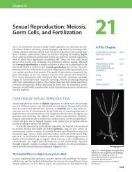
The Questions of Developmental Biology
The Questions of Developmental Biology
The Questions of Developmental Biology

Create successful ePaper yourself
Turn your PDF publications into a flip-book with our unique Google optimized e-Paper software.
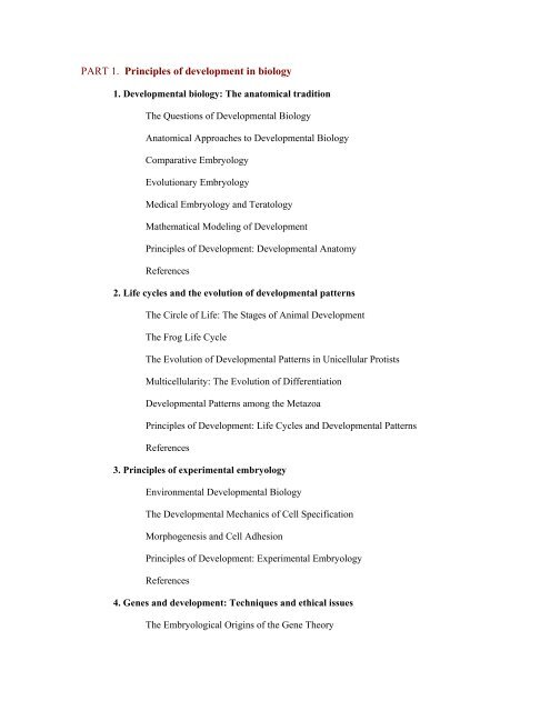
PART 1. Principles <strong>of</strong> development in biology<br />
1. <strong>Developmental</strong> biology: <strong>The</strong> anatomical tradition<br />
<strong>The</strong> <strong>Questions</strong> <strong>of</strong> <strong>Developmental</strong> <strong>Biology</strong><br />
Anatomical Approaches to <strong>Developmental</strong> <strong>Biology</strong><br />
Comparative Embryology<br />
Evolutionary Embryology<br />
Medical Embryology and Teratology<br />
Mathematical Modeling <strong>of</strong> Development<br />
Principles <strong>of</strong> Development: <strong>Developmental</strong> Anatomy<br />
References<br />
2. Life cycles and the evolution <strong>of</strong> developmental patterns<br />
<strong>The</strong> Circle <strong>of</strong> Life: <strong>The</strong> Stages <strong>of</strong> Animal Development<br />
<strong>The</strong> Frog Life Cycle<br />
<strong>The</strong> Evolution <strong>of</strong> <strong>Developmental</strong> Patterns in Unicellular Protists<br />
Multicellularity: <strong>The</strong> Evolution <strong>of</strong> Differentiation<br />
<strong>Developmental</strong> Patterns among the Metazoa<br />
Principles <strong>of</strong> Development: Life Cycles and <strong>Developmental</strong> Patterns<br />
References<br />
3. Principles <strong>of</strong> experimental embryology<br />
Environmental <strong>Developmental</strong> <strong>Biology</strong><br />
<strong>The</strong> <strong>Developmental</strong> Mechanics <strong>of</strong> Cell Specification<br />
Morphogenesis and Cell Adhesion<br />
Principles <strong>of</strong> Development: Experimental Embryology<br />
References<br />
4. Genes and development: Techniques and ethical issues<br />
<strong>The</strong> Embryological Origins <strong>of</strong> the Gene <strong>The</strong>ory
Evidence for Genomic Equivalence<br />
Differential Gene Expression<br />
RNA Localization Techniques<br />
Determining the Function <strong>of</strong> Genes during Development<br />
Identifying the Genes for Human <strong>Developmental</strong> Anomalies<br />
Principles <strong>of</strong> Development: Genes and Development<br />
References<br />
5. <strong>The</strong> genetic core <strong>of</strong> development: Differential gene expression<br />
Differential Gene Transcription<br />
Methylation Pattern and the Control <strong>of</strong> Transcription<br />
Transcriptional Regulation <strong>of</strong> an Entire Chromosome: Dosage Compensation<br />
Differential RNA Processing<br />
Control <strong>of</strong> Gene Expression at the Level <strong>of</strong> Translation<br />
Epilogue: Posttranslational Gene Regulation<br />
Principles <strong>of</strong> Development: <strong>Developmental</strong> Genetics<br />
References<br />
6. Cell-cell communication in development<br />
Induction and Competence<br />
Paracrine Factors<br />
Cell Surface Receptors and <strong>The</strong>ir Signal Transduction Pathways<br />
<strong>The</strong> Cell Death Pathways<br />
Juxtacrine Signaling<br />
Cross-Talk between Pathways<br />
Coda<br />
Principles <strong>of</strong> Development:Cell-Cell Communication<br />
References
PART 2: Early embryonic development<br />
7. Fertilization: Beginning a new organism<br />
Structure <strong>of</strong> the Gametes<br />
Recognition <strong>of</strong> Egg and Sperm<br />
Gamete Fusion and the Prevention <strong>of</strong> Polyspermy<br />
<strong>The</strong> Activation <strong>of</strong> Egg Metabolism<br />
Fusion <strong>of</strong> the Genetic Material<br />
Rearrangement <strong>of</strong> the Egg Cytoplasm<br />
Snapshot Summary: Fertilization<br />
References<br />
8. Early development in selected invertebrates<br />
An Introduction to Early <strong>Developmental</strong> Processes<br />
<strong>The</strong> Early Development <strong>of</strong> Sea Urchins<br />
<strong>The</strong> Early Development <strong>of</strong> Snails<br />
Early Development in Tunicates<br />
Early Development <strong>of</strong> the Nematode Caenorhabditis elegans<br />
References<br />
9. <strong>The</strong> genetics <strong>of</strong> axis specification in Drosophila<br />
Early Drosophila Development<br />
<strong>The</strong> Origins <strong>of</strong> Anterior-Posterior Polarity<br />
<strong>The</strong> Generation <strong>of</strong> Dorsal-Ventral Polarity<br />
References<br />
10. Early development and axis formation in amphibians<br />
Early Amphibian Development<br />
Axis Formation in Amphibians: <strong>The</strong> Phenomenon <strong>of</strong> the Organizer<br />
References
11. <strong>The</strong> early development <strong>of</strong> vertebrates: Fish, birds, and mammals<br />
Early Development in Fish<br />
Early Development in Birds<br />
Early Mammalian Development<br />
References<br />
PART 3: Later embryonic development<br />
12. <strong>The</strong> central nervous system and the epidermis<br />
Formation <strong>of</strong> the Neural Tube<br />
Differentiation <strong>of</strong> the Neural Tube<br />
Tissue Architecture <strong>of</strong> the Central Nervous System<br />
Neuronal Types<br />
Development <strong>of</strong> the Vertebrate Eye<br />
<strong>The</strong> Epidermis and the Origin <strong>of</strong> Cutaneous Structures<br />
Snapshot Summary: Central Nervous System and Epidermis<br />
References<br />
13. Neural crest cells and axonal specificity<br />
<strong>The</strong> Neural Crest<br />
Neuronal Specification and Axonal Specificity<br />
References<br />
14. Paraxial and intermediate mesoderm<br />
Paraxial Mesoderm: <strong>The</strong> Somites and <strong>The</strong>ir Derivatives<br />
Myogenesis: <strong>The</strong> Development <strong>of</strong> Muscle<br />
Osteogenesis: <strong>The</strong> Development <strong>of</strong> Bones<br />
Intermediate Mesoderm<br />
Snapshot Summary: Paraxial and Intermediate Mesoderm<br />
References
15. Lateral plate mesoderm and endoderm<br />
Lateral Plate Mesoderm<br />
Endoderm<br />
References<br />
16. Development <strong>of</strong> the tetrapod limb<br />
Formation <strong>of</strong> the Limb Bud<br />
Generating the Proximal-Distal Axis <strong>of</strong> the Limb<br />
Specification <strong>of</strong> the Anterior-Posterior Limb Axis<br />
<strong>The</strong> Generation <strong>of</strong> the Dorsal-Ventral Axis<br />
Coordination among the Three Axes<br />
Cell Death and the Formation <strong>of</strong> Digits and Joints<br />
Snapshot Summary: <strong>The</strong> Tetrapod Limb<br />
References<br />
17. Sex determination<br />
Chromosomal Sex Determination in Mammals<br />
Chromosomal Sex Determination in Drosophila<br />
Environmental Sex Determination<br />
Snapshot Summary: Sex Determination<br />
References<br />
18. Metamorphosis, regeneration, and aging<br />
Metamorphosis: <strong>The</strong> Hormonal Reactivation <strong>of</strong> Development<br />
Regeneration<br />
Aging: <strong>The</strong> <strong>Biology</strong> <strong>of</strong> Senescence<br />
References<br />
19. <strong>The</strong> saga <strong>of</strong> the germ line<br />
Germ Plasm and the Determination <strong>of</strong> the Primordial Germ Cells
Germ Cell Migration<br />
Meiosis<br />
Spermatogenesis<br />
Oogenesis<br />
Snapshot Summary: <strong>The</strong> Germ Line<br />
References<br />
PART 4: Ramifications <strong>of</strong> developmental biology<br />
20. An overview <strong>of</strong> plant development<br />
Plant Life Cycles<br />
Gamete Production in Angiosperms<br />
Pollination<br />
Fertilization<br />
Embryonic Development<br />
Dormancy<br />
Germination<br />
Vegetative Growth<br />
<strong>The</strong> Vegetative-to-Reproductive Transition<br />
Senescence<br />
Snapshot Summary: Plant Development<br />
References<br />
21. Environmental regulation <strong>of</strong> animal development<br />
Environmental Regulation <strong>of</strong> Normal Development<br />
Environmental Disruption <strong>of</strong> Normal Development<br />
References<br />
22. <strong>Developmental</strong> mechanisms <strong>of</strong> evolutionary change<br />
"Unity <strong>of</strong> Type" and "Conditions <strong>of</strong> Existence"
Hox Genes: Descent with Modification<br />
Homologous Pathways <strong>of</strong> Development<br />
Modularity: <strong>The</strong> Prerequisite for Evolution through Development<br />
<strong>Developmental</strong> Correlation<br />
<strong>Developmental</strong> Constraints<br />
A New Evolutionary Synthesis<br />
Snapshot Summary: Evolutionary <strong>Developmental</strong> <strong>Biology</strong><br />
References<br />
Appendix
PARTE 1. Principles <strong>of</strong> development in biology<br />
1. <strong>Developmental</strong> biology: <strong>The</strong> anatomical tradition<br />
<strong>The</strong> <strong>Questions</strong> <strong>of</strong> <strong>Developmental</strong> <strong>Biology</strong><br />
According to Aristotle, the first embryologist known to history, science begins with<br />
wonder: "It is owing to wonder that people began to philosophize, and wonder remains the<br />
beginning <strong>of</strong> knowledge." <strong>The</strong> development <strong>of</strong> an animal from an egg has been a source <strong>of</strong><br />
wonder throughout history. <strong>The</strong> simple procedure <strong>of</strong> cracking open a chick egg on each<br />
successive day <strong>of</strong> its 3-week incubation provides a remarkable experience as a thin band <strong>of</strong> cells<br />
is seen to give rise to an entire bird. Aristotle performed this procedure and noted the formation <strong>of</strong><br />
the major organs. Anyone can wonder at this remarkable yet commonplace phenomenon, but<br />
the scientist seeks to discover how development actually occurs. And rather than dissipating<br />
wonder, new understanding increases it.<br />
Multicellular organisms do not spring forth fully formed. Rather, they arise by a<br />
relatively slow process <strong>of</strong> progressive change that we call development. In nearly all cases, the<br />
development <strong>of</strong> a multicellular organism begins with a single cell the fertilized egg, or zygote,<br />
which divides mitotically to produce all the cells <strong>of</strong> the body. <strong>The</strong> study <strong>of</strong> animal development<br />
has traditionally been called embryology, from that stage <strong>of</strong> an organism that exists between<br />
fertilization and birth. But development does not stop at birth, or even at adulthood. Most<br />
organisms never stop developing. Each day we replace more than a gram <strong>of</strong> skin cells (the older<br />
cells being sloughed <strong>of</strong>f as we move), and our bone marrow sustains the development <strong>of</strong> millions<br />
<strong>of</strong> new red blood cells every minute <strong>of</strong> our lives. In addition, some animals can regenerate<br />
severed parts, and many species undero metamorphosis (such as the transformation <strong>of</strong> a tadpole<br />
into a frog, or a caterpillar into a butterfly). <strong>The</strong>refore, in recent years it has become customary to<br />
speak <strong>of</strong> developmental biology as the discipline that studies embryonic and other<br />
developmental processes.<br />
Development accomplishes two major objectives: it generates cellular diversity and order<br />
within each generation, and it ensures the continuity <strong>of</strong> life from one generation to the next. Thus,<br />
there are two fundamental questions in developmental biology: How does the fertilized egg give<br />
rise to the adult body, and how does that adult body produce yet another body? <strong>The</strong>se two huge<br />
questions have been subdivided into six general questions scrutinized by developmental<br />
biologists:<br />
<strong>The</strong> question <strong>of</strong> differentiation. A single cell, the fertilized egg, gives rise to hundreds <strong>of</strong><br />
different cell types muscle cells, epidermal cells, neurons, lens cells, lymphocytes, blood cells,<br />
fat cells, and so on (Figure 1.1). This generation <strong>of</strong> cellular diversity is called differentiation.<br />
Since each cell <strong>of</strong> the body (with very few exceptions) contains the same set <strong>of</strong> genes, we need to<br />
understand how this same set <strong>of</strong> genetic instructions can produce different types <strong>of</strong> cells. How can<br />
the fertilized egg generate so many different cell types?<br />
<strong>The</strong> question <strong>of</strong> morphogenesis. Our differentiated cells are not randomly distributed. Rather,<br />
they are organized into intricate tissues and organs. <strong>The</strong>se organs are arranged in a given way: the<br />
fingers are always at the tips <strong>of</strong> our hands, never in the middle; the eyes are always in our heads,<br />
not in our toes or gut. This creation <strong>of</strong> ordered form is called morphogenesis. How can the cells<br />
form such ordered structures?
<strong>The</strong> question <strong>of</strong> growth. How do our cells know when to stop dividing? If each cell in our face<br />
were to undergo just one more cell division, we would be considered horribly malformed. If each<br />
cell in our arms underwent just one more round <strong>of</strong> cell division, we could tie our shoelaces<br />
without bending over. Our arms are generally the same size on both sides <strong>of</strong> the body. How is cell<br />
division so tightly regulated?<br />
<strong>The</strong> question <strong>of</strong> reproduction. <strong>The</strong> sperm and egg are very specialized cells. Only they can<br />
transmit the instructions for making an organism from one generation to the next. How are these<br />
cells set apart to form the next generation, and what are the instructions in the nucleus and<br />
cytoplasm that allow them to function this way?<br />
<strong>The</strong> question <strong>of</strong> evolution. Evolution involves inherited changes in development. When we say<br />
that today's one-toed horse had a five-toed ancestor, we are saying that changes in the<br />
development <strong>of</strong> cartilage and muscles occurred over many generations in the embryos <strong>of</strong> the<br />
horse's ancestors. How do changes in development create new body forms? Which heritable<br />
changes are possible, given the constraints imposed by the necessity <strong>of</strong> the organism to survive as<br />
it develops?<br />
<strong>The</strong> question <strong>of</strong> environmental integration. <strong>The</strong> development <strong>of</strong> many organisms is<br />
influenced by cues from the environment. Certain butterflies, for instance, inherit the ability to<br />
produce different wing colors based on the temperature or the amount <strong>of</strong> daylight experienced by<br />
the caterpillar before it undergoes metamorphosis. How is the development <strong>of</strong> an organism<br />
integrated into the larger context <strong>of</strong> its habitat?<br />
Anatomical Approaches to <strong>Developmental</strong> <strong>Biology</strong><br />
A field <strong>of</strong> science is defined by the questions it seeks to answer, and most <strong>of</strong> the<br />
questions in developmental biology have been bequeathed to it through its embryological<br />
heritage. <strong>The</strong>re are numerous strands <strong>of</strong> embryology, each predominating during a different era.<br />
Sometimes they are very distinct traditions, and sometimes they blend. We can identify three<br />
major ways <strong>of</strong> studying embryology:<br />
Anatomical approaches<br />
Experimental approaches<br />
Genetic approaches<br />
While it is true that anatomical approaches gave rise to experimental approaches, and that<br />
genetic approaches built on the foundations <strong>of</strong> the earlier two approaches, all three traditions<br />
persist to this day and continue to play a major role in developmental biology. Chapter 3 <strong>of</strong> this<br />
text discusses experimental approaches, and Chapters 4 and 5 examine the genetic approaches in<br />
greater depth. In recent years, each <strong>of</strong> these traditions has become joined with molecular genetics<br />
to produce a vigorous and multifaceted science <strong>of</strong> developmental biology.<br />
But the basis <strong>of</strong> all research in developmental biology is the changing anatomy <strong>of</strong> the<br />
organism. What parts <strong>of</strong> the embryo form the heart? How do the cells that form the retina position<br />
themselves the proper distance from the cells that form the lens? How do the tissues that form the<br />
bird wing relate to the tissues that form the fish fin or the human hand?
<strong>The</strong>re are several strands that weave together to form the anatomical approaches to<br />
development. <strong>The</strong> first strand is comparative embryology, the study <strong>of</strong> how anatomy changes<br />
during the development <strong>of</strong> different organisms. For instance, a comparative embryologist may<br />
study which tissues form the nervous system in the fly or in the frog. <strong>The</strong> second strand, based on<br />
the first, is evolutionary embryology, the study <strong>of</strong> how changes in development may cause<br />
evolutionary changes and <strong>of</strong> how an organism's ancestry may constrain the types <strong>of</strong> changes that<br />
are possible. <strong>The</strong> third anatomical approach to developmental biology is teratology, the study <strong>of</strong><br />
birth defects. <strong>The</strong>se anatomical abnormalities may be caused by mutant genes or by substances in<br />
the environment that interfere with development. <strong>The</strong> study <strong>of</strong> abnormalities is <strong>of</strong>ten used to<br />
discover how normal development occurs. <strong>The</strong> fourth anatomical approach is mathematical<br />
modeling, which seeks to describe developmental phenomena in terms <strong>of</strong> equations. Certain<br />
patterns <strong>of</strong> growth and differentiation can be explained by interactions whose results are<br />
mathematically predictable. <strong>The</strong> revolution in graphics technology has enabled scientists to model<br />
certain types <strong>of</strong> development on the computer and to identify mathematical principles upon which<br />
those developmental processes are based.<br />
Evolutionary Embryology<br />
Charles Darwin's theory <strong>of</strong> evolution restructured comparative embryology and gave it a<br />
new focus. After reading Johannes Müller's summary <strong>of</strong> von Baer's laws in 1842, Darwin saw<br />
that embryonic resemblances would be a very strong argument in favor <strong>of</strong> the genetic<br />
connectedness <strong>of</strong> different animal groups. "Community <strong>of</strong> embryonic structure reveals<br />
community <strong>of</strong> descent," he would conclude in On the Origin <strong>of</strong> Species in 1859.<br />
Larval forms had been used for taxonomic classification even before Darwin. J. V.<br />
Thompson, for instance, had demonstrated that larval barnacles were almost identical to larval<br />
crabs, and he therefore counted barnacles as arthropods, not molluscs (Figure 1.12; Winsor 1969).<br />
Darwin, an expert on barnacle taxonomy, celebrated this finding: "Even the illustrious Cuvier did<br />
not perceive that a barnacle is a crustacean, but a glance at the larva shows this in an<br />
unmistakable manner." Darwin's evolutionary interpretation <strong>of</strong> von Baer's laws established a<br />
paradigm that was to be followed for many decades, namely, that relationships between groups<br />
can be discovered by finding common embryonic or larval forms. Kowalevsky (1871) would<br />
soon make a similar type <strong>of</strong> discovery (publicized in Darwin's Descent <strong>of</strong> Man) that tunicate<br />
larvae have notochords and form their neural tubes and other organs in a manner very similar to<br />
that <strong>of</strong> the primitive chordate Amphioxus. <strong>The</strong> tunicates, another enigma <strong>of</strong> classification schemes<br />
(formerly placed, along with barnacles, among the molluscs), thereby found a home with the<br />
chordates.<br />
Darwin also noted that embryonic organisms sometimes make structures that are<br />
inappropriate for their adult form but that show their relatedness to other animals. He pointed out<br />
the existence <strong>of</strong> eyes in embryonic moles, pelvic rudiments in embryonic snakes, and teeth in<br />
embryonic baleen whales.<br />
Darwin also argued that adaptations that depart from the "type" and allow an organism to<br />
survive in its particular environment develop late in the embryo.* He noted that the differences<br />
between species within genera become greater as development persists, as predicted by von<br />
Baer's laws. Thus, Darwin recognized two ways <strong>of</strong> looking at "descent with modification." One<br />
could emphasize the common descent by pointing out embryonic similarities between two or<br />
more groups <strong>of</strong> animals, or one could emphasize the modifications by showing how development<br />
was altered to produce structures that enabled animals to adapt to particular conditions.
Embryonic homologies<br />
One <strong>of</strong> the most important distinctions made by the evolutionary embryologists was the<br />
difference between analogy and homology. Both terms refer to structures that appear to be<br />
similar. Homologous structures are those organs whose underlying similarity arises from their<br />
being derived from a common ancestral structure. For example, the wing <strong>of</strong> a bird and the<br />
forelimb <strong>of</strong> a human are homologous. Moreover, their respective parts are homologous (Figure<br />
1.13). Analogous structures are those whose similarity comes from their performing a similar<br />
function, rather than their arising from a common ancestor. <strong>The</strong>refore, for example, the wing <strong>of</strong> a<br />
butterfly and the wing <strong>of</strong> a bird are analogous. <strong>The</strong> two types <strong>of</strong> wings share a common function<br />
(and therefore are both called wings), but the bird wing and insect wing did not arise from an<br />
original ancestral structure that became modified through evolution into bird wings and butterfly<br />
wings.<br />
Homologies must be made carefully and must always refer to the level <strong>of</strong> organization<br />
being compared. For instance, the bird wing and the bat wing are homologous as forelimbs, but<br />
not as wings. In other words, they share a common underlying structure <strong>of</strong> forelimb bones<br />
because birds and mammals share a common ancestry. However, the bird wing developed<br />
independently from the bat wing. Bats descended from a long line <strong>of</strong> nonwinged mammals, and<br />
the structure <strong>of</strong> the bat wing is markedly different from that <strong>of</strong> a bird wing.<br />
One <strong>of</strong> the most celebrated cases <strong>of</strong> embryonic homology is that <strong>of</strong> the fish gill cartilage,<br />
the reptilian jaw, and the mammalian middle ear (reviewed in Gould 1990). First, the gill arches<br />
<strong>of</strong> jawless (agnathan) fishes became modified to form the jaw <strong>of</strong> the jawed fishes. In the jawless<br />
fishes, a series <strong>of</strong> gills opened behind the jawless mouth. When the gill slits became supported by<br />
cartilaginous elements, the first set <strong>of</strong> these gill supports surrounded the mouth to form the jaw.<br />
<strong>The</strong>re is ample evidence that jaws are modified gill supports. First, both these sets <strong>of</strong> bones are<br />
made from neural crest cells. (Most other bones come from mesodermal tissue.) Second, both<br />
structures form from upper and lower bars that bend forward and are hinged in the middle. Third,<br />
the jaw musculature seems to be homologous to the original gill support musculature. Thus, the<br />
vertebrate jaw appears to be homologous to the gill arches <strong>of</strong> jawless fishes.<br />
But the story does not end here. <strong>The</strong> upper portion <strong>of</strong> the second embryonic arch<br />
supporting the gill became the hyomandibular bone <strong>of</strong> jawed fishes. This element supports the<br />
skull and links the jaw to the cranium (Figure 1.14A). As vertebrates came up onto land, they had<br />
a new problem: how to hear in a medium as thin as air. <strong>The</strong> hyomandibular bone happens to be<br />
near the otic (ear) capsule, and bony material is excellent for transmitting sound. Thus, while still<br />
functioning as a cranial brace, the hyomandibular bone <strong>of</strong> the first amphibians also began<br />
functioning as a sound transducer (Clack 1989). As the terrestrial vertebrates altered their<br />
locomotion, jaw structure, and posture, the cranium became firmly attached to the rest <strong>of</strong> the skull<br />
and did not need the hyomandibular brace. <strong>The</strong> hyomandibular bone then seems to have become<br />
specialized into the stapes bone <strong>of</strong> the middle ear. What had been this bone's secondary function<br />
became its primary function.<br />
<strong>The</strong> original jaw bones changed also. <strong>The</strong> first embryonic arch generates the jaw<br />
apparatus. In amphibians, reptiles, and birds, the posterior portion <strong>of</strong> this cartilage forms the<br />
quadrate bone <strong>of</strong> the upper jaw and the articular bone <strong>of</strong> the lower jaw. <strong>The</strong>se bones connect to<br />
each other and are responsible for articulating the upper and lower jaws. However, in mammals,<br />
this articulation occurs at another region (the dentary and squamosal bones), thereby "freeing"<br />
these bony elements to acquire new functions. <strong>The</strong> quadrate bone <strong>of</strong> the reptilian upper jaw
evolved into the mammalian incus bone <strong>of</strong> the middle ear, and the articular bone <strong>of</strong> the reptile's<br />
lower jaw has become our malleus. This latter process was first described by Reichert in 1837,<br />
when he observed in the pig embryo that the mandible (jawbone) ossifies on the side <strong>of</strong> Meckel's<br />
cartilage, while the posterior region <strong>of</strong> Meckel's cartilage ossifies, detaches from the rest <strong>of</strong> the<br />
cartilage, and enters the region <strong>of</strong> the middle ear to become the malleus (Figure 1.14B,C). Thus,<br />
the middle ear bones <strong>of</strong> the mammal are homologous to the posterior lower jaw <strong>of</strong> the reptile and<br />
to the gill arches <strong>of</strong> agnathan fishes. Chapter 22 will detail more recent information concerning<br />
the relationship <strong>of</strong> development to evolution.<br />
Medical Embryology and Teratology<br />
While embryologists could look at embryos to describe the evolution <strong>of</strong> life and how<br />
different animals form their organs, physicians became interested in embryos for more practical<br />
reasons. About 2% <strong>of</strong> human infants are born with a readily observable anatomical abnormality<br />
(Thorogood 1997). <strong>The</strong>se abnormalities may include missing limbs, missing or extra digits, cleft<br />
palate, eyes that lack certain parts, hearts that lack valves, and so forth. Physicians need know the<br />
causes <strong>of</strong> these birth defects in order to counsel parents as to the risk <strong>of</strong> having another<br />
malformed infant. In addition, the different birth defects can tell us how the human body is<br />
normally formed. In the absence <strong>of</strong> experimental data on human embryos, we <strong>of</strong>ten must rely on<br />
nature's "experiments" to learn how the human body becomes organized.* Some birth defects are<br />
produced by mutant genes or chromosomes, and some are produced by environmental factors that<br />
impede development.<br />
Abnormalities caused by genetic events (gene mutations, chromosomal aneuploidies and<br />
translocations) are called malformations. Malformations <strong>of</strong>ten appear as syndromes (from the<br />
Greek, "running together"), where several abnormalities are seen concurrently. For instance, a<br />
human malformation called piebaldism, shown in Figure 1.15A, is due to a dominant mutation in<br />
a gene (KIT) on the long arm <strong>of</strong> chromosome 4 (Halleban and Moellmann 1993). <strong>The</strong> syndrome<br />
includes anemia, sterility, unpigmented regions <strong>of</strong> the skin and hair, deafness, and the absence <strong>of</strong><br />
the nerves that cause peristalsis in the gut. <strong>The</strong> common feature underlying these conditions is<br />
that the KIT gene encodes a protein that is expressed in the neural crest cells and in the precursors<br />
<strong>of</strong> blood cells and germ cells. <strong>The</strong> Kit protein enables these cells to proliferate. Without this<br />
protein, the neural crest cells which generate the pigment cells, certain ear cells, and the gut<br />
neurons do not multiply as much as they should (resulting in underpigmentation, deafness, and<br />
gut malformations), nor do the precursors <strong>of</strong> the blood cells (resulting in anemia) or the germ<br />
cells (resulting in sterility).<br />
<strong>Developmental</strong> biologists and clinical geneticists <strong>of</strong>ten study human syndromes (and<br />
determine their causes) by studying animals that display the same syndrome. <strong>The</strong>se are called<br />
animal models <strong>of</strong> the disease; the mouse model for piebaldism is shown in Figure 1.15B. It has a<br />
phenotype very similar to that <strong>of</strong> the human condition, and it is caused by a mutation in the Kit<br />
gene <strong>of</strong> the mouse.<br />
Abnormalities due to exogenous agents (certain chemicals or viruses, radiation, or<br />
hyperthermia) are called disruptions. <strong>The</strong> agents responsible for these disruptions are called<br />
teratogens (Greek, "monster-formers"), and the study <strong>of</strong> how environmental agents disrupt<br />
normal development is called teratology. In 1961, Lenz and McBride independently accumulated<br />
evidence that thalidomide, prescribed as a mild sedative to many pregnant women, caused an<br />
enormous increase in a previously rare syndrome <strong>of</strong> congenital anomalies. <strong>The</strong> most noticeable <strong>of</strong><br />
these anomalies was phocomelia, a condition in which the long bones <strong>of</strong> the limbs are deficient or
absent (Figure 1.16A). Over 7000 affected infants were born to women who took this drug, and a<br />
woman need only have taken one tablet to produce children with all four limbs deformed (Lenz<br />
1962, 1966; Toms 1962). Other abnormalities induced by the ingestion <strong>of</strong> thalidomide included<br />
heart defects, absence <strong>of</strong> the external ears, and malformed intestines.<br />
Nowack (1965) documented the period <strong>of</strong> susceptibility during which thalidomide caused<br />
these abnormalities. <strong>The</strong> drug was found to be teratogenic only during days 34 50 after the last<br />
menstruation (about 20 to 36 days postconception). <strong>The</strong> specificity <strong>of</strong> thalidomide action is<br />
shown in Figure 1.16B. From day 34 to day 38, no limb abnormalities are seen. During this<br />
period, thalidomide can cause the absence or deficiency <strong>of</strong> ear components. Malformations <strong>of</strong><br />
upper limbs are seen before those <strong>of</strong> the lower limbs, since the arms form slightly before the legs<br />
during development. <strong>The</strong> only animal models for thalidomide, however, are primates, and we still<br />
do not know the mechanisms by which thalidomide causes human developmental disruptions.<br />
Thalidomide was withdrawn from the market in November 1961, but it is beginning to be<br />
prescribed again, this time as a potential anti-tumor and anti-autoimmunity drug (Raje and<br />
Anderson 1999).<br />
<strong>The</strong> integration <strong>of</strong> anatomical information about congenital malformations with our new<br />
knowledge concerning the genes responsible for development has had a revolutionary effect and<br />
is currently restructuring medicine. This integration is allowing us to discover the genes<br />
responsible for inherited malformations, and it permits us to identify the steps in development<br />
being disrupted by teratogens. We will see examples <strong>of</strong> this integration throughout this text, and<br />
Chapter 21 will detail some <strong>of</strong> the remarkable new discoveries in teratology.<br />
*<strong>The</strong> word "monster," used frequently in textbooks prior to the mid-twentieth century to describe<br />
malformed infants, comes from the Latin monstrare, "to show or point out." This is also the root<br />
<strong>of</strong> our word "demonstrate." It was realized by Meckel (<strong>of</strong> jaw cartilage fame) that syndromes <strong>of</strong><br />
congenital anomalies demonstrated certain principles about normal development. Parts <strong>of</strong> the<br />
body that were affected together must have some common developmental origin or mechanism<br />
that was being affected.<br />
Mathematical Modeling <strong>of</strong> Development<br />
<strong>Developmental</strong> biology has been described as the last refuge <strong>of</strong> the mathematically<br />
incompetent scientist. This phenomenon, however, is not going to last. While most embryologists<br />
have been content trying to analyze specific instances <strong>of</strong> development or even formulating some<br />
general principles <strong>of</strong> embryology, some researchers are now seeking the laws <strong>of</strong> development.<br />
<strong>The</strong> goal <strong>of</strong> these investigators is to base embryology on formal mathematical or physical<br />
principles (see Held 1992; Webster and Goodwin 1996). Pattern formation and growth are two<br />
areas in which such mathematical modeling has given biologists interesting insights into some<br />
underlying laws <strong>of</strong> animal development.
<strong>The</strong> mathematics <strong>of</strong> organismal growth<br />
Most animals grow by increasing their volume while retaining their proportions.<br />
<strong>The</strong>oretically, an animal that increases its weight (volume) tw<strong>of</strong>old will increase its length only<br />
1.26 times (as 1.26 3 = 2). W. K. Brooks (1886) observed that this ratio was frequently seen in<br />
nature, and he noted that the deep-sea arthropods collected by the Challenger expedition<br />
increased about 1.25 times between molts. In 1904, Przibram and his colleagues performed a<br />
detailed study <strong>of</strong> mantises and found that the increase <strong>of</strong> size between molts was almost exactly<br />
1.26 (see Przibram 1931). Even the hexagonal facets <strong>of</strong> the arthropod eye (which grow by cell<br />
expansion, not by cell division) increased by that ratio.<br />
D'Arcy Thompson (1942) similarly showed<br />
that the spiral growth <strong>of</strong> shells (and fingernails) can<br />
be expressed mathematically (r = a ), and that the<br />
ratio <strong>of</strong> the widths between two whorls <strong>of</strong> a shell can<br />
be calculated by the formula r = e 2cot (Figure 1.17;<br />
Table 1.1).<br />
Thus, if a whorl were 1 inch in breadth at<br />
one point on a radius and the angle <strong>of</strong> the spiral were<br />
80°, the next whorl would have a width <strong>of</strong> 3 inches<br />
on the same radius. Most gastropod (snail) and<br />
nautiloid molluscs have an angle <strong>of</strong> curvature between 80° and 85°.* Lower-angle curvatures are<br />
seen in some shells (mostly bivalves) and are common in teeth and claws.<br />
Constant angle <strong>of</strong> an equiangular spiral and the ratio <strong>of</strong> widths between whorls<br />
Constant angle<br />
Ratio <strong>of</strong> widths a<br />
90° 1.0<br />
89°8´ 1.1<br />
86°18´ 1.5<br />
83°42´ 2.0<br />
80°5´ 3.0<br />
75°38´ 5.0<br />
69°53´ 10.0<br />
64°31´ 20.0<br />
58°5´ 50.0<br />
53°46´ 10 2<br />
42°17´ 10 3<br />
34°19´ 10 4<br />
28°37´ 10 5<br />
24°28´ 10 6<br />
Source: From Thompson 1942.<br />
a <strong>The</strong> ratio <strong>of</strong> widths is calculated by dividing the width <strong>of</strong> one whorl by the width <strong>of</strong> the
Such growth, in which the shape is preserved because all components grow at the same<br />
rate, is called isometric growth. In many organisms, growth is not a uniform phenomenon. It is<br />
obvious that there are some periods in an organism's life during which growth is more rapid than<br />
in others. Physical growth during the first 10 years <strong>of</strong> person's existence is much more dramatic<br />
than in the 10 years following one's graduation from college. Moreover, not all parts <strong>of</strong> the body<br />
grow at the same rate. This phenomenon <strong>of</strong> the different growth rates <strong>of</strong> parts within the same<br />
organism is called allometric growth (or allometry). Human allometry is depicted in Figure<br />
1.18.<br />
Our arms and legs grow at a faster rate than our torso and head, such that adult<br />
proportions differ markedly from those <strong>of</strong> infants. Julian Huxley (1932) likened allometry to<br />
putting money in the bank at two different continuous interest rates.<br />
<strong>The</strong> formula for allometric growth (or for comparing moneys invested at two different<br />
interest rates) is y = bx a/c , where a and c are the growth rates <strong>of</strong> two body parts, and b is the value<br />
<strong>of</strong> y when x = 1. If a/c > 1, then that part <strong>of</strong> the body represented by a is growing faster than that<br />
part <strong>of</strong> the body represented by c. In logarithmic terms (which are much easier to graph), log y =<br />
log b + (a/c)log x.<br />
One <strong>of</strong> the most vivid examples <strong>of</strong> allometric growth is seen in the male fiddler crab, Uca<br />
pugnax. In small males, the two claws are <strong>of</strong> equal weight, each constituting about 8% <strong>of</strong> the<br />
crab's total weight. As the crab grows larger, its chela (the large crushing claw) grows even more<br />
rapidly, eventually constituting about 38% <strong>of</strong> the<br />
crab's weight (Figure 1.19)<br />
When these data are plotted on double<br />
logarithmic plots (the body mass on the x axis, the<br />
chela mass on the y axis), one obtains a straight<br />
line whose slope is the a/c ratio. In the male Uca<br />
pugnax (whose name is derived from the huge<br />
claw), the a/c ratio is 6:1. This means that the mass<br />
<strong>of</strong> the chela increases six times faster than the mass<br />
<strong>of</strong> the rest <strong>of</strong> the body. In females <strong>of</strong> the species,<br />
the claw remains about 8% <strong>of</strong> the body weight<br />
throughout growth. It is only in the males (who use<br />
the claw for defense and display) that this<br />
allometry occurs.
<strong>The</strong> mathematics <strong>of</strong> patterning<br />
One <strong>of</strong> the most important mathematical models in developmental biology has been that<br />
formulated by Alan Turing (1952), one <strong>of</strong> the founders <strong>of</strong> computer science (and the<br />
mathematician who cracked the German "Enigma" code during World War II). He proposed a<br />
model wherein two homogeneously distributed solutions would interact to produce stable patterns<br />
during morphogenesis. <strong>The</strong>se patterns would represent regional differences in the concentrations<br />
<strong>of</strong> the two substances. <strong>The</strong>ir interactions would produce an ordered structure out <strong>of</strong> random<br />
chaos.<br />
Turing's reaction-diffusion model involves two substances. One <strong>of</strong> them, substance S,<br />
inhibits the production <strong>of</strong> the other, substance P. Substance P promotes the production <strong>of</strong> more<br />
substance P as well as more substance S. Turing's mathematics show that if S diffuses more<br />
readily than P, sharp waves <strong>of</strong> concentration differences will be generated for substance P (Figure<br />
1.20). <strong>The</strong>se waves have been observed in certain chemical reactions (Prigogine and Nicolis<br />
1967; Winfree 1974).<br />
<strong>The</strong> reaction-diffusion model predicts alternating areas <strong>of</strong> high and low concentrations <strong>of</strong><br />
some substance. When the concentration <strong>of</strong> such a substance is above a certain threshold level, a<br />
cell (or group <strong>of</strong> cells) may be instructed to differentiate in a certain way. An important feature <strong>of</strong><br />
Turing's model is that particular chemical wavelengths will be amplified while all others will be<br />
suppressed. As local concentrations <strong>of</strong> P increase, the values <strong>of</strong> S form a peak centering on the P<br />
peak, but becoming broader and shallower because <strong>of</strong> S's more rapid diffusion. <strong>The</strong>se S peaks<br />
inhibit other P peaks from forming. But which <strong>of</strong> the many P peaks will survive? That depends on<br />
the size and shape <strong>of</strong> the tissues in which the oscillating reaction is occurring. (This pattern is<br />
analogous to the harmonics <strong>of</strong> vibrating strings, as in a guitar. Only certain resonance vibrations<br />
are permitted, based on the boundaries <strong>of</strong> the string.)<br />
<strong>The</strong> mathematics describing which particular wavelengths are selected consist <strong>of</strong><br />
complex polynomial equations. Such functions have been used to model the spiral patterning <strong>of</strong><br />
slime molds, the polar organization <strong>of</strong> the limb, and the pigment patterns <strong>of</strong> mammals, fish, and<br />
snails (Figures 1.21 and 1.22; Kondo and Asai 1995; Meinhardt 1998). A computer simulation<br />
based on a Turing reaction-diffusion system can successfully predict such patterns, given the<br />
starting shapes and sizes <strong>of</strong> the elements involved.
One way to search for the chemicals predicted by Turing's model is to find genetic<br />
mutations in which the ordered structure <strong>of</strong> a pattern has been altered. <strong>The</strong> wild-type alleles <strong>of</strong><br />
these genes may be responsible for generating the normal pattern. Such a candidate is the leopard<br />
gene <strong>of</strong> zebrafish (Asai et al. 1999). Zebrafish usually have five parallel stripes along their flanks.<br />
However, in the different mutations, the stripes are broken into spots <strong>of</strong> different sizes and<br />
densities. Figure 1.22 shows fish homozygous for four different alleles <strong>of</strong> the leopard gene. If the<br />
leopard gene encodes an enzyme that catalyzes one <strong>of</strong> the reactions <strong>of</strong> the reaction-diffusion<br />
system, the different mutations <strong>of</strong> this gene may change the kinetics <strong>of</strong> synthesis or degradation.<br />
Indeed, all the mutant patterns (and those <strong>of</strong> their heterozygotes) can be computer-generated by<br />
changing a single<br />
parameter in the reactiondiffusion<br />
equation.<br />
<strong>The</strong> cloning <strong>of</strong> this gene<br />
should enable further<br />
cooperation between<br />
theoretical biology and<br />
developmental anatomy.<br />
*If the angle were 90°, the shell would form a circle rather than a spiral, and growth would cease.<br />
If the angle were 60°, however, the next whorl would be 4 feet on that radius, and if the angle<br />
were 17°, the next whorl would occupy a distance <strong>of</strong> some 15,000 miles!<br />
Principles <strong>of</strong> Development: <strong>Developmental</strong> Anatomy<br />
1. Organisms must function as they form their organs. <strong>The</strong>y have to use one set <strong>of</strong> structures<br />
while constructing others.<br />
2. <strong>The</strong> main question <strong>of</strong> development is, How does the egg becomes an adult? This question can<br />
be broken down into the component problems <strong>of</strong> differentiation (How do cells become different<br />
from one another and from their precursors?), morphogenesis (How is ordered form is<br />
generated?), growth (How is size regulated?), reproduction (How does one generation create<br />
another generation?), and evolution (How do changes in developmental processes create new<br />
anatomical structures?).<br />
3. Epigenesis happens. New organisms are created de novo each generation from the relatively<br />
disordered cytoplasm <strong>of</strong> the egg.<br />
4. Preformation is not in the anatomical structures, but in the instructions to form them. <strong>The</strong><br />
inheritance <strong>of</strong> the fertilized egg includes the genetic potentials <strong>of</strong> the organism.<br />
5. <strong>The</strong> preformed nuclear instructions include the ability to respond to environmental stimuli in<br />
specific ways.
6. <strong>The</strong> ectoderm gives rise to the epidermis, nervous system, and pigment cells.<br />
7. <strong>The</strong> mesoderm generates the kidneys, gonads, bones, heart, and blood cells.<br />
8. <strong>The</strong> endoderm forms the lining <strong>of</strong> the digestive tube and the respiratory system.<br />
9. Karl von Baer's principles state that the general features <strong>of</strong> a large group <strong>of</strong> animals appear<br />
earlier in the embryo than do the specialized features <strong>of</strong> a smaller group. As each embryo <strong>of</strong> a<br />
given species develops, it diverges from the adult forms <strong>of</strong> other species. <strong>The</strong> early embryo <strong>of</strong> a<br />
"higher" animal species is not like the adult <strong>of</strong> a "lower" animal.<br />
10. Labeling cells with dyes shows that some cells differentiate where they form, while others<br />
migrate from their original sites and differentiate in their new locations. Migratory cells include<br />
neural crest cells and the precursors <strong>of</strong> germ cells and blood cells.<br />
11. "Community <strong>of</strong> embryonic structure reveals community <strong>of</strong> descent" (Charles Darwin).<br />
12. Homologous structures in different species are those organs whose similarity is due to their<br />
sharing a common ancestral structure. Analogous structures are those organs whose similarity<br />
comes from their serving a similar function (but which are not derived from a common ancestral<br />
structure).<br />
13. Congenital anomalies can be caused by genetic factors (mutations, aneuploidies,<br />
translocations) or by environmental agents (certain chemicals, certain viruses, radiation).<br />
14. Syndromes consists <strong>of</strong> sets <strong>of</strong> developmental abnormalities that "run together."<br />
15. Organs that are linked in developmental syndromes share either a common origin or a<br />
common mechanism <strong>of</strong> formation.<br />
16. If growth is isometric, a tw<strong>of</strong>old change in weight will cause a 1.26-fold expansion in length.<br />
17. Allometric growth can create dramatic changes in the structure <strong>of</strong> organisms.<br />
18. Complex patterns may be self-generated by reaction-diffusion events, wherein the activator <strong>of</strong><br />
a local phenomenon stimulates the production <strong>of</strong> more <strong>of</strong> itself as well as the production <strong>of</strong> a<br />
more diffusible inhibitor.<br />
2. Life cycles and the evolution <strong>of</strong> developmental patterns<br />
Traditional ways <strong>of</strong> classifying catalog animals according to their adult structure. But, as<br />
J. T. Bonner (1965) pointed out, this is a very artificial method, because what we consider an<br />
individual is usually just a brief slice <strong>of</strong> its life cycle. When we consider a dog, for instance, we<br />
usually picture an adult. But the dog is a "dog" from the moment <strong>of</strong> fertilization <strong>of</strong> a dog egg by a<br />
dog sperm. It remains a dog even as a senescent dying hound. <strong>The</strong>refore, the dog is actually the<br />
entire life cycle <strong>of</strong> the animal, from fertilization through death.<br />
<strong>The</strong> life cycle has to be adapted to its environment, which is composed <strong>of</strong> nonliving<br />
objects as well as other life cycles. Take, for example, the life cycle <strong>of</strong> Clunio marinus, a small<br />
fly that inhabits tidal waters along the coast <strong>of</strong> western Europe. Females <strong>of</strong> this species live only
2 3 hours as adults, and they must mate and lay their eggs within this short time. To make<br />
matters even more precarious, egg laying is confined to red algae mats that are exposed only<br />
during the lowest ebbing <strong>of</strong> the spring tide. Such low tides occur on four successive days shortly<br />
after the new and full moons (i.e., at about 15-day intervals). <strong>The</strong>refore, the life cycle <strong>of</strong> these<br />
insects must be coordinated with the tidal rhythms as well as the daily rhythms such that the<br />
insects emerge from their pupal cases during the few days <strong>of</strong> the spring tide and at the correct<br />
hour for its ebb (Beck 1980; Neumann and Spindler 1991).<br />
One <strong>of</strong> the major triumphs <strong>of</strong> descriptive embryology was the idea <strong>of</strong> a generalizable life<br />
cycle. Each animal, whether an earthworm, an eagle, or a beagle, passes through similar stages <strong>of</strong><br />
development. <strong>The</strong> major stages <strong>of</strong> animal development are illustrated in Figure 2.1. <strong>The</strong> life <strong>of</strong> a<br />
new individual is initiated by the fusion <strong>of</strong> genetic material from the two gametes the sperm<br />
and the egg. This fusion, called fertilization, stimulates the egg to begin development. <strong>The</strong> stages<br />
<strong>of</strong> development between fertilization and hatching are collectively called embryogenesis.<br />
<strong>The</strong><br />
Circle <strong>of</strong> Life: <strong>The</strong> Stages <strong>of</strong> Animal Development<br />
Throughout the animal kingdom, an incredible variety <strong>of</strong> embryonic types exist, but most<br />
patterns <strong>of</strong> embryogenesis are variations on five themes:<br />
1. Immediately following fertilization, cleavage occurs. Cleavage is a series <strong>of</strong> extremely rapid<br />
mitotic divisions wherein the enormous volume <strong>of</strong> zygote cytoplasm is divided into numerous<br />
smaller cells. <strong>The</strong>se cells are called blastomeres, and by the end <strong>of</strong> cleavage, they generally form<br />
a sphere known as a blastula.<br />
2. After the rate <strong>of</strong> mitotic division has slowed down, the blastomeres undergo dramatic<br />
movements wherein they change their positions relative to one another. This series <strong>of</strong> extensive<br />
cell rearrangements is called gastrulation, and the embryo is said to be in the gastrula stage. As<br />
a result <strong>of</strong> gastrulation, the embryo contains three germ layers: the ectoderm, the endoderm, and<br />
the mesoderm.
3. Once the three germ layers are established, the cells interact with one another and rearrange<br />
themselves to produce tissues and organs. This process is called organogenesis. Many organs<br />
contain cells from more than one germ layer, and it is not unusual for the outside <strong>of</strong> an organ to<br />
be derived from one layer and the inside from another. For example, the outer layer <strong>of</strong> skin comes<br />
from the ectoderm, while the inner layer (the dermis) comes from the mesoderm. Also during<br />
organogenesis, certain cells undergo long migrations from their place <strong>of</strong> origin to their final<br />
location. <strong>The</strong>se migrating cells include the precursors <strong>of</strong> blood cells, lymph cells, pigment cells,<br />
and gametes. Most <strong>of</strong> the bones <strong>of</strong> our face are derived from cells that have migrated ventrally<br />
from the dorsal region <strong>of</strong> the head.<br />
4. As seen in Figure 2.1, in many species a specialized portion <strong>of</strong> egg cytoplasm gives rise to cells<br />
that are the precursors <strong>of</strong> the gametes (the sperm and egg). <strong>The</strong> gametes and their precursor cells<br />
are collectively called germ cells, and they are set aside for reproductive function. All the other<br />
cells <strong>of</strong> the body are called somatic cells. This separation <strong>of</strong> somatic cells (which give rise to the<br />
individual body) and germ cells (which contribute to the formation <strong>of</strong> a new generation) is <strong>of</strong>ten<br />
one <strong>of</strong> the first differentiations to occur during animal development. <strong>The</strong> germ cells eventually<br />
migrate to the gonads, where they differentiate into gametes. <strong>The</strong> development <strong>of</strong> gametes, called<br />
gametogenesis, is usually not completed until the organism has become physically mature. At<br />
maturity, the gametes may be released and participate in fertilization to begin a new embryo. <strong>The</strong><br />
adult organism eventually undergoes senescence and dies.<br />
5. In many species, the organism that hatches from the egg or is born into the world is not<br />
sexually mature. Indeed, in most animals, the young organism is a larva that may look<br />
significantly different from the adult. Larvae <strong>of</strong>ten constitute the stage <strong>of</strong> life that is used for<br />
feeding or dispersal. In many species, the larval stage is the one that lasts the longest, and the<br />
adult is a brief stage solely for reproduction. In the silkworm moths, for instance, the adults do<br />
not have mouthparts and cannot feed. <strong>The</strong> larvae must eat enough for the adult to survive and<br />
mate. Indeed, most female moths mate as soon as they eclose from their pupa, and they fly only<br />
once to lay their eggs. <strong>The</strong>n they die<br />
<strong>The</strong> Frog Life Cycle<br />
Figure 2.1 uses the development <strong>of</strong> a frog to show a representative life cycle. Let us look at this<br />
life cycle in a bit more detail. First, in most frogs, gametogenesis and fertilization are seasonal<br />
events for this animal, because its life depends upon the plants and insects in the pond where it<br />
lives and on the temperature <strong>of</strong> the air and water. A combination <strong>of</strong> photoperiod (hours <strong>of</strong><br />
daylight) and temperature tells the pituitary gland <strong>of</strong> the female frog that it is spring.<br />
If the frog is mature, the pituitary gland secretes hormones that stimulate the ovary to make<br />
estrogen. Estrogen is a hormone that can instruct the liver to make and secrete the yolk proteins,<br />
which are then transported through the blood into the enlarging eggs in the ovary.* <strong>The</strong> yolk is<br />
transported into the bottom portion <strong>of</strong> the egg (Figure 2.2A).<br />
Another ovarian hormone, progesterone, signals the egg to resume its meiotic<br />
division.This is necessary because the egg had been "frozen" in the metaphase <strong>of</strong> its first meiosis.<br />
When it has completed this first meiotic division, the egg is released from the ovary and can be<br />
fertilized. In many species, the eggs are enclosed in a jelly coat that acts to enhance their size (so<br />
they won't be as easily eaten), to protect them against bacteria, and to attract and activate sperm.<br />
Sperm also occur on a seasonal basis. <strong>The</strong> male leopard frogs make their sperm in the summer,<br />
and by the time they begin hibernation in autumn, they have all the sperm that are to be available
for the following spring's breeding season. In most species <strong>of</strong> frogs, fertilization is external. <strong>The</strong><br />
male frog grabs the female's back and fertilizes the eggs as the female frog releases them (Figure<br />
2.2B). Rana pipiens usually lays around 2500 eggs, while the bullfrog, Rana catesbiana, can lay<br />
as many as 20,000. Some species lay their eggs in pond vegetation, and the jelly adheres to the<br />
plants and anchors the eggs (Figure 2.2C). Other species float their eggs into the center <strong>of</strong> the<br />
pond without any support.<br />
Fertilization accomplishes several<br />
things. First, it allows the egg to complete its<br />
second meiotic division, which provides the egg<br />
with a haploid pronucleus. <strong>The</strong> egg pronucleus<br />
and the sperm pronucleus will meet in the egg<br />
cytoplasm to form the diploid zygotic nucleus.<br />
Second, fertilization causes the cytoplasm <strong>of</strong> the<br />
egg to move such that different parts <strong>of</strong> the<br />
cytoplasm find themselves in new locations<br />
(Figure 2.2D). Third, fertilization activates those<br />
molecules necessary to begin cell cleavage and<br />
development (Rugh 1950). <strong>The</strong> sperm and egg<br />
die quickly unless fertilization occurs.<br />
During cleavage, the volume <strong>of</strong> the frog<br />
egg stays the same, but it is divided into tens <strong>of</strong><br />
thousands <strong>of</strong> cells (Figure 2.2E-H). <strong>The</strong> animal<br />
hemisphere <strong>of</strong> the egg divides faster than the<br />
vegetal hemisphere does, and the cells <strong>of</strong> the<br />
vegetal hemisphere become progressively larger<br />
the more vegetal the cytoplasm. A fluid-filled<br />
cavity, the blastocoel, forms in the animal<br />
hemisphere (Figure 2.2H). This cavity will be important for allowing cell movements to occur<br />
during gastrulation.<br />
Gastrulation in the frog begins at a point on the embryo surface roughly 180 degrees<br />
opposite the point <strong>of</strong> sperm entry with the formation <strong>of</strong> a dimple, called the blastopore. Cells<br />
migrate through the blastopore and toward the animal pole (Figure 2.3A,B). <strong>The</strong>se cells become<br />
the dorsal mesoderm. <strong>The</strong> blastopore expands into a circle (Figure 2.3C), and cells migrating<br />
through this circle become the lateral and ventral mesoderm. <strong>The</strong> cells remaining on the outside<br />
become the ectoderm, and this outer layer expands vegetally to enclose the entire embryo. <strong>The</strong><br />
large yolky cells that remain at the vegetal hemisphere (until they are<br />
encircled by the ectoderm) become the endoderm. Thus, at the end <strong>of</strong><br />
gastrulation, the ectoderm (the precursor <strong>of</strong> the epidermis and nerves)<br />
is on the outside <strong>of</strong> the embryo, the endoderm (the precursor <strong>of</strong> the gut<br />
lining) is on the inside <strong>of</strong> the embryo, and the mesoderm (the<br />
precursor <strong>of</strong> connective tissue, blood, skeleton, gonads, and kidneys)<br />
is between them.
Organogenesis begins when the notochord a rod <strong>of</strong><br />
mesodermal cells in the most dorsal portion <strong>of</strong> the embryo tells<br />
the ectodermal cells above it that they are not going to<br />
become skin. Rather, these dorsal ectoderm cells are to form a<br />
tube and become the nervous system. At this stage, the embryo<br />
is called a neurula. <strong>The</strong> neural precursor cells elongate, stretch,<br />
and fold into the embryo (Figure 2.3A-D), forming the neural tube.<br />
<strong>The</strong> future back epidermal cells cover them. <strong>The</strong> cells that had connected the neural tube<br />
to the epidermis become the neural crest cells. <strong>The</strong> neural crest cells are almost like a fourth<br />
germ layer. <strong>The</strong>y give rise to the pigment cells <strong>of</strong> the body (the melanocytes), the peripheral<br />
neurons, and the cartilage <strong>of</strong> the face. Once the neural tube has formed, it induces changes in its<br />
neighbors, and organogenesis continues. <strong>The</strong> mesodermal tissue adjacent to the notochord<br />
becomes segmented into somites, the precursors <strong>of</strong> the frog's back muscles, spinal cord, and<br />
dermis (the inner portion <strong>of</strong> the skin).<br />
<strong>The</strong>se somites appear as blocks <strong>of</strong> mesodermal tissue (Figure 2.3F,G). <strong>The</strong> embryo<br />
develops a mouth and an anus, and it elongates into the typical tadpole structure. <strong>The</strong> neurons<br />
make their connections to the muscles and to other neurons, the gills form, and the larva is ready<br />
to hatch from its egg jelly. <strong>The</strong> hatched tadpole will soon feed for itself once the yolk supply<br />
given it by its mother is exhausted (Figure 2.3H).
VADE MECUM<br />
Amphibian development. <strong>The</strong> development <strong>of</strong> frogs is best portrayed in time-lapse movies and<br />
3-D models. This CD-ROM segment follows amphibian development from fertilization through<br />
metamorphosis. [Click on Amphibian]<br />
Metamorphosis <strong>of</strong> the tadpole larva into an adult frog is one <strong>of</strong> the most striking<br />
transformations in all <strong>of</strong> biology (Figure 2.4). In amphibians, metamorphosis is initiated by<br />
hormones from the tadpole's thyroid gland, and these changes prepare an aquatic organism for a<br />
terrestrial existence. (<strong>The</strong> mechanisms by which thyroid hormones accomplish these changes will<br />
be discussed in Chapter 18.)<br />
In anurans (frogs and toads), the metamorphic changes are<br />
most striking, and almost every organ is subject to modification.<br />
<strong>The</strong> changes in form are very obvious. For locomotion, the hindlimbs<br />
and forelimbs differentiate as the paddle tail recedes. <strong>The</strong> cartilaginous<br />
skull <strong>of</strong> the tadpole is replaced by the predominantly bony skull <strong>of</strong> the<br />
young frog. <strong>The</strong> horny teeth the tadpole uses to tear up pond plants<br />
disappear as the mouth and jaw take a new shape, and the fly-catching<br />
tongue muscle <strong>of</strong> the frog develops. Meanwhile the large intestine<br />
characteristic <strong>of</strong> herbivores shortens to suit the more carnivorous diet <strong>of</strong> the adult frog.<br />
<strong>The</strong> gills regress, and the lungs enlarge.<br />
As metamorphosis ends, the development <strong>of</strong> the first germ cells begins. In Rana pipiens,<br />
egg development lasts 3 years. At that time, the frog is sexually mature and can produce <strong>of</strong>fspring<br />
<strong>of</strong> her own. <strong>The</strong> speed <strong>of</strong> metamorphosis is carefully keyed to environmental pressures. In<br />
temperate regions, for instance, metamorphosis must occur before the pond becomes frozen. A<br />
Rana pipiens frog can burrow into the mud and survive the winter; its tadpole cannot.<br />
Since the bottom half <strong>of</strong> the egg usually contains the yolk, it divides more slowly<br />
(because the large yolk deposits interfere with cleavage). This portion is the vegetal hemisphere<br />
<strong>of</strong> the egg. Conversely, the upper half <strong>of</strong> the egg usually has less yolk and divides faster. This<br />
upper portion is called the animal hemisphere <strong>of</strong> the egg.
*As we will see in later chapters, there are numerous ways by which the synthesis <strong>of</strong> a new<br />
protein can be induced. Estrogen stimulates the production <strong>of</strong> vitellogenin protein in two ways.<br />
First, it uses transcriptional regulation to make new vitellogenin mRNA. Before estrogen<br />
stimulation, no vitellogenin message can be seen in the liver cells. After stimulation, there are<br />
over 50,000 vitellogenin mRNA molecules in these cells. Estrogen also uses translational<br />
regulation to stabilize these particular messages, increasing their half-life from 16 hours to 3<br />
weeks. In this way, more protein can be translated from each message.<br />
<strong>The</strong> terms animal and vegetal reflect the movements <strong>of</strong> cells seen in some embryos (such as<br />
those <strong>of</strong> frogs). <strong>The</strong> cells derived from the upper portion <strong>of</strong> the egg are actively mobile (hence,<br />
animated), while the yolk-filled cells were seen as being immobile (hence, like plants).<br />
<strong>The</strong> Evolution <strong>of</strong> <strong>Developmental</strong> Patterns in Unicellular Protists<br />
Every living organism develops. Development can be seen even among the unicellular<br />
organisms. Moreover, by studying the development <strong>of</strong> unicellular protists, we can see the<br />
simplest forms <strong>of</strong> cell differentiation and sexual reproduction.<br />
Control <strong>of</strong> developmental morphogenesis: <strong>The</strong> role <strong>of</strong> the nucleus<br />
A century ago, it had not yet been proved that the nucleus contained hereditary or<br />
developmental information. Some <strong>of</strong> the best evidence for this theory came from studies in which<br />
unicellular organisms were fragmented into nucleate and anucleate pieces (reviewed in Wilson<br />
1896). When various protists were cut into fragments, nearly all the pieces died. However, the<br />
fragments containing nuclei were able to live and to regenerate entire complex cellular structures.<br />
Nuclear control <strong>of</strong> cell morphogenesis and the interaction<br />
<strong>of</strong> nucleus and cytoplasm are beautifully demonstrated in studies <strong>of</strong><br />
Acetabularia. This enormous single cell (2 4 cm long) consists <strong>of</strong> three parts:<br />
a cap, a stalk, and a rhizoid (Figure 2.5A; Mandoli 1998).<br />
<strong>The</strong> rhizoid is located at the base <strong>of</strong> the cell and holds it to the<br />
substrate. <strong>The</strong> single nucleus <strong>of</strong> the cell resides within the rhizoid.<br />
<strong>The</strong> size <strong>of</strong> Acetabularia and the location <strong>of</strong> its nucleus allow<br />
investigators to remove the nucleus<br />
from one cell and replace it with a<br />
nucleus from another cell. In the 1930s,<br />
J. Hämmerling took advantage <strong>of</strong> these<br />
unique features and exchanged nuclei<br />
between two morphologically distinct<br />
species, A. mediterranea* and<br />
A. crenulata. As Figure 2.5A shows,<br />
these two species have very different<br />
cap structures. Hämmerling found that<br />
when he transferred the nucleus from<br />
one species into the stalk <strong>of</strong> another<br />
species, the newly formed cap eventually<br />
assumed the form associated with the donor nucleus (Figure 2.5B). Thus, the nucleus was seen to<br />
control Acetabularia development.
<strong>The</strong> formation <strong>of</strong> a cap is a complex morphogenic event involving the synthesis <strong>of</strong><br />
numerous proteins, which must be accumulated in a certain portion <strong>of</strong> the cell and then assembled<br />
into complex, species-specific structures. <strong>The</strong> transplanted nucleus does indeed direct the<br />
synthesis <strong>of</strong> its species-specific cap, but it takes several weeks to do so. Moreover, if the nucleus<br />
is removed from an Acetabularia cell early in development, before it first forms a cap, a normal<br />
cap is formed weeks later, even though the organism will eventually die. <strong>The</strong>se studies suggest<br />
that (1) the nucleus contains information specifying the type <strong>of</strong> cap produced (i.e., it contains the<br />
genetic information that specifies the proteins required for the production <strong>of</strong> a certain type <strong>of</strong><br />
cap), and (2) material containing this information enters the cytoplasm long before cap production<br />
occurs. This information in the cytoplasm is not used for several weeks.<br />
One current hypothesis proposed to explain these observations is that the nucleus<br />
synthesizes a stable mRNA that lies dormant<br />
in the cytoplasm until the time <strong>of</strong> cap<br />
formation. This hypothesis is supported by an<br />
observation that Hämmerling published in<br />
1934. Hämmerling fractionated young<br />
Acetabularia into several parts (Figure 2.6).<br />
<strong>The</strong> portion with the nucleus<br />
eventually formed a new cap, as expected; so<br />
did the apical tip <strong>of</strong> the stalk. However, the<br />
intermediate portion <strong>of</strong> the stalk did not form<br />
a cap. Thus, Hämmerling postulated (nearly<br />
30 years before the existence <strong>of</strong> mRNA was<br />
known) that the instructions for cap<br />
formation originated in the nucleus and were<br />
somehow stored in a dormant form near the<br />
tip <strong>of</strong> the stalk. Many years later, researchers<br />
established that nucleus-derived mRNA does<br />
accumulate in the tip <strong>of</strong> the stalk, and that the<br />
destruction <strong>of</strong> this mRNA or the inhibition <strong>of</strong><br />
protein synthesis in this region prevents cap<br />
formation (Kloppstech and Schweiger 1975;<br />
Garcia and Dazy 1986).<br />
It is clear from the preceding discussion that nuclear transcription plays an important role<br />
in the formation <strong>of</strong> the Acetabularia cap. But note that the cytoplasm also plays an essential role<br />
in cap formation. <strong>The</strong> mRNAs are not translated for weeks, even though they are in the<br />
cytoplasm. Something in the cytoplasm controls when the message is utilized. Hence, the<br />
expression <strong>of</strong> the cap is controlled not only by nuclear transcription, but also by the translation <strong>of</strong><br />
the cytoplasmic RNA. In this unicellular organism, "development" is controlled at both the<br />
transcriptional and translational levels.<br />
Unicellular protists and the origins <strong>of</strong> sexual reproduction<br />
Sexual reproduction is another invention <strong>of</strong> the protists that has had a pr<strong>of</strong>ound effect on<br />
more complex organisms. It should be noted that sex and reproduction are two distinct and<br />
separable processes. Reproduction involves the creation <strong>of</strong> new individuals.
Sex involves the combining <strong>of</strong> genes from two different individuals into new<br />
arrangements. Reproduction in the absence <strong>of</strong> sex is characteristic <strong>of</strong> organisms that reproduce by<br />
fission (i.e., splitting into two); there is no sorting <strong>of</strong> genes when an amoeba divides or when a<br />
hydra buds <strong>of</strong>f cells to form a new colony.<br />
Sex without reproduction is also common among unicellular organisms. Bacteria are able<br />
to transmit genes from one individual to another by means <strong>of</strong> sex pili. This transmission is<br />
separate from reproduction. Protists are also able to reassort genes without reproduction.<br />
Paramecia, for instance, reproduce by fission, but sex is accomplished by conjugation. When two<br />
paramecia join together, they link their oral apparatuses and form a cytoplasmic connection<br />
through which they can exchange genetic material (Figure 2.7).<br />
Each macronucleus (which controls the metabolism <strong>of</strong> the<br />
organism) degenerates, while each micronucleus undergoes meiosis to<br />
produce eight haploid micronuclei, <strong>of</strong> which all but one degenerate.<br />
<strong>The</strong> remaining micronucleus divides once more to form a<br />
stationary micronucleus and a migratory micronucleus.<br />
Each migratory micronucleus crosses the cytoplasmic<br />
bridge and fuses with ("fertilizes") the stationary micronucleus,<br />
thereby creating a new diploid nucleus in each cell. This diploid nucleus<br />
then divides mitotically to give rise to a new micronucleus and a new<br />
macronucleus as the two partners disengage. <strong>The</strong>refore, no<br />
reproduction has occurred, only sex.<br />
<strong>The</strong> union <strong>of</strong> these two distinct processes, sex and<br />
reproduction, into sexual reproduction is seen in unicellular<br />
eukaryotes. Figure 2.8 shows the life cycle <strong>of</strong> Chlamydomonas.<br />
This organism is usually haploid, having just one copy <strong>of</strong> each<br />
chromosome (like a mammalian gamete). <strong>The</strong> individuals <strong>of</strong> each<br />
species, however, are divided into two mating types: plus and minus.<br />
When a plus and a minus meet, they join their cytoplasms, and their nuclei fuse to form a diploid<br />
zygote. This zygote is the only diploid cell in the life cycle, and it eventually undergoes meiosis<br />
to form four new Chlamydomonas cells. This is true sexual reproduction, for chromosomes are<br />
reassorted during the meiotic divisions and more individuals are formed. Note that in this protist<br />
type <strong>of</strong> sexual reproduction, the gametes are morphologically identical; the distinction between<br />
sperm and egg has not yet been made.<br />
In evolving sexual reproduction, two important advances had to be achieved. <strong>The</strong> first<br />
was the mechanism <strong>of</strong> meiosis (Figure 2.9), whereby the diploid complement <strong>of</strong> chromosomes is<br />
reduced to the haploid state (discussed in detail in Chapter 19). <strong>The</strong> second was a mechanism<br />
whereby the two different mating types could recognize each other.
In Chlamydomonas, recognition occurs first on<br />
the flagellar membranes (Figure 2.10; Bergman et al.<br />
1975; Goodenough and Weiss 1975). <strong>The</strong> flagella <strong>of</strong> two<br />
individuals twist around each other, enabling specific<br />
regions <strong>of</strong> the cell membranes to come together. <strong>The</strong>se<br />
specialized regions contain mating type-specific<br />
components that enable the cytoplasms to fuse.<br />
Following flagellar agglutination, the plus individuals<br />
initiate fusion by extending a fertilization tube. This tube<br />
contacts and fuses with a specific site on the minus<br />
individual. Interestingly, the mechanism used to extend<br />
this tube the polymerization <strong>of</strong> the protein actin is<br />
also used to extend processes <strong>of</strong> sea urchin eggs and<br />
sperm. In Chapter 7, we will see that the recognition and<br />
fusion <strong>of</strong> sperm and egg occur in an amazingly similar<br />
manner.<br />
Unicellular eukaryotes appear to possess the basic<br />
elements <strong>of</strong> the developmental processes that<br />
characterize the more complex organisms:<br />
protein sinthesis is<br />
controlled such that<br />
certain proteins are<br />
made only at certain<br />
times and in certain<br />
places; the structures <strong>of</strong><br />
individual genes and<br />
chromosomes are as<br />
they will be throughout<br />
eukaryotic evolution;<br />
mitosis and meiosis<br />
have been perfected;<br />
and sexual reproduction<br />
exists, involving<br />
cooperation between<br />
individual cells. Such<br />
intercellular cooperation<br />
even more important with the<br />
evolution <strong>of</strong> multicellular organisms.<br />
*After a recent formal name change,<br />
this species is now called<br />
Acetabularia acetabulum. For the<br />
sake <strong>of</strong> simplicity, however, we will<br />
use Hämmerling's designations here.
Multicellularity: <strong>The</strong> Evolution <strong>of</strong> Differentiation<br />
One <strong>of</strong> evolution's most important experiments was the creation <strong>of</strong> multicellular organisms. <strong>The</strong>re<br />
appear to be several paths by which single cells evolved multicellular arrangements; we will<br />
discuss only two <strong>of</strong> them here (see Chapter 22 for a fuller discussion). <strong>The</strong> first path involves the<br />
orderly division <strong>of</strong> the reproductive cell and the subsequent differentiation <strong>of</strong> its progeny into<br />
different cell types. This path to multicellularity can be seen in a remarkable series <strong>of</strong><br />
multicellular organisms collectively referred to as the family Volvocaceae, or the volvocaceans<br />
(Kirk 1999).<br />
<strong>The</strong> Volvocaceans<br />
<strong>The</strong> simpler organisms among the<br />
volvocaceans are ordered assemblies <strong>of</strong><br />
numerous cells, each resembling the<br />
unicellular protist Chlamydomonas, to<br />
which they are related (Figure 2.11A).<br />
A single organism <strong>of</strong> the volvocacean<br />
genus Gonium (Figure 2.11B), for<br />
example, consists <strong>of</strong> a flat plate <strong>of</strong> 4 to 16<br />
cells, each with its own flagellum.<br />
In a related genus, Pandorina, the 16 cells<br />
form a sphere (Figure 2.11C); and in<br />
Eudorina, the sphere contains 32 or 64<br />
cells arranged in a regular pattern (Figure 2.11D). In these organisms, then, a very important<br />
developmental principle has been worked out: the ordered division <strong>of</strong> one cell to generate a<br />
number <strong>of</strong> cells that are organized in a predictable fashion. As occurs during cleavage in most<br />
animal embryos, the cell divisions by which a single volvocacean cell produces an organism <strong>of</strong> 4<br />
to 64 cells occur in very rapid sequence and in the absence <strong>of</strong> cell growth.<br />
<strong>The</strong> next two genera <strong>of</strong> the volvocacean series exhibit another important principle <strong>of</strong><br />
development: the differentiation <strong>of</strong> cell types within an individual organism. <strong>The</strong> reproductive<br />
cells become differentiated from the somatic cells. In all the genera mentioned earlier, every cell<br />
can, and normally does, produce a complete new organism by mitosis. In the genera Pleodorina<br />
and Volvox, however, relatively few cells can reproduce. In Pleodorina californica (Figure<br />
2.11E), the cells in the anterior region are restricted to a somatic function; only those cells on the<br />
posterior side can reproduce. In P. californica, a colony usually has 128 or 64 cells, and the ratio<br />
<strong>of</strong> the number <strong>of</strong> somatic cells to the number <strong>of</strong> reproductive cells is usually 3:5. Thus, a 128-cell<br />
colony typically has 48 somatic cells, and a 64-cell colony has 24.<br />
In Volvox, almost all the cells are somatic, and very few <strong>of</strong> the cells are able to produce<br />
new individuals. In some species <strong>of</strong> Volvox, reproductive cells, as in Pleodorina, are derived from<br />
cells that originally look and function like somatic cells before they enlarge and divide to form<br />
new progeny. However, in other members <strong>of</strong> the genus, such as V. carteri, there is a complete<br />
division <strong>of</strong> labor: the reproductive cells that will create the next generation are set aside during<br />
the division <strong>of</strong> the original cell that is forming a new individual. <strong>The</strong> reproductive cells never<br />
develop functional flagella and never contribute to motility or other somatic functions <strong>of</strong> the<br />
individual; they are entirely specialized for reproduction. Thus, although the simpler<br />
volvocaceans may be thought <strong>of</strong> as colonial organisms (because each cell is capable <strong>of</strong><br />
independent existence and <strong>of</strong> perpetuating the species), in V. carteri we have a truly multicellular
organism with two distinct and interdependent cell types (somatic and reproductive), both <strong>of</strong><br />
which are required for perpetuation <strong>of</strong> the species (Figure 2.11F). Although not all animals set<br />
aside the reproductive cells from the somatic cells (and plants hardly ever do), this separation <strong>of</strong><br />
germ cells from somatic cells early in development is characteristic <strong>of</strong> many animal phyla and<br />
will be discussed in more detail in Chapter 19.<br />
Although all the volvocaceans, like their unicellular relative Chlamydomonas, reproduce<br />
predominantly by asexual means, they are also capable <strong>of</strong> sexual reproduction, which involves<br />
the production and fusion <strong>of</strong> haploid gametes. In many species <strong>of</strong> Chlamydomonas, including the<br />
one illustrated in Figure 2.10, sexual reproduction is isogamous ("the same gametes"), since the<br />
haploid gametes that meet are similar in size, structure, and motility. However, in other species <strong>of</strong><br />
Chlamydomonas as well as many species <strong>of</strong> colonial volvocaceans swimming gametes <strong>of</strong><br />
very different sizes are produced by the different mating types. This pattern is called heterogamy<br />
("different gametes"). But the larger volvocaceans have evolved a specialized form <strong>of</strong><br />
heterogamy, called oogamy, which involves the production <strong>of</strong> large, relatively immotile eggs by<br />
one mating type and small, motile sperm by the other (see Sidelights and Speculations). Here we<br />
see one type <strong>of</strong> gamete specialized for the retention <strong>of</strong> nutritional and developmental resources<br />
and the other type <strong>of</strong> gamete specialized for the transport <strong>of</strong> nuclei. Thus, the volvocaceans<br />
include the simplest organisms that have distinguishable male and female members <strong>of</strong> the species<br />
and that have distinct developmental pathways for the production <strong>of</strong> eggs or sperm. In all the<br />
volvocaceans, the fertilization reaction resembles that <strong>of</strong> Chlamydomonas in that it results in the<br />
production <strong>of</strong> a dormant diploid zygote, which is capable <strong>of</strong> surviving harsh environmental<br />
conditions. When conditions allow the zygote to germinate, it first undergoes meiosis to produce<br />
haploid <strong>of</strong>fspring <strong>of</strong> the two different mating types in equal numbers.<br />
Differentiation and Morphogenesis in Dictyostelium: Cell Adhesion<br />
<strong>The</strong> life cycle <strong>of</strong> dictyostelium<br />
Another type <strong>of</strong> multicellular organization derived from unicellular organisms is found in<br />
Dictyostelium discoideum.* <strong>The</strong> life cycle <strong>of</strong> this fascinating organism is illustrated in Figure<br />
2.17A. In its asexual cycle, solitary haploid amoebae (called myxamoebae or "social amoebae" to<br />
distinguish them from amoeba species that always remain solitary) live on decaying logs, eating<br />
bacteria and reproducing by binary fission. When they have exhausted their food supply, tens <strong>of</strong><br />
thousands <strong>of</strong> these myxamoebae join together to<br />
form moving streams <strong>of</strong> cells that converge at a<br />
central point. Here they pile atop one another to<br />
produce a conical mound called a tight<br />
aggregate. Subsequently, a tip arises at the top <strong>of</strong><br />
this mound, and the tight aggregate bends over<br />
to produce the migrating slug (with the tip at the<br />
front). <strong>The</strong> slug (<strong>of</strong>ten given the more dignified<br />
title <strong>of</strong> pseudoplasmodium or grex) is usually 2<br />
4 mm long and is encased in a slimy sheath. <strong>The</strong><br />
grex begins to migrate (if the environment is<br />
dark and moist) with its anterior tip slightly<br />
raised. When it reaches an illuminated area,<br />
migration ceases, and the grex differentiates into<br />
a fruiting body composed <strong>of</strong> spore cells and a<br />
stalk. <strong>The</strong> anterior cells, representing 15 20% <strong>of</strong>
the entire cellular population, form the tubed stalk. This process begins as some <strong>of</strong> the central<br />
anterior cells, the prestalk cells, begin secreting an extracellular coat and extending a tube through<br />
the grex. As the prestalk cells differentiate, they form vacuoles and enlarge, lifting up the mass <strong>of</strong><br />
prespore cells that had made up the posterior four-fifths <strong>of</strong> the grex (Jermyn and Williams 1991).<br />
<strong>The</strong> stalk cells die, but the prespore cells, elevated above the stalk, become spore cells. <strong>The</strong>se<br />
spore cells disperse, each one becoming a new myxamoeba.<br />
In addition to this asexual cycle, there is a possibility for sex in Dictyostelium. Two<br />
myxamoebae can fuse to create a giant cell, which digests all the other cells <strong>of</strong> the aggregate.<br />
When it has eaten all its neighbors, it encysts itself in a thick wall and undergoes meiotic and<br />
mitotic divisions; eventually, new myxamoebae are liberated.<br />
Dictyostelium has been a wonderful experimental organism for developmental biologists<br />
because initially identical cells are differentiated into one <strong>of</strong> two alternative cell types, spore and<br />
stalk. It is also an organism wherein individual cells come together to form a cohesive structure<br />
composed <strong>of</strong> differentiated cell types, akin to tissue formation in more complex organisms. <strong>The</strong><br />
aggregation <strong>of</strong> thousands <strong>of</strong> myxamoebae into a single organism is an incredible feat <strong>of</strong><br />
organization that invites experimentation to answer questions about the mechanisms involved.<br />
<strong>The</strong> first question is,<br />
What causes the myxamoebae<br />
to aggregate?<br />
Time-lapse microcinematography has shown that no directed movement occurs during<br />
the first 4 5 hours following nutrient starvation. During the next 5 hours, however, the cells can<br />
be seen moving at about 20 μm/min for 100 seconds. This movement ceases for about 4 minutes,<br />
then resumes. Although the movement is directed toward a central point, it is not a simple radial<br />
movement. Rather, cells join with one another to form streams; the streams converge into larger<br />
streams, and eventually all streams merge at the center. Bonner (1947) and Shaffer (1953)<br />
showed that this movement is due to chemotaxis: the cells are guided to aggregation centers by a<br />
soluble substance. This substance was later identified as cyclic adenosine 3´,5´-monophosphate<br />
(cAMP) (Konijn et al. 1967; Bonner et al. 1969), the chemical structure <strong>of</strong> which is shown in<br />
Figure 2.18A.
Aggregation is initiated as each <strong>of</strong> the cells begins to synthesize cAMP. <strong>The</strong>re are no dominant<br />
cells that begin the secretion or control the others. Rather, the sites <strong>of</strong> aggregation are determined<br />
by the distribution <strong>of</strong> myxamoebae (Keller and Segal 1970; Tyson and Murray 1989).<br />
Neighboring cells respond to cAMP in two ways: they initiate a movement toward the cAMP<br />
pulse, and they release cAMP <strong>of</strong> their own (Robertson et al. 1972; Shaffer 1975). After this, the<br />
cell is unresponsive to further cAMP pulses for several minutes. <strong>The</strong> result is a rotating spiral<br />
wave <strong>of</strong> cAMP that is propagated throughout the population <strong>of</strong> cells (Figure 2.18B-D). As each<br />
wave arrives, the cells take another step toward the center.<br />
<strong>The</strong> differentiation <strong>of</strong> individual myxamoebae into either stalk (somatic) or spore<br />
(reproductive) cells is a complex matter. Raper (1940) and Bonner (1957) demonstrated that the<br />
anterior cells normally become stalk, while the remaining, posterior cells are usually destined to<br />
form spores. However, surgically removing the anterior part <strong>of</strong> the slug does not abolish its<br />
ability to form a stalk. Rather, the cells that now find themselves at the anterior end (and which<br />
originally had been destined to produce spores) now form the stalk (Raper 1940). Somehow a<br />
decision is made so that whichever cells are anterior become stalk cells and whichever are<br />
posterior become spores. This ability <strong>of</strong> cells to change their developmental fates according to<br />
their location within the whole organism and thereby compensate for missing parts is called<br />
regulation. We will see this phenomenon in many embryos, including those <strong>of</strong> mammals.<br />
Cell adhesion molecules in dictyostelium<br />
How do individual cells stick together to form a cohesive organism? This problem is the same<br />
one that embryonic cells face, and the solution that evolved in the protists is the same one used by<br />
embryos: developmentally regulated cell adhesion molecules.<br />
While growing mitotically on bacteria, Dictyostelium cells do not adhere to one another.<br />
However, once cell division stops, the cells become increasingly adhesive, reaching a plateau <strong>of</strong><br />
maximum cohesiveness around 8 hours after starvation. <strong>The</strong> initial cell-cell adhesion is mediated<br />
by a 24,000-Da (24-kDa) glycoprotein that is absent in myxamoebae but appears shortly after<br />
division ceases (Figure 2.19; Knecht et al. 1987; Loomis 1988). This protein is synthesized from<br />
newly transcribed mRNA and becomes localized in the cell membranes <strong>of</strong> the myxamoebae. If<br />
myxamoebae are treated with antibodies that bind to and mask this protein, they will not stick to<br />
one another, and all subsequent development ceases.<br />
Once this initial aggregation has occurred, it is<br />
stabilized by a second cell adhesion molecule.<br />
This 80-kDa glycoprotein is also synthesized during the<br />
aggregation phase. If it is defective or absent in the cells,<br />
small slugs will form, and their fruiting bodies will be only<br />
about one-third the normal size. Thus, the second cell<br />
adhesion system seems to be needed for retaining a large<br />
enough number <strong>of</strong> cells to form large fruiting bodies (Müller and Gerisch 1978; Loomis 1988). In<br />
addition, a third cell adhesion system is activated late in development, while the slug is migrating.<br />
This protein appears to be important in the movement <strong>of</strong> the prestalk cells to the apex <strong>of</strong> the<br />
mound (Ginger et al. 1998). Thus, Dictyostelium has evolved three developmentally regulated<br />
systems <strong>of</strong> cell-cell adhesion that are necessary for the morphogenesis <strong>of</strong> individual cells into a<br />
coherent organism. As we will see in subsequent chapters, metazoan cells also use cell adhesion<br />
molecules to form the tissues and organs <strong>of</strong> the embryo.
Dictyostelium is a "part-time multicellular organism" that does not form many cell types<br />
(Kay et al. 1989), and the more complex multicellular organisms do not form by the aggregation<br />
<strong>of</strong> formerly independent cells. Nevertheless, many <strong>of</strong> the principles <strong>of</strong> development demonstrated<br />
by this "simple" organism also appear in embryos <strong>of</strong> more complex phyla (see Loomis and Insall<br />
1999). <strong>The</strong> ability <strong>of</strong> individual cells to sense a chemical gradient (as in the myxamoeba's<br />
response to cAMP) is very important for cell migration and morphogenesis during animal<br />
development. Moreover, the role <strong>of</strong> cell surface proteins in cell cohesiveness is seen throughout<br />
the animal kingdom, and differentiation-inducing molecules are beginning to be isolated in<br />
metazoan organisms.<br />
Differentiation in dictyostelium<br />
Differentiation into stalk cell or spore cell reflects another major phenomenon <strong>of</strong><br />
embryogenesis: the cell's selection <strong>of</strong> a developmental pathway. Cells <strong>of</strong>ten select a particular<br />
developmental fate when alternatives are available. A particular cell in a vertebrate embryo, for<br />
instance, can become either an epidermal skin cell or a neuron. In Dictyostelium, we see a simple<br />
dichotomous decision, because only two cell types are possible. How is it that a given cell<br />
becomes a stalk cell or a spore cell? Although the details are not fully known, a cell's fate appears<br />
to be regulated by certain diffusible molecules. <strong>The</strong> two major candidates are differentiationinducing<br />
factor (DIF) and cAMP. DIF appears to be necessary for stalk cell differentiation. This<br />
factor, like the sex-inducing factor <strong>of</strong> Volvox, is effective at very low concentrations (10 -10 M);<br />
and, like the Volvox protein, it appears to induce differentiation into a particular type <strong>of</strong> cell.<br />
When added to isolated myxamoebae or even to prespore (posterior) cells, it causes them to form<br />
stalk cells. <strong>The</strong> synthesis <strong>of</strong> this low molecular weight lipid is genetically regulated, for there are<br />
mutant strains <strong>of</strong> Dictyostelium that form only spore precursors and no stalk cells. When DIF is<br />
added to these mutant cultures, stalk cells are able to differentiate (Kay and Jermyn 1983; Morris<br />
et al. 1987), and new prestalk-specific mRNAs are seen in the cell cytoplasm (Williams et al.<br />
1987). While the mechanisms by which DIF induces 20% <strong>of</strong> the grex cells to become stalk tissue<br />
are still controversial (see Early et al. 1995), DIF may act by releasing calcium ions from<br />
intracellular compartments within the cell (Shaulsky and Loomis 1995).<br />
Although DIF stimulates myxamoebae to become<br />
prestalk cells, the differentiation <strong>of</strong> prespore cells is most likely<br />
controlled by the continuing pulses <strong>of</strong> cAMP. High<br />
concentrations <strong>of</strong> cAMP initiate the expression <strong>of</strong> presporespecific<br />
mRNAs in aggregated myxamoebae. Moreover, when<br />
slugs are placed in a medium containing an enzyme that destroys<br />
extracellular cAMP, the prespore cells lose their differentiated<br />
characteristics (Figure 2.20; Schaap and van Driel 1985; Wang<br />
et al. 1988a,b).<br />
*Though colloquially called a "cellular slime mold," Dictyostelium is not a<br />
mold, nor is it consistently slimy. It is perhaps best to think <strong>of</strong> Dictyostelium as<br />
a social amoeba.<br />
<strong>The</strong> biochemistry <strong>of</strong> this reaction involves a receptor that binds cAMP. When this binding<br />
occurs, specific gene transcription takes place, motility toward the source <strong>of</strong> the cAMP is<br />
initiated, and adenyl cyclase enzymes (which synthesize cAMP from ATP) are activated. <strong>The</strong><br />
newly formed cAMP activates the cell's own receptors, as well as those <strong>of</strong> its neighbors. <strong>The</strong> cells<br />
in the area remain insensitive to new waves <strong>of</strong> cAMP until the bound cAMP is removed from the<br />
receptors by another cell surface enzyme, phosphodiesterase (Johnson et al. 1989).
<strong>The</strong> mathematics <strong>of</strong> such oscillation reactions predict that the diffusion <strong>of</strong> cAMP should initially<br />
be circular. However, as cAMP interacts with the cells that receive and propagate the signal, the<br />
cells that receive the front part <strong>of</strong> the wave begin to migrate at a different rate than the cells<br />
behind them (see Nanjundiah 1997, 1998). <strong>The</strong> result is the rotating spiral <strong>of</strong> cAMP and<br />
migration seen in Figure 2.18. Interestingly, the same mathematical formulas predict the behavior<br />
<strong>of</strong> certain chemical reactions and the formation <strong>of</strong> new stars in rotating spiral galaxies (Tyson and<br />
Murray 1989).<br />
Sex and Individuality in Volvox<br />
Simple as it is, Volvox shares many features that characterize the life cycles and<br />
developmental histories <strong>of</strong> much more complex organisms, including ourselves. As already<br />
mentioned, Volvox is among the simplest organisms to exhibit a division <strong>of</strong> labor between two<br />
completely different cell types. As a consequence, it is among the simplest organisms to include<br />
death as a regular, genetically regulated part <strong>of</strong> its life history.<br />
Death and Differentiation<br />
Unicellular organisms that reproduce by simple cell division, such as amoebae, are<br />
potentially immortal. <strong>The</strong> amoeba you see today under the microscope has no dead ancestors.<br />
When an amoeba divides, neither <strong>of</strong> the two resulting cells can be considered either ancestor or<br />
<strong>of</strong>fspring; they are siblings. Death comes to an amoeba only if it is eaten or meets with a fatal<br />
accident, and when it does, the dead cell leaves no <strong>of</strong>fspring.<br />
Death becomes an<br />
essential part <strong>of</strong> life, however,<br />
for any multicellular organism<br />
that establishes a division <strong>of</strong><br />
labor between somatic (body)<br />
cells and germ (reproductive)<br />
cells. Consider the life history<br />
<strong>of</strong> Volvox carteri when it is<br />
reproducing asexually (Figure<br />
2.12).<br />
Each asexual adult is a spheroid containing some 2000 small, biflagellated somatic cells along its<br />
periphery and about 16 large, asexual reproductive cells, called gonidia, toward one end <strong>of</strong> the<br />
interior. When mature, each gonidium divides rapidly 11 or 12 times. Certain <strong>of</strong> these divisions<br />
are asymmetrical and produce the 16 large cells that will become a new set <strong>of</strong> gonidia in the next<br />
generation. At the end <strong>of</strong> cleavage, all the cells that will be present in an adult have been<br />
produced from the gonidium. But the embryo is "inside out": it is now a hollow sphere with its<br />
gonidia on the outside and the flagella <strong>of</strong> its somatic cells pointing toward the interior.<br />
This predicament is corrected by a<br />
process called inversion, in which<br />
the embryo turns itself right side out<br />
by a set <strong>of</strong> cell movements that<br />
resemble the gastrulation<br />
movements <strong>of</strong> animal embryos<br />
(Figure 2.13).
Clusters <strong>of</strong> bottle-shaped cells open a hole at one end <strong>of</strong> the embryo by producing tension<br />
on the interconnected cell sheet (Figure 2.14). <strong>The</strong> embryo everts through this hole and then<br />
closes it up. About a day after this is done, the juvenile Volvox are enzymatically released from<br />
the parent and swim away.<br />
What happens to the somatic cells <strong>of</strong> the "parent" Volvox now<br />
that its young have "left home"? Having produced <strong>of</strong>fspring and<br />
being incapable <strong>of</strong> further reproduction, these somatic cells die.<br />
Actually, these cells commit suicide, synthesizing a set <strong>of</strong> proteins<br />
that cause the death and dissolution <strong>of</strong> the cells that make them<br />
(Pommerville and Kochert 1982). Moreover, in death, the cells<br />
release for the use <strong>of</strong> others, including their own <strong>of</strong>fspring, all the<br />
nutrients that they had stored during life. "Thus emerges," notes<br />
David Kirk, "one <strong>of</strong> the great themes <strong>of</strong> life on planet Earth: 'Some<br />
die that others may live.'"<br />
In V. carteri, a specific gene, regA, plays a central role in regulating cell death has been<br />
identified (Kirk 1988; Kirk et al. 1999). In laboratory strains possessing mutations <strong>of</strong> this gene,<br />
somatic cells abandon their suicidal ways, gain the ability to reproduce asexually, and become<br />
potentially immortal (Figure 2.15). <strong>The</strong> fact that such mutants have never been found in nature<br />
indicates that cell death most likely plays an important role<br />
in the survival <strong>of</strong> V. carteri under natural conditions.<br />
Enter Sex<br />
Although V. carteri reproduces asexually much <strong>of</strong><br />
the time, in nature it reproduces sexually once each year.<br />
When it does, one generation <strong>of</strong> individuals passes away and a new and genetically different<br />
generation is produced. <strong>The</strong> naturalist Joseph Wood Krutch (1956, pp. 28 29) put it more<br />
poetically:<br />
<strong>The</strong> amoeba and the paramecium are potentially immortal. . . . But for Volvox, death seems to be<br />
as inevitable as it is in a mouse or in a man. Volvox must die as Leeuwenhoek saw it die because<br />
it had children and is no longer needed. When its time comes it drops quietly to the bottom and<br />
joins its ancestors. As Hegner, the Johns Hopkins zoologist, once wrote, "This is the first advent<br />
<strong>of</strong> inevitable natural death in the animal kingdom and all for the sake <strong>of</strong> sex." And he asked: "Is it<br />
worth it?"<br />
For Volvox carteri, it most assuredly is worth it. V. carteri lives in shallow temporary<br />
ponds that fill with spring rains but dry out in the heat <strong>of</strong> late summer. During most <strong>of</strong> that time,<br />
V. carteri swims about, reproducing asexually. <strong>The</strong>se asexual volvoxes would die in minutes once<br />
the pond dried up. V. carteri is able to survive by turning sexual shortly before the pond dries up,<br />
producing dormant zygotes that survive the heat and drought <strong>of</strong> late summer and the cold <strong>of</strong><br />
winter. When rain fills the pond in spring, the zygotes break their dormancy and hatch out a new<br />
generation <strong>of</strong> individuals to reproduce asexually until the pond is about to dry up once more.<br />
How do these simple organisms predict the coming <strong>of</strong> adverse conditions with sufficient<br />
accuracy to produce a sexual generation just in time, year after year? <strong>The</strong> stimulus for switching<br />
from the asexual to the sexual mode <strong>of</strong> reproduction in V. carteri is known to be a 30-kDa sexual<br />
inducer protein. This protein is so powerful that concentrations as low as 6 × 10 -17 M cause
gonidia to undergo a modified pattern <strong>of</strong> embryonic development that results in the production <strong>of</strong><br />
eggs or sperm, depending on the genetic sex <strong>of</strong> the individual (Sumper et al. 1993). <strong>The</strong> sperm are<br />
released and swim to a female, where they fertilize eggs to produce the dormant zygotes (Figure<br />
2.16).<br />
What is the source <strong>of</strong> this sexual inducer protein? Kirk and Kirk (1986) discovered that<br />
the sexual cycle could be initiated by heating dishes <strong>of</strong> V. carteri to temperatures that might be<br />
expected in a shallow pond in late summer. When this was done, the somatic cells <strong>of</strong> the asexual<br />
volvoxes produced the sexual inducer protein. Since the amount <strong>of</strong> sexual inducer protein<br />
secreted by one individual is sufficient to initiate sexual development in over 500 million asexual<br />
volvoxes, a single inducing volvox can convert the entire pond to sexuality. This discovery<br />
explained an observation made nearly 90 years ago that "in the full blaze <strong>of</strong> Nebraska sunlight,<br />
Volvox is able to appear, multiply, and riot in sexual reproduction in pools <strong>of</strong> rainwater <strong>of</strong><br />
scarcely a fortnight's duration" (Powers 1908). Thus, in temporary ponds formed by spring rains<br />
and dried up by summer's heat, Volvox has found a means <strong>of</strong> survival: it uses that heat to induce<br />
the formation <strong>of</strong> sexual individuals whose mating produces zygotes capable <strong>of</strong> surviving<br />
conditions that kill the adult organism. We see, too, that development is critically linked to the<br />
ecosystem in which the organism has adapted to survive<br />
Evidence and Antibodies<br />
<strong>Biology</strong>, like any other science, does not deal with Facts; rather, it deals with evidence.<br />
Several types <strong>of</strong> evidence will be presented in this book, and they are not equivalent in strength.<br />
As an example, we will use the analysis <strong>of</strong> cell adhesion in Dictyostelium. <strong>The</strong> first, and weakest,<br />
type <strong>of</strong> evidence is correlative evidence. Here, correlations are made between two or more<br />
events, and there is an inference that one event causes the other. As we have seen, fluorescently<br />
labeled antibodies to a certain 24-kDa glycoprotein do not bind to dividing myxamoebae, but they<br />
do find this protein in myxamoeba cell membranes soon after the cells stop dividing and become<br />
competent to aggregate (see Figure 2.19). Thus, there is a correlation between the presence <strong>of</strong> this<br />
cell membrane glycoprotein and the ability to aggregate.<br />
Correlative evidence gives a starting point to investigations, but one cannot say with<br />
certainty that one event causes the other based solely on correlations. Although one might infer<br />
that the synthesis <strong>of</strong> this 24-kDa glycoprotein caused the adhesion <strong>of</strong> the cells, it is also possible<br />
that cell adhesion caused the cells to synthesize this new glycoprotein, or that cell adhesion and<br />
the synthesis <strong>of</strong> the glycoprotein are separate events initiated by the same underlying cause. <strong>The</strong><br />
simultaneous occurrence <strong>of</strong> the two events could even be coincidental, the events having no<br />
relationship to each other.*
How, then, does one get beyond mere correlation? In the study <strong>of</strong> cell adhesion in<br />
Dictyostelium, the next step was to use the antibodies that bound to the 24-kDa glycoprotein to<br />
block the adhesion <strong>of</strong> myxamoebae. Using a technique pioneered by Gerisch's laboratory (Beug<br />
et al. 1970), Knecht and co-workers (1987) isolated the antibodies' antigen-binding sites (the<br />
portions <strong>of</strong> the antibody molecule that actually recognize the antigen). This was necessary<br />
because the whole antibody molecule contains two antigen-binding sites and would therefore<br />
artificially crosslink and agglutinate the myxamoebae. When these antigen-binding fragments<br />
(called Fab fragments) were added to aggregation-competent cells, the cells could not aggregate.<br />
<strong>The</strong> antibody fragments inhibited the cells' adhering together, presumably by binding to the 24-<br />
kDa glycoprotein and blocking its function. This type <strong>of</strong> evidence is called loss-<strong>of</strong>-function<br />
evidence. While stronger than correlative evidence, it still does not make other inferences<br />
impossible. For instance, perhaps the antibodies killed the cells (as might have been the case if<br />
the 24-kDa glycoprotein were a critical transport channel). This would also stop the cells from<br />
adhering. Or perhaps the 24-kDa glycoprotein has nothing to do with adhesion itself but is<br />
necessary for the real adhesive molecule to function (perhaps, for example, it stabilizes<br />
membrane proteins in general). In this case, blocking the glycoprotein would similarly cause the<br />
inhibition <strong>of</strong> cell aggregation. Thus, loss-<strong>of</strong>-function evidence must be bolstered by many<br />
controls demonstrating that the agents causing the loss <strong>of</strong> function specifically knock out the<br />
particular function and nothing else.<br />
<strong>The</strong> strongest type <strong>of</strong> evidence is gain-<strong>of</strong>-function evidence. Here, the initiation <strong>of</strong> the<br />
first event causes the second event to happen even in instances where neither event usually<br />
occurs. For instance, da Silva and Klein (1990) and Faix and co-workers (1990) obtained such<br />
evidence to show that the 80-kDa glycoprotein <strong>of</strong> Dictyostelium is an adhesive molecule. <strong>The</strong>y<br />
isolated the gene for the 80-kDa protein and modified it in a way that would cause it to be<br />
expressed all the time. <strong>The</strong>y then placed it back into well-fed, dividing myxamoebae, which do<br />
not usually express this protein and are not usually able to adhere to one another. <strong>The</strong> presence <strong>of</strong><br />
this protein on the cell membrane <strong>of</strong> these dividing cells was confirmed by antibody labeling.<br />
Moreover, the treated cells now adhered to one another even in the proliferative stages (when<br />
they normally do not). Thus, they had gained an adhesive function solely upon expressing this<br />
particular glycoprotein on their cell surfaces. This gain-<strong>of</strong>-function evidence is more convincing<br />
than other types <strong>of</strong> evidence. Similar experiments have recently been performed on mammalian<br />
cells to demonstrate the presence <strong>of</strong> particular cell adhesion molecules in the developing embryo.<br />
Evidence must also be taken together. "Every scientist," writes Fleck (1979), "knows just<br />
how little a single experiment can prove or convince. To establish pro<strong>of</strong>, an entire system <strong>of</strong><br />
experiments and controls is needed." Science is a communal endeavor, and it is doubtful that any<br />
great discovery is the achievement <strong>of</strong> a single experiment, or <strong>of</strong> any individual. Correlative, loss<strong>of</strong>-function,<br />
and gain-<strong>of</strong>-function evidence must consistently support each other to establish and<br />
solidify a conclusion.<br />
<strong>Developmental</strong> Patterns among the Metazoa<br />
Since the remainder <strong>of</strong> this book concerns the development <strong>of</strong> metazoans multicellular<br />
animals* that pass through embryonic stages <strong>of</strong> development we will present an overview <strong>of</strong><br />
their developmental patterns here. Figure 2.21 illustrates the major evolutionary trends <strong>of</strong><br />
metazoan development. <strong>The</strong> most striking pattern is that life has not evolved in a straight line;<br />
rather, there are several branching evolutionary paths. We can see that metazoans belong to one<br />
<strong>of</strong> three major branches: Diploblasts, protostomes, and deuterostomes.
Sponges develop in a<br />
manner so different from that<br />
<strong>of</strong> any other animal group that<br />
some taxonomists do not<br />
consider them metazoans at<br />
all, and call them "parazoans."<br />
A sponge has three major<br />
types <strong>of</strong> somatic cells, but one<br />
<strong>of</strong> these, the archeocyte, can<br />
differentiate into all the other<br />
cell types in the body.<br />
Individual cells <strong>of</strong> a sponge<br />
passed through a sieve can<br />
reaggregate to form new<br />
sponges. Moreover, in some<br />
instances, such reaggregation<br />
is species-specific: if<br />
individual sponge cells from<br />
two different species are<br />
mixed together, each <strong>of</strong> the<br />
sponges that re-forms contains<br />
cells from only one species<br />
(Wilson 1907). In these cases,<br />
it is thought that the motile<br />
archeocytes collect cells from<br />
their own species and not from<br />
others (Turner 1978). Sponges<br />
contain no mesoderm, so the Porifera have no true organ systems; nor do they have a digestive<br />
tube or circulatory system, nerves, or muscles. Thus, even though they pass through an embryonic<br />
and a larval stage, sponges are very unlike most metazoans (Fell 1997). However, sponges do<br />
share many features <strong>of</strong> development (including gene regulatory proteins and signaling cascades)<br />
with all the other animal phyla, suggesting that they share a common origin (Coutinho et al.<br />
1998).<br />
Diploblasts<br />
Diploblastic animals are those who have ectoderm and endoderm, but no true mesoderm.<br />
<strong>The</strong>se include the cnidarians (jellyfish and hydras) and the ctenophores (comb jellies).<br />
Cnidrians and ctenophores constitute the Radiata, so called because they have radial<br />
symmetry, like that <strong>of</strong> a tube or a wheel. In these animals, the mesoderm is rudimentary,<br />
consisting <strong>of</strong> sparsely scattered cells in a gelatinous matrix.<br />
Protostomes and deuterostomes<br />
Most metazoans have bilateral symmetry and three germ layers. <strong>The</strong> animals <strong>of</strong> these<br />
phyla, known collectively as the Bilatera, are classified as either protostomes or deuterostomes.<br />
All Bilateria are thought to have descended from a primitive type <strong>of</strong> flatworm. <strong>The</strong>se flatworms<br />
were the first to have a true mesoderm (although it was not hollowed out to form a body cavity),<br />
and they may have resembled the larvae <strong>of</strong> certain contemporary coelenterates.
<strong>The</strong>re are two divisions <strong>of</strong> bilaterian phyla, the protostomes and the deuterostomes.<br />
Protostomes (Greek, "mouth first"), which include the mollusc, arthropod, and worm phyla, are<br />
so called because the mouth is formed first, at or near the opening to the gut, which is produced<br />
during gastrulation. <strong>The</strong> anus forms later at another location. <strong>The</strong> coelom, or body cavity, <strong>of</strong> these<br />
animals forms from the hollowing out <strong>of</strong> a previously solid cord <strong>of</strong> mesodermal cells.<br />
<strong>The</strong>re are two major branches <strong>of</strong> the protostomes. <strong>The</strong> ecdysozoa includes those animals<br />
that molt. Its major constituent is Arthropoda, a phylum containing insects, arachnids, mites,<br />
crustaceans, and millipedes. <strong>The</strong> second major group <strong>of</strong> protostomes are the lophotrochozoa.<br />
<strong>The</strong>y are characterized by a common type <strong>of</strong> cleavage (spiral), a common larval form, and a<br />
distinctive feeding apparatus. <strong>The</strong>se phyla include annelids, molluscs, and flatworms.<br />
Phyla in the deuterostome lineage include the chordates and echinoderms. Although it<br />
may seem strange to classify humans, fish, and frogs in the same group as starfish and sea<br />
urchins, certain embryological features stress this kinship. First, in deuterostomes ("mouth<br />
second"), the mouth opening is formed after the anal opening. Also, whereas protostomes<br />
generally form their body cavities by hollowing out a solid mesodermal block (schizocoelous<br />
formation <strong>of</strong> the body cavity), most deuterostomes form their body cavities from mesodermal<br />
pouches extending from the gut (enterocoelous formation <strong>of</strong> the body cavity). It should be<br />
mentioned that there are many exceptions to these generalizations.<br />
<strong>The</strong> evolution <strong>of</strong> organisms depends on inherited changes in their development. One <strong>of</strong> the<br />
greatest evolutionary advances the amniote egg occurred among the deuterostomes. This<br />
type <strong>of</strong> egg, exemplified by that <strong>of</strong> a chicken (Figure 2.22), is thought to have originated in the<br />
amphibian ancestors <strong>of</strong> reptiles about 255 million years ago.<br />
<strong>The</strong> amniote egg allowed<br />
vertebrates to roam on land, far<br />
from existing ponds. Whereas most<br />
amphibians must return to water to<br />
lay their eggs, the amniote egg<br />
carries its own water and food<br />
supplies. It is fertilized internally<br />
and contains yolk to nourish the<br />
developing embryo. Moreover, the<br />
amniote egg contains four sacs: the<br />
yolk sac, which stores nutritive<br />
proteins; the amnion, which<br />
contains the fluid bathing the<br />
embryo; the allantois, in which<br />
waste materials from embryonic<br />
metabolism collect; and the chorion,<br />
which interacts with the outside<br />
environment, selectively allowing<br />
materials to reach the embryo. <strong>The</strong> entire structure is encased in a shell that allows the diffusion<br />
<strong>of</strong> oxygen but is hard enough to protect the embryo from environmental assaults and dehydration.<br />
A similar development <strong>of</strong> egg casings enabled arthropods to be the first terrestrial invertebrates.<br />
Thus, the final crossing <strong>of</strong> the boundary between water and land occurred with the modification<br />
<strong>of</strong> the earliest stage in development: the egg.
Embryology provides an endless assortment <strong>of</strong> fascinating animals and problems to study.<br />
In this text, we will use but a small sample <strong>of</strong> them to illustrate the major principles <strong>of</strong> animal<br />
development. This sample is an incredibly small collection. We are merely observing a small<br />
tidepool within our reach, while the whole ocean <strong>of</strong> developmental phenomena lies before us.<br />
After a brief outline <strong>of</strong> the experimental and genetic approaches to developmental biology,<br />
we will investigate the early stages <strong>of</strong> animal embryogenesis: fertilization, cleavage, gastrulation,<br />
and the establishment <strong>of</strong> the body axes. Later chapters will concentrate on the genetic and cellular<br />
mechanisms by which animal bodies are constructed. Although an attempt has been made to<br />
survey the important variations throughout the animal kingdom, a certain deuterostome<br />
chauvinism may be apparent. (For a more comprehensive survey <strong>of</strong> the diversity <strong>of</strong> animal<br />
development across the phyla, see Gilbert and Raunio 1997.)<br />
*Plants undergo equally complex and fascinating patterns <strong>of</strong> embryonic and postembryonic development. However,<br />
plant development differs significantly from that <strong>of</strong> animals, and the decision was made to focus this text on the<br />
development <strong>of</strong> animals. Readers who wish to discover some <strong>of</strong> the differences are referred to Chapter 20, which<br />
provides an overview <strong>of</strong> plant life cycles and the patterns <strong>of</strong> angiosperm (seed plant) development.<br />
In mammals, the chorion is modified to form the placenta<br />
to produce evolutionary change.<br />
another example <strong>of</strong> the modification <strong>of</strong> development<br />
Principles <strong>of</strong> Development: Life Cycles and <strong>Developmental</strong> Patterns<br />
1. <strong>The</strong> life cycle can be considered a central unit in biology. <strong>The</strong> adult form need not be<br />
paramount. In a sense, the life cycle is the organism.<br />
2. <strong>The</strong> basic life cycle consists <strong>of</strong> fertilization, cleavage, gastrulation, germ layer formation,<br />
organogenesis, metamorphosis, adulthood, and senescence.<br />
3. Reproduction need not be sexual. Some organisms, such as Volvox and Dictyostelium, exhibit<br />
both asexual reproduction and sexual reproduction.<br />
4. Cleavage divides the zygote into numerous cells called blastomeres.<br />
5. In animal development, gastrulation rearranges the blastomeres and forms the three germ<br />
layers.<br />
6. Organogenesis <strong>of</strong>ten involves interactions between germ layers to produce distinct organs.<br />
7. Germ cells are the precursors <strong>of</strong> the gametes. Gametogenesis forms the sperm and the eggs.<br />
8. <strong>The</strong>re are three main ways to provide nutrition to the developing embryo: (1) supply the<br />
embryo with yolk; (2) form a larval feeding stage between the embryo and the adult; or (3) create<br />
a placenta between the mother and the embryo.<br />
9. Life cycles must be adapted to the nonliving environment and interwoven with other life<br />
cycles.
10. Don't regress your tail until you've formed your hindlimbs.<br />
11. <strong>The</strong>re are several types <strong>of</strong> evidence. Correlation between phenomenon A and phenomenon B<br />
does not imply that A causes B or that B causes A. Loss-<strong>of</strong>-function data (if A is experimentally<br />
removed, B does not occur) suggests that A causes B, but other explanations are possible. Gain<strong>of</strong>-function<br />
data (if A happens where or when it does not usually occur, then B also happens in<br />
this new time or place) is most convincing.<br />
12. Protostomes and deuterostomes represent two different sets <strong>of</strong> variations on development.<br />
Protostomes form the mouth first, while deuterostomes form the anus first.<br />
3. Principles <strong>of</strong> experimental embryology<br />
3. Principles <strong>of</strong> experimental embryology<br />
Descriptive embryology and evolutionary embryology both had their roots in anatomy.<br />
At the end <strong>of</strong> the nineteenth century, however, the new biological science <strong>of</strong> physiology made<br />
inroads into embryological research. <strong>The</strong> questions <strong>of</strong> "what?" became questions <strong>of</strong> "how?" A<br />
new generation <strong>of</strong> embryologists felt that embryology should not merely be a guide to the study<br />
<strong>of</strong> anatomy and evolution, but should answer the question, "How does an egg become an adult?"<br />
Embryologists were to study the mechanisms <strong>of</strong> organ formation (morphogenesis) and<br />
differentiation. This new program was called Entwicklungsmechanik, <strong>of</strong>ten translated as "causal<br />
embryology," "physiological embryology," or "developmental mechanics." Its goals were to find<br />
the molecules and processes that caused the visible changes in embryos. Experimentation was to<br />
supplement observation in the study <strong>of</strong> embryos, and embryologists were expected to discover the<br />
properties <strong>of</strong> the embryo by seeing how the embryonic cells responded to perturbations and<br />
disruptions. Wilhelm Roux (1894), one <strong>of</strong> the founders <strong>of</strong> this branch <strong>of</strong> embryology, saw it as a<br />
grand undertaking:<br />
We must not hide from ourselves the fact that the causal investigation <strong>of</strong> organisms is one <strong>of</strong> the<br />
most difficult, if not the most difficult, problem which the human intellect has attempted to solve .<br />
. . since every new cause ascertained only gives rise to fresh questions regarding the cause <strong>of</strong> this<br />
cause.<br />
In this chapter, we will discuss three <strong>of</strong> the major research programs in experimental<br />
embryology. <strong>The</strong> first concerns how forces outside the embryo influence its development. <strong>The</strong><br />
second concerns how forces within the embryo cause the differentiation <strong>of</strong> its cells. <strong>The</strong> third<br />
looks at how the cells order themselves into tissues and organs.<br />
Environmental <strong>Developmental</strong> <strong>Biology</strong><br />
<strong>The</strong> developing embryo is not isolated from its environment. In numerous instances,<br />
environmental cues are a fundamental part <strong>of</strong> the organism's life cycle. Moreover, removing or<br />
altering these environmental parameters can alter development.
Environmental sex determination<br />
Sex determination in an echiuroid worm: bonellia<br />
When developmental mechanics was first<br />
formulated, some <strong>of</strong> the obvious variables to manipulate<br />
were the temperature and media in which embryos were<br />
developing. <strong>The</strong>se early studies initiated several<br />
experimental programs on the effects <strong>of</strong> the environment<br />
on development. For instance, Baltzer (1914) showed<br />
that the sex <strong>of</strong> the echiuroid worm Bonellia viridis<br />
depended on where the Bonellia larva settled. <strong>The</strong> female Bonellia worm is a marine, rockdwelling<br />
animal, with a body about 10 cm long (Figure 3.1).<br />
She has a proboscis that can extend over a meter in length. <strong>The</strong> male Bonellia, however,<br />
is only 1 3 mm long and resides within the uterus <strong>of</strong> the female, fertilizing her eggs. Baltzer<br />
showed that if a Bonellia larva settles on the seafloor, it becomes a female. However, should a<br />
larva land on a female's proboscis (which apparently<br />
emits chemical signals that attract the larva), it enters the<br />
female's mouth, migrates into her uterus, and<br />
differentiates into a male. Thus, if a larva lands on the<br />
seafloor, it becomes female; if it settles on a proboscis, it<br />
becomes male. Baltzer (1914) and Leutert (1974) were<br />
able to duplicate this phenomenon in the laboratory,<br />
incubating larvae in either the absence or presence <strong>of</strong><br />
adult females (Figure 3.2).<br />
Sex determination in a vertebrate: alligator<br />
Recent research has shown that the effects <strong>of</strong> the environment on development can have<br />
important consequences. Such research has shown that the sex <strong>of</strong> the alligators, crocodiles, and<br />
many other reptiles depends not on chromosomes, but on temperature. After studying the sex<br />
determination <strong>of</strong> the Mississippi alligator both in the laboratory and in the field, Ferguson and<br />
Joanen (1982) concluded that sex is determined by the temperature <strong>of</strong> the egg during the second<br />
and third weeks <strong>of</strong> incubation. Eggs incubated at 30°C or below during this time period produce<br />
female alligators, whereas those eggs incubated at 34°C or above produce males. (At 32°C, 87%<br />
<strong>of</strong> the hatchlings were female.) Moreover, nests constructed on levees (close to 34°C) give rise to<br />
males, whereas nests built in wet marshes (close to 30°C) produce females. <strong>The</strong>se findings are<br />
obviously important to wildlife managers and farmers who wish to breed this species; but they<br />
also raise questions <strong>of</strong> environmental policy. <strong>The</strong> shade <strong>of</strong> buildings or the heat <strong>of</strong> thermal<br />
effluents can have dramatic effects on the sex ratios <strong>of</strong> reptiles. We will discuss the mechanisms<br />
<strong>of</strong> temperature-dependent sex determination further in Chapter 17.
Adaptation <strong>of</strong> embryos and larvae to their environments<br />
Norms <strong>of</strong> reaction<br />
Another program <strong>of</strong> environmental developmental biology concerns how the embryo<br />
adapts to its particular environment. August Weismann (1875) pioneered the study <strong>of</strong> larval<br />
adaptations, and recent research in this area has provided some fascinating insights into how an<br />
organism's development is keyed to its environment. Weismann noted that butterflies that hatched<br />
during different seasons were colored differently, and that this season-dependent coloration could<br />
be mimicked by incubating larvae at different temperatures.<br />
Phenotypic variations caused by environmental differences are <strong>of</strong>ten<br />
called morphs. One example <strong>of</strong> such seasonal variation is the<br />
European map butterfly, Araschnia levana, which has two seasonal<br />
phenotypes so different that Linnaeus classified them as two different<br />
species (van der Weele 1995). <strong>The</strong> spring morph is bright orange<br />
with black spots, while the summer form is mostly black with a white<br />
band (Figure 3.3). <strong>The</strong> change from spring to summer morph is<br />
controlled by changes in both day length and temperature during the<br />
larval period. When researchers experimentally mimic spring<br />
conditions, summer caterpillars can give rise to "spring" butterflies<br />
(Koch and Buchmann 1987; Nijhout 1991).<br />
Another dramatic example <strong>of</strong> seasonal change in development occurs in the moth<br />
Nemoria arizonaria. This moth has a fairly typical insect life cycle. Eggs hatch in the spring, and<br />
the caterpillars feed on young oak flowers (catkins). <strong>The</strong>se larvae metamorphose in the late<br />
spring, mate in the summer, and produce another brood <strong>of</strong> caterpillars on the oak trees. <strong>The</strong>se<br />
caterpillars eat the oak leaves, metamorphose, and mate. <strong>The</strong>ir eggs overwinter to start the cycle<br />
over again next spring. What is remarkable is that the caterpillars that hatch in the spring look<br />
nothing like their progeny that hatch in the summer. <strong>The</strong> caterpillars that hatch in the spring and<br />
eat oak catkins (flowers) are yellow-brown, rugose, and beaded, resembling nothing else but an<br />
oak catkin. <strong>The</strong>y are magnificently camouflaged against predation. But what <strong>of</strong> the caterpillars<br />
that hatch in the summer, after all the catkins are gone? <strong>The</strong>y, too, are well camouflaged,<br />
resembling year-old oak twigs. What controls this difference? By doing reciprocal feeding<br />
experiments, Greene (1989) was able to convert spring caterpillars into summer morphs by<br />
feeding them oak leaves. <strong>The</strong> reciprocal experiment did not turn the summer morphs into catkinlike<br />
caterpillars. Thus, it appears that the catkin form is the "default state" and that something<br />
induces the twiglike morphology. That something is probably a tannin that is concentrated in oak<br />
leaves as they mature.<br />
Embryologists have emphasized that what gets inherited is not a deterministic genotype,<br />
but rather a genotype that encodes a potential range <strong>of</strong> phenotypes. <strong>The</strong> environment is <strong>of</strong>ten able<br />
to select the phenotype that is adaptive for that season or habitat. This continuous range <strong>of</strong><br />
phenotypes expressed by a single genotype across a range <strong>of</strong> environmental conditions is called<br />
the reaction norm (Woltereck 1909; Schmalhausen 1949; Stearns et al. 1991; Schlichting and<br />
Pigliucci 1998). <strong>The</strong> reaction norm is thus a property <strong>of</strong> the genome and is also subject to<br />
selection. Different genotypes will be expected to differ in the direction and amount <strong>of</strong> plasticity<br />
that they are able to express (Gotthard and Nylin 1995; Via et al. 1995).
Protection <strong>of</strong> the egg by sunscreens and repair enzymes<br />
<strong>The</strong> survival <strong>of</strong> embryos in their environments poses major problems. Indeed, as Darwin<br />
clearly noted, most eggs and embryos fail to survive. A sea urchin may broadcast tens <strong>of</strong><br />
thousands <strong>of</strong> eggs into the seawater, but only one or two <strong>of</strong> the resulting embryos will become<br />
adult urchins. Most become food for other organisms. Moreover, if the environment changes,<br />
embryonic survival may be increased or decreased dramatically. For instance, many eggs and<br />
early embryos lie in direct sunlight for long periods. If we lie in the sun for hours without<br />
sunscreen, we get sunburn from the ultraviolet rays <strong>of</strong> the sun; this radiation is harmful to our<br />
DNA. How can eggs survive all those hours <strong>of</strong> constant exposure to the sun (<strong>of</strong>ten on the same<br />
beaches where we sun ourselves)? First, it seems that many eggs have evolved natural sunscreens.<br />
<strong>The</strong> eggs <strong>of</strong> many marine organisms possess high concentrations <strong>of</strong> mycosporine amino acid<br />
pigments, which absorb ultraviolet radiation (UV-B). Moreover, just like our melanin pigment,<br />
these pigments can be induced by exposure to UV-B radiation (Jokiel and York 1982; Siebeck<br />
1988). <strong>The</strong> eggs <strong>of</strong> tunicates are very resistant to UV-B radiation, and much <strong>of</strong> this resistance<br />
comes from extracellular coats that are enriched with mycosporine compounds (Mead and Epel<br />
1995). Similarly, Adams and Shick (1996) experimentally modulated the amount <strong>of</strong> mycosporine<br />
amino acids in sea urchin eggs and found that embryos from eggs with more <strong>of</strong> these compounds<br />
were better protected from UV damage than embryos with less. So some <strong>of</strong> the beautiful<br />
pigments found in marine eggs have a very practical function.<br />
VADE MECUM<br />
Sea urchins and UV radiation. This segment presents data documenting the protection <strong>of</strong> sea<br />
urchin embryos by mycosporine amino acids. This type <strong>of</strong> research is linking developmental<br />
biology with ecology and conservation biology. [<br />
<strong>The</strong> possibility exists that the recent global decline <strong>of</strong> amphibian populations may be caused by<br />
increasing amounts <strong>of</strong> UV-B reaching the Earth's surface. Populations <strong>of</strong> amphibians in widely<br />
scattered locations have been drastically reduced in the past decade, and some <strong>of</strong> these species<br />
(such as the golden toad <strong>of</strong> Costa Rica) have recently become extinct. While no single cause for<br />
these declines has been identified (and the extinctions may be due to the convergence <strong>of</strong> more<br />
than one cause), the fact that they have occurred in undisturbed areas and throughout the world<br />
has prompted considerations <strong>of</strong> global phenomena (see Phillips 1994).<br />
Blaustein and his colleagues (1994) have looked at levels<br />
<strong>of</strong> photolyase, an enzyme that repairs UV damage to DNA<br />
by excising and replacing damaged thymidine residues<br />
(Figure 3.5), in amphibian eggs and oocytes. Levels <strong>of</strong><br />
photolyase varied 80-fold among the tested species and<br />
correlates with the site <strong>of</strong> egg laying. Those eggs more<br />
exposed to the sun had higher levels <strong>of</strong> photolyase<br />
(Table 3.1)<br />
<strong>The</strong>se levels also correlated with whether or not the species was suffering population decline. <strong>The</strong><br />
highest photolyase levels were in those species (such as the Pacific tree frog, Hyla regilla) whose<br />
populations were not seen to be in decline. <strong>The</strong> lowest levels were seen in those species (such as<br />
the Western toad, Bufo boreas, and the Cascades frog, Rana cascadae) whose populations had<br />
declined dramatically.
Table 3.1. Photolyase activity correlated with exposure <strong>of</strong> eggs to ultraviolet radiation in 10<br />
amphibian species<br />
Species Photolyase activity a Mode <strong>of</strong> egg-laying Exposure to sunlight<br />
Plethodon dunni
<strong>The</strong> <strong>Developmental</strong> Mechanics <strong>of</strong> Cell Specification<br />
An embryo's environment may be a tide pool, a pond, or a uterus. As we saw above, the<br />
embryo interacts with its environment, and its developmental trajectory can be guided by<br />
information from its surroundings. On a smaller scale, the environment <strong>of</strong> an embryonic cell<br />
consists <strong>of</strong> the surrounding tissues within the embryo, and the fate <strong>of</strong> that cell (for instance,<br />
whether it becomes part <strong>of</strong> the skin or part <strong>of</strong> the lens) <strong>of</strong>ten depends upon its interactions with<br />
other components <strong>of</strong> its immediate "ecosystem."<br />
Thus, a second research program <strong>of</strong> experimental embryology studies how interactions<br />
between embryonic cells generate the embryo. <strong>The</strong> development <strong>of</strong> specialized cell types is called<br />
differentiation (Table 3.2). <strong>The</strong>se overt changes in cellular biochemistry and function are<br />
preceded by a process involving the commitment <strong>of</strong> the cell to a certain fate. At this point, even<br />
though the cell or tissue does not appear phenotypically different from its uncommitted state, its<br />
developmental fate has become restricted.<br />
Table 3.2. Some differentiated cell types and their major produts<br />
Cell type Differentiated cell product Specialized function <strong>of</strong> cell<br />
Keratinocyte (epidermal Keratin<br />
Protection against abrasion, desiccation<br />
cell)<br />
Erythrocyte (red blood Hemoglobin<br />
Transport <strong>of</strong> oxygen<br />
cell)<br />
Lens cell Crystallins Transmission <strong>of</strong> light<br />
B lymphocyte Immunoglobulins Antibody synthesis<br />
T lymphocyte Cell-surface antigens (lymphokines) Destruction <strong>of</strong> foreign cells; regulation <strong>of</strong> immune<br />
response<br />
Melanocyte Melanin Pigment production<br />
Pancreatic islet cells Insulin Regulation <strong>of</strong> carbohydrate metabolism<br />
Leydig cell ( ) Testosterone Male sexual characteristics<br />
Chondrocyte (cartilage Chondroitin sulfate; type II collagen Tendons and ligaments<br />
cell)<br />
Osteoblast (bone-forming Bone matrix<br />
Skeletal support<br />
cell)<br />
Myocyte (muscle cell) Muscle actin and myosin Contraction<br />
Hepatocyte (liver cell) Serum albumin; numerous enzymes Production <strong>of</strong> serum proteins and numerous<br />
enzymatic functions<br />
Neurons Neurotransmitters (acetylcholine Transmission <strong>of</strong> electrical impulses<br />
epinephrine, etc.)<br />
Tubule cell ( ) <strong>of</strong> hen Ovalbumin<br />
Egg white proteins for nutrition and protection <strong>of</strong><br />
oviduct<br />
embryo<br />
Follicle cell ( ) <strong>of</strong> insect Chorion proteins<br />
Eggshell proteins for protection <strong>of</strong> embryo<br />
ovary<br />
<strong>The</strong> process <strong>of</strong> commitment can be divided into two stages (Harrison 1933; Slack 1991).<br />
<strong>The</strong> first stage is a labile phase called specification. <strong>The</strong> fate <strong>of</strong> a cell or a tissue is said to be<br />
specified when it is capable <strong>of</strong> differentiating autonomously when placed in a neutral<br />
environment such as a petri dish or test tube. (<strong>The</strong> environment is neutral with respect to the<br />
developmental pathway.) At this stage, the commitment is still capable <strong>of</strong> being reversed. <strong>The</strong><br />
second stage <strong>of</strong> commitment is determination. A cell or tissue is said to be determined when it is
capable <strong>of</strong> differentiating autonomously even when placed into another region <strong>of</strong> the embryo. If it<br />
is able to differentiate according to its original fate even under these circumstances, it is assumed<br />
that the commitment is irreversible.*<br />
Autonomous Specification<br />
Three basic modes <strong>of</strong> commitment have been described (Table 3.3; Davidson 1991). <strong>The</strong> first is<br />
called autonomous specification. In this case, if a particular blastomere is removed from an<br />
embryo early in its development, that isolated blastomere will produce the same cells that it<br />
would have made if it were still part <strong>of</strong> the embryo (Figure 3.7). Moreover, the embryo from<br />
which that cell is taken will lack those cells (and only those cells) that would have been produced<br />
by the missing blastomere. Autonomous specification gives rise to a pattern <strong>of</strong> development<br />
referred to as mosaic development, since the<br />
embryo appears to be constructed like a tile mosaic<br />
<strong>of</strong> independent self-differentiating parts.<br />
Invertebrate embryos, especially those <strong>of</strong> molluscs,<br />
annelids, and tunicates, <strong>of</strong>ten use autonomous<br />
specification to determine the fate <strong>of</strong> their cells.<br />
In these embryos, morphogenetic determinants<br />
(certain proteins or messenger RNAs) are placed in<br />
different regions <strong>of</strong> the egg cytoplasm and are<br />
apportioned to the different cells as the embryo<br />
divides.<br />
<strong>The</strong>se morphogenetic determinants specify the cell type.<br />
Table 3.3. Modes <strong>of</strong> cell type specification and their characteristics<br />
I. Autonomous specification<br />
Characteristic <strong>of</strong> most invertebrates.<br />
Specification by differential acquisition <strong>of</strong> certain cytoplasmic molecules present in the egg.<br />
Invariant cleavages produce the same lineages in each embryo <strong>of</strong> the species. Blasto- mere fates are generally<br />
invariant.<br />
Cell type specification precedes any large-scale embryonic cell migration.<br />
Produces "mosaic" ("determinative") development: cells cannot change fate if a blasto mere is lost.<br />
II. Conditional specification<br />
Characteristic <strong>of</strong> all vertebrates and few invertebrates.<br />
Specification by interactions between cells. Relative positions are important.<br />
Variable cleavages produce no invariant fate assignments to cells.<br />
Massive cell rearrangements and migrations precede or accompany specification.<br />
Capacity for "regulative" development: allows cells to acquire different functions.<br />
III. Syncytial specification<br />
Characteristic <strong>of</strong> most insect classes.<br />
Specification <strong>of</strong> body regions by interactions between cytoplasmic regions prior to cellularization <strong>of</strong> the<br />
blastoderm.<br />
Variable cleavage produces no rigid cell fates for particular nuclei.<br />
After cellularization, conditional specification is most <strong>of</strong>ten seen.<br />
Source: After Davidson 1991.
Autonomous specification was first demonstrated in 1887 by a French medical student,<br />
Laurent Chabry. Chabry desired to know the causes <strong>of</strong> birth defects, and he reasoned that such<br />
malformations might be caused by the lack <strong>of</strong> certain cells. He decided to perform experiments on<br />
tunicate embryos, since they have relatively large cells and were abundant in a nearby bay. This<br />
was a fortunate choice, because tunicate embryos develop rapidly into larvae with relatively few<br />
cells and cell types (Chabry 1887; Fischer 1991). Chabry set out to produce specific<br />
malformations by isolating or lancing specific blastomeres <strong>of</strong> the cleaving tunicate embryo. He<br />
discovered that each blastomere was responsible for producing a particular set <strong>of</strong> larval tissues<br />
(Figure 3.8). In the absence <strong>of</strong> particular blastomeres, the larva lacked just those structures<br />
normally formed by those cells. Moreover, he observed that when particular cells were isolated<br />
from the rest <strong>of</strong> the embryo, they formed their characteristic structure apart from the context <strong>of</strong><br />
the other cells. Thus, each <strong>of</strong> the tunicate cells appeared to be<br />
developing autonomously.<br />
Recent studies have confirmed that when particular<br />
cells <strong>of</strong> the 8-cell tunicate embryo are removed, the embryo<br />
lacks those structures normally produced by the missing cells,<br />
and the isolated cells produce these structures away from the<br />
embryo. J. R. Whittaker provided dramatic biochemical<br />
confirmation <strong>of</strong> the cytoplasmic segregation <strong>of</strong> the<br />
morphogenetic determinants responsible for this pattern.<br />
Whittaker (1973) stained blastomeres for the presence <strong>of</strong> the<br />
enzyme acetylcholinesterase. This enzyme is found only in<br />
muscle tissue and is involved in enabling larval muscles to<br />
respond to repeated nerve impulses. From the cell lineage<br />
studies <strong>of</strong> Conklin and others (see Chapter 1), it was known that only one pair <strong>of</strong> blastomeres (the<br />
posterior vegetal pair, B4.1) in the 8-cell tunicate embryo is capable <strong>of</strong> producing tail muscle<br />
tissue. (As discussed in Chapter 1, the B4.1 blastomere pair contains the yellow crescent<br />
cytoplasm that correlates with muscle determination.) When Whittaker removed these two cells<br />
and placed them in isolation, they produced muscle tissue that stained positively for the presence<br />
<strong>of</strong> acetylcholinesterase (Figure 3.9). When he transferred some <strong>of</strong> the yellow crescent cytoplasm<br />
<strong>of</strong> the B4.1 (muscle-forming) blastomere into the b4.2 (ectoderm-forming) blastomere <strong>of</strong> an 8-<br />
cell tunicate embryo, the ectoderm-forming blastomere<br />
generated muscle cells as well as its normal ectodermal<br />
progeny (Figure 3.10; Whittaker 1982).
Conditional specification<br />
<strong>The</strong> phenomenon <strong>of</strong> conditional specification<br />
A second mode <strong>of</strong> commitment involves<br />
interactions with neighboring cells. In this type <strong>of</strong><br />
specification, each cell originally has the ability to<br />
become many different cell types. However, the<br />
interactions <strong>of</strong> the cell with other cells restricts the fate <strong>of</strong><br />
one or both <strong>of</strong> the participants. This mode <strong>of</strong> commitment<br />
is sometimes called conditional specification, because<br />
the fate <strong>of</strong> a cell depends upon the conditions in which<br />
the cell finds itself. If a blastomere is removed from an<br />
early embryo that uses conditional specification, the<br />
remaining embryonic cells alter their fates so that the<br />
roles <strong>of</strong> the missing cells can be taken over. This ability<br />
<strong>of</strong> the embryonic cells to change their fates to<br />
compensate for the missing parts is called regulation<br />
(Figure 3.11). <strong>The</strong> isolated blastomere can also give rise<br />
to a wide variety <strong>of</strong> cell types (and sometimes generates<br />
cell types that the cell would normally not have made if it<br />
were part <strong>of</strong> the embryo). Thus, conditional specification<br />
gives rise to a pattern <strong>of</strong> embryogenesis called regulative<br />
development. Regulative development is seen in most<br />
vertebrate embryos, and it is obviously critical in the development <strong>of</strong> identical twins. In the<br />
formation <strong>of</strong> such twins, the cleavage-stage cells <strong>of</strong> a single embryo<br />
divide into two groups, and each group <strong>of</strong> cells produces a fully<br />
developed individual (Figure 3.12).<br />
<strong>The</strong> research leading to the discovery <strong>of</strong> conditional specification<br />
began with the testing <strong>of</strong> a hypothesis claiming that there was no such<br />
thing. In 1883, August Weismann proposed the first testable model <strong>of</strong> cell<br />
specification, the germ plasm theory.<br />
Based on the scant knowledge <strong>of</strong> fertilization available at that time, Weismann boldly proposed<br />
that the sperm and egg provided equal chromosomal contributions, both quantitatively and<br />
qualitatively, to the new organism. Moreover, he postulated that the chromosomes carried the<br />
inherited potentials <strong>of</strong> this new organism. However, not all the determinants on the<br />
chromosomes were thought to enter every cell <strong>of</strong> the embryo. Instead <strong>of</strong> dividing equally, the<br />
chromosomes were hypothesized to divide in such a way that different chromosomal<br />
determinants entered different cells. Whereas the fertilized egg would carry the full complement<br />
<strong>of</strong> determinants, certain somatic cells would retain the "blood-forming" determinants while others<br />
would retain the "muscle-forming" determinants (Figure 3.13). Only in the nuclei <strong>of</strong> those cells<br />
destined to become gametes (the germ cells) were all types <strong>of</strong> determinants thought to be<br />
retained. <strong>The</strong> nuclei <strong>of</strong> all other cells would have only a subset <strong>of</strong> the original determinant types.
In postulating this model,<br />
Weismann had proposed a<br />
hypothesis <strong>of</strong> development<br />
that could be tested<br />
immediately. Based on the<br />
fate map <strong>of</strong> the frog embryo,<br />
Weismann claimed that when the<br />
first cleavage division separated the<br />
future right half <strong>of</strong> the embryo<br />
from the future left half,<br />
there would be a<br />
separation <strong>of</strong> "right"<br />
determinants from "left" determinants in the resulting blastomeres. <strong>The</strong> testing <strong>of</strong> this hypothesis<br />
pioneered three <strong>of</strong> the four major techniques involved in experimental embryology:<br />
<strong>The</strong> defect experiment, wherein one destroys a portion <strong>of</strong> the embryo and then observes the<br />
development <strong>of</strong> the impaired embryo.<br />
<strong>The</strong> isolation experiment, wherein one removes a portion <strong>of</strong> the embryo and then observes the<br />
development <strong>of</strong> the partial embryo and the isolated part.<br />
<strong>The</strong> recombination experiment, wherein one observes the development <strong>of</strong> the embryo after<br />
replacing an original part with a part from a different region <strong>of</strong> the embryo.<br />
<strong>The</strong> transplantation experiment, wherein one portion <strong>of</strong> the embryo is replaced by a portion<br />
from a different embryo. This fourth technique was used by some <strong>of</strong> the same scientists when<br />
they first constructed fate maps <strong>of</strong> early embryos (see Chapter 1).<br />
One <strong>of</strong> the first scientists to test Weismann's hypothesis was Wilhelm Roux, a young<br />
German embryologist. In 1888, Roux published the results <strong>of</strong> a series <strong>of</strong> defect experiments in<br />
which he took 2- and 4-cell frog embryos and destroyed some <strong>of</strong> the cells <strong>of</strong> each embryo with a<br />
hot needle. Weismann's hypothesis predicted the formation <strong>of</strong> right or left half-embryos. Roux<br />
obtained half-blastulae, just as Weismann had predicted (Figure 3.14). <strong>The</strong>se developed into halfneurulae<br />
having a complete right or left side, with one neural fold, one ear pit, and so on. He<br />
therefore concluded that the frog embryo was a mosaic <strong>of</strong> self-differentiating parts and that it was<br />
likely that each cell received a specific set <strong>of</strong> determinants and differentiated accordingly.<br />
Nobody appreciated Roux's work and the experimental approach to embryology more<br />
than Hans Driesch. Driesch's goal was to explain development in terms <strong>of</strong> the laws <strong>of</strong> physics and<br />
mathematics. His initial investigations were similar to those <strong>of</strong> Roux. However, while Roux's<br />
studies were defect experiments that answered the question <strong>of</strong> how the remaining blastomeres <strong>of</strong><br />
an embryo would develop when a subset <strong>of</strong> them was destroyed, Driesch (1892) sought to extend
this research by performing isolation experiments. He separated sea urchin blastomeres from each<br />
other by vigorous shaking (or, later, by placing them in calcium-free seawater). To Driesch's<br />
surprise, each <strong>of</strong> the blastomeres from a 2-cell embryo developed into a complete larva.<br />
Similarly, when Driesch separated the blastomeres from 4- and 8-cell embryos, some <strong>of</strong> the<br />
isolated cells produced entire pluteus larvae (Figure 3.15). Here was a result drastically different<br />
from the predictions <strong>of</strong> Weismann or Roux. Rather than self-differentiating into its future<br />
embryonic part, each isolated blastomere regulated its development so as to produce a complete<br />
organism. Moreover, these experiments provided the first experimentally observable instance <strong>of</strong><br />
regulative development.<br />
Driesch confirmed regulative development in<br />
sea urchin embryos by performing an intricate<br />
recombination experiment. In sea urchin eggs, the<br />
first two cleavage planes are meridional, passing<br />
through both the animal and vegetal poles, whereas<br />
the third division is equatorial, dividing the embryo<br />
into four upper and four lower cells. Driesch (1893)<br />
changed the direction <strong>of</strong> the third cleavage by gently<br />
compressing early embryos between two glass plates,<br />
thus causing the third division to be meridional like<br />
the preceding two. After he released the pressure, the<br />
fourth division was equatorial. This procedure<br />
reshuffled the nuclei, causing a nucleus that normally<br />
would be in the region destined to form endoderm to<br />
now be in the presumptive ectoderm region. Some nuclei that would normally have produced<br />
dorsal structures were now found in the ventral cells (Figure 3.16).<br />
If segregation <strong>of</strong> nuclear determinants had occurred (as had been proposed by Weismann and<br />
Roux), the resulting embryo<br />
should have been strangely<br />
disordered.<br />
However, Driesch obtained<br />
normal larvae from these<br />
embryos.<br />
He<br />
concluded, "<strong>The</strong> relative<br />
position <strong>of</strong> a blastomere<br />
within the whole will<br />
probably in a general way<br />
determine what shall come from it."<br />
<strong>The</strong> consequences <strong>of</strong> these experiments were momentous, both for embryology and for<br />
Driesch personally. First, Driesch had demonstrated that the prospective potency <strong>of</strong> an isolated<br />
blastomere (those cell types it was possible for it to form) is greater than its prospective fate<br />
(those cell types it would normally give rise to over the unaltered course <strong>of</strong> its development).<br />
According to Weismann and Roux, the prospective potency and the prospective fate <strong>of</strong> a<br />
blastomere should be identical. Second, Driesch concluded that the sea urchin embryo is a<br />
"harmonious equipotential system" because all <strong>of</strong> its potentially independent parts functioned<br />
together to form a single organism. Third, he concluded that the fate <strong>of</strong> a nucleus depended solely<br />
on its location in the embryo. Driesch (1894) hypothesized a series <strong>of</strong> events wherein<br />
development proceeded by the interactions <strong>of</strong> the nucleus and cytoplasm:
Ins<strong>of</strong>ar as it contains a nucleus, every cell, during development, carries the totality <strong>of</strong> all<br />
primordia; ins<strong>of</strong>ar as it contains a specific cytoplasmic cell body, it is specifically enabled by this<br />
to respond to specific effects only. . . .When nuclear material is activated, then, under its<br />
guidance, the cytoplasm <strong>of</strong> its cell that had first influenced the nucleus is in turn changed, and<br />
thus the basis is established for a new elementary process, which itself is not only the result but<br />
also a cause.<br />
This strikingly modern concept <strong>of</strong> nuclear-cytoplasmic interaction and nuclear<br />
equivalence eventually caused Driesch to abandon science. Because the embryo could be<br />
subdivided into parts that were each capable <strong>of</strong> re-forming the entire organism, he could no<br />
longer envision it as a physical machine. In other words, Driesch had come to believe that<br />
development could not be explained by physical forces. Harking back to Aristotle, he invoked a<br />
vital force, entelechy ("internal goal-directed force"), to explain how development proceeds.<br />
Essentially, he believed that the embryo was imbued with an internal psyche and wisdom to<br />
accomplish its goals despite the obstacles embryologists placed in its path. Unable to explain his<br />
results in terms <strong>of</strong> the physics <strong>of</strong> his day, Driesch renounced the study <strong>of</strong> developmental<br />
physiology and became a philosophy pr<strong>of</strong>essor, proclaiming vitalism (the doctrine that living<br />
things cannot be explained by physical forces alone) until his death in 1941. Others, especially<br />
Oscar Hertwig (1894), were able to incorporate Driesch's experiments into a more sophisticated<br />
experimental embryology.<br />
<strong>The</strong> differences between Roux's experiments and those <strong>of</strong> Driesch are summarized in<br />
Table 3.4. <strong>The</strong> difference between isolation and defect experiments and the importance <strong>of</strong> the<br />
interactions among blastomeres were highlighted in 1910, when J. F. McClendon showed that<br />
isolated frog blastomeres behave just like separated sea urchin cells. <strong>The</strong>refore, the mosaic-like<br />
development <strong>of</strong> the first two frog blastomeres in Roux's study was an artifact <strong>of</strong> the defect<br />
experiment. Something in or on the dead blastomere still informed the live cells that it existed.<br />
<strong>The</strong>refore, even though Weismann and Roux pioneered the study <strong>of</strong> developmental physiology,<br />
their proposition that differentiation is caused by the segregation <strong>of</strong> nuclear determinants was<br />
soon shown to be incorrect.<br />
Table 3.4. Experimental procedures and results <strong>of</strong> Roux and Dreisch<br />
Investigator Organism Type <strong>of</strong><br />
experiment<br />
Conclusion Interpretation concerning<br />
potency and fate<br />
Roux (1888) Frog (Rana fusca) Defect Mosaic development<br />
(autonomous)<br />
Driesch<br />
(1892)<br />
Driesch<br />
(1893)<br />
Sea urchin (Echinus<br />
microtuberculatus)<br />
Sea urchin (Echinus and<br />
Paracentrotus)<br />
Isolation<br />
Recombination<br />
Regulative development<br />
(conditional)<br />
Regulative development<br />
(conditional)<br />
Prospective potency equals<br />
prospective fate<br />
Prospective potency is greater<br />
than prospective fate<br />
Prospective potency is greater<br />
than prospective fate
<strong>The</strong> influence <strong>of</strong> neighboring cells<br />
Driesch referred to the embryo as an "harmonious equipotential system" because each <strong>of</strong><br />
the composite cells had surrendered most <strong>of</strong> its potential in order to form part <strong>of</strong> a single<br />
complete organism. Each cell could have become a complete animal on its own, yet didn't. What<br />
made the cells cooperate instead <strong>of</strong> becoming autonomous entities? Recent evidence suggests that<br />
the "harmonious equipotential system" is the result <strong>of</strong> negative induction events that mutually<br />
restrict the fates <strong>of</strong> neighboring cells. Jon Henry and colleagues in Rudolf Raff's laboratory<br />
(1989) showed that if one isolates pairs <strong>of</strong> cells from the animal cap <strong>of</strong> a 16-cell sea urchin<br />
embryo, those cells can give rise to both ectodermal and mesodermal components. However, their<br />
capacity to form mesoderm is severely restricted if they are aggregated with other animal cap<br />
pairs. Thus, the presence <strong>of</strong> neighbor cells, even <strong>of</strong> the same kind, restricts the potencies <strong>of</strong> both<br />
partners (Figure 3.17). Ettensohn and McClay (1988) and Khaner and Wilt 1990,1991 showed<br />
that potency is also restricted when a cell is combined with its neighbors along the animal-vegetal<br />
axis. First, they demonstrated that the number <strong>of</strong> skeletal mesoderm cells<br />
is fixed and can be regulated by changes in the cells that normally<br />
produce the gut endoderm. If all the 60 skeletal mesoderm cells <strong>of</strong> the<br />
sea urchin Lytechinus variegatus are removed from the early gastrula, an<br />
equal number <strong>of</strong> skeletal cells is produced from cells that would<br />
otherwisehave become part <strong>of</strong> the gut. If one removes 20 skeletal<br />
precursor cells, about 20 gut precursor cells become skeletal mesoderm.<br />
Thus, the skeletal mesoderm cells have a restrictive influence,<br />
preventing the formation <strong>of</strong> new skeletal mesoderm cells from the gut<br />
precursors.<br />
Morphogen gradients<br />
Cell fates may be specified by neighboring cells, but cell fates can also be specified by<br />
specific amounts <strong>of</strong> soluble molecules secreted at a distance from the target cells. Such a soluble<br />
molecule is called a morphogen, and a morphogen may specify more than one cell type by<br />
forming a concentration gradient. <strong>The</strong> concept <strong>of</strong> morphogen gradients had been used to model<br />
another phenomenon <strong>of</strong> regulative development: regeneration. It had been known since the<br />
1700s that when hydras and planarian flatworms were cut in half, the head half would regenerate<br />
a tail from the wound site, while the tail half would regenerate a head. Allman (1864) had called<br />
attention to the fact that this phenomenon indicated a polarity in the organization <strong>of</strong> the hydra. It<br />
was not until 1905, however, that Thomas Hunt Morgan 1905,1906 realized that such polarity<br />
indicated an important principle in development. He pointed out that if the head and tail were<br />
both cut <strong>of</strong>f a flatworm, leaving only the medial segment, this segment would regenerate a head<br />
from the former anterior end and a tail from the former posterior end never the reverse (Figure<br />
3.18A,B). Moreover, if the medial segment were sufficiently small, the regenerating portions<br />
would be abnormal (Figure 3.18C). Morgan postulated a gradient <strong>of</strong> anterior-producing materials<br />
concentrated in the head region. <strong>The</strong> middle segment would be told what to regenerate at both<br />
ends by the concentration gradient <strong>of</strong> these materials. If the piece were too small, however, the<br />
gradient would not be sensed within the segment. (It is possible that there are actually two<br />
gradients in the flatworm, one to instruct the formation <strong>of</strong> a head and one to instruct the<br />
production <strong>of</strong> a tail. Regeneration will be discussed in more detail in Chapter 18.)
Other tissues<br />
may use the same<br />
gradient system, but<br />
respond to the gradient<br />
in a different way. If<br />
cells that would<br />
normally become the<br />
middle segment <strong>of</strong> a Drosophila leg are removed from the legforming<br />
area <strong>of</strong> the larva and placed into the region that will<br />
become the tip <strong>of</strong> the fly's antenna, they differentiate into claws.<br />
<strong>The</strong>se cells retain their committed status as leg cells, but respond<br />
to the positional information <strong>of</strong> their environment. <strong>The</strong>reby, they<br />
became leg tip cells claws. This phenomenon, said Wolpert, is<br />
analogous to reciprocally transplanting portions <strong>of</strong> American and<br />
French flags into each other. <strong>The</strong> segments will retain their<br />
identity (French or American), but will be positionally specified<br />
(develop colors) appropriate to their new positions.<br />
In the 1930s through the 1950s, gradient models were<br />
used to explain conditional cell specification in sea<br />
urchin and amphibian embryos (Hörstadius and Wolsky<br />
1936; Hörstadius 1939; Toivonen and Saxén 1955). In<br />
the 1960s, these gradient models were extended to<br />
explain how cells might be told their position along an<br />
embryonic axis (Lawrence 1966; Stumpf 1966; Wolpert<br />
(1968, 1969)). In such models, a soluble substance the<br />
morphogen is posited to diffuse from its site <strong>of</strong><br />
synthesis (source) to its site <strong>of</strong> degradation (sink).<br />
Wolpert (1968) illustrated this type <strong>of</strong> positional<br />
information using "the French flag analogy." Imagine a<br />
row <strong>of</strong> "flag cells," each <strong>of</strong> which is capable <strong>of</strong><br />
differentiating into a red, white, or blue cell. <strong>The</strong>n<br />
imagine a morphogen whose source is on the left-hand<br />
edge <strong>of</strong> the blue stripe, and whose sink is at the other<br />
end <strong>of</strong> the flag, on the right-hand edge <strong>of</strong> the red stripe.<br />
A concentration gradient is thus formed, being highest at<br />
one end <strong>of</strong> the "flag tissue" and lowest at the other. <strong>The</strong><br />
specification <strong>of</strong> what type <strong>of</strong> cell any <strong>of</strong> the<br />
multipotential cells in this tissue will become is<br />
accomplished by the concentration <strong>of</strong> the morphogen.<br />
Cells sensing a high concentration <strong>of</strong> the morphogen<br />
become blue. <strong>The</strong>n there is a threshold <strong>of</strong> morphogen<br />
concentration below which cells become white. As the<br />
declining concentration <strong>of</strong> morphogen falls below<br />
another threshold, the cells become red (Figure 3.19).<br />
<strong>The</strong> molecules involved in establishing such gradients are beginning to be identified. For<br />
a diffusible molecule to be considered a morphogen, it must be demonstrated that cells respond<br />
directly to that molecule and that the differentiation <strong>of</strong> those cells depends upon the concentration<br />
<strong>of</strong> that molecule. One such system currently being analyzed concerns the ability <strong>of</strong> different<br />
concentrations <strong>of</strong> the protein activin to specify different fates in the frog Xenopus. In the Xenopus
lastula, the cells in the middle <strong>of</strong> the embryo become mesodermal by responding to activin (or<br />
an activin-like compound) produced in the vegetal hemisphere. Makoto Asashima and his<br />
colleagues (Fukui and Asashima 1994; Ariizumi and Asashima 1994) have shown that the animal<br />
cap <strong>of</strong> the Xenopus embryo (which normally becomes ectoderm, but which can be induced to<br />
form mesoderm if transplanted into other regions within the embryo) responds differently to<br />
different concentrations <strong>of</strong> activin. If left untreated in saline solution, animal cap blastomeres<br />
form an epidermis-like mass <strong>of</strong> cells. However, if exposed to small amounts <strong>of</strong> activin, they form<br />
ventral mesodermal tissue blood and connective tissue. Progressively higher concentrations <strong>of</strong><br />
activin will cause the animal cap cells to develop into other types <strong>of</strong> mesodermal cells: muscles,<br />
notochord cells, and heart cells (Figure 3.20).<br />
John Gurdon's laboratory has<br />
shown that these animal cap cells<br />
respond to activin by changing the<br />
expression <strong>of</strong> particular genes (Figure<br />
3.21; Gurdon et al. (1994,1995),).<br />
Gurdon and his colleagues placed<br />
activin-releasing beads or control<br />
beads into "sandwiches" <strong>of</strong> Xenopus<br />
animal cap cells. <strong>The</strong>y found that cells<br />
exposed to little or no activin failed to<br />
express any <strong>of</strong> the genes associated<br />
with mesodermal tissues. <strong>The</strong>se cells<br />
differentiated into ectoderm. Higher<br />
concentrations <strong>of</strong> activin turned on<br />
genes such as Brachyury, which are responsible for instructing cells to become mesoderm. Still<br />
higher concentrations <strong>of</strong> activin caused the cells to express genes such as goosecoid, which are<br />
associated with the most dorsal mesodermal structure, the notochord. <strong>The</strong> expression <strong>of</strong><br />
the Brachyury and goosecoid genes has been correlated with the number <strong>of</strong> activin receptors on<br />
each cell that are bound by activin. Each cell has about 500 activin receptors. If about 100 <strong>of</strong><br />
them are bound, this activates Brachyury expression, and the tissue becomes ventrolateral<br />
mesoderm, such as blood and connective tissue cells. If about 300 <strong>of</strong> these receptors are occupied,<br />
the cell turns on its goosecoid gene and differentiates into a more dorsal mesodermal cell type<br />
such as notochord (Figure 3.22; Dyson and Gurdon 1998; Shimizu and Gurdon 1999).<br />
Morphogenetic fields<br />
One <strong>of</strong> the most interesting ideas to come from experimental embryology has been that <strong>of</strong><br />
the morphogenetic field. A morphogenetic field can be described as a group <strong>of</strong> cells whose<br />
position and fate are specified with respect to the same set <strong>of</strong> boundaries (Weiss 1939; Wolpert<br />
1977). <strong>The</strong> general fate <strong>of</strong> a morphogenetic field is determined; thus, a particular field <strong>of</strong> cells
will give rise to its particular organ (forelimb, eye, heart, etc.) even when transplanted to a<br />
different part <strong>of</strong> the embryo. However, the the individual cells within the field are not committed,<br />
and the cells <strong>of</strong> the field can regulate their fates to make up for missing cells in the field (Huxley<br />
and De Beer 1934; Opitz 1985; De Robertis et al. 1991). Moreover, as described earlier (i.e., the<br />
case <strong>of</strong> presumptive Drosophila leg cells transposed to the tip region <strong>of</strong> the presumptive antennal<br />
field), if cells from one field are placed within another field, they can use the positional cues <strong>of</strong><br />
their new location, even if they retain their organ-specific commitment.<br />
One <strong>of</strong> the first morphogenetic fields identified was the limb field. <strong>The</strong> mesodermal cells<br />
that give rise to a vertebrate limb can be identified by (1) removing certain groups <strong>of</strong> cells and<br />
observing that a limb does not develop in their absence (Detwiler 1918; Harrison 1918),<br />
(2) transplanting these cells to new locations and observing that they form a limb in this new<br />
place (Hertwig 1925), and (3) marking groups <strong>of</strong> cells with dyes or radioactive precursors and<br />
observing that their descendants partake in limb development (Rosenquist 1971).<br />
Figure 3.23 shows the prospective forelimb area in the tailbud<br />
stage <strong>of</strong> the salamander Ambystoma maculatum. <strong>The</strong> center <strong>of</strong><br />
this disc normally gives rise to the limb itself. Adjacent to it<br />
are the cells that will form the peribrachial flank tissue and the<br />
shoulder girdle. However, if all these cells are extirpated from<br />
the embryo, a limb will still form, albeit somewhat later, from<br />
an additional ring <strong>of</strong> cells that surrounds this area (and which<br />
would not normally form a limb). If this last ring <strong>of</strong> cells is<br />
included in the extirpated tissue, no limb will develop.<br />
This larger region, representing all the cells in the area capable <strong>of</strong> forming a limb, is called the<br />
limb field.<br />
When it first forms, the limb field has the ability to regulate for lost or added parts. In the<br />
tailbud stage <strong>of</strong> Ambystoma, any half <strong>of</strong> the limb disc is able to regenerate the entire limb when<br />
grafted to a new site (Harrison 1918). This potential can<br />
also be shown by splitting the limb disc vertically into two<br />
or more segments and placing thin barriers between the<br />
segments to prevent their reunion. When this is done, each<br />
segment develops into a full limb. <strong>The</strong> regulative ability <strong>of</strong><br />
the limb bud has recently been highlighted by a remarkable<br />
experiment <strong>of</strong> nature. In several ponds in the United States,<br />
numerous multilegged frogs and salamanders have been<br />
found (Figure 3.24). <strong>The</strong> presence <strong>of</strong> these extra<br />
appendages has been linked to the infestation <strong>of</strong> the larval<br />
abdomen by parasitic trematode worms. <strong>The</strong> eggs <strong>of</strong> these<br />
worms apparently split the limb buds in several places while the tadpoles were first forming these<br />
structures (Sessions and Ruth 1990; Sessions et al. 1999). Thus, like an early sea urchin embryo,<br />
the limb field represents a "harmonious equipotential system" wherein a cell can be instructed to<br />
form any part <strong>of</strong> the limb.<br />
<strong>The</strong> morphogenetic field has been referred to as a "field <strong>of</strong> organization" (Spemann 1921)<br />
and as a "cellular ecosystem" (1923, 1939). <strong>The</strong> ecosystem metaphor is quite appropriate, in that<br />
recent studies have shown that there are webs <strong>of</strong> interactions among the cells in different regions<br />
<strong>of</strong> a morphogenetic field. <strong>The</strong> molecular connections among the various cells <strong>of</strong> such fields are<br />
now being studied in the limb, eye, and heart fields <strong>of</strong> several vertebrates, as well as in the<br />
imaginal discs that form the eyes, antennae, legs, wings, and balancers <strong>of</strong> insects.
Syncytial specification<br />
Many insects also use a third means, known as syncitial<br />
specification, to commit cells to their fates. Here,<br />
interactions occur not between cells, but between parts <strong>of</strong><br />
one cell. In early embryos <strong>of</strong> these insects, cell division<br />
is not complete. Rather, the nuclei divide within the egg<br />
cytoplasm. This creates many nuclei in the large egg cell.<br />
A cytoplasm that contains many nuclei is called a<br />
syncytium. <strong>The</strong> egg cytoplasm, however, is not uniform.<br />
Rather, the anterior <strong>of</strong> the egg cytoplasm is markedly<br />
different from the posterior. In Drosophila, for instance,<br />
the anteriormost portion <strong>of</strong> the egg contains an mRNA<br />
that encodes a protein called Bicoid.<br />
<strong>The</strong><br />
posteriormost portion <strong>of</strong> the egg contains an mRNA that<br />
encodes a protein called Nanos. When the egg is laid and<br />
fertilized, these two mRNAs are translated into their<br />
respective proteins. <strong>The</strong> concentration <strong>of</strong> Bicoid protein<br />
is highest in the anterior and declines toward the posterior; that <strong>of</strong> Nanos protein is highest in the<br />
posterior and declines as it diffuses anteriorly. Thus, the long axis <strong>of</strong> the Drosophila egg is<br />
spanned by two opposing gradients one <strong>of</strong> Bicoid protein coming from the anterior, and one <strong>of</strong><br />
Nanos protein coming from the posterior.<br />
<strong>The</strong> Bicoid and Nanos proteins form a coordinate system based on their ratios, such that each<br />
region <strong>of</strong> the embryo will be distinguished by a different ratio <strong>of</strong> the two proteins. As the nuclei<br />
divide and enter different regions <strong>of</strong> the egg cytoplasm, they will be instructed by these ratios as<br />
to their position along the anterior-posterior axis. Those nuclei in regions containing high<br />
amounts <strong>of</strong> Bicoid and little Nanos will be instructed to activate those genes necessary for<br />
producing the head. Those nuclei in regions with slightly less Bicoid but with a small amount <strong>of</strong><br />
Nanos will be instructed to activate those genes that generate the thorax. Those nuclei in regions<br />
that have little or no Bicoid and plenty <strong>of</strong> Nanos will be instructed to form the abdominal<br />
structures (Figure 3.25; Nüsslein-Volhard et al. 1987). <strong>The</strong> mechanisms <strong>of</strong> syncytial specification<br />
will be detailed in Chapter 9.<br />
No embryo uses only autonomous, conditional, or syncytial mechanisms to specify its<br />
cells. One finds autonomous specification even in a "regulative embryo" such as the sea urchin,<br />
and the nervous system and some musculature <strong>of</strong> the "autonomously developing" tunicate have<br />
been shown to come from regulative interactions between its cells. Insects such as Drosophila use<br />
all three modes <strong>of</strong> specification to commit their cells to particular fates. Later chapters will detail<br />
the mechanisms by which cell fates are committed in these species.<br />
*This irreversibility <strong>of</strong> commitment is only with regard to normal development. As Dolly and other cloned animals<br />
have recently shown, the nucleus <strong>of</strong> a differentiated cell can be reprogrammed experimentally to give rise to any cell<br />
type in the body. We will discuss this in detail in the next chapter.<br />
This was not the answer Chabry expected, nor the one he had hoped to find. In nineteenth-century France,<br />
conservatives favored preformationist views, which were interpreted to support hereditary inequalities between<br />
members <strong>of</strong> a community. What you were was determined by your lineage. Liberals, especially Socialists, favored<br />
epigenetic views, which were interpreted to indicate that everyone started <strong>of</strong>f with an equal hereditary endowment, and<br />
that no one had a "right" to a higher position than any other person. Chabry, a Socialist who hated the inherited rights<br />
<strong>of</strong> the aristocrats, took pains not to extrapolate his data to anything beyond tunicate embryos (see Fischer 1991).
Sydney Brenner (quoted in Wilkins 1993) has remarked that animal development can proceed according to either the<br />
American or the European plan. Under the European plan (autonomous specification), you are what your progenitors<br />
were. Lineage is important. Under the American plan (conditional specification), the cells start <strong>of</strong>f undetermined, but<br />
with certain biases. <strong>The</strong>re is a great deal <strong>of</strong> mixing, lineages are not critical, and one tends to becomes what one's<br />
neighbors are.<br />
Embryologists were thinking in these terms some 15 years before the rediscovery <strong>of</strong> Mendel's work. Weismann<br />
1892,1893 also speculated that these nuclear determinants <strong>of</strong> inheritance functioned by elaborating substances that<br />
became active in the cytoplasm!<br />
Driesch also became an outspoken opponent <strong>of</strong> the Nazis, and was one <strong>of</strong> the first non-Jewish pr<strong>of</strong>essors to be<br />
forcibly retired when Hitler came to power (Harrington 1996). Hertwig used Driesch's experiments and some <strong>of</strong> his<br />
own to strengthen within embryology a type <strong>of</strong> materialistic philosophy called wholist organicism. This philosophy<br />
embraces the views that (1) the properties <strong>of</strong> the whole cannot be predicted solely from the properties <strong>of</strong> the component<br />
parts, and (2) the properties <strong>of</strong> the parts are informed by their relationship to the whole. As an analogy, the meaning <strong>of</strong><br />
a sentence obviously depends on the meanings <strong>of</strong> its component parts, words. However, the meaning <strong>of</strong> each word<br />
depends on the entire sentence. In the sentence, "<strong>The</strong> party leaders were split on the platform," the possible meanings <strong>of</strong><br />
each noun and verb are limited by the meaning <strong>of</strong> the entire sentence and by their relationships to other words within<br />
the sentence. Similarly, the phenotype <strong>of</strong> a cell in the embryo depends on its interactions within the entire embryo. <strong>The</strong><br />
opposite materialist view is reductionism, which maintains that the properties <strong>of</strong> the whole can be known if all the<br />
properties <strong>of</strong> the parts are known. Embryology has traditionally espoused wholist organicism as its ontology (model <strong>of</strong><br />
reality) while maintaining a reductionist methodology (experimental procedures) (Needham 1943; Haraway 1976;<br />
Hamburger 1988;Gilbert and Faber 1996).<br />
Morphogenesis and Cell Adhesion<br />
A body is more than a collection <strong>of</strong> randomly distributed cell types. Development<br />
involves not only the differentiation <strong>of</strong> cells, but also their organization into multicellular<br />
arrangements such as tissues and organs. When we observe the detailed anatomy <strong>of</strong> a tissue such<br />
as the neural retina <strong>of</strong> the eye, we see an intricate and precise arrangement <strong>of</strong> many types <strong>of</strong> cells.<br />
How can matter organize itself so as to create a complex structure such as a limb or an eye?<br />
<strong>The</strong>re are five major questions for embryologists who study morphogenesis:<br />
1. How are tissues formed from populations <strong>of</strong> cells? For example, how do neural retina cells<br />
stick to other neural retina cells and not become integrated into the pigmented retina or iris cells<br />
next to them? How are the various cell types within the retina (the three distinct layers <strong>of</strong><br />
photoreceptors, bipolar neurons, and ganglion cells) arranged so that the retina is functional?<br />
2. How are organs constructed from tissues? <strong>The</strong> retina <strong>of</strong> the eye forms at a precise distance<br />
behind the cornea and the lens. <strong>The</strong> retina would be useless if it developed behind a bone or in the<br />
middle <strong>of</strong> the kidney. Moreover, neurons from the retina must enter the brain to innervate the<br />
regions <strong>of</strong> the brain cortex that analyze visual information. All these connections must be<br />
precisely<br />
ordered.<br />
3. How do organs form in particular locations, and how do migrating cells reach their<br />
destinations? Eyes develop only in the head and nowhere else. What stops an eye from forming<br />
in some other area <strong>of</strong> the body? Some cells for instance, the precursors <strong>of</strong> our pigment cells,<br />
germ cells, and blood cells must travel long distances to reach their final destinations. How are<br />
cells instructed to travel along certain routes in our embryonic bodies, and how are they told to<br />
stop once they have reached their appropriate destinations?
4. How do organs and their cells grow, and how is their growth coordinated throughout<br />
development? <strong>The</strong> cells <strong>of</strong> all the tissues in the eye must grow in a coordinated fashion if one is<br />
to see. Some cells, including most neurons, do not divide after birth. In contrast, the intestine is<br />
constantly shedding cells, and new intestinal cells are regenerated each day. <strong>The</strong> mitotic rate <strong>of</strong><br />
this tissue must be carefully regulated. If the intestine generated more cells than it sloughed <strong>of</strong>f, it<br />
could produce tumorous outgrowths. If it produced fewer cells than it sloughed <strong>of</strong>f, it would soon<br />
become nonfunctional. What controls the rate <strong>of</strong> mitosis in the intestine?<br />
5. How do organs achieve polarity? If one were to look at a cross section <strong>of</strong> the fingers, one<br />
would see a certain organized collection <strong>of</strong> tissues bone, cartilage, muscle, fat, dermis,<br />
epidermis, blood, and neurons. Looking at a cross section <strong>of</strong> the forearm, one would find the<br />
same collection <strong>of</strong> tissues. But they are arranged very differently in different parts <strong>of</strong> the arm.<br />
How is it that the same cell types can be arranged in different ways in different parts <strong>of</strong> the same<br />
structure?<br />
All these questions concern aspects <strong>of</strong> cell behavior. <strong>The</strong>re are two major types <strong>of</strong> cell<br />
arrangements in the embryo: epithelial cells, which are tightly connected to one another in sheets<br />
or tubes, and mesenchymal cells, which are unconnected to one another and which operate as<br />
independent units. Morphogenesis is brought about through a limited repertoire <strong>of</strong> variations in<br />
cellular processes within these two types <strong>of</strong> arrangements: (1) the direction and number <strong>of</strong> cell<br />
divisions; (2) cell shape changes; (3) cell movement; (4) cell growth; (5) cell death; and (6)<br />
changes in the composition <strong>of</strong> the cell membrane or secreted products. We will discuss the last <strong>of</strong><br />
these considerations here.<br />
Differential cell affinity<br />
Many <strong>of</strong> the answers to our questions about morphogenesis involve the properties <strong>of</strong> the<br />
cell surface. <strong>The</strong> cell surface looks pretty much the same in all cell types, and many early<br />
investigators thought that the cell surface was not even a living part <strong>of</strong> the cell. We now know<br />
that each type <strong>of</strong> cell has a different set <strong>of</strong> proteins in its surfaces, and that some <strong>of</strong> these<br />
differences are responsible for forming the structure <strong>of</strong> the tissues and organs during<br />
development. Observations <strong>of</strong> fertilization and early embryonic development made by E. E. Just<br />
(1939) suggested that the cell membrane differed among cell types, but the modern analysis <strong>of</strong><br />
morphogenesis began with the experiments <strong>of</strong> Townes and Holtfreter in 1955. Taking advantage<br />
<strong>of</strong> the discovery that amphibian tissues become dissociated into single cells when placed in<br />
alkaline solutions, they prepared single-cell suspensions from each <strong>of</strong> the three germ layers <strong>of</strong><br />
amphibian embryos soon after the neural tube had formed. Two or more <strong>of</strong> these single-cell<br />
suspensions could be combined in various ways, and when the pH was normalized, the cells<br />
adhered to one another, forming<br />
aggregates on agar-coated petri dishes.<br />
By using embryos from species having<br />
cells <strong>of</strong> different sizes and colors,<br />
Townes and Holtfreter were able to<br />
follow the behavior <strong>of</strong> the recombined<br />
cells (Figure 3.26).
<strong>The</strong> results <strong>of</strong> their experiments were striking. First, they found that reaggregated cells<br />
become spatially segregated. That is, instead <strong>of</strong> the two cell types remaining mixed, each cell type<br />
sorts out into its own region. Thus, when epidermal (ectodermal) and mesodermal cells are<br />
brought together to form a mixed aggregate, the epidermal cells move to the periphery <strong>of</strong> the<br />
aggregate and the mesodermal cells move to the inside. In no case do the recombined cells remain<br />
randomly mixed, and in most cases, one tissue type completely envelops the other.<br />
Second, the researchers found that the final positions <strong>of</strong> the reaggregated cells reflect their<br />
embryonic positions. <strong>The</strong> mesoderm migrates centrally with respect to the epidermis, adhering to<br />
the inner epidermal surface (Figure 3.27A). <strong>The</strong> mesoderm also migrates centrally with respect to<br />
the gut or endoderm (Figure 3.27B). However, when the three germ layers are mixed together, the<br />
endoderm separates from the ectoderm and mesoderm and is then enveloped by them (Figure<br />
3.27C). In its final configuration, the ectoderm is on the periphery, the endoderm is internal, and<br />
the mesoderm lies in the region between them. Holtfreter interpreted this finding in terms <strong>of</strong><br />
selective affinity. <strong>The</strong> inner surface <strong>of</strong> the ectoderm has a positive affinity for mesodermal cells<br />
and a negative affinity for the endoderm, while the mesoderm has positive affinities for both<br />
ectodermal and endodermal cells. Mimicry <strong>of</strong> normal embryonic structure by cell aggregates is<br />
also seen in the recombination <strong>of</strong> epidermis and neural plate cells (Figures 3.26 and 3.27D). <strong>The</strong><br />
presumptive epidermal cells<br />
migrate to the periphery as<br />
before; the neural plate cells<br />
migrate inward, forming a<br />
structure reminiscent <strong>of</strong> the<br />
neural tube. When axial<br />
mesoderm (notochord) cells<br />
are added to a suspension <strong>of</strong><br />
presumptive epidermal and<br />
presumptive neural cells, cell<br />
segregation results in an<br />
external epidermal layer, a<br />
centrally located neural<br />
tissue, and a layer <strong>of</strong><br />
mesodermal tissue between<br />
them (Figure 3.27E).<br />
Somehow, the cells are able<br />
to sort out into their proper<br />
embryonic positions.<br />
Such selective affinities were also noted by Boucaut (1974), who injected individual cells<br />
from specific germ layers into the body cavity <strong>of</strong> amphibian gastrulae. He found that these cells<br />
migrated back to their appropriate germ layer. Endodermal cells found positions in the host<br />
endoderm, whereas ectodermal cells were found only in host ectoderm. Thus, selective affinity<br />
appears to be important for imparting positional information to embryonic cells.
<strong>The</strong> third conclusion <strong>of</strong> Holtfreter and his colleagues was that selective affinities change<br />
during development. This should be expected, because embryonic cells do not retain a single<br />
stable relationship with other cell types. For development to occur, cells must interact differently<br />
with other cell populations at specific times. Such changes in cell affinity are extremely important<br />
in the processes <strong>of</strong> morphogenesis.<br />
<strong>The</strong> reconstruction <strong>of</strong> aggregates from cells <strong>of</strong> later embryos <strong>of</strong><br />
birds and mammals was accomplished by the use <strong>of</strong> the protease trypsin to<br />
dissociate the cells from one another (Moscona 1952). When the resulting<br />
single cells were mixed together in a flask and swirled so that the shear<br />
force would break any nonspecific adhesions, the cells sorted themselves<br />
out according to their cell type. In so doing, they reconstructed the<br />
organization <strong>of</strong> the original tissue (Moscona 1961; Giudice 1962). Figure<br />
3.28 shows the "reconstruction" <strong>of</strong> skin tissue from a 15-day embryonic<br />
mouse. <strong>The</strong> skin cells are separated by proteolytic enzymes and then<br />
aggregated in a rotary culture. <strong>The</strong> epidermal cells <strong>of</strong> each aggregate<br />
migrate to the periphery, and the dermal cells migrate toward the center. In<br />
72 hours, the epidermis has been reconstituted, a keratin layer has formed,<br />
and interactions between these tissues form hair follicles in the dermal<br />
region. Such reconstruction <strong>of</strong> complex tissues from individual cells is<br />
called histotypic aggregation.<br />
<strong>The</strong> thermodynamic model <strong>of</strong> cell interactions<br />
Cells, then, do not sort randomly, but can actively move to create tissue<br />
organization. What forces direct cell movement during morphogenesis? In<br />
1964, Malcolm Steinberg proposed the differential adhesion hypothesis,<br />
a model that explained patterns <strong>of</strong> cell sorting based on thermodynamic<br />
principles. Using cells derived from trypsinized embryonic tissues,<br />
Steinberg showed that certain cell types always migrate centrally when<br />
combined with some cell types, but migrate peripherally when combined<br />
with others. Figure 3.29 illustrates the interactions between pigmented<br />
retina cells and neural retina cells. When single-cell suspensions <strong>of</strong> these two cell types are mixed<br />
together, they form aggregates <strong>of</strong> randomly arranged cells. However, after several hours, the<br />
pigmented retina cells are no longer seen on the periphery <strong>of</strong> the aggregates, and after 2 days, two<br />
distinct layers are seen, with the pigmented retina cells lying<br />
internal to the neural retina cells. Moreover, such<br />
interactions form a hierarchy (Steinberg 1970). If the final<br />
position <strong>of</strong> one cell type, A, is internal to a second cell type,<br />
B, and the final position <strong>of</strong> B is internal to a third cell type,<br />
C, then the final position <strong>of</strong> A will always be internal to C.<br />
For example, pigmented retina cells migrate internally to<br />
neural retina cells, and heart cells migrate internally to<br />
pigmented retina cells. <strong>The</strong>refore, heart cells migrate<br />
internally to neural retina cells.
This observation led Steinberg to propose that cells interact so as to form an aggregate<br />
with the smallest interfacial free energy. In other words, the cells rearrange themselves into the<br />
most thermodynamically stable pattern. If cell types A and B have different strengths <strong>of</strong> adhesion,<br />
and if the strength <strong>of</strong> A-A connections is greater than the strength <strong>of</strong> A-B or B-B connections,<br />
sorting will occur, with the A cells becoming central. On the other hand, if the strength <strong>of</strong> A-A<br />
connections is less than or equal to the strength <strong>of</strong> A-B connections, then the aggregate will<br />
remain as a random mix <strong>of</strong> cells. Finally, if the strength <strong>of</strong> A-A connections is far greater than the<br />
strength <strong>of</strong> A-B connections in other words, if A and B cells show essentially no adhesivity<br />
toward one another then A cells and B cells will form separate aggregates. According to this<br />
hypothesis, the early embryo can be viewed as existing in an equilibrium state until some change<br />
in gene activity changes the cell surface molecules. <strong>The</strong> movements that result seek to restore the<br />
cells to a new equilibrium configuration.<br />
All that is needed for sorting to occur is that cell types<br />
differ in the strengths <strong>of</strong> their adhesion. In 1996, Foty and his<br />
colleagues in Steinberg's laboratory demonstrated that this was<br />
indeed the case: the cell types that had greater surface cohesion<br />
sorted within those cells that had less surface tension (Figure<br />
3.30; Foty et al. 1996). In the simplest form <strong>of</strong> this model, all<br />
cells could have the same type <strong>of</strong> "glue" on the cell surface.<br />
<strong>The</strong> amount <strong>of</strong> this cell surface product, or the cellular<br />
architecture that allows the substance to be differentially<br />
distributed across the surface, made between cell types. In a<br />
more specific version <strong>of</strong> this model, the thermodynamic<br />
differences could be caused by different types <strong>of</strong> adhesion<br />
molecules (see Moscona 1974). When Holtfreter's studies were<br />
revisited using modern techniques, Davis and colleagues<br />
(1997) found that the tissue surface tensions <strong>of</strong> the individual<br />
germ layers were precisely those required for the sorting<br />
patterns observed<br />
both in vitro and in<br />
vivo.<br />
Cadherins and cell adhesion<br />
Recent evidence shows that boundaries<br />
between tissues can indeed be created both by (1)<br />
different cell types having different types <strong>of</strong> cell<br />
adhesion molecules and (2) different cell types<br />
having different amounts <strong>of</strong> cell adhesion molecules.<br />
<strong>The</strong>re are several classes <strong>of</strong> molecules that can<br />
mediate cell adhesion. <strong>The</strong> major cell adhesion<br />
molecules appear to be the cadherins. As their name<br />
suggests, they are calcium-dependent adhesion<br />
molecules. Cadherins are critical for establishing and<br />
maintaining intercellular connections, and they<br />
appear to be crucial to the spatial segregation <strong>of</strong> cell<br />
types and to the organization <strong>of</strong> animal form
(Takeichi 1987). Cadherins interact with other cadherins on adjacent cells, and they are anchored<br />
into the cell by a complex <strong>of</strong> proteins called catenins (Figure 3.31). <strong>The</strong> cadherin-catenin<br />
complex forms the classic adherens junctions that connect epithelial cells together. Moreover,<br />
since the catenins bind to the actin cytoskeleton <strong>of</strong> the cell, they integrate the epithelial cells<br />
together into a mechanical unit.<br />
In vertebrate embryos, several major cadherin classes have been identified:<br />
E-cadherin (epithelial cadherin, also called uvomorulin and L-CAM) is expressed on all early<br />
mammalian embryonic cells, even at the 1-cell stage. Later, this molecule is restricted to<br />
epithelial tissues <strong>of</strong> embryos and adults.<br />
P-cadherin (placental cadherin) appears to be expressed primarily on the trophoblast cells<br />
(those placental cells <strong>of</strong> the mammalian embryo that contact the uterine wall) and on the uterine<br />
wall epithelium (Nose and Takeichi 1986). It is possible that P-cadherin facilitates the connection<br />
<strong>of</strong> the embryo to the uterus, since P-cadherin on the uterine cells is seen to contact P-cadherin on<br />
the trophoblast cells <strong>of</strong> mouse embryos (Kadokawa et al. 1989).<br />
N-cadherin (neural cadherin) is first seen on mesodermal cells in the gastrulating embryo as<br />
they lose their E-cadherin expression. It is also highly expressed on the cells <strong>of</strong> the developing<br />
central nervous system (Figure 3.32; Hatta and Takeichi 1986).<br />
Figure 3.32. Localization <strong>of</strong> two different cadherins during the formation <strong>of</strong> the mouse neural tube. (A) Double immun<strong>of</strong>luorescent<br />
staining was used to localize E-cadherin (B) and N-cadherin (C) in the same transverse section <strong>of</strong> an 8.5-day embryonic mouse<br />
hindbrain. Antibodies to E-cadherin were labeled with one type <strong>of</strong> fluorescent dye (which fluoresces under one set <strong>of</strong> wavelengths),<br />
while antibodies to N-cadherin were marked with a second type <strong>of</strong> fluorescent dye (which emits its color at other wavelengths).<br />
Photographs taken at the different wavelengths reveal that the outer ectoderm expresses predominantly E-cadherin, while the<br />
invaginating neural plate ceases E-cadherin expression and instead expresses N-cadherin. (D) When the neural tube has formed, it<br />
expresses N-cadherin, the epidermis expresses E-cadherin, and the neural crest cells between them express neither. (B, C photographs<br />
by K. Shimamura and H. Matsunami, courtesy <strong>of</strong> M. Takeichi; D after Rutishauser et al. 1988.)<br />
EP-cadherin (C-cadherin) has been found to be critical for<br />
maintaining adhesion between the blastomeres <strong>of</strong> the Xenopus<br />
blastula and is required for the normal movements <strong>of</strong> gastrulation<br />
(Figure 3.33; Heasman et al. 1994; Lee and Gumbiner 1995).<br />
Protocadherins are calcium-dependent adhesion proteins that<br />
differ from the classic cadherins in that they lack connections to the cytoskeleton through<br />
catenins. Protocadherins have been found to be very important in separating the notochord from<br />
the other mesodermal tissues during Xenopus gastrulation (Chapter 10).<br />
Cadherins join cells together by binding to the same type <strong>of</strong> cadherin on another cell.<br />
Thus, cells with E-cadherin stick best to other cells with E-cadherin, and they will sort out from<br />
cells containing N-cadherin in their membranes. This pattern is called homophilic binding. Cells<br />
expressing N-cadherin readily sort out from N-cadherin-negative cells in vitro, and univalent<br />
(Fab) antibodies against cadherins will convert a three-dimensional, histotypic aggregate <strong>of</strong> cells
into a single layer <strong>of</strong> cells (Takeichi et al. 1979). Moreover, when activated E-cadherin genes are<br />
added to and expressed in cultured mouse fibroblasts (mesenchymal cells that usually do not<br />
express this protein), E-cadherin is seen on their cell surfaces, and the treated fibroblasts become<br />
tightly connected to one another (Nagafuchi et al. 1987). In fact, these cells begin acting like<br />
epithelial cells. <strong>The</strong> sorting out <strong>of</strong> cells can be explained by the amounts and types <strong>of</strong> cadherins<br />
on their cell surfaces. Fibroblasts made to express E-cadherin adhere to other E-cadherin-bearing<br />
fibroblasts, while fibroblasts made to express P-cadherin stick to other fibroblasts expressing P-<br />
cadherin (Takeichi 1987; Nose et al. 1988).<br />
<strong>The</strong>se adhesion patterns may have important consequences in the embryo. In the gastrula<br />
<strong>of</strong> the frog Xenopus, the neural tube expresses N-cadherin, while the epidermis expresses E-<br />
cadherin. Normally, these two tissues separate from each other<br />
such that the neural tube is inside the body and the epidermis<br />
covers the body (see Figure 3.32). If the epidermis is<br />
experimentally manipulated to remove its E-cadherin, the<br />
epidermal epithelium cannot hold together. If the epidermis is<br />
made to express N-cadherin, or if the neural cells are made to<br />
lose it, the neural tube will not separate from the epidermis<br />
(Figure 3.34; Detrick et al. 1990; Fujimori et al. 1990).<br />
<strong>The</strong> amount <strong>of</strong> cadherin can also mediate the formation <strong>of</strong> embryonic structures. This was<br />
first shown to be a possibility when Steinberg and Takeichi (1994) collaborated on an experiment<br />
using two cell lines that were identical except that they synthesized different amounts <strong>of</strong> P-<br />
cadherin. When these two groups <strong>of</strong> cells were mixed, the cells that expressed more cadherin had<br />
a higher surface cohesion and sorted out within the lower-expressing group <strong>of</strong> cells. Recent<br />
studies show that differences in the degree <strong>of</strong> cell adhesion may be critical in the development <strong>of</strong><br />
the fruit fly embryo. Within the Drosophila ovary, the developing egg, or oocyte, is always found<br />
at the most posterior side <strong>of</strong> the egg chamber, or follicle. <strong>The</strong> oocyte's nurse cells (which export<br />
messenger RNA and ribosomes into the oocyte) are found more anteriorly (Figure 3.35).<br />
This pattern reflects the distribution <strong>of</strong> E-cadherin in these cells. Although all follicle cells, nurse<br />
cells, and the oocyte<br />
express E-cadherin, the<br />
oocyte and the posterior<br />
follicle cells express it at<br />
far higher levels than the<br />
other cells (Godt and<br />
Tepass 1998; González-<br />
Reyes and St. Johnston<br />
1998).<br />
Moreover, when E-cadherin<br />
was experimentally<br />
removed from the oocyte<br />
and nurse cells (or from the<br />
follicle cells), the position<br />
<strong>of</strong> the oocyte became<br />
random.
During development, the cadherins <strong>of</strong>ten work with other adhesion systems. For instance,<br />
one <strong>of</strong> the most critical times in a mammal's life is when the embryo is passing through the<br />
uterus. If development is to continue, the embryo must adhere to the uterus and embed itself in<br />
the uterine wall. That is why the first differentiation event in mammalian development<br />
distinguishes the trophoblast cells (the outer cells that bind to the uterus) from the inner cell<br />
mass (those cells that will generate the adult organism). This process occurs as the embryo travels<br />
down from the upper regions <strong>of</strong> the oviduct on its way to the uterus. <strong>The</strong> trophoblast cells are<br />
endowed with several adhesion molecules to anchor the embryo to the uterine wall. First, they<br />
contain both E-cadherins and P-cadherins (Kadokawa et al. 1989), and these cadherins recognize<br />
similar cadherins on the uterine cells. Second, they have receptors (the integrin proteins) for the<br />
collagen and the heparan sulfate glycoproteins <strong>of</strong> the uterine wall (Farach et al. 1987; Carson et<br />
al. 1988; 1993; Cross et al. 1994). Third, the trophoblast cells also have a modified<br />
glycosyltransferase enzyme that extends out from the membrane and that can bind to specific<br />
carbohydrate residues on uterine glycoproteins (Dutt et al. 1987). For something as important as<br />
the implantation <strong>of</strong> the mammalian embryo, it is not surprising that several cell adhesion systems<br />
appear to be working together.<br />
As the psalmist said, "I am fearfully and wonderfully made." <strong>The</strong> questions <strong>of</strong><br />
morphogenesis remain some <strong>of</strong> the most fascinating <strong>of</strong> all developmental biology. Think, for<br />
example, <strong>of</strong> the thousands <strong>of</strong> specific connections made by the millions <strong>of</strong> cells within the human<br />
brain; or ponder the mechanisms by which the heart chambers form on the correct sides and<br />
become connected to the appropriate arteries and veins. <strong>The</strong>se and other questions will be<br />
specifically addressed in later chapters.<br />
Principles <strong>of</strong> Development: Experimental Embryology<br />
1. <strong>The</strong>re are norms <strong>of</strong> reaction that describe an embryo's inherited ability to develop a range <strong>of</strong><br />
phenotypes. <strong>The</strong> environment can play a role in selecting which phenotype is expressed.<br />
(Examples include temperature-dependent sex determination and seasonal phenotypic changes in<br />
caterpillars and butterflies.)<br />
2. Developing organisms are adapted to the ecological niches in which they develop. (Examples<br />
include the ability <strong>of</strong> frog eggs exposed to sunlight to repair DNA damage.)<br />
3. Before cells overtly differentiate into the many cell types <strong>of</strong> the body, they undergo a "covert"<br />
commitment to a certain fate. This commitment is first labile (the specification step) but later<br />
becomes irreversible (the determination step).<br />
4. In autonomous specification, removal <strong>of</strong> a blastomere from an embryo causes the absence in<br />
the embryo <strong>of</strong> those tissues formed by that blastomere. This mechanism <strong>of</strong> specification produces<br />
a mosaic pattern <strong>of</strong> development. (Examples include early snail and tunicate embryos.)<br />
5. In autonomous specification, morphogenetic determinants are apportioned to different<br />
blastomeres during cell cleavage. (An example is the yellow crescent cytoplasm that is found in<br />
the muscle-forming cells <strong>of</strong> tunicate embryos.)
6. In conditional specification, the removal <strong>of</strong> a blastomere from the embryo can be compensated<br />
for by the other cells' changing their fates. Each cell can potentially give rise to more cell types<br />
than it normally does. This produces a regulative pattern <strong>of</strong> development wherein cell fates are<br />
determined relatively late. (Examples include frog and mammalian embryos.)<br />
7. In conditional specification, the fate <strong>of</strong> a cell <strong>of</strong>ten depends upon its neighbors ("whom it<br />
meets").<br />
8. In conditional specification, groups <strong>of</strong> cells can have their fates determined according to a<br />
concentration gradient <strong>of</strong> morphogen. <strong>The</strong> cells specified by such a morphogen can constitute a<br />
field.<br />
9. In syncytial specification, the fates <strong>of</strong> cells can be determined by gradients <strong>of</strong> morphogens<br />
within the egg cytoplasm.<br />
10. Different cell types can sort themselves into regions by means <strong>of</strong> cell surface molecules such<br />
as cadherins. <strong>The</strong>se molecules can be critical in patterning cells into tissues and organs.<br />
4. Genes and development: Techniques and ethical issues<br />
"Between the characters that furnish the data for the theory, and the postulated genes, to which the<br />
characters are referred, lies the whole field <strong>of</strong> embryonic development." Here Thomas Hunt Morgan noted<br />
in 1926 that the only way to get from genotype to phenotype is through developmental processes.<br />
In the early twentieth century, embryology and genetics were not considered separate sciences. <strong>The</strong>y<br />
diverged in the 1920s, when Morgan redefined genetics as the science studying the transmission <strong>of</strong> traits,<br />
as opposed to embryology, the science studying the expression <strong>of</strong> traits. Within the past decade, however,<br />
the techniques <strong>of</strong> molecular biology have effected a rapprochement <strong>of</strong> embryology and genetics. In fact, the<br />
two fields have become linked to a degree that makes it necessary to discuss molecular genetics early in<br />
this text. Problems in animal development that could not be addressed a decade ago are now being solved<br />
by a set <strong>of</strong> techniques involving nucleic acid synthesis and hybridization. This chapter seeks to place these<br />
new techniques within the context <strong>of</strong> the ongoing dialogues between genetics and embryology<br />
<strong>The</strong> Embryological Origins <strong>of</strong> the Gene <strong>The</strong>ory<br />
Nucleus or cytoplasm: Which controls heredity?<br />
Mendel called them bildungsfähigen Elemente, "form-building elements"; we call them<br />
genes. It is in Mendel's term, however, that we see how closely intertwined were the concepts <strong>of</strong><br />
inheritance and development in the nineteenth century. Mendel's observations, however, did not<br />
indicate where these hereditary elements existed in the cell, or how they came to be expressed.<br />
<strong>The</strong> gene theory that was to become the cornerstone <strong>of</strong> modern genetics originated from a<br />
controversy within the field <strong>of</strong> physiological embryology (see Chapter 3). In the late 1800s, a<br />
group <strong>of</strong> scientists began to study the mechanisms by which fertilized eggs give rise to adult<br />
organisms. Two young American embryologists, Edmund Beecher Wilson and Thomas Hunt<br />
Morgan (Figure 4.1), became part <strong>of</strong> this group <strong>of</strong> "physiological embryologists," and each<br />
became a partisan in the controversy over which <strong>of</strong> the two compartments <strong>of</strong> the fertilized egg<br />
the nucleus or the cytoplasm controls inheritance. Morgan allied himself with those<br />
embryologists who thought the control <strong>of</strong> development lay within the cytoplasm, while Wilson<br />
allied himself with <strong>The</strong>odor Boveri, one <strong>of</strong> the biologists who felt that the nucleus contained the<br />
instructions for development. In fact, Wilson 1896, p. 262 declared that the processes <strong>of</strong> meiosis,
mitosis, fertilization, and unicellular regeneration (only from the fragment containing the nucleus;<br />
see Chapter 2) "converge to the conclusion that the chromatin is the most essential element in<br />
development."* He did not shrink from the consequences <strong>of</strong> this belief. Years before the<br />
rediscovery <strong>of</strong> Mendel or the gene theory, Wilson 1895, p. 4 noted, "Now, chromatin is known to<br />
be closely similar, if not identical with, a substance known as nuclein . . . which analysis shows to<br />
be a tolerably definite chemical composed <strong>of</strong> a nucleic acid (a complex organic acid rich in<br />
phosphorus) and albumin. And thus we reach the remarkable conclusion that inheritance may,<br />
perhaps, be effected by the physical transmission <strong>of</strong> a particular chemical compound from parent<br />
to <strong>of</strong>fspring."<br />
Some <strong>of</strong> the major support for the chromosomal hypothesis <strong>of</strong> inheritance was coming<br />
from the embryological studies <strong>of</strong> <strong>The</strong>odor Boveri (Figure 4.2A), a researcher at the Naples<br />
Zoological Station. Boveri fertilized sea urchin eggs with large concentrations <strong>of</strong> their sperm and<br />
obtained eggs that had been fertilized by two sperm. At first cleavage, these eggs formed four<br />
mitotic poles and divided into four cells instead <strong>of</strong> two (see Chapter 7). Boveri then separated the<br />
blastomeres and demonstrated that each cell developed abnormally, and in a different way, as a<br />
result <strong>of</strong> each <strong>of</strong> the cells having different types <strong>of</strong> chromosomes. Thus, Boveri claimed that each<br />
chromosome had an individual nature and controlled different vital processes.<br />
Adding to Boveri's evidence, E. B. Wilson (1905) and Nettie Stevens 1905a,Nettie<br />
Stevens 1905b; Figure 4.2B) demonstrated a critical correlation between nuclear chromosomes<br />
and organismal development: XO or XY embryos became male; XX embryos became female.<br />
Here was a nuclear property that correlated with development. Eventually, Morgan began to<br />
obtain mutations that correlated with sex and with the X chromosome, and he began to view the<br />
genes as being physically linked to one another on the chromosomes. <strong>The</strong> embryologist Morgan<br />
had shown that nuclear chromosomes are responsible for the development <strong>of</strong> inherited<br />
characters.<br />
<strong>The</strong> split between embryology and genetics<br />
Morgan's evidence provided a material basis for the concept <strong>of</strong> the gene. Originally, this<br />
type <strong>of</strong> genetics was seen as being part <strong>of</strong> embryology, but by the 1930s, genetics became its own<br />
discipline, developing its own vocabulary, journals, societies, favored research organisms,<br />
pr<strong>of</strong>essorships, and rules <strong>of</strong> evidence. Hostility between embryology and genetics also emerged.<br />
Geneticists believed that the embryologists were old-fashioned and that development would be<br />
completely explained as the result <strong>of</strong> gene expression. Conversely, the embryologists regarded the<br />
geneticists as uninformed about how organisms actually developed and felt that genetics was<br />
irrelevant to embryological questions. Embryologists such as Frank Lillie (1927), Ross Granville<br />
Harrison (1937), Hans Spemann (1938), and Ernest E. Just (1939) (Figure 4.3) claimed that there<br />
could be no genetic theory <strong>of</strong> development until at least three major challenges had been met by<br />
the geneticists:<br />
1. Geneticists had to explain how chromosomes which were thought to be identical in every<br />
cell <strong>of</strong> the organism produce different and changing types <strong>of</strong> cell cytoplasms.<br />
2. Geneticists had to provide evidence that genes control the early stages <strong>of</strong> embryogenesis.<br />
Almost all the genes known at the time affected the final modeling steps in development (eye<br />
color, bristle shape, wing venation in Drosophila). As Just said (quoted in Harrison 1937),<br />
embryologists were interested in how a fly forms its back, not in the number <strong>of</strong> bristles on its<br />
back.
3. Geneticists had to explain phenomena such as sex determination in certain invertebrates (and<br />
vertebrates such as reptiles), in which the environment determines sexual phenotype.<br />
<strong>The</strong> debate became quite vehement. In rhetoric reflecting the political anxieties <strong>of</strong> the late<br />
1930s, Harrison (1937) warned:<br />
Now that the necessity <strong>of</strong> relating the data <strong>of</strong> genetics to embryology is generally recognized and<br />
the Wanderlust <strong>of</strong> geneticists is beginning to urge them in our direction, it may not be<br />
inappropriate to point out a danger <strong>of</strong> this threatened invasion. <strong>The</strong> prestige <strong>of</strong> success enjoyed<br />
by the gene theory might easily become a hindrance to the understanding <strong>of</strong> development by<br />
directing our attention solely to the genom, whereas cell movements, differentiation, and in fact<br />
all <strong>of</strong> developmental processes are actually effected by cytoplasm. Already we have theories that<br />
refer the processes <strong>of</strong> development to gene action and regard the whole performance as no more<br />
than the realization <strong>of</strong> the potencies <strong>of</strong> genes. Such theories are altogether too one-sided.<br />
Until geneticists could demonstrate the existence <strong>of</strong> inherited variants during early<br />
development, and until geneticists had a well-documented theory for how the same chromosomes<br />
could produce different cell types, embryologists generally felt no need to ground their science in<br />
gene action.<br />
Early attempts at developmental genetics<br />
Some scientists, however, felt that neither embryology nor genetics was complete without<br />
the other. <strong>The</strong>re were several attempts to synthesize the two disciplines, but the first successful<br />
reintegration <strong>of</strong> genetics and embryology came in the late 1930s from two embryologists, Salome<br />
Gluecksohn-Schoenheimer (now S. Gluecksohn Waelsch) and Conrad Hal Waddington (Figure<br />
4.4). Both were trained in European embryology and had learned genetics in the United States<br />
from Morgan's students. Gluecksohn-Schoenheimer and Waddington attempted to find mutations<br />
that affected early development and to discover the processes that these genes<br />
affected.Gluecksohn-Schoenheimer 1938,Gluecksohn-Schoenheimer 1940 showed that mutations<br />
in the Brachyury genes <strong>of</strong> the mouse caused the aberrant development <strong>of</strong> the posterior portion <strong>of</strong><br />
the embryo, and she traced the effects <strong>of</strong> these mutant genes to the notochord, which would<br />
normally have helped induce the dorsal axis.<br />
At the same time, Waddington (1939) isolated several genes that caused wing<br />
malformations in fruit flies (Drosophila). He, too, analyzed these mutations in terms <strong>of</strong> how the<br />
genes might affect the developmental primordia that give rise to these structures. <strong>The</strong> Drosophila<br />
wing, he correctly claimed, "appears favorable for investigations on the developmental action <strong>of</strong><br />
genes." Thus, one <strong>of</strong> the main objections <strong>of</strong> embryologists to the genetic model <strong>of</strong><br />
development that genes appear to be working only on the final modeling <strong>of</strong> the embryo and not<br />
on its major outlines was countered.<br />
*Notice that Wilson was writing about form-building units in chromatin in 1896 before the rediscovery <strong>of</strong> Mendel's<br />
paper or the founding <strong>of</strong> the gene theory. For further analysis <strong>of</strong> the interactions between Morgan and Wilson that led<br />
to the gene theory, seeGilbert 1978,Gilbert 1987 andAllen 1986.<br />
Morgan's evidence for nuclear control <strong>of</strong> development went against his expectations; until 1910, he was the leading<br />
proponent <strong>of</strong> the cytoplasm. Wilson was one <strong>of</strong> Morgan's closest friends, and Morgan considered Stevens his best<br />
graduate student at that time. Both were against Morgan on this issue. Even though they disagreed, Morgan<br />
wholeheartedly supported Stevens's request for research funds, saying that her qualifications were the best possible.<br />
Wilson wrote an equally laudatory letter <strong>of</strong> support, even though she would be a rival researcher (see Brush 1978).
Gluecksohn-Schoenheimer's observations took 60 years to be confirmed by DNA hybridization. However, when the<br />
Brachyury (T-locus) gene was cloned and its expression detected by the in situ hybridization technique (discussed later<br />
in this chapter), Wilkinson and co-workers 1990 found that "the expression <strong>of</strong> the T gene has a direct role in the early<br />
events <strong>of</strong> mesoderm formation and in the morphogenesis <strong>of</strong> the notochord." While a comprehensive history <strong>of</strong> early<br />
developmental genetics needs to be written, more information on its turbulent origins can be found inOppenheimer<br />
1981;Sander 1986;Gilbert 1988,Gilbert 1991,Gilbert 1996;Burian et al. 1991;Harwood 1993;Keller 1995; andMorange<br />
1996<br />
Evidence for Genomic Equivalence<br />
<strong>The</strong> other major objection to a genetically based embryology still remained: How could<br />
nuclear genes direct development when they were the same in every cell type? <strong>The</strong> existence <strong>of</strong><br />
this genomic equivalence was not so much proved as assumed (because every cell is the mitotic<br />
descendant <strong>of</strong> the fertilized egg), so one <strong>of</strong> the first problems <strong>of</strong> developmental genetics was to<br />
determine whether every cell <strong>of</strong> an organism indeed had the same set <strong>of</strong> genes, or genome, as<br />
every other cell.<br />
Metaplasia<br />
<strong>The</strong> first evidence for genomic equivalence<br />
came from embryologists studying the regeneration <strong>of</strong><br />
excised tissues. <strong>The</strong> study <strong>of</strong> salamander eye<br />
regeneration demonstrated that even adult<br />
differentiated cells can retain their potential to<br />
produce other cell types. <strong>The</strong>refore, the genes for<br />
these other cell types' products must still be present in<br />
the cells, though not normally expressed. In the<br />
salamander eye, removal <strong>of</strong> the neural retina promotes<br />
its regeneration from the pigmented retina, and if the<br />
lens is removed, a new lens can be formed from the<br />
cells <strong>of</strong> the dorsal iris. <strong>The</strong> regeneration <strong>of</strong> lens tissue<br />
from the iris (called Wolffian regeneration after the<br />
person who first observed it, in 1894) has been<br />
intensively studied. Yamada and his colleagues<br />
(Yamada 1966; Dumont and Yamada 1972) found that<br />
a series <strong>of</strong> events leads to the production <strong>of</strong> a new lens<br />
from the iris (Figure 4.5). <strong>The</strong> nuclei <strong>of</strong> the dorsal side<br />
<strong>of</strong> the iris begin to synthesize enormous numbers <strong>of</strong><br />
ribosomes, their DNA replicates, and a series <strong>of</strong><br />
mitotic divisions ensues. <strong>The</strong> pigmented iris cells then<br />
begin to dedifferentiate by throwing out their<br />
melanosomes (the pigmented granules that give the eye its color) <strong>The</strong>se melanosomes are<br />
ingested by macrophages that enter the wound site. <strong>The</strong> dorsal iris cells continue to divide,<br />
forming a globe <strong>of</strong> dedifferentiated tissue in the region <strong>of</strong> the removed lens. <strong>The</strong>se cells then start<br />
synthesizing the products <strong>of</strong> differentiated lens cells, the crystallin proteins. <strong>The</strong>se crystallins are<br />
made in the same order as in normal lens development. Once a new lens has formed, the cells on<br />
the dorsal side <strong>of</strong> the iris cease mitosis (Mikhailov et al. 1997).
<strong>The</strong>se events are not the normal route by which the vertebrate lens is formed. As<br />
mentioned in Chapter 3, the lens normally develops from a layer <strong>of</strong> head epithelial cells induced<br />
by the underlying retinal precursor cells. <strong>The</strong> formation <strong>of</strong> the lens by the differentiated cells <strong>of</strong><br />
the iris is an example <strong>of</strong> metaplasia (or transdifferentiation), the transformation <strong>of</strong> one<br />
differentiated cell type into another (Okada 1991). <strong>The</strong> salamander iris, then, has not lost any <strong>of</strong><br />
the genes that are used to differentiate the cells <strong>of</strong> the lens.<br />
Amphibian cloning: <strong>The</strong> restriction <strong>of</strong> nuclear potency<br />
<strong>The</strong> ultimate test <strong>of</strong> whether the nucleus <strong>of</strong> a differentiated cell has undergone any<br />
irreversible functional restriction is to have that nucleus generate every other type <strong>of</strong><br />
differentiated cell in the body. If each cell's nucleus is identical to the zygote nucleus, then each<br />
cell's nucleus should be totipotent (capable <strong>of</strong> directing the entire development <strong>of</strong> the organism)<br />
when transplanted into an activated enucleated egg. Before such an experiment could be done,<br />
however, three techniques for transplanting nuclei into eggs had to be perfected:<br />
(1) a method for enucleating host eggs without destroying them; (2) a method for isolating intact<br />
donor nuclei; and (3) a method for transferring such nuclei into the host egg without damaging<br />
either the nucleus or the oocyte.<br />
All three techniques were developed in the 1950s by<br />
Robert Briggs and Thomas King. First, they combined the<br />
enucleation <strong>of</strong> the host egg with its activation. When an oocyte<br />
from the leopard frog (Rana pipiens) is pricked with a clean<br />
glass needle, the egg undergoes all the cytological and<br />
biochemical changes associated with fertilization. <strong>The</strong> internal<br />
cytoplasmic rearrangements <strong>of</strong> fertilization occur, and the<br />
completion <strong>of</strong> meiosis takes place near the animal pole <strong>of</strong> the<br />
cell. <strong>The</strong> meiotic spindle can easily be located as it pushes<br />
away the pigment granules at the animal pole, and puncturing<br />
the oocyte at this site causes the spindle and its chromosomes<br />
to flow outside the egg (Figure 4.6). <strong>The</strong> host egg is now<br />
considered both activated (the fertilization reactions necessary<br />
to initiate development have been completed) and enucleated.<br />
<strong>The</strong> transfer <strong>of</strong> a nucleus into the egg is accomplished by<br />
disrupting a donor cell and transferring the released nucleus<br />
into the oocyte through a micropipette. Some cytoplasm<br />
accompanies the nucleus to its new home, but the ratio <strong>of</strong> donor<br />
to recipient cytoplasm is only 1:10 5 , and the donor cytoplasm<br />
does not seem to affect the outcome <strong>of</strong> the experiments. In<br />
1952, Briggs and King, using these techniques, demonstrated<br />
that blastula cell nuclei could direct the development <strong>of</strong><br />
complete tadpoles when transferred into the oocyte cytoplasm.<br />
What happens when nuclei from more advanced<br />
developmental stages are transferred into activated enucleated<br />
oocytes? King and Briggs (1956) found that whereas most<br />
blastula nuclei could produce entire tadpoles, there was a<br />
dramatic decrease in the ability <strong>of</strong> nuclei from later stages to<br />
direct development to the tadpole stage (Figure 4.7).
When nuclei from the somatic cells <strong>of</strong> tailbudstage<br />
tadpoles were used as donors, normal<br />
development did not occur. However, nuclei from the<br />
germ cells <strong>of</strong> tailbud-stage tadpoles (which could give<br />
rise to a complete organism after fertilization) were<br />
capable <strong>of</strong> directing normal development in 40% <strong>of</strong><br />
the blastulae that developed (Smith 1956). Thus, most<br />
somatic cells appeared to lose their ability to direct<br />
development as they became determined and<br />
differentiated.<br />
Amphibian cloning: <strong>The</strong> pluripotency <strong>of</strong> somatic cells<br />
Is it possible that some differentiatied cell nuclei differ from<br />
others in their ability to direct development? John Gurdon and his<br />
colleagues, using slightly different methods <strong>of</strong> nuclear<br />
transplantation on the frog Xenopus, obtained results suggesting that<br />
the nuclei <strong>of</strong> some differentiated cells can remain totipotent.<br />
Gurdon, too, found a progressive loss <strong>of</strong> potency with increasing<br />
developmental age, although Xenopus cells retained their potencies<br />
for a longer period than did the cells <strong>of</strong> Rana (Figure 4.8). <strong>The</strong><br />
exceptions to this rule, however, proved very interesting. Gurdon<br />
transferred nuclei from the intestinal endoderm <strong>of</strong> feeding Xenopus<br />
tadpoles into activated enucleated eggs.<br />
<strong>The</strong>se donor nuclei contained a genetic marker (one nucleolus per<br />
cell instead <strong>of</strong> the usual two)<br />
that distinguished them from<br />
host nuclei. Out <strong>of</strong> 726 nuclei<br />
transferred, only 10 (1.4%) promoted development to the<br />
feeding tadpole stage. Serial transplantation (transplanting an<br />
intestinal nucleus into an egg and, when the egg had become<br />
a blastula, transferring the nuclei <strong>of</strong> the blastula cells into<br />
several more eggs) increased the yield to 7% (Gurdon 1962).<br />
In some instances, nuclei from tadpole intestinal cells were<br />
capable <strong>of</strong> generating all the cell lineages neurons, blood<br />
cells, nerves, and so forth <strong>of</strong> a living tadpole.<br />
Moreover, seven <strong>of</strong> these tadpoles (from two original nuclei)<br />
metamorphosed into fertile adult frogs (Gurdon and<br />
Uehlinger 1966); these two nuclei were totipotent (Figure<br />
4.9). King and his colleagues criticized these experiments,<br />
pointing out that (1) not enough care was taken to make<br />
certain that primordial germ cells, which can migrate through<br />
to the gut, were not used as sources <strong>of</strong> nuclei, and (2) the<br />
intestinal epithelial cells <strong>of</strong> such a young tadpole may not<br />
qualify as a truly differentiated cell type because such cells<br />
<strong>of</strong> feeding tadpoles still contain yolk platelets (Di Berardino<br />
and King 1967;McKinnell 1978; Briggs 1979).<br />
To answer these criticisms, Gurdon and his colleagues<br />
cultured epithelial cells from adult frog foot webbing. <strong>The</strong>se cells were shown to be
differentiated; each <strong>of</strong> them contained a specific keratin, the characteristic protein <strong>of</strong> adult skin<br />
cells. When nuclei from these cells were transferred into activated, enucleated Xenopus oocytes,<br />
none <strong>of</strong> the first-generation transfers progressed further than the formation <strong>of</strong> the neural tube<br />
shortly after gastrulation. By serial transplantation, however, numerous tadpoles were generated<br />
(Gurdon et al. 1975). Although these tadpoles all died prior to feeding, they showed that a single<br />
differentiated cell nucleus still retained incredible potencies.<br />
Cloning mammals<br />
In 1997, Ian Wilmut announced that a sheep had been cloned from a somatic cell nucleus<br />
from an adult female sheep. This was the first time that an adult vertebrate had been successfully<br />
cloned from another adult. To do this, Wilmut and his colleagues 1997 took cells from the<br />
mammary gland <strong>of</strong> an adult (6-year-old) pregnant ewe and put them into culture.<br />
<strong>The</strong> culture medium was formulated to keep the nuclei in these cells at the resting stage <strong>of</strong> the cell<br />
cycle (G 0 ). <strong>The</strong>y then obtained oocytes (the maturing egg cell) from a different strain <strong>of</strong> sheep<br />
and removed their nuclei. <strong>The</strong> fusion <strong>of</strong> the donor cell and the enucleated oocyte was<br />
accomplished by bringing the two cells together and sending electrical pulses through them. <strong>The</strong><br />
electric pulses destabilized the cell membranes, allowing the cells to fuse together. Moreover, the<br />
same pulses that fused the cells activated the egg to begin development. <strong>The</strong> resulting embryos<br />
were eventually transferred into the uteri <strong>of</strong> pregnant sheep. Of the 434 sheep oocytes originally<br />
used in this experiment, only one survived: Dolly (Figure 4.10A).<br />
DNA analysis confirmed that the nuclei<br />
<strong>of</strong> Dolly's cells were derived from the<br />
strain <strong>of</strong> sheep from which the donor<br />
nucleus was taken (Ashworth et al. 1998;<br />
Signer et al. 1998). Thus, it appears that<br />
the nuclei <strong>of</strong> adult somatic cells can be<br />
totipotent. No genes necessary for<br />
development have been lost or mutated<br />
in a way that would make them<br />
nonfunctional. This result has been<br />
confirmed in cows (Kato et al. 1998) and<br />
mice (Wakayama et al. 1998). In mice,<br />
somatic cell nuclei from the cumulus<br />
cells <strong>of</strong> the ovary were injected directly<br />
into enucleated oocytes. <strong>The</strong>se renucleated<br />
oocytes were able to develop<br />
into mice at a frequency <strong>of</strong> 2.5% (Figure<br />
4.10B,Figure 4.10C). Interestingly,<br />
nuclei from other somatic cells (such as<br />
neurons or Sertoli cells) that are similarly<br />
blocked at the G 0 stage did not generate<br />
any live mice. Cumulus cell nuclei from<br />
cows have also directed the complete<br />
development <strong>of</strong> oocytes into mature cows (Kato et al. 1998).
Why Clone Mammals?<br />
Given that we already knew from amphibian studies in the 1960s that nuclei were<br />
pluripotent, why clone mammals? Many <strong>of</strong> the reasons are medical and commercial, and there are<br />
good reasons why these techniques were first developed by pharmaceutical companies rather than<br />
at universities. Cloning is <strong>of</strong> interest to some developmental biologists who study the<br />
relationships between the nucleus and cytoplasm during fertilization or who study aging (and the<br />
loss <strong>of</strong> totipotency that appears to accompany it), but cloned mammals are <strong>of</strong> special interest to<br />
those people concerned with protein pharmaceuticals. Protein drugs such as human insulin,<br />
protease inhibitor, and clotting factors are difficult to manufacture. Due to immunological<br />
rejection problems, the human proteins are usually much better tolerated by patients than proteins<br />
from other animals. So the problem becomes how to obtain large amounts <strong>of</strong> the human protein.<br />
One <strong>of</strong> the most efficient ways <strong>of</strong> producing these proteins is to insert the human genes encoding<br />
them into the oocyte DNA <strong>of</strong> sheep, goats, or cows. Animals containing a gene from another<br />
individual (<strong>of</strong>ten <strong>of</strong> a different species) a transgene are called transgenic animals. A<br />
transgenic female sheep or cow might not only contain the gene for the human protein, but might<br />
also be able to express the gene in her mammary tissue and thereby secrete the protein in her milk<br />
(Figure 4.11; Prather 1991). Thus, shortly after the announcement <strong>of</strong> Dolly, the same laboratory<br />
announced the birth <strong>of</strong> Polly (Schnieke et al. 1997). Polly was cloned from transgenic fetal sheep<br />
fibroblasts that contained the gene for human clotting factor IX, a gene whose function is<br />
deficient in hereditary hemophilia.<br />
Producing transgenic sheep, cows, or goats is not an efficient undertaking. Only 20% <strong>of</strong><br />
the treated eggs survive the technique. Of these, only about 5% express the human gene. And <strong>of</strong><br />
those transgenic animals expressing the human gene, only half are female, and only a small<br />
percentage <strong>of</strong> these actually secrete a high level <strong>of</strong> the protein into their milk. (And it <strong>of</strong>ten takes<br />
years for them to first produce milk). Moreover, after several years <strong>of</strong> milk production, they die,<br />
and their <strong>of</strong>fspring are usually not as good at secreting the human protein as the originals.<br />
Cloning would enable pharmaceutical companies to make numerous copies <strong>of</strong> such an "elite<br />
transgenic animal," all <strong>of</strong> which should produce high yields <strong>of</strong> the human protein in their milk.<br />
<strong>The</strong> medical importance <strong>of</strong> such a technology would be great, since such proteins could become<br />
much cheaper for the patients who need them for survival. <strong>The</strong> economic incentives for cloning<br />
are therefore enormous (Meade 1997).<br />
Why Not Clone Humans?<br />
Attorney John Robertson 1998a,John Robertson 1998b has listed several reasons why<br />
cloning <strong>of</strong> humans should be legal. First, it would provide infertile couples with a chance to have<br />
a genetically related child. <strong>The</strong> government, Robertson says, should not interfere with the ability<br />
<strong>of</strong> any couple to have a genetically related child, and if cloning technologies are available, they<br />
should be used toward that end. Second, cloning could provide a source <strong>of</strong> body parts for<br />
transplantation (liver, pancreas, etc.) that would not be immunologically rejected. Third, in<br />
Robertson's view, no harm would come to the individual or society. Fourth, the techniques for<br />
cloning are not that different from other techniques <strong>of</strong> assisted reproduction, and the clone is<br />
merely a "late-born twin."<br />
In 1997, the Society for <strong>Developmental</strong> <strong>Biology</strong>, representing some <strong>of</strong> the developmental<br />
biologists who pioneered cloning technology, argued against each <strong>of</strong> these positions and passed a<br />
voluntary moratorium on human cloning for 5 years. This moratorium was subsequently adopted<br />
by the Federation <strong>of</strong> Societies <strong>of</strong> Experimental <strong>Biology</strong>, and it has served as the basis for the
federal proscription <strong>of</strong> human cloning. Some <strong>of</strong> the reasons cited by scientists and ethicists<br />
against Robertson's human cloning arguments are:<br />
1. Cloning is not a good technique for giving couples a chance to have genetically related<br />
children,* for scientific reasons that include the following: (a) <strong>The</strong> resulting child would be<br />
genetically related to only one member <strong>of</strong> the couple (which could cause enormous problems with<br />
divorce and custody laws). (b) With a success rate so far from 100%, a woman could not produce<br />
enough oocytes for the procedure to have a good chance <strong>of</strong> success. (c) This high failure rate<br />
predicts that many cloned fetuses would abort or be born malformed. As Marie DiBerardino and<br />
Robert McKinnell, two <strong>of</strong> the pioneers <strong>of</strong> cloning, have pointed out, "In nearly 100% <strong>of</strong> the cases,<br />
[human cloning] would give rise to abnormal embryos and fetuses, almost all <strong>of</strong> which would<br />
abort. And what an atrocity if an abnormal child were born!" (DiBerardino and McKinnell 1997).<br />
2. Moral issues loom tremendous against the "spare parts" argument. A person cannot "own"<br />
another person; a clone might not wish to be used for spare parts. Scenarios that involve the<br />
creation <strong>of</strong> "brain-dead" clones kept "in storage" for spare parts are not acceptable to most <strong>of</strong> us<br />
(see Krauthammer 1998). In addition, using cloning in this manner would extend the already<br />
great social inequalities caused by differential access to medical care. Those with enough<br />
resources could use cloned body parts to extend their lives to an unprecedented duration, creating<br />
a situation wherein economic advantage becomes an overwhelming biological advantage.<br />
3. No one knows the community or personal harm in being raised by a genetically identical<br />
parent. Genetic uniqueness has always been assumed. (Or, as one student remarked, it's bad<br />
enough being told what to do by a regular parent.)<br />
4. Biologically speaking, clones are not "late-born twins." First, they fail the genetic definition <strong>of</strong><br />
identical twins, since their mitochondrial genes (derived from the eggs <strong>of</strong> different women) would<br />
be different. Second, they fail the embryological criteria for twins in that they are not derived<br />
from the same zygote and do not share the same uterus.<br />
5. <strong>The</strong> government already sets limits on procreation rights. (You cannot legally marry your<br />
sibling or your child, for instance.) It does so by consent <strong>of</strong> the government. A ban on human<br />
cloning would not be a new role for the government.<br />
<strong>The</strong> Society for <strong>Developmental</strong> <strong>Biology</strong> and other scientific societies have taken the stand that<br />
researchers should be allowed to study the early human embryo in attempts to understand<br />
diseases and development and to enhance fertility. <strong>The</strong>y also do not wish to block pharmaceutical<br />
companies from developing their products. However, these groups feel that we are not technically<br />
or morally prepared to start cloning human beings.<br />
*In earlier times, adoption <strong>of</strong> a niece or nephew was a fairly common means <strong>of</strong> raising a genetically related child. K. E.<br />
von Baer, for instance, was raised by his childless aunt and uncle.<br />
<strong>The</strong> Exception to the Rule: Immunoglobulin Genes<br />
While the rule is that the genome is the same in every cell in the body, some white blood<br />
cells that function as part <strong>of</strong> the immune system provide exceptions to that rule. <strong>The</strong> B<br />
lymphocyte ("B cell") is able to synthesize proteins called immunoglobulins that can function as<br />
antibodies. For decades, immunologists puzzled over how the immune system could possibly<br />
generate so many different types <strong>of</strong> antibodies. Could all the 10 7 different types <strong>of</strong> antibody<br />
proteins be encoded in the genome?
This would take up an enormous amount <strong>of</strong> chromosomal space. Moreover, how could<br />
the immune system "know" how to make an antibody to some foreign molecule (antigen) that<br />
isn't even found outside the laboratory? <strong>The</strong>y eventually discovered that the genome <strong>of</strong> the B cell<br />
does not contain DNA encoding for any <strong>of</strong> the antibody proteins. Rather, the DNA is rearranged<br />
during the development <strong>of</strong> the B cell to create the antibody-encoding genes. Moreover, while the<br />
mammalian organism has the ability to synthesize over 10 million different types <strong>of</strong> antibody<br />
proteins, each B cell can synthesize only one.<br />
All immunoglobulins secreted from the B cells have a very similar structure. Each<br />
consists <strong>of</strong> two pairs <strong>of</strong> polypeptide subunits. <strong>The</strong>re are two identical heavy chains and two<br />
identical light chains; the chains are linked together by disulfide bonds (Figure 4.12). <strong>The</strong><br />
specificity <strong>of</strong> the immunoglobulin molecule (i.e., whether it will bind to a poliovirus, an E. coli<br />
cell, or some other antigen) is determined by the amino acid sequence <strong>of</strong> the variable regions at<br />
the amino-terminal ends <strong>of</strong> the heavy and light chains. <strong>The</strong> variable regions <strong>of</strong> the<br />
immunoglobulin molecule are attached to constant regions that give the antibody its effector<br />
properties needed for inactivating the antigen.<br />
<strong>The</strong> genes that encode the immunoglobulin heavy and light chains are organized in<br />
segments. Mammalian light chain genes contain three segments (Figure 4.12). <strong>The</strong> first gene<br />
segment, V, encodes the first 97 amino acids <strong>of</strong> the light chain variable region. <strong>The</strong>re are about<br />
300 different V sequences linked tandemly on the mouse genome. <strong>The</strong> second segment, J,<br />
consists <strong>of</strong> 4 or 5 possible DNA sequences for the last 15 17 residues <strong>of</strong> the variable region. <strong>The</strong><br />
third segment, C, encodes the constant region <strong>of</strong> the light chain. During B cell differentiation,<br />
which occurs as the B cells are maturing in the bone marrow, one <strong>of</strong> the 300 V segments and one<br />
<strong>of</strong> the 5 J segments combine to form the variable region <strong>of</strong> the antibody gene. This is done by<br />
moving a V segment sequence next to a J segment sequence, a rearrangement that eliminates the<br />
intervening DNA (Hozumi and Tonegawa 1976).<br />
<strong>The</strong> heavy chain genes contain even more segments than the light chain genes. Heavy<br />
chain genes include a V segment (200 different sequences for the first 97 amino acids), a D<br />
segment (10 15 different sequences encoding 3 14 amino acids), and a J segment (4 sequences<br />
for the last 15 17 amino acids <strong>of</strong> the variable region). <strong>The</strong> next segment, C, codes for the<br />
constant region. <strong>The</strong> heavy chain variable region is formed by adjoining one V segment and one<br />
D segment to one J segment (Figure 4.12). This VDJ variable region sequence is now adjacent to<br />
the first constant region <strong>of</strong> the heavy chain genes the C m region, which is specific for antibodies<br />
that can be inserted into the plasma membrane.<br />
Thus, an immunoglobulin molecule is formed from two genes created during the antigenindependent<br />
stage <strong>of</strong> B lymphocyte development. About 10 3 different light chain genes and about<br />
10 4 different heavy chain genes can be formed. Since each is formed independently <strong>of</strong> the other,<br />
about 10 7 types <strong>of</strong> immunoglobulins can be created from the union <strong>of</strong> the light chain and the<br />
heavy chain within a cell. Each cell makes only one <strong>of</strong> these 10 7 antibody types.<br />
<strong>The</strong> B cell is not the only cell type that alters its genome during differentiation. <strong>The</strong> other<br />
major cell type <strong>of</strong> the immune system, the T cell, also recombines and deletes a portion <strong>of</strong> its<br />
genome in the construction <strong>of</strong> its antigen receptor (Fujimoto and Yamagishi 1987). <strong>The</strong> enzymes<br />
responsible for mediating these DNA recombination events appear to be the same in the B and T<br />
cell lineages. Called recombinases (Agrawal et al. 1998), these two proteins recognize the signal<br />
regions <strong>of</strong> DNA immediately upstream from the recombinable DNA segments and form a<br />
complex there that initiates the double-stranded breaks. Moreover, the genes for these
ecombinase enzymes are active only in pre-B cells and pre-T cells, where the genes are being<br />
recombined.* Mutations that elimate the function <strong>of</strong> either <strong>of</strong> the recombinases lead to severe<br />
immunodeficiency syndromes that are manifest at birth (Schwarz et al. 1996; Villa et al. 1998).<br />
*Recombinase proteins were once thought to be found solely in lymphocytes, but there is evidence (Chun et al. 1991;<br />
Matsuoka et al. 1991) that recombination events and recombinases exist In brain tissue as well. It is not known what<br />
their function might be in neural cells, but it is fascinating to speculate that some <strong>of</strong> the receptors that bind a nerve cell<br />
axon to its specific target might be made by the recombination <strong>of</strong> several gene regions.<br />
Differential Gene Expression<br />
If the genome is the same in all somatic cells within an organism (with the exception <strong>of</strong><br />
the above-mentioned lymphocytes), how do the cells become different from one another? If every<br />
cell in the body contains the genes for hemoglobin and insulin proteins, how are the hemoglobin<br />
proteins made only in the red blood cells, the insulin proteins made only in certain pancreas cells,<br />
and neither made in the kidney or nervous system? Based on the embryological evidence for<br />
genomic equivalence (and on bacterial models <strong>of</strong> gene regulation), a consensus emerged in the<br />
1960s that cells differentiate through differential gene expression. <strong>The</strong> three postulates <strong>of</strong><br />
differential gene expression are as follows:<br />
1. Every cell nucleus contains the complete genome established in the fertilized egg. In molecular<br />
terms, the DNAs <strong>of</strong> all differentiated cells are identical.<br />
2. <strong>The</strong> unused genes in differentiated cells are not destroyed or mutated, and they retain the<br />
potential for being expressed.<br />
3. Only a small percentage <strong>of</strong> the genome is expressed in each cell, and a portion <strong>of</strong> the RNA<br />
synthesized in the cell is specific for that cell type.<br />
<strong>The</strong> first two postulates have already been discussed. <strong>The</strong> third postulate that only a<br />
small portion <strong>of</strong> the genome is active in making tissue-specific products was first tested in<br />
insect larvae. Fruit fly larvae have certain cells whose chromosomes become polytene. <strong>The</strong>se<br />
chromosomes, beloved by Drosophila geneticists, undergo DNA replication in the absence <strong>of</strong><br />
mitosis and therefore contain 512 (2 9 ), 1024 (2 10 ), or even more parallel DNA double helices<br />
instead <strong>of</strong> just one (Figure 4.13A,Figure 4.13B). <strong>The</strong>se cells do not undergo mitosis, and they<br />
grow by expanding to about 150 times their original volume. Beermann (1952) showed that the<br />
banding patterns <strong>of</strong> polytene chromosomes were identical throughout the larva, and that no loss<br />
or addition <strong>of</strong> any chromosomal region was seen when<br />
different cell types were compared. However, he and<br />
others showed that in different tissues, different regions<br />
<strong>of</strong> these chromosomes were making organ-specific<br />
RNA. In certain cell types, particular regions <strong>of</strong> the<br />
chromosomes would loosen up, "puff" out, and<br />
transcribe mRNA. In other cell types, these regions<br />
would be "silent," but other regions would puff out and<br />
synthesize mRNA.
<strong>The</strong> idea that the genes <strong>of</strong> chromosomes were differentially expressed in different cell<br />
types was confirmed using DNA-RNA hybridization (Figure 4.13C). This technique involves<br />
annealing single-stranded pieces <strong>of</strong> RNA and DNA to allow complementary strands to form<br />
double-stranded hybrids. While some mRNAs from one cell type were also found in other cell<br />
types (as expected for mRNAs encoding enzymes concerned with cell metabolism), many<br />
mRNAs were found to be specific for a particular type <strong>of</strong> cell and were not expressed in other cell<br />
types, even though the genes encoding them were present (Wetmur and Davidson 1968). Thus,<br />
differential gene expression was shown to be the way a single genome derived from the fertilized<br />
egg could generate the hundreds <strong>of</strong> different cell types in the body. <strong>The</strong> question then became,<br />
How does this differential gene expression occur? <strong>The</strong> answers to that question will be the topic<br />
<strong>of</strong> the next chapter. To understand the results that will be presented there, however, one must<br />
become familiar with some <strong>of</strong> the techniques <strong>of</strong> molecular biology that are being applied to the<br />
study <strong>of</strong> development. <strong>The</strong>se include techniques to determine the spatial and temporal location <strong>of</strong><br />
specific mRNAs, as well as techniques to determine the functions <strong>of</strong> these messages.<br />
RNA Localization Techniques<br />
Northern blotting<br />
We can determine the temporal and spatial locations where RNAs are expressed by<br />
running an RNA blot (<strong>of</strong>ten called a Northern blot). First, one extracts the RNA from embryos at<br />
different stages <strong>of</strong> development, or from different organs <strong>of</strong> the same embryo. <strong>The</strong> investigator<br />
then places the RNA samples side by side at one end <strong>of</strong> a gel and runs an electrical current<br />
through the gel. <strong>The</strong> smaller the RNA, the faster it moves through the gel. Thus, different RNAs<br />
are separated by their sizes. This technique is called electrophoresis.* <strong>The</strong> investigator then<br />
transfers the separated RNAs to a nitrocellulose paper or nylon membrane filter, and incubates<br />
the RNA-containing filter in a solution containing a radioactive single-stranded DNA fragment<br />
from a particular gene (Figure 4.14A). This radioactively labeled DNA probe binds only to<br />
regions <strong>of</strong> the filter where the complementary RNA is located. If the mRNA for that gene is<br />
present in a sample, the labeled DNA will bind to it and can be detected by autoradiography.<br />
X-ray film is placed above the filter and<br />
incubated in the dark.<br />
<strong>The</strong> localized radioactivity in the probe<br />
reduces the silver in the X-ray film, and<br />
grains form. <strong>The</strong> resulting spots, which<br />
appear directly above the places where<br />
the radioactive DNA has bound, appear<br />
black when viewed directly.<br />
Autoradiographs <strong>of</strong> this type, in which RNAs from several stages<br />
or tissues are compared simultaneously, are called developmental<br />
Northern blots.<br />
Figure 4.14F shows a developmental Northern blot used to<br />
investigate the expression <strong>of</strong> Pax6 expression in the mammalian embryo.<br />
Pax6 is a protein that is critical for normal eye development; mutations in<br />
this gene give small eyes (in heterozygous mice) or no eyes or nose<br />
(in mice or humans homozygous for the loss-<strong>of</strong>-function mutation).<br />
<strong>The</strong> Northern blot shows that this gene is expressed in the embryo in<br />
the brain, eyes, and pancreas, but in no other tissue.
In situ hybridization<br />
But where was this gene expressed in the eye? Northern blot analysis (which uses mRNA<br />
extracted from pieces <strong>of</strong> tissue that have been removed from the embryo) can give only an<br />
approximate location and time for gene expression. A more detailed map <strong>of</strong> gene expression<br />
patterns can be obtained by using a process called in situ hybridization. Instead <strong>of</strong> using a DNA<br />
probe to seek mRNA on a filter, the probe is hybridized with the mRNA in the organ itself.<br />
Embryos or organs are fixed to preserve their structure and to prevent the RNA from being<br />
degraded, then sectioned for microscopy and placed on a slide. When the radioactive DNA probe<br />
is added, it binds only where the complementary mRNA is present. After any unbound probe is<br />
washed <strong>of</strong>f, the slide is covered with a transparent photographic emulsion for autoradiography.<br />
By means <strong>of</strong> computer-mediated bright-field imaging, the reduced silver grains can be shown in a<br />
color that contrasts with the background stain. Thus, we can visualize those cells (or even regions<br />
within cells) that have accumulated a specific type <strong>of</strong> mRNA. Figure 4.15 shows an in situ<br />
hybridization for Pax6 mRNA in mice. One can see that Pax6 mRNA is found in the region<br />
where the presumptive retina meets the presumptive lens tissue. As development proceeds, it is<br />
seen in the developing retina, lens, and cornea <strong>of</strong> the eye.<br />
<strong>The</strong> polymerase chain reaction<br />
<strong>The</strong> polymerase chain reaction (PCR) is a method <strong>of</strong> in vitro gene cloning that can<br />
generate enormous quantities <strong>of</strong> a specific DNA fragment from a small amount <strong>of</strong> starting<br />
material (Saiki et al. 1985). It can be used for cloning a specific gene or for determining whether<br />
a specific gene is actively transcribing RNA in a particular organ or cell type. <strong>The</strong> standard<br />
methods <strong>of</strong> cloning use living microorganisms to amplify recombinant DNA. PCR, however, can<br />
amplify a single region <strong>of</strong> a DNA molecule several million times in a few hours, and can do it in<br />
a test tube. This technique has been extremely useful in cases in which there is very little nucleic<br />
acid to study. Preimplantation mouse embryos, for instance, have very little mRNA, and we<br />
cannot obtain millions <strong>of</strong> such embryos to study. If we wanted to know whether the<br />
preimplantation mouse embryo contained the mRNA for a particular protein, it would be very<br />
difficult to find out using standard methods. We would have to lyse thousands <strong>of</strong> mouse embryos<br />
to get enough mRNA. However, the PCR technique allows us to find this message in a few<br />
embryos by specifically amplifying only that message millions <strong>of</strong> times (Rappolee et al. 1988).<br />
<strong>The</strong> use <strong>of</strong> PCR for finding rare mRNAs is illustrated in Figure 4.17. First, the mRNAs<br />
from a group <strong>of</strong> cells are purified and converted into complementary DNA (cDNA) using an<br />
enzyme called reverse transcriptase. By using DNA polymerase and S1 nuclease, this<br />
population <strong>of</strong> single-stranded cDNAs is then made into double-stranded cDNAs. Next a specific<br />
cDNA is targeted for amplification. In preparation for this step, the cDNA double helices are<br />
separated, or denatured, by heating them.
Two small oligonucleotide primers that are complementary to a portion <strong>of</strong> the message<br />
being looked for are then added to the denatured cDNA. Oligonucleotides are relatively short<br />
stretches <strong>of</strong> DNA (about 20 bases). If the oligonucleotides recognize sequences in the cDNA, this<br />
means that the mRNA being sought was present in the source <strong>of</strong> the original DNA. <strong>The</strong><br />
oligonucleotide primers are made so that they hybridize to opposite strands at opposite ends <strong>of</strong><br />
the targeted sequence. (If we are trying to isolate the gene or mRNA for a specific protein <strong>of</strong><br />
known sequence, we can synthesize oligonucleotides that are complementary to the amino end<br />
and the carboxyl end <strong>of</strong> the protein.) <strong>The</strong> 3 ends <strong>of</strong> these primers face each other, so that<br />
replication is through the target DNA. Once the first primer has hybridized with the DNA, DNA<br />
polymerase can be used to synthesize a new strand.<br />
<strong>The</strong> DNA polymerase used in this process is not normal E. coli DNA polymerase,<br />
however; it is DNA polymerase from bacteria such as <strong>The</strong>rmus aquaticus or <strong>The</strong>rmococcus<br />
littoralis. <strong>The</strong>se bacteria live in hot springs (such as those in Yellowstone National Park) or in<br />
submarine thermal vents, where the temperature reaches nearly 90°C. <strong>The</strong>se DNA polymerases<br />
can withstand temperatures near boiling. PCR takes advantage <strong>of</strong> this evolutionary adaptation.<br />
Once the second strand is made, it is heat-denatured from its complement. (<strong>The</strong> temperatures used<br />
would inactivate the E. coli DNA polymerase, but the thermostable polymerases are not
damaged.) <strong>The</strong> second primer is added, and now both strands can synthesize new DNA. Repeated<br />
cycles <strong>of</strong> denaturation and synthesis amplify this region <strong>of</strong> DNA in geometric fashion. After 20<br />
such rounds, that specific region has been amplified 2 20 (a little more than a million) times. When<br />
the DNA is subjected to electrophoresis, the presence <strong>of</strong> such an amplified fragment is easily<br />
detected. Its presence shows that in the original messenger RNA population there was an mRNA<br />
with the sequence <strong>of</strong> interest.<br />
*Given the same charge-to-mass ratio, smaller RNA fragments obtain a faster velocity than larger ones when propelled<br />
by the same energy (i.e., larger fragments will move more slowly than smaller fragments). This is a function <strong>of</strong> the<br />
kinetic energy equation, E = 1/2mv 2 . Solving for velocity, we find that velocity is inversely proportional to the square<br />
<strong>of</strong> the mass.<br />
<strong>The</strong> role that Pax6 is playing in this process will be discussed in Chapter 6.<br />
Gene cloning making numerous copies <strong>of</strong> the same DNA sequence should not be confused with organism<br />
cloning (making numerous genetically identical copies <strong>of</strong> the same organism).<br />
Whole-Mount In Situ Hybridization<br />
Working with radioactive probes and emulsions necessitates the use <strong>of</strong> finely sliced<br />
microscopic sections. A more recent technique for in situ hybridization uses probes that bind<br />
colored reagents. This technique allows us to look at entire organs (and organisms) without<br />
sectioning them, thereby observing large regions <strong>of</strong> gene expression.<br />
Figure 4.16 shows an in situ hybridization performed on a whole chick embryos that have<br />
been fixed without being<br />
sectioned. It has also been<br />
permeabilized by lipid<br />
and protein solvents so<br />
that the probe can get in<br />
and out <strong>of</strong> its cells.<br />
<strong>The</strong> probe used in this experiment recognizes the mRNA encoding<br />
Pax6 in the chick embryo. This probe is labeled not with a<br />
radioactive isotope, but with a modified UTP. To create this probe,<br />
a region <strong>of</strong> the cloned Pax6 gene was transcribed into mRNA, but<br />
with two important modifications. First, in addition to regular UTP,<br />
the nucleotide mix also contained UTP conjugated with<br />
digoxigenin. This does not interfere with the coding properties <strong>of</strong><br />
the resulting mRNA, but does make it recognizably different from<br />
any other RNA in the cell. (Digoxigenin is a compound made by<br />
particular groups <strong>of</strong> plants and is not found in animal cells.)<br />
Second, the gene was reversed with respect to its promoter. By manipulating how a<br />
cloned piece <strong>of</strong> DNA gets into its vector, one can clone a gene in its reversed orientation.<br />
An "antisense" mRNA can be transcribed from the reversed gene and used as a probe, since it<br />
will recognize the "sense" mRNA in the cell.
<strong>The</strong> digoxigenin-labeled probe is then incubated with the embryo. After several hours,<br />
numerous washes remove any probe that has not bound to the embryo. <strong>The</strong>n the embryo is<br />
incubated in a solution containing an antibody against digoxigenin. <strong>The</strong> only places where<br />
digoxigenin should exist is where the probe has bound (i.e., where it recognized its RNA), so the<br />
antibody sticks there. After repeated washes to remove all the unbound antibody, the embryo is<br />
incubated in a different antibody solution. This second antibody, which is covalently linked to an<br />
enzyme such as alkaline phosphatase, recognizes and binds to the first antibody.<br />
If the first antibody has bound to the probe, the second antibody will find it. After any excess<br />
antibody is washed away, the presence <strong>of</strong> the second antibody is assayed by using a dye that is<br />
activated by the phosphatase enzyme attached to that antibody. A dark blue precipitate forms in<br />
every cell that contains the second antibody. And the second antibody should be present only<br />
where the particular mRNA encoding chick Pax6 is present. This message is found in the ro<strong>of</strong> <strong>of</strong><br />
the brain region that will form the eyes, as well as in the head ectoderm that will form the lenses.<br />
It is also expressed in other, more caudal, regions <strong>of</strong> the neural tube.<br />
Determining the Function <strong>of</strong> Genes during Development<br />
Transgenic cells and organisms<br />
While it is important to know the sequence <strong>of</strong> a gene and its temporal-spatial pattern <strong>of</strong><br />
expression, what's really crucial is to know the functions <strong>of</strong> that gene during development.<br />
Recently developed techniques have enabled us to study gene function by moving certain genes<br />
into and out <strong>of</strong> embryonic cells.<br />
Inserting new DNA into a cell.<br />
Cloned pieces <strong>of</strong> DNA can be isolated, modified (if<br />
so desired), and placed into cells by several means.<br />
One very direct technique is microinjection, in which a<br />
solution containing the cloned gene is injected very carefully<br />
into the nucleus <strong>of</strong> a cell (Capecchi 1980). This is an<br />
especially useful technique for injecting genes into newly<br />
fertilized eggs, since the haploid nuclei <strong>of</strong> the sperm and egg<br />
are relatively large (Figure 4.18). In transfection, DNA is incorporated directly into cells by<br />
incubating them in a solution that makes them "drink" it in. <strong>The</strong> chances <strong>of</strong> a DNA fragment<br />
being incorporated into the chromosomes in this way are relatively small, however, so the DNA<br />
<strong>of</strong> interest is usually mixed with another gene, such as a gene encoding resistance to a particular<br />
antibiotic, that enables those rare cells that incorporate the DNA to survive under culture<br />
conditions that will kill all the other cells (Perucho et al. 1980; Robins et al. 1981). Another<br />
technique is electroporation, in which a high-voltage pulse "pushes" the DNA into the cells.<br />
A more "natural" way <strong>of</strong> getting genes into cells is to put the cloned gene into a<br />
transposable element or retroviral vector. <strong>The</strong>se are naturally occurring mobile regions <strong>of</strong><br />
DNA that can integrate themselves into the genome <strong>of</strong> an organism. Retroviruses are RNAcontaining<br />
viruses. Within a host cell, they make a DNA copy <strong>of</strong> themselves (using their own<br />
virally encoded reverse transcriptase); the copy then becomes double-stranded and integrates<br />
itself into a host chromosome. <strong>The</strong> integration is accomplished by two identical sequences (long<br />
terminal repeats) at the ends <strong>of</strong> the retroviral DNA. Retroviral vectors are made by removing the<br />
viral packaging genes (needed for the exit <strong>of</strong> viruses from the cell) from the center <strong>of</strong> a mouse<br />
retrovirus. This extraction creates a vacant site where other genes can be placed.
By using the appropriate restriction enzymes, researchers can insert an isolated gene<br />
(such as a gene isolated by PCR) and insert it into a retroviral vector. <strong>The</strong>se retroviral vectors<br />
infect mouse cells with an efficiency approaching 100%. Similarly, in Drosophila, new genes can<br />
be carried into a fly via P elements. <strong>The</strong>se DNA sequences are naturally occurring transposable<br />
elements that can integrate like viruses into any region <strong>of</strong> the Drosophila genome. Moreover, they<br />
can be isolated, and cloned genes can be inserted into the center <strong>of</strong> the P element. When the<br />
recombined P element is injected into a Drosophila oocyte, it can integrate into the DNA and<br />
provide the embryo with the new gene (Spradling and Rubin 1982).<br />
Chimeric mice.<br />
<strong>The</strong> techniques described above have been used to transfer<br />
genes into every cell <strong>of</strong> the mouse embryo (Figure 4.19).<br />
During early mouse development, there is a stage when only<br />
two cell types are present: the outer trophoblast cells, which<br />
will form the fetal portion <strong>of</strong> the placenta, and the inner cell<br />
mass, whose cells will give rise to the embryo itself. <strong>The</strong>se<br />
inner cells are the cells whose separation can lead to twins<br />
(Chapters 3 and 11), and if an inner cell mass blastomere <strong>of</strong><br />
one mouse is transferred into the embryo <strong>of</strong> a second mouse,<br />
that donor cell can contribute to every organ <strong>of</strong> the host<br />
embryo. Inner cell mass blastomeres can be isolated from the<br />
embryo and cultured in vitro; such cultured cells are called<br />
embryonic stem cells (ES cells). ES cells retain their<br />
totipotency, and each <strong>of</strong> them can contribute to all organs if<br />
injected into a host embryo (Gardner 1968; Moustafa and<br />
Brinster 1972). Moreover, once in culture, these cells can be<br />
treated as described in the preceding section so that they will<br />
incorporate new DNA. A treated ES cell (the entire cell, not<br />
just the DNA) can then be injected into another early-stage<br />
mouse embryo, and will integrate into the host embryo. <strong>The</strong><br />
result is a chimeric mouse.* Some <strong>of</strong> this mouse's cells will<br />
be derived from the host embryonic stem cells, but some<br />
portion <strong>of</strong> its cells will be derived from the treated embryonic<br />
stem cell. If the treated cells become part <strong>of</strong> the germ line <strong>of</strong><br />
the mouse, some <strong>of</strong> its gametes will be derived from the<br />
donor cell.<br />
If such a chimeric mouse is mated with a wild-type mouse, some <strong>of</strong> its progeny will carry<br />
one copy <strong>of</strong> the inserted gene. When these heterozygous progeny are mated to one another, about<br />
25% <strong>of</strong> the resulting <strong>of</strong>fspring will carry two copies <strong>of</strong> the inserted gene in every cell <strong>of</strong> their
odies (Gossler et al. 1986). Thus, in three generations the chimeric mouse, the heterozygous<br />
mouse, and the homozygous mouse a gene that was cloned from some other organism will be<br />
present in both copies <strong>of</strong> the chromosomes within the mouse genome. Strains <strong>of</strong> such transgenic<br />
mice have been particularly useful in determining how genes are regulated during development.<br />
Gene targeting ("knockout") experiments.<br />
<strong>The</strong> analysis <strong>of</strong> early mammalian embryos has long been hindered by our inability to<br />
breed and select animals with mutations that affect early embryonic development. This block has<br />
been circumvented by the techniques <strong>of</strong> gene targeting (or, as it is sometimes called, gene<br />
knockout). <strong>The</strong>se techniques are similar to those that generate transgenic mice, but instead <strong>of</strong><br />
adding genes, gene targeting replaces wild-type alleles with mutant ones. As an example, we will<br />
look at the gene knockout <strong>of</strong> bone morphogenetic protein 7 (BMP7). Bone morphogenetic<br />
proteins are involved in numerous developmental interactions whereby one set <strong>of</strong> cells interacts<br />
with other neighboring cells to alter their properties. BMP7 has been implicated as a protein that<br />
prevents cell death and promotes cell division in several developing organs. Dudley and his<br />
colleagues 1995 used gene targeting to find the function <strong>of</strong> BMP7 in the development <strong>of</strong> the<br />
mouse. First, they isolated the BMP7 gene, cut it at one site with a restriction enzyme, and<br />
inserted a bacterial gene for neomycin resistance into that site (Figure 4.20).<br />
In other words, they<br />
mutated the BMP7 gene<br />
by inserting into it a large<br />
piece <strong>of</strong> foreign DNA,<br />
destroying the ability <strong>of</strong><br />
the BMP7 protein to<br />
function.<br />
<strong>The</strong>se mutant BMP7 genes were electroporated into ES<br />
cells that were sensitive to neomycin. Once inside the<br />
nucleus <strong>of</strong> an ES cell, the mutated BMP7 gene may replace<br />
a normal allele <strong>of</strong> BMP7 by a process called homologous<br />
recombination. In this process, the enzymes involved in<br />
DNA repair and replication incorporate the mutant gene in<br />
the place <strong>of</strong> the normal copy. It's a rare event, but such cells<br />
can be selected by growing the ES cells in neomycin. Most<br />
<strong>of</strong> the cells are killed by the drug, but the ones that have<br />
acquired resistance from the incorporated gene survive.<br />
<strong>The</strong> resulting cells have one normal BMP7 gene and one<br />
mutated BMP7 gene. <strong>The</strong>se heterozygous ES cells were<br />
then microinjected into mouse blastocysts, where they were<br />
integrated into the cells <strong>of</strong> the embryo. <strong>The</strong> resulting mice<br />
were chimeras composed <strong>of</strong> wild-type cells from the host<br />
embryo and heterozygous BMP7-containing cells from the<br />
donor ES cells. <strong>The</strong> chimeras were mated to wild-type<br />
mice, producing progeny that were heterozygous for the<br />
BMP7 gene. <strong>The</strong>se heterozygous mice were then bred with<br />
each other, and about 25% <strong>of</strong> their progeny carried two<br />
copies <strong>of</strong> the mutated BMP7 gene.
<strong>The</strong>se homozygous mutant mice lacked eyes and kidneys (Figure 4.21). In the absence <strong>of</strong><br />
BMP7, it appears that many <strong>of</strong> the cells that normally form these two organs stop dividing and<br />
die. In this way, gene targeting can be used to analyze the roles <strong>of</strong> particular genes during<br />
mammalian development.<br />
Determining the function <strong>of</strong> a message: Antisense RNA<br />
Another method for determining the function <strong>of</strong> a gene is to use "antisense" copies <strong>of</strong> its<br />
message to block the function <strong>of</strong> that message. Antisense RNA allows developmental biologists<br />
to determine the function <strong>of</strong> genes during development and to analyze the action <strong>of</strong> genes that<br />
would otherwise be inaccessible for genetic analysis.<br />
Antisense messages can be generated by cloning DNA into vectors that have promoters at<br />
both ends <strong>of</strong> the inserted gene.When incubated with a particular RNA polymerase and nucleotide<br />
triphosphates, the promoter will initiate transcription <strong>of</strong> the message "in the wrong direction." In<br />
so doing, it synthesizes a transcript that is complementary to the natural one (Figure 4.23A).<br />
This complementary transcript is<br />
called antisense RNA because it is<br />
the complement <strong>of</strong> the original<br />
("sense") message. When large<br />
amounts <strong>of</strong> antisense RNA are<br />
injected or transfected into cells<br />
containing the normal mRNA from<br />
the same gene, the antisense RNA<br />
binds to the normal message, and the<br />
resulting double-stranded nucleic<br />
acid is degraded. (Cells have<br />
enzymes to digest double-stranded<br />
nucleic acids in the cytoplasm.) This<br />
causes a functional deletion <strong>of</strong> the message, just as if there were a deletion mutation for that gene.<br />
<strong>The</strong> similarities between the phenotypes produced by a loss-<strong>of</strong>-function mutation and by<br />
antisense RNA treatment were seen when antisense RNA was made to the Krüppel gene <strong>of</strong><br />
Drosophila.Krüppel is critical for forming the thorax and abdomen <strong>of</strong> the fly. If this gene is<br />
absent, fly larvae die because they lack thoracic and anterior abdominal segments (Figure 4.23B).<br />
A similar situation is created when large amounts <strong>of</strong> antisense RNA against the Krüppel message<br />
are injected into early fly embryos (Rosenberg et al. 1985).
*It is critical to note the difference between a chimera and a hybrid. A hybrid results from the union <strong>of</strong> two different<br />
genomes within the same cell: the <strong>of</strong>fspring <strong>of</strong> an AA genotype parent and an aa genotype parent is an Aa hybrid. A<br />
chimera results when cells <strong>of</strong> different genetic constitution appear in the same organism. <strong>The</strong> term is apt: it refers to a<br />
mythical beast with a lion's head, a goat's body, and a serpent's tail.<br />
Human Somatic and Germ Line Gene <strong>The</strong>rapy<br />
Embryonic Stem Cells In 1998, two laboratories (Gearhart 1998; Thomson et al. 1998)<br />
announced that they had derived human embryonic stem cells. In some instances, these cells were<br />
derived from inner cell masses <strong>of</strong> embryos that were not implanted into infertility patients. In<br />
other cases, they were generated from germ cells derived from spontaneously aborted fetuses. In<br />
both instances, the embryonic stem cells were pluripotent, since they were able to differentiate in<br />
culture to form more restricted stem cells that produced neurons and blood when injected into<br />
immunodeficient mice. <strong>The</strong> hope is that human ES cells can be used to produce new neurons for<br />
patients with degenerative brain disorders or spinal cord injuries, and to produce new blood cells<br />
for people with anemias (Figure 4.22A).<br />
Such therapy has already worked in<br />
mice, where ES cells were cultured in conditions<br />
causing them to form glial stem cells. <strong>The</strong>se<br />
glial stem cells were transplanted into mice that<br />
had a genetic deficiency <strong>of</strong> glial function, and<br />
cured the defect (Figure 4.22B; Brüstle et al.<br />
1999). Similarly, neural stem cells derived from<br />
mouse ES cells were able to divide and<br />
differentiate into functional neurons when<br />
injected into a damaged rodent nervous system<br />
(McDonald et al. 1999). But laboratory mice are<br />
inbred, and humans are not. To get around the<br />
problem <strong>of</strong> host rejection, human ES cells might<br />
have to be modified, or doctors might be able to<br />
clone an early embryo from a patient's somatic<br />
cells and then generate his or her own ES cells.
Phenotype Manipulation<br />
This technology also means that we may someday have the ability to genetically modify<br />
our bodies (and the bodies <strong>of</strong> our children). <strong>The</strong> ability to insert new genes into a fertilized egg<br />
and the ability to produce human embryonic stem cells have caused concern that the human<br />
genome could be manipulated. Some people look forward to the time when genetic diseases<br />
might be treated by gene therapy, "fixing" the deleterious gene in germ cells and thereby<br />
eliminating it from subsequent generations as well. Other people (and in some cases, the same<br />
people) worry that this ability to manipulate germ line genes will result in misguided attempts to<br />
enhance human physical or mental abilities.<br />
<strong>The</strong>re is relatively little concern about genetic therapy being applied to somatic cells.<br />
This type <strong>of</strong> gene therapy is currently being tested at several medical centers. In such cases, a<br />
stem cell (such as a blood stem cell) is cultured, given the new gene, and reinserted into the body<br />
(Anderson 1998; Gage 1998; Ye et al. 1999). This procedure is akin to standard medical<br />
treatment wherein the individual is treated. Germ line gene therapy, in contrast, would seek to<br />
eliminate "bad" genes from the population.<br />
When would germ line genetic manipulation be justified? Wivel and Walters (1993) see<br />
two possible cases. First, such manipulation may be justified when both parents are afflicted with<br />
a recessive autosomal disorder, so that 100% <strong>of</strong> their <strong>of</strong>fspring would be expected to have it. This<br />
is an exceptionally rare situation. More common is the case in which both parents are<br />
heterozygotes for a recessive genetic disorder. <strong>The</strong>se parents have a 75% chance <strong>of</strong> having a<br />
phenotypically normal child, and screening can be carried out during pregnancy, followed by<br />
selective abortion if the fetus is found to be homozygous for the mutant allele. Germ line genetic<br />
modification is seen as an alternative to screening and selective abortion. Wivel and Walters view<br />
such monogenic deficiency diseases as Lesch-Nyhan syndrome, Tay-Sachs disease, and<br />
metachromatic leukodystrophy as candidates for this type <strong>of</strong> genetic therapy.<br />
<strong>The</strong> arguments against germ line gene therapy come from several sources. Danks (1994)<br />
can see very little medical good coming from germ line gene therapy. First, he finds that there are<br />
very few candidates for such a procedure. <strong>The</strong> case <strong>of</strong> two homozygous parents is extraordinarily<br />
rare. Moreover, in the case <strong>of</strong> the two heterozygous parents, the procedures that would be used<br />
for germ line therapy would include in vitro fertilization, a technique that discards several viable<br />
embryos and is seen by many as equivalent to abortion. Indeed, the same techniques used to<br />
identify the affected <strong>of</strong>fspring <strong>of</strong> two heterozygous parents would also be able to identify those<br />
embryos that are not at risk. "Are we to propose discarding those ova that are shown to be<br />
unaffected in order to correct an affected one and reimplant it? Surely one should discard the<br />
affected and reimplant one <strong>of</strong> the unaffected."<br />
Danks also argues against the view that germ line gene therapy would result in the<br />
correction <strong>of</strong> genetic disease in future generations. It might even lead to more mutations. No one<br />
can target a gene to a particular location in the egg nucleus. So a new gene added to a cell could<br />
become inserted within a normal gene and mutate it. To achieve heritable correction, it will be<br />
necessary to achieve replacement <strong>of</strong> the defective gene by the inserted gene, and this has not yet<br />
been done. Zanjani and Anderson (1999) have proposed that somatic gene therapy in utero might<br />
be a worthwhile alternative to germ line gene therapy.
<strong>The</strong>re are important social issues also at stake in germ line gene therapy. In the recent<br />
symposium on Engineering the Human Genome (UCLA 1998), James D. Watson was explicit<br />
about using gene therapy techniques for enhancement: "I mean, if we could make better human<br />
beings by knowing how to add genes, why shouldn't we do it?" Critics and commentators<br />
(Haraway 1997; Silver 1997; Rifkin 1998) claim that if this technology were successful, it would<br />
cause a large gap between the haves and have-nots. Those who could afford it could have<br />
"prettier," healthier, perhaps even smarter children. Those who couldn't afford it would be left out<br />
<strong>of</strong> this bright future. Thus, genetic engineering <strong>of</strong> the germ line could exacerbate social<br />
stratification, reify economic differences into biological ones, and create a genetic underclass.*<br />
Moreover, what we think <strong>of</strong> today as being a good trait might not turn out to be so good in a<br />
future environment.<br />
Another reason for caution concerns individuality. Until this moment in history, each<br />
person could see himself or herself as an individual whose genetic capabilities were the products<br />
<strong>of</strong> a chance union <strong>of</strong> a particular sperm and egg. But what if you knew that your good looks and<br />
abilities were given to you by your parents through the insertion <strong>of</strong> their choice <strong>of</strong> particular<br />
alleles into your zygote chromatin? What if you knew that you could have been taller or thinner if<br />
your parents had been richer when you were conceived? As Stan Lee wrote in 1963, "With great<br />
power comes great responsibility."<br />
*If this sounds like science fiction, remember that we have been just been discussing cloning mammals<br />
that was science fiction until 1997.<br />
something<br />
Identifying the Genes for Human <strong>Developmental</strong> Anomalies<br />
One cannot experiment on human embryos, nor can one selectively breed humans to<br />
express a particular mutant phenotype. How, then, can we find the genes that are involved in<br />
normal human development and whose mutations cause congenital (inborn) developmental<br />
malformations? Two approaches have revolutionized human embryology within the past five<br />
years.<br />
<strong>The</strong> first approach is positional gene cloning (Collins 1992; Scambler 1997). Here,<br />
pedigree analysis highlights a region <strong>of</strong> the genome where a particular mutant gene is thought to<br />
reside. By making a DNA map <strong>of</strong> that area, one can hope to find a region <strong>of</strong> DNA that differs<br />
between people who have the mutation and people who lack it. <strong>The</strong>n, by sequencing that region<br />
<strong>of</strong> the genome, the gene can be located. As an example, we will look at the discovery <strong>of</strong> the<br />
human Aniridia gene. Ton and his colleagues 1991 attempted to find the gene whose absence<br />
caused defects in normal eye development. People heterozygous for this gene, Aniridia, lack the<br />
irises <strong>of</strong> their eyes. Fetuses homozygous for this condition are stillborn and have no eyes or nose.<br />
Using DNA from individuals with aniridia having chromosomal breaks in this region, Ton and<br />
co-workers found a region <strong>of</strong> chromosome 11 that was present in wild-type individuals but<br />
completely or partially absent in individuals with aniridia. Moreover, this region <strong>of</strong> DNA<br />
contained a region that could be a gene (i.e., it had a promoter site, and sequences connoting<br />
introns and exons). To determine whether this sequence was a gene that was active in eye<br />
development, they did a Northern blot, using this region <strong>of</strong> DNA as a probe. As Figure 4.14F<br />
shows, this probe found complementary mRNA primarily in the brain and eye regions <strong>of</strong> the<br />
body. This fragment <strong>of</strong> DNA contained a gene that was indeed expressed in the brain and eye.<br />
Sequencing this fragment showed that it was the human PAX6 gene: the human homologue <strong>of</strong> the<br />
Pax6 gene already known in mice. This was confirmed through in situ hybridization (Figure<br />
4.24).
<strong>The</strong> second approach is candidate gene<br />
mapping. This approach is similar to<br />
positional cloning, but here, one starts<br />
with a correlation between the genetic mapping <strong>of</strong> a<br />
particular syndrome and the genetic mapping for a<br />
particular gene. As an example, we will take<br />
another condition that produces small eyes:<br />
Waardenburg syndrome type 2. This autosomal dominant<br />
condition is characterized by deafness, heterochromatic<br />
(multicolored) irises, and a white forelock (Figure 4.25).<br />
By analyzing the pedigrees <strong>of</strong> several families whose members had<br />
this condition, Hughes and her colleagues 1994 showed that it is<br />
caused by a mutation in a gene on the small arm <strong>of</strong> chromosome 3,<br />
between bands 12.3 and 14.4. A strikingly similar condition is found in<br />
mice, in which it is called microphthalmia. Mutations <strong>of</strong> the<br />
microphthalmia gene (mi) cause a dominant syndrome involving deafness, a white patch <strong>of</strong> fur,<br />
and eye abnormalities (Figure 4.25). Could Waardenburg syndrome type 2 be caused by<br />
mutations in the human equivalent <strong>of</strong> the microphthalmia gene? <strong>The</strong> mouse mi gene was cloned,<br />
and was found (by sequencing and in situ<br />
hybridization) to encode a DNA-binding<br />
protein that is expressed in the pigment<br />
cells <strong>of</strong> the eyes, ears, and hair follicles <strong>of</strong><br />
embryonic mice (Hemesath et al. 1994;<br />
Hodgkinson et al. 1994; Nakayama et al.<br />
1998). DNA-binding proteins are important<br />
because they <strong>of</strong>ten act to regulate the<br />
transcription <strong>of</strong> other genes (as will be<br />
detailed in the next two chapters).<br />
<strong>The</strong> mouse mi gene was used to<br />
make a probe to look for similar genes in<br />
the human genome. Indeed, the probe<br />
found a human homologue <strong>of</strong> the mouse<br />
microphthalmia gene, and this homologous<br />
sequence (MITF) mapped to the exact same<br />
region as Waardenburg syndrome type 2<br />
(Tachibana et al. 1994; Tassabehji et al.<br />
1994). When Waardenburg syndrome<br />
patients were studied, it was found that<br />
they each had mutations <strong>of</strong> the MITF gene.
Thus, Waardenburg syndrome type 2 was correlated with mutations in a DNA-binding protein<br />
encoded by the human MITF locus.<br />
Candidate gene and positional gene mapping techniques have brought together medical<br />
embryology and medical genetics. This fusion has enabled scientists and physicians to understand<br />
normal human development and the causes <strong>of</strong> many malformations.<br />
Principles <strong>of</strong> Development: Genes and Development<br />
1. Development connects genotype and phenotype.<br />
2. Nuclear genes are not lost or mutated during development. <strong>The</strong> genome <strong>of</strong> every cell is<br />
equivalent.<br />
3. <strong>The</strong> exceptions to the rule <strong>of</strong> genomic equivalence are the lymphocytes. During differentiation,<br />
these cells rearrange their DNA to create new immunoglobulin and antigen receptor genes.<br />
4. Genomic equivalence is implied by metaplasia, in which one differentiated cell type becomes<br />
another differentiated cell type. An example is the transdifferentiation <strong>of</strong> the salamander dorsal<br />
iris into a lens when the lens is removed.<br />
5. <strong>The</strong> ability <strong>of</strong> nuclei from differentiated cells to direct the development <strong>of</strong> complete adult<br />
organisms has recently confirmed the principle <strong>of</strong> genomic equivalence.<br />
6. <strong>The</strong> cloning <strong>of</strong> human beings, as well as regenerating damaged organs or enhancing physical<br />
abilities, may soon be possible through cloning technology and the use <strong>of</strong> embryonic stem cells.<br />
7. Only a small percentage <strong>of</strong> the genome is expressed in any particular cell.<br />
8. Polytene chromosomes, in which the DNA has replicated without separating (as in larval<br />
Drosophila salivary glands), show regions where DNA is being transcribed. Different cell types<br />
show different regions <strong>of</strong> DNA being transcribed.<br />
9. Northern blots, in situ hybridization, and the polymerase chain reaction can show which cells<br />
are transcribing particular genes.<br />
10. <strong>The</strong> functions <strong>of</strong> a gene <strong>of</strong>ten can be ascertained by antisense mRNA, transgenic expression,<br />
or (in the case <strong>of</strong> mammals) gene knockouts.<br />
11. Knowledge <strong>of</strong> gene activity in humans can be obtained by candidate gene mapping or<br />
positional cloning<br />
5. <strong>The</strong> genetic core <strong>of</strong> development: Differential gene expression<br />
Different cell types make different sets <strong>of</strong> proteins, even though their genomes are<br />
identical. Each human being has roughly 150,000 genes in each nucleus, but each cell uses only a<br />
small subset <strong>of</strong> these genes. Moreover, different cell types use different subsets <strong>of</strong> these genes.<br />
Red blood cells make globins, lens cells make crystallins, melanocytes make melanin, and<br />
endocrine glands make their specific hormones. <strong>Developmental</strong> genetics is the discipline that<br />
examines how the genotype is transformed into the phenotype, and the major paradigm <strong>of</strong>
developmental genetics is differential gene expression from the same nuclear repertoire. <strong>The</strong><br />
regulation <strong>of</strong> gene expression can be accomplished at several levels:<br />
Differential gene transcription, regulating which <strong>of</strong> the nuclear genes are transcribed into<br />
RNA<br />
Selective nuclear RNA processing, regulating which <strong>of</strong> the transcribed RNAs (or which parts<br />
<strong>of</strong> such a nuclear RNA) enter into the cytoplasm to become messenger RNAs<br />
Selective messenger RNA translation, regulating which <strong>of</strong> the mRNAs in the cytoplasm<br />
become translated into proteins<br />
Differential protein modification, regulating which proteins are allowed to remain or function<br />
in the cell<br />
Some genes (such as those coding for the globin proteins <strong>of</strong> hemoglobin) are regulated at<br />
each <strong>of</strong> these levels. This chapter will discuss the mechanisms <strong>of</strong> cell differentiation: how<br />
different genes from the same genetic repertoire are activated or repressed at particular times and<br />
places to cause cells to become different from one another<br />
Differential Gene Transcription<br />
Anatomy <strong>of</strong> the gene: Exons and introns<br />
<strong>The</strong>re are two fundamental differences distinguishing most eukaryotic genes from most<br />
prokaryotic genes. First, eukaryotic genes are contained within a complex <strong>of</strong> DNA and protein<br />
called chromatin. <strong>The</strong> protein component constitutes about half the weight <strong>of</strong> chromatin and is<br />
composed largely <strong>of</strong> nucleosomes. <strong>The</strong> nucleosome is the basic unit <strong>of</strong> chromatin structure. It is<br />
composed <strong>of</strong> an octamer <strong>of</strong> histone proteins (two molecules each <strong>of</strong> histones H2A-H2B and<br />
histones H3-H4) wrapped with two loops containing approximately 140 base pairs <strong>of</strong> DNA<br />
(Figure 5.1; Kornberg and Thomas 1974). Chromatin can thus be visualized as a string <strong>of</strong><br />
nucleosome beads linked by<br />
ribbons <strong>of</strong> DNA. While classic<br />
geneticists have likened genes<br />
to "beads on a string,"<br />
molecular geneticists liken<br />
genes to "string on the beads."<br />
Most <strong>of</strong> the time, the<br />
nucleosomes are themselves<br />
wound into tight "solenoids"<br />
that are stabilized by histone<br />
H1.<br />
Histone H1 is found in the 60 or so base pairs <strong>of</strong> "linker" DNA between the nucleosomes<br />
(Weintraub 1984). This H1-dependent conformation <strong>of</strong> nucleosomes inhibits the transcription <strong>of</strong><br />
genes in somatic cells by packing adjacent nucleosomes together into tight arrays that prohibit the<br />
access <strong>of</strong> transcription factors and RNA polymerases to the genes (Thoma et al. 1979; Schlissel<br />
and Brown 1984). It is generally thought, then, that the "default" condition <strong>of</strong> chromatin is a<br />
repressed state, and that tissue-specific genes become activated by local interruption <strong>of</strong> this<br />
repression (Weintraub 1985).
<strong>The</strong> second difference is that eukaryotic genes are not co-linear with their peptide<br />
products. Rather, the single nucleic acid strand <strong>of</strong> eukaryotic mRNA comes from noncontiguous<br />
regions on the chromosome. Between the regions <strong>of</strong> DNA coding for a protein exons are<br />
intervening sequences introns that have nothing whatsoever to do with the amino acid<br />
sequence <strong>of</strong> the protein.* <strong>The</strong> structure <strong>of</strong> a typical eukaryotic gene can be represented by the<br />
human β-globin gene, shown in Figure 5.2.
This gene consists <strong>of</strong> the<br />
following elements:<br />
1. A promoter region, which is<br />
responsible for the binding <strong>of</strong><br />
RNA polymerase and for the<br />
subsequent initiation <strong>of</strong><br />
transcription. <strong>The</strong> promoter<br />
region <strong>of</strong> the human β-globin<br />
gene has three distinct units and<br />
extends from 95 to 26 base pairs<br />
before ("upstream from") the<br />
transcription initiation site (i.e.,<br />
from -95 to -26).<br />
2. <strong>The</strong> transcription initiation site, which for human β-globin is ACATTTG. This site is <strong>of</strong>ten<br />
called the cap sequence because it represents the 5´ end <strong>of</strong> the RNA, which will receive a "cap"<br />
<strong>of</strong> modified nucleotides soon after it is transcribed. <strong>The</strong> specific cap sequence varies among<br />
genes.<br />
3. <strong>The</strong> translation initiation site, ATG. This codon (which becomes AUG in the mRNA) is<br />
located 50 base pairs after the transcription initiation site in the human β-globin gene (although<br />
this distance differs greatly among different genes). <strong>The</strong> intervening sequence <strong>of</strong> 50 base pairs<br />
between the initiation points <strong>of</strong> transcription and translation is the 5´ untranslated region, <strong>of</strong>ten<br />
called the 5´ UTR or leader sequence. <strong>The</strong> 5´ UTR can determine the rate at which translation is<br />
initiated.<br />
4. <strong>The</strong> first exon, which contains 90 base pairs coding for amino acids 1 30 <strong>of</strong> human β-globin.<br />
5. An intron containing 130 base pairs with no coding sequences for the globin protein. <strong>The</strong><br />
structure <strong>of</strong> this intron is important in enabling the RNA to be processed into messenger RNA and<br />
exit from the nucleus.<br />
6. An exon containing 222 base pairs coding for amino acids 31 104.<br />
7. A large intron 850 base pairs having nothing to do with the globin protein structure.<br />
8. An exon containing 126 base pairs coding for amino acids 105 146.<br />
9. A translation termination codon, TAA. This codon becomes UAA in the mRNA. <strong>The</strong><br />
ribosome dissociates at this codon, and the protein is released.<br />
10. A 3´ untranslated region that, (3´ UTR) although transcribed, is not translated into protein.<br />
This region includes the sequence AATAAA, which is needed for polyadenylation: the lacement<br />
<strong>of</strong> a "tail" <strong>of</strong> some 200 to 300 adenylate residues on the RNA transcript. This poly(A) tail<br />
(1) confers stability on the mRNA, (2) allows the mRNA to exit the nucleus, and (3) permits the<br />
mRNA to be translated into protein.<strong>The</strong> poly(A) tail is inserted into the RNA about 20 bases<br />
downstream <strong>of</strong> the AAUAAA sequence. Transcription continues beyond the AATAAA site for<br />
about 1000 nucleotides before being terminated.
<strong>The</strong> original nuclear RNA transcript for such a gene contains the capping sequence, the<br />
5´ untranslated region, the exons, the introns, and the 3´ untranslated region (Figure 5.3). In<br />
addition, both its ends become modified. A cap consisting <strong>of</strong> methylated guanosine is placed on<br />
the 5´ end <strong>of</strong> the RNA in opposite polarity to the RNA itself. This means that there is no free 5´<br />
phosphate group on the nuclear RNA. <strong>The</strong> 5´ cap is necessary for the binding <strong>of</strong> mRNA to the<br />
ribosome and for subsequent translation (Shatkin 1976). <strong>The</strong> 3´ terminus is usually modified in<br />
the nucleus by the addition <strong>of</strong> a poly(A) tail. <strong>The</strong>se adenylate residues are put together<br />
enzymatically and are added to the transcript; they are not part <strong>of</strong> the gene sequence. Both the 5´<br />
and 3´ modifications may protect the RNA from exonucleases that would otherwise digest the<br />
mRNA (Sheiness and Darnell 1973; Gedamu and Dixon 1978). <strong>The</strong> modifications thus stabilize<br />
the message and its precursor.<br />
Anatomy <strong>of</strong> the gene: Promoters and enhancers<br />
In addition to the protein-encoding<br />
region <strong>of</strong> the gene, there are regulatory sequences<br />
that can be on either end <strong>of</strong> the gene (or even<br />
within it). <strong>The</strong>se sequences the promoters and<br />
enhancers are necessary for controlling where<br />
and when a particular gene is transcribed.<br />
Promoters are the sites where RNA polymerase binds to the DNA to initiate<br />
transcription. Promoters <strong>of</strong> genes that synthesize messenger RNAs (i.e., genes that encode<br />
proteins ) are typically located immediately upstream from the site where the RNA polymerase<br />
initiates transcription. Most <strong>of</strong> these promoters contain the sequence TATA, where RNA<br />
polymerase will be bound (Figure 5.4). This site, known as the TATA box, is usually about 30<br />
base pairs upstream from the site where the first base is transcribed. Eukaryotic RNA<br />
polymerases, however, will not bind to this naked DNA sequence. Rather, they require additional<br />
protein factors to bind efficiently to the promoter. <strong>The</strong> protein-encoding genes are transcribed by<br />
RNA polymerase II, and at least six nuclear proteins have been shown to be necessary for the<br />
proper initiation <strong>of</strong> transcription by RNA polymerase II (Buratowski et al. 1989; Sopta et al.<br />
1989). <strong>The</strong>se proteins are called basal transcription factors. <strong>The</strong> first <strong>of</strong> these, TFIID,<br />
recognizes the TATA box through one <strong>of</strong> its subunits, TATA-binding protein (TBP). TFIID<br />
serves as the foundation <strong>of</strong> the transcription initiation complex, and it also serves to keep<br />
nucleosomes from forming in this region. Once TFIID is stabilized by TFIIA, it becomes able to<br />
bind TFIIB. Once TFIIB is in place, RNA polymerase can bind to this complex. Other<br />
transcription factors (TFIIE, F, and H) are then used to release RNA polymerase from the<br />
complex so that it can transcribe the gene, and to unwind the DNA helix so that the RNA<br />
polymerase will have a free template from which to transcribe.
In addition to these basal transcription factors,<br />
which are found in each nucleus, there is also a set <strong>of</strong><br />
transcription factors called TBP-associated factors, or<br />
TAFs (Figure 5.5; Buratowski 1997; Lee and Young<br />
1998), which can stabilize the TBP.<br />
This function is critical for gene transcription, for if the<br />
TBP is not stabilized, it can fall <strong>of</strong>f the small TATA<br />
sequence. <strong>The</strong> TAFs are bound by upstream promoter<br />
elements on the DNA. <strong>The</strong>se DNA sequences are near<br />
the TATA sequence, and usually upstream from it. <strong>The</strong>se<br />
TAFs need not be in every cell <strong>of</strong> the body, however.<br />
Cell-specific transcription factors (such as the Pax6<br />
and microphthalmia proteins mentioned in Chapter 4) can<br />
also activate the gene by stabilizing the transcription<br />
initiation complex. <strong>The</strong>y can do so by binding to the<br />
TAFs, by binding directly to other factors such as TFIIB,<br />
or (as we will see soon) by destabilizing nucleosomes.<br />
An enhancer is a DNA sequence that can activate the utilization <strong>of</strong> a promoter,<br />
controlling the efficiency and rate <strong>of</strong> transcription from that particular promoter. Enhancers can<br />
activate only cis-linked promoters (i.e., promoters on the same chromosome ), but they can do so<br />
at great distances (some as great as 50 kilobases away from the promoter). Moreover, enhancers<br />
do not need to be on the 5´ (upstream) side <strong>of</strong> the gene. <strong>The</strong>y can also be at the 3´ end, in the<br />
introns, or even on the complementary DNA strand (Maniatis et al. 1987). <strong>The</strong> human β-globin<br />
gene has an enhancer in its 3´ UTR, roughly 700 base pairs downstream from the AATAAA site.<br />
This sequence is necessary for the temporal- and tissue-specific expression <strong>of</strong> the β-globin gene<br />
in adult red blood cell precursors (Trudel and Constantini 1987). Like promoters, enhancers<br />
function by binding specific regulatory proteins called transcription factors.<br />
Enhancers can regulate the temporal and tissue-specific expression <strong>of</strong> any differentially<br />
regulated gene, but different types <strong>of</strong> genes normally have different enhancers. In the pancreas,<br />
for instance, the exocrine protein genes (for the digestive proteins chymotrypsin, amylase, and<br />
trypsin) have enhancers different from that <strong>of</strong> the gene for the endocrine protein insulin. <strong>The</strong>se<br />
enhancers both lie in the 5´ flanking sequences <strong>of</strong> their genes. Walker and colleagues et al. 1983<br />
created transgenes by placing flanking regions from the genes for chymotrypsin and insulin onto<br />
the gene for bacterial chloramphenicol acetyltransferase (CAT), an enzyme that is not found in<br />
mammalian cells. CAT activity is easy to assay in mammalian cells, so the bacterial CAT gene
can be used as a reporter gene to tell investigators whether a particular enhancer is functioning.<br />
<strong>The</strong> researchers then transfected the transgenes into (1) ovary cells (which do not secrete either<br />
insulin or chymotrypsin), (2) an insulin-secreting cell line, and (3) a chymotrypsin-secreting cell<br />
line, and measured the activity <strong>of</strong> CAT in each <strong>of</strong> these cells. As shown in Figure 5.6, neither<br />
enhancer sequence caused the enzyme to be made in the ovarian cells. In the insulin-secreting<br />
cell, however, the 5´ flanking region from the insulin gene enabled the CAT gene to be expressed,<br />
but the 5´ flanking region <strong>of</strong> the chymotrypsin gene did not. Conversely, when the clones were<br />
placed into the exocrine pancreatic cell line, the chymotrypsin 5´ flanking sequence allowed CAT<br />
expression, while the insulin enhancer did not. <strong>The</strong> enhancers for 10 different exocrine proteins<br />
share a 20-base-pair consensus sequence, suggesting that these similar sequences play a role in<br />
activating an entire set <strong>of</strong> genes specifically expressed in the exocrine cells <strong>of</strong> the pancreas<br />
(Boulet et al. 1986). Thus, the expression <strong>of</strong> genes in exocrine and in endocrine cells <strong>of</strong> the<br />
pancreas appears to be controlled by different enhancers.<br />
By taking an enhancer from one gene and fusing it to another gene, it has been shown<br />
that enhancers can direct the expression <strong>of</strong> any gene sequence. For instance, the β-galactosidase<br />
gene from E. coli (the lacZ gene) can be used as a reporter gene and placed onto an enhancer that<br />
normally directs a particular mouse gene to become expressed in muscles. If the resulting<br />
transgene is injected into a newly fertilized mouse egg and gets incorporated into its DNA, the β-<br />
galactosidase gene will be expressed in the muscle cells. By staining for the presence <strong>of</strong> β-<br />
galactosidase, the expression pattern <strong>of</strong> that muscle-specific gene can be seen (Figure 5.7A).<br />
Similarly, if the gene for green fluorescent protein (GFP, a reporter protein that is usually made<br />
only in jellyfish) is placed on the enhancer <strong>of</strong> genes encoding the crystallin proteins <strong>of</strong> the eye<br />
lens, GFP expression is seen solely in the lens (Figure 5.7B).
Enhancers are critical in the regulation <strong>of</strong> normal development. Over the past decade, six<br />
generalizations that emphasize their importance for differential gene expression have been made:<br />
1. Most genes require enhancers for their transcription.<br />
2. Enhancers are the major determinant <strong>of</strong> differential transcription in space (cell type) and time.<br />
3. <strong>The</strong> ability <strong>of</strong> an enhancer to function while far from the promoter means that there can be<br />
multiple signals to determine whether a given gene is transcribed. A given gene can have several<br />
enhancer sites linked to it, and each enhancer can be bound by more than one transcription factor.<br />
4. <strong>The</strong> interaction between the proteins bound to the enhancer sites and the transcription initiation<br />
complex assembled at the promoter is thought to regulate transcription. <strong>The</strong> mechanism <strong>of</strong> this<br />
association is not fully known, nor do we comprehend how the promoter integrates all these<br />
signals.<br />
5. Enhancers are modular. <strong>The</strong>re are various DNA elements that regulate temporal and spatial<br />
gene expression, and these can be mixed and matched. As we will see, the enhancers for<br />
endocrine hormones such as insulin and for lens-specific proteins such as crystallins both have<br />
sites that bind Pax6 protein. But Pax6 doesn't tell the lens to make insulin or the pancreas to make<br />
crystallins, because there are other transcription factor proteins that also must bind. It is the<br />
combination <strong>of</strong> transcription factors that causes particular genes to be transcribed.<br />
6. A gene can have several enhancer elements, each turning it on in a different set <strong>of</strong> cells<br />
7. Enhancers can also be used to inhibit transcription. In some cases, the same transcription<br />
factors that activate the transcription <strong>of</strong> one gene can be used to repress the transcription <strong>of</strong> other<br />
genes. <strong>The</strong>se "negative enhancers " are also called silencers.<br />
Transcription factors<br />
Transcription factors are proteins that bind to enhancer or promoter regions and interact to<br />
activate or repress the transcription <strong>of</strong> a particular gene. Most transcription factors can bind to<br />
specific DNA sequences. <strong>The</strong>se proteins can be grouped together in families based on similarities<br />
in structure (Table 5.1). <strong>The</strong> transcription factors within such a family share a common<br />
framework structure in their DNA-binding sites, and slight differences in the amino acids at the<br />
binding site can alter the sequence <strong>of</strong> the DNA to which the factor binds.<br />
Table 5.1. Some major transcription factor families and subfamilies<br />
Family Representative transcription factors Some functions<br />
Homeodomain:<br />
Hox Hoxa-1, Hoxb-2, etc. Axis formation<br />
POU Pit-1, Unc-86, Oct-2 Pituitary development; neural fate<br />
LIM Lim-1, Forkhead Head development<br />
Pax Pax1, 2, 3, etc. Neural specification; eye development<br />
Basic helix-loophelix<br />
MyoD, achaete, daughterless<br />
Muscle and nerve specification; Drosophila<br />
(bHLH)<br />
sex determination<br />
Basic leucine C/EBP, AP1 Liver differentiation; fat cell specification
zipper (bZip)<br />
Zinc finger:<br />
Standard WT1, Krüppel, Engrailed Kidney, gonad, and macrophage development;<br />
Drosophila segmentation<br />
Nuclear hormone Glucocorticoid receptor, estrogen receptor, Secondary sex determination; crani<strong>of</strong>acial<br />
receptors testosterone receptor, retinoic acid receptors development; limb development<br />
Sry-Sox Sry, SoxD, Sox2 Bend DNA; mammalian primary sex determination;<br />
ectoderm differentiation<br />
Transcription factors have three major domains. <strong>The</strong> first is a DNA-binding domain that<br />
recognizes a particular DNA sequence. <strong>The</strong> second is a trans-activating domain that activates or<br />
suppresses the transcription <strong>of</strong> the gene whose promoter or enhancer it has bound. Usually, this<br />
trans-activating domain enables the transcription factor to interact with proteins involved in<br />
binding RNA polymerase (such as TFIIB or TFIIE; see Sauer et al. 1995). In addition, there may<br />
be a protein-protein interaction domain that allows the transcription factor's activity to be<br />
modulated by TAFs or other transcription factors.<br />
Examples <strong>of</strong> transcription factors: MITF and Pax6<br />
As examples <strong>of</strong> transcription factors, we can look at the Pax6 and<br />
microphthalmia proteins mentioned in Chapter 4. <strong>The</strong> microphthalmia<br />
(MITF) protein is necessary for the production <strong>of</strong> pigment cells and their<br />
pigments. <strong>The</strong>re are three functionally important domains <strong>of</strong> the MITF<br />
protein. First, MITF has a protein-protein interaction domain that enables<br />
it to dimerize with another MITF protein (Ferré-D'Amaré et al. 1993).<br />
This homodimer (two microphthalmia proteins bound together)<br />
forms the functional protein that can bind to DNA and activate the<br />
transcription <strong>of</strong> certain genes (Figure 5.8). <strong>The</strong> second region, the<br />
DNA-binding domain, is close to the amino-terminal end <strong>of</strong> the<br />
protein and contains numerous basic amino acids that make<br />
contact with the DNA (Hemesath et al. 1994;<br />
Steingrímsson et al. 1994). This assignment was<br />
confirmed by the discovery <strong>of</strong> various human and<br />
mouse mutations that map within the DNA-binding site<br />
for MITF and prevent the attachment <strong>of</strong> the MITF<br />
protein to the DNA. Sites for MITF binding have been<br />
found in the promoter regions for three pigment-cellspecific<br />
proteins <strong>of</strong> the tyrosinase family (Bentley et al.<br />
1994; Yasumoto et al. 1997). Without MITF, these<br />
proteins are not synthesized properly (Figure 5.9) and<br />
the melanin pigment is not made. <strong>The</strong>se promoters all<br />
contain the same 11-base-pair sequence, including the<br />
core sequence CATGTG.
<strong>The</strong> third functional region <strong>of</strong> MITF is its trans-activating domain. This domain includes<br />
a long stretch <strong>of</strong> amino acids in the center <strong>of</strong> the protein. When the MITF dimer is bound to its<br />
target element in a promoter or enhancer, the trans-activating region is able to bind a TAF called<br />
p300/CBP. <strong>The</strong> p300/CPB protein is a histone acetyltransferase enzyme that can transfer acetyl<br />
groups to each histone in the nucleosomes (Ogryzko et al. 1996; Price et al. 1998). This<br />
acetylation <strong>of</strong> the nucleosomes destabilizes them and allows the genes for pigment-forming<br />
enzymes to be expressed. Recent discoveries have shown that numerous transcription factors<br />
operate by recruiting histone acetyltransferases. It is thought that once the nucleosomes are<br />
destabilized, other transcription factors and RNA polymerase can bind more easily to the DNA in<br />
that region.<br />
<strong>The</strong> Pax6 transcription factor, which is needed for mammalian eye, nervous system, and<br />
pancreas development, contains two potential DNA-binding domains. <strong>The</strong> major DNA-binding<br />
site <strong>of</strong> the Pax6 protein resides at its amino-terminal end, and these amino acids interact with a<br />
specific 20 26-base-pair sequence <strong>of</strong> DNA (Figure 5.10; Xu et al. 1995). Such Pax6-binding<br />
sequences have been found in the enhancers <strong>of</strong> vertebrate lens crystallin genes and in the genes<br />
expressed in the endocrine cells <strong>of</strong> the pancreas (insulin, glucagon, and somatostatin) (Cvekl and<br />
Piatigorsky 1996; Andersen 1999). When Pax6 binds to a<br />
particular site in an enhancer or promoter, it can either<br />
activate or repress that gene. <strong>The</strong> trans-activating domain <strong>of</strong><br />
Pax6 is rich in proline, threonine, and serine. Mutations in<br />
this region cause severe nervous system, pancreatic, and optic<br />
abnormalities in humans Glaser et al. 1994.<br />
<strong>The</strong> use <strong>of</strong> Pax6 by<br />
different organs<br />
demonstrates the modular<br />
nature <strong>of</strong> transcriptional<br />
regulatory units.<br />
Figure 5.11 shows two gene regulatory regions that use Pax6. <strong>The</strong> first is that <strong>of</strong> the chick 1<br />
lens crystallin gene. This gene has a promoter containing a site for TBP binding and an upstream<br />
site that binds Sp1, a general transcriptional activator found in all cells. <strong>The</strong> gene also has an<br />
enhancer in its third intron that controls the time and place <strong>of</strong> crystallin gene expression. This<br />
enhancer has two Pax6-binding sites. <strong>The</strong> crystallin gene will not be expressed unless Pax6 is<br />
present in the nucleus and bound to these enhancer sites. As mentioned in Chapter 4, Pax6 is<br />
present during early development in the central nervous system and head surface ectoderm <strong>of</strong> the<br />
chick. Moreover, this enhancer has a site for another transcription factor, the Sox2 protein. Sox2<br />
is not usually found in the outer ectoderm, but it appears in those outer ectodermal cells that will<br />
become lens by virtue <strong>of</strong> their being induced by the optic vesicle evaginating from the brain<br />
(Kamachi et al. 1998). Thus, only those cells that contain both Sox2 and Pax6 can express the<br />
lens crystallin gene. In addition, there is a third site that can bind either an activator (the EF3<br />
protein) or a repressor (the EF1 protein) <strong>of</strong> transcription. It is thought that the repressor may be<br />
critical in preventing crystallin expression in the nervous system. Thus, enhancers function in a<br />
combinatorial manner, wherein several transcription factors work together to promote or inhibit<br />
transcription.
Another set <strong>of</strong> regulatory regions that use Pax6 are the enhancers regulating the<br />
transcription <strong>of</strong> the insulin, glucagon, and somatostatin genes <strong>of</strong> the pancreas (Figure 5.11B).<br />
Here, Pax6 is essential for gene expression, and it works in cooperation with other transcription<br />
factors such as Pdx1 (specific for the pancreatic region <strong>of</strong> the endoderm) and Pbx1 (Andersen et<br />
al. 1999; Hussain and Habener 1999). In the absence <strong>of</strong> Pax6 (as in the homozygous small eye<br />
mutation in mice and rats), the endocrine parts <strong>of</strong> the pancreas do not develop properly, and the<br />
production <strong>of</strong> these proteins from those cells is deficient (Sander et al. 1997). One can see that the<br />
genes for specific proteins use numerous transcription factors in various combinations. In this<br />
way, transcription factors can regulate the timing and place <strong>of</strong> gene expression.<br />
<strong>The</strong>re are other genes that are activated by Pax6 binding, and one <strong>of</strong> them is the Pax6<br />
gene itself. Pax6 protein can bind to the Pax6 gene promoter (Plaza et al. 1993). This means that<br />
once the Pax6 gene is turned on, it will be continue to be expressed, even if the signal that<br />
originally activated it is no longer given.<br />
Within the past decade, our knowledge <strong>of</strong> transcription factors has progressed<br />
enormously, giving us a new, dynamic view <strong>of</strong> gene expression. <strong>The</strong> gene itself is no longer seen<br />
as an independent entity controlling the synthesis <strong>of</strong> proteins. Rather, the gene both directs and is<br />
directed by protein synthesis. Natalie Angier (1992) has written, "A series <strong>of</strong> new discoveries<br />
suggests that DNA is more like a certain type <strong>of</strong> politician, surrounded by a flock <strong>of</strong> protein<br />
handlers and advisers that must vigorously massage it, twist it and, on occasion, reinvent it before<br />
the grand blueprint <strong>of</strong> the body can make any sense at all."<br />
Cascades <strong>of</strong> transcription factors<br />
We now know that the tissue-specific expression <strong>of</strong> a particular gene, such as the gene<br />
encoding the 1 crystallin <strong>of</strong> the lens, is the result <strong>of</strong> the presence <strong>of</strong> a particular constellation <strong>of</strong><br />
transcription factors in the nucleus. In the case <strong>of</strong> this lens crystalline, the Pax6 and Sox2<br />
transcription factors are especially important. It is important to see that this combinatorial mode<br />
<strong>of</strong> operation means that none <strong>of</strong> these transcription factors has to be cell type-specific.<br />
But how do the transcription factors themselves get to be expressed in a tissue-specific<br />
manner? In many cases, the genes for transcription factors are activated by other transcription<br />
factors. Let us look again at the Pax6 gene as an example. Not only does the Pax6 protein<br />
regulate other genes (such as the ones for insulin and crystallins); it is itself regulated. <strong>The</strong><br />
regulatory regions <strong>of</strong> the mouse Pax6 gene were discovered by taking regions from its 5´ flanking<br />
sequence and introns and fusing them to a β-galactosidase reporter gene.<br />
This transgene was then microinjected into newly fertilized<br />
mouse pronuclei, and the resulting embryos were stained for β-<br />
galactosidase. (Figure 5.15A; Kammandel et al. 1998; Williams et al.<br />
1998). <strong>The</strong> results are summarized in Figure 5.15B. An enhancer farthest<br />
upstream from the promoter contains the regions necessary for Pax6<br />
expression in the pancreas, while a second enhancer activates Pax6<br />
expression in surface ectoderm (lens, cornea, and conjunctiva). A third<br />
enhancer resides in the leader sequence, and it contained the sequences<br />
that direct Pax6 expression in the neural tube.
A fourth enhancer sequence, located in an intron shortly after the translation initiation<br />
sequence, determines the expression <strong>of</strong> Pax6 in the retina. <strong>The</strong> search is on now for those<br />
transcription factors that activate the gene for the Pax6 transcription factor. Wilhelm Roux (1894)<br />
described this situation eloquently in his manifesto for experimental embryology when he stated<br />
that the causal analysis <strong>of</strong> development may be the greatest problem the human intellect has<br />
attempted to solve, "since every new cause ascertained only gives rise to fresh questions<br />
concerning the cause <strong>of</strong> this cause."<br />
Silencers<br />
Some sequences act specifically to block transcription. <strong>The</strong>se silencer domains are useful<br />
in restricting the transcription <strong>of</strong> a particular gene to a particular group <strong>of</strong> cells or for regulating<br />
the timing <strong>of</strong> the gene's expression. For example, the fetal mouse liver makes serum albumin, but<br />
only after a certain stage <strong>of</strong> gut development. At first, the endodermal cells that will form the<br />
liver do not transcribe this albumin gene. However, when the endodermal tube contacts the<br />
cardiac mesoderm (which is in the process <strong>of</strong> forming a heart), the heart precursors are able to<br />
instruct the endodermal tube to begin forming<br />
the liver and to start transcribing liver-specific<br />
genes (Le Douarin 1964; Gualdi et al. 1996).<br />
This contact is thought to release a transcription<br />
factor bound to a silencer region in the serum<br />
albumin gene. This silencer site is occupied<br />
prior to the contact <strong>of</strong> the endoderm with the<br />
cardiac mesoderm, but it becomes vacant in the<br />
endodermal tube immediately after contact with<br />
the heart-forming cells (Figure 5.16; Gualdi et<br />
al. 1996).<br />
Another example <strong>of</strong> a silencer is found<br />
in certain neural genes. <strong>The</strong>re is a sequence<br />
found in certain promoters that prevents the<br />
promoter's activation in any tissue except<br />
neurons. This sequence was given the name neural restrictive silencer element (NRSE), and it<br />
has been found in several mouse genes whose expression is limited to the nervous system: those<br />
for synapsin I, sodium channel type II, brain-derived neurotrophic factor, Ng-CAM, and L1. <strong>The</strong><br />
protein that binds to the NRSE is a zinc finger transcription factor called neural restrictive<br />
silencer factor (NRSF). NRSF appears to be expressed in every cell that is not a mature neuron<br />
(Chong et al. 1995; Schoenherr and Anderson 1995). Thus, the down-regulation <strong>of</strong> NRSF seems<br />
to be a key event in allowing the expression <strong>of</strong> several genes that are critical to neural function.<br />
To test the hypothesis that NRSE was necessary in the normal repression <strong>of</strong> neural genes<br />
in non-neural cells, lacZ transgenes were made by fusing a β-galactosidase gene (lacZ) with part<br />
<strong>of</strong> the L1 neural cell adhesion gene. (L1 is a protein whose function is critical for brain<br />
development, as we will see in later chapters.) In one case, the L1 gene, from its promoter<br />
through the fourth exon, was fused to the lacZ sequences. A second transgene was made just like<br />
the first, except that the NRSE had been deleted from the L1 promoter. <strong>The</strong> two transgenes were<br />
separately inserted into the pronuclei <strong>of</strong> fertilized oocytes, and the resulting transgenic mice were<br />
analyzed for the expression <strong>of</strong> β-galactosidase (Kallunki et al. 1995, 1997).
In the embryos receiving the complete transgene<br />
(which included the NSRE), expression was seen only<br />
in the nervous system (Figure 5.17A). However, in those<br />
mice whose transgene lacked the NRSE, expression was<br />
seen in the heart, limb mesenchyme and limb ectoderm,<br />
kidney mesoderm, ventral body wall, and cephalic mesenchyme<br />
(Figure 5.17B).<br />
Locus control regions in globin genes<br />
<strong>The</strong>re are some regions <strong>of</strong> DNA called locus control regions<br />
(LCRs), which function as "super-enhancers." <strong>The</strong>se LCRs establish an "open"<br />
chromatin configuration, inhibiting the normal repression <strong>of</strong> transcription over an area spanning<br />
several genes. <strong>The</strong> mechanism by which the LCR opens up the chromatin is not yet known.<br />
One <strong>of</strong> the best-studied<br />
LCRs is that regulating the<br />
tissue-specific expression <strong>of</strong> the<br />
β-globin family <strong>of</strong> genes in<br />
humans, mice, and chicks. In<br />
many species, including chicks<br />
and humans, the embryonic or<br />
fetal hemoglobin differs from<br />
that found in adult red blood<br />
cells (Figure 5.18).<br />
Human hemoglobin consists<br />
largely <strong>of</strong> four globin chains <strong>of</strong><br />
two different types and four<br />
molecules <strong>of</strong> heme. Human embryonic hemoglobin has two zeta () globin chains and two<br />
epsilon () globin chains (and four molecules <strong>of</strong> heme). During the second month <strong>of</strong> human<br />
gestation, - and -globin synthesis abruptly ceases, while alpha () and gamma () globin<br />
synthesis increases. <strong>The</strong> association <strong>of</strong> two -globin chains with two -globin chains<br />
produces fetal hemoglobin ( 2 2 ). (<strong>The</strong> physiological importance <strong>of</strong> the -globin chain in fetal<br />
hemoglobin is examined in Chapter 14.) At 3 months gestation, the beta (β) globin and delta ()<br />
globin genes begin to be active, and their products slowly increase, while -globin levels<br />
gradually decline. This switchover is greatly accelerated after birth, and fetal hemoglobin is<br />
replaced by adult hemoglobin ( 2 β 2 ). <strong>The</strong> normal adult hemoglobin pr<strong>of</strong>ile is 97% 2 β 2 , 2 3%<br />
2 2 , and 1% 2 2 .<br />
A schematic diagram <strong>of</strong> human hemoglobin types and the genes that code for them is<br />
shown in Figure 5.19. In humans, the - and -globin genes are located on chromosome 16, but<br />
the -, -, -, and β-globin genes (known as the β-globin gene family) are linked together, in<br />
order <strong>of</strong> appearance, on chromosome 11. It appears, then, that there is a mechanism that directs<br />
the sequential switching <strong>of</strong> the chromosome 11 genes from embryonic, to fetal, to adult globins.
<strong>The</strong> discovery <strong>of</strong> the human globin<br />
LCR came from studies <strong>of</strong> the genetic disease<br />
β-thalassemia. This anemic condition results<br />
from a lack <strong>of</strong> β-globin and can be caused in<br />
several ways. <strong>The</strong> usual causes <strong>of</strong> β-thalassemia<br />
involve deletions or mutations in either the<br />
coding region <strong>of</strong> the β-globin gene or its<br />
promoter. However, in some patients, there is a<br />
deletion in a region upstream from the β-globin<br />
gene family, while the genes themselves are<br />
normal. Moreover, without this upstream<br />
region, the β-globin family DNA was found to<br />
be DNase I-insensitive (van der Ploeg et al.<br />
1980; Kioussis et al. 1983). DNase I treatment<br />
is used to see whether the DNA in chromatin is<br />
accessible to transcription factors. If the DNA<br />
in chromatin is not digested by DNase I, it<br />
means that DNase I cannot reach it, and<br />
therefore, transcription factors couldn't reach it either. Promoters are usually DNase I-sensitive in<br />
the cells where they function; and they are usually DNase I-insensitive in those cells where they<br />
are not active (Weintraub and Groudine 1976; Stalder et al. 1980). So it appeared that there was a<br />
region <strong>of</strong> DNA upstream from the β-globin gene cluster that was responsible for "opening up" the<br />
chromatin <strong>of</strong> the genes, making them accessible to transcription factors. This region <strong>of</strong> DNA was<br />
termed the β-globin locus control region.<br />
<strong>The</strong> locus control region for the β-globin gene complex is located far upstream from its<br />
most 5´ member (). This LCR contains four sites that are DNase I-hypersensitive only in<br />
erythroid precursor cells. Sites are said to be DNase-hypersensitive when the DNA in the<br />
chromatin can be digested there with only small amounts <strong>of</strong> DNase I. In most instances, a site is<br />
thought to be DNase I hypersensitive when it lacks nucleosomes (Elgin 1988). <strong>The</strong>se sites in the<br />
LCR are therefore within nucleosome coils in most cells' nuclei, but in the precursors <strong>of</strong> the red<br />
blood cells, this DNA is exposed. <strong>The</strong> entire LCR is necessary for activating high levels <strong>of</strong><br />
erythroid cell-specific transcription <strong>of</strong> the entire β-globin gene family on human chromosome 11<br />
(Grosveld et al. 1987). Deletion or mutation <strong>of</strong> the LCR causes the silencing <strong>of</strong> all these genes.<br />
Conversely, if the LCR is placed adjacent to a gene that is not usually expressed in red blood cells<br />
(such as the T cell-specific thy-1 gene) and then transfected into erythroid precursor cells, the<br />
additional gene will be expressed in the red blood cells. This effect is specific for red blood cell<br />
precursors, since only they have the appropriate transcription factors to bind to the LCR (Blom<br />
van Assendelft et al. 1989; Fiering et al. 1993). If any <strong>of</strong> the globin genes are separated from the<br />
LCR, they are repressed, even in the erythroid cells that would normally be transcribing them.<br />
<strong>The</strong> locus control region is crammed with binding sites for transcription factors. As Gary<br />
Felsenfeld (1992) observed, "<strong>The</strong> domains look as though they were put together by an<br />
overenthusiastic student determined to construct a powerful cis-acting element." He suggested<br />
that one <strong>of</strong> the functions <strong>of</strong> the LCR is to loop around to one <strong>of</strong> the globin promoter regions<br />
during DNA replication and bind to it in a manner that prevents nucleosomes from forming on<br />
that promoter. Indeed, the globin promoters are not DNase I-hypersensitive (i.e., accessible to<br />
transcription factors) except in the presence <strong>of</strong> the LCR.
It is not known why the genes closest to the LCR are transcribed earlier than the genes<br />
farther from it. Interestingly, experiments have shown that the distance between the LCR and the<br />
globin genes does affect their activation (Hanscombe et al. 1991; Dillon et al. 1997). When linked<br />
closely to the LCR, the human β-globin gene becomes expressed in transgenic mouse embryonic<br />
cells. Its correct activation (in adult red cells only) is restored only when it is placed farther away<br />
from the LCR. Similarly, the human -globin gene is repressed earlier (like the normal β-globin<br />
gene) when it is placed farther from the LCR. <strong>The</strong>se findings suggest that the interaction between<br />
the LCR and the globin genes is polarized: those globin genes closest to the LCR are turned on<br />
earliest, while those genes more distal are turned on later. Presumably, there is physical contact<br />
between the LCR and the gene-specific promoters and enhancers.<br />
LCRs have been found for other genes, such as the human growth hormone locus, the<br />
macrophage-specific lysozyme gene, and the CD2 and CD4 genes expressed in T lymphocytes<br />
(see Kioussis and Festenstein 1997; Fraser and Grosveld 1998).<br />
*<strong>The</strong> term exon refers to a nucleotide sequence whose RNA "exits" the nucleus. It has taken on the functional<br />
definition <strong>of</strong> a protein-encoding nucleotide sequence. Leader sequences and 3´ untranslated sequences are also derived<br />
from exons, even though they are not translated into protein.<br />
By convention, upstream, downstream, 5´, and 3´ directions are specified in relation to the RNA. Thus, the promoter<br />
is upstream <strong>of</strong> the gene, near its 5´ end.<br />
<strong>The</strong>re are several types <strong>of</strong> RNA that do not encode proteins. <strong>The</strong>se include the ribosomal RNAs and transfer RNAs<br />
(which are used in protein synthesis) and the small nuclear RNAs (which are used in RNA processing). In addition,<br />
there are regulatory RNAs (such as Xist and lin-4, which we will discuss later in this chapter) that are involved in<br />
regulating gene expression (and which do not encode proteins).<br />
TF stands for transcription factor; II indicates that the factor was first found to be needed for RNA polymerase II;<br />
and the letter designations refer to the active fractions from the phosphocellulose columns used to purify these proteins.<br />
Cis- and trans-regulatory elements are so named by analogy with E. coli genetics. <strong>The</strong>re, cis-factors are regulatory<br />
elements that reside on the same strand <strong>of</strong> DNA (cis-, meaning "on the same side as"), while trans-elements are those<br />
that could be supplied from another chromosome (trans-, meaning "on the other side <strong>of</strong>"). Cis-regulatory elements now<br />
refers to those DNA sequences that regulate a gene on the same stretch <strong>of</strong> DNA (i.e., the promoters and enhancers).<br />
Trans-regulatory factors are soluble molecules whose genes are located elsewhere in the genome and which bind to the<br />
cis-regulatory elements. <strong>The</strong>y are usually transcription factors.<br />
Studying DNA Regulatory Elements<br />
Identifying DNA regulatory elements How do we know that a particular DNA fragment binds a<br />
transcription factor? One <strong>of</strong> the simplest ways is to perform a gel mobility shift assay. <strong>The</strong> basis<br />
for this assay is gel electrophoresis. Fragments <strong>of</strong> DNA can be placed into a depression at one end<br />
<strong>of</strong> a gel, and an electrical current run through the gel. <strong>The</strong> DNA will be moved toward the<br />
negative pole, and the distance it travels in a given time will depend upon its mass and<br />
conformation. Larger fragments will run more slowly than smaller fragments. If the fragments are<br />
incubated in a solution <strong>of</strong> Pax6 protein before they are placed in the gel, two things can happen. If<br />
the Pax6 does not recognize a sequence in a DNA fragment, the DNA will not be bound, and the<br />
fragment will migrate as it normally would in the gel. Alternatively, if the Pax6 protein does<br />
recognize a sequence in the DNA, it will bind to it. This will increase the mass <strong>of</strong> the fragment<br />
and cause it to run more slowly in the gel. Figure 5.12A shows such a gel used to find the binding<br />
site <strong>of</strong> Pax6 protein.
<strong>The</strong> results <strong>of</strong> this procedure are <strong>of</strong>ten confirmed by a<br />
DNase protection assay. If a DNA-binding protein such as Pax6<br />
finds its target sequence in a DNA fragment, it will bind to it. If<br />
the fragment is then placed into a solution <strong>of</strong> DNase I (which<br />
cleaves DNA randomly), the transcription factor will protect that<br />
specific region <strong>of</strong> DNA from being cleaved. Figure 5.12B shows<br />
the results <strong>of</strong> one such assay for Pax6 binding.<br />
Reporter genes such as the lacZ gene mentioned above are<br />
also used to determine the function <strong>of</strong> various DNA fragments. If a<br />
sequence <strong>of</strong> DNA is thought to contain an enhancer region, it can<br />
be fused to a reporter gene and injected into an egg by various<br />
means (see Chapter 4). (This can be done only in certain species,<br />
such as fruit flies or mice, for which techniques <strong>of</strong> gene insertion<br />
have been established.) If the tested sequence is able to direct the<br />
expression <strong>of</strong> the reporter gene in the appropriate tissues, it is<br />
assumed to contain an enhancer.<br />
Using enhancers to activate genes<br />
<strong>The</strong> ability <strong>of</strong> an enhancer from one gene<br />
to activate other genes has been used by scientists<br />
to find new enhancers and the genes regulated by<br />
them. To do this, one makes an enhancer trap.<br />
<strong>The</strong> trap consists <strong>of</strong> a reporter gene (such as the<br />
lacZ gene for E. coliβ-galactosidase or the jellyfish<br />
gene for green fluorescent protein, GFP) fused to a<br />
relatively weak eukaryotic promoter. <strong>The</strong> weak<br />
promoter will not initiate the transcription <strong>of</strong> the<br />
reporter gene without the help <strong>of</strong> an enhancer. This<br />
recombinant enhancer trap is then introduced into<br />
an egg or oocyte, where it integrates randomly into<br />
the genome. If the reporter gene<br />
becomes expressed, that means that it has come<br />
within the domain <strong>of</strong> an active enhancer (Figure<br />
5.13). By isolating this<br />
activated region <strong>of</strong> the genome in wild-type flies or<br />
mice, the normal gene activated by this enhancer can be discovered (O'Kane and Gehring 1987).<br />
One <strong>of</strong> the most powerful uses <strong>of</strong> genetic technology has been to activate regulatory<br />
genes such as Pax6 in new places. Using Drosophila embryos, Halder and his colleagues (1995)<br />
in Walter Gehring's laboratory placed a gene encoding the yeast GAL4 transcriptional activator<br />
protein downstream from an enhancer that was known to function in the antennal imaginal discs<br />
(those parts <strong>of</strong> the Drosophila larva that become the adult antennae). In other words, the gene for<br />
the GAL4 transcription factor was placed next to an enhancer for genes normally expressed in the<br />
developing antennae. <strong>The</strong>refore, GAL4 should also be expressed in antennal tissue. Halder and<br />
his colleagues then constructed a second transgenic fly, placing the cDNA for the Drosophila<br />
Pax6 gene downstream from a sequence composed <strong>of</strong> five GAL4-binding sites. <strong>The</strong> GAL4<br />
protein should be made only in a particular group <strong>of</strong> cells destined to become the antennae, and<br />
when that protein was made, it should cause the transcription <strong>of</strong> the Pax6 gene in those particular
cells (Figure 5.14A). In flies in which the Pax6 gene was expressed in the incipient antennal cells,<br />
part <strong>of</strong> the antennae gave rise to eyes (Figure 5.14B). Pax6 in Drosophila and frogs (but not in<br />
mice) is able to turn several developing tissue types into eyes (Chou et al. 1999). It appears that in<br />
Drosophila, Pax6 not only activates those genes that are necessary for the construction <strong>of</strong> eyes,<br />
but also represses those genes that are used to construct other organs.<br />
Methylation Pattern and the Control <strong>of</strong> Transcription<br />
DNA methylation and gene activity<br />
How does a pattern <strong>of</strong> transcription become stable? How can a lens cell continue to<br />
remain a lens cell and not activate muscle-specific genes? How can cells undergo rounds <strong>of</strong><br />
mitosis and still maintain their differentiated characteristics? <strong>The</strong> answer appears to be DNA<br />
methylation. <strong>The</strong> promoters <strong>of</strong> inactive genes become methylated at certain cytosine residues,<br />
and the resulting methylcytosine stabilizes nucleosomes and prevents transcription factors from<br />
binding.<br />
It is <strong>of</strong>ten assumed that a gene contains exactly the same nucleotides whether it is active<br />
or inactive; that is, that a β-globin gene in a red blood cell precursor has the same nucleotides as a<br />
β-globin gene in a fibroblast or retinal cell <strong>of</strong> the same animal. <strong>The</strong>re is, however, a subtle<br />
difference in the DNA. In 1948, R. D. Hotchkiss discovered a "fifth base" in DNA, 5-<br />
methylcytosine. In vertebrates, this base is made enzymatically after DNA is replicated, at which<br />
time about 5% <strong>of</strong> the cytosines in mammalian DNA are converted to 5-methylcytosine (Figure<br />
5.20A). This conversion can occur only when the cytosine residue is followed by a guanosine.<br />
Recent studies have shown that the degree to which the cytosines <strong>of</strong> a gene are methylated can<br />
control the level <strong>of</strong> the gene's transcription. Cytosine methylation appears to be a major<br />
mechanism <strong>of</strong> transcriptional regulation in vertebrates. However, Drosophila, nematodes, and<br />
perhaps most invertebrates do not methylate their DNA.
In vertebrates, the presence <strong>of</strong> methylated cytosines in the promoter <strong>of</strong> a gene correlates<br />
with the repression <strong>of</strong> transcription from that gene. In developing human and chick red blood<br />
cells, the DNA <strong>of</strong> the globin promoters is almost completely unmethylated, whereas the same<br />
promoters are highly methylated in cells that do not produce globin. Moreover, the methylation<br />
pattern changes during development (Figure 5.20B). <strong>The</strong> cells that produce hemoglobin in the<br />
human embryo have unmethylated promoters for the genes encoding the -globins <strong>of</strong> embryonic<br />
hemoglobin. <strong>The</strong>se promoters become methylated in the fetal tissue (van der Ploeg and Flavell<br />
1980; Groudine and Weintraub 1981; Mavilio et al. 1983). Similarly, when the fetal globin gives<br />
way to adult globin, the -globin gene promoters become methylated. <strong>The</strong> correlation between<br />
methylated cytosines and transcriptional repression has been confirmed experimentally. By<br />
adding transgenes to cells and giving them different patterns <strong>of</strong> methylation, Busslinger and coworkers<br />
(1983) showed that methylation in the promoter or enhancer <strong>of</strong> a gene correlates<br />
extremely well with the repression <strong>of</strong> gene transcription. In vertebrate development, the absence<br />
<strong>of</strong> DNA methylation correlates well with the tissue-specific expression <strong>of</strong> many genes.<br />
Possible mechanisms by which methylation represses gene transcription<br />
How is methylation involved in repressing genes? One hypothesis is that methylated<br />
DNA stabilizes nucleosomes. Here, DNA methylation is linked to histone deacetylation. Whereas<br />
acetylated histones are relatively unstable and cause the nucleosomes to disperse (see above),<br />
deacetylated histones form a stable nucleosome. <strong>The</strong> protein MeCP2 selectively binds to<br />
methylated regions <strong>of</strong> DNA; it also binds to histone deacetylases. Thus, when MeCP2 binds to<br />
methylated DNA it can stabilize the nucleosomes in that particular region <strong>of</strong> chromatin (Keshet et<br />
al. 1986; Jones et al. 1998; Nan et al. 1998). Methylated DNA may also be preferentially bound<br />
by histone H1, the histone that associates nucleosomes into higher-order folded complexes<br />
(McArthur and Thomas 1996). <strong>The</strong>se patterns are maintained through cell division by the enzyme<br />
DNA (cytosine-5)-methyltransferase. During replication, one strand <strong>of</strong> the DNA (the template<br />
strand) retains the methylation pattern, while the newly synthesized strand does not. However, the<br />
enzyme DNA (cytosine-5)-methyltransferase has a strong preference for DNA that has one<br />
methylated strand, and when it sees a methyl-CpG on one side <strong>of</strong> the DNA, it methylates the new<br />
C on the other side (Gruenbaum et al. 1982; Bestor and Ingram 1983).<br />
Genomic Imprinting<br />
It is usually assumed that the genes one inherits from one's father and the genes one<br />
inherits from one's mother are equivalent. In fact, the basis for Mendelian ratios (and the Punnett<br />
square analyses used to teach them) is that it does not matter whether the genes came from the<br />
sperm or from the egg. But in some cases, it does matter. In certain mutations <strong>of</strong> mice and<br />
humans, a severe or lethal condition arises if the mutant gene is derived from one parent, but that<br />
same mutant gene has no deleterious effects if inherited from the other parent. For instance, in<br />
mice, the gene for insulin-like growth factor II (Igf-2) on chromosome 7 is active in early<br />
embryos only on the chromosome transmitted from the father. Conversely, the gene (Igf-2r) for a<br />
protein that binds this growth factor, located on chromosome 17, is active only in the<br />
chromosome transmitted from the mother (Barlow et al. 1991; DeChiara et al. 1991; Bartolomei<br />
and Tilghman 1997). <strong>The</strong> Igf-2r protein acts to bind and degrade excess Igf-2. A mouse pup that<br />
inherits a deletion <strong>of</strong> the Igf-2r gene from its father is normal, but if the same deletion is inherited<br />
from the mother, the fetus experiences a 30% increase in growth and dies late in gestation.*<br />
In humans, the loss <strong>of</strong> a particular segment <strong>of</strong> the long arm <strong>of</strong> chromosome 15 results in<br />
different phenotypes, depending on whether the loss is in the male- or the female-derived
chromosome (Figure 5.21). If the chromosome with the defective or missing segment comes from<br />
the father, the child is born with Prader-Willi syndrome, a disease associated with mild mental<br />
retardation, obesity, small gonads, and short stature. If the defective or missing segment comes<br />
from the mother, the child has Angelman syndrome,<br />
characterized by severe mental retardation, seizures, lack <strong>of</strong><br />
speech, and inappropriate laughter (Knoll et al. 1989; Nicholls et<br />
al. 1998). Angelman syndrome is thought to occur through the<br />
loss <strong>of</strong> the gene for E6-AP ubiquitin protein ligase (UBE3A), a<br />
gene that is found in this 4-kilobase deletion (see Jiang et al.<br />
1998).<br />
<strong>The</strong>se differences involve methylation. In the primordial<br />
germ cells that give rise to the sperm and egg, all methylation<br />
differences are wiped out. <strong>The</strong> DNA is almost entirely<br />
unmethylated (Monk et al. 1987; Driscoll and Migeon 1990).<br />
However, as the germ cells develop into sperm or eggs, their<br />
genes undergo extensive methylation. Moreover, the pattern <strong>of</strong> methylation on a given gene can<br />
differ between egg and sperm. <strong>The</strong>se gene-specific methylation differences can be seen in the<br />
chromosomes <strong>of</strong> embryonic cells (Sanford et al. 1987; Chaillet et al. 1991; Kafri et al. 1992).<br />
Thus, methylation differences may reveal whether a gene came from the father or from the<br />
mother. Methylation also determines whether the gene will be active or inactive. Thus, the Igf-2<br />
gene is methylated in the mouse egg and unmethylated in the mouse sperm. <strong>The</strong> expression <strong>of</strong><br />
UBE3A in the brain comes only from the unmethylated maternally derived gene. <strong>The</strong> paternal<br />
gene for UBE3A in the brain is methylated (Zeschingk et al. 1997). This ability to mark a gene as<br />
coming either from the father or the mother is called genomic imprinting. Imprinting adds<br />
additional information to the inherited genome, information that may regulate spatial and<br />
temporal gene activity. It also provides a reminder that the organism cannot be explained solely<br />
by its genes. One needs knowledge <strong>of</strong> developmental parameters as well as genetic ones.<br />
*<strong>The</strong> increase in growth is caused by the excess <strong>of</strong> Igf-2. <strong>The</strong> lethality is probably due to lysosomal defects, since the<br />
Igf-2 receptor also serves to target lysosomal enzymes into that organelle.<br />
Transcriptional Regulation <strong>of</strong> an Entire Chromosome: Dosage Compensation<br />
In Drosophila and mammals, females are characterized as having two X chromosomes<br />
per cell, while males are characterized as having a single X chromosome per cell. Unlike the Y<br />
chromosome, the X chromosome contains thousands <strong>of</strong> genes that are essential for cell activity.<br />
Yet, despite the female's cells having double the number <strong>of</strong> X chromosomes the male's have, male<br />
and female cells contain approximately equal amounts <strong>of</strong> X chromosome-encoded gene products.<br />
This equalization is called dosage compensation. <strong>The</strong> transcription rates <strong>of</strong> the X chromosomes<br />
are altered so that male and female cells transcribe the same amount <strong>of</strong> RNAs from their X<br />
chromosomes. In Drosophila, both X chromosomes in the female are active, but there is<br />
increased transcription from the male's X chromosome, so that the single X chromosome <strong>of</strong> male<br />
cells produces as much product as the two X chromosomes in female cells (Lucchesi and<br />
Manning 1987). This is accomplished by the binding <strong>of</strong> particular transcription factors to<br />
hundreds <strong>of</strong> sites along the male X chromosome (Kuroda et al. 1991).<br />
In mammals, X chromosome dosage compensation occurs through the inactivation <strong>of</strong> one<br />
X chromosome in each female cell. Thus, each mammalian somatic cell, whether male or female,<br />
has only one functioning X chromosome. This phenomenon is called X chromosome<br />
inactivation. <strong>The</strong> chromatin <strong>of</strong> the inactive X chromosome is converted into heterochromatin
chromatin that remains condensed throughout most<br />
<strong>of</strong> the cell cycle and replicates later than most <strong>of</strong> the<br />
other chromatin (the euchromatin) <strong>of</strong> the nucleus.<br />
<strong>The</strong> heterochromatic (inactive) X chromosome can<br />
<strong>of</strong>ten be seen on the nuclear envelope <strong>of</strong> female cells<br />
and is referred to as a Barr body (Figure 5.22; Barr<br />
and Bertram 1949). X chromosome inactivation must<br />
occur early in development. Using a mutated X<br />
chromosome that would not inactivate, Tagaki and Abe (1990) showed that the expression <strong>of</strong> two<br />
X chromosomes per cell in mouse embryos leads to ectodermal cell death and the absence <strong>of</strong><br />
mesoderm formation, eventually causing embryonic death at day 10 <strong>of</strong> gestation.<br />
<strong>The</strong> early inactivation <strong>of</strong> one X chromosome per cell has important<br />
phenotypic consequences. One <strong>of</strong> the earliest analyses <strong>of</strong> X chromosome<br />
inactivation was performed by Mary Lyon (1961), who observed coat<br />
color patterns in mice. If a mouse is heterozygous for an autosomal gene<br />
controlling hair pigmentation, then it resembles one <strong>of</strong> its two parents, or<br />
has a color intermediate between the two. In either case, the mouse is a<br />
single color. But if a female mouse is heterozygous for a pigmentation<br />
gene on the X chromosome, a different result is seen: patches <strong>of</strong> one<br />
parental color alternate with patches <strong>of</strong> the other parental color (Figure<br />
5.23).<br />
Lyon proposed the following hypothesis to account for these results:<br />
1. Very early in the development <strong>of</strong> female mammals, both X chromosomes are active.<br />
2. As development proceeds, one X chromosome is turned <strong>of</strong>f in each cell.<br />
3. This inactivation is random. In some cells, the paternally derived X chromosome is inactivated;<br />
in other cells, the maternally derived X chromosome is shut <strong>of</strong>f.<br />
4. This process is irreversible. Once an X chromosome has been inactivated, the same X<br />
chromosome is inactivated in all that cell's progeny. Since X inactivation happens relatively early<br />
in development, an entire region <strong>of</strong> cells derived from a single cell may all have the same X<br />
chromosome inactivated. Thus, all tissues in female mammals are mosaics <strong>of</strong> two cell types.<br />
<strong>The</strong> Lyon hypothesis <strong>of</strong> X chromosome inactivation provides an excellent account <strong>of</strong><br />
differential gene inactivation at the level <strong>of</strong> transcription. Some interesting exceptions to the<br />
general rules further show its importance. First, X chromosome inactivation holds true only for
somatic cells, not germ cells. In female germ cells, the inactive X chromosome is reactivated<br />
shortly before the cells enter meiosis (Gartler et al. 1973; Migeon and Jelalian 1977). Thus, in<br />
early oocytes, both X chromosomes are unmethylated (and active). In each generation, X<br />
chromosome inactivation has to be established anew.<br />
Second, there are some exceptions to the rule <strong>of</strong> randomness in the inactivation pattern.<br />
For instance, the first X chromosome inactivation in the mouse is seen in the fetal portion <strong>of</strong> the<br />
placenta, where the paternally derived X chromosome is specifically inactivated (Tagaki 1974).<br />
Third, X chromosome inactivation does not extend to every gene on the human X chromosome.<br />
<strong>The</strong>re are a few genes (such as that encoding steroid sulfatase) on both arms <strong>of</strong> the X<br />
chromosome that "escape" X inactivation (Brown et al. 1997).<br />
<strong>The</strong> fourth exception really ends up proving the rule. <strong>The</strong>re are a few male mammals with<br />
coat color patterns we would not expect to find unless the animals exhibited X chromosome<br />
inactivation. Male calico and tortoiseshell cats are among these examples. <strong>The</strong>se orange and<br />
black spotted coat patterns are normally seen in females and are thought to result from random X<br />
chromosome inactivation.* But rare males exhibit these coat patterns as well. How can this be? It<br />
turns out that these cats are XXY. <strong>The</strong> Y chromosome makes them male, but one X chromosome<br />
undergoes inactivation, just as in females, so there is only one active X per cell (Centerwall and<br />
Benirschke 1973). Thus, these cats have cells with a Barr body and random X chromosome<br />
inactivation. It is clear, then, that one mechanism for transcription-level control <strong>of</strong> gene regulation<br />
is to make a large number <strong>of</strong> genes heterochromatic and thus transcriptionally inert.<br />
*Although the terms calico and tortoiseshell are sometimes used synonymously, calico cats usually have white patches<br />
as well (where no pigment is found). If the cat has an allele for orange pigment on one X chromosome and an allele for<br />
black pigment on the other X chromosome, X inactivation will give patches <strong>of</strong> orange and black fur<br />
<strong>The</strong> Mechanisms <strong>of</strong> X Chromosome Inactivation<br />
<strong>The</strong> mechanisms <strong>of</strong> X chromosome inactivation are still poorly understood, but new<br />
research is giving us some indication <strong>of</strong> the factors that may be involved in initiating and<br />
maintaining a heterochromatic X chromosome.<br />
Initiation <strong>of</strong> X Chromosome Inactivation: <strong>The</strong> Xist Gene<br />
In 1991, Brown and her colleagues found an RNA transcript that was<br />
made solely from the inactive X chromosome <strong>of</strong> humans. This transcript,<br />
XIST, does not encode a protein. Rather, It stays within the nucleus and<br />
interacts with the inactive X chromatin, forming an XIST-Barr body<br />
complex (Brown et al. 1992). A similar situation exists in the mouse, in<br />
which the transcript <strong>of</strong> the Xist gene is seen to coat the inactive X<br />
chromosome (Figure 5.24; Borsani et al. 1991; Brockdorrf et al. 1992).<br />
<strong>The</strong> Xist gene is an excellent candidate for the initiator <strong>of</strong> X inactivation. First, the<br />
transcripts from the Xist gene are seen in mouse embryos prior to X chromosome inactivation,<br />
which would be expected if this gene plays a role in initiating inactivation (Kay et al. 1993).
Second, knocking out one Xist locus in an XX cell prevents X inactivation from occurring on that<br />
particular chromosome (Penny et al. 1996). Third, the transfer <strong>of</strong> a 450-kilobase segment<br />
containing the mouse Xist gene into an autosome <strong>of</strong> male embryonic stem cells causes the random<br />
inactivation <strong>of</strong> either that autosome or the endogenous X chromosome (Lee et al. 1996). <strong>The</strong><br />
autosome is "counted" as an X chromosome. Fourth, Xist appears to be involved in "choosing"<br />
which X chromosome is inactivated. Female mice heterozygous for a deletion <strong>of</strong> a particular<br />
region <strong>of</strong> the Xist gene will preferentially inactivate the wild-type chromosome (Marahrens et al.<br />
1998). Xist expression is needed only for the initiation <strong>of</strong> X chromosome inactivation; once<br />
inactivation occurs, Xist transcription is dispensible (Brown and Willard 1994).<br />
It is still not known what Xist RNA does to inactivate the chromosome. <strong>The</strong> Xist RNA<br />
works only in cis; that is, on the chromosome that made it. In the preimplantation mouse embryo,<br />
both X chromosomes synthesize Xist RNA, but this RNA is quickly degraded. When the cells<br />
begin to differentiate, the Xist RNA is stabilized on one <strong>of</strong> the two X chromosomes (Figure<br />
5.24B-D; Sheardown et al. 1997; Panning et al. 1997). Xist RNA appears to be critical in the<br />
counting, selection, and intrachromosomal spreading <strong>of</strong> X chromosome inactivation.<br />
Maintaining X Chromosome Inactivation<br />
Once Xist initiates the inactivation <strong>of</strong> an X chromosome,<br />
the silencing <strong>of</strong> that chromosome is maintained in at least two<br />
ways. <strong>The</strong> first way involves methylation. <strong>The</strong> Xist locus on the<br />
active X chromosome becomes methylated, while the active Xist<br />
gene (on the inactive X chromosome) remains unmethylated<br />
(Norris et al. 1994). Conversely, the promoter regions <strong>of</strong><br />
numerous genes are methylated on the inactive X chromosome<br />
and unmethylated on the active X chromosome (Wolf et al.<br />
1984; Keith et al. 1986; Migeon et al. 1991).<br />
This pattern may be reflected in the observation that the inactive X chromosomes <strong>of</strong><br />
humans and mice have hardly any acetylated histone 4 (Figure 5.25; Jeppesen and Turner 1993).<br />
Conversely, the inactive X chromosome becomes associated with a higher concentration <strong>of</strong> the<br />
histone 2A variant macroH2A1 (Costanzi and Pehrson 1998). <strong>The</strong>se nucleosome changes may<br />
create the heterochromatin that is characteristic <strong>of</strong> the Barr body.<br />
We are still ignorant <strong>of</strong> the mechanisms by which the Xist transcript regulates the state <strong>of</strong><br />
the chromatin and by which the spreading <strong>of</strong> inactivation occurs. We still donot understand the<br />
ways in which Xist transcription is linked to DNA methylation. We do not yet know how the<br />
choice between the two X chromosomes is originally made, nor how the Xist RNA is transcribed<br />
from a region surrounded by inactivated genes.* <strong>The</strong>re is still much to learn about dosage
compensation in mammals, as well as in other animals that have chromosomal sex determination.<br />
*Lee and Lu (1999) provide a fascinating clue to Xist regulation. <strong>The</strong>y have shown that Xist may be regulated by a<br />
natural antisense RNA that binds to it. This RNA (called Tsix) is transcribed from the X chromosome that remains<br />
active<br />
Differential RNA Processing<br />
<strong>The</strong> regulation <strong>of</strong> gene expression is not confined to the differential transcription <strong>of</strong><br />
DNA. Even if a particular RNA transcript is synthesized, there is no guarantee that it will create a<br />
functional protein in the cell. To become an active protein, the RNA must be (1) processed into a<br />
messenger RNA by the removal <strong>of</strong> introns, (2) translocated from the nucleus to the cytoplasm,<br />
and (3) translated by the protein-synthesizing apparatus. In some cases, the synthesized protein is<br />
not in its mature form and (4) must be posttranslationally modified to become active. Regulation<br />
can occur at any <strong>of</strong> these steps during development.<br />
<strong>The</strong> essence <strong>of</strong> differentiation is the production <strong>of</strong> different sets <strong>of</strong> proteins in different<br />
types <strong>of</strong> cells. In bacteria, differential gene expression can be effected at the levels <strong>of</strong><br />
transcription, translation, and protein modification. In eukaryotes, however, another possible level<br />
<strong>of</strong> regulation exists namely, control at the level <strong>of</strong> RNA processing and transport. <strong>The</strong>re are two<br />
major ways in which differential RNA processing can regulate development. <strong>The</strong> first involves<br />
the "censoring" <strong>of</strong> which nuclear transcripts are processed into cytoplasmic messages. Here,<br />
different cells can select different nuclear transcripts to be processed and sent to the cytoplasm as<br />
messenger RNA. <strong>The</strong> same pool <strong>of</strong> nuclear transcripts can thereby give rise to different<br />
populations <strong>of</strong> cytoplasmic mRNAs in<br />
different cell types (Figure 5.26A).<br />
<strong>The</strong> second mode <strong>of</strong> differential RNA<br />
processing is the splicing <strong>of</strong> the mRNA<br />
precursors into messages for different<br />
proteins by using different<br />
combinations <strong>of</strong> potential exons.<br />
If an mRNA precursor had five<br />
potential exons, one cell might use<br />
exons 1, 2, 4, and 5; a different cell<br />
might utilize exons 1, 2, and 3; and yet<br />
another cell type might use yet another<br />
combination (Figure 5.26B).<br />
Thus, one gene can create a family <strong>of</strong><br />
related proteins.<br />
Control <strong>of</strong> early development by nuclear RNA selection<br />
In the late 1970s, numerous investigators found that mRNA was not the primary<br />
transcript from the genes. Rather, the genes transcribed nuclear RNA (nRNA), sometimes called<br />
heterogeneous nuclear RNA (hnRNA) or pre-messenger RNA (pre-mRNA). This nRNA is<br />
usually many times longer than the messenger RNA because the nuclear RNA contains introns<br />
that get spliced out during the passage from nucleus to cytoplasm. Originally, investigators<br />
thought that whatever RNA was transcribed in the nucleus was processed into cytoplasmic<br />
mRNA. But studies <strong>of</strong> sea urchins showed that different cell types could be transcribing the same<br />
type <strong>of</strong> nuclear RNA, but processing different subsets <strong>of</strong> this population into mRNA in different
types <strong>of</strong> cells (Kleene and Humphreys 1977, 1985). Wold and her colleagues (1978) showed that<br />
sequences present in sea urchin blastula messenger RNA, but absent in gastrula and adult tissue<br />
mRNA, were nonetheless present in the nuclear RNA <strong>of</strong> the gastrula and adult tissues.<br />
More genes are transcribed in the nucleus than are allowed to become mRNAs in the<br />
cytoplasm. This censoring <strong>of</strong> RNA transcripts has been confirmed for specific messages by<br />
probing for the introns and exons <strong>of</strong> specific genes. Gagnon and his colleagues (1992) performed<br />
such an analysis on the transcripts from the CyIIIa genes <strong>of</strong> the sea urchin Strongylocentrotus<br />
purpuratus. <strong>The</strong>se genes encode calcium-binding and actin proteins, respectively, that are<br />
expressed only in a particular part <strong>of</strong> the ectoderm <strong>of</strong> the sea urchin larva. Using probes that<br />
bound to an exon (which is included in the mRNA) and to an intron (which is not included in the<br />
mRNA), they found that these genes were being transcribed not only in the ectodermal cells, but<br />
also in the mesoderm and endoderm. <strong>The</strong> analysis <strong>of</strong> the CyIIIa gene showed that the<br />
concentration <strong>of</strong> introns was the same in both<br />
the gastrula ectoderm and in the<br />
mesoderm/endoderm samples, suggesting that<br />
this gene was being transcribed at the same rate<br />
in the nuclei <strong>of</strong> all cell types, but was made<br />
into cytoplasmic mRNA only in ectodermal<br />
cells (Figure 5.27). <strong>The</strong> unprocessed nRNA for<br />
CyIIIa is degraded while still in the nuclei <strong>of</strong><br />
the endodermal and mesodermal cells.<br />
Creating families <strong>of</strong> proteins through differential nRNA splicing<br />
<strong>The</strong> average vertebrate nRNA consists <strong>of</strong> relatively short exons (averaging about 140<br />
bases) separated by introns that are usually much longer. Most mammalian nRNAs contain<br />
numerous exons. By splicing together different sets <strong>of</strong> exons, different cells can make different<br />
types <strong>of</strong> mRNAs, and hence, different proteins. Whether a sequence <strong>of</strong> RNA is recognized as an<br />
exon or as an intron is a crucial step in gene regulation. What is an intron in one cell's nucleus<br />
may be an exon in another cell's nucleus.<br />
Alternative nRNA splicing is based on<br />
determining which sequences can be spliced out as<br />
introns. This can occur in several ways (Figure 5.28).<br />
Cells can differ in their ability to recognize the 5´ splice<br />
site (at the beginning <strong>of</strong> the intron) or the 3´ splice site (at<br />
the end <strong>of</strong> the intron). Or some cells could fail to<br />
recognize a sequence as an intron at all, retaining it<br />
within the message. <strong>The</strong> splicing <strong>of</strong> nRNA is mediated<br />
through a complex called a spliceosome, made up <strong>of</strong><br />
small nuclear RNAs (snRNA) and proteins, that<br />
assembles at a splice site. Whether a spliceosome<br />
recognizes the splice sites depends on certain factors in<br />
the nucleus that can interact with those sites and compete<br />
or cooperate with the proteins that direct spliceosome
formation. <strong>The</strong> 5´ splice site is normally recognized by small nuclear RNA U1 (U1 snRNA) and<br />
splicing factor 2 (SF2; also known as alternative splicing factor). <strong>The</strong> choice <strong>of</strong> alternative 3´<br />
splice sites is <strong>of</strong>ten controlled by which splice site can best bind a protein called U2AF. <strong>The</strong><br />
spliceosome forms when the 5´ and 3´ splice sites are brought together and the intervening RNA<br />
is cut out.<br />
Differential RNA processing has<br />
been found to control the alternative forms <strong>of</strong><br />
expression <strong>of</strong> genes encoding over 100<br />
proteins. <strong>The</strong> deletion <strong>of</strong> certain potential<br />
exons in some cells but not in others enables<br />
one gene to create a family <strong>of</strong> closely related<br />
proteins. Instead <strong>of</strong> one gene-one<br />
polypeptide, one can have one gene-one<br />
family <strong>of</strong> proteins. For instance, alternative<br />
RNA splicing enables the -tropomyosin<br />
gene to encode brain, liver, skeletal muscle,<br />
smooth muscle, and fibroblast forms <strong>of</strong> this<br />
protein (Figure 5.29; Breitbart et al. 1987).<br />
<strong>The</strong> nuclear RNA for -tropomyosin<br />
contains 11 potential exons, but different sets<br />
<strong>of</strong> exons are used in different cells. Such different proteins encoded by the same gene are called<br />
splicing is<strong>of</strong>orms <strong>of</strong> the protein.<br />
In some instances, alternatively spliced RNAs yield proteins that play similar, yet<br />
distinguishable, roles in the same cell. <strong>The</strong> nuclear RNA for the Pax6 transcription factor is<br />
actually spliced to yield two types <strong>of</strong> Pax6 proteins. In about half the mRNAs, there is an added<br />
exon that interrupts one major DNA-binding site and enables another to be used (Epstein et al.<br />
1994). <strong>The</strong> two forms <strong>of</strong> Pax 6 appear to be made at similar rates in all the cells expressing the<br />
Pax6 gene. If the human gene for PAX6 is mutated such that the 5´ splicing site becomes more<br />
efficient, the Pax6 is<strong>of</strong>orm containing the amino acids encoded by the alternatively spliced exon<br />
is made in excess. <strong>The</strong> eyes <strong>of</strong> people with this mutation have defects in their lenses, corneas, and<br />
pupils.<br />
If you think that differential splicing means that certain genes with dozens <strong>of</strong> introns can<br />
create thousands <strong>of</strong> different related proteins, you are probably correct. Proteins derived from the<br />
neurexin genes, for example, are found on the cell surfaces <strong>of</strong> developing neurons, and they may<br />
be important in specifying the connections that these neurons make.* <strong>The</strong>se genes can be<br />
alternatively spliced at several different sites, creating hundreds <strong>of</strong> proteins from the same gene<br />
(Ullrich et al. 1995; Ichtchenko et al. 1995).<br />
*<strong>The</strong> neurexins are cell recognition molecules and appear to be involved in neuron-neuron adhesion and recognition.<br />
<strong>The</strong> venom <strong>of</strong> the black widow spider works by binding to neurexins, causing massive neurotransmitter release<br />
(Rosenthal and Meldolesi 1989).<br />
Differential nRNA Processing and Drosophila Sex Determination<br />
Sex determination in Drosophila is regulated by a cascade <strong>of</strong> RNA processing events (see<br />
Baker et al. 1987; MacDougall et al. 1995). As we will see in Chapter 17, the development <strong>of</strong> the<br />
sexual phenotype in Drosophila is mediated by the ratio <strong>of</strong> X chromosomes to autosomes (nonsex<br />
chromosomes). <strong>The</strong> X chromosomes produce transcription factors that activate the Sex-lethal
(Sxl) gene,* while the autosomes produce transcription<br />
factors that compete for these activators. When the X-toautosome<br />
ratio is 1 (i.e., when there are two X<br />
chromosomes per diploid cell), the Sxl gene is active,<br />
and the embryo develops into a female fly. When the<br />
ratio is 0.5 (i.e., when the fly is XY with only one X<br />
chromosome per diploid cell), the Sxl gene is not active,<br />
and the embryo develops into a male (Figure 5.30).<br />
But what is the Sxl protein doing to determine<br />
sex? It is acting as a differential splicing factor on the<br />
nRNA <strong>of</strong> the transformer (tra) gene. Throughout the<br />
larval period, the tra gene actively synthesizes a<br />
transcript that is processed into either a general mRNA<br />
(found in both females and males) or a female-specific<br />
mRNA. <strong>The</strong> female-specific message is made when Sxl<br />
binds to the nRNA and inhibits spliceosome formation<br />
on the general 3´ splice site <strong>of</strong> the first intron<br />
(Sosnowski et al. 1989; Valcárcel et al. 1993).<br />
Instead, the spliceosome forms on another, less efficient<br />
3´ site and allows splicing to occur there. As a result, the female form <strong>of</strong> the transformer mRNA<br />
lacks a portion <strong>of</strong> RNA that is found in the general form. And that is a crucial difference, for this<br />
portion contains a translational stop codon (UGA) that causes the message to make a small,<br />
nonfunctional, protein. <strong>The</strong>refore, the general transcript has no bearing on sex determination<br />
(Belote et al. 1989). However, in the female-specific message, the UGA codon is spliced out<br />
during mRNA formation and does not interfere with the translation <strong>of</strong> the message. In other<br />
words, the female transformer transcript is the only functional transcript <strong>of</strong> this gene.<br />
<strong>The</strong> Transformer protein is, itself, an alternative splicing factor that regulates the splicing<br />
<strong>of</strong> the nuclear transcript <strong>of</strong> the doublesex (dsx) gene. This gene is needed for the production <strong>of</strong><br />
either sexual phenotype, and mutations <strong>of</strong> dsx can reverse the expected sexual phenotype, causing<br />
XX embryos to become males or XY embryos to become females. During pupation, the<br />
doublesex gene makes a transcript that can be processed in two alternative ways. It can generate a<br />
female-specific mRNA or a male-specific mRNA (Nagoshi et al. 1988). In females and males, the<br />
first three exons <strong>of</strong> the doublesex mRNA are the same (Tian and Maniatis 1992, 1993). But if the<br />
Transformer protein is present, it converts a weak 3´ splicing site into a strong site (a more<br />
efficient binder <strong>of</strong> U2AF), and exon 4 is retained, resulting in female-specific doublesex mRNA.<br />
If Transformer protein is not present, U2AF will not bind to this 3´ site, and exon 4 will not be<br />
included, resulting in the male-specific doublesex message. <strong>The</strong> Doublesex proteins made by the<br />
male and female mRNAs are both transcription factors, and they recognize the same sequence <strong>of</strong><br />
DNA. However, while the female Doublesex protein activates female-specific enhancers (such as<br />
those on the genes encoding yolk proteins), the male Doublesex protein inhibits transcription<br />
from those same enhancers (Coschigano and Wensink 1993; Jursnich and Burtis 1993).<br />
Conversely, the female Doublesex protein can inhibit transcription from genes that are otherwise<br />
activated by the male Doublesex protein. Research into Drosophila sex determination shows that<br />
differential RNA processing plays enormously important roles throughout development.<br />
*<strong>The</strong> name <strong>of</strong> this gene, Sex-lethal, comes from the deadly decoupling <strong>of</strong> the dosage compensation mechanism that<br />
arises when this gene is mutated. When this happens, the fly will become male, even if it is XX. However, its two X<br />
chromosomes will be instructed to transcribe their genes at the higher (male) rate. This will create regulatory defects<br />
that will kill the embryo (Cline 1986).
Control <strong>of</strong> Gene Expression at the Level <strong>of</strong> Translation<br />
Once the RNA has reached the cytoplasm, there is still no guarantee that it will be<br />
translated. <strong>The</strong> control <strong>of</strong> gene expression at the level <strong>of</strong> translation can occur by many means;<br />
some <strong>of</strong> the most important <strong>of</strong> these are described below.<br />
Differential mRNA longevity<br />
<strong>The</strong> longer an mRNA persists, the more protein can be translated from it. If a message<br />
with a relatively short half-life were selectively stabilized in certain cells at certain times, it would<br />
make large amounts <strong>of</strong> its particular protein only at those times and places. <strong>The</strong> stability <strong>of</strong> a<br />
message is <strong>of</strong>ten dependent upon the length <strong>of</strong> its poly(A) tail. This, in turn, appears to depend<br />
upon sequences in the 3´ untranslated region. Certain 3´ UTR sequences allow longer poly(A)<br />
tails than others. If these regions are experimentally traded, the half-lives <strong>of</strong> the resulting mRNAs<br />
will be altered: usually long-lived messages will decay rapidly, while normally short-lived<br />
mRNAs will remain around longer<br />
(Shaw and Kamen 1986; Wilson and<br />
Treisman 1988; Decker and Parker<br />
1994). In some instances, messenger<br />
RNAs can be selectively stabilized at<br />
specific times in specific cells.<br />
<strong>The</strong> mRNA for casein, the major<br />
protein <strong>of</strong> milk, has a half-life <strong>of</strong> 1.1<br />
hours in rat mammary gland tissue.<br />
However, during periods <strong>of</strong> lactation, the<br />
presence <strong>of</strong> the hormone prolactin<br />
increases this half-life to 28.5 hours<br />
(Figure 5.31; Guyette et al. 1979).<br />
Selective inhibition <strong>of</strong> mRNA translation<br />
Some <strong>of</strong> the most remarkable cases <strong>of</strong> translational regulation <strong>of</strong> gene expression occur in<br />
the oocyte. <strong>The</strong> oocyte <strong>of</strong>ten makes and stores mRNAs that will be used only after fertilization<br />
occurs. <strong>The</strong>se messages stay in a dormant state until they are actived by ionic signals (to be<br />
discussed in Chapter 7) that spread through the egg during ovulation or sperm binding. Table 5.3<br />
gives a partial list <strong>of</strong> some <strong>of</strong> the mRNAs that are stored in the oocyte cytoplasm. Some <strong>of</strong> these<br />
stored mRNAs are for proteins that will be needed during cleavage, when the embryo makes<br />
enormous amounts <strong>of</strong> chromatin, cell membranes, and cytoskeletal components. Some <strong>of</strong> them<br />
encode cyclin proteins that regulate the timing <strong>of</strong> early cell division (Rosenthal et al. 1980;<br />
Standart et al. 1986). Indeed, in many species (including sea urchins and Drosophila),<br />
maintenance <strong>of</strong> the normal rate and pattern <strong>of</strong> early cell divisions does not require a nucleus;<br />
rather, it requires continued protein synthesis from stored maternal mRNAs (Wagenaar and<br />
Mazia 1978; Edgar et al. 1989). Other stored messages encode proteins that determine the fates <strong>of</strong><br />
cells. <strong>The</strong>se include the bicoid and nanos messages that provide positional information in the<br />
Drosophila embryo, as we saw in Chapter 3, and the glp-1 mRNA <strong>of</strong> the nematode C. elegans.
Table 5.3. Some mRNAs stored in oocyte cytoplasm and translated at or near fertilization<br />
mRNAs encoding Function(s) Organism(s)<br />
Cyclins Cell division regulation Sea urchin, clam, starfish, frog<br />
Actin Cell movement and contraction Mouse, starfish<br />
Tubulin<br />
Formation mitotic spindles, cilia, flagella Clam, mouse<br />
Small subunit <strong>of</strong> ribonucleotide reductase DNA synthesis Sea urchin, clam, starfish<br />
Hypoxanthine phosphoribosyl-transferase Purine synthesis Mouse<br />
Vg1 Mesodermal determination(?) Frog<br />
Histones Chromatin formation Sea urchin, frog, clam<br />
Cadherins Blastomere adhesion Frog<br />
Metalloproteinases Implantation in uterus Mouse<br />
Growth factors Cell growth; uterine cell growth(?) Mouse<br />
Sex determination factor FEM-3 Sperm formation C. elegans<br />
PAR gene products Segregate morphogenetic determinants C. elegans<br />
SKN-1 morphogen Blastomere fate determination C. elegans<br />
Hunchback morphogen Anterior fate determination Drosophila<br />
Caudal morphogen Posterior fate determination Drosophila<br />
Bicoid morphogen Anterior fate determination Drosophila<br />
Nanos morphogen Posterior fate determination Drosophila<br />
GLP-1 morphogen Anterior fate determination C. elegans<br />
Germ cell-less protein Germ cell determination Drosophila<br />
Oskar protein Germ cell localization Drosophila<br />
Ornithine transcarbamylase Urea cycle Frog<br />
Elongation factor 1 Protein synthesis Frog<br />
Ribosomal proteins Protein synthesis Frog, Drosophila<br />
Sources: Compiled from numerous sources, including Raff 1980; Shiokawa et al. 1983; Rappoll et al. 1988; Brenner et<br />
al. 1989; Standart 1992.<br />
In some instances, these messages are prevented from being translated by the binding <strong>of</strong><br />
some inhibitory protein. For instance, the smaug protein binds to the 3´ UTR <strong>of</strong> the Drosophila<br />
nanos message, and this prevents the translation <strong>of</strong> nanos mRNA in the oocyte until fertilization<br />
(Smibert et al. 1996). (At that time, the Nanos protein will become critical for determining which<br />
part <strong>of</strong> the fly will be its abdomen.) In other instances, the translatability <strong>of</strong> the mRNA is<br />
regulated by the length <strong>of</strong> its poly(A) tail. In oocytes, having a short poly(A) tail does not lead to<br />
the degradation <strong>of</strong> the message; however, such messages are not translated. In the Drosophila<br />
oocyte, the bicoid message remains untranslated until signals at fertilization allow the Cortex and<br />
Grauzone proteins to add poly(A) residues to the bicoid mRNA (Sallés et al. 1994; Lieberfarb et<br />
al. 1996). At that point, the bicoid message becomes translatable (and its product determines<br />
which part <strong>of</strong> the embryo becomes the head and thorax). Other organisms use ingenious ways <strong>of</strong><br />
regulating the translatability <strong>of</strong> their messages. <strong>The</strong> oocyte <strong>of</strong> the tobacco hornworm moth makes<br />
some <strong>of</strong> its mRNAs without their methylated 5´ caps. In this state, they cannot be efficiently<br />
translated. However, at fertilization, a methyltransferase completes the formation <strong>of</strong> the caps, and<br />
these mRNAs can be translated (Kastern et al. 1982).
One <strong>of</strong> the most remarkable ways <strong>of</strong> regulating<br />
the translation <strong>of</strong> a specific message is seen in<br />
Caenorhabditis elegans. This nematode lives up to its<br />
name, having evolved a particularly elegant solution to<br />
the problem <strong>of</strong> controlling larval gene expression<br />
(Lee et al. 1993; Wightman et al. 1993).<br />
It makes a naturally occuring antisense mRNA to one<br />
<strong>of</strong> its own messages. High levels <strong>of</strong> the LIN-14<br />
transcription factor are important in the development <strong>of</strong><br />
early larval organs. <strong>The</strong>reafter, the LIN-14 protein is no<br />
longer seen, although lin-14 messages can be detected<br />
throughout development. C. elegans is able to inhibit<br />
the synthesis <strong>of</strong> LIN-14 from these messages by<br />
activating the lin-4 gene. <strong>The</strong> lin-4 gene does not<br />
encode a protein. Rather, it encodes two small RNAs<br />
(the most abundant being 25 nucleotides long, the other<br />
continuing for 40 more nucleotides) that are<br />
complementary to an imperfectly repeated site in the<br />
lin-14 3´ UTR. Figure 5.32 shows a hypothetical sketch<br />
<strong>of</strong> what might be happening. It appears that the binding<br />
<strong>of</strong> the lin-4 transcripts to the lin-14 mRNA 3´ UTR<br />
does not signal the destruction <strong>of</strong> the message, but<br />
rather prevents the message from being translated.<br />
Control <strong>of</strong> RNA expression by cytoplasmic localization<br />
Not only is the time <strong>of</strong> mRNA translation regulated, but so is the place <strong>of</strong> RNA<br />
expression. Just like the selective repression <strong>of</strong> mRNA translation, the selective localization <strong>of</strong><br />
messages is also <strong>of</strong>ten accomplished through their 3´ UTRs, and it is also <strong>of</strong>ten performed in<br />
oocytes. Rebagliati and colleagues (1985) showed that there are certain<br />
mRNAs in Xenopus embryos that are selectively transported to the<br />
vegetal pole <strong>of</strong> the frog oocyte (Figure 5.33). After fertilization, these<br />
messages make proteins that are found only in the vegetal blastomeres.<br />
In Drosophila, the bicoid and nanos messages are each localized to<br />
different ends <strong>of</strong> the oocyte. <strong>The</strong> 3´ UTR <strong>of</strong> the bicoid mRNA allows<br />
this message to bind to the microtubules through its association with<br />
two other proteins (swallow and staufen). If the bicoid 3´ UTR is<br />
attached to some other message, that mRNA also will be bound to the<br />
anterior pole <strong>of</strong> the oocyte (Driever and Nüsslein-Volhard 1988a, b;<br />
Ferrandon et al. 1994). <strong>The</strong> 3´ UTR <strong>of</strong> the nanos message similarly<br />
allows it to be transported to the posterior pole <strong>of</strong> the egg, where it will be bound to the<br />
cytoskeleton (Gavis and Lehmann 1994).<br />
As we saw in Chapter 3, this localization<br />
allows the Bicoid protein to form a gradient<br />
wherein the highest amount <strong>of</strong> it is at the anterior<br />
pole, while the Nanos protein forms a gradient<br />
with its peak at the posterior pole (Figure 5.34).<br />
<strong>The</strong> ratio <strong>of</strong> these two proteins will eventually
determine the anterior-posterior axis <strong>of</strong> the Drosophila embryo and adult. (<strong>The</strong> ability <strong>of</strong> cells to<br />
be specified by gradients <strong>of</strong> proteins is a critical phenomenon and is discussed in more detail in<br />
Chapters 3 and 9.)<br />
Epilogue: Posttranslational Gene Regulation<br />
When a protein is synthesized, the story is still not over. Once a protein is made, it<br />
becomes part <strong>of</strong> a larger level <strong>of</strong> organization. For instance, it may become part <strong>of</strong> the structural<br />
framework <strong>of</strong> the cell, or it may become involved in one <strong>of</strong> the myriad enzymatic pathways for<br />
the synthesis or breakdown <strong>of</strong> cellular metabolites. In any case, the individual protein is now part<br />
<strong>of</strong> a complex "ecosystem" that integrates it into a relationship with numerous other proteins.<br />
Thus, several changes can still take place that determine whether or not the protein will be active.<br />
Some newly synthesized proteins are inactive without the cleaving away <strong>of</strong> certain inhibitory<br />
sections. This is what happens when insulin is made from its larger protein precursor. Some<br />
proteins must be "addressed" to their specific intracellular destinations in order to function.<br />
Proteins are <strong>of</strong>ten sequestered in certain regions, such as membranes, lysosomes, nuclei, or<br />
mitochondria. Some proteins need to assemble with other proteins to form a functional unit. <strong>The</strong><br />
hemoglobin protein, the microtubule, and the ribosome are all examples <strong>of</strong> numerous proteins<br />
joining together to form a functional unit. And some proteins are not active unless they bind an<br />
ion such as calcium, or are modified by the covalent addition <strong>of</strong> a phosphate or acetate group.<br />
This last type <strong>of</strong> protein modification will become very important in the next chapter, since many<br />
important proteins in embryonic cells are just sitting there until some signal activates them. We<br />
turn next to how the embryo develops by activating certain proteins in specific cells.<br />
Principles <strong>of</strong> Development: <strong>Developmental</strong> Genetics<br />
1. Differential gene expression from genetically identical nuclei creates different cell types.<br />
Differential gene expression can occur at the levels <strong>of</strong> gene transcription, nuclear RNA<br />
processing, mRNA translation, and protein modification.<br />
2. Genes are usually repressed. Activation <strong>of</strong> a gene <strong>of</strong>ten means inhibiting its repressor. This<br />
leads to thinking in double and triple negatives: Activation is <strong>of</strong>ten the inhibition <strong>of</strong> the inhibitor;<br />
repression is the inhibition <strong>of</strong> the inhibitor <strong>of</strong> the inhibitor.<br />
3. Eukaryotic genes contain promoter sequences to which RNA polymerase can bind to initiate<br />
transcription. <strong>The</strong> eukaryotic RNA polymerases are bound by a series <strong>of</strong> proteins called basal<br />
transcription factors.<br />
4. Eukaryotic genes expressed in specific cell types contain enhancer sequences that regulate their<br />
transcription in time and space.<br />
5. Specific transcription factors can recognize specific sequences <strong>of</strong> DNA in the promoter and<br />
enhancer regions. <strong>The</strong>y activate or repress transcription from the genes to which they have bound.<br />
6. Enhancers work in a combinatorial fashion. <strong>The</strong> binding <strong>of</strong> several transcription factors can act<br />
to promote or inhibit transcription from a certain promoter. In some cases transcription is<br />
activated only if both factor A and factor B are present, while in other cases, transcription is<br />
activated if either factor A or factor B is present.
7. A gene encoding a transcription factor can keep itself activated if the transcription factor it<br />
encodes also activates its own promoter. Thus, a transcription factor gene can have one set <strong>of</strong><br />
enhancer sites to initiate its activation and a second set <strong>of</strong> enhancer sites (that bind the encoded<br />
transcription factor) to maintain its activation.<br />
8. Often, the same transcription factors that are used during the differentiation <strong>of</strong> a particular cell<br />
type are also used to activate the genes for that cell type's specific products. For instance, Pax6 is<br />
needed both for the differentiation <strong>of</strong> the lens and for the transcription <strong>of</strong> the lens crystallin genes,<br />
and Mitf is needed for pigment cell differentiation and for the transcription <strong>of</strong> the genes whose<br />
products catalyze the synthesis <strong>of</strong> melanin.<br />
9. Enhancers can act as silencers to suppress the transcription <strong>of</strong> a gene in inappropriate cell<br />
types.<br />
10. Locus control regions may function by making relatively large portions <strong>of</strong> a chromosome<br />
accessible to transcription factors.<br />
11. Transcription factors act in different ways to regulate RNA synthesis. Some transcription<br />
factors stabilize RNA polymerase binding to the DNA, some disrupt nucleosomes, and some<br />
increase the efficiency <strong>of</strong> transcription.<br />
12. Transcription correlates with a lack <strong>of</strong> methylation on the promoter and enhancer regions <strong>of</strong><br />
genes. Methylation differences can account for examples <strong>of</strong> genomic imprinting, wherein a gene<br />
transmitted through the sperm is expressed differently from the same gene transmitted through<br />
the egg.<br />
13. Dosage compensation enables the X chromosome-derived products <strong>of</strong> males (which have one<br />
X chromosome per cell in fruit flies and mammals) to equal the X chromosome-derived products<br />
<strong>of</strong> females (which have two X chromosomes per cell). This compensation is accomplished at the<br />
level <strong>of</strong> transcription, either by accelerating transcription from the lone X chromosome in males<br />
(Drosophila) or by inactivating a large portion <strong>of</strong> one <strong>of</strong> the two X chromosomes in females<br />
(mammals).<br />
14. X chromosome inactivation in placental mammals is generally random and involves the<br />
activation <strong>of</strong> the Xist gene on the chromosome that will be inactivated.<br />
15. Differential RNA selection can allow certain transcripts to enter the cytoplasm while<br />
preventing other transcripts from leaving the nucleus.<br />
16. Differential RNA splicing can create a family <strong>of</strong> related proteins by causing different regions<br />
<strong>of</strong> the nRNA to be read as exons and introns. What is an exon in one set <strong>of</strong> circumstances may be<br />
an intron in another.<br />
17. Some messages are translated only at certain times. <strong>The</strong> oocyte, in particular, uses<br />
translational regulation to set aside certain messages that it transcribes during egg development<br />
but uses only after the egg is fertilized. This activation is <strong>of</strong>ten accomplished either by the<br />
removal <strong>of</strong> inhibitory proteins or by the polyadenylation <strong>of</strong> the message.<br />
18. Many messenger RNAs are localized to particular regions <strong>of</strong> the oocyte or other cells. This<br />
localization appears to be regulated by the 3´ untranslated region <strong>of</strong> the mRNA.
6. Cell-cell communication in development<br />
<strong>The</strong> formation <strong>of</strong> organized bodies has been one <strong>of</strong> the great sources <strong>of</strong> wonder for<br />
humankind. Indeed, the "miracle <strong>of</strong> life" seems just that matter has become organized in such a<br />
way that it lives.* While each organism starts <strong>of</strong>f as a single cell, the progeny <strong>of</strong> that cell form<br />
complex structures tissues and organs that are themselves integrated into larger systems.<br />
Probably no one better recognizes how remarkable life actually is than the developmental<br />
biologists who get to study how all this complexity arises. In the past decade, developmental<br />
biologists have started to answer some <strong>of</strong> the most important questions <strong>of</strong> natural science: We<br />
have begun to understand how organs form.<br />
*<strong>The</strong> twelfth-century rabbi and physician Maimonides (1190) framed the question <strong>of</strong> morphogenesis beautifully when<br />
he noted that the pious persons <strong>of</strong> his day believed that an angel <strong>of</strong> God had to enter the womb to form the organs <strong>of</strong> the<br />
embryo. How much more powerful a miracle would life be, he asked, if the Deity had made matter such that it could<br />
generate such remarkable order without a matter-molding angel having to intervene in every pregnancy? <strong>The</strong> idea <strong>of</strong> an<br />
angel was still part <strong>of</strong> the embryology <strong>of</strong> the Renaissance. <strong>The</strong> problem addressed today is the secular version <strong>of</strong><br />
Maimonides' question: How can matter alone construct the organized tissues <strong>of</strong> the embryo?<br />
Induction and Competence<br />
Organs are complex structures composed <strong>of</strong> numerous types <strong>of</strong> tissues. In the vertebrate<br />
eye, for example, light is transmitted through the transparent corneal tissue and focused by the<br />
lens tissue (the diameter <strong>of</strong> which is controlled by muscle tissue), eventually impinging on the<br />
tissue <strong>of</strong> the neural retina. <strong>The</strong> precise arrangement <strong>of</strong> tissues in this organ cannot be disturbed<br />
without impairing its function. Such coordination in the construction <strong>of</strong> organs is accomplished<br />
by one group <strong>of</strong> cells changing the behavior <strong>of</strong> an adjacent set <strong>of</strong> cells, thereby causing them to<br />
change their shape, mitotic rate, or fate. This kind <strong>of</strong> interaction at close range between two or<br />
more cells or tissues <strong>of</strong> different history and properties is called proximate interaction, or<br />
induction*. <strong>The</strong>re are at least two components to every inductive interaction.<br />
<strong>The</strong> first component is the inducer: the tissue that produces a signal (or signals) that changes the<br />
cellular behavior <strong>of</strong> the other tissue. <strong>The</strong> second component, the tissue being induced, is the<br />
responder.<br />
Not all tissues can respond<br />
to the signal being produced by<br />
the inducer. For instance, if the<br />
optic vesicle (presumptive<br />
retina) <strong>of</strong> Xenopus laevis is<br />
placed in an ectopic location<br />
(i.e., in a different place from<br />
where it normally forms)<br />
underneath the head ectoderm,<br />
it will induce that ectoderm to<br />
form lens tissue.<br />
Only the optic vesicle appears to be able to do this; therefore, it is an inducer. However, if the<br />
optic vesicle is placed beneath ectoderm in the flank or abdomen <strong>of</strong> the same organism, that<br />
ectoderm will not be able to respond. Only the head ectoderm is competent to respond to the<br />
signals from the optic vesicle by producing a lens (Figure 6.1; Saha et al. 1989; Grainger 1992).
This ability to respond<br />
to a specific inductive signal<br />
is called competence<br />
(Waddington 1940).<br />
Competence is not a passive<br />
state, but an actively acquired<br />
condition. For example, in the<br />
developing chick and<br />
mammalian eye, the Pax6<br />
protein appears to be<br />
important in making the<br />
ectoderm competent to<br />
respond to the inductive<br />
signal from the optic vesicle.<br />
Pax6 expression is seen in the<br />
head ectoderm, which can<br />
respond to the optic vesicle by<br />
forming lenses, and it is not<br />
seen in other regions <strong>of</strong> the<br />
surface ectoderm (See Figure 4.16 ; Li et al. 1994). Moreover, the importance <strong>of</strong> Pax6 as a<br />
competence factor was demonstrated by recombination experiments using embryonic rat eye<br />
tissue (Fujiwara et al. 1994). <strong>The</strong> homozygous Pax6-mutant rat has a phenotype similar to the<br />
homozygous Pax6-mutant mouse (see Chapter 4), lacking eyes and nose.<br />
It has been shown that part <strong>of</strong> this phenotype is due to the<br />
failure <strong>of</strong> lens induction (Figure 6.2). But<br />
which is the defective component the optic<br />
vesicle or the surface ectoderm?<br />
When head ectoderm from Pax6-mutant rat<br />
embryos was combined with a wild-type optic<br />
vesicle, no lenses were formed. However,<br />
when the head ectoderm from wild-type rat<br />
embryos was combined with a Pax6-mutant<br />
optic vesicle, lenses formed normally (Figure<br />
6.3). <strong>The</strong>refore, Pax6 is needed for the surface<br />
ectoderm to respond to the inductive signal<br />
from the optic vesicle.
<strong>The</strong> inducing tissue does not need it. It is not known how Pax6 becomes expressed in the<br />
anterior ectoderm <strong>of</strong> the embryo, although it is thought that its expression is induced by the<br />
anterior regions <strong>of</strong> the neural plate. Competence to respond to the optic vesicle inducer can be<br />
conferred on ectodermal tissue by incubating it next to anterior neural plate tissue (Henry and<br />
Grainger 1990; Li et al. 1994; Zygar et al. 1998). Thus, there is no single inducer <strong>of</strong> the lens.<br />
Studies on amphibians suggest that the first inducers may be the pharyngeal endoderm and heartforming<br />
mesoderm that underlie the lens-forming ectoderm during the early- and mid-gastrula<br />
stages (Jacobson 1963, 1966). <strong>The</strong> anterior neural plate may produce the next signals, including a<br />
signal that promotes the synthesis <strong>of</strong> Pax6 in the anterior ectoderm (Zygar et al. 1998; Figure<br />
6.4). Thus, the optic vesicle appears to be the inducer, but the anterior ectoderm has already been<br />
induced by at least two other factors. (<strong>The</strong> situation is like that <strong>of</strong> the player who kicks the<br />
"winning goal" <strong>of</strong> a soccer match.)
Cascades <strong>of</strong> induction: Reciprocal and sequential inductive events<br />
Another feature <strong>of</strong> induction is the reciprocal nature <strong>of</strong> many inductive interactions. Once<br />
the lens has formed, it can then induce other tissues. One <strong>of</strong> these responding tissues is the optic<br />
vesicle itself. Now the inducer becomes the induced. Under the influence <strong>of</strong> factors secreted by<br />
the lens, the optic vesicle becomes the optic cup, and the wall <strong>of</strong> the optic cup differentiates into<br />
two layers, the pigmented retina and the neural retina (Figure 6.5; Cvekl and Piatigorsky 1996).<br />
Such interactions are called reciprocal inductions.<br />
At the same time, the lens is also inducing the ectoderm above it to become the cornea.<br />
Like the lens-forming ectoderm, the cornea-forming ectoderm has achieved a particular<br />
competence to respond to inductive signals, in this case the signals from the lens (Meier 1977).<br />
Under the influence <strong>of</strong> the lens, the corneal ectodermal cells become columnar and secrete<br />
multiple layers <strong>of</strong> collagen. Mesenchymal cells from the neural crest use this collagen matrix to<br />
enter the area and secrete a set <strong>of</strong> proteins (including the enzyme hyaluronidase) that further<br />
differentiate the cornea. A third signal, the hormone thyroxine, dehydrates the tissue and makes it<br />
transparent (see Hay 1980; Bard 1990). Thus, there are sequential inductive events, and multiple<br />
causes for each induction.
Instructive and permissive interactions<br />
Howard Holtzer (1968) distinguished two major modes <strong>of</strong> inductive interaction.<br />
In instructive interaction, a signal from the inducing cell is necessary for initiating new gene<br />
expression in the responding cell. Without the inducing cell, the responding cell would not be<br />
capable <strong>of</strong> differentiating in that particular way. For example, when the optic vesicle is<br />
experimentally placed under a new region <strong>of</strong> the head ectoderm and causes that region <strong>of</strong> the<br />
ectoderm to form a lens, that is an instructive interaction. Wessells (1977) has proposed three<br />
general principles characteristic <strong>of</strong> most instructive interactions:<br />
1. In the presence <strong>of</strong> tissue A, responding tissue B develops in a certain way.<br />
2. In the absence <strong>of</strong> tissue A, responding tissue B does not develop in that way.<br />
3. In the absence <strong>of</strong> tissue A, but in the presence <strong>of</strong> tissue C, tissue B does not develop in that<br />
way.<br />
<strong>The</strong> second type <strong>of</strong> induction is permissive interaction. Here, the responding tissue<br />
contains all the potentials that are to be expressed, and needs only an environment that allows the<br />
expression <strong>of</strong> these traits. For instance, many tissues need a solid substrate containing<br />
fibronectin or laminin in order to develop. <strong>The</strong> fibronectin or laminin does not alter the type <strong>of</strong><br />
cell that is to be produced, but only enables what has been determined to be expressed.<br />
Epithelial-mesenchymal interactions<br />
Some <strong>of</strong> the best-studied cases <strong>of</strong> induction are those involving the interactions <strong>of</strong> sheets<br />
<strong>of</strong> epithelial cells with adjacent mesenchymal cells. <strong>The</strong>se interactions are called epithelialmesenchymal<br />
interactions. Epithelia are sheets or tubes <strong>of</strong> connected cells; they can originate<br />
from any germ layer. Mesenchyme refers to loosely packed, unconnected cells. Mesenchymal<br />
cells are derived from the mesoderm or neural crest. All organs consist <strong>of</strong> an epithelium and an<br />
associated mesenchyme, so epithelial-mesenchymal interactions are among the most important<br />
phenomena in nature. Some examples are listed in Table 6.1.<br />
Table 6.1. Some epithelial-mesenchymal interactions<br />
Organ Epithelial component Mesenchymal<br />
component<br />
Cutaneous structures (hair, feathers, sweat glands, Epidermis (ectoderm) Dermis (mesoderm)<br />
mammary glands)<br />
Limb Epidermis (ectoderm) Mesenchyme<br />
(mesoderm)<br />
Gut organs (liver, pancreas, salivary glands) Epithelium (endoderm) Mesenchyme<br />
(mesoderm)<br />
Pharyngeal and respiratory associated organs (lungs,<br />
thymus, thyroid)<br />
Epithelium (endoderm)<br />
Kidney Ureteric bud epithelim<br />
(mesoderm)<br />
Mesenchyme<br />
(mesoderm)<br />
Mesenchyme<br />
(mesoderm)<br />
Tooth Jaw epithelium (ectoderm) Mesenchyme (neural<br />
crest)
Regional specificity <strong>of</strong> induction.<br />
Using the induction <strong>of</strong> cutaneous structures as our examples, we will look at the properties<br />
<strong>of</strong> epithelial-mesenchymal interactions. <strong>The</strong> first <strong>of</strong> these properties is the regional specificity <strong>of</strong><br />
induction. Skin is composed <strong>of</strong> two main tissues: an outer epidermis, an epithelial tissue derived<br />
from ectoderm, and a dermis, a mesenchymal tissue derived from mesoderm. <strong>The</strong> chick epidermis<br />
signals the underlying dermal cells to form condensations (probably by secreting Sonic hedgehog<br />
and TGF- β 2 proteins, which will be discussed below), and the condensed dermal mesenchyme<br />
responds by secreting factors that cause the epidermis to form regionally specific cutaneous<br />
structures (Figure 6.6; Nohno et al. 1995, Ting-Berreth and Chuong 1996). <strong>The</strong>se structures can<br />
be the broad feathers <strong>of</strong> the wing, the narrow feathers <strong>of</strong> the thigh, or the scales and claws <strong>of</strong> the<br />
feet. Researchers can separate the embryonic epithelium and mesenchyme from each other and<br />
recombine them in different ways (Saunders et al. 1957).<br />
As Figure 6.7 demonstrates, the dermal<br />
mesenchyme is responsible for the regional<br />
specificity <strong>of</strong> induction in the competent<br />
epidermal epithelium. <strong>The</strong> same type <strong>of</strong><br />
epithelium develops cutaneous structures<br />
according to the region from which the<br />
mesenchyme was taken. Here, the mesenchyme<br />
plays an instructive role, calling into play<br />
different sets <strong>of</strong> genes in the responding<br />
epithelial cells.<br />
Genetic specificity <strong>of</strong> induction.<br />
<strong>The</strong> second property <strong>of</strong> epithelial-mesenchymal<br />
interactions is the genetic specificity <strong>of</strong> induction. Whereas the mesenchyme may instruct the<br />
epithelium as to what sets <strong>of</strong> genes to activate, the responding epithelium can comply with these<br />
instructions only so far as its genome permits. This property was discovered through experiments<br />
involving the transplantation <strong>of</strong> tissues from one species to another. In one <strong>of</strong> the most dramatic<br />
examples <strong>of</strong> interspecific induction, Hans Spemann and Oscar Schotté (1932) transplanted flank<br />
ectoderm from an early frog gastrula to the region <strong>of</strong> a newt gastrula destined to become parts <strong>of</strong><br />
the mouth. Similarly, they placed presumptive flank ectodermal tissue from a newt gastrula into<br />
the presumptive oral regions <strong>of</strong> frog embryos. <strong>The</strong> structures <strong>of</strong> the mouth region differ greatly<br />
between salamander and frog larvae. <strong>The</strong> salamander larva has club-shaped balancers beneath its
mouth, whereas the frog tadpole produces mucus-secreting glands and suckers (Figure8).<br />
<strong>The</strong> frog tadpole also has a horny jaw<br />
without teeth, whereas the salamander<br />
has a set <strong>of</strong> calcareous teeth in its jaw.<br />
<strong>The</strong> larvae resulting from the<br />
transplants were chimeras. <strong>The</strong><br />
salamander larvae had froglike<br />
mouths, and the frog tadpoles had<br />
salamander teeth and balancers. In<br />
other words, the mesodermal cells<br />
instructed the ectoderm to make a<br />
mouth, but the ectoderm responded by<br />
making the only kind <strong>of</strong> mouth it<br />
"knew" how to make, no matter how<br />
inappropriate.<br />
Thus, the instructions sent by the<br />
mesenchymal tissue can cross species<br />
barriers. Salamanders respond to frog<br />
signals, and chick tissue responds to<br />
mammalian inducers. <strong>The</strong> response <strong>of</strong><br />
the epithelium, however, is speciesspecific.<br />
So, whereas organ type<br />
specificity (e.g., feather or claw) is<br />
usually controlled by the mesenchyme<br />
within a species, species specificity is usually controlled by the responding epithelium. As we<br />
will see in Chapters 21 and 22, large evolutionary changes can be brought about by changing the<br />
response to a particular inducer.<br />
*Often, these inductions are called "secondary" inductions, whereas the tissue interactions that generate the neural tube<br />
are called "primary embryonic induction." However, there is no difference in the molecular nature <strong>of</strong> "primary" and<br />
"secondary" inductions. Primary embryonic induction will be detailed separately in Chapters 10 and 11.<br />
When describing lens induction, one has to be careful to mention which species one is studying because there are<br />
numerous species-specific differences. In some species, induction will not occur at certain temperatures. In other<br />
species, the entire ectoderm can respond to the optic vesicle by forming lenses. <strong>The</strong>se species-specific differences have<br />
made this area very difficult to study (Jacobson and Sater 1988; Saha et al. 1989; Saha et al. 1991).<br />
It is easy to distinguish permissive and instructive interactions by an analogy with a more familiar situation. This<br />
textbook is made possible by both permissive and instructive interactions. <strong>The</strong> reviewers can convince me to change<br />
the material in the chapters. This is an instructive interaction, as the information in the book is changed from what it<br />
would have been. However, the information in the book could not be expressed without permissive interactions with<br />
the publisher and printer.<br />
Spemann is reported to have put it this way: "<strong>The</strong> ectoderm says to the inducer, 'you tell me to make a mouth; all<br />
right, I'll do so, but I can't make your kind <strong>of</strong> mouth; I can make my own and I'll do that'" (quoted in Harrison 1933).
Paracrine Factors<br />
How are the signals between inducer and responder transmitted? While studying the<br />
mechanisms <strong>of</strong> induction that produce the kidney tubules and teeth, Grobstein (1956) and others<br />
(Saxén et al. 1976; Slavkin and Bringas 1976) found that some inductive events could occur<br />
despite a filter separating the epithelial and mesenchymal cells. Other inductions, however, were<br />
blocked by the filter. <strong>The</strong> researchers therefore concluded that some <strong>of</strong> the inductive molecules<br />
were soluble factors that could pass through the small pores <strong>of</strong> the filter, and that other inductive<br />
events required physical contact between the epithelial and mesenchymal cells (Figure 6.9).<br />
When cell membrane proteins on one cell surface interact with receptor proteins on adjacent cell<br />
surfaces, these events are called juxtacrine interactions (since the cell membranes are<br />
juxtaposed). When proteins<br />
synthesized by one cell can<br />
diffuse over small distances to<br />
induce changes in neighboring<br />
cells, the event is called a<br />
paracrine interaction, and the<br />
diffusible proteins are called<br />
paracrine factors or growth<br />
and differentiation factors<br />
(GDFs). We will consider<br />
paracrine interactions first and<br />
then return to juxtacrine<br />
interactions later in the chapter.<br />
Whereas endocrine factors (hormones) travel through the blood to exert their effects,<br />
paracrine factors are secreted into the immediate spaces around the cell producing them.* <strong>The</strong>se<br />
proteins are the "inducing factors" <strong>of</strong> the classic experimental embryologists. During the past<br />
decade, developmental biologists have discovered that the induction <strong>of</strong> numerous organs is<br />
actually effected by a relatively small set <strong>of</strong> paracrine factors.<br />
<strong>The</strong> embryo inherits a rather compact "tool kit" and uses many <strong>of</strong> the same proteins to<br />
construct the heart, the kidneys, the teeth, the eyes, and other organs. Moreover, the same proteins
are utilized throughout the animal kingdom; the factors active in creating the Drosophila eye or<br />
heart are very similar to those used in generating mammalian organs. Many <strong>of</strong> these paracrine<br />
factors can be grouped into four major families on the basis <strong>of</strong> their structures. <strong>The</strong>se families are<br />
the fibroblast growth factor (FGF) family, the Hedgehog family, the Wingless (Wnt) family, and<br />
the TGF-β superfamily.<br />
<strong>The</strong> fibroblast growth factors<br />
<strong>The</strong> fibroblast growth factor (FGF) family currently has over a dozen structurally<br />
related members. FGF1 is also known as acidic FGF; FGF2 is sometimes called basic FGF; and<br />
FGF7 sometimes goes by the name <strong>of</strong> keratinocyte growth factor. Over a dozen distinct FGF<br />
genes are known in vertebrates, and they can generate hundreds <strong>of</strong> protein is<strong>of</strong>orms by varying<br />
their RNA splicing or initiation codons in different tissues (Lappi 1995). FGFs can activate a set<br />
<strong>of</strong> receptor tyrosine kinases called the fibroblast growth factor receptors (FGFRs). As we will<br />
discuss later in this chapter, receptor tyrosine kinases are proteins that extend through the cell<br />
membrane (Figure 6.10A). On the extracellular side is the portion <strong>of</strong> the protein that binds the<br />
paracrine factor. On the intracellular side is a dormant tyrosine kinase (i.e., a protein that can<br />
phosphorylate another protein by splitting ATP). When the FGF receptor binds an FGF (and only<br />
when it binds an FGF), the dormant kinase is activated, and it phosphorylates certain proteins<br />
within the responding cell. <strong>The</strong> proteins are now activated and can perform new functions. FGFs<br />
are associated with several developmental functions, including angiogenesis (blood vessel<br />
formation), mesoderm formation, and axon extension. While FGFs can <strong>of</strong>ten substitute for one<br />
another, their expression patterns give them separate functions. FGF2 is especially important in<br />
angiogenesis, and FGF8 is important for the development <strong>of</strong> the midbrain and limbs (Figure<br />
6.10B; Crossley et al. 1996).<br />
<strong>The</strong> Hedgehog family<br />
<strong>The</strong> Hedgehog proteins constitute a family <strong>of</strong> paracrine factors that are <strong>of</strong>ten used by the<br />
embryo to induce particular cell types and to create boundaries between tissues. Vertebrates have<br />
at least three homologues <strong>of</strong> the Drosophila hedgehog gene: sonic hedgehog (shh), desert<br />
hedgehog (dhh), and indian hedgehog (ihh). Desert hedgehog is expressed in the Sertoli cells <strong>of</strong><br />
the testes, and mice homozygous for a null allele <strong>of</strong> dhh exhibit defective spermatogenesis. Indian<br />
hedgehog is expressed in the gut and in cartilage and is important in postnatal bone growth<br />
(Bitgood and McMahon 1995; Bitgood et al. 1996).
Sonic hedgehog is the most widely used <strong>of</strong> the three vertebrate homologues. Made by<br />
the notochord, it is processed so that only the amino-terminal two-thirds <strong>of</strong> the molecule is<br />
secreted. This peptide is responsible for patterning the neural tube such that motor neurons are<br />
formed from the ventral neurons and sensory neurons are formed from the dorsal neurons (see<br />
Chapter 12; Yamada et al. 1993). Sonic hedgehog is also responsible for patterning the somites so<br />
that the portion <strong>of</strong> the somite closest to the notochord becomes the cartilage <strong>of</strong> the spine (Fan and<br />
Tessier-Lavigne 1994; Johnson et al. 1994). As we will see in later chapters, Sonic hedgehog has<br />
been shown to mediate the formation <strong>of</strong> the left-right axis in chicks, to initiate the anteriorposterior<br />
axis in limbs, to induce the regionally specific differentiation <strong>of</strong> the digestive tube, and<br />
to induce feather formation (see Figures 6.11<br />
and 6.6). Sonic hedgehog <strong>of</strong>ten works with<br />
other paracrine factors, such as Wnt and FGF<br />
proteins. In the developing tooth, Sonic<br />
hedgehog, FGF4, and other paracrine factors<br />
are concentrated in the region where cell<br />
interactions are creating the cusps <strong>of</strong> the teeth<br />
(see Figure 13.9; Vaahtokari et al. 1996a).<br />
<strong>The</strong> Wnt family<br />
<strong>The</strong> Wnts constitute a family <strong>of</strong> cysteinerich<br />
glycoproteins. <strong>The</strong>re are at least 15 members<br />
<strong>of</strong> this family in vertebrates. <strong>The</strong>ir name comes<br />
from fusing the name <strong>of</strong> the Drosophila segment<br />
polarity gene wingless with the name <strong>of</strong> one <strong>of</strong> its<br />
vertebrate homologues, integrated.<br />
While Sonic hedgehog is important in patterning<br />
the ventral portion <strong>of</strong> the somites (causing the<br />
cells to become cartilage), Wnt1 appears to be<br />
active in inducing the dorsal cells <strong>of</strong> the somites to<br />
become muscle (McMahon and Bradley 1990;<br />
Stern et al. 1995). Wnt proteins also are critical in<br />
establishing the polarity <strong>of</strong> insect and vertebrate<br />
limbs, and they are used in several steps <strong>of</strong><br />
urogenital system development (Figure 6.12).<br />
<strong>The</strong> TGF-β superfamily<br />
<strong>The</strong>re are over 30 structurally related members <strong>of</strong> the TGF-b superfamily, and they<br />
regulate some <strong>of</strong> the most important interactions in development (Figure 6.13). <strong>The</strong> proteins<br />
encoded by TGF-β superfamily genes are processed such that the carboxy-terminal region<br />
contains the mature peptide. <strong>The</strong>se peptides are dimerized into homodimers (with themselves) or<br />
heterodimers (with other TGF-β peptides) and are secreted from the cell. <strong>The</strong> TGF-β superfamily
includes the TGF-β family, the activin family, the bone<br />
morphogenetic proteins (BMPs), the Vg1 family, and<br />
other proteins, including glial-derived neurotrophic<br />
factor (necessary for kidney and enteric neuron<br />
differentiation) and Müllerian inhibitory factor (which is<br />
involved in mammalian sex determination).<br />
TGF-β family members TGF-β1, 2, 3, and 5 are<br />
important in regulating the formation <strong>of</strong> the extracellular<br />
matrix between cells and for regulating cell division<br />
(both positively and negatively). TGF-β1 increases the<br />
amount <strong>of</strong> extracellular matrix epithelial cells make<br />
(both by stimulating collagen and fibronectin synthesis<br />
and by inhibiting matrix degradation). TGF-βs may be<br />
critical in controlling where and when epithelia can<br />
branch to form the ducts <strong>of</strong> kidneys, lungs, and salivary<br />
glands (Daniel 1989; Hardman et al. 1994; Ritvos et al.<br />
1995). <strong>The</strong> effects <strong>of</strong> the individual TGF-β family<br />
members are difficult to sort out, because members <strong>of</strong><br />
the TGF-β family appear to function similarly and can<br />
compensate for losses <strong>of</strong> the others when expressed<br />
together. Moreover, targeted deletions <strong>of</strong> the Tgf-β1 gene<br />
in mice are difficult to interpret, since the mother can<br />
supply this factor through the placenta and milk (Letterio<br />
et al. 1994).<br />
<strong>The</strong> members <strong>of</strong> the BMP family were originally<br />
discovered by their ability to induce bone formation;<br />
hence, they are the bone morphogenetic proteins. Bone formation, however, is only one <strong>of</strong> their<br />
many functions, and they have been found to regulate cell division, apoptosis (programmed cell<br />
death), cell migration, and differentiation (Hogan 1996). BMPs can be distinguished from other<br />
members <strong>of</strong> the TGF-β superfamily by their having seven, rather than nine, conserved cysteines<br />
in the mature polypeptide. <strong>The</strong> BMPs include proteins such as Nodal (responsible for left-right<br />
axis formation) and BMP4 (important in neural tube polarity, eye development, and cell death;<br />
see Figure 4.21 ). (As it turns out, BMP1 is not a member <strong>of</strong> the family; it is a protease.) <strong>The</strong><br />
Drosophila Decapentaplegic protein is homologous to the vertebrate BMP4, and human BMP4<br />
can replace the Drosophila homologue, rescuing those flies deficient in Dpp (Padgett et al. 1993).<br />
Other paracrine factors<br />
Although most <strong>of</strong> the paracrine factors are members <strong>of</strong> the above-mentioned four<br />
families, some have few or no close relatives. Factors such as epidermal growth factor,<br />
hepatocyte growth factor, neurotrophins, and stem cell factor are not in the above-mentioned<br />
families, but each plays important roles during development. In addition, there are numerous<br />
factors involved almost exclusively with developing blood cells: erythropoietin, the cytokines,<br />
and the interleukins. <strong>The</strong>se factors will be discussed when we detail blood cell formation in<br />
Chapter 14.
*<strong>The</strong>re is considerable debate as to how far paracrine factors can operate. Activin, for instance, can diffuse over many<br />
cell diameters and can induce different sets <strong>of</strong> genes at different concentrations (Gurdon et al. 1994, 1995). <strong>The</strong> Vg1,<br />
BMP4, and Nodal proteins, however, probably work only on their adjacent neighbors (Jones et al. 1996; Reilly and<br />
Melton 1996). <strong>The</strong>se factors may induce the expression <strong>of</strong> other short-range factors from these neighbors, and a<br />
cascade <strong>of</strong> paracrine inductions can be initiated.<br />
In addition to endocrine, paracrine, and juxtacrine regulation, there is also autocrine regulation. Autocrine regulation<br />
occurs when the same cells that secrete paracrine factors also respond to them. In this case, the cell synthesizes a<br />
molecule for which it has its own receptor. Although autocrine regulation is not common, it is seen in placental<br />
cytotrophoblast cells; these cells synthesize and secrete platelet-derived growth factor, whose receptor is on the<br />
cytotrophoblast cell membrane (Goustin et al. 1985). <strong>The</strong> result is the explosive proliferation <strong>of</strong> that tissue.<br />
Yes, it is named after the Sega Genesis character. <strong>The</strong> original hedgehog gene was found in Drosophila, in which<br />
genes are named after their mutant phenotype. <strong>The</strong> loss-<strong>of</strong>-function hedgehog mutation in Drosophila causes the fly<br />
embryo to be covered with pointy denticles on its cuticle. Hence, it looks like a hedgehog. <strong>The</strong> vertebrate hedgehog<br />
genes were discovered by searching chick gene libraries with probes that would find sequences similar to that <strong>of</strong> the<br />
fruit fly hedgehog gene. Riddle and his colleagues in Cliff Tabin's laboratory (1993) discovered three genes<br />
homologous to the Drosophila hedgehog. Two were named after species <strong>of</strong> hedgehogs, the third was named after the<br />
cartoon character.<br />
TGF stands for transforming growth factor. <strong>The</strong> designation superfamily is <strong>of</strong>ten given when each <strong>of</strong> the different<br />
classes <strong>of</strong> molecules constitutes a "family." <strong>The</strong> members <strong>of</strong> a superfamily all have similar structures, but are not as<br />
close as the molecules within a family are to one another.<br />
Cell Surface Receptors and <strong>The</strong>ir Signal Transduction Pathways<br />
<strong>The</strong> paracrine factors are inducer proteins. We now turn to the molecules involved in the<br />
response to induction. <strong>The</strong>se molecules include the receptors in the membrane <strong>of</strong> the responding<br />
cell, which binds the paracrine factor, and the cascade <strong>of</strong> interacting proteins that transmit a<br />
signal through a pathway from the bound receptor to the nucleus. <strong>The</strong>se pathways between the<br />
cell membrane and the genome are called signal transduction pathways. Several types <strong>of</strong> signal<br />
transduction pathways have been discovered, and we will outline some <strong>of</strong> the major ones here. As<br />
you will see, they appear to be variations on a common and rather elegant theme: Each receptor<br />
spans the cell membrane and has an extracellular region, a transmembrane region, and a<br />
cytoplasmic region. When a ligand (the paracrine factor) binds its receptor in the extracellular<br />
region, the ligand induces a conformational change in the receptor's structure. This shape change<br />
is transmitted through the membrane and changes the shape <strong>of</strong> the cytoplasmic domains. <strong>The</strong><br />
conformational change in the cytoplasmic domains gives them enzymatic activity usually a<br />
kinase activity that can use ATP to phosphorylate proteins, including the receptor molecule itself.<br />
<strong>The</strong> active receptor can now catalyze reactions that phosphorylate other proteins, and this<br />
phosphorylation activates their latent activities in turn. Eventually, the cascade <strong>of</strong><br />
phosphorylation activates a dormant transcription factor, which activates (or represses) a<br />
particular set <strong>of</strong> genes.<br />
<strong>The</strong> RTK pathway<br />
<strong>The</strong> RTK signal transduction pathway was one <strong>of</strong> the first pathways to unite various areas<br />
<strong>of</strong> developmental biology. Researchers studying Drosophila eyes, nematode vulvae, and human<br />
cancers found that they were all studying the same genes. <strong>The</strong> RTK-Ras pathway begins at the<br />
cell surface, where a receptor tyrosine kinase (RTK) binds its specific ligand. Ligands that bind<br />
to RTKs include the fibroblast growth factors, epidermal growth factors, platelet-derived growth<br />
factors, and stem cell factor. Each RTK can bind only one or a small set <strong>of</strong> these ligands. (Stem<br />
cell factor, for instance, will bind to only one RTK, the Kit protein.)
<strong>The</strong> RTK spans the cell membrane, and when it binds its ligand, it undergoes a<br />
conformational change that enables it to dimerize with another RTK. This conformational change<br />
activates the latent kinase activity <strong>of</strong> each RTK, and these receptors phosphorylate each other on<br />
particular tyrosine residues. Thus, the binding <strong>of</strong> the ligand to the receptor causes the<br />
autophosphorylation <strong>of</strong> the cytoplasmic domain <strong>of</strong> the receptor.<br />
<strong>The</strong> phosphorylated tyrosine on the receptor is then recognized by an adaptor protein<br />
(Figure 6.14). <strong>The</strong> adaptor protein serves as a bridge that links the phosphorylated RTK to a<br />
powerful intracellular signaling system. While binding to the phosphorylated RTK through one <strong>of</strong><br />
its cytoplasmic domains, the adaptor protein also activates a G protein. Normally, the G protein<br />
is in an inactive, GDP-bound state. <strong>The</strong> activated receptor stimulates the adaptor protein to<br />
activate the guanine nucleotide releasing factor . This protein exchanges a phosphate from a<br />
GTP to transform the bound GDP into GTP. <strong>The</strong> GTP-bound G protein is an active form that<br />
transmits the signal. After delivering the signal, the GTP on the G protein is hydrolyzed back into<br />
GDP. This catalysis is greatly stimulated by the complexing <strong>of</strong> the Ras protein with the GTPaseactivating<br />
protein (GAP). In this way, the G protein is returned to its inactive state, where it can<br />
await further signaling.<br />
One <strong>of</strong> the major G proteins is called Ras; mutations in the RAS gene account for a large<br />
proportion <strong>of</strong> human tumors (Shih and Weinberg 1982), and the mutations <strong>of</strong> RAS that make it<br />
oncogenic all inhibit the binding <strong>of</strong> the GAP protein. Without the GAP protein, Ras protein<br />
cannot catalyze GTP well, and so remains in its active configuration (Cales et al. 1988;<br />
McCormick 1989).<br />
<strong>The</strong> active G protein associates with a kinase called Raf. <strong>The</strong> G protein recruits the<br />
inactive Raf protein to the cell membrane, where it becomes active (Leevers et al. 1994; Stokoe et<br />
al. 1994). <strong>The</strong> Raf protein is a kinase that activates the MEK protein by phosphorylating it.
MEK is itself a kinase, which activates ERK by phosphorylation. And ERK is a kinase<br />
that can enter the nucleus and phosphorylate certain transcription factors. This pathway is critical<br />
in numerous developmental processes.<br />
In the migrating neural crest cells <strong>of</strong> humans and mice, the RTK pathway is important in<br />
activating the microphthalmia transcription factor (Mitf) to produce the pigment cells. We have<br />
been following the Mitf transcription factor for the past two chapters. It is transcribed in the<br />
pigment-forming melanoblast cells that migrate from the neural crest into the skin and in the<br />
melanin-forming cells <strong>of</strong> the pigmented retina. But we have not yet discussed what proteins<br />
signal this transcription factor to become active. <strong>The</strong> clue lay in two mouse mutants whose<br />
phenotypes resemble those <strong>of</strong> mice homozygous for microphthalmia mutations. Like those mice,<br />
homozygous White mice and homozygous Steel mice are white because their pigment cells have<br />
failed to migrate. Perhaps all three genes (Mitf,Steel, and White) are on the same developmental<br />
pathway. In 1990, several laboratories demonstrated that the Steel gene encodes a paracrine<br />
protein called stem cell factor (see Witte 1990). Stem cell factor<br />
binds to and activates the Kit receptor tyrosine kinase encoded<br />
by the White gene (Spritz et al. 1992; Wu et al. 2000). <strong>The</strong><br />
binding <strong>of</strong> stem cell factor to the Kit RTK dimerizes the Kit<br />
protein, causing it to become phosphorylated. <strong>The</strong><br />
phosphorylated Kit activates the pathway whereby<br />
phosphorylated ERK is able to phosphorylate the Mitf<br />
transcription factor (Hsu et al. 1997; Hemesath et al. 1998). Only<br />
the phosphorylated form <strong>of</strong> Mitf is able to bind the p300/CBP<br />
coactivator protein that enables it to activate transcription <strong>of</strong> the<br />
genes encoding tyrosinase and other proteins <strong>of</strong> the melaninformation<br />
pathway (Figure 6.15; Price et al. 1998).<br />
<strong>The</strong> Smad pathway<br />
Members <strong>of</strong> the TFG-β superfamily <strong>of</strong> paracrine factors activate members <strong>of</strong> the Smad<br />
family <strong>of</strong> transcription factors (Figure 6.20; Heldin et al. 1997). <strong>The</strong> TGF-β ligand binds to a type<br />
II TGF-β receptor, which allows that receptor to bind to a type I TGF-β receptor. Once the two<br />
receptors are in close contact, the type II receptor phosphorylates a serine or threonine on the type<br />
I receptor, thereby activating it. <strong>The</strong> activated type I receptor can now phosphorylate the Smad<br />
proteins. (Researchers named the Smad proteins by eliding the names <strong>of</strong> the first identified
members <strong>of</strong> this family: the C. elegans Sma protein and the Drosophila Mad protein.) Smads 1<br />
and 5 are activated by the BMP family <strong>of</strong> TGF-β factors, while the receptors binding activin and<br />
the TGF-β family phosphorylate Smads 2 and 3. <strong>The</strong>se phosphorylated Smads bind to Smad 4<br />
and form the transcription factor complex that will enter the nucleus. In vertebrates, the TGF-β<br />
superfamily ligand Nodal appears to activate the Smads pathway in those cells responsible for the<br />
formation <strong>of</strong> the mesoderm and for specifying the left-right axis in vertebrates (Graff et al. 1996;<br />
Nomura and Li 1998).<br />
<strong>The</strong> JAK-STAT pathway<br />
Another important pathway transducing information on the cell membrane to the nucleus<br />
is the JAK-STAT pathway. Here the set <strong>of</strong> transcription factors consists <strong>of</strong> the STAT (signal<br />
transducers and activators <strong>of</strong> transcription) proteins (Ihle 1995Ihle 1996). STATs are<br />
phosphorylated by certain receptor tyrosine kinases, including fibroblast growth factor receptors<br />
and the JAK family <strong>of</strong> tyrosine kinases. <strong>The</strong> JAK-STAT pathway is extremely important in the<br />
differentiation <strong>of</strong> blood cells and in the activation <strong>of</strong> the casein gene during milk production<br />
(Briscoe et al. 1994; Groner and Gouilleux 1995). <strong>The</strong> role <strong>of</strong> this pathway in casein production is<br />
shown in Figure 6.21. Here, the endocrine factor prolactin binds to the extracellular regions <strong>of</strong><br />
prolactin receptors, causing them to dimerize. A JAK protein kinase is bound to each <strong>of</strong> the<br />
receptors (in their respective cytoplasmic regions), and these JAK proteins are now brought<br />
together, where they can phosphorylate the receptors at several sites. <strong>The</strong> receptors are now<br />
activated and have their own protein kinase activity. <strong>The</strong>refore, the JAK proteins convert a<br />
receptor into a receptor tyrosine kinase. <strong>The</strong> activated receptors can now phosphorylate particular<br />
inactive STATs and cause them to dimerize. <strong>The</strong>se dimers are the active form <strong>of</strong> the STAT<br />
transcription factors, and they are translocated into the nucleus, where they bind to specific<br />
regions <strong>of</strong> DNA. In this case, they bind to the upstream promoter elements <strong>of</strong> the casein gene,<br />
causing it to be transcribed.
<strong>The</strong> STAT pathway is very important in the regulation <strong>of</strong> human fetal bone growth.<br />
Mutations that prematurely activate the STAT pathway have been implicated in some severe<br />
forms <strong>of</strong> dwarfism such as the lethal thanatophoric dysplasia, wherein the growth plates <strong>of</strong> the<br />
rib and limb bones fail to proliferate. <strong>The</strong> short-limbed newborn dies because its ribs cannot<br />
support breathing. <strong>The</strong> genetic lesion resides in the gene encoding fibroblast growth factor<br />
receptor 3 (FGFR3) (Figure 6.22; Rousseau et al. 1994; Shiang et al. 1994). This protein is<br />
expressed in the cartilage precursor cells known as chondrocytes in the growth plates <strong>of</strong> the<br />
long bones. Normally, the FGFR3 protein is activated by a fibroblast growth factor, and it signals<br />
the chondrocytes to stop dividing and begin differentiating into cartilage. This signal is mediated<br />
by the STAT1 protein, which is phosphorylated by the activated FGFR3 and then translocated<br />
into the nucleus. Inside the nucleus, this transcription factor activates the genes encoding a cell<br />
cycle inhibitor, the p21 protein (Su et al. 1997). <strong>The</strong> mutations causing thanatophoric dwarfism<br />
result in a gain-<strong>of</strong>-function phenotype, wherein the mutant FGFR3 is active constitutively that<br />
is, without the need to be activated by an FGF (Deng et al. 1996; Webster and Donoghue 1996).<br />
This causes the chondrocytes to stop proliferating shortly after they are formed, and the bones fail<br />
to grow. Mutations that activate FGFR3 to a lesser degree produce achondroplasic (short-limbed)<br />
dwarfism, the most prevalent human dominant syndrome.
<strong>The</strong> Wnt pathway<br />
Members <strong>of</strong> the Wnt family <strong>of</strong> paracrine factors interact with transmembrane receptors <strong>of</strong><br />
the Frizzled family. In most instances, the binding <strong>of</strong> Wnt by the Frizzled protein causes the<br />
Frizzled protein to activate the Disheveled protein. Once the Disheveled protein is activated, it<br />
inhibits the activity <strong>of</strong> the glycogen synthase kinase-3 enzyme. GSK-3, if it were active, would<br />
prevent the dissociation <strong>of</strong> the β-catenin protein from the APC protein, which targets β-catenin<br />
for degradation. However, when the Wnt signal is given and GSK-3 is inhibited, β-catenin can<br />
dissociate from the APC protein and enter the nucleus. Once inside the nucleus, it can form a<br />
heterodimer with an LEF or TCF DNA-binding protein, becoming a transcription factor. This<br />
complex binds to and activates the Wnt-responsive genes (Figure 6.23A; Behrens et al. 1996;<br />
Cadigan and Nusse 1997).<br />
This model is undoubtedly an oversimplification, because different cells use this pathway<br />
in different ways (see Cox and Peifer 1998). Moreover, its components can have more than one<br />
function in the cell. In addition to being part <strong>of</strong> the Wnt signal transduction cascade, GSK-3 is<br />
also a metabolic enzyme regulating glycogen metabolism. <strong>The</strong> β-catenin protein was first<br />
recognized as being part <strong>of</strong> the cell adhesion complex on the cell surface before it was also found<br />
to be a transcription factor. <strong>The</strong> APC protein also functions as a tumor suppressor in adults. <strong>The</strong><br />
transformation <strong>of</strong> normal colon cells into colon cancer is thought to occur when the APC gene is
mutated and can no longer keep the β-catenin protein out <strong>of</strong> the nucleus (Figure 6.23B; Korinek<br />
et al. 1997; He et al. 1998). Once in the nucleus, β-catenin can bind with another transcription<br />
factor and activate genes for cell division.<br />
One principle that is readily seen in the Wnt pathway (and which is also evident in the<br />
Hedgehog pathway) is that activation is <strong>of</strong>ten accomplished by inhibiting an inhibitor. Thus, the<br />
GSK-3 protein is an inhibitor that is itself repressed by the Wnt signal.<br />
<strong>The</strong> Hedgehog pathway<br />
Members <strong>of</strong> the Hedgehog protein family function by binding to a receptor called<br />
Patched. <strong>The</strong> Patched protein, however, is not a signal transducer. Rather, it is bound to a signal<br />
transducer, the Smoothened protein. <strong>The</strong> Patched protein prevents the Smoothened protein from<br />
functioning. In the absence <strong>of</strong> Hedgehog binding to Patched, the Smoothened protein is inactive,<br />
and the Cubitus interruptus (Ci) protein is tethered to the microtubules <strong>of</strong> the responding cell.<br />
While on the microtubules, it is cleaved in such a way that a portion <strong>of</strong> it enters the nucleus and<br />
acts as a transcriptional repressor. This portion <strong>of</strong> the Ci protein binds to the promoters and<br />
enhancers <strong>of</strong> particular genes and acts as an inhibitor <strong>of</strong> transcription. When Hedgehog binds to<br />
the Patched protein, the Patched protein's shape is altered such that it no longer inhibits<br />
Smoothened. <strong>The</strong> Smoothened protein acts (probably by phosphorylation) to release the Ci<br />
protein from the microtubules and to prevent its being cleaved. <strong>The</strong> intact Ci protein can now<br />
enter the nucleus, where it acts as a transcriptional activator <strong>of</strong> the same genes it used to repress<br />
(Figure 6.24; Aza-Blanc et al. 1997).
<strong>The</strong> Hedgehog pathway is extremely important in limb and neural differentiation in<br />
vertebrates. When mice were made homozygous for a mutant allele <strong>of</strong> Sonic hedgehog, they had<br />
major limb abnormalities as well as cyclopia a single eye in the center <strong>of</strong> the forehead (Chiang<br />
et al. 1996). <strong>The</strong> vertebrate homologues <strong>of</strong> the Ci protein in Drosophila are the Gli proteins.<br />
Severe truncations <strong>of</strong> the human GLI3 gene produce a nonfunctional protein that gives rise to<br />
Grieg cephalopoly-syndactyly, a condition involving a high forehead and extra digits. A less<br />
severe truncation retains the DNA binding domain <strong>of</strong> the GLI3 protein, but deletes the activion<br />
region. Thus, this mutant GLI3 protein can act only as a repressor. This protein is found in<br />
patients with Pallister-Hall syndrome, a much more severe syndrome (indeed, lethal soon after<br />
birth) involving not only extra digits, but also poor development <strong>of</strong> the pituitary gland,<br />
hypothalamus, anus, and kidneys (see Shin et al. 1999). While mutations that inactivate the<br />
Hedgehog pathway can cause malformations, mutations that activate the pathway ectopically can<br />
cause cancers. If the Patched protein is mutated in somatic tissues such that it can no longer<br />
inhibit Smoothened, it can cause basal cell carcinomas, tumors <strong>of</strong> the basal cell layer <strong>of</strong> the<br />
epidermis. Heritable mutations <strong>of</strong> the patched gene cause basal cell nevus syndrome, a rare<br />
autosomal dominant condition characterized by both developmental anomalies (fused fingers, rib<br />
and facial abnormalities) and multiple malignant tumors such as basal cell carcinoma (Hahn et al.<br />
1996; Johnson et al. 1996).<br />
One remarkable feature <strong>of</strong> the Hedgehog signal transduction pathway is the importance<br />
<strong>of</strong> cholesterol. First, cholesterol is critical for the catalytic cleavage <strong>of</strong> Sonic hedgehog protein.<br />
Only the amino-terminal portion <strong>of</strong> the protein is functional and secreted. Second, the Patched<br />
protein that binds the Sonic hedgehog protein also needs cholesterol in order to function. It has<br />
recently been found (Kelley et al. 1996; Roessler et al. 1996) that some human cyclopia<br />
syndromes are caused by mutations in genes that encode either Sonic hedgehog or the enzymes<br />
that synthesize cholesterol. Moreover, certain chemicals<br />
that induce cyclopia do so by interfering with the<br />
cholesterol biosynthetic enzymes (Figure 6.25; Beachy et<br />
al. 1997; Cooper et al. 1998). Environmental factors that<br />
cause developmental anomalies are called teratogens<br />
(from the Greek, meaning "monster-former"), and they<br />
will discussed in more detail in Chapter 21.<br />
Two teratogens known to cause cyclopia in vertebrates<br />
are jervine and cyclopamine. Both substances are found<br />
in the plant Veratrum californicum (Keeler and Binns et<br />
al. 1968), and both block the synthesis <strong>of</strong> cholesterol<br />
<strong>The</strong> RTK Pathway and Cell-to-Cell Induction<br />
Recent research into the development <strong>of</strong> Drosophila and Caenorhab-ditis elegans has<br />
shown that induction does indeed occur on the cell-to-cell level. Some <strong>of</strong> the best-studied<br />
examples involve the formation <strong>of</strong> the retinal photoreceptors in the Drosophila eye and the<br />
formation <strong>of</strong> the vulva in C. elegans. Remarkably, the signal transduction pathways involved turn<br />
out to be the same in both cases; only the targeted transcription factors are different. In both<br />
cases, an epidermal growth factor-like inducer activates the RTK pathway.
Photoreceptor induction in Drosophila<br />
<strong>The</strong> Drosophila retina consists <strong>of</strong> about 800 units called ommatidia<br />
(Figure 6.16). Each ommatidium is composed <strong>of</strong> 20 cells arranged in a<br />
precise pattern. Eight <strong>of</strong> those cells are photoreceptors; the rest are lens cells.<br />
<strong>The</strong> eye develops in the flat epithelial layer <strong>of</strong> the eye imaginal disc <strong>of</strong> the<br />
larva. <strong>The</strong>re are no cells directly above or below this layer, so the<br />
interactions are confined to neighboring cells in the same plane. <strong>The</strong><br />
differentiation <strong>of</strong> these randomly arranged epithelial cells into the retinal<br />
photoreceptors and their surrounding lens tissue occurs during the last (third)<br />
larval stage. An indentation forms at the posterior margin <strong>of</strong> the imaginal<br />
disc, and this morphogenetic furrow begins to travel forward toward the anterior <strong>of</strong> the<br />
epithelium (Figure 6.17). <strong>The</strong> movement <strong>of</strong> the furrow depends on the interactions between two<br />
paracrine factors, Hedgehog and Decapentaplegic. Hedgehog is expressed by the cells<br />
immediately posterior to the furrow (i.e., those that have just differentiated), and it induces the<br />
expression <strong>of</strong> the Decapentaplegic protein within the furrow (Heberlein et al. 1993; Ma et al.<br />
1993). Thus, as retinal cells begin to differentiate behind the furrow, they secrete the Hedgehog<br />
protein, which drives the furrow anteriorly (Brown et al. 1995).<br />
As the morphogenetic furrow passes through a region <strong>of</strong> cells, those cells begin to<br />
differentiate in a specific order. <strong>The</strong> first cell to differentiate is the central (R8) photoreceptor<br />
(Chen and Chien 1999). (It is not yet known how the furrow instructs certain cells to become R8<br />
photoreceptors, but it is possible that the Decapentaplegic and Hedgehog proteins in the furrow<br />
region induce R8 determination.) <strong>The</strong> R8 cell is thought to induce the cell before it and the cell<br />
after it (with respect to the furrow) to become the R2 and R5 photoreceptors, respectively.<br />
<strong>The</strong> R2 and R5 photoreceptors are functionally equivalent, so the signal from R8 is<br />
probably the same to both cells (Tomlinson and Ready 1987). Signals from these cells induce<br />
four more adjacent cells to become the R3, R4, and then the R1 and R6 photoreceptors. Last, the<br />
R7 photoreceptor appears. <strong>The</strong> other cells around these photoreceptors become the lens cells.<br />
Lens determination is the "default" condition if the cells are not induced.
A series <strong>of</strong> mutations has been found that blocks some <strong>of</strong> the steps <strong>of</strong> this induction<br />
cascade. Mutations in the sevenless (sev) gene or in the bride <strong>of</strong> sevenless (boss) gene can each<br />
prevent the R7 cell from differentiating into a photoreceptor. (It becomes a lens cell instead.)<br />
Analysis <strong>of</strong> these mutations has shown that they affect the inductive process. <strong>The</strong> sev gene is<br />
required in the R7 cell itself. If mosaic embryos are made such that some <strong>of</strong> the cells <strong>of</strong> the eye<br />
imaginal disc are heterozygous (normal) and some are homozygous for the sevenless mutation,<br />
the R7 photoreceptor develops only if the R7 precursor cell has the wild-type sev allele (Basler<br />
and Hafen 1989; Bowtell et al. 1989). Antibodies to the Sevenless protein have found it in the cell<br />
membrane, and the sequence <strong>of</strong> the sev gene suggests that it encodes a transmembrane protein<br />
with a tyrosine kinase site in its cytoplasmic domain (Banerjee et al. 1987; Hafen et al. 1987).<br />
This finding is consistent with the protein's being a receptor for some signal an RTK.<br />
<strong>The</strong> signal that tells the R7 precursor to differentiate into an R7 photoreceptor comes<br />
from a protein encoded by the wild-type bride-<strong>of</strong>-sevenless gene. Flies homozygous for the boss<br />
mutation also lack R7 photoreceptors. Genetic mosaic studies wherein some <strong>of</strong> the cells <strong>of</strong> the<br />
eye imaginal disc are normal and some are homozygous for the boss mutation show that the wildtype<br />
boss gene is not needed in the R7 precursor cell itself. Rather, the R7 photoreceptor<br />
differentiates only if the wild-type boss gene is expressed in the R8 cell. Thus, the boss gene<br />
encodes some protein whose existence in the R8 cell is necessary for the differentiation <strong>of</strong> the R7<br />
cell. In fact, the Boss protein is the ligand for the Sev RTK. <strong>The</strong> Boss protein probably works in a<br />
juxtacrine fashion, and its extracellular domain binds to the the extracellular domain <strong>of</strong> the Sev<br />
protein (Reinke and Zipursky 1988; Hart et al. 1993). In Drosophila eyes, the RTK cascade<br />
initiated by the binding <strong>of</strong> Boss to Sev activates the Sevenless-in-Absentia (Sina) transcription<br />
factor, whose activity is necessary for the differentiation <strong>of</strong> photoreceptor R7 (Carthew and Rubin<br />
1990; Dickson et al. 1992). Once R7 is induced, it reciprocally induces the expression <strong>of</strong> opsin<br />
proteins in the R8 cell (Chou et al. 1999). A summary <strong>of</strong> the some <strong>of</strong> the cell-to-cell inductions in<br />
the Drosophila retina (Figure 6.18) shows that individual cells are able<br />
to induce other individual cells to create the precise arrangement <strong>of</strong><br />
cells in particular tissues.<br />
Vulval Induction in Caenorhabditis elegans<br />
Most C. elegans individuals are hermaphrodites. In their early<br />
development, they are male, and the gonad produces sperm, which is stored for later use. As they<br />
grow older, they develop ovaries. <strong>The</strong> eggs "roll" through the region <strong>of</strong> sperm storage, are<br />
fertilized inside the nematode, and then pass out <strong>of</strong> the body through the vulva.<br />
<strong>The</strong> vulva <strong>of</strong> Caenorhabditis elegans represents a case in which one inductive signal<br />
generates a variety <strong>of</strong> cell types. This organ forms during the larval stage from six cells called the<br />
vulval precursor cells (VPCs). <strong>The</strong> cell connecting the overlying gonad to the vulval precursor<br />
cells is called the anchor cell. <strong>The</strong> anchor cell secretes the LIN-3 protein, a relative <strong>of</strong> epidermal<br />
growth factor (EGF) and the Boss protein (Hill and Sternberg 1992). If the anchor cell is<br />
destroyed (or if the lin-3 gene is mutated), the VPCs will not form a vulva; they will instead<br />
become part <strong>of</strong> the hypodermis (skin) (Kimble 1981). <strong>The</strong> six VPCs influenced by the anchor cell<br />
form an equivalence group. Each member <strong>of</strong> this group is competent to become induced by the<br />
anchor cell and can assume any <strong>of</strong> three fates, depending on its proximity to the anchor cell<br />
(Figure 6.19). <strong>The</strong> cell directly beneath the anchor cell divides to form the central vulval cells.<br />
<strong>The</strong> two cells flanking that central cell divide to become the lateral vulval cells, while the three<br />
cells farther away from the anchor cell generate hypodermal cells. If the anchor cell is destroyed,
all six cells <strong>of</strong> the equivalence group divide once and contribute to the hypodermal tissue. If the<br />
three central VPCs are destroyed, the three outer cells, which normally form hypodermal cells,<br />
generate vulval cells instead. <strong>The</strong> LIN-3 protein is received by the LET-23 receptor tyrosine<br />
kinase on the VPCs, and the signal is transferred to the nucleus through the RTK-Ras pathway.<br />
<strong>The</strong> target <strong>of</strong> the kinase cascade is the LIN-31 protein (Tan et al. 1998). When this protein is<br />
phosphorylated in the nucleus, it loses its inhibitory protein partner and is able to function as a<br />
transcription factor, promoting vulval cell fates.<br />
Two major mechanisms coordinate the formation <strong>of</strong> the vulva through this induction<br />
(Figure 6.19D; Katz and Sternberg 1996):<br />
1. <strong>The</strong> LIN-3 protein forms a concentration gradient. Here, the VPC closest to the anchor cell<br />
(i.e., the P6.p cell) receives the highest concentration <strong>of</strong> LIN-3 protein and generates the central<br />
vulval cells. <strong>The</strong> two VPCs adjacent to it (P5.p and P7.p) receive a lower amount <strong>of</strong> LIN-3 and<br />
become the lateral vulval cells. <strong>The</strong> VPCs farther away from the anchor cell do not receive<br />
enough LIN-3 to have an effect, so they become hypodermis (Katz et al. 1995).<br />
2. In addition to forming the central vulval lineage, the VPC closest to the anchor cell also signals<br />
laterally to the two adjacent cells and instructs them to generate the lateral vulval lineages. <strong>The</strong>se<br />
lateral cells do not instruct the peripheral VPCs to do anything, so they become hypodermis<br />
(Koga and Oshima 1995; Simske and Kim 1995). This lateral inhibition <strong>of</strong> the "secondary" vulval<br />
precursor cells by the "primary" VPC is accomplished through the LIN-12 proteins (which are<br />
discussed more thoroughly below; Sternberg 1988).<br />
Both <strong>of</strong> these mechanisms function during normal development. As Kenyon (1995)<br />
notes, "together they could produce the ever-perfect tiny vulvae that C. elegans is so famous for."
<strong>The</strong> Nature <strong>of</strong> Human Syndromes<br />
As we have seen, human infants are sometimes born with congenital malformations that<br />
range from life-threatening to relatively benign. Congenital means "at birth," and this term<br />
reflects the fact that these malformations are errors <strong>of</strong> development. Often these malformations<br />
are linked into syndromes (see Chapter 1).<br />
Pleiotropy<br />
<strong>The</strong> production <strong>of</strong> several effects by one gene is called pleiotropy. For instance, in<br />
humans, heterozygosity for MITF causes a condition called Waardenburg syndrome type 2, as we<br />
saw in Chapter 4 (Figure 4.25). This syndrome involves iris defects, pigmentation abnormalities,<br />
deafness, and inability to produce the normal number <strong>of</strong> mast cells (a type <strong>of</strong> blood cell). <strong>The</strong> skin<br />
pigment, the iris <strong>of</strong> the eye, the inner ear tissue, and the mast cells <strong>of</strong> the blood are not related to<br />
one another in such a way that the absence <strong>of</strong> one would produce the absence <strong>of</strong> the others.<br />
Rather, all four parts <strong>of</strong> the body independently use the MITF protein as a transcription factor.<br />
This type <strong>of</strong> pleiotropy has been called mosaic pleiotropy, because the affected organ systems<br />
are separately affected by the abnormal gene function.<br />
While the eye pigment, body pigment, and mast cell features <strong>of</strong> Waardenburg syndrome<br />
type 2 are separate events, other parts <strong>of</strong> the syndrome are not. For instance, the failure <strong>of</strong> MITF<br />
expression in the pigmented retina prevents this structure from fully differentiating. This in turn<br />
causes a malformation <strong>of</strong> the choroid fissure <strong>of</strong> the eye, resulting in the drainage <strong>of</strong> vitreous<br />
humor fluid. Without this fluid, the eye fails to enlarge (hence the name microphthalmia, which<br />
means "small eye"). This phenomenon, in which<br />
several developing tissues are affected by the<br />
mutation even though they do not express the<br />
mutated gene, is called relational pleiotropy (see<br />
Gruneberg 1938).<br />
Genetic heterogeneity<br />
Another important feature <strong>of</strong> syndromes is<br />
that mutations in different genes can produce the<br />
same phenotype. If the genes are part <strong>of</strong> the same<br />
signal transduction pathway, a mutation in any <strong>of</strong><br />
them can give a similar result. <strong>The</strong> phenomenon<br />
whereby mutations in different genes produce<br />
similar phenotypes is called genetic heterogeneity.<br />
For example, cyclopia can be produced by mutations<br />
in the Sonic hedgehog gene or by mutations in<br />
cholesterol synthesis genes. Since they are in the<br />
same pathway, mutations in one gene generate a<br />
phenotype similar or identical to mutations in the<br />
other genes. Similarly, as we saw above, mutations<br />
in the stem cell factor (Steel) gene produce a<br />
syndrome resembling that produced by mutations in<br />
the gene for its receptor, the Kit protein (White).<br />
Since mutations <strong>of</strong> either <strong>of</strong> these genes prevent Mitf<br />
from being activated, they produce a phenotype
similar to that <strong>of</strong> the Mitf-deficient mouse (Figures 6.26 and 1.15B). But here, there is a<br />
difference. Since the Kit and stem cell factor proteins are also used by migrating germ cells and<br />
blood cell precursors (which do not use Mitf), mice that are mutant for Kit and stem cell factor<br />
also have fewer gametes and blood cells. Albinism, which is produced by a loss-<strong>of</strong>-function<br />
mutation <strong>of</strong> the tyrosinase gene, also gives a white phenotype, but it does not preclude mast cell<br />
function or fertility.<br />
Phenotypic heterogeneity<br />
Not only can different mutations produce the same phenotype, but the same mutation can<br />
produce a different phenotype in different individuals (Wolf 1995, 1997; Nijhout and Paulsen<br />
1997). This phenomenon is called phenotypic heterogeneity (see Wolf 1995, 1997). It is caused<br />
by the existence <strong>of</strong> the signal transduction pathways and the integration <strong>of</strong> genes into complex<br />
networks. Genes are not autonomous entities. Rather, they interact with other genes and gene<br />
products to make pathways and networks. For instance, Bellus and colleagues 1996 have<br />
analyzed the phenotypes derived from the same mutation in the gene for FGFR3 in ten<br />
independent human families. <strong>The</strong>se phenotypes range from relatively mild anomalies to<br />
potentially lethal malformations. Similarly, Freire-Maia 1975 reported that within one family, a<br />
mutant gene affecting limb development caused phenotypes ranging from severe phocomelia<br />
(lack <strong>of</strong> limb development shortly after the most proximal bone, ending in a "flipper-like stump")<br />
to a mild abnormality <strong>of</strong> the thumb. <strong>The</strong> severity <strong>of</strong> a mutant gene's effect <strong>of</strong>ten depends on the<br />
other genes in the pathway.<br />
Mechanisms <strong>of</strong> dominance<br />
Whether a syndrome is dominant or recessive can now be explained at a molecular level.<br />
First, it must be recognized that there are many syndromes that are referred to as dominant only<br />
because the homozygous condition is lethal to the embryo and the fetus is never born. <strong>The</strong>refore,<br />
the homozygous condition never exists. Second, there are at least three ways <strong>of</strong> achieving a<br />
"dominant" phenotype.<br />
<strong>The</strong> first mechanism <strong>of</strong> dominance is haploinsufficiency. This merely means that one<br />
copy <strong>of</strong> the gene (the haploid condition) is not enough to produce the required amount <strong>of</strong> product<br />
for normal development. For example, individuals with Waardenburg syndrome type 2 have<br />
roughly half the wild-type amount <strong>of</strong> MITF. This is not enough for full pigment cell proliferation,<br />
mast cell differentiation, or inner ear development. Thus, an aberrant phenotype results when only<br />
one <strong>of</strong> the two copies <strong>of</strong> this gene is absent or nonfunctional.<br />
<strong>The</strong> second mechanism <strong>of</strong> dominance is gain-<strong>of</strong>-function mutations. Thus,<br />
thanatophoric dysplasia (as well as milder forms <strong>of</strong> dwarfism such as achondroplasia) results<br />
from a mutation causing the FGF receptor to be constitutively active. This activity is enough to<br />
cause an anomalous phenotype to develop.<br />
<strong>The</strong> third mechanism <strong>of</strong> dominance is a dominant negative allele. This situation can<br />
occur when the active form <strong>of</strong> the protein is a multimer and all proteins <strong>of</strong> the multimer have to<br />
be wild-type in order for the protein to function. A dominant negative allele is the cause <strong>of</strong><br />
Marfan syndrome, a disorder <strong>of</strong> the extracellular matrix. Marfan syndrome is associated with<br />
joint and connective tissue anomalies, not all <strong>of</strong> which are necessarily disadvantageous. Increased<br />
height, disproportionately long limbs and digits, and mild to moderate joint laxity are<br />
characteristic <strong>of</strong> this syndrome. However, patients with Marfan syndrome also experience
vertebral column deformities, myopia, loose lenses, and (most importantly) aortic problems that<br />
may lead to aneurysm (bursting <strong>of</strong> the aorta) later in life. <strong>The</strong> mutation is in the gene for fibrillin,<br />
a secreted glycoprotein that forms multimeric micr<strong>of</strong>ibrils in elastic connective tissue. <strong>The</strong><br />
presence <strong>of</strong> even small amounts <strong>of</strong> mutant fibrillin prohibits the association <strong>of</strong> wild-type fibrillin<br />
into micr<strong>of</strong>ibrils. Eldadah and colleagues 1995 have shown that when a mutant human gene for<br />
fibrillin is transfected into fibroblast cells that already contain two wild-type genes, the<br />
incorporation <strong>of</strong> fibrillin into the matrix is inhibited.<br />
We are beginning to understand the molecular bases for many <strong>of</strong> the inherited congenital<br />
malformation syndromes in humans. This understanding constitutes a critically important<br />
synthesis <strong>of</strong> developmental biology, human genetics, and pediatric medicine<br />
<strong>The</strong> Cell Death Pathways<br />
"To be, or not to be: that is the question." While we all are poised at life-or-death<br />
decisions, this existential dichotomy is exceptionally stark for embryonic cells. Programmed cell<br />
death, called apoptosis,* is a normal part <strong>of</strong> development. In the nematode C. elegans, in which<br />
we can count the number <strong>of</strong> cells, exactly 131 cells die according to the normal developmental<br />
pattern. All the cells <strong>of</strong> this nematode are "programmed" to die unless they are actively told not to<br />
undergo apoptosis. In humans, as many as 10 11 cells die in each adult each day and are replaced<br />
by other cells. (Indeed, the mass <strong>of</strong> cells we lose each year through normal cell death is close to<br />
our entire body weight!) Within the uterus, we were constantly making and destroying cells, and<br />
we generated about three times as many neurons as we eventually ended up with when we were<br />
born. Lewis Thomas (1992) has aptly noted,<br />
By the time I was born, more <strong>of</strong> me had died than survived. It was no wonder I cannot remember;<br />
during that time I went through brain after brain for nine months, finally contriving the one<br />
model that could be human, equipped for language.<br />
Apoptosis is necessary not only for the proper spacing and orientation <strong>of</strong> neurons, but<br />
also for generating the middle ear space, the vaginal opening, and the spaces between our fingers<br />
and toes Saunders and Fallon 1966Roberts and Miller 1998;Rodriguez et al. 1997.<br />
Apoptosis prunes away unneeded structures, controls the number <strong>of</strong> cells in particular tissues, and<br />
sculpts complex organs.<br />
Different tissues use different signals for apoptosis. One <strong>of</strong> the signals <strong>of</strong>ten used in<br />
vertebrates is bone morphogenetic protein 4 (BMP4). Some tissues, such as connective tissue,<br />
respond to BMP4 by differentiating into bone. Others, such as the frog gastrula ectoderm,<br />
respond to BMP4 by differentiating into skin. Still others, such as neural crest cells and tooth<br />
primordia, respond by degrading their DNA and dying. In the developing tooth, for instance,<br />
numerous growth and differentiation factors are secreted by the enamel knot. After the cusp has<br />
grown, the enamel knot synthesizes BMP4 and shuts itself down by apoptosis (see Chapter 13;<br />
Vaahtokari et al. 1996b).<br />
In other tissues, the cells are "programmed" to die, and they will remain alive only if<br />
some growth or differentiation factor is present to "rescue" them. This happens during the<br />
development <strong>of</strong> mammalian red blood cells. <strong>The</strong> red blood cell precursors in the mouse liver need<br />
the hormone erythropoietin in order to survive. If they do not receive it, they undergo apoptosis.<br />
<strong>The</strong> erythropoietin receptor works through the JAK-STAT pathway, activating the Stat5<br />
transcription factor. In this way, the amount <strong>of</strong> erythropoietin present can determine how many<br />
red blood cells enter the circulation.
One <strong>of</strong> the pathways for apoptosis was largely delineated through genetic studies <strong>of</strong> C.<br />
elegans. It was found that the proteins encoded by the ced-3 and ced-4 genes were essential for<br />
apoptosis, but that in the cells that did not undergo apoptosis, those genes were turned <strong>of</strong>f by the<br />
product <strong>of</strong> the ced-9 gene (Figure 6.27A; Hengartner et al. 1992).<br />
<strong>The</strong> CED-4 protein is a protease activating factor that activates CED-3, a protease that<br />
initiates the destruction <strong>of</strong> the cell. Mutations that inactivate the CED-9 protein cause numerous<br />
cells that would normally survive to activate their ced-3 and ced-4 genes and die. This leads to the<br />
death <strong>of</strong> the entire embryo. Conversely, gain-<strong>of</strong>-function mutations <strong>of</strong> ced-9 cause CED-9 protein<br />
to be made in cells that would otherwise die. Thus, the ced-9 gene appears to be a binary switch<br />
that regulates the choice between life and death on the cellular level. It is possible that every cell<br />
in the nematode embryo is poised to die, and those cells that survive are rescued by the activation<br />
<strong>of</strong> the ced-9 gene.<br />
<strong>The</strong> CED-3 and CED-4 proteins form the center <strong>of</strong> the apoptosis pathway that is common<br />
to all animals studied. <strong>The</strong> trigger for apoptosis can be a developmental cue such as a particular<br />
molecule (such as BMP4 or glucocorticoids) or the loss <strong>of</strong> adhesion to a matrix. Either type <strong>of</strong><br />
cue can activate the CED-3 or CED-4 proteins or inactivate the CED-9 molecules. In mammals,<br />
the homologues <strong>of</strong> the CED-9 protein are members <strong>of</strong> the Bcl-2 family <strong>of</strong> genes. This family<br />
includes Bcl-2,Bcl-X, and similar genes. <strong>The</strong> functional similarities are so strong that if an active<br />
human BCL-2 gene is placed into C. elegans embryos, it prevents normally occurring cell deaths<br />
in the nematode embryos (Vaux et al. 1992). In vertebrate red blood cell development (mentioned<br />
above), the Stat5 transcription factor activated by erythropoietin functions by binding to the<br />
promoter <strong>of</strong> the Bcl-X gene, where it activates the synthesis <strong>of</strong> that anti-apoptosis protein<br />
(Socolovsky et al. 1999).
<strong>The</strong> mammalian homologue <strong>of</strong> CED-4 is called Apaf-1 (apoptotic protease activating<br />
factor-1), and it participates in the cytochrome c-dependent activation <strong>of</strong> the mammalian CED-3<br />
homologues, the proteases caspase-9 and caspase-3 (Shaham and Horvitz 1996; Cecconi et al.<br />
1998; Yoshida et al. 1998). <strong>The</strong> activation <strong>of</strong> the caspases causes the autodigestion <strong>of</strong> the cell.<br />
Caspases are strong proteases, and they digest the cell from within. <strong>The</strong> cellular proteins are<br />
cleaved and the DNA is fragmented.<br />
While apoptosis-deficient<br />
nematodes deficient for CED-4<br />
are viable (despite their having<br />
15% more cells than wild-type<br />
worms), mice with loss-<strong>of</strong>function<br />
mutations for either<br />
caspase-3 or caspase-9 die<br />
around birth from massive cell<br />
overgrowth in the nervous<br />
system (Figure 6.28; Kuida et<br />
al. 1996, 1998; Jacobson et al.<br />
1997). Mice homozygous for<br />
targeted deletions <strong>of</strong> Apaf-1<br />
have severe crani<strong>of</strong>acial<br />
abnormalities, brain<br />
overgrowth, and webbing<br />
between their toes.<br />
In mammals, there is more than one pathway to apoptosis. <strong>The</strong> apoptosis <strong>of</strong> the<br />
lymphocytes, for instance, is not affected by the deletion <strong>of</strong> Apaf-1 or caspase-9, and works by a<br />
separate pathway initiated by the CD95 protein (Figure 6.27B,C) Different caspases may be<br />
functioning in different cell types to mediate the apoptotic signals (Hakem et al. 1998; Kuida et<br />
al. 1998).<br />
*Apoptosis (both "p"s are pronounced) comes from the Greek word for the natural processes <strong>of</strong> leaves falling from<br />
trees or petals from flowers. It is active and can be evolutionarily selected. <strong>The</strong> other type <strong>of</strong> cell death, necrosis, is a<br />
pathological death caused by external factors such as inflammation or toxic injury.<br />
<strong>The</strong>re is some evidence (see Barinaga 1998a,b; Saudou et al. 1998) that activation <strong>of</strong> the apoptosis pathway in adult<br />
neurons may be responsible for the pathology <strong>of</strong> Alzheimer's disease and stroke. Fragmentation <strong>of</strong> DNA is one <strong>of</strong> the<br />
major ways that apoptosis is recognized, and it is seen in regions <strong>of</strong> the brain affected by these diseases. Its<br />
fragmentation into specific sized pieces (protected and held together by nucleosomes) may be caused by the digestion<br />
<strong>of</strong> poly(ADP-ribose) polymerase (PARP) by caspase-3 (Lazebnik et al. 1994). PARP recognizes and repairs DNA<br />
breaks. For more on other pathways leading to apoptosis, see Green 1998.
Cell-Cell Interactions and Chance in the Determination <strong>of</strong> Cell Types<br />
<strong>The</strong> development <strong>of</strong> the vulva in C. elegans <strong>of</strong>fers several examples <strong>of</strong> induction on the<br />
cellular level. We have already discussed the reception <strong>of</strong> the EGF-like LIN-3 signal by the cells<br />
<strong>of</strong> the equivalence group that can form the vulva. But before this induction occurs, there is an<br />
earlier interaction that forms the anchor cell. <strong>The</strong> formation <strong>of</strong> the anchor cell is mediated by the<br />
lin-12 gene, the C. elegans homologue <strong>of</strong> the Notch gene. In wild-type C. elegans<br />
hermaphrodites, two adjacent cells, Z1.ppp and Z4.aaa, have the potential to become the anchor<br />
cell. <strong>The</strong>y interact in a manner that causes one <strong>of</strong> them to become the anchor cell while the other<br />
one becomes the precursor <strong>of</strong> the uterine tissue. In recessive lin-12 mutants, both cells become<br />
anchor cells, while in dominant mutations, both cells become uterine precursors (Greenwald et al.<br />
1983). Studies using genetic mosaics and cell ablations have shown that this decision is made in<br />
the second larval stage, and that the lin-12 gene only needs to function in that cell destined to<br />
become the uterine precursor cell. <strong>The</strong> presumptive anchor cell does not need it. Seydoux and<br />
Greenwald (1989) speculate that these two cells originally synthesize both the signal for uterine<br />
differentiation (the LAG-2 protein, homologous to Delta in<br />
Drosophila) and the receptor for this molecule (the LIN-12<br />
protein, homologous to Notch) (Figure 6.30; Wilkinson et al.<br />
1994). During a particular time in larval development, the cell<br />
that, by chance, is secreting more LAG-2 causes its neighbor<br />
to cease its production <strong>of</strong> this differentiation signal and to<br />
increase its production <strong>of</strong> LIN-12 protein.<br />
<strong>The</strong> cell secreting LAG-2 becomes the gonadal anchor cell,<br />
while the cell receiving the signal through its LIN-12 protein<br />
becomes the ventral uterine precursor cell. Thus, the two cells<br />
are thought to determine each other prior to their respective<br />
differentiation events.<br />
<strong>The</strong> anchor cell/ventral uterine precursor decision<br />
illustrates two important aspects <strong>of</strong> determination in two<br />
originally equivalent cells. First, the initial difference<br />
between the two cells is created by chance. Second, this<br />
initial difference is reinforced by feedback.<br />
Such a mechanism is also seen in the determination <strong>of</strong> which<br />
<strong>of</strong> the originally equivalent epidermal cells <strong>of</strong> the insect<br />
embryo will generate the neurons <strong>of</strong> the peripheral nervous<br />
system. Here, the choice is between becoming a skin<br />
(hypodermal) cell or a neural precursor cell (a neuroblast). <strong>The</strong> Notch gene <strong>of</strong> Drosophila, like<br />
its C. elegans homologue, lin-12, channels a bipotential cell into one <strong>of</strong> two alternative paths.<br />
<strong>The</strong> LIN-12 protein will be used again during vulva formation. It is activated by the primary<br />
vulval lineage to stop the lateral vulval cells from forming the central vulval phenotype (Figure<br />
6.19).<br />
Soon after gastrulation, a region <strong>of</strong> about 1800 ectodermal cells lies along the ventral<br />
midline <strong>of</strong> the Drosophila embryo. <strong>The</strong>se cells, known as neurogenic ectodermal cells, all have<br />
the potential to form the ventral nerve cord <strong>of</strong> the insect. About one-quarter <strong>of</strong> these cells will<br />
become neuroblasts, while the rest will become the precursors <strong>of</strong> the hypodermis. <strong>The</strong> cells that<br />
give rise to neuroblasts are intermingled with those that become hypodermal precursors. Thus,<br />
each neurogenic ectoderm cell can give rise to either hypodermal or neural precursor cells<br />
(Hartenstein and Campos-Ortega 1984).
In the absence <strong>of</strong> Notch gene transcription in the embryo, these cells develop exclusively<br />
into neuroblasts, rather than into a mixture <strong>of</strong> hypodermal and neural precursor cells (Artavanis-<br />
Tsakonis et al. 1983; Lehmann et al. 1983). <strong>The</strong>se embryos die, having a gross excess <strong>of</strong> neural<br />
cells at the expense <strong>of</strong> the ventral and head hypodermis (Poulson 1937; Hoppe and Greenspan<br />
1986).<br />
Heitzler and Simpson (1991) have proposed that the Notch protein, like LIN-12, serves as<br />
a receptor for intercellular signals involved in differentiating equivalent cells. Moreover, they<br />
provided evidence that Delta is the ligand for Notch. Genetic mosaics show that whereas Notch is<br />
needed in the cells that are to become hypodermis, Delta is needed in the cells that induce the<br />
hypodermal phenotype.<br />
Greenwald and Rubin (1992) have proposed a model based on the LIN-12 hypothesis to<br />
explain the spacing <strong>of</strong> neuroblasts in these proneural clusters <strong>of</strong> epidermal and neural precursors<br />
(Figure 6.31). Initially, all the cells have equal potentials and produce the same signals. However,<br />
when one <strong>of</strong> the cells, by chance, produces more signal (say, Delta protein), it activates the<br />
receptors on adjacent cells and reduces their signaling level. Since the signaling levels on<br />
adjacent cells are lowered, the neighbors <strong>of</strong> those low-signaling cells will tend to become highlevel<br />
signalers. In this way, a spacing <strong>of</strong> neuroblasts is produced.<br />
Juxtacrine Signaling<br />
In juxtacrine interactions, proteins from the inducing cell interact with receptor proteins<br />
<strong>of</strong> adjacent responding cells. <strong>The</strong> inducer does not diffuse from the cell producing it. <strong>The</strong>re are<br />
three types <strong>of</strong> juxtacrine interactions. In the first type, a protein on one cell binds to its receptor<br />
on the adjacent cell. We saw this type <strong>of</strong> juxtacrine interaction when we discussed the interaction<br />
between the Bride <strong>of</strong> sevenless protein and its receptor, Sevenless. In the second type, a receptor<br />
on one cell binds to its ligand on the extracellular matrix secreted by another cell. In the third<br />
type, the signal is transmitted directly from the cytoplasm <strong>of</strong> one cell through small conduits into<br />
the cytoplasm <strong>of</strong> an adjacent cell.<br />
<strong>The</strong> Notch pathway: Juxtaposed ligands and receptors<br />
While most known regulators <strong>of</strong> induction are diffusible proteins, some inducing proteins<br />
remain bound to the inducing cell surface. In one such pathway, cells expressing the Delta,<br />
Jagged, or Serrate proteins in their cell membranes activate neighboring cells that contain the<br />
Notch protein in their cell membranes. Notch extends through the cell membrane, and its external<br />
surface contacts Delta, Jagged, or Serrate proteins extending out from an adjacent cell. When<br />
complexed to one <strong>of</strong> these ligands, Notch undergoes a conformational change that enables it to be<br />
cut by a protease. <strong>The</strong> cleaved portion enters the nucleus and binds to a dormant transcription<br />
factor <strong>of</strong> the CSL family. When bound to the Notch protein, the CSL transcription factors activate<br />
their target genes (Figure 6.29A; Lecourtois and Schweisguth 1998; Schroeder et al. 1998; Struhl<br />
and Adachi 1998 ).
Notch proteins are extremely important receptors in the nervous system. In both the<br />
vertebrate and Drosophila nervous system, the binding <strong>of</strong> Delta to Notch tells the receiving cell<br />
not to become neural (Figure 6.29B; Chitnis et al. 1995; Wang et al. 1998). In the vertebrate eye,<br />
the interactions between Notch and its ligands seem to regulate which cells become optic neurons<br />
and which become glial cells (Dorsky et al. 1997; Wang et al. 1998).<br />
<strong>The</strong> extracellular matrix as a source <strong>of</strong> critical developmental signals<br />
Proteins and functions <strong>of</strong> the extracellular matrix.<br />
<strong>The</strong> extracellular matrix consists <strong>of</strong> macromolecules secreted by cells into their<br />
immediate environment. <strong>The</strong>se macromolecules form a region <strong>of</strong> noncellular material in the<br />
interstices between the cells. <strong>The</strong> extracellular matrix is a critical region for much <strong>of</strong> animal<br />
development. Cell adhesion, cell migration, and the formation <strong>of</strong> epithelial sheets and tubes all<br />
depend on the ability <strong>of</strong> cells to form attachments to extracellular matrices. In some cases, as in<br />
the formation <strong>of</strong> epithelia, these attachments have to be extremely strong. In other instances, as<br />
when cells migrate, attachments have to be made, broken, and made again. In some cases, the<br />
extracellular matrix merely serves as a permissive substrate to which cells can adhere, or upon<br />
which they can migrate. In other cases, it provides the directions for cell movement or the signal<br />
for a developmental event.<br />
Extracellular matrices are made up <strong>of</strong> collagen, proteoglycans, and a variety <strong>of</strong><br />
specialized glycoprotein molecules, such as fibronectin and laminin. <strong>The</strong>se large glycoproteins<br />
are responsible for organizing the matrix and cells into an ordered structure. Fibronectin is a very<br />
large (460-kDa) glycoprotein dimer synthesized by numerous cell types. One function <strong>of</strong><br />
fibronectin is to serve as a general adhesive molecule, linking cells to one another and to other<br />
substrates such as collagen and proteoglycans. Fibronectin has several distinct binding sites, and<br />
their interaction with the appropriate molecules results in the proper alignment <strong>of</strong> cells with their
extracellular matrix (Figure 6.32). As we will see in later<br />
chapters, fibronectin also has an important role in cell migration.<br />
<strong>The</strong> "roads" over which certain migrating cells travel are paved<br />
with this protein. Fibronectin paths lead germ cells to the gonads<br />
and lead heart cells to the midline <strong>of</strong> the embryo. If chick<br />
embryos are injected with antibodies to fibronectin, the heartforming<br />
cells fail to reach the midline, and two separate hearts<br />
develop (Heasman et al. 1981; Linask and Lash 1988).<br />
Laminin and type IV collagen are major components <strong>of</strong> a type <strong>of</strong> extracellular matrix called the<br />
basal lamina (Figure 6.33). This basal lamina is characteristic <strong>of</strong> the closely knit sheets that<br />
surround epithelial tissue. <strong>The</strong> adhesion <strong>of</strong> epithelial cells to laminin (upon which they sit) is<br />
much greater than the affinity <strong>of</strong> mesenchymal cells for fibronectin (to which they must bind and<br />
release if they are to migrate).<br />
Like fibronectin, laminin plays a role in<br />
assembling the extracellular matrix,<br />
promoting cell adhesion and growth,<br />
changing cell shape, and permitting cell<br />
migration (Hakamori et al. 1984).<br />
Integrins, the cell receptors for<br />
extracellular matrix molecules.<br />
<strong>The</strong> ability <strong>of</strong> a cell to bind to adhesive<br />
glycoproteins depends on its expressing a cell<br />
membrane receptor for the cell-binding site <strong>of</strong><br />
these large molecules. <strong>The</strong> main fibronectin<br />
receptors were identified by using antibodies that<br />
block the attachment <strong>of</strong> cells to fibronectin<br />
(Chen et al. 1985; Knudsen et al. 1985).<br />
<strong>The</strong> fibronectin receptor complex was found not<br />
only to bind fibronectin on the outside <strong>of</strong> the cell,<br />
but also to bind cytoskeletal proteins on the inside<br />
<strong>of</strong> the cell. Thus, the fibronectin receptor complex<br />
appears to span the cell membrane and unite two<br />
types <strong>of</strong> matrices. On the outside <strong>of</strong> the cell, it<br />
binds to the fibronectin <strong>of</strong> the extracellular matrix;<br />
on the inside <strong>of</strong> the cell, it serves as an anchorage<br />
site for the actin micr<strong>of</strong>ilaments that move the cell
(Figure 6.34). Horwitz and co-workers 1986; Tamkun et al. 1986) have called this family <strong>of</strong><br />
receptor proteins integrins because they integrate the extracellular and intracellular scaffolds,<br />
allowing them to work together. On the extracellular side, integrins bind to the sequence arginineglycine-aspartate<br />
(RGD), found in several adhesive proteins in extracellular matrices, including<br />
fibronectin, vitronectin (found in the basal lamina <strong>of</strong> the eye), and laminin (Ruoslahti and<br />
Pierschbacher 1987). On the cytoplasmic side, integrins bind to talin and -actinin, two proteins<br />
that connect to actin micr<strong>of</strong>ilaments. This dual binding enables the cell to move by contracting<br />
the actin micr<strong>of</strong>ilaments against the fixed extracellular matrix.<br />
Bissell and her colleagues (1982; Martins-Green and Bissell 1995) have proposed that the<br />
extracellular matrix is capable <strong>of</strong> inducing specific gene expression in developing tissues,<br />
especially those <strong>of</strong> the liver, testis, and mammary gland, in which the induction <strong>of</strong> specific<br />
transcription factors depends on cell-substrate binding (Figure 6.35; Liu et al. 1991; Streuli et al.<br />
1991; Notenboom et al. 1996). Often, the presence <strong>of</strong> bound<br />
integrin prevents the activation <strong>of</strong> genes that specify<br />
apoptosis (Montgomery et al. 1994; Frisch and Ruoslahti<br />
1997). <strong>The</strong> chondrocytes that produce the cartilage <strong>of</strong> our<br />
vertebrae and limbs can survive and differentiate only if they<br />
are surrounded by an extracellular matrix and are joined to<br />
that matrix through their integrins (Hirsch et al. 1997).<br />
If chondrocytes from the developing chick sternum are<br />
incubated with antibodies that block the binding <strong>of</strong> integrins<br />
to the extracellular matrix, they shrivel up and die.<br />
While the mechanisms by which bound integrins inhibit<br />
apoptosis remain controversial (see Howe et al. 1998), the<br />
extracellular matrix is obviously an important source <strong>of</strong><br />
signals that can be transduced into the nucleus to<br />
produce specific gene expression.<br />
Some <strong>of</strong> the genes induced by matrix<br />
attachment are being identified. When plated onto<br />
tissue culture plastic, mouse mammary gland cells<br />
will divide (Figure 6.36). Indeed, the genes for cell<br />
division (c-myc,cyclinD1) are expressed, while the<br />
genes for the differentiated products <strong>of</strong> the mammary<br />
gland (casein, lact<strong>of</strong>errin, whey acidic protein) are<br />
not expressed. If the same cells are plated onto<br />
plastic coated with a laminin-containing basement<br />
membrane, the cells stop dividing and the<br />
differentiated genes <strong>of</strong> the mammary gland are<br />
expressed. This happens only after the integrins <strong>of</strong><br />
the mammary gland cells bind to the laminin <strong>of</strong> the<br />
extracellular basement membrane. <strong>The</strong>n the gene for<br />
lact<strong>of</strong>errin is expressed, as is the gene for p21, a cell<br />
division inhibitor. <strong>The</strong> c-myc and cyclinD1 genes<br />
become silent. Eventually, all the genes for the<br />
developmental products <strong>of</strong> the mammary gland are<br />
expressed and the cell division genes remain turned<br />
<strong>of</strong>f. At this time, the mammary cells will have<br />
enveloped themselves in the basement membrane,<br />
forming a secretory epithelium reminiscent <strong>of</strong> the
mammary gland tissue. <strong>The</strong> binding <strong>of</strong> integrins to laminin is essential for the transcription <strong>of</strong> the<br />
casein gene, and the integrins act in concert with prolactin (see Figure 6.21) to activate that gene's<br />
expression (Roskelley et al. 1994; Muschler et al. 1999).<br />
Recent studies have shown that the binding <strong>of</strong> integrins to the extracellular matrix can<br />
stimulate the RTK-Ras pathway. When an integrin on the cell membrane <strong>of</strong> one cell binds to the<br />
fibronectin or collagen secreted by a neighboring cell, the integrin can activate the tyrosine kinase<br />
cascade through an adaptor protein-like complex that connects the integrin to the Ras G protein<br />
(Figure 6.37A; Wary et al. 1998). Cadherins and other cell adhesion molecules can also transmit<br />
signals by "hijacking" the FGF receptors (Williams et al. 1994b; Clark and Brugge 1995).<br />
Cadherins, for example, are able to bind to the intramembrane region <strong>of</strong> FGF receptors and<br />
thereby dimerize these receptors just like the normal FGF ligands (Figure 6.37B; Williams et al.<br />
1994a; Doherty et al. 1995).<br />
Direct transmission <strong>of</strong> signals through gap junctions<br />
Throughout this chapter<br />
we have been<br />
discussing the reception<br />
<strong>of</strong> signals by cell<br />
membrane receptors. In<br />
these mechanisms, the<br />
receptor is in some<br />
manner altered by binding the ligand, so that its<br />
cytoplasmic domain transmits the signal into the<br />
cell. Another mechanism transmits small,<br />
soluble signals directly through the cell<br />
membrane, for the membrane is not continuous<br />
in all places. <strong>The</strong>re are regions called gap<br />
junctions that serve as communication channels<br />
between adjacent cells (Figure 6.38A,B). Cells<br />
so linked are said to be "coupled," and small<br />
molecules (molecular weight
In most embryos, at least some <strong>of</strong> the early blastomeres are connected by gap junctions.<br />
<strong>The</strong> ability <strong>of</strong> cells to form gap junctions with some cells and not with others creates<br />
physiological "compartments" within the developing embryo (Figure 6.38C).<br />
<strong>The</strong> gap junction channels are made <strong>of</strong> connexin proteins. In each cell, six identical<br />
connexins in the membrane group together to form a transmembrane channel containing a central<br />
pore. <strong>The</strong> channel complex <strong>of</strong> one cell connects to the channel complex <strong>of</strong> another cell, enabling<br />
the cytoplasms <strong>of</strong> both cells to be joined (Figure 6.38D). Different types <strong>of</strong> connexin proteins<br />
have separate, but overlapping, roles in normal development. For example, connexin-43 is found<br />
throughout the developing mouse embryo, in nearly every tissue. However, if the connexin-43<br />
genes are knocked out by gene targeting, the mouse embryo will still develop. It appears that<br />
most <strong>of</strong> the functions <strong>of</strong> the connexin-43 protein can be taken over by other related connexin<br />
proteins but not all. Shortly after birth, connexin-43-deficient mice take gasping breaths, turn<br />
bluish, and die. Autopsies <strong>of</strong> these mice show that the right ventricle the chamber that pumps<br />
blood through the pulmonary artery to the lungs is filled with tissue that occludes the chamber<br />
and obstructs blood flow (Reaume et al. 1995; Huang et al. 1998). Although loss <strong>of</strong> the connexin-<br />
43 protein can be compensated for in many tissues, it appears to be critical for normal heart<br />
development.<br />
<strong>The</strong> importance <strong>of</strong> gap<br />
junctions in development has<br />
been demonstrated in amphibian<br />
and mammalian embryos<br />
(Warner et al. 1984).<br />
When antibodies to connexins<br />
were microinjected into one<br />
specific cell <strong>of</strong> an 8-cell Xenopus blastula, the progeny <strong>of</strong> that cell, which are usually coupled<br />
through gap junctions, could no longer pass ions or small molecules from cell to cell.<br />
Moreover, the tadpoles that resulted from such treated blastulae showed defects specifically<br />
relating to the developmental fate <strong>of</strong> the injected cell (Figure 6.39). <strong>The</strong> progeny <strong>of</strong> the injected<br />
cell did not die, but they were unable to develop normally (Warner et al. 1984). In the mouse<br />
embryo, the first eight blastomeres are also connected to one another by gap junctions. Although<br />
loosely associated with one another, these eight cells move together to form a compacted embryo.<br />
If compaction is inhibited by antibodies against connexins, the treated blastomeres continue to<br />
divide, but further development ceases (Lo and Gilula 1979; Lee et al. 1987). If antisense RNA to<br />
connexin messages is injected into one <strong>of</strong> the blastomeres <strong>of</strong> a normal mouse embryo, that cell<br />
will not form gap junctions and will not be included in the embryo (Bevilacqua et al. 1989).<br />
Cross-Talk between Pathways<br />
We have been representing the major signal transduction pathways as if they were linear<br />
chains, through which information flows in a single conduit. However, these pathways are just<br />
the major highways <strong>of</strong> information flow. Between them, avenues and streets connect one pathway<br />
with another. (This may be why there are so many steps between the cell surface and the nucleus.<br />
Each step is a potential regulatory point as well as a potential intersection.) This cross-talk can be<br />
seen in numerous tissues, wherein two signaling pathways reinforce each other. We must<br />
remember that a cell has numerous receptors and is constantly receiving many signals<br />
simultaneously.
In some cells, gene transcription requires two signals. This pattern is seen during<br />
lymphocyte differentiation, for which two signals are needed, each one producing one <strong>of</strong> the two<br />
peptides <strong>of</strong> a transcription factor needed for the production <strong>of</strong> interleukin 2 (IL-2, also known as<br />
T cell growth factor). One peptide, c-Fos, is<br />
produced by the binding <strong>of</strong> the T cell receptor<br />
to an antigen (Figure 6.40).<br />
This signal activates the Ras pathway,<br />
creating a transcription factor, Elk-1, that<br />
activates the c-fos gene to synthesize c-Fos.<br />
<strong>The</strong> second signal comes from the B7<br />
glycoprotein on the surface <strong>of</strong> the cell<br />
presenting the antigen. This signal activates a<br />
second cascade <strong>of</strong> kinases, eventually<br />
producing c-Jun. <strong>The</strong> two peptides, c-Fos and<br />
c-Jun, can join to make the AP-1 protein, a<br />
transcription factor that binds to the IL-2<br />
enhancer and activates its expression (Li et al.<br />
1996).<br />
We also have seen that one receptor<br />
can activate several different pathways.<br />
<strong>The</strong> fibroblast growth factors, for instance,<br />
can activate the RTK pathway, the STAT<br />
pathway, or even a third pathway that<br />
involves lipid turnover and increases the<br />
levels <strong>of</strong> calcium ions in the cell.<br />
Coda<br />
In 1782, the French essayist Denis Diderot posed the question <strong>of</strong> morphogenesis in the fevered<br />
dream <strong>of</strong> a noted physicist. This character could imagine that the body was formed from myriads<br />
<strong>of</strong> "tiny sensitive bodies" that collected together to form an aggregate, but he could not envision<br />
how this aggregate could become an animal. A hundred years later, Charles Darwin wrote that the<br />
eye must have evolved by small useful variations, but he did not know the mechanisms by which<br />
the intricate coordination <strong>of</strong> eye development occurred.<br />
Recent studies have shown that this coordination <strong>of</strong> cells to form organs and organisms is due to<br />
the molecules on the embryonic cell surfaces. Inducers and their competent responders interact<br />
with one another to instruct and coordinate the further development <strong>of</strong> the component parts. In<br />
subsequent chapters, we will look more closely at some <strong>of</strong> these morphogenetic interactions. As<br />
we continue our study <strong>of</strong> animal development, we will find that induction and competence<br />
provide the core <strong>of</strong> morphogenesis. Moreover, we will find these signal transduction pathways<br />
wherever we look, both throughout the animal kingdom and within each developing embryo. We<br />
are now at the stage where we can begin our study <strong>of</strong> early embryogenesis and see the integration<br />
<strong>of</strong> the organismal, genetic, and cellular processes <strong>of</strong> animal development
Principles <strong>of</strong> Development:Cell-Cell Communication<br />
1. Inductive tissue interaction involves inducer and responding tissues.<br />
2. <strong>The</strong> ability to respond to inductive signals depends upon the competence <strong>of</strong> the responding<br />
cells.<br />
3. Reciprocal induction occurs when the two interacting tissues are both inducers and are<br />
competent to respond to each other's signals.<br />
4. Cascades <strong>of</strong> inductive events are responsible for organ formation.<br />
5. Regionally specific inductions can generate different structures from the same tissue.<br />
6. <strong>The</strong> ability to respond to inducers is determined by the genetic state <strong>of</strong> the responding tissue.<br />
7. Juxtacrine interactions are inductions that occur between the cell membranes <strong>of</strong> adjacent cells or between<br />
a cell membrane and an extracellular matrix secreted by another cell.<br />
8. Paracrine interactions occur when a cell or tissue secretes proteins that induce changes in neighboring<br />
cells.<br />
9. Paracrine factors are inducing proteins that bind to cell membrane receptors in competent responding<br />
cells.<br />
10. Competent cells respond to paracrine factors through signal transduction pathways. Competence is the<br />
ability to bind and to respond to the inducers, and it is <strong>of</strong>ten the result <strong>of</strong> a prior induction.<br />
11. Signal transduction pathways begin with the paracrine or juxtacrine factor causing a conformational<br />
change in its cell membrane receptor. <strong>The</strong> new shape results in enzymatic activity in the cytoplasmic<br />
domains <strong>of</strong> the receptor protein. This allows the receptor to phosphorylate other cytoplasmic proteins,<br />
thereby activating a dormant kinase activity. Eventually, a transcription factor (or set <strong>of</strong> factors) is activated<br />
that activates or represses specific gene activity.<br />
12. Pleiotropy is the phenomenon <strong>of</strong> many phenotypic changes being caused by one mutation. Mosaic<br />
pleiotropy results when the mutant gene is used in different parts <strong>of</strong> the body and each part is separately<br />
altered. Relational pleiotropy occurs when a particular defect caused by the mutant gene affects other parts<br />
<strong>of</strong> the body that do not express the gene.<br />
13. Genetic heterogeneity results when multiple genes are needed to create a particular phenotype. Often<br />
mutant genes for a paracrine factor cause syndromes similar to those generated by mutant genes for the<br />
factor's receptor.<br />
14. Phenotypic heterogeneity results when the same mutation produces different phenotypic effects in<br />
different individuals. It is caused by the interactions between gene products.<br />
15. Dominant mutations (in which only one mutant gene <strong>of</strong> the diploid pair is necessary to produce an<br />
abnormal phenotype) can be caused by haploinsufficiency, gain-<strong>of</strong>-function mutations, or dominant<br />
negative alleles.<br />
16. Programmed cell death is one possible response to inductive stimuli. Apoptosis is a critical part <strong>of</strong> life.<br />
17. <strong>The</strong>re is cross-talk between signal transduction pathways, which allows the cell to respond to multiple<br />
inputs simultaneously.
PARTE 2. Early embryonic development<br />
7. Fertilization: Beginning a new organism<br />
Fertilization is the process whereby two sex cells (gametes) fuse together to create a new<br />
individual with genetic potentials derived from both parents. Fertilization accomplishes two<br />
separate ends: sex (the combining <strong>of</strong> genes derived from the two parents) and reproduction (the<br />
creation <strong>of</strong> new organisms). Thus, the first function <strong>of</strong> fertilization is to transmit genes from<br />
parent to <strong>of</strong>fspring, and the second is to initiate in the egg cytoplasm those reactions that permit<br />
development to proceed.<br />
Although the details <strong>of</strong> fertilization vary from species to species, conception generally<br />
consists <strong>of</strong> four major events:<br />
1. Contact and recognition between sperm and egg. In most cases, this ensures that the sperm and<br />
egg are <strong>of</strong> the same species.<br />
2. Regulation <strong>of</strong> sperm entry into the egg. Only one sperm can ultimately fertilize the egg. This is<br />
usually accomplished by allowing only one sperm to enter the egg and inhibiting any others from<br />
entering.<br />
3. Fusion <strong>of</strong> the genetic material <strong>of</strong> sperm and egg.<br />
4. Activation <strong>of</strong> egg metabolism to start development.<br />
Structure <strong>of</strong> the Gametes<br />
A complex dialogue exists between egg and sperm. <strong>The</strong> egg activates the sperm<br />
metabolism that is essential for fertilization, and the sperm reciprocates by activating the egg<br />
metabolism needed for the onset <strong>of</strong> development. But before we investigate these aspects <strong>of</strong><br />
fertilization, we need to consider the structures <strong>of</strong> the sperm and egg the two cell types<br />
specialized for fertilization.<br />
Sperm<br />
It is only within the past century that the sperm's role in fertilization has been known.<br />
Anton van Leeuwenhoek, the Dutch microscopist who co-discovered sperm in 1678, first<br />
believed them to be parasitic animals living within the semen (hence the term spermatozoa,<br />
meaning "sperm animals"). He originally assumed that they had nothing at all to do with<br />
reproducing the organism in which they were found, but he later came to believe that each sperm<br />
contained a preformed embryo. Leeuwenhoek (1685) wrote that sperm were seeds (both sperma<br />
and semen mean "seed") and that the female merely provided the nutrient soil in which the seeds<br />
were planted. In this, he was returning to a notion <strong>of</strong> procreation promulgated by Aristotle 2000<br />
years earlier. Try as he might, Leeuwenhoek was continually disappointed in his attempts to find<br />
the preformed embryo within the spermatozoa. Nicolas Hartsoeker, the other co-discoverer <strong>of</strong><br />
sperm, drew a picture <strong>of</strong> what he hoped to find: a preformed human ("homunculus") within the<br />
human sperm (Figure 7.1). This belief that the sperm contained the entire embryonic organism<br />
never gained much acceptance, as it implied an enormous waste <strong>of</strong> potential life. Most<br />
investigators regarded the sperm as unimportant. (See Pinto-Correia 1997 for details <strong>of</strong> this<br />
remarkable story.)
<strong>The</strong> first evidence suggesting the importance <strong>of</strong> sperm in reproduction<br />
came from a series <strong>of</strong> experiments performed by Lazzaro Spallanzani in the<br />
late 1700s. Spallanzani demonstrated that filtered toad semen devoid <strong>of</strong> sperm<br />
would not fertilize eggs. He concluded, however, that the viscous fluid retained<br />
by the filter paper, and not the sperm, was the agent <strong>of</strong> fertilization.<br />
He, like many others, felt that the spermatic "animals" were parasites.<br />
<strong>The</strong> combination <strong>of</strong> better microscopic lenses and the cell theory led to<br />
a new appreciation <strong>of</strong> spermatic function. In 1824, J. L. Prevost and J. B.<br />
Dumas claimed that sperm were not parasites, but rather the active agents <strong>of</strong><br />
fertilization. <strong>The</strong>y noted the universal existence <strong>of</strong> sperm in sexually mature<br />
males and their absence in immature and aged individuals. <strong>The</strong>se observations,<br />
coupled with the known absence <strong>of</strong> spermatozoa in the sterile mule, convinced<br />
them that "there exists an intimate relation between their presence in the organs<br />
and the fecundating capacity <strong>of</strong> the animal." <strong>The</strong>y proposed that the sperm<br />
entered the egg and contributed materially to the next generation.<br />
<strong>The</strong>se claims were largely disregarded until the 1840s, when A. von<br />
Kolliker described the formation <strong>of</strong> sperm from cells within the adult testes. He<br />
ridiculed the idea that the semen could be normal and yet support such an<br />
enormous number <strong>of</strong> parasites. Even so, von Kolliker denied that there was any physical contact<br />
between sperm and egg. He believed that the sperm excited the egg to develop, much as a magnet<br />
communicates its presence to iron. It was only in 1876 that Oscar Hertwig and Herman Fol<br />
independently demonstrated sperm entry into the egg and the union <strong>of</strong> the two cells' nuclei.<br />
Hertwig had sought an organism suitable for detailed microscopic observations, and he found that<br />
the Mediterranean sea urchin, Toxopneustes lividus, was perfect. Not only was it common<br />
throughout the region and sexually mature throughout most <strong>of</strong> the year, but its eggs were<br />
available in large numbers and were transparent even at high magnifications. After mixing sperm<br />
and egg suspensions together, Hertwig repeatedly observed a sperm entering an egg and saw the<br />
two nuclei unite. He also noted that only one sperm was seen to enter each egg, and that all the<br />
nuclei <strong>of</strong> the embryo were derived from the fused nucleus created at fertilization. Fol made<br />
similar observations and detailed the mechanism <strong>of</strong> sperm entry. Fertilization was at last<br />
recognized as the union <strong>of</strong> sperm and egg, and the union <strong>of</strong> sea urchin gametes remains one <strong>of</strong> the<br />
best-studied examples <strong>of</strong> fertilization.<br />
Each sperm consists <strong>of</strong> a haploid nucleus, a propulsion system to move the nucleus, and a<br />
sac <strong>of</strong> enzymes that enable the nucleus to enter the egg. Most <strong>of</strong> the sperm's cytoplasm is<br />
eliminated during maturation, leaving only certain organelles that are modified for spermatic<br />
function (Figure 7.2). During the course <strong>of</strong> sperm maturation, the haploid nucleus becomes very<br />
streamlined, and its DNA becomes tightly compressed. In front <strong>of</strong> this compressed haploid<br />
nucleus lies the acrosomal vesicle, or acrosome, which is derived from the Golgi apparatus and<br />
contains enzymes that digest proteins and complex sugars; thus, it can be considered a modified<br />
secretory vesicle. <strong>The</strong>se stored enzymes are used to lyse the outer coverings <strong>of</strong> the egg. In many<br />
species, such as sea urchins, a region <strong>of</strong> globular actin molecules lies between the nucleus and the<br />
acrosomal vesicle. <strong>The</strong>se proteins are used to extend a fingerlike acrosomal process from the<br />
sperm during the early stages <strong>of</strong> fertilization. In sea urchins and several other species, recognition<br />
between sperm and egg involves molecules on the acrosomal process. Together, the acrosome<br />
and nucleus constitute the head <strong>of</strong> the sperm.
<strong>The</strong> means by which sperm are propelled vary according to how the species has adapted<br />
to environmental conditions. In some species (such as the parasitic roundworm Ascaris), the<br />
sperm travel by the amoeboid motion <strong>of</strong> lamellipodial extensions <strong>of</strong> the cell membrane. In most<br />
species, however, each sperm is able to travel long distances by whipping its flagellum. Flagella<br />
are complex structures. <strong>The</strong> major motor portion <strong>of</strong> the flagellum is called the axoneme. It is<br />
formed by microtubules emanating from the centriole at the base <strong>of</strong> the sperm nucleus (Figures<br />
7.2 and 7.3). <strong>The</strong> core <strong>of</strong> the axoneme consists <strong>of</strong> two central microtubules surrounded by a row<br />
<strong>of</strong> nine doublet microtubules. Actually, only one microtubule <strong>of</strong> each doublet is complete, having<br />
13 prot<strong>of</strong>ilaments; the other is C-shaped and has only 11 prot<strong>of</strong>ilaments (Figure 7.3B). A threedimensional<br />
model <strong>of</strong> a complete microtubule is shown in Figure 7.3C. Here we can see the 13<br />
interconnected prot<strong>of</strong>ilaments, which are made exclusively <strong>of</strong> the dimeric protein tubulin.<br />
Although tubulin is the basis for the structure <strong>of</strong> the flagellum, other proteins are also critical for<br />
flagellar function. <strong>The</strong> force for sperm propulsion is provided by dynein, a protein that is<br />
attached to the microtubules (Figure 7.3B). Dynein hydrolyzes molecules <strong>of</strong> ATP and can convert<br />
the released chemical energy into the mechanical energy that propels the sperm.<br />
This energy allows the active sliding <strong>of</strong> the outer doublet microtubules, causing the flagellum to<br />
bend (Ogawa et al. 1977; Shinyoji et al. 1998). <strong>The</strong> importance <strong>of</strong> dynein can be seen in<br />
individuals with the genetic syndrome called the Kartagener triad. <strong>The</strong>se individuals lack dynein<br />
on all their ciliated and flagellated cells, rendering these structures immotile.
Males with this disease are sterile<br />
(immotile sperm), are susceptible to<br />
bronchial infections (immotile<br />
respiratory cilia), and have a 50% chance<br />
<strong>of</strong> having the heart on the right side <strong>of</strong><br />
the body (Afzelius 1976). Another<br />
important flagellar protein appears to be<br />
histone H1. This protein is<br />
usually found inside the nucleus, where it<br />
folds the chromatin into tight clusters.<br />
However, Multigner and colleagues<br />
(1992) found that this same protein<br />
stabilizes the flagellar microtubules so<br />
that they do not disassemble.<br />
<strong>The</strong> "9 + 2" microtubule<br />
arrangement with the dynein arms has<br />
been conserved in axonemes throughout<br />
the eukaryotic kingdoms, suggesting that<br />
this arrangement is extremely well suited<br />
for transmitting energy for movement.<br />
<strong>The</strong> ATP needed to whip the flagellum<br />
and propel the sperm comes from rings<br />
<strong>of</strong> mitochondria located in the neck<br />
region <strong>of</strong> the sperm (see Figure 7.2).<br />
In many species (notably mammals), a<br />
layer <strong>of</strong> dense fibers has interposed itself<br />
between the mitochondrial sheath and the<br />
axoneme. This fiber layer stiffens the<br />
sperm tail. Because the thickness <strong>of</strong> this<br />
layer decreases toward the tip, the fibers<br />
probably prevent the sperm head from<br />
being whipped around too suddenly.<br />
Thus, the sperm has undergone extensive<br />
modification for the transport <strong>of</strong> its<br />
nucleus to the egg.<br />
<strong>The</strong> differentiation <strong>of</strong><br />
mammalian sperm is not completed in<br />
the testes. After being expelled into the<br />
lumen <strong>of</strong> the seminiferous tubules, the<br />
sperm are stored in the epididymis,<br />
where they acquire the ability to move.<br />
Motility is achieved through changes in<br />
the ATP-generating system (possibly<br />
through modification <strong>of</strong> dynein) as well<br />
as changes in the plasma membrane that<br />
make it more fluid (Yanagimachi 1994).
<strong>The</strong> sperm released during ejaculation are able to move, yet they do not yet have the capacity to<br />
bind to and fertilize an egg. <strong>The</strong>se final stages <strong>of</strong> sperm maturation (called capacitation) do not<br />
occur until the sperm has been inside the female reproductive tract for a certain period <strong>of</strong> time.<br />
<strong>The</strong> egg<br />
All the material necessary for the<br />
beginning <strong>of</strong> growth and development must<br />
be stored in the mature egg (the ovum).<br />
Whereas the sperm has eliminated most <strong>of</strong><br />
its cytoplasm, the developing egg (called<br />
the oocyte before it reaches the stage <strong>of</strong><br />
meiosis at which it is fertilized) not only<br />
conserves its material, but is actively involved<br />
in accumulating more. <strong>The</strong> meiotic divisions<br />
that form the oocyte conserve its cytoplasm<br />
(rather than giving half <strong>of</strong> it away), and the oocyte<br />
either synthesizes or absorbs proteins, such as yolk,<br />
that act as food reservoirs for the developing embryo.<br />
Thus, birds' eggs are enormous single cells, swollen with their accumulated yolk. Even eggs with<br />
relatively sparse yolk are comparatively large. <strong>The</strong> volume <strong>of</strong> a sea urchin egg is about 200<br />
picoliters (2 × 10 4 mm 3 , more than 10,000 times the volume <strong>of</strong> the sperm) (Figure 7.4). So,<br />
while sperm and egg have equal haploid nuclear components, the egg also has a remarkable<br />
cytoplasmic storehouse that it has accumulated during its maturation. This cytoplasmic trove<br />
includes the following:*<br />
Proteins. It will be a long while before the embryo is able to feed itself or obtain food from its<br />
mother. <strong>The</strong> early embryonic cells need a supply <strong>of</strong> energy and amino acids. In many species, this<br />
is accomplished by accumulating yolk proteins in the egg. Many <strong>of</strong> the yolk proteins are made in<br />
other organs (liver, fat body) and travel through the maternal blood to the egg.<br />
Ribosomes and tRNA. <strong>The</strong> early embryo needs to make many <strong>of</strong> its own proteins, and in some<br />
species, there is a burst <strong>of</strong> protein synthesis soon after fertilization. Protein synthesis is<br />
accomplished by ribosomes and tRNA, which exist in the egg. <strong>The</strong> developing egg has special<br />
mechanisms to synthesize ribosomes, and certain amphibian oocytes produce as many as 10 12<br />
ribosomes during their meiotic prophase.<br />
Messenger RNA. In most organisms, the instructions for proteins made during early<br />
development are already packaged in the oocyte. It is estimated that the eggs <strong>of</strong> sea urchins<br />
contain 25,000 to 50,000 different types <strong>of</strong> mRNA. This mRNA, however, remains dormant until<br />
after fertilization (see Chapter 5).<br />
Morphogenetic factors. Molecules that direct the differentiation <strong>of</strong> cells into certain cell types<br />
are present in the egg. <strong>The</strong>y appear to be localized in different regions <strong>of</strong> the egg and become<br />
segregated into different cells during cleavage (see Chapter 8).<br />
Protective chemicals. <strong>The</strong> embryo cannot run away from predators or move to a safer<br />
environment, so it must come equipped to deal with threats. Many eggs contain ultraviolet filters<br />
and DNA repair enzymes that protect them from sunlight; some eggs contain molecules that<br />
potential predators find distasteful; and the yolk <strong>of</strong> bird eggs even contains antibodies.
Within this enormous volume <strong>of</strong> cytoplasm resides a large nucleus. In some species (e.g.,<br />
sea urchins), the nucleus is already haploid at the time <strong>of</strong> fertilization. In other species (including<br />
many worms and most mammals), the egg nucleus is still diploid, and the sperm enters before the<br />
meiotic divisions are completed. <strong>The</strong> stage <strong>of</strong> the egg nucleus at the time <strong>of</strong> sperm entry in<br />
different species is illustrated in Figure 7.5.<br />
Enclosing the cytoplasm is the egg plasma membrane. This membrane must regulate the<br />
flow <strong>of</strong> certain ions during fertilization and must be capable <strong>of</strong> fusing with the sperm plasma<br />
membrane. Outside the plasma membrane is the vitelline envelope (Figure 7.6), which forms a<br />
fibrous mat around the egg. This envelope contains at least eight different glycoproteins and is<br />
<strong>of</strong>ten involved in sperm-egg recognition (Correia and Carroll 1997). It is supplemented by<br />
extensions <strong>of</strong> membrane glycoproteins from the plasma membrane and by proteinaceous vitelline<br />
posts that adhere the vitelline envelope to the membrane (Mozingo and Chandler 1991).<br />
<strong>The</strong> vitelline envelope is essential for the species-specific binding <strong>of</strong> sperm. In mammals, the<br />
vitelline envelope is a separate and thick extracellular matrix called the zona pellucida.<br />
<strong>The</strong> mammalian egg is also surrounded by a layer <strong>of</strong> cells called the cumulus (Figure 7.7), which<br />
is made up <strong>of</strong> the<br />
ovarian follicular cells<br />
that were nurturing the<br />
egg at the time <strong>of</strong> its<br />
release from the ovary.<br />
Mammalian sperm<br />
have to get past these<br />
cells to fertilize the<br />
egg. <strong>The</strong> innermost<br />
layer <strong>of</strong> cumulus cells,<br />
immediately adjacent<br />
to the zona pellucida,<br />
is called the corona radiata.
Lying immediately beneath the plasma membrane <strong>of</strong> the egg is a thin shell (about 5 μm)<br />
<strong>of</strong> gel-like cytoplasm called the cortex. <strong>The</strong> cytoplasm in this region is stiffer than the internal<br />
cytoplasm and contains high concentrations <strong>of</strong> globular actin molecules. During fertilization,<br />
these actin molecules polymerize to form long cables <strong>of</strong> actin known as micr<strong>of</strong>ilaments.<br />
Micr<strong>of</strong>ilaments are necessary for cell division, and they also are used to extend the egg surface<br />
into small projections called microvilli, which may aid sperm entry into the cell (see Figure 7.6B;<br />
also see Figure 7.19). Also within the cortex are the cortical granules (see Figures 7.4 and 7.6B).<br />
<strong>The</strong>se membrane-bound structures, which are homologous to the acrosomal vesicle <strong>of</strong> the sperm,<br />
are Golgi-derived organelles containing proteolytic enzymes. However, whereas each sperm<br />
contains one acrosomal vesicle, each sea urchin egg contains approximately 15,000 cortical<br />
granules. Moreover, in addition to digestive enzymes, the cortical granules contain<br />
mucopolysaccharides, adhesive glycoproteins, and hyalin protein. <strong>The</strong> enzymes and<br />
mucopolysaccharides are active in preventing other sperm from entering the egg after the first<br />
sperm has entered, and the hyalin and adhesive glycoproteins surround the early embryo and<br />
provide support for the cleavage-stage blastomeres.<br />
Many types <strong>of</strong> eggs also have an egg jelly outside the vitelline envelope (see Figure 7.4).<br />
This glycoprotein meshwork can have numerous functions, but most commonly is used either to<br />
attract or to activate sperm. <strong>The</strong> egg, then, is a cell specialized for receiving sperm and initiating<br />
development.<br />
*<strong>The</strong> contents <strong>of</strong> the egg vary greatly from species to species. <strong>The</strong> synthesis and placement <strong>of</strong> these materials will be<br />
addressed in Chapter 19, when we discuss the differentiation <strong>of</strong> germ cells<br />
Recognition <strong>of</strong> Egg and Sperm<br />
<strong>The</strong> interaction <strong>of</strong> sperm and egg generally proceeds according to five basic steps<br />
(Figure 7.8; Vacquier 1998):<br />
1. <strong>The</strong> chemoattraction <strong>of</strong> the sperm to the egg by soluble molecules secreted by the egg<br />
2. <strong>The</strong> exocytosis <strong>of</strong> the acrosomal vesicle to release its enzymes<br />
3. <strong>The</strong> binding <strong>of</strong> the sperm to the extracellular envelope (vitelline layer or zona pellucida) <strong>of</strong> the<br />
egg<br />
4. <strong>The</strong> passing <strong>of</strong> the sperm through this extracellular envelope<br />
5. Fusion <strong>of</strong> egg and sperm cell plasma membranes
Sometimes steps 2 and 3 are reversed (as in mammalian fertilization) and the sperm binds to the<br />
egg before releasing the contents <strong>of</strong> the acrosome. After these five steps are accomplished, the<br />
haploid sperm and egg nuclei can meet, and the reactions that initiate development can begin.<br />
In many species, the meeting <strong>of</strong> sperm and egg is not a simple matter. Many marine<br />
organisms release their gametes into the environment. That environment may be as small as a tide<br />
pool or as large as an ocean. Moreover, it is shared with other species that may shed their sex<br />
cells at the same time. <strong>The</strong>se organisms are faced with two problems: How can sperm and eggs<br />
meet in such a dilute concentration, and how can sperm be prevented from trying to fertilize eggs<br />
<strong>of</strong> another species? Two major mechanisms have evolved to solve these problems: speciesspecific<br />
attraction <strong>of</strong> sperm and species-specific sperm activation.<br />
Sperm attraction: Action at a distance<br />
Species-specific sperm attraction has been documented in numerous species, including<br />
cnidarians, molluscs, echinoderms, and urochordates (Miller 1985; Yoshida et al. 1993). In many<br />
species, sperm are attracted toward eggs <strong>of</strong> their species by chemotaxis, that is, by following a<br />
gradient <strong>of</strong> a chemical secreted by the egg. In 1978, Miller demonstrated that the eggs <strong>of</strong> the<br />
cnidarian Orthopyxis caliculata not only secrete a chemotactic factor but also regulate the timing<br />
<strong>of</strong> its release. Developing oocytes at various stages in their maturation were fixed on microscope<br />
slides, and sperm were released at a certain distance from the eggs. Miller found that when sperm<br />
were added to oocytes that had not yet completed their second meiotic division, there was no<br />
attraction <strong>of</strong> sperm to eggs. However, after the second meiotic division was finished and the eggs<br />
were ready to be fertilized, the sperm migrated toward them. Thus, these oocytes control not only<br />
the type <strong>of</strong> sperm they attract, but also the time at which they attract them.
<strong>The</strong> mechanisms <strong>of</strong> chemotaxis differamong species (see Metz 1978; Ward and Kopf<br />
1993). One chemotactic molecule, a 14-amino acid peptide called resact, has been isolated from<br />
the egg jelly <strong>of</strong> the sea urchin Arbacia punctulata (Ward et al. 1985). Resact diffuses readily in<br />
seawater and has a pr<strong>of</strong>ound effect at very low concentrations when added to a suspension <strong>of</strong><br />
Arbacia sperm (Figure 7.9). When a drop <strong>of</strong><br />
seawater containing Arbacia sperm is placed on a<br />
microscope slide, the sperm generally swim in<br />
circles about 50 μm in diameter. Within seconds<br />
after a minute amount <strong>of</strong> resact is injected into<br />
the drop, sperm migrate into the region <strong>of</strong> the<br />
injection and congregate there.<br />
As resact continues to diffuse from the area <strong>of</strong><br />
injection, more sperm are recruited into the<br />
growing cluster. Resact is specific for A.<br />
punctulata and does not attract sperm <strong>of</strong> other<br />
species. A. punctulata sperm have receptors in<br />
their plasma membranes that bind resact<br />
(Ramarao and Garbers 1985; Bentley et al. 1986)<br />
and can swim up a concentration gradient <strong>of</strong> this<br />
compound until they reach the egg.<br />
Resact also acts as a sperm-activating<br />
peptide. Sperm-activating peptides cause dramatic and immediate increases in mitochondrial<br />
respiration and sperm motility (Tombes and Shapiro 1985; Hardy et al. 1994). <strong>The</strong> sperm receptor<br />
for resact is a transmembrane protein, and when it binds resact on the extracellular side, a<br />
conformational change on the cytoplasmic side activates the receptor's enzymatic activity. This<br />
activates the mitochondrial ATP-generating apparatus as well as the dynein ATPase that<br />
stimulates flagellar movement in the sperm (Shimomura et al. 1986; Cook and Babcock 1993).<br />
<strong>The</strong> acrosomal reaction in sea urchins<br />
A second interaction between sperm and egg is the acrosomal reaction. In most marine<br />
invertebrates, the acrosomal reaction has two components: the fusion <strong>of</strong> the acrosomal vesicle<br />
with the sperm plasma membrane (an exocytosis that results in the release <strong>of</strong> the contents <strong>of</strong> the<br />
acrosomal vesicle) and the extension <strong>of</strong> the acrosomal process (Colwin and Colwin 1963). <strong>The</strong><br />
acrosomal reaction in sea urchins is initiated by contact <strong>of</strong> the sperm with the egg jelly. Contact<br />
with egg jelly causes the exocytosis <strong>of</strong> the sperm's acrosomal vesicle and the release <strong>of</strong><br />
proteolytic enzymes that can digest a path through the jelly coat to the egg surface (Dan 1967;<br />
Franklin 1970; Levine et al. 1978). <strong>The</strong> sequence <strong>of</strong> these events is outlined in Figure 7.10.
In sea urchins, the acrosomal reaction is thought to be initiated by a fucose-containing<br />
polysaccharide in the egg jelly that binds to the sperm and allows calcium to enter into the sperm<br />
head (Schackmann and Shapiro 1981; Alves et al. 1997; Vacquier and Moy 1997). <strong>The</strong><br />
exocytosis <strong>of</strong> the acrosomal vesicle is caused by the calcium-mediated fusion <strong>of</strong> the acrosomal<br />
membrane with the adjacent sperm plasma membrane (Figures 7.10 and 7.11). <strong>The</strong> egg jelly<br />
factors that initiate the acrosomal reaction in sea urchins are <strong>of</strong>ten highly specific to each species*<br />
(Summers and Hylander 1975).<br />
<strong>The</strong> second part <strong>of</strong> the acrosomal reaction involves the extension <strong>of</strong> the acrosomal<br />
process (see Figure 7.10). This protrusion arises through the polymerization <strong>of</strong> globular actin<br />
molecules into actin filaments (Tilney et al. 1978).<br />
Species-specific recognition in sea urchins
Once the sea urchin sperm has penetrated the egg jelly, the acrosomal process <strong>of</strong> the<br />
sperm contacts the surface <strong>of</strong> the egg (Figure 7.14A). A major species-specific recognition step<br />
occurs at this point. <strong>The</strong> acrosomal protein mediating this recognition is called bindin. In 1977,<br />
Vacquier and co-workers isolated this nonsoluble 30,500-Da protein from the acrosome <strong>of</strong><br />
Strongylocentrotus purpuratus and found it to be capable <strong>of</strong> binding to dejellied eggs <strong>of</strong> the same<br />
species (Figure 7.14B; Vacquier and Moy 1977). Further, its interaction with eggs is relatively<br />
species-specific (Glabe and Vacquier 1977; Glabe and Lennarz 1979): bindin isolated from the<br />
acrosomes <strong>of</strong> S. purpuratus<br />
binds to its own dejellied eggs,<br />
but not to those <strong>of</strong> Arbacia<br />
punctulata.<br />
Using immunological<br />
techniques, Moy and Vacquier<br />
(1979) demonstrated that<br />
bindin is located specifically<br />
on the acrosomal process<br />
exactly where it should be for<br />
sperm-egg recognition (Figure<br />
7.15).<br />
Biochemical studies have shown that the bindins <strong>of</strong> closely related sea urchin species are indeed<br />
different. This finding implies the existence <strong>of</strong> species-specific bindin receptors on the egg,<br />
vitelline envelope, or plasma membrane. Such receptors were also suggested by the experiments<br />
<strong>of</strong> Vacquier and Payne (1973), who saturated sea urchin eggs with sperm. As seen in Figure<br />
7.16A, sperm binding does not occur over the entire egg surface. Even at saturating numbers <strong>of</strong><br />
sperm (approximately 1500), there appears to be room on the ovum for more sperm heads,<br />
implying a limiting number <strong>of</strong> sperm-binding sites. <strong>The</strong> bindin receptor on the egg has recently<br />
. been isolated (Giusti et al. 1997; Stears and Lennarz 1997).<br />
This 350-kDa protein may have several regions that .<br />
. interact with bindin. At least one <strong>of</strong> these sites .<br />
…<br />
recognizes only the bindin <strong>of</strong> the same species. <strong>The</strong><br />
.. other site or sites appear to recognize a . . ..<br />
N general bindin structure and can recognize .<br />
M<br />
the bindin <strong>of</strong> many species. <strong>The</strong> bindin<br />
B<br />
receptors are thought to be aggregated into complexes on the egg<br />
M<br />
cell surface, and hundreds <strong>of</strong> these complexes may be needed to<br />
M t ether the sperm to the egg (Figure 7.16B).<br />
Thus, species-specific recognition <strong>of</strong> sea urchin gametes occurs at the levels <strong>of</strong> sperm<br />
attraction, sperm activation, and sperm adhesion to the egg surface.
Gamete binding and recognition in mammals<br />
ZP3: the sperm-binding protein <strong>of</strong> the mouse zona pellucida.<br />
<strong>The</strong> zona pellucida in mammals plays a role analogous to that <strong>of</strong> the vitelline envelope in<br />
invertebrates. This glycoprotein matrix, which is synthesized and secreted by the growing oocyte,<br />
plays two major roles during fertilization: it binds the sperm, and it initiates the acrosomal<br />
reaction after the sperm is bound (Saling et al. 1979; Florman and Storey 1982; Cherr et al. 1986).<br />
<strong>The</strong> binding <strong>of</strong> sperm to the zona is relatively, but not absolutely, species-specific. (Speciesspecific<br />
gamete recognition is not a major problem when fertilization occurs internally.)<br />
<strong>The</strong> binding <strong>of</strong> mouse sperm to the mouse zona pellucida can be inhibited by first<br />
incubating the sperm with zona glycoproteins. Bleil and Wassarman (1980, 1986 1988) isolated<br />
an 83-kDa glycoprotein, ZP3, from the mouse zona that was the active competitor for binding in<br />
this inhibition assay. <strong>The</strong> other two zona glycoproteins they found, ZP1 and ZP2, failed to<br />
compete for sperm binding (Figure 7.17). Moreover, they found that radiolabeled ZP3 bound to<br />
the heads <strong>of</strong> mouse sperm with intact acrosomes. Thus, ZP3 is the specific glycoprotein in the<br />
mouse zona pellucida to which the sperm bind. ZP3 also initiates the acrosomal reaction after<br />
sperm have bound to it. <strong>The</strong> mouse sperm can thereby concentrate its proteolytic enzymes<br />
directly at the point <strong>of</strong> attachment at the zona pellucida.<br />
<strong>The</strong> molecular mechanism by which the zona pellucida and the mammalian sperm<br />
recognize each other is presently being studied. <strong>The</strong> current hypothesis <strong>of</strong> mammalian gamete<br />
binding postulates a set <strong>of</strong> proteins on the sperm capable <strong>of</strong> recognizing specific carbohydrate<br />
regions <strong>of</strong> ZP3 (Figure 7.18A; Florman et al. 1984; Florman and Wassarman 1985; Wassarman<br />
1987; Saling 1989).
Removal <strong>of</strong> these<br />
threonine- or serine-linked<br />
carbohydrate groups from<br />
ZP3 abolishes its ability to<br />
bind sperm. Several proteins<br />
have been identified on the<br />
sperm cell surface that<br />
specifically bind to the ZP3 carbohydrates. Moreover, the deletion<br />
<strong>of</strong> these proteins from the sperm can inhibit or eliminate sperm-zona<br />
binding (see Kopf 1998).<br />
Induction <strong>of</strong> the mammalian acrosomal reaction by ZP3.<br />
Unlike the sea urchin acrosomal reaction, the acrosomal reaction in mammals occurs only<br />
after the sperm has bound to the zona pellucida (Figure 7.8). <strong>The</strong> mouse sperm acrosomal<br />
reaction is induced by the crosslinking <strong>of</strong> ZP3 with the receptors for it on the sperm membrane<br />
(Endo et al. 1987; Leyton and Saling 1989). This crosslinking opens calcium channels to increase<br />
the concentration <strong>of</strong> calcium in the sperm (Leyton and Saling 1992; Florman et al. 1998). <strong>The</strong><br />
mechanism by which ZP3 induces the opening <strong>of</strong> the calcium channels and the subsequent<br />
exocytosis <strong>of</strong> the acrosome remains controversial, but it may involve the receptor's activating a<br />
cation channel (for sodium, potassium, or calcium), which would change the resting potential <strong>of</strong><br />
the sperm plasma membrane. <strong>The</strong> calcium channels in the membrane would be sensitive to this<br />
change in membrane potential, allowing calcium to enter the sperm.<br />
<strong>The</strong> difference between the acrosomal reaction in sea urchins and mammals may be due<br />
to the thickness <strong>of</strong> the extracellular envelopes surrounding the egg. In the sea urchin, the vitelline<br />
envelope is very thin and porous. Once a sperm has bound there, it is very close to the egg plasma<br />
membrane, and, indeed, the bindin receptor may extend through the vitelline envelope. In<br />
mammals, however, the zona pellucida is a very thick matrix, so the sperm is far removed from<br />
the egg. By undergoing the acrosomal reaction directly on the zona, the sperm is able to<br />
concentrate its proteolytic enzymes to lyse a hole in this envelope. Indeed, sperm that undergo the<br />
acrosomal reaction before they reach the zona pellucida are unable to penetrate it (Florman et al.<br />
1998).<br />
Secondary binding <strong>of</strong> sperm to the zona pellucida.<br />
During the acrosomal reaction, the anterior portion <strong>of</strong> the sperm plasma membrane is<br />
shed from the sperm (see Figure 7.11). This region is where the ZP3-binding proteins are located,<br />
and yet the sperm must still remain bound to the zona in order to lyse a path through it. In mice, it<br />
appears that secondary binding to the zona is accomplished by proteins in the inner acrosomal<br />
membrane that bind specifically to ZP2 (Bleil et al. 1988). Whereas acrosome-intact sperm will<br />
not bind to ZP2, acrosome-reacted sperm will. Moreover, antibodies against the ZP2 glycoprotein<br />
will not prevent the binding <strong>of</strong> acrosome-intact sperm to the zona, but will inhibit the attachment<br />
<strong>of</strong> acrosome-reacted sperm. <strong>The</strong> structure <strong>of</strong> the zona consists <strong>of</strong> repeating units <strong>of</strong> ZP3 and ZP2,<br />
occasionally crosslinked by ZP1 (Figure 7.18). It appears that the acrosome-reacted sperm<br />
transfer their binding from ZP3 to the adjacent ZP2 molecules. After a mouse sperm has entered<br />
the egg, the egg cortical granules release their contents. One <strong>of</strong> the proteins released by these<br />
granules is a protease that specifically alters ZP2 (Moller and Wassarman 1989). This inhibits<br />
other acrosome-reacted sperm from moving closer toward the egg.
In guinea pigs, secondary binding to the zona is thought to be mediated by the protein<br />
PH-20. Moreover, when this inner acrosomal membrane protein was injected into adult male or<br />
female guinea pigs, 100% <strong>of</strong> them became sterile for several months (Primak<strong>of</strong>f et al. 1988). <strong>The</strong><br />
blood sera <strong>of</strong> these sterile guinea pigs had extremely high concentrations <strong>of</strong> antibodies to PH-20.<br />
<strong>The</strong> antiserum from guinea pigs sterilized in this manner not only bound specifically to PH-20,<br />
but also blocked sperm-zona adhesion in vitro. <strong>The</strong> contraceptive effect lasted several months,<br />
after which fertility was restored. <strong>The</strong>se experiments show that the principle <strong>of</strong> immunological<br />
contraception is well founded.<br />
*Such exocytotic reactions are seen in the release <strong>of</strong> insulin from pancreatic cells and in the release <strong>of</strong><br />
neurotransmitters from synaptic terminals. In all cases, there is a calcium-mediated fusion between the secretory vesicle<br />
and the cell membrane. Indeed, the similarity <strong>of</strong> acrosomal vesicle exocytosis and synaptic vesicle exocytosis may<br />
actually be quite deep. Studies <strong>of</strong> acrosomal reactions in sea urchins and mammals (Florman et al. 1992; González-<br />
Martínez et al. 1992) suggest that when the receptors for the sperm-activating ligands bind these molecules, they cause<br />
a depolarization <strong>of</strong> the membrane that would open voltage-dependent calcium ion channels in a manner reminiscent <strong>of</strong><br />
synaptic transmission. <strong>The</strong> proteins that dock the cortical granules <strong>of</strong> the egg to the plasma membrane also appear to be<br />
homologous to those used in the axon tip (Bi et al. 1995).<br />
Bindin is probably the fastest evolving protein known. Closely related species may have near-identity <strong>of</strong> every other<br />
protein, but their bindins may have diverged significantly. For more information on bindin evolution, see Chapter 22<br />
Action at a Distance: Mammalian Gametes<br />
It is very difficult to study the interactions that might be occurring between mammalian<br />
gametes prior to sperm-egg contact. One obvious reason for this is that mammalian fertilization<br />
occurs inside the oviducts <strong>of</strong> the female. While it is relatively easy to mimic the conditions<br />
surrounding sea urchin fertilization (using either natural or artificial seawater), we do not yet<br />
know the components <strong>of</strong> the various natural environments that mammalian sperm encounter as<br />
they travel to the egg. A second reason for this difficulty is that the sperm population ejaculated<br />
into the female is probably very heterogeneous, containing spermatozoa at different stages <strong>of</strong><br />
maturation. Of the 280 × 10 6 human sperm normally ejaculated into the vagina, only about 200<br />
reach the ampullary region <strong>of</strong> the oviduct, where fertilization takes place (Ralt et al. 1991). Since<br />
fewer than 1 in 10,000 sperm get close to the egg, it is difficult to assay those molecules that<br />
might enable the sperm to swim toward the egg and become activated. <strong>The</strong>re is a great deal <strong>of</strong><br />
controversy concerning the mechanisms underlying the translocation <strong>of</strong> mammalian sperm to the<br />
oviduct, the possibility that the egg may be attracting the sperm through chemotaxis, and the<br />
capacitation and hyperactivation reactions that appear necessary for some species' sperm to bind<br />
with the egg.<br />
Translocation and Capacitation<br />
<strong>The</strong> reproductive tract <strong>of</strong> female mammals plays a very active role in the mammalian<br />
fertilization process. While sperm motility is required for mouse sperm to encounter the egg once<br />
it is in the oviduct, sperm motility is probably a minor factor in getting the sperm into the oviduct<br />
in the first place. Sperm are found in the oviducts <strong>of</strong> mice, hamsters, guinea pigs, cows, and<br />
humans within 30 minutes <strong>of</strong> sperm deposition in the vagina, a time "too short to have been<br />
attained by even the most Olympian sperm relying on their own flagellar power" (Storey 1995).<br />
Rather, the sperm appear to be transported to the oviduct by the muscular activity <strong>of</strong> the uterus.<br />
By whatever means, mammalian sperm pass through the uterus and oviduct, interacting<br />
with the cells and secretions <strong>of</strong> the female reproductive tract as they do so. <strong>The</strong>se interactions are<br />
critical for their ability to interact with the egg. Newly ejaculated mammalian sperm are unable to
undergo the acrosomal reaction without residing for some time in the female reproductive tract<br />
(Chang 1951; Austin 1952). <strong>The</strong> set <strong>of</strong> physiological changes that allow the sperm to be<br />
competent to fertilize the egg is called capacitation. <strong>The</strong> requirement for capacitation varies from<br />
species to species (Gwatkin 1976). Capacitation can be mimicked in vitro by incubating sperm in<br />
tissue culture media (containing calcium ions, bicarbonate, and serum albumin) or in fluid from<br />
the oviducts. Sperm that are not capacitated are "held up" in the cumulus and so do not reach the<br />
egg (Austin 1960; Corselli and Talbot 1987).<br />
As mentioned above (and contrary to the opening scenes <strong>of</strong> the Look Who's Talking<br />
movies), "the race is not always to the swiftest." Although some human sperm reach the<br />
ampullary region <strong>of</strong> the oviduct within a half hour after intercourse, those sperm may have little<br />
chance <strong>of</strong> fertilizing the egg. Wilcox and colleagues (1995) found that nearly all human<br />
pregnancies result from sexual intercourse during a 6-day period ending on the day <strong>of</strong> ovulation.<br />
This means that the fertilizing sperm could have taken as long as 6 days to make the journey.<br />
Eisenbach (1995) has proposed a hypothesis wherein capacitation is a transient event, and sperm<br />
are given a relatively brief window <strong>of</strong> competence in which they can successfully fertilize the<br />
egg. As the sperm reach the ampulla, they acquire competence, but if they stay around too long,<br />
they lose it. Sperm may also have different survival rates depending on their location within the<br />
reproductive tract, and this may allow some sperm to arrive late but with better chance <strong>of</strong> success<br />
than those that have arrived days earlier.<br />
<strong>The</strong> molecular changes that account for capacitation are still unknown, but there are four<br />
sets <strong>of</strong> molecular changes that may be important. First, the fluidity <strong>of</strong> the sperm plasma<br />
membrane is altered by the removal <strong>of</strong> cholesterol by albumin proteins found in the female<br />
reproductive tract. If serum albumin is experimentally preloaded with cholesterol, it will not<br />
permit capacitation to occur in vitro. Second, particular proteins or carbohydrates on the sperm<br />
surface are lost during capacitation (Lopez et al. 1985; Wilson and Oliphant 1987). It is possible<br />
that these compounds block the recognition sites for the proteins that bind to the zona pellucida.<br />
Third, the membrane potential <strong>of</strong><br />
the sperm becomes more negative<br />
as potassium ions leave the sperm.<br />
This change in membrane potential<br />
may allow calcium channels to be<br />
opened and permit calcium to enter<br />
the sperm. Calcium and<br />
bicarbonate ions may be critical in<br />
activating cAMP production and in<br />
facilitating the membrane fusion<br />
events <strong>of</strong> the acrosomal reaction<br />
(Visconti et al. 1995; Arnoult et al.<br />
1999). Fourth, protein<br />
phosphorylation occurs (Galantino-<br />
Homer et al. 1997). However, it is<br />
still uncertain whether these events<br />
are independent <strong>of</strong> one another and<br />
to what extent each <strong>of</strong> them causes<br />
sperm capacitation (Figure 7.12).
<strong>The</strong>re may be an important connection between sperm translocation and capacitation.<br />
Timothy Smith (1998) and Susan Suarez (1998) have documented that before entering the<br />
ampulla <strong>of</strong> the oviduct (where mammalian fertilization occurs), the uncapacitated sperm bind<br />
actively to the membranes <strong>of</strong> the oviduct cells in the narrow passage (isthmus) preceding it<br />
(Figure 7.13). This binding is temporary and appears to be broken when the sperm become<br />
capacitated. Moreover, the life span <strong>of</strong> the sperm is significantly lengthened by this binding, and<br />
its capacitation is slowed down. This restriction <strong>of</strong> sperm<br />
entry into the ampulla, the slowing down <strong>of</strong> capacitation, and<br />
the expansion <strong>of</strong> sperm life span may have very important<br />
consequences. First, this binding may function as a block to<br />
polyspermy by preventing many sperm from reaching the egg<br />
at the same time. If the isthmus is excised in cows, a much<br />
higher rate <strong>of</strong> polyspermy results. Second, slowing the rate <strong>of</strong><br />
sperm capacitation and extending the active life <strong>of</strong> sperm may<br />
maximize the probability <strong>of</strong> there being some sperm in the<br />
ampulla to meet the egg if ejaculation does not occur at the<br />
same time as ovulation.<br />
Hyperactivation and Chemotaxis<br />
Different regions <strong>of</strong> the female reproductive tract may secrete different, regionally<br />
specific molecules. <strong>The</strong>se factors may influence sperm motility as well as capacitation. For<br />
instance, when sperm <strong>of</strong> certain mammals (especially hamsters, guinea pigs, and some strains <strong>of</strong><br />
mice) pass from the uterus into the oviducts, they become hyperactivated, swimming at higher<br />
velocities and generating greater force than before. Suarez and co-workers (1991) have shown<br />
that while this behavior is not conducive to traveling in low-viscosity fluids, it appears to be<br />
extremely well suited for linear sperm movement in the viscous fluid that sperm might encounter<br />
in the oviduct.<br />
In addition to increasing the activity <strong>of</strong> sperm, soluble factors in the oviduct may also<br />
provide the directional component <strong>of</strong> sperm movement. <strong>The</strong>re has been speculation that the ovum<br />
(or, more likely, the ovarian follicle in which it developed) may secrete chemotactic substances<br />
that attract the sperm toward the egg during the last stages <strong>of</strong> sperm migration (see Hunter 1989).<br />
Ralt and colleagues (1991) tested this hypothesis using follicular fluid from human follicles<br />
whose eggs were being used for in vitro fertilization. Performing an experiment similar to the one<br />
described earlier with sea urchins, they microinjected a drop <strong>of</strong> follicular fluid into a larger drop<br />
<strong>of</strong> sperm suspension. When they did this, some <strong>of</strong> the sperm changed their direction to migrate<br />
toward the source <strong>of</strong> follicular fluid. Microinjection <strong>of</strong> other solutions did not have this effect.<br />
<strong>The</strong>se studies did not rule out the possibility that the effect was due to a general stimulation <strong>of</strong><br />
sperm movement or metabolism. However, these investigations uncovered a fascinating<br />
correlation: the fluid from only about half the follicles tested showed a chemotactic effect, and in<br />
nearly every case, the egg was fertilizable if, and only if, the fluid showed chemotactic ability (P<br />
< 0.0001). It is possible, then, that like certain invertebrate eggs, the human egg secretes a<br />
chemotactic factor only when it is capable <strong>of</strong> being fertilized.<br />
<strong>The</strong> female reproductive tract, then, is not a passive conduit through which the sperm<br />
race, but a highly specialized set <strong>of</strong> tissues that regulate the timing <strong>of</strong> sperm capacitation and<br />
access to the egg.
Gamete Fusion and the Prevention <strong>of</strong> Polyspermy<br />
Fusion <strong>of</strong> the egg and sperm plasma membranes<br />
Recognition <strong>of</strong> sperm by the vitelline envelope or zona pellucida<br />
is followed by the lysis <strong>of</strong> that portion <strong>of</strong> the envelope or zona in the<br />
region <strong>of</strong> the sperm head by the acrosomal enzymes (Colwin and Colwin<br />
1960; Epel 1980). This lysis is followed by the fusion <strong>of</strong> the sperm<br />
plasma membrane with the plasma membrane <strong>of</strong> the egg. <strong>The</strong> entry <strong>of</strong> a<br />
sperm into a sea urchin egg is illustrated in Figure 7.19. Sperm-egg<br />
binding appears to cause the extension <strong>of</strong> several microvilli to form the<br />
fertilization cone (Summers et al. 1975; Schatten and Schatten 1980,<br />
1983).Homology between the egg and the sperm is again demonstrated,<br />
because the transitory fertilization cone, like the acrosomal process, appears to be extended by the<br />
polymerization <strong>of</strong> actin. <strong>The</strong> sperm and egg plasma membranes then join together, and material<br />
from the sperm membrane can later be found on the egg membrane (Gundersen et al. 1986). <strong>The</strong><br />
sperm nucleus and tail pass through the resulting cytoplasmic bridge, which is widened by the<br />
actin polymerization. A similar process occurs during the fusion <strong>of</strong> mammalian gametes<br />
(Yanagimachi and Noda 1970; Figure 7.20).<br />
In the sea urchin, all regions <strong>of</strong> the egg plasma membrane are capable <strong>of</strong> fusing with<br />
sperm. In several other species, certain regions <strong>of</strong> the membrane are specialized for sperm<br />
recognition and fusion (Vacquier 1979). Fusion is an active process, <strong>of</strong>ten mediated by specific<br />
"fusogenic" proteins. It seems that bindin plays a second role as a fusogenic protein. Glabe (1985)<br />
has shown that sea urchin bindin will cause phospholipid vesicles to fuse together and that, like<br />
viral fusogenic proteins, bindin contains a long stretch <strong>of</strong> hydrophobic amino acids near its amino<br />
terminus. This region is able to fuse phospholipid vesicles (Ulrich et al. 1998).<br />
In mammals, the fertilin proteins in the sperm plasma membrane are essential for sperm<br />
membrane-egg membrane fusion (Primak<strong>of</strong>f et al. 1987; Blobel et al. 1992; Myles et al. 1994).<br />
Mouse fertilin is localized to the posterior plasma membrane <strong>of</strong> the sperm head (Hunnicut et al.<br />
1997). It adheres the sperm to the egg by binding to the 6β1 integrin protein on the egg plasma<br />
membrane (Evans et al. 1997; Chen and Sampson 1999). Moreover, like sea urchin bindin (to<br />
which it is not structurally related), fertilin has a hydrophobic region that could potentially<br />
mediate the union <strong>of</strong> the two membranes (Almeida et al. 1995). Thus, fertilin appears to bind the<br />
sperm plasma membrane to the egg plasma membrane and then to fuse the two <strong>of</strong> them together.<br />
Mice homozygous for mutant fertilin have sperm with several defects, one <strong>of</strong> them being the<br />
inability to fuse with the egg plasma membrane (Cho et al. 1998). When the membranes are<br />
fused, the sperm nucleus, mitochondria, centriole, and flagellum can enter the egg.
Figure 7.20<br />
<strong>The</strong> prevention <strong>of</strong> polyspermy<br />
As soon as one sperm has entered the egg, the fusibility <strong>of</strong> the<br />
egg membrane, which was so necessary to get the sperm inside<br />
the egg, becomes a dangerous liability. In sea urchins, as in most<br />
animals studied, any sperm that enters the egg can provide a haploid<br />
nucleus and a centriole to the egg. In normal monospermy, in<br />
which only one sperm enters the egg, a haploid sperm nucleus and a<br />
haploid egg nucleus combine to form the diploid nucleus <strong>of</strong> the<br />
fertilized egg (zygote), thus restoring the chromosome number<br />
appropriate for the species. <strong>The</strong> centriole, which is provided by the<br />
sperm, will divide to form the two poles <strong>of</strong> the mitotic spindle during<br />
cleavage.<br />
<strong>The</strong> entrance <strong>of</strong> multiple sperm polyspermy leads to<br />
disastrous consequences in most organisms. In the sea urchin, fertilization by two sperm results in<br />
a triploid nucleus, in which each chromosome is represented three times rather than twice. Worse,<br />
since each sperm's centriole divides to form the two poles <strong>of</strong> a mitotic apparatus, instead <strong>of</strong> a<br />
bipolar mitotic spindle separating the chromosomes into two cells, the triploid chromosomes may<br />
be divided into as many as four cells. Because there is no mechanism to ensure that each <strong>of</strong> the<br />
four cells receives the proper number and type <strong>of</strong> chromosomes, the chromosomes would be<br />
apportioned unequally. Some cells receive extra copies <strong>of</strong> certain chromosomes and other cells<br />
lack them. <strong>The</strong>odor Boveri demonstrated in 1902 that such cells either die or develop abnormally<br />
(Figure 7.21).<br />
Species have evolved ways to prevent the union <strong>of</strong> more than two haploid nuclei. <strong>The</strong><br />
most common way is to prevent the entry <strong>of</strong> more than one sperm into the egg. <strong>The</strong> sea urchin<br />
egg has two mechanisms to avoid polyspermy: a fast reaction, accomplished by an electric<br />
change in the egg plasma membrane, and a slower reaction, caused by the exocytosis <strong>of</strong> the<br />
cortical granules (Just 1919).
<strong>The</strong> fast block to polyspermy.<br />
<strong>The</strong> fast block to poly-spermy is achieved by<br />
changing the electric potential <strong>of</strong> the egg plasma<br />
membrane. This membrane provides a selective barrier<br />
between the egg cytoplasm and the outside environment,<br />
and the ionic concentration <strong>of</strong> the egg differs greatly<br />
from that <strong>of</strong> its surroundings. This concentration<br />
difference is especially significant for sodium and<br />
potassium ions. Seawater has a particularly high sodium<br />
ion concentration, whereas the egg cytoplasm contains<br />
relatively little sodium. <strong>The</strong> reverse is the case with<br />
potassium ions. This condition is maintained by the<br />
plasma membrane, which steadfastly inhibits the entry <strong>of</strong><br />
sodium ions into the oocyte and prevents potassium ions<br />
from leaking out into the environment. If we insert an<br />
electrode into an egg and place a second electrode<br />
outside it, we can measure the constant difference in<br />
charge across the egg plasma membrane. This resting<br />
membrane potential is generally about 70 mV, usually<br />
expressed as 70 mV because the inside <strong>of</strong> the cell is<br />
negatively charged with respect to the exterior.<br />
Within 1 3 seconds after the binding <strong>of</strong> the first sperm, the membrane potential shifts to<br />
a positive level, about +20 mV (Longo et al. 1986). This change is caused by a small influx <strong>of</strong><br />
sodium ions into the egg (Figure 7.22A). Although sperm can fuse with membranes having a<br />
resting potential <strong>of</strong> 70 mV, they cannot fuse with membranes having a positive resting potential,<br />
so no more sperm can fuse to the egg. It is not known whether the increased sodium permeability<br />
is due to the binding <strong>of</strong> the first sperm or to the fusion <strong>of</strong> the first sperm with the egg (Gould and<br />
Stephano 1987, 1991; McCulloh and Chambers 1992).
<strong>The</strong> importance <strong>of</strong> sodium ions and the change in resting potential was demonstrated by<br />
Laurinda Jaffe and colleagues. <strong>The</strong>y found that polyspermy can be induced if sea urchin eggs are<br />
artificially supplied with an electric current that keeps their membrane potential negative.<br />
Conversely, fertilization can be prevented entirely by artificially keeping the membrane potential<br />
<strong>of</strong> eggs positive (Jaffe 1976). <strong>The</strong> fast block to polyspermy can also be circumvented by lowering<br />
the concentration <strong>of</strong> sodium ions in the water (Figure 7.22B D). If the supply <strong>of</strong> sodium ions is<br />
not sufficient to cause the positive shift in membrane potential, polyspermy occurs (Gould-<br />
Somero et al. 1979; Jaffe 1980). It is not known how the change in membrane potential acts on<br />
the sperm to block secondary fertilization. Most likely, the sperm carry a voltage-sensitive<br />
component (possibly a positively charged fusogenic protein), and the insertion <strong>of</strong> this component<br />
into the egg plasma membrane could be regulated by the electric charge across the membrane<br />
(Iwao and Jaffe 1989). An electric block to polyspermy also occurs in frogs (Cross and Elinson<br />
1980), but probably not in most mammals (Jaffe and Cross 1983).<br />
<strong>The</strong> slow block to polyspermy.<br />
<strong>The</strong> eggs <strong>of</strong> sea urchins (and many other animals) have a second mechanism to ensure<br />
that multiple sperm do not enter the egg cytoplasm (Just 1919). <strong>The</strong> fast block to polyspermy is<br />
transient, since the membrane potential <strong>of</strong> the sea urchin egg remains positive for only about a<br />
minute. This brief potential shift is not sufficient to prevent polyspermy, which can still occur if<br />
the sperm bound to the vitelline envelope are not somehow removed (Carroll and Epel 1975).<br />
This removal is accomplished by the cortical granule reaction, a slower, mechanical block to<br />
polyspermy that becomes active about a minute after the first successful sperm-egg attachment.<br />
Directly beneath the sea urchin egg plasma membrane are about 15,000 cortical granules,<br />
each about 1 μm in diameter (see Figure 7.6B). Upon sperm entry, these cortical granules fuse<br />
with the egg plasma membrane and release their contents into the space between the plasma<br />
membrane and the fibrous mat <strong>of</strong> vitelline envelope proteins. Several proteins are released by this<br />
cortical granule exocytosis. <strong>The</strong> first are proteases. <strong>The</strong>se enzymes dissolve the protein posts that<br />
connect the vitelline envelope proteins to the cell membrane, and they clip <strong>of</strong>f the bindin receptor<br />
and any sperm attached to it (Vacquier et al. 1973; Glabe and Vacquier 1978).<br />
Mucopolysaccharides released by the cortical granules produce an osmotic gradient that causes<br />
water to rush into the space between the plasma membrane and the vitelline envelope, causing the
envelope to expand and become the fertilization envelope (Figures 7.23 and 7.24). A third<br />
protein released by the cortical granules, a peroxidase enzyme, hardens the fertilization envelope<br />
by crosslinking tyrosine residues on adjacent proteins (Foerder and Shapiro 1977; Mozingo and<br />
Chandler 1991). As shown in Figure 7.23, the fertilization<br />
envelope starts to form at the site <strong>of</strong> sperm entry and<br />
continues its expansion around the egg. As it forms, bound<br />
sperm are released from the envelope. This process starts<br />
about 20 seconds after sperm attachment and is complete by<br />
the end <strong>of</strong> the first minute <strong>of</strong> fertilization.<br />
Finally, a fourth cortical granule protein, hyalin, forms a<br />
coating around the egg (Hylander and Summers 1982).<br />
<strong>The</strong> egg extends elongated microvilli whose tips attach to<br />
this hyaline layer. This layer provides support for the<br />
blastomeres during cleavage.<br />
In mammals, the cortical granule reaction does not create a fertilization envelope, but its<br />
ultimate effect is the same. Released enzymes modify the zona pellucida sperm receptors such<br />
that they can no longer bind sperm (Bleil and Wassarman 1980). During this process, called the<br />
zona reaction, both ZP3 and ZP2 are modified. Florman and Wassarman (1985) have proposed<br />
that the cortical granules <strong>of</strong> mouse eggs contain an enzyme that clips <strong>of</strong>f the terminal sugar<br />
residues <strong>of</strong> ZP3, thereby releasing bound sperm from the zona and preventing the attachment <strong>of</strong><br />
other sperm. Cortical granules <strong>of</strong> mouse eggs have been found to contain<br />
N-acetylglucosaminidase enzymes capable <strong>of</strong> cleaving N-acetylglucosamine from ZP3<br />
carbohydrate chains. N-acetylglucosamine is one <strong>of</strong> the carbohydrate groups that sperm can bind<br />
to, and Miller and co-workers (1992, 1993) have demonstrated that when the N-
acetylglucosamine residues are removed at fertilization, ZP3 will no longer serve as a substrate<br />
for the binding <strong>of</strong> other sperm. ZP2 is clipped by the cortical granule proteases and loses its<br />
ability to bind sperm as well (Moller and Wassarman 1989). Thus, once a sperm has entered the<br />
egg, other sperm can no longer initiate or maintain their binding to the zona pellucida and are<br />
rapidly shed.<br />
Calcium as the initiator <strong>of</strong> the cortical granule reaction.<br />
<strong>The</strong> mechanism <strong>of</strong> the cortical granule reaction is similar to that <strong>of</strong> the acrosomal<br />
reaction. Upon fertilization, the intracellular calcium ion concentration <strong>of</strong> the egg increases<br />
greatly. In this high-calcium environment, the cortical granule membranes fuse with the egg<br />
plasma membrane, releasing their contents (see Figure 7.24). Once the fusion <strong>of</strong> the cortical<br />
granules begins near the point <strong>of</strong> sperm entry, a wave <strong>of</strong> cortical granule exocytosis propagates<br />
around the cortex to the opposite side <strong>of</strong> the egg.<br />
In sea urchins and mammals, the rise in calcium concentration responsible for the cortical<br />
granule reaction is not due to an influx <strong>of</strong> calcium into the egg, but rather comes from within the<br />
egg itself. <strong>The</strong> release <strong>of</strong> calcium from intracellular storage can be monitored visually using<br />
calcium-activated luminescent dyes such as aequorin (isolated from luminescent jellyfish) or<br />
fluorescent dyes such as fura-2. <strong>The</strong>se dyes emit light when they bind free calcium ions. When a<br />
sea urchin egg is injected with dye and then fertilized, a striking wave <strong>of</strong> calcium release<br />
propagates across the egg (Figure 7.25). Starting at the point <strong>of</strong> sperm entry, a band <strong>of</strong> light<br />
traverses the cell (Steinhardt et al. 1977; Gilkey et al. 1978; Hafner et al. 1988). <strong>The</strong> calcium ions<br />
do not merely diffuse across the egg from the point <strong>of</strong> sperm entry. Rather, the release <strong>of</strong> calcium<br />
ions starts at one end <strong>of</strong> the cell<br />
and proceeds actively to the other<br />
end. <strong>The</strong> entire release <strong>of</strong> calcium<br />
ions is complete in roughly 30<br />
seconds in sea urchin eggs, and the<br />
free calcium ions are resequestered<br />
shortly after they are released.<br />
If two sperm enter the egg<br />
cytoplasm, calcium ion release can<br />
be seen starting at the two separate<br />
points <strong>of</strong> entry on the cell surface<br />
(Hafner et al. 1988).<br />
Several experiments have demonstrated that calcium ions are directly responsible for<br />
propagating the cortical granule reaction, and that these calcium ions are stored within the egg<br />
itself. <strong>The</strong> drug A23187 is a calcium ionophore (a compound that transports free calcium ions<br />
across lipid membranes, allowing these cations to traverse otherwise impermeable barriers).<br />
Placing unfertilized sea urchin eggs into seawater containing A23187 causes the cortical granule<br />
reaction and the elevation <strong>of</strong> the fertilization envelope. Moreover, this reaction occurs in the<br />
absence <strong>of</strong> any calcium ions in the surrounding water. <strong>The</strong>refore, A23187 must be causing the<br />
release <strong>of</strong> calcium ions already sequestered in organelles within the egg (Chambers et al. 1974;<br />
Steinhardt and Epel 1974).
<strong>The</strong> calcium ions responsible for the cortical granule reaction are stored in the<br />
endoplasmic reticulum <strong>of</strong> the egg (Eisen and Reynolds 1985; Terasaki and Sardet 1991). In sea<br />
urchins and frogs, this reticulum is pronounced in the cortex and surrounds the cortical granules<br />
(Figure 7.26; Gardiner and Grey 1983; Luttmer and Longo 1985). In Xenopus, the cortical<br />
endoplasmic reticulum becomes ten times more abundant during the maturation <strong>of</strong> the egg and<br />
disappears locally within a minute after the wave <strong>of</strong> cortical granule exocytosis occurs in any<br />
region <strong>of</strong> the cortex. Once initiated, the<br />
release <strong>of</strong> calcium is self-propagating.<br />
Free calcium is able to release<br />
sequestered calcium from its storage<br />
sites, thus causing a wave <strong>of</strong> calcium<br />
ion release and cortical granule<br />
exocytosis.<br />
<strong>The</strong> Activation <strong>of</strong> Egg Metabolism<br />
Although fertilization is <strong>of</strong>ten depicted as merely the means to merge two haploid nuclei,<br />
it has an equally important role in initiating the processes that begin development. <strong>The</strong>se events<br />
happen in the cytoplasm and occur without the involvement <strong>of</strong> the nuclei.*<br />
<strong>The</strong> mature sea urchin egg is a metabolically sluggish cell that is activated by the sperm.<br />
This activation is merely a stimulus, however; it sets into action a preprogrammed set <strong>of</strong><br />
metabolic events. <strong>The</strong> responses <strong>of</strong> the egg to the sperm can be divided into "early" responses,<br />
which occur within seconds <strong>of</strong> the cortical reaction, and "late" responses, which take place<br />
several minutes after fertilization begins (Table 7.1; Figure 7.27).<br />
Early responses<br />
As we have seen, contact between sea urchin sperm and egg activates the two major<br />
blocks to polyspermy: the fast block, initiated by sodium influx into the cell, and the slow block,<br />
initiated by the intracellular release <strong>of</strong> calcium ions. <strong>The</strong> activation <strong>of</strong> all eggs appears to depend<br />
on an increase in the concentration <strong>of</strong> free calcium ions within the egg. Such an increase can<br />
occur in two ways: calcium ions can enter the egg from outside, or calcium ions can be released<br />
from the endoplasmic reticulum within the egg.
Both mechanisms are used to different degrees in different species. In snails and worms,<br />
much <strong>of</strong> the calcium probably enters the egg from outside, while in fishes, frogs, sea urchins, and<br />
mammals, most <strong>of</strong> the calcium ions probably come from the endoplasmic reticulum. In both<br />
cases, a wave <strong>of</strong> calcium ions sweeps across the egg, beginning at the site <strong>of</strong> sperm-egg fusion<br />
(Jaffe 1983; Terasaki and Sardet 1991).<br />
Table 7.1. Events <strong>of</strong> sea urchin fertilization<br />
Event<br />
Approximate time postinsemination a<br />
EARLY RESPONSES<br />
Sperm-egg binding<br />
0 seconds<br />
Fertilization potential rise (fast block to polyspermy)<br />
within 1 sec<br />
Sperm-egg membrane fusion<br />
within 6 sec<br />
Calcium increase first detected<br />
6 sec<br />
Cortical vesicle exocytosis (slow block to polyspermy)<br />
15 60 sec<br />
LATE RESPONSES<br />
Activation <strong>of</strong> NAD kinase<br />
starts at 1 min<br />
Increase in NADH and NADPH<br />
starts at 1 min<br />
Increase in O 2 consumption<br />
starts at 1 min<br />
Sperm entry<br />
1 2 min<br />
Acid efflux<br />
1 5 min<br />
Increase in pH (remains high)<br />
1 5 min<br />
Sperm chromatin decondensation<br />
2 12 min<br />
Sperm nucleus migration to egg center 2 12 min<br />
Egg nucleus migration to sperm nucleus<br />
5 10 min<br />
Activation <strong>of</strong> protein synthesis<br />
starts at 5 10 min<br />
Activation <strong>of</strong> amino acid transport<br />
starts at 5 10 min<br />
Initiation <strong>of</strong> DNA synthesis<br />
20 40 min<br />
Mitosis<br />
60 80 min<br />
First cleavage<br />
85 95 min<br />
Main sources: Whitaker and Steinhardt 1985; Mohri et al. 1995.<br />
a Approximate times based on data from S. purpuratus (15 17°C), L. pictus (16 18°C), A. punctulata (18 20°C), and L. variegatus<br />
(22 24°C). <strong>The</strong> timing <strong>of</strong> events within the first minute is best known for Lytechinus variegatus, so times are listed for that species.<br />
<strong>The</strong> presence <strong>of</strong> calcium ions is essential for activating the development <strong>of</strong> the embryo.<br />
If the calcium-chelating chemical EGTA is injected into the sea urchin egg, there is no cortical<br />
granule reaction, no change in membrane resting potential, and no reinitiation <strong>of</strong> cell division<br />
(Kline 1988). Conversely, eggs can be activated artificially in the absence <strong>of</strong> sperm by procedures<br />
that release free calcium into the oocyte. Steinhardt and Epel (1974) found that injection <strong>of</strong><br />
micromolar amounts <strong>of</strong> the calcium ionophore A23187 into a sea urchin egg elicits most <strong>of</strong> the<br />
responses characteristic <strong>of</strong> a normally fertilized egg. <strong>The</strong> elevation <strong>of</strong> the fertilization envelope, a<br />
rise <strong>of</strong> intracellular pH, a burst <strong>of</strong> oxygen utilization, and increases in protein and DNA synthesis<br />
are all generated in their proper order. In most <strong>of</strong> these cases, development ceases before the first<br />
mitosis because the egg is still haploid and lacks the sperm centriole needed for division.<br />
Calcium release activates a series <strong>of</strong> metabolic reactions (Figure 7.27). One <strong>of</strong> these is the<br />
activation <strong>of</strong> the enzyme NAD + kinase, which converts NAD + to NADP + (Epel et al. 1981).<br />
This change may have important consequences for lipid metabolism, since NADP + (but not<br />
NAD + ) can be used as a coenzyme for lipid biosynthesis.
Thus, the conversion <strong>of</strong> NAD + to NADP + may be important in the construction <strong>of</strong> the<br />
many new cell membranes required during cleavage. Another effect <strong>of</strong> calcium release involves<br />
oxygen consumption. A burst <strong>of</strong> oxygen reduction (to hydrogen peroxide) is seen during<br />
fertilization, and much <strong>of</strong> this "respiratory burst" is used to crosslink the fertilization envelope.<br />
<strong>The</strong> enzyme responsible for this reduction <strong>of</strong> oxygen is also NADPH-dependent (Heinecke and<br />
Shapiro 1989). Lastly, NADPH helps regenerate glutathione and ovothiols, which may be crucial<br />
for scavenging free radicals that could otherwise damage the DNA <strong>of</strong> the egg and early embryo<br />
(Mead and Epel 1995).<br />
Late responses<br />
Shortly after the calcium ion levels rise in a sea urchin egg, its intracellular pH also<br />
increases. <strong>The</strong> rise in intracellular pH begins with a second influx <strong>of</strong> sodium ions, which causes<br />
a 1:1 exchange between sodium ions from the seawater and hydrogen ions from the egg. This loss<br />
<strong>of</strong> hydrogen ions causes the pH to rise (Shen and Steinhardt 1978). It is thought that the pH<br />
increase and the calcium ion elevation act together to stimulate new protein synthesis and DNA<br />
synthesis (Winkler et al. 1980; Whitaker and Steinhardt 1982; Rees et al. 1995). If one<br />
experimentally elevates the pH <strong>of</strong> an unfertilized egg to a level similar to that <strong>of</strong> a fertilized egg,<br />
DNA synthesis and nuclear envelope breakdown ensue, just as if the egg were fertilized (Miller<br />
and Epel 1999).<br />
<strong>The</strong> late responses <strong>of</strong> fertilization<br />
brought about by these ionic changes include the<br />
activation <strong>of</strong> DNA synthesis and protein<br />
synthesis. In sea urchins, a burst <strong>of</strong> protein<br />
synthesis usually occurs within several minutes<br />
after sperm entry. This protein synthesis does not<br />
depend on the synthesis <strong>of</strong> new messenger RNA;<br />
rather, it utilizes mRNAs already present in the<br />
oocyte cytoplasm (Figure 7.30; see Table 5.3).<br />
<strong>The</strong>se messages include mRNAs encoding<br />
proteins such as histones, tubulins, actins, and<br />
morphogenetic factors that are utilized during<br />
early development. Such a burst <strong>of</strong> protein<br />
synthesis can be induced by artificially raising<br />
the pH <strong>of</strong> the cytoplasm using ammonium ions<br />
(Winkler et al. 1980).<br />
*In certain salamanders, this developmental function <strong>of</strong> fertilization has been totally divorced from the genetic<br />
function. <strong>The</strong> silver salamander (Ambystoma platineum) is a hybrid subspecies consisting solely <strong>of</strong> females. Each<br />
female produces an egg with an unreduced chromosome number. This egg, however, cannot develop on its own, so the<br />
silver salamander mates with a male Jefferson salamander (A. jeffersonianum). <strong>The</strong> sperm from the male Jefferson<br />
salamander only stimulates the egg's development; it does not contribute genetic material (Uzzell 1964). For details <strong>of</strong><br />
this complex mechanism <strong>of</strong> procreation, see Bogart et al. 1989.<br />
Again, species-to-species variation is rampant. In the much smaller egg <strong>of</strong> the mouse, there is no elevation <strong>of</strong> pH after<br />
fertilization. Similarly in the mouse, there is no dramatic increase in protein synthesis immediately following<br />
fertilization (Ben-Yosef et al. 1996).
<strong>The</strong> Activation <strong>of</strong> Gamete Metabolism<br />
If calcium ion release is necessary for the activation <strong>of</strong> the oocyte, how does the sperm<br />
cause it to occur? We do not really know. As one investigator (Berridge 1993) stated, "Just how<br />
the sperm triggers the explosive release <strong>of</strong> calcium in the egg is still something <strong>of</strong> a mystery."<br />
Recent data suggest that the production <strong>of</strong> inositol 1,4,5-trisphosphate (IP 3 ) is the primary<br />
mechanism for releasing calcium ions from their intracellular storage.<br />
<strong>The</strong> IP 3 pathway is shown in Figure 7.28. <strong>The</strong> membrane phospholipid<br />
phosphatidylinositol 4,5-bisphosphate (PIP 2 ) is split by the enzyme phospholipase C (PLC) to<br />
yield two active compounds: IP 3 and diacylglycerol (DAG). IP 3 is able to release calcium ions<br />
into the cytoplasm by opening the calcium ion channels <strong>of</strong> the endoplasmic reticulum. DAG<br />
activates protein kinase C, which in turn activates a protein that exchanges sodium ions for<br />
hydrogen ions, raising the pH <strong>of</strong> the egg (Swann and Whitaker 1986; Nishizuka 1986). This<br />
Na + /H + exchange pump also needs calcium ions for activity. <strong>The</strong> result <strong>of</strong> PLC activation is<br />
therefore the liberation <strong>of</strong> calcium ions and the alkalinization <strong>of</strong> the egg, and both <strong>of</strong> the proteins<br />
it creates, IP 3 and DAG, are involved in the initiation <strong>of</strong> development.<br />
IP 3 is formed at the site <strong>of</strong> sperm entry in sea urchin eggs, and can be detected within<br />
seconds <strong>of</strong> their being fertilized. <strong>The</strong> inhibition <strong>of</strong> IP 3 synthesis prevents calcium release (Lee and<br />
Shen 1998), while injected IP 3 can release sequestered calcium ions in sea urchin eggs, leading to<br />
the cortical granule reaction (Whitaker and Irvine 1984; Busa et al. 1985). Moreover, these IP 3 -<br />
mediated effects can be thwarted by preinjecting the egg with calcium-chelating agents (Turner et<br />
al. 1986).<br />
IP 3 -responsive calcium channels have been found in the egg endoplasmic reticulum. <strong>The</strong><br />
IP 3 formed at the site <strong>of</strong> sperm entry is thought to bind to the IP 3 receptors <strong>of</strong> these channels,<br />
effecting a local release <strong>of</strong> calcium (Ferris et al. 1989; Furuichi et al. 1989; Terasaki and Sardet<br />
1991). Once released, the calcium ions can diffuse directly, or they can facilitate the release <strong>of</strong><br />
more calcium ions by binding to calcium-sensitive receptors located in the cortical endoplasmic
eticulum (McPherson et al. 1992). <strong>The</strong> binding <strong>of</strong> calcium ions to these receptors releases more<br />
calcium, and this released calcium binds to more receptors, and so on. <strong>The</strong> resulting wave <strong>of</strong><br />
calcium release is propagated throughout the cell, starting at the point <strong>of</strong> sperm entry; and the<br />
cortical granules, which fuse with the cell membrane in the presence <strong>of</strong> high calcium<br />
concentrations, respond in a wave <strong>of</strong> exocytosis that follows the calcium ions. Mohri and<br />
colleagues (1995) have shown that IP 3 -released calcium is both necessary and sufficient for<br />
initiating the wave <strong>of</strong> calcium release.<br />
IP 3 is similarly found to release calcium ions in vertebrate eggs. As in sea urchins, waves<br />
<strong>of</strong> IP 3 are thought to mediate calcium release from sites within the endoplasmic reticulum<br />
(Lechleiter and Clapham 1992; Miyazaki et al. 1992; Ayabe et al. 1995). Blocking the IP 3<br />
receptor in hamster eggs prevents the release <strong>of</strong> calcium at fertilization. Xu and colleagues (1994)<br />
found that blocking the IP 3 -mediated calcium release blocks every aspect <strong>of</strong> sperm-induced egg<br />
activation, including cortical granule exocytosis, mRNA recruitment, and cell cycle resumption.<br />
<strong>The</strong> question then becomes, what initiates the production <strong>of</strong> IP 3 ? In other words, what<br />
activates the phospholipase C enzymes? This question has not been easy to address, since (1)<br />
there are numerous types <strong>of</strong> PLC, (2) they can be activated through different pathways, and (3)<br />
different species can use different mechanisms to activate them. Results from recent studies <strong>of</strong><br />
sea urchin eggs suggest that the active PLC is a member <strong>of</strong> the family <strong>of</strong> PLCs and is activated<br />
by a protein tyrosine kinase<br />
(Carroll et al. 1997, 1999;<br />
Williams et al. 1998).<br />
<strong>The</strong> next question, then,<br />
is, what protein tyrosine kinase<br />
is activated? This question has<br />
not yet been satisfactorily<br />
answered. According to one<br />
model, the sperm receptor<br />
protein crosses the egg plasma<br />
membrane and has a protein<br />
tyrosine kinase activity in its<br />
cytoplasmic domain (Figure<br />
7.29A).
This structure would make it a classic receptor tyrosine kinase (see Chapter 5). However,<br />
the sequence <strong>of</strong> the putative bindin receptor reveals neither transmembrane nor kinase domains<br />
(Just and Lennarz 1997). According to a second model, the bindin receptor is linked to a protein<br />
tyrosine kinase and can activate the kinase, perhaps as a consequence <strong>of</strong> receptor crosslinking by<br />
the sperm (Figure 7.29B; see Giusti et al. 1999). A third possibility is that the activation <strong>of</strong> the IP 3<br />
pathway is caused not by the binding <strong>of</strong> sperm and egg, but by the fusion <strong>of</strong> the sperm and egg<br />
plasma membranes. McCulloh and Chambers (1992) have electrophysiological evidence that sea<br />
urchin egg activation does not occur until after sperm and egg cytoplasms are joined.<br />
<strong>The</strong>y suggest that the egg-activating components are located on the sperm plasma membrane or in<br />
the cytoplasm. It is even possible that when the fusion <strong>of</strong> gamete membranes occurs, the sperm<br />
receptor tyrosine kinases (activated by the egg jelly to initiate the acrosomal reaction) activate the<br />
IP 3 cascade for calcium release in the egg (see Gilbert 1994). In this scenario, shown in Figure<br />
7.29C, bindin serves for cell-cell adhesion and membrane fusion, but not for signaling. Rather,<br />
the egg "activates itself" through the sperm.<br />
Still another possibility is that the agent active in releasing the sequestered calcium<br />
comes from the sperm cytosol (Figure 17.29D). Support for the sperm's carrying an activating<br />
molecule comes from the clinical procedure <strong>of</strong> intracytoplasmic sperm injection (ICSI) used to<br />
treat infertility when a man's sperm count is low. Here, a single intact human sperm is injected<br />
directly into the cytoplasm <strong>of</strong> the egg. This results in egg activation, the formation <strong>of</strong> a male<br />
pronucleus, and normal embryonic development (Van Steirtinghem 1994). Kimura and<br />
colleagues (1998) have shown that the isolated head <strong>of</strong> a mouse sperm is capable <strong>of</strong> activating the<br />
mouse oocyte, and that the active portion <strong>of</strong> the sperm head appears to be the proteins<br />
surrounding the haploid nucleus. It is not known what role these perinuclear components may<br />
play in the normal physiology <strong>of</strong> egg activation.<br />
Fusion <strong>of</strong> the Genetic Material<br />
Fusion <strong>of</strong> genetic material in sea urchins<br />
In sea urchins, the sperm nucleus enters the egg perpendicular to the egg surface. After<br />
fusion <strong>of</strong> the sperm and egg plasma membranes, the sperm nucleus and its centriole separate from<br />
the mitochondria and the flagellum. <strong>The</strong> mitochondria and the flagellum disintegrate inside the<br />
egg, so very few, if any, sperm-derived mitochondria are found in developing or adult organisms<br />
(Dawid and Blackler 1972; Sutovsky et al. 1999). In mice, it is estimated that only 1 out <strong>of</strong> every<br />
10,000 mitochondria is sperm-derived (Gyllensten et al. 1991). Thus, although each gamete<br />
contributes a haploid genome to the zygote, the mitochondrial genome is transmitted primarily by<br />
the maternal parent. Conversely, in almost all animals studied (the mouse being the major<br />
exception), the centrosome needed to produce the mitotic spindle <strong>of</strong> the subsequent divisions is<br />
derived from the sperm centriole (Sluder et al. 1989, 1993).<br />
<strong>The</strong> egg nucleus, once it is haploid, is called the female pronucleus. Once inside the egg,<br />
the sperm nucleus decondenses to form the male pronucleus. <strong>The</strong> sperm nucleus undergoes a<br />
dramatic transformation. <strong>The</strong> nuclear envelope vesiculates into small packets, thereby exposing<br />
the compact sperm chromatin to the egg cytoplasm (Longo and Kunkle 1978; Poccia and Collas<br />
1997). <strong>The</strong> proteins holding the sperm chromatin in its condensed, inactive state are exchanged<br />
for other proteins derived from the egg cytoplasm. This exchange permits the decondensation <strong>of</strong><br />
the sperm chromatin. In sea urchins, decondensation appears to be initiated by the<br />
phosphorylation <strong>of</strong> two sperm-specific histones that bind tightly to the DNA. This process begins<br />
when the sperm comes into contact with a glycoprotein in the egg jelly that elevates the level <strong>of</strong>
cAMP-dependent protein kinase activity. <strong>The</strong>se protein kinases phosphorylate several <strong>of</strong> the basic<br />
residues <strong>of</strong> the sperm-specific histones and thereby interfere with their binding to DNA (Garbers<br />
et al. 1980, 1983; Porter and Vacquier 1986). This loosening is thought to facilitate the<br />
replacement <strong>of</strong> the sperm-specific histones with other histones that have been stored in the oocyte<br />
cytoplasm (Poccia et al. 1981; Green and Poccia 1985). Once decondensed, the DNA can begin<br />
transcription and replication.<br />
After the sea urchin sperm enters the egg cytoplasm, the male pronucleus rotates 180° so<br />
that the sperm centriole is between the sperm pronucleus and the egg pronucleus. <strong>The</strong> sperm<br />
centriole then acts as a microtubule organizing center, extending its own microtubules and<br />
integrating them with egg microtubules to form an aster.*<br />
<strong>The</strong>se microtubules<br />
extend throughout the egg and<br />
contact the female pronucleus,<br />
and the two pronuclei migrate<br />
toward each other (Hamaguchi<br />
and Hiramoto 1980; Bestor and<br />
Schatten 1981).<br />
Fusion <strong>of</strong> genetic material in mammals<br />
<strong>The</strong>ir fusion forms the diploid zygote nucleus (Figure<br />
7.31). <strong>The</strong> initiation <strong>of</strong> DNA synthesis can occur either in the<br />
pronuclear stage (during migration) or after the formation <strong>of</strong> the<br />
zygote nucleus.<br />
In mammals, the process <strong>of</strong> pronuclear migration takes about 12 hours, compared with<br />
less than 1 hour in the sea urchin. <strong>The</strong> mammalian sperm enters almost tangentially to the surface<br />
<strong>of</strong> the egg rather than approaching it perpendicularly, and it fuses with numerous microvilli (see<br />
Figure 7.20). <strong>The</strong> mammalian sperm nucleus also breaks down as its chromatin decondenses and<br />
is then reconstructed by coalescing vesicles. <strong>The</strong> DNA <strong>of</strong> the sperm nucleus is bound by basic<br />
proteins called protamines, and these nuclear proteins are tightly compacted through disulfide<br />
bonds. In the egg cytoplasm, glutathione reduces these disulfide bonds and allows the uncoiling<br />
<strong>of</strong> the sperm chromatin (Calvin and Bedford 1971; Kvist et al. 1980; Perreault et al. 1988). <strong>The</strong><br />
mammalian male pronucleus enlarges while the oocyte nucleus completes its second meiotic<br />
division (Figure 7.32A). <strong>The</strong> centrosome (new centriole) accompanying the male pronucleus<br />
produces its asters (largely from proteins stored in the oocyte) and contacts the female<br />
pronucleus. <strong>The</strong>n each pronucleus migrates toward the other, replicating its DNA as it travels.<br />
Upon meeting, the two nuclear envelopes break down (Figure 7.32B). However, instead <strong>of</strong><br />
producing a common zygote nucleus (as happens in sea urchin fertilization), the chromatin<br />
condenses into chromosomes that orient themselves on a common mitotic spindle (Figure 7.32C).<br />
Thus, a true diploid nucleus in mammals is first seen not in the zygote, but at the 2-cell stage.
*When Oscar Hertwig observed this radial array <strong>of</strong> sperm asters forming in his newly fertilized sea urchin eggs, he<br />
called it "the sun in the egg," and he thought it the happy indication <strong>of</strong> a successful fertilization (Hertwig 1877). More<br />
recently, Simerly and co-workers (1994) found that certain types <strong>of</strong> human male infertility are due to defects in the<br />
centriole's ability to form these microtubular asters. This deficiency causes the failure <strong>of</strong> pronuclear migration and the<br />
cessation <strong>of</strong> further development.<br />
<strong>The</strong> Nonequivalence <strong>of</strong> Mammalian Pronuclei<br />
It is generally assumed that males and females carry equivalent haploid genomes. Indeed,<br />
one <strong>of</strong> the fundamental tenets <strong>of</strong> Mendelian genetics is that genes derived from the sperm are<br />
functionally equivalent to those derived from the egg. However, recent studies show that in<br />
mammals, the sperm-derived genome and the egg-derived genome may be functionally different<br />
and play complementary roles during certain stages <strong>of</strong> development. <strong>The</strong> first evidence for this<br />
nonequivalence came from studies <strong>of</strong> a human tumor called a hydatidiform mole, which<br />
resembles placental tissue. A majority <strong>of</strong> such moles have been shown to arise from a haploid<br />
sperm fertilizing an egg in which the female pronucleus is absent. After entering the egg, the<br />
sperm chromosomes duplicate themselves, thereby restoring the diploid chromosome number.<br />
Thus, the entire genome is derived from the sperm (Jacobs et al. 1980; Ohama et al. 1981). Here<br />
we see a situation in which the cells survive, divide, and have a normal chromosome number, but<br />
development is abnormal. Instead <strong>of</strong> forming an embryo, the egg becomes a mass <strong>of</strong> placenta-like<br />
cells. Normal development does not occur when the entire genome comes from the male parent.<br />
Evidence for the nonequivalence <strong>of</strong> mammalian pronuclei also comes from attempts to<br />
get ova to develop in the absence <strong>of</strong> sperm. <strong>The</strong> ability to develop an embryo without spermatic<br />
contribution is called parthenogenesis (Greek, "virgin birth"), and the eggs <strong>of</strong> many invertebrates<br />
and some vertebrates are capable <strong>of</strong> developing normally in the absence <strong>of</strong> sperm (see Chapters 2<br />
and 19). Mammals, however, do not exhibit parthenogenesis. Placing mouse oocytes in a culture<br />
medium that artificially activates the oocyte while suppressing the formation <strong>of</strong> the second polar<br />
body produces diploid mouse eggs whose inheritance is derived from the egg alone (Kaufman et<br />
al. 1977).
<strong>The</strong>se cells divide to form embryos with spinal cords, muscles, skeletons, and organs,<br />
including beating hearts. However, development does not continue, and by day 10 or 11 (halfway<br />
through the mouse's gestation), the parthenogenetic embryos deteriorate and become grossly<br />
disorganized. Neither human nor mouse development can be completed solely with egg-derived<br />
chromosomes.<br />
<strong>The</strong> hypothesis that male and female pronuclei are different also gains support from pronuclear<br />
transplantation experiments (Surani and Barton 1983; Surani et al. 1986; McGrath and Solter<br />
1984). Either male or female pronuclei <strong>of</strong> recently fertilized mouse eggs can be removed and<br />
added to other recently fertilized eggs. (<strong>The</strong> two pronuclei can be distinguished at this stage<br />
because the female pronucleus is the one beneath the polar bodies.) Thus, zygotes with two male<br />
or two female pronuclei can be constructed. Although embryonic cleavage occurs, neither <strong>of</strong><br />
these types <strong>of</strong> eggs develops to birth, whereas some control eggs (containing one male pronucleus<br />
and one female pronucleus from different zygotes) undergoing such transplantation develop<br />
normally (Table 7.2). Moreover, the bimaternal or bipaternal embryos cease development at the<br />
same time as parthenogenetic embryos. Thus, although the two pronuclei are equivalent in many<br />
animals, in mammals there are important functional differences between them.<br />
Table 7.2. Pronuclear transplantation experiments<br />
Class <strong>of</strong> reconstructed zygotes Operation<br />
Number <strong>of</strong> successful transplants Number <strong>of</strong> progeny surviving<br />
Bimaternal 339 0<br />
Bipaternal 328 0<br />
Control 348 18<br />
Source: McGrath and Solter 1984.<br />
<strong>The</strong> reason for these embryonic deaths is that in some cells, only the maternally derived<br />
allele <strong>of</strong> certain genes is active, while in other cells, only the paternally derived allele <strong>of</strong> those<br />
genes is functional. (In most genes, <strong>of</strong> course, the male-derived and female-derived alleles are<br />
equivalent and are activated to the same degree in every cell. We are dealing here with exceptions<br />
to that Mendelian rule.) For instance, insulin-like growth factor II (Igf-2) promotes the growth <strong>of</strong><br />
embryonic and fetal organs. In embryonic mice, the paternally derived allele <strong>of</strong> Igf-2 is active<br />
throughout the embryo, whereas the maternally derived allele is usually inactive (except in a few<br />
neural cells). Thus, if a mouse inherits a mutant Igf-2 allele from its mother, the mouse will<br />
develop to a normal size (since the maternally derived allele is not expressed), but if the same<br />
mutant allele is inherited from its father, the mouse will have stunted growth (DeChiara et al.<br />
1991). <strong>The</strong> opposite pattern <strong>of</strong> allele expression is found for one <strong>of</strong> the receptors <strong>of</strong> Igf-2. Here,<br />
the paternal gene for the receptor is poorly transcribed, while the maternal allele is active (Barlow<br />
et al. 1991). <strong>The</strong> differences between the active and inactive alleles are caused by differential<br />
methylation <strong>of</strong> DNA in the egg and sperm nuclei (see Chapter 5). Because certain<br />
developmentally important genes are active only if they come from the sperm and others are<br />
active only if they come from the egg, maternal and paternal pronuclei are both necessary for the<br />
completion <strong>of</strong> mammalian development
Rearrangement <strong>of</strong> the Egg Cytoplasm<br />
Fertilization can initiate radical displacements <strong>of</strong> the egg's cytoplasmic materials. While<br />
these cytoplasmic movements are not obvious in mammalian or sea urchin eggs, there are several<br />
species in which these rearrangements <strong>of</strong> oocyte cytoplasm are crucial for cell differentiation later<br />
in development. In the eggs <strong>of</strong> tunicates, as we will see in Chapter 8, cytoplasmic rearrangements<br />
are particularly obvious because <strong>of</strong> the differing pigmentation <strong>of</strong> the different regions <strong>of</strong> the egg.<br />
Such cytoplasmic movements are also easy to see in amphibian eggs. In frogs, a single sperm can<br />
enter anywhere on the animal hemisphere <strong>of</strong> the egg; when it does, it changes the cytoplasmic<br />
pattern <strong>of</strong> the egg. Originally, the egg is radially symmetrical about the animal-vegetal axis. After<br />
sperm entry, however, the cortical (outer) cytoplasm shifts about 30° toward the point <strong>of</strong> sperm<br />
entry, relative to the inner cytoplasm (Manes and Elinson 1980; Vincent et al. 1986). In some<br />
frogs (such as Rana), a region <strong>of</strong> the egg that was formerly covered by the dark cortical<br />
cytoplasm <strong>of</strong> the animal hemisphere is now exposed (Figure 7.33). This underlying cytoplasm,<br />
located near the equator on the side opposite the point <strong>of</strong> sperm entry, contains diffuse pigment<br />
granules and therefore appears gray. Thus, this region has been referred to as the gray crescent<br />
(Roux 1887; Ancel and Vintenberger 1948). As we will see in subsequent chapters, the gray<br />
crescent marks the region where gastrulation is initiated in amphibian embryos.<br />
In frogs such as Xenopus, in which no gray crescent appears, dye labeling confirms that<br />
the cortical cytoplasm rotates 30° relative to the internal, subcortical cytoplasm (Vincent et al.<br />
1986). <strong>The</strong> motor for these cytoplasmic movements in amphibian eggs appears to be an array <strong>of</strong><br />
parallel microtubules that form between the cortical and inner cytoplasm <strong>of</strong> the vegetal<br />
hemisphere parallel to the direction <strong>of</strong> cytoplasmic rotation. <strong>The</strong>se microtubular tracks are first<br />
seen immediately before the rotation commences, and they disappear when rotation ceases
(Figure 7.34; Elinson and Rowning 1988). Treating the egg with colchicine or ultraviolet<br />
radiation at the beginning <strong>of</strong> rotation stops the formation <strong>of</strong> these microtubules, thereby inhibiting<br />
the cytoplasmic rotation. Using antibodies that bind to the microtubules, Houliston and Elinson<br />
(1991a) found that these tracks are formed from both sperm- and egg-derived microtubules, and<br />
that the sperm centriole directs the polymerization <strong>of</strong> the microtubules so that they grow into the<br />
vegetal region <strong>of</strong> the egg. Upon reaching the vegetal cortex, the microtubules angle away from<br />
the point <strong>of</strong> sperm entry, toward the vegetal pole.<br />
<strong>The</strong> <strong>of</strong>f-center position <strong>of</strong> the sperm centriole as it<br />
initiates microtubule polymerization provides the<br />
directionality to the rotation. <strong>The</strong> motive force for<br />
the rotation may be provided by the ATPase<br />
kinesin. Like dynein and myosin, kinesin is able to<br />
attach to fibers and produce energy through ATP<br />
hydrolysis. This ATPase is located on the vegetal<br />
microtubules and the membranes <strong>of</strong> the cortical<br />
endoplasmic reticulum (Houliston and Elinson<br />
1991b).<br />
<strong>The</strong> movement <strong>of</strong> the cortical cytoplasm<br />
with respect to the inner cytoplasm causes pr<strong>of</strong>ound<br />
changes within the inner cytoplasm. Danilchik and<br />
Denegre (1991) have labeled yolk platelets with<br />
Nile blue and watched their movement by<br />
fluorescent microscopy (the bound dye fluoresces<br />
red). During the middle part <strong>of</strong> the first cell cycle, a<br />
mass <strong>of</strong> central egg cytoplasm flows from the presumptive ventral (belly) to the future dorsal<br />
(back) side <strong>of</strong> the embryo (Figure 7.35). By the end <strong>of</strong> first division, the cytoplasm <strong>of</strong> the<br />
prospective dorsal side <strong>of</strong> the embryo is distinctly different from that <strong>of</strong> the prospective ventral<br />
side. What had been a radially symmetrical embryo is now a bilaterally symmetrical embryo. As<br />
we will see in Chapter 10, these cytoplasmic movements initiate a cascade <strong>of</strong> events that<br />
determine the dorsal-ventral axis <strong>of</strong> the frog. Indeed, the parallel microtubules that allow these<br />
rearrangements to stretch along what will become the dorsal-ventral axis <strong>of</strong> the frog (Klag and<br />
Ubbels 1975; Gerhart et al. 1983).
Preparation for cleavage<br />
<strong>The</strong> increase in intracellular free calcium ions that activates DNA and protein synthesis<br />
also sets in motion the apparatus for cell division. <strong>The</strong> mechanisms by which cleavage is initiated<br />
probably differ among species, depending on the stage <strong>of</strong> meiosis at which fertilization occurs.<br />
However, in all species studied, the rhythm <strong>of</strong> cell divisions is regulated by the synthesis and<br />
degradation <strong>of</strong> a protein called cyclin. As we will see in Chapters 8 and 19, cyclin keeps cells in<br />
metaphase, and the breakdown <strong>of</strong> cyclin enables the cells to return to interphase. In addition to<br />
their other activities, calcium ions appear to initiate the degradation <strong>of</strong> cyclin (Watanabe et al.<br />
1991; Tokumoto et al. 1997). Once the cyclin is degraded, the cycles <strong>of</strong> cell division can begin<br />
anew.<br />
Cleavage has a special relationship to the egg regions established by the cytoplasmic<br />
movements described above. In tunicate embryos, the first cleavage bisects the egg, with its<br />
established cytoplasmic pattern, into mirror-image duplicates. From that stage on, every division<br />
on one side <strong>of</strong> the cleavage furrow has a mirror-image division on the opposite side. Similarly,<br />
the gray crescent is bisected by the first cleavage furrow in amphibian eggs (Figure 7.33D). Thus,<br />
the position <strong>of</strong> the first cleavage is not random, but tends to be specified by the point <strong>of</strong> sperm<br />
entry and the subsequent rotation <strong>of</strong> the egg cytoplasm. <strong>The</strong> coordination <strong>of</strong> cleavage plane and<br />
cytoplasmic rearrangements is probably mediated through the microtubules <strong>of</strong> the sperm aster<br />
(Manes et al. 1978; Gerhart et al. 1981; Elinson 1985).<br />
Toward the end <strong>of</strong> the first cell cycle, then, the cytoplasm is rearranged, the pronuclei<br />
have met, DNA is replicating, and new proteins are being translated. <strong>The</strong> stage is set for the<br />
development <strong>of</strong> a multicellular organism.<br />
Snapshot Summary: Fertilization<br />
1. Fertilization accomplishes two separate activities: sex (the combining <strong>of</strong> genes derived from<br />
two parents) and reproduction (the creation <strong>of</strong> a new organism).<br />
2. <strong>The</strong> events <strong>of</strong> conception usually include: (1) contact and recognition between sperm and egg;<br />
(2) regulation <strong>of</strong> sperm entry into the egg; (3) fusion <strong>of</strong> genetic material from the two gametes;<br />
and (4) activation <strong>of</strong> egg metabolism to start development.<br />
3. <strong>The</strong> sperm head consists <strong>of</strong> a haploid nucleus and an acrosome. <strong>The</strong> acrosome is derived from<br />
the Golgi apparatus and contains enzymes needed to digest extracellular coats surrounding the<br />
egg. <strong>The</strong> neck <strong>of</strong> the sperm contains the mitochondria and the centriole that generates the<br />
microtubules <strong>of</strong> the flagellum. Energy for flagellar motion comes from mitochondrial ATP and a<br />
dynein ATPase in the flagellum.<br />
4. <strong>The</strong> egg contains a haploid nucleus, and an enlarged cytoplasm storing ribosomes, mRNAs,<br />
and nutritive proteins. Other mRNAs and proteins, used as morphogenetic factors, are also stored<br />
in the egg. Cortical granules lie beneath the egg's plasma membrane. Many eggs also contain<br />
protective agents needed for survival in their particular environment.<br />
5. Surrounding the egg plasma membrane is an extracellular layer <strong>of</strong>ten used in sperm<br />
recognition. In most animals, this extracellular layer is the vitelline envelope. In mammals, it is<br />
the much thicker zona pellucida.
6. In many species, eggs secrete diffusible molecules that attract and activate the sperm.<br />
7. In sea urchins, the acrosome reaction is initiated by compounds in the egg jelly. <strong>The</strong> acrosomal<br />
vesicle undergoes exocytosis to release its enzymes. Globular actin polymerizes to extend the<br />
acrosomal process. Bindin on the acrosomal process is recognized by a protein complex on the<br />
sea urchin egg surface.<br />
8. In mammals, sperm must be capacitated in the female reproductive tract before they are<br />
capable <strong>of</strong> fertilizing the egg.<br />
9. Mammalian sperm bind to the zona pellucida before undergoing the acrosome reaction. In the<br />
mouse, this binding is mediated by ZP3 (zona protein 3) and one or many sperm proteins that<br />
recognize it. <strong>The</strong> mammalian acrosome reaction is initiated on the zona pellucida, and the<br />
acrosomal enzymes are concentrated there.<br />
10. Fusion between sperm and egg is mediated by protein molecules whose hydrophobic groups<br />
can merge the sperm and egg plasma membranes. In sea urchins, bindin may mediate gamete<br />
fusion. In mammals, fertilin proteins in the sperm bind to integrins in the egg and allow the<br />
membranes to fuse.<br />
11. Polyspermy results when two sperm fertilize the egg. It is usually lethal, since it results in<br />
three sets <strong>of</strong> chromosomes divided among four cells.<br />
12. <strong>The</strong>re are <strong>of</strong>ten two blocks to polyspermy. <strong>The</strong> fast block is electrical and is mediated by<br />
sodium ions: the egg membrane resting potential rises, and sperm can no longer fuse with the egg.<br />
<strong>The</strong> slow block is physical and is mediated by calcium ions. A wave <strong>of</strong> calcium ions propagates<br />
from the point <strong>of</strong> sperm entry, causing the cortical granules to fuse with the egg cell membrane.<br />
<strong>The</strong> released contents <strong>of</strong> the granules cause the vitelline membrane to rise and to harden into the<br />
fertilization envelope.<br />
13. In mammals, blocks to polyspermy include the modification <strong>of</strong> the zona proteins by the<br />
contents <strong>of</strong> the cortical granules. Sperm can no longer bind to the zona.<br />
14. Inositol 1,4,5-triphosphate (IP 3 ) is believed to be responsible for releasing calcium ions from<br />
storage in the endoplasmic reticulum. DAG (diacylglycerol) is thought to initiate the rise in egg<br />
pH. <strong>The</strong> free calcium ions, supported by the alkalization <strong>of</strong> the egg, activate egg metabolism,<br />
protein synthesis, and DNA synthesis.<br />
15. <strong>The</strong> male pronucleus and the female pronucleus migrate toward each other, replicating DNA<br />
as they move.<br />
16. In sea urchins, the two pronuclei merge and a diploid zygote nucleus is formed. In mammals,<br />
the pronuclei disintegrate as they approach each other, and their chromosomes gather around a<br />
common metaphase plate.<br />
17. Some genes are transmitted differently depending on whether they are from the egg or the<br />
sperm. Methylation differences determine if these genes are to be expressed in the early embryo.<br />
18. Microtubular changes cause cytoplasmic movements. <strong>The</strong>se rearrangements <strong>of</strong> cytoplasm can<br />
be critical in specifying which portions <strong>of</strong> the egg are going to develop into which organs.
8. Early development in selected invertebrates<br />
Remarkable as it is, fertilization is but the initiating step in development. <strong>The</strong> zygote,<br />
with its new genetic potential and its new arrangement <strong>of</strong> cytoplasm, now begins the production<br />
<strong>of</strong> a multicellular organism. Between these events <strong>of</strong> fertilization and the events <strong>of</strong> organ<br />
formation are two critical stages: cleavage and gastrulation. During cleavage, rapid cell divisions<br />
divide the cytoplasm <strong>of</strong> the fertilized egg into numerous cells. <strong>The</strong>se cells then undergo dramatic<br />
displacements during gastrulation, a process whereby they move to different parts <strong>of</strong> the embryo<br />
and acquire new neighbors (see Chapter 2). During cleavage and gastrulation, the major axes <strong>of</strong><br />
the embryo are determined, and the cells begin to acquire their respective fates.<br />
While cleavage always precedes gastrulation, axis formation can begin as early as oocyte<br />
formation. It can be completed during cleavage (as is the case with Drosophila) or extend all the<br />
way through gastrulation (as it does in Xenopus). <strong>The</strong>re are three axes that need to be specified:<br />
the anterior-posterior (head-anus) axis, the dorsal-ventral (back-belly) axis, and the left-right axis.<br />
Different species specify these axes at different times, using different mechanisms<br />
An Introduction to Early <strong>Developmental</strong> Processes<br />
Cleavage<br />
After fertilization, the development <strong>of</strong> a multicellular organism proceeds by a process<br />
called cleavage, a series <strong>of</strong> mitotic divisions whereby the enormous volume <strong>of</strong> egg cytoplasm is<br />
divided into numerous smaller, nucleated cells. <strong>The</strong>se cleavage-stage cells are called<br />
blastomeres. In most species (mammals being the chief exception), the rate <strong>of</strong> cell division and<br />
the placement <strong>of</strong> the blastomeres with respect to one another is completely under the control <strong>of</strong><br />
the proteins and mRNAs stored in the oocyte by the mother. <strong>The</strong> zygotic genome, transmitted by<br />
mitosis to all the new cells, does not function in early-cleavage embryos. Few, if any, mRNAs are<br />
made until relatively late in cleavage, and the embryo can divide properly even when chemicals<br />
are used experimentally to inhibit transcription. During cleavage, however, cytoplasmic volume<br />
does not increase. Rather, the enormous volume <strong>of</strong> zygote cytoplasm is divided into increasingly<br />
smaller cells. First the egg is divided in half, then quarters, then eighths, and so forth. This<br />
division <strong>of</strong> egg cytoplasm without increasing its volume is accomplished by abolishing the<br />
growth period between cell divisions (that is, the G 1 and G 2 phases <strong>of</strong> the cell cycle). Meanwhile,<br />
the cleavage <strong>of</strong> nuclei occurs at a rapid rate never seen again (not even in tumor cells). A frog<br />
egg, for example, can divide into 37,000 cells in just 43 hours. Mitosis in cleavage-stage<br />
Drosophila embryos occurs every 10 minutes for over 2 hours and in just 12 hours forms some<br />
50,000 cells.<br />
This dramatic increase in cell<br />
number can be appreciated by comparing<br />
cleavage with other stages <strong>of</strong> development.<br />
Figure 8.1 shows the logarithm <strong>of</strong> cell<br />
number in a frog embryo plotted against the<br />
time <strong>of</strong> development (Sze 1953). It illustrates<br />
a sharp discontinuity between cleavage and<br />
gastrulation.
One consequence <strong>of</strong> this rapid cell division is that the ratio <strong>of</strong> cytoplasmic to nuclear<br />
volume gets increasingly smaller as cleavage progresses. In many types <strong>of</strong> embryos (such as<br />
those <strong>of</strong> Xenopus and Drosophila, but not those <strong>of</strong> C. elegans or mammals), this decrease in the<br />
cytoplasmic to nuclear volume ratio is crucial in timing the activation <strong>of</strong> certain genes. For<br />
example, in the frog Xenopus laevis, transcription <strong>of</strong> new messages is not activated until after 12<br />
divisions. At that time, the rate <strong>of</strong> cleavage decreases, the blastomeres become motile, and<br />
nuclear genes begin to be transcribed. This stage is called the mid-blastula transition. It is<br />
thought that some factor in the egg is being titrated by the newly made chromatin, because the<br />
time <strong>of</strong> this transition can be changed by experimentally altering the ratio <strong>of</strong> chromatin to<br />
cytoplasm in the cell (Newport and Kirschner 1982a,b; Edgar et al. 1986). Thus, cleavage begins<br />
soon after fertilization and ends shortly after the stage when the embryo achieves a new balance<br />
between nucleus and cytoplasm.<br />
From fertilization to cleavage<br />
<strong>The</strong> transition from fertilization to cleavage is caused by the activation <strong>of</strong> mitosis<br />
promoting factor (MPF). MPF was first discovered as the major factor responsible for the<br />
resumption <strong>of</strong> meiotic cell divisions in the ovulated frog egg. It continues to play a role after<br />
fertilization, regulating the biphasic cell cycle <strong>of</strong> early blastomeres. Blastomeres generally<br />
progress through a cell cycle consisting <strong>of</strong> just two steps: M (mitosis) and S (DNA synthesis)<br />
(Figure 8.2). Gerhart and co-workers (1984) showed that MPF undergoes cyclical changes in its<br />
level <strong>of</strong> activity in mitotic cells. <strong>The</strong> MPF activity <strong>of</strong> early blastomeres is highest during M and<br />
undetectable during S. Newport and Kirschner (1984) demonstrated that DNA replication (S) and<br />
mitosis (M) are driven solely by the gain and loss <strong>of</strong> MPF activity. Cleaving cells can be<br />
experimentally trapped in S phase by incubating them in an inhibitor <strong>of</strong> protein synthesis. When<br />
MPF is microinjected into these cells, they enter M. <strong>The</strong>ir nuclear envelope breaks down and<br />
their chromatin condenses into chromosomes. After an hour, MPF is degraded and the<br />
chromosomes return to S phase.<br />
What causes this cyclic activity <strong>of</strong> MPF? Mitosis-promoting factor contains two subunits.<br />
<strong>The</strong> large subunit is called cyclin B. It is this component that shows a periodic behavior,<br />
accumulating during S and then being degraded after the cells have reached M (Evans et al. 1983;<br />
Swenson et al. 1986). Cyclin B is <strong>of</strong>ten encoded by mRNAs stored in the oocyte cytoplasm, and<br />
if the translation <strong>of</strong> this message is specifically inhibited, the cell will not enter mitosis (Minshull<br />
et al. 1989). <strong>The</strong> presence <strong>of</strong> cyclin B depends upon its synthesis and its degradation. Cyclin B<br />
regulates the small subunit <strong>of</strong> MPF, the cyclin-dependent kinase.
This kinase activates mitosis by phosphorylating several target proteins, including<br />
histones, the nuclear envelope lamin proteins, and the regulatory subunit <strong>of</strong> cytoplasmic myosin.<br />
This brings about chromatin condensation, nuclear envelope depolymerization, and the<br />
organization <strong>of</strong> the mitotic spindle.<br />
Without cyclin, the cyclin-dependent kinase will not function. <strong>The</strong> presence <strong>of</strong> cyclin is<br />
controlled by several proteins that ensure its periodic synthesis and degradation. In most species<br />
studied, the regulators <strong>of</strong> cyclin (and thus, <strong>of</strong> MPF) are stored in the egg cytoplasm. <strong>The</strong>refore,<br />
the cell cycle is independent <strong>of</strong> the nuclear genome for numerous cell divisions. <strong>The</strong>se early<br />
divisions tend to be rapid and synchronous. However, as the cytoplasmic components are used<br />
up, the nucleus begins to synthesize them. <strong>The</strong> embryo now enters the mid-blastula transition, in<br />
which several new phenomena are added to the biphasic cell divisions <strong>of</strong> the embryo. First, the<br />
growth stages (G 1 and G 2 ) are added to the cell cycle, permitting the cells to grow. Before this<br />
time, the egg cytoplasm was being divided into smaller and smaller cells, but the total volume <strong>of</strong><br />
the organism remained unchanged. Xenopus embryos add those phases to the cell cycle shortly<br />
after the twelfth cleavage. Drosophila adds G 2 during cycle 14 and G 1 during cycle 17 (Newport<br />
and Kirschner 1982a; Edgar et al. 1986). Second, the synchronicity <strong>of</strong> cell division is lost, as<br />
different cells synthesize different regulators <strong>of</strong> MPF. Third, new mRNAs are transcribed. Many<br />
<strong>of</strong> these messages encode proteins that will become necessary for gastrulation. If transcription is<br />
blocked, cell division will occur at normal rates and at normal times in many species, but the<br />
embryo will not be able to initiate gastrulation.<br />
<strong>The</strong> cytoskeletal mechanisms <strong>of</strong> mitosis<br />
Cleavage is<br />
actually the result <strong>of</strong><br />
two coordinated<br />
processes. <strong>The</strong> first <strong>of</strong><br />
these cyclic processes is<br />
karyokinesis the<br />
mitotic division <strong>of</strong> the<br />
nucleus.<br />
<strong>The</strong> mechanical agent <strong>of</strong> this<br />
division is the mitotic spindle, with its<br />
microtubules composed <strong>of</strong> tubulin (the<br />
same type <strong>of</strong> protein that makes up the<br />
sperm flagellum). <strong>The</strong> second process is<br />
cytokinesis the division <strong>of</strong> the cell.<br />
<strong>The</strong> mechanical agent <strong>of</strong> cytokinesis is a<br />
contractile ring <strong>of</strong> micr<strong>of</strong>ilaments<br />
made <strong>of</strong> actin (the same type <strong>of</strong> protein<br />
that extends the egg microvilli and the<br />
sperm acrosomal process). Table 8.1<br />
presents a comparison <strong>of</strong> these agents <strong>of</strong><br />
cell division.
Table 8.1. Karyokinesis and cytokinesis<br />
Process Mechanical agent Major protein composition Location Major disruptive drug<br />
Karyokinesis Mitotic spindle Tubulin microtubules Central cytoplasm Colchicine, nocodazole a<br />
Cytokinesis Contractile ring Actin micr<strong>of</strong>ilaments Cortical cytoplasm Cytochalasin B<br />
a Because colchicine has been found to independently inhibit several membrane functions, including osmoregulation<br />
and the transport <strong>of</strong> ions and nucleosides, nocodazole has become the major drug used to inhibit microtubule-mediated<br />
processes (see Hardin 1987).<br />
<strong>The</strong> relationship and coordination between these two systems during cleavage is depicted<br />
in Figure 8.3A, in which a sea urchin egg is shown undergoing first cleavage. <strong>The</strong> mitotic spindle<br />
and contractile ring are perpendicular to each other, and the spindle is internal to the contractile<br />
ring. <strong>The</strong> contractile ring creates a cleavage furrow, which eventually bisects the plane <strong>of</strong><br />
mitosis, thereby creating two genetically equivalent blastomeres.<br />
<strong>The</strong> actin micr<strong>of</strong>ilaments are found in the cortex <strong>of</strong> the egg<br />
rather than in the central cytoplasm. Under the electron microscope,<br />
the ring <strong>of</strong> micr<strong>of</strong>ilaments can be seen forming a distinct cortical<br />
band 0.1 μm wide (Figure 8.3B). This contractile ring exists only<br />
during cleavage and extends 8 10 μm into the center <strong>of</strong> the egg. It is<br />
responsible for exerting the force that splits the zygote into<br />
blastomeres; for if it is disrupted, cytokinesis stops. Schroeder<br />
(1973) has proposed a model <strong>of</strong> cleavage wherein the contractile<br />
ring splits the egg like an "intercellular purse-string," tightening<br />
about the egg as cleavage continues. This tightening <strong>of</strong> the<br />
micr<strong>of</strong>ilamentous ring creates the cleavage furrow. Microtubules are<br />
also seen near the cleavage furrow (in addition to their role in<br />
creating the mitotic spindles), since they are needed to bring<br />
membrane material to the site <strong>of</strong> membrane addition (Figure 8.4;<br />
Danilchik et al. 1998).<br />
Although karyokinesis and cytokinesis are usually coordinated, they are sometimes<br />
separated by natural or experimental conditions. In insect eggs, karyokinesis occurs several times<br />
before cytokinesis takes place. Another way to produce this state is to treat embryos with the drug<br />
cytochalasin B. This drug inhibits the formation and organization <strong>of</strong> micr<strong>of</strong>ilaments in the<br />
contractile ring, thereby stopping cleavage without stopping karyokinesis (Schroeder 1972).<br />
Patterns <strong>of</strong> embryonic cleavage<br />
In 1923, embryologist E. B. Wilson reflected on how little we knew about cleavage: "To<br />
our limited intelligence, it would seem a simple task to divide a nucleus into equal parts. <strong>The</strong> cell,<br />
manifestly, entertains a very different opinion." Indeed, different organisms undergo cleavage in<br />
distinctly different ways. <strong>The</strong> pattern <strong>of</strong> embryonic cleavage particular to a species is determined<br />
by two major parameters: the amount and distribution <strong>of</strong> yolk protein within the cytoplasm, and<br />
factors in the egg cytoplasm that influence the angle <strong>of</strong> the mitotic spindle and the timing <strong>of</strong> its<br />
formation.
<strong>The</strong> amount and distribution <strong>of</strong> yolk determines where cleavage can occur and the<br />
relative size <strong>of</strong> the blastomeres. When one pole <strong>of</strong> the egg is relatively yolk-free, the cellular<br />
divisions occur there at a faster rate than at the opposite pole. <strong>The</strong> yolk-rich pole is referred to as<br />
the vegetal pole; the yolk concentration in the animal pole is relatively low. <strong>The</strong> zygote nucleus<br />
is frequently displaced toward the animal pole. In general, yolk inhibits cleavage. Figure 8.5
provides a classification <strong>of</strong> cleavage types and shows the influence <strong>of</strong> yolk on cleavage symmetry<br />
and pattern.<br />
At one extreme are the eggs <strong>of</strong> sea urchins, mammals, and snails. <strong>The</strong>se eggs have sparse,<br />
equally spaced yolk and are thus isolecithal (Greek, "equal yolk"). In these species, cleavage is<br />
holoblastic (Greek holos, "complete"). meaning that the cleavage furrow extends through the<br />
entire egg. <strong>The</strong>se embryos must have some other way <strong>of</strong> obtaining food. Most will generate a<br />
voracious larval form, while mammals get their nutrition from the placenta.<br />
At the other extreme are the eggs <strong>of</strong> insects, fishes, reptiles, and birds. Most <strong>of</strong> their cell<br />
volumes are made up <strong>of</strong> yolk. <strong>The</strong> yolk must be sufficient to nourish these animals. Zygotes<br />
containing large accumulations <strong>of</strong> yolk undergo meroblastic cleavage, wherein only a portion <strong>of</strong><br />
the cytoplasm is cleaved. <strong>The</strong> cleavage furrow does not penetrate into the yolky portion <strong>of</strong> the<br />
cytoplasm. <strong>The</strong> eggs <strong>of</strong> insects have their yolk in the center (i.e., they are centrolecithal), and the<br />
divisions <strong>of</strong> the cytoplasm occur only in the rim <strong>of</strong> cytoplasm around the periphery <strong>of</strong> the cell<br />
(i.e., superficial cleavage). <strong>The</strong> eggs <strong>of</strong> birds and fishes have only one small area <strong>of</strong> the egg that<br />
is free <strong>of</strong> yolk (telolecithal eggs), and therefore, the cell divisions occur only in this small disc <strong>of</strong><br />
cytoplasm, giving rise to the discoidal pattern <strong>of</strong> cleavage. <strong>The</strong>se are general rules, however, and<br />
closely related species can evolve different patterns <strong>of</strong> cleavage in a different environment.<br />
However, the yolk is just one factor influencing a species' pattern <strong>of</strong> cleavage. <strong>The</strong>re are<br />
also inherited patterns <strong>of</strong> cell division that are superimposed upon the constraints <strong>of</strong> the yolk. This<br />
can readily be seen in isolecithal eggs, in which very little yolk is present. In the absence <strong>of</strong> a<br />
large concentration <strong>of</strong> yolk, four major cleavage types can be observed: radial holoblastic, spiral<br />
holoblastic, bilateral holoblastic, and rotational holoblastic cleavage. We will see examples <strong>of</strong><br />
these cleavage patterns below when we take a more detailed look at the early development <strong>of</strong> four<br />
different invertebrate groups.<br />
Gastrulation<br />
Gastrulation is the process <strong>of</strong> highly coordinated cell and tissue movements whereby the<br />
cells <strong>of</strong> the blastula are dramatically rearranged. <strong>The</strong> blastula consists <strong>of</strong> numerous cells, the<br />
positions <strong>of</strong> which were established during cleavage. During gastrulation, these cells are given<br />
new positions and new neighbors, and the multilayered body plan <strong>of</strong> the organism is established.<br />
<strong>The</strong> cells that will form the endodermal and mesodermal organs are brought inside the embryo,<br />
while the cells that will form the skin and nervous system are spread over its outside surface.<br />
Thus, the three germ layers outer ectoderm, inner endoderm, and interstitial mesoderm are<br />
first produced during gastrulation. In addition, the stage is set for the interactions <strong>of</strong> these newly<br />
positioned tissues.<br />
<strong>The</strong> movements <strong>of</strong> gastrulation involve the entire embryo, and cell migrations in one part<br />
<strong>of</strong> the gastrulating embryo must be intimately coordinated with other movements occurring<br />
simultaneously. Although the patterns <strong>of</strong> gastrulation vary enormously throughout the animal<br />
kingdom, there are only a few basic types <strong>of</strong> cell movements. Gastrulation usually involves some<br />
combination <strong>of</strong> the following types <strong>of</strong> movements (Figure 8.6):
Invagination. <strong>The</strong> infolding<br />
<strong>of</strong> a region <strong>of</strong> cells, much like<br />
the indenting <strong>of</strong> a s<strong>of</strong>t rubber<br />
ball when it is poked.<br />
Involution. <strong>The</strong> inturning or<br />
inward movement <strong>of</strong> an<br />
expanding outer layer so that it<br />
spreads over the internal surface<br />
<strong>of</strong> the remaining external cells.<br />
Ingression. <strong>The</strong> migration <strong>of</strong> individual cells from the<br />
surface layer into the interior <strong>of</strong> the embryo.<br />
Delamination. <strong>The</strong> splitting <strong>of</strong> one cellular sheet into<br />
two more or less parallel sheets.<br />
Epiboly. <strong>The</strong> movement <strong>of</strong> epithelial sheets (usually<br />
<strong>of</strong> ectodermal cells) that spread as a unit, rather than<br />
individually, to enclose the deeper layers <strong>of</strong> the embryo.<br />
As we look at gastrulation in different types <strong>of</strong> embryos, we should keep in mind the<br />
following questions (Trinkaus 1984):<br />
What is the unit <strong>of</strong> migration? Is migration dependent on the movements <strong>of</strong> individual cells,<br />
or are the cells part <strong>of</strong> a migrating sheet or region?<br />
Is the spreading or folding <strong>of</strong> a cell sheet due to intrinsic factors within the sheet or to<br />
extrinsic forces stretching or distorting it? It is essential to know the answer to this question if<br />
we are to understand how the various cell movements <strong>of</strong> gastrulation are integrated. For instance,<br />
do involuting cells pull epibolizing cells down toward them, or are the two movements<br />
independent?<br />
Is there active spreading <strong>of</strong> the whole tissue, or does the leading edge expand and drag the<br />
rest <strong>of</strong> a cell sheet passively along?<br />
Are changes in cell shape and motility during gastrulation the consequence <strong>of</strong> changes in<br />
cell surface properties, such as adhesiveness to the substrate or to other cells?<br />
Contrary to expectations, some regional migrational properties may be totally controlled by<br />
cytoplasmic factors that are independent <strong>of</strong> cellularization. F. R. Lillie (1902) was able to<br />
parthenogenetically activate eggs <strong>of</strong> the annelid Chaetopterus and suppress their cleavage. Many<br />
<strong>of</strong> the events <strong>of</strong> early development occurred even in the absence <strong>of</strong> cells. <strong>The</strong> cytoplasm <strong>of</strong> the<br />
zygote separated into defined regions, and cilia differentiated in the appropriate parts <strong>of</strong> the egg.<br />
Moreover, the outermost clear cytoplasm migrated down over the vegetal regions in a manner<br />
specifically reminiscent <strong>of</strong> the epiboly <strong>of</strong> animal hemisphere cells during normal development.
This occurred at precisely the time that epiboly would have taken place during normal<br />
gastrulation. Thus, epiboly may be (at least in some respects) independent <strong>of</strong> the cells that form<br />
the migrating region.<br />
Axis Formation<br />
Some <strong>of</strong> the most important phenomena<br />
in development concern the formation <strong>of</strong><br />
embryonic axes (Figure 8.7). Embryos<br />
must develop three very important axes<br />
that are the foundations <strong>of</strong> the body: the<br />
anterior-posterior axis, the dorsalventral<br />
axis, and the right-left axis.<br />
<strong>The</strong> anterior-posterior (or<br />
anteroposterior) axis is the line<br />
extending from head to tail (or mouth to<br />
anus in those organisms that lack heads<br />
and tails).<br />
<strong>The</strong> dorsal-ventral (dorsoventral) axis is the<br />
line extending from back (dorsum) to belly (ventrum).<br />
For instance, in vertebrates, the neural tube is a dorsal<br />
structure. In insects, the neural cord is a ventral<br />
structure. <strong>The</strong> right-left axis is a line between the two<br />
lateral sides <strong>of</strong> the body. Although we may look<br />
symmetrical, recall that in most <strong>of</strong> us, the heart and liver<br />
are in the left half <strong>of</strong> the body only. Somehow, the<br />
embryo knows that some organs go on one side and<br />
other organs go on the other.<br />
In this chapter, we will look at how four selected invertebrates a sea urchin, a tunicate,<br />
a snail, and a nematode undergo cleavage, gastrulation, axis specification, and cell fate<br />
determination. <strong>The</strong>se four invertebrates were chosen because they have been important model<br />
systems for developmental biologists. In other words, they can be studied easily in the laboratory,<br />
and they have special properties that allow their mechanisms <strong>of</strong> development to be readily<br />
observed. <strong>The</strong>y also represent a wide variety <strong>of</strong> cleavage types, patterns <strong>of</strong> gastrulation, and ways<br />
<strong>of</strong> specifying axes and cell fates.*<br />
*However, model systems by their very ability to develop in the laboratory preclude our asking certain questions<br />
concerning the relationship <strong>of</strong> development to its habitat. <strong>The</strong>se questions will be addressed in Chapter 21<br />
<strong>The</strong> Early Development <strong>of</strong> Sea Urchins<br />
Cleavage in Sea Urchins<br />
Sea urchins exhibit radial holoblastic cleavage. <strong>The</strong> first and second cleavages are both<br />
meridional and are perpendicular to each other. That is to say, the cleavage furrows pass through<br />
the animal and vegetal poles. <strong>The</strong> third cleavage is equatorial, perpendicular to the first two<br />
cleavage planes, and separates the animal and vegetal hemispheres from one another (Figures 8.8<br />
and 8.9). <strong>The</strong> fourth cleavage, however, is very different from the first three.
<strong>The</strong> four cells <strong>of</strong> the animal tier divide meridionally into eight blastomeres, each with the<br />
same volume. <strong>The</strong>se cells are called mesomeres. <strong>The</strong> vegetal tier, however, undergoes an<br />
unequal equatorial cleavage to produce four large cells, the macromeres, and four smaller<br />
micromeres at the vegetal pole (8.10;Summers et al. 1993). As the 16-cell embryo cleaves, the<br />
eight mesomeres divide to produce two "animal" tiers, an 1 and an 2 , one staggered above the other.<br />
<strong>The</strong> macromeres divide meridionally, forming a tier <strong>of</strong> eight cells below an 2 . <strong>The</strong> micromeres<br />
also divide, albeit somewhat later, producing a small cluster beneath the larger tier. All the<br />
cleavage furrows <strong>of</strong> the sixth division are equatorial, and the seventh division is meridional,<br />
producing a 128-cell blastula.<br />
Blastula formation<br />
<strong>The</strong> blastula stage <strong>of</strong> sea urchin development begins at the 128-cell stage. Here the cells<br />
form a hollow sphere surrounding a central cavity, or blastocoel (Figure 8.11A). By this time, all<br />
the cells are the same size, the micromeres having slowed down their cell division. Every cell is<br />
in contact with the proteinaceous fluid <strong>of</strong> the blastocoel on the inside and with the hyaline layer<br />
on the outside. At this time, tight junctions unite the once loosely connected blastomeres into a<br />
seamless epithelial sheet that completely encircles the blastocoel (Figure 8.11B; Dan-Sohkawa<br />
and Fujisawa 1980). As the cells continue to divide, the blastula remains one cell layer thick,<br />
thinning out as it expands. This is accomplished by the adhesion <strong>of</strong> the blastomeres to the hyaline<br />
layer and by an influx <strong>of</strong> water that expands the blastocoel (Dan 1960; Wolpert and Gustafson<br />
1961; Ettensohn and Ingersoll 1992).
<strong>The</strong>se rapid and invariant cell<br />
cleavages last through the ninth or tenth<br />
cell division, depending upon the species.<br />
After that time, there is a mid-blastula<br />
transition, when the synchrony <strong>of</strong> cell<br />
division ends, new genes become<br />
expressed, and many <strong>of</strong> the nondividing cells develop cilia on their outer surfaces; Masuda and<br />
Sato 1984). <strong>The</strong> ciliated blastula begins to rotate within the fertilization envelope. Soon<br />
afterward, differences are seen in the cells. <strong>The</strong> cells at the vegetal pole <strong>of</strong> the blastula begin to<br />
thicken, forming a vegetal plate. <strong>The</strong> cells <strong>of</strong> the animal half synthesize and secrete a hatching<br />
enzyme that digests the fertilization envelope (Lepage et al. 1992; Figure 8.11C). <strong>The</strong> embryo is<br />
now a free-swimming hatched blastula.<br />
Fate maps and the determination <strong>of</strong> sea urchin blastomeres<br />
Cell fate determination<br />
<strong>The</strong> fate map <strong>of</strong> the sea urchin embryo was originally created by observing each <strong>of</strong> the cell layers<br />
and what its descendants became. More recent investigations have refined these maps by<br />
following the fates <strong>of</strong> individual cells injected with fluorescent dyes such as diI (see Chapter 1).<br />
<strong>The</strong>se studies have shown that by the 60-cell stage, most<br />
<strong>of</strong> the embryonic cell fates are specified, but that the cells are not<br />
irreversibly committed.<br />
In other words, particular blastomeres<br />
consistently produce the same cell types in each<br />
embryo, but these cells remain pluripotent and<br />
can give rise to other cell types if experimentally<br />
placed in a different part <strong>of</strong> the embryo.
A fate map <strong>of</strong> the 60-cell sea urchin embryo is shown in Figure 8.12 (Logan and McClay<br />
1999; Wray 1999). <strong>The</strong> animal half <strong>of</strong> the embryo consistently gives rise to the ectoderm the<br />
larval skin and its neurons. <strong>The</strong> veg 1 layer produces cells that can enter into either the ectodermal<br />
or endodermal organs. <strong>The</strong> veg 2 layer gives rise to cells that can populate three different<br />
structures the endoderm, the coelom (body wall), and secondary mesenchyme (pigment cells,<br />
immunocytes, and muscle cells). <strong>The</strong> first tier <strong>of</strong> micromeres produces the primary mesenchyme<br />
cells that form the larval skeleton, while the second tier <strong>of</strong> micromeres contributes cells to the<br />
coelom (Logan and McClay 1997, 1999).<br />
Although the early blastomeres have consistent<br />
fates in the larva, most <strong>of</strong> these fates are achieved by<br />
conditional specification. <strong>The</strong> only cells whose fates are<br />
determined autonomously are the skeletogenic<br />
micromeres. If these micromeres are isolated from the<br />
embryo and placed in test tubes, they will still form<br />
skeletal spicules. Moreover, if these micromeres are<br />
transplanted into the animal region <strong>of</strong> the blastula,<br />
not only will their descendants form<br />
skeletal spicules, but the transplanted<br />
micromeres will alter the fates <strong>of</strong> nearby<br />
cells by inducing a secondary site for<br />
gastrulation.<br />
Cells that would normally have<br />
produced ectodermal skin cells will be<br />
respecified as endoderm and will<br />
produce a secondary gut (Figure 8.13;<br />
Hörstadius 1973; Ransick and Davidson<br />
1993). <strong>The</strong> micromeres appear to<br />
produce a signal that tells the cells<br />
adjacent to them to become endoderm<br />
and induces them to invaginate into the<br />
embryo.<br />
<strong>The</strong>ir ability to reorganize the embryonic cells is so pronounced that if the isolated<br />
micromeres are recombined with an isolated animal cap (the top two animal tiers), the animal cap<br />
cells will generate endoderm, and a more or less normal larva will develop (Figure 8.14;<br />
Hörstadius 1939).<br />
In a normal embryo, the veg 2 cells become specified by the micromeres, and they, in turn,<br />
help specify the veg 1 layer. Without the veg 2 layer, the veg 1 cells are able to produce endoderm,<br />
but the endoderm is not specified as foregut, midgut, or hindgut. Thus, there appears to be a<br />
cascade wherein the vegetal pole micromeres induce the cells above them to become the veg 2<br />
cells, and the veg 2 cells induce the cells above them to assume the veg 1 fates.Thus, the<br />
micromeres undergo autonomous specification to become skeletogenic mesenchyme, and these<br />
micromeres produce the initial signals that specify the other tiers <strong>of</strong> cells.
<strong>The</strong> identities <strong>of</strong> the signaling<br />
molecules involved in this process are just<br />
now becoming known (Sherwood and<br />
McClay 1997). <strong>The</strong> molecule responsible<br />
for specifying the micromeres (and their<br />
ability to induce the neighboring cells)<br />
appears to be β-catenin. As we saw in<br />
Chapter 6, β-catenin is a transcription<br />
factor that is <strong>of</strong>ten activated by the Wnt<br />
pathway, and several pieces <strong>of</strong> evidence<br />
suggest it for this role.<br />
First, during normal sea urchin<br />
development, β-catenin accumulates in the<br />
nuclei <strong>of</strong> those cells fated to become<br />
endoderm and mesoderm (Figure 8.15A).<br />
This accumulation is autonomous and can<br />
occur even if the micromere precursors are<br />
separated from the rest <strong>of</strong> the embryo.<br />
Second, this accumulation appears to be<br />
responsible for specifying the vegetal half<br />
<strong>of</strong> the embryo.<br />
Treating sea urchin embryos with lithium chloride causes the accumulation <strong>of</strong> β-catenin<br />
in all their cells, and this treatment transforms the presumptive ectoderm into endoderm.<br />
Conversely, experimental procedures that inhibit β-catenin accumulation in the vegetal cell nuclei<br />
prevent the formation <strong>of</strong> endoderm and mesoderm (Figure 8.15B,C; Logan et al. 1998;<br />
Wikramanayake et al. 1998). It is possible that the levels <strong>of</strong> nuclear β-catenin accumulation help<br />
to determine the mesodermal and endodermal fates <strong>of</strong> the vegetal cells. Third, β-catenin is<br />
essential for giving the micromeres their inductive ability.* Experiments described above<br />
demonstrated that the micromeres were able to induce a second embryonic axis when<br />
transplanted to the<br />
animal hemisphere.<br />
However, micromeres<br />
from embryos in<br />
which β-catenin was<br />
prevented from<br />
entering the nucleus<br />
were unable to induce<br />
the animal hemisphere<br />
cells to form<br />
endoderm, and a<br />
second axis was not<br />
formed (Logan et al.<br />
1998).
It appears that the specification <strong>of</strong> cell fates in sea urchins involves a cascade initiated by<br />
the micromeres. It has been hypothesized (Davidson 1989) that the signal from the micromeres<br />
induces a post-translational modification in some factor in the veg 2 layer. Similarly, the signal<br />
from the veg 2 layer probably modifies some protein (possibly a transcription factor) in the veg 1<br />
layer. In this way, the different tiers are assigned different fates.<br />
Axis specification<br />
In the sea urchin blastula, the cell fates line up along the animal-vegetal axis established<br />
in the egg cytoplasm prior to fertilization. <strong>The</strong> animal-vegetal axis also appears to structure the<br />
future anterior-posterior axis, with the vegetal region sequestering those maternal components<br />
necessary for posterior development (Boveri 1901; Maruyama et al. 1985).<br />
In most sea urchins, the dorsal-ventral and left-right axes are specified after fertilization,<br />
but the manner <strong>of</strong> their specification is not well understood. Since the first cleavage plane can be<br />
either parallel, perpendicular, or oblique with respect to the eventual dorsal-ventral axis, it is<br />
probable that the dorsal-ventral axis is not specified until the 8-cell stage, when there are cell<br />
boundaries that correspond to these positions (Kominami 1983; Henry et al. 1992). Interestingly,<br />
in those sea urchins that bypass the larval stage to develop directly into juveniles, the dorsalventral<br />
axis is specified maternally in the egg cytoplasm (Henry and Raff 1990).<br />
Sea Urchin Gastrulation<br />
<strong>The</strong> late sea urchin blastula consists <strong>of</strong> single layer <strong>of</strong> about 1000 cells that form a hollow<br />
ball, somewhat flattened at the vegetal end. <strong>The</strong> blastomeres, derived from different regions <strong>of</strong><br />
the zygote, have different sizes and properties. Figures 8.16 and 8.17 show the fates <strong>of</strong> the various<br />
regions <strong>of</strong> the blastula as it develops through gastrulation to the pluteus larva stage characteristic<br />
<strong>of</strong> sea urchins. <strong>The</strong> fate <strong>of</strong> each cell layer can be seen through its movements during gastrulation.
Ingression <strong>of</strong> primary mesenchyme<br />
Function <strong>of</strong> primary mesenchyme cells<br />
Shortly after the blastula hatches from<br />
its fertilization envelope, the vegetal side <strong>of</strong> the<br />
spherical blastula begins to thicken and flatten<br />
(Figure 8.17, 9 hours). At the center <strong>of</strong> this flat<br />
vegetal plate, a cluster <strong>of</strong> small cells begins to<br />
change. <strong>The</strong>se cells begin extending and<br />
contracting long, thin (30 × 5 μm) processes<br />
called filopodia from their inner surfaces. <strong>The</strong><br />
cells then dissociate from the epithelial<br />
monolayer and ingress into the blastocoel<br />
(Figure 8.17, 9 10 hours). <strong>The</strong>se cells, derived<br />
from the micromeres, are called the primary<br />
mesenchyme. <strong>The</strong>y will form the larval<br />
skeleton, so they are sometimes called the<br />
skeletogenic mesenchyme.<br />
At first the cells appear to move randomly along<br />
the inner blastocoel surface, actively making and<br />
breaking filopodial connections to the wall <strong>of</strong><br />
the blastocoel. Eventually, however, they<br />
become localized within the prospective<br />
ventrolateral region <strong>of</strong> the blastocoel. Here they<br />
fuse into syncytial cables, which will form the<br />
axis <strong>of</strong> the calcium carbonate spicules <strong>of</strong> the<br />
larval skeleton (Figure 8.18).<br />
Importance <strong>of</strong> extracellular lamina inside the blastocoel<br />
<strong>The</strong> ingression <strong>of</strong> the micromere descendants into the blastocoel<br />
is caused by these cells losing their affinity for their neighbors and for<br />
the hyaline membrane and acquiring a strong affinity for a group <strong>of</strong><br />
proteins that line the blastocoel. This model was first proposed by<br />
Gustafson and Wolpert (1967) and was confirmed in 1985, when Rachel<br />
Fink and David McClay measured the strengths <strong>of</strong> sea urchin blastomere<br />
adhesion to the hyaline layer, to the basal lamina lining the blastocoel,<br />
and to other blastomeres. Originally, all the cells <strong>of</strong> the blastula are<br />
connected on their outer surface to the hyaline layer and on their inner<br />
surface to a basal lamina secreted by the cells.
On their lateral sides, each cell has another cell for a neighbor. Fink and McClay found that the<br />
prospective ectoderm and endoderm cells (descendants <strong>of</strong> the mesomeres and macromeres,<br />
respectively) bind tightly to one another and to the hyaline layer, but adhere only loosely to the<br />
basal lamina (Table 8.2).<br />
Table 8.2. Affinities <strong>of</strong> mesenchymal and nonmesenchymal cells to cellular and extracellular<br />
components a<br />
Dislodgment force (in dynes)<br />
Cell type Hyaline Gastrula cell monolayers Basal lamina<br />
16-cell-stage micromeres 5.8 × 10 -5 6.8 × 10 -5 4.8 × 10 -7<br />
Migratory-stage mesenchyme cells 1.2 × 10 -7 1.2 × 10 -7 1.5 × 10 -5<br />
Gastrula ectoderm and endoderm 5.0 × 10 -5 5.0 × 10 -5 5.0 × 10 -7<br />
Source: After Fink and McClay 1985.<br />
a Tested cells were allowed to adhere to plates containing hyaline, extracellular basal lamina, or cell monolayers. <strong>The</strong><br />
plates were inverted and centrifuged at various strengths to dislodge the cells. <strong>The</strong> dislodgement force is calculated<br />
from the centrifugal force needed to remove the test cells from the substrate.<br />
<strong>The</strong> micromeres originally display a similar pattern <strong>of</strong> binding. However, the micromere<br />
pattern changes at gastrulation. Whereas the other cells retain their tight binding to the hyaline<br />
layer and to their neighbors, the primary mesenchyme precursors lose their affinity for these<br />
structures (which drops to about 2% <strong>of</strong> its original value) while their affinity for components <strong>of</strong><br />
the basal lamina and extracellular matrix (such as fibronectin) increases a hundredfold. This<br />
change in affinity causes the micromeres to release their attachments to the external hyaline layer<br />
and their neighboring cells and, drawn in by the basal lamina, migrate up into the blastocoel<br />
(Figures 8.18, 8.19). <strong>The</strong>se changes in affinity have been correlated with changes in cell surface<br />
molecules that occur during this time (Wessel and McClay 1985).<br />
As shown in Figure 8.18, there is a heavy concentration <strong>of</strong> extracellular material around<br />
the ingressing primary mesenchyme cells (Galileo and Morrill 1985; Cherr et al. 1992). Once<br />
inside the blastocoel, the primary mesenchyme cells appear to migrate along the extracellular<br />
matrix <strong>of</strong> the blastocoel wall, extending their filopodia in front <strong>of</strong> them (Galileo and Morrill 1985;<br />
Karp and Solursh 1985). Several proteins (including fibronectin and a particular sulfated<br />
glycoprotein) are necessary to initiate and maintain this migration (Wessel et al. 1984; Sugiyama<br />
1972; Lane and Solursh 1991; Berg et al. 1996).
But these guidance cues cannot be sufficient, since the migrating cells "know" when to<br />
stop their movement and form spicules near the equator <strong>of</strong> the blastocoel. <strong>The</strong> primary<br />
mesenchyme cells arrange themselves in a ring at a specific position along the animal-vegetal<br />
axis. At two sites near the future ventral side <strong>of</strong> the larva, many <strong>of</strong> these primary mesenchyme<br />
cells cluster together, fuse with each other, and initiate spicule formation (Figure 8.18A; Hodor<br />
and Ettensohn 1998). If a labeled micromere from another embryo is injected into the blastocoel<br />
<strong>of</strong> a gastrulating sea urchin embryo, it migrates to the correct location and contributes to the<br />
formation <strong>of</strong> the embryonic spicules (Figure 8.20; Ettensohn<br />
1990). It is thought that this positional information is provided by<br />
the prospective ectodermal cells and their basal laminae (von<br />
Übisch 1939; Harkey and Whiteley 1980). Only the primary<br />
mesenchyme cells (and not other cell types or latex beads) are<br />
capable <strong>of</strong> responding to these patterning cues (Ettensohn and<br />
McClay 1986). Miller and colleagues (1995) have reported the<br />
existence <strong>of</strong> extremely fine (0.3-μm diameter) filopodia on the<br />
skeleton-forming mesenchyme. <strong>The</strong>se filopodia are not thought<br />
to function in locomotion; rather, they appear to explore and<br />
sense the blastocoel wall and may be responsible for picking up<br />
dorsal-ventral and animal-vegetal patterning cues from the<br />
ectoderm (Figure 8.20; Malinda et al. 1995).<br />
First stage <strong>of</strong> archenteron invagination<br />
As the ring <strong>of</strong> primary mesenchyme cells leaves the<br />
vegetal region <strong>of</strong> the blastocoel, important changes are occurring<br />
in the cells that remain at the vegetal plate. <strong>The</strong>se cells remain bound to one another and to the<br />
hyaline layer <strong>of</strong> the egg, and they move to fill the gaps caused by the ingression <strong>of</strong> the primary<br />
mesenchyme. Moreover, the vegetal plate bends inward and invaginates about one-fourth to onehalf<br />
the way into the blastocoel (Figure 8.21A; also see Figure 8.17, 10.5 11.5 hours). <strong>The</strong>n<br />
invagination suddenly ceases. <strong>The</strong> invaginated region is called the archenteron (primitive gut),<br />
and the opening <strong>of</strong> the archenteron at the vegetal region is called the blastopore.<br />
Lane and co-workers (1993) have provided evidence that the mechansim <strong>of</strong> this<br />
invagination is similar to that <strong>of</strong> the buckling produced by heating a bimetallic strip. <strong>The</strong> hyaline<br />
layer is actually made up <strong>of</strong> two layers, an outer lamina made primarily <strong>of</strong> hyalin protein and an<br />
inner lamina composed <strong>of</strong> fibropellin proteins (Hall and Vacquier 1982; Bisgrove et al. 1991).
Fibropellins are stored in secretory granules within the oocyte, and are secreted from<br />
those granules after cortical granule exocytosis releases the hyalin protein. By the blastula stage,<br />
the fibropellins have formed a meshlike network over the embryo surface. At the time <strong>of</strong><br />
invagination, the vegetal plate cells (and only those cells) secrete a chondroitin sulfate<br />
proteoglycan into the inner lamina <strong>of</strong> the hyaline layer directly beneath them. This hygroscopic<br />
(water-absorbing) molecule swells the inner lamina, but not the outer lamina. This causes the<br />
vegetal region <strong>of</strong> the hyaline layer to buckle (Figure 8.21B,C). Slightly later, a second force<br />
arising from the movements <strong>of</strong> epithelial cells adjacent to the vegetal plate may facilitate this<br />
invagination by drawing the buckled layer inward (Burke et al. 1991).<br />
At the stage when the skeletogenic mesenchyme cells begin ingressing into the<br />
blastocoel, the fates <strong>of</strong> the vegetal plate cells have already been specified (Figure 8.22; Ruffins<br />
and Ettensohn 1996). <strong>The</strong> endodermal cells adjacent to the micromere-derived mesenchyme<br />
become foregut, migrating the farthest distance into the blastocoel. <strong>The</strong> next layer <strong>of</strong> endodermal<br />
cells becomes midgut, and the last circumferential row to invaginate forms the hindgut and anus.<br />
Second and third stages <strong>of</strong> archenteron invagination<br />
<strong>The</strong> invagination <strong>of</strong> the vegetal cells occurs in three discrete stages. After a brief pause,<br />
the second phase <strong>of</strong> archenteron formation begins. During this time, the archenteron extends<br />
dramatically, sometimes tripling its length. In this process <strong>of</strong> extension, the wide, short gut<br />
rudiment is transformed into a long, thin tube (see Figure 8.17, 12 hours; Figure 8.23). To<br />
accomplish this extension, the cells <strong>of</strong> the archenteron rearrange themselves by migrating over<br />
one another and by flattening themselves (Ettensohn 1985; Hardin and Cheng 1986). This<br />
phenomenon, wherein cells intercalate to narrow the tissue and at the same time move it forward,<br />
is called convergent extension. Moreover, cell division continues, producing more endodermal<br />
and secondary mesenchyme cells as the archenteron extends (Figure 8.24; Martins et al. 1998).<br />
In at least some species <strong>of</strong> sea urchins, a third stage <strong>of</strong> archenteron elongation occurs.<br />
This last phase is initiated by the tension provided by secondary mesenchyme cells, which form at<br />
the tip <strong>of</strong> the archenteron and remain there (see Figure 8.17, 13 hours; Figure 8.25). Filopodia are<br />
extended from these cells through the blastocoel fluid to contact the inner surface <strong>of</strong> the
lastocoel wall (Dan and Okazaki 1956; Schroeder 1981). <strong>The</strong> filopodia attach to the wall at the<br />
junctions between the blastoderm cells and then shorten, pulling up the archenteron. Hardin<br />
(1988) ablated the secondary mesenchyme cells with a laser, with the result that the archenteron<br />
could elongate to only about two-thirds <strong>of</strong> the normal length. If a few secondary mesenchyme<br />
cells were left, elongation continued,<br />
although at a slower rate. <strong>The</strong><br />
secondary mesenchyme cells, then,<br />
play an essential role in pulling the<br />
archenteron up to the blastocoel wall<br />
during the last phase <strong>of</strong> invagination.<br />
But can the secondary<br />
mesenchyme filopodia attach to any part <strong>of</strong> the blastocoel wall, or is there a specific target in the<br />
animal hemisphere that must be present for attachment to occur? Is there a region <strong>of</strong> the<br />
blastocoel wall that is already committed to becoming the ventral side <strong>of</strong> the larva? Studies by<br />
Hardin and McClay (1990) show that there is a specific "target" site for the filopodia that differs<br />
from other regions <strong>of</strong> the animal hemisphere. <strong>The</strong> filopodia extend, touch the blastocoel wall at<br />
random sites, and then retract. However, when the filopodia contact a particular region <strong>of</strong> the<br />
wall, they remain attached there, flatten out against this region, and pull the archenteron toward it.<br />
When Hardin and McClay poked in the other side <strong>of</strong> the blastocoel wall so that the contacts were<br />
made most readily with that region, the filopodia continued to extend and retract after touching it.<br />
Only when the filopodia found their "target" did they cease these movements. If the gastrula was<br />
constricted so that filopodia never reached the target area, the secondary mesenchyme cells<br />
continued to explore until they eventually moved <strong>of</strong>f the archenteron and found the target tissue<br />
as freely migrating cells. <strong>The</strong>re appears, then, to be a target region on what is to become the<br />
ventral side <strong>of</strong> the larva that is recognized by the secondary mesenchyme cells, and which<br />
positions the archenteron in the region where the mouth will form.<br />
As the top <strong>of</strong> the archenteron meets the blastocoel wall in the target region, the secondary<br />
mesenchyme cells disperse into the blastocoel, where they proliferate to form the mesodermal<br />
organs (see Figure 8.17, 13.5 hours). Where the archenteron contacts the wall, a mouth is<br />
eventually formed. <strong>The</strong> mouth fuses with the archenteron to create a continuous digestive tube.<br />
Thus, as is characteristic <strong>of</strong> deuterostomes, the blastopore marks the position <strong>of</strong> the anus.<br />
*<strong>The</strong>y probably activate the genes necessary for producing the inducing signal, and a Notch ligand (such as Delta) may<br />
be one <strong>of</strong> the signal's components<br />
<strong>The</strong> Early Development <strong>of</strong> Snails<br />
Cleavage in Snail Eggs<br />
Spiral holoblastic cleavage is characteristic <strong>of</strong> several animal groups, including annelid<br />
worms, some flatworms, and most molluscs. It differs from radial cleavage in numerous ways.<br />
First, the cleavage planes are not parallel or perpendicular to the animal-vegetal axis <strong>of</strong> the egg;<br />
rather, cleavage is at oblique angles, forming a "spiral" arrangement <strong>of</strong> daughter blastomeres.<br />
Second, the cells touch one another at more places than do those <strong>of</strong> radially cleaving embryos. In<br />
fact, they assume the most thermodynamically stable packing orientation, much like that <strong>of</strong><br />
adjacent soap bubbles. Third, spirally cleaving embryos usually undergo fewer divisions before
they begin gastrulation, making it possible to follow the fate <strong>of</strong> each cell <strong>of</strong> the blastula. When the<br />
fates <strong>of</strong> the individual blastomeres from annelid, flatworm, and mollusc embryos were compared,<br />
many <strong>of</strong> the same cells were seen in the same places, and their general fates were identical<br />
(Wilson 1898). Blastulae produced by radial cleavage have no blastocoel and are called<br />
stereoblastulae.<br />
Figures 8.26 and 8.27 depict the cleavage <strong>of</strong> mollusc embryos. <strong>The</strong> first two cleavages<br />
are nearly meridional, producing four large macromeres (labeled A, B, C, and D). In many<br />
species, the blastomeres are different sizes (D being the largest), a characteristic that allows them<br />
to be individually identified. In each successive cleavage, each macromere buds <strong>of</strong>f a small<br />
micromere at its animal pole. Each successive quartet <strong>of</strong> micromeres is displaced to the right or to<br />
the left <strong>of</strong> its sister macromere, creating the characteristic spiral pattern. Looking down on the<br />
embryo from the animal pole, the upper ends <strong>of</strong> the mitotic spindles appear to alternate clockwise<br />
and counterclockwise. This causes alternate micromeres to form obliquely to the left and to the<br />
right <strong>of</strong> their macromere. At the third cleavage, the A macromere gives rise to two daughter cells,<br />
macromere 1A and micromere 1a. <strong>The</strong> B, C, and D cells behave similarly, producing the first<br />
quartet <strong>of</strong> micromeres. In most species, the micromeres are to the right <strong>of</strong> their macromeres<br />
(looking down on the animal pole). At the fourth cleavage, macromere 1A divides to form<br />
macromere 2A and micromere 2a; and micromere 1a divides to form two more micromeres, 1a 1<br />
and 1a 2 . Further cleavage yields blastomeres 3A and 3a from macromere 2A, and micromere 1a 2<br />
divides to produce cells 1a 21 and 1a 22 .
<strong>The</strong> orientation <strong>of</strong> the cleavage plane to the left or to the right is controlled by<br />
cytoplasmic factors within the oocyte. This was discovered by analyzing mutations <strong>of</strong> snail<br />
coiling. Some snails have their coils opening to the right <strong>of</strong> their shells (dextral coiling), whereas<br />
other snails have their coils opening to the left (sinistral coiling). Usually, the direction <strong>of</strong> coiling<br />
is the same for all members <strong>of</strong> a given species. Occasionally, though, mutants are found. For<br />
instance, in species in which the coils open on the right, some individuals will be found with coils<br />
that open on the left. Crampton (1894) analyzed the embryos <strong>of</strong> such aberrant snails and found<br />
that their early cleavage differed from the norm. <strong>The</strong> orientation <strong>of</strong> the cells after the second<br />
cleavage was different in the sinistrally coiling snails owing to a different orientation <strong>of</strong> the<br />
mitotic apparatus (Figure 8.28). All subsequent divisions in left-coiling embryos are mirror<br />
images <strong>of</strong> those in dextrally coiling embryos. In Figure 8.28, one can see that the position <strong>of</strong> the<br />
4d blastomere (which is extremely important, as its progeny will form the mesodermal organs) is<br />
different in the two types <strong>of</strong> spiraling embryos. Eventually, two snails are formed, with their<br />
bodies on different sides <strong>of</strong> the coil opening.<br />
<strong>The</strong> direction <strong>of</strong> snail shell coiling is controlled by a single pair <strong>of</strong> genes (Sturtevant<br />
1923; Boycott et al. 1930). In the snail Limnaea peregra, most individuals are dextrally coiled.<br />
Rare mutants exhibiting sinistral coiling were found and mated with wild-type snails. <strong>The</strong>se<br />
matings showed that there is a right-coiling allele D, which is dominant to the left-coiling allele d.<br />
However, the direction <strong>of</strong> cleavage is determined not by the genotype <strong>of</strong> the developing snail, but<br />
by the genotype <strong>of</strong> the snail's mother. A dd female snail can produce only sinistrally coiling<br />
<strong>of</strong>fspring, even if the <strong>of</strong>fspring's genotype is Dd. A Dd individual will coil either left or right,<br />
depending on the genotype <strong>of</strong> its mother. Such matings produce a chart like this:<br />
<strong>The</strong> genetic factors involved in snail coiling are brought to the embryo by the oocyte<br />
cytoplasm. It is the genotype <strong>of</strong> the ovary in which the oocyte develops that determines which<br />
orientation cleavage will take. When Freeman and Lundelius (1982) injected a small amount <strong>of</strong><br />
cytoplasm from dextrally coiling snails into the eggs <strong>of</strong> dd mothers, the resulting embryos coiled
to the right. Cytoplasm from sinistrally coiling snails did not affect the right-coiling embryos.<br />
<strong>The</strong>se findings confirmed that the wild-type mothers were placing a factor into their eggs that was<br />
absent or defective in the dd mothers.<br />
<strong>The</strong> polar lobe: cell determination and axis formation<br />
Molluscs provide some <strong>of</strong> the most impressive examples <strong>of</strong> mosaic development, in<br />
which the blastomeres are specified autonomously, and <strong>of</strong> cytoplasmic localization, wherein<br />
morphogenetic determinants are placed in a specific region <strong>of</strong> the oocyte (see Chapter 3). Mosaic<br />
development is widespread throughout the animal kingdom, especially in protostomal organisms<br />
such as annelids, nematodes, and molluscs, all <strong>of</strong> which initiate gastrulation at the future anterior<br />
end after only a few cell divisions. Moreover, the cytoplasmic factors responsible for<br />
specification are actively moved to one pole <strong>of</strong> the cell so that a blastomere containing these<br />
factors can restrict their transmission to only one <strong>of</strong> its two daughter cells. <strong>The</strong> fate <strong>of</strong> the two<br />
daughter cells is thus changed by which one <strong>of</strong> them gets the morphogenetic determinant.<br />
E. B. Wilson demonstrated that mosaic development characterizes the early snail embryo<br />
(see Figure 3.7). He also was able to demonstrate that such development is predicated on the<br />
segregation <strong>of</strong> specific morphogenetic determinants into specific blastomeres. .<br />
M<br />
Certain spirally cleaving embryos (mostly in the mollusc and annelid phyla)<br />
. extrude a bulb <strong>of</strong> cytoplasm immediately before first cleavage (Figure 8.31).<br />
This protrusion is called<br />
the polar lobe.<br />
In certain species <strong>of</strong><br />
snails, the region<br />
uniting the polar lobe to<br />
the rest <strong>of</strong> the egg<br />
becomes a fine tube.<br />
<strong>The</strong> first cleavage splits<br />
the<br />
zygote<br />
asymmetrically, so that<br />
the polar lobe is<br />
connected only to the<br />
CD blastomere.<br />
In several species, nearly one-third <strong>of</strong> the total cytoplasmic volume is present in this<br />
anucleate lobe, giving it the appearance <strong>of</strong> another cell. This three-lobed structure is <strong>of</strong>ten<br />
referred to as the trefoil-stage embryo (Figure 8.32). <strong>The</strong> CD blastomere then absorbs the polar<br />
lobe material, but extrudes it again prior to second cleavage. After this division, the polar lobe is<br />
attached only to the D blastomere, which absorbs its material. <strong>The</strong>reafter, no polar lobe is formed.<br />
Wilson (1904) showed that if<br />
one removes the polar lobe at the trefoil
stage, the remaining cells divide<br />
normally.<br />
However, instead <strong>of</strong> producing a normal trochophore<br />
(snail) larva, they produce an incomplete larva, wholly lacking<br />
its mesodermal organs muscles, mouth, shell gland,* and foot.<br />
Moreover, Wilson demonstrated that the same type <strong>of</strong> abnormal embryo can be produced<br />
by removing the D blastomere from the 4-cell embryo. Wilson concluded that the polar lobe<br />
cytoplasm contains the mesodermal determinants, and that these determinants give the D<br />
blastomere its mesoderm-forming capacity. Wilson also showed that the localization <strong>of</strong> the<br />
mesodermal determinants is established shortly after fertilization, thereby demonstrating that a<br />
specific cytoplasmic region <strong>of</strong> the egg, destined for inclusion in the D blastomere, contains<br />
whatever factors are necessary for the special cleavage rhythms <strong>of</strong> the D blastomere and for the<br />
differentiation <strong>of</strong> the mesoderm.<br />
<strong>The</strong> morphogenetic determinants sequestered within the polar lobe are probably located<br />
in the cytoskeleton or cortex, not in the diffusible cytoplasm <strong>of</strong> the embryo. Evidence for this<br />
localization came from the studies <strong>of</strong> A. C. Clement (1968). When he separated the animal<br />
hemisphere <strong>of</strong> the egg <strong>of</strong> the snail Ilyanassa obsoleta from the vegetal hemisphere, the animal<br />
hemisphere formed ectodermal organs that resembled those formed by lobeless embryos. Clement<br />
then took embryos that had begun resorbing their second polar lobe and placed them into gelatin<br />
slabs. He centrifuged the embedded embryos, forcing the fluid, yolky cytoplasm from the vegetal<br />
part <strong>of</strong> the cell into the animal hemisphere. By centrifuging these embryos in a second, viscous<br />
medium, he caused the separation <strong>of</strong> the animal and vegetal hemispheres. <strong>The</strong> animal halves from<br />
such centrifuged embryos did not develop any more mesodermal or endodermal structures than<br />
those <strong>of</strong> uncentrifuged eggs. Thus, the determinants <strong>of</strong> the polar lobe were not transferred to the<br />
animal hemisphere in the fluid contents <strong>of</strong> the vegetal hemisphere. Van den Biggelaar (1977)<br />
obtained similar results when he removed the cytoplasm from the polar lobe with a micropipette.<br />
Cytoplasm from other regions <strong>of</strong> the cell flowed into the polar lobe, replacing the portion that he<br />
had removed. <strong>The</strong> subsequent development <strong>of</strong> these embryos was normal. In addition, when he<br />
added the soluble polar lobe cytoplasm to the B blastomere, duplications <strong>of</strong> structures were not<br />
seen (Verdonk and Cather 1983). <strong>The</strong>refore, the diffusible part <strong>of</strong> the cytoplasm does not contain<br />
the morphogenetic determinants. <strong>The</strong>y probably reside in the nonfluid cortical cytoplasm or on<br />
the cytoskeleton.
Clement (1962) also analyzed the<br />
further development <strong>of</strong> the D blastomere<br />
in order to observe the further<br />
appropriation <strong>of</strong> these determinants. <strong>The</strong><br />
development <strong>of</strong> the D blastomere is<br />
illustrated in Figure 8.27. This<br />
macromere, having received the contents<br />
<strong>of</strong> the polar lobe, is larger than the other<br />
three. When one removes the D<br />
blastomere or its first or second<br />
macromere derivatives (1D or 2D), one<br />
obtains an incomplete larva, lacking heart, intestine, velum (the ciliated border <strong>of</strong> the larva), shell<br />
gland, eyes, and foot. When one removes the 3D blastomere (after the division <strong>of</strong> the 2D cell to<br />
form the 3D and 3d blastomeres), one obtains an almost normal embryo, having eyes, foot,<br />
velum, and some shell gland, but no heart or intestine (Figure 8.33). <strong>The</strong>refore, some <strong>of</strong> the<br />
morphogenetic determinants originally present in the D blastomere must have been apportioned<br />
to the 3d cell. After the 4d cell is given <strong>of</strong>f (by the division <strong>of</strong> the 3D blastomere), removal <strong>of</strong> the<br />
D derivative (the 4D cell) produces no qualitative difference in development. In fact, all the<br />
essential determinants for heart and intestine formation are now in the 4d blastomere, and<br />
removal <strong>of</strong> that cell results in a heartless and gutless larva (Clement 1986). <strong>The</strong> 4d blastomere is<br />
responsible for forming (at its next division) the two mesentoblasts, the cells that give rise to<br />
both the mesodermal (heart) and endodermal (intestine) organs.<br />
<strong>The</strong> material in the polar lobe is also responsible for organizing the dorsal-ventral (backbelly)<br />
polarity <strong>of</strong> the embryo. When polar lobe material is forced to pass into the AB blastomere<br />
as well as into the CD blastomere, twin larvae are formed that are joined at their ventral surfaces<br />
(Figure 8.34; Guerrier et al. 1978; Henry and<br />
Martindale 1987).<br />
Thus, experiments have demonstrated that<br />
the nondiffusible polar lobe cytoplasm is extremely<br />
important in normal mollusc development for a<br />
number <strong>of</strong> reasons:<br />
It contains the determinants for the proper cleavage<br />
rhythm and cleavage orientation <strong>of</strong> the D blastomere.<br />
It contains certain determinants (those entering the 4d blastomere and hence leading to the<br />
mesentoblasts) for mesodermal and intestinal differentiation.<br />
It is responsible for permitting the inductive interactions (through the material entering the 3d<br />
blastomere) leading to the formation <strong>of</strong> the shell gland and eye.<br />
It contains determinants needed for specifying the dorsal-ventral axis <strong>of</strong> the embryo.<br />
Although the polar lobe is clearly important for normal snail development, we still do not<br />
know the mechanisms <strong>of</strong> its effects. <strong>The</strong>re appear to be no major differences in mRNA or protein<br />
synthesis between lobed and lobeless embryos (Brandhorst and Newrock 1981; Collier 1983,<br />
1984). One possible clue has been provided by Atkinson (1987), who has observed differentiated<br />
cells <strong>of</strong> the velum, digestive system, and shell gland within lobeless embryos. Thus, lobeless
embryos can produce these cells, but they appear unable to organize them into functional tissues<br />
and organs. Tissues <strong>of</strong> the digestive tract can be found, but they are not connected; muscle cells<br />
are scattered around the lobeless larva, but are not organized into a functional muscle tissue.<br />
Thus, the developmental functions <strong>of</strong> the polar lobe may be very complex.<br />
Fate map <strong>of</strong> Ilyanassa obsoleta<br />
As detailed above, fate maps <strong>of</strong> the snail Ilyanassa have been made means <strong>of</strong> ablation<br />
experiments. More recently, Joanne Render (1997) has constructed a more detailed fate map by<br />
injecting specific micromeres with beads containing the fluorescent dye Lucifer Yellow.<br />
<strong>The</strong> fluorescence is<br />
maintained over the period <strong>of</strong><br />
embryogenesis and can be seen in<br />
the larval tissue derived from the<br />
injected cells. Figure 8.35 shows the<br />
new fate map <strong>of</strong> Ilyanassa obsoleta.<br />
<strong>The</strong> second quartet micromeres (2ad)<br />
generally contribute to the shellforming<br />
mantle, the velum, the<br />
mouth, and the heart. <strong>The</strong> third<br />
quartet micromeres (3a-d) generate<br />
large regions <strong>of</strong> the foot, velum,<br />
esophagus, and heart. <strong>The</strong> 4d cell,<br />
the mesentoblast, contributes to the<br />
larval kidney, heart, retractor<br />
muscles, and intestine.<br />
Gastrulation in Snails<br />
<strong>The</strong> snail stereoblastula is<br />
relatively small, and its cell fates<br />
have already been determined by<br />
the D series <strong>of</strong> macromeres.<br />
Gastrulation is accomplished<br />
primarily by epiboly, wherein the<br />
micromeres at the animal cap<br />
multiply and "overgrow" the vegetal<br />
macromeres. Eventually, the<br />
micromeres will cover the entire<br />
embryo, leaving a small slit at the<br />
vegetal pole (Figure 8.36; Collier<br />
1997).
*<strong>The</strong> shell gland is an ectodermal organ<br />
formed through induction by mesodermal cells. Without the mesoderm,<br />
no cells are present to induce the competent ectoderm. Here we see an example <strong>of</strong> limited<br />
induction within a mosaic embryo. For more information on the formation <strong>of</strong> snail embryos, see Collier 1997.<br />
Adaptation by Modifying Embryonic Cleavage<br />
Evolution is caused by the hereditary alteration <strong>of</strong> embryonic development. Sometimes<br />
we are able to identify a modification <strong>of</strong> embryogenesis that has enabled the organism to survive<br />
in an otherwise inhospitable environment. One such modification, discovered by Frank Lillie in<br />
1898, is brought about by altering the typical pattern <strong>of</strong> spiral cleavage in the unionid family <strong>of</strong><br />
clams.<br />
Unlike most clams, Unio and its relatives live in swift-flowing streams. Streams create a<br />
problem for the dispersal <strong>of</strong> larvae: because the adults are sedentary, free-swimming larvae would<br />
always be carried downstream by the current. <strong>The</strong>se clams, however, have adapted to this<br />
environment by effecting two changes in their development. <strong>The</strong> first alters embryonic cleavage.<br />
In the typical cleavage <strong>of</strong> molluscs, either all the macromeres are equal in size or the 2D<br />
blastomere is the largest cell at that embryonic stage. However, the division <strong>of</strong> Unio is such that<br />
the 2d blastomere gets the largest amount <strong>of</strong> cytoplasm (Figure 8.29).<br />
This cell divides to produce most <strong>of</strong> the larval structures, including a gland<br />
capable <strong>of</strong> producing a large shell.<br />
<strong>The</strong> resulting larvae (called glochidia) resemble tiny bear traps; they have sensitive hairs<br />
that cause the valves <strong>of</strong> the shell to snap shut when they are touched by the gills or fins <strong>of</strong> a<br />
wandering fish. <strong>The</strong>y attach themselves to the fish and "hitchhike" with it until they are ready to<br />
drop <strong>of</strong>f and metamorphose into adult clams. In this manner, they can spread upstream.<br />
In some species, glochidia are released from the female's brood pouch and merely wait<br />
for a fish to come wandering by. Some other species, such as Lampsilis ventricosa, have<br />
increased the chances <strong>of</strong> their larvae finding a fish by yet another modification <strong>of</strong> their<br />
development (Welsh 1969). Many clams develop a thin mantle that flaps around the shell and<br />
surrounds the brood pouch. In some unionids, the shape <strong>of</strong> the brood pouch (marsupium) and the<br />
undulations <strong>of</strong> the mantle mimic the shape and swimming behavior <strong>of</strong> a minnow. To make the<br />
deception all the better, they develop a black "eyespot" on one end and a flaring "tail" on the<br />
other. <strong>The</strong> "fish" seen in Figure 8.30 is not a fish at all, but the brood pouch and mantle <strong>of</strong> the<br />
clam beneath it. When a predatory fish is lured within range, the clam discharges the glochidia<br />
from the brood pouch. Thus, the modification <strong>of</strong> existing developmental patterns has permitted<br />
unionid clams to survive in challenging environments
Early Development in Tunicates<br />
Tunicate Cleavage<br />
Ascidians, members <strong>of</strong> the tunicate subphylum, are fascinating animals for several<br />
reasons, but the foremost is that they are invertebrate chordates. <strong>The</strong>y have a notochord as larvae<br />
(and therefore are chordates), but they lack vertebrae. As larvae, they are free-swimming<br />
tadpoles; but when the tadpole undergoes metamorphosis, it sticks to the sea floor, its nerve cord<br />
and notochord degenerate, and it secrete a cellulose tunic (which gave the name "tunicates" to<br />
these creatures). <strong>The</strong>se animals are characterized by bilateral holoblastic cleavage, a pattern<br />
found primarily in tunicates (Figure 8.37). <strong>The</strong> most striking phenomenon in this type <strong>of</strong> cleavage<br />
is that the first cleavage plane establishes the earliest axis <strong>of</strong> symmetry in the embryo, separating<br />
the embryo into its future right and left sides.<br />
Each successive division orients itself to this plane <strong>of</strong> symmetry, and the half-embryo<br />
formed on one side <strong>of</strong> the first cleavage plane is the mirror image <strong>of</strong> the half-embryo on the other<br />
side. <strong>The</strong> second cleavage is meridional, like the first, but unlike the first division, it does not pass<br />
through the center <strong>of</strong> the egg. Rather, it creates two large anterior cells (the A and D blastomeres)<br />
and two smaller posterior cells (blastomeres B and C). Each side now has a large and a small<br />
blastomere. During the next three divisions, differences in cell size and shape highlight the<br />
bilateral symmetry <strong>of</strong> these embryos. At the 32-cell stage, a small blastocoel is formed, and<br />
gastrulation begins. <strong>The</strong> cell lineages <strong>of</strong> the tunicate Styela partita are shown in Figure 1.7.<br />
<strong>The</strong> tunicate fate map<br />
As mentioned in Chapter 3, early tunicate cells are specified autonomously, each cell<br />
acquiring a specific type <strong>of</strong> cytoplasm that will determine its fate. In tunicates such as Styela<br />
partita, the different regions <strong>of</strong> cytoplasm have distinct pigmentation, and the cell fates can easily<br />
be seen to correspond to the type <strong>of</strong> cytoplasm taken up by each cell. <strong>The</strong>se cytoplasmic regions<br />
are apportioned to the egg during fertilization. In the unfertilized egg <strong>of</strong> Styela partita, a central<br />
gray cytoplasm is enveloped by a cortical layer containing yellow lipid inclusions (Figure 8.38A).<br />
During meiosis, the breakdown <strong>of</strong> the nucleus releases a clear substance that accumulates in the<br />
animal hemisphere <strong>of</strong> the egg. Within 5 minutes <strong>of</strong> sperm entry, the inner clear and cortical<br />
yellow cytoplasms migrate into the vegetal (lower) hemisphere <strong>of</strong> the egg. As the male<br />
pronucleus migrates from the vegetal pole to the equator <strong>of</strong> the cell along the future posterior side
<strong>of</strong> the embryo, the yellow lipid inclusions migrate with it. This migration forms a yellow<br />
crescent, extending from the vegetal pole to the equator (Figure 8.38B-D); this region will<br />
produce most <strong>of</strong> the tail muscles <strong>of</strong> the tunicate larva. <strong>The</strong> movement <strong>of</strong> these cytoplasmic<br />
regions depends on microtubules that are generated by the sperm centriole and on the wave <strong>of</strong><br />
calcium ions that contracts the animal pole cytoplasm (Sawada and Schatten 1989; Speksnijder et<br />
al. 1990; Roegiers et al. 1995).<br />
Edwin Conklin (1905) took advantage <strong>of</strong> the differing coloration <strong>of</strong> these regions <strong>of</strong><br />
cytoplasm to follow each <strong>of</strong> the cells <strong>of</strong> the tunicate embryo to its fate in the larva (Figure 1.7).<br />
He found that cells receiving clear cytoplasm become ectoderm; those containing yellow<br />
cytoplasm give rise to mesodermal cells; those that incorporate slate-gray inclusions become<br />
endoderm; and light gray cells become the neural tube and notochord. <strong>The</strong> cytoplasmic regions<br />
are localized bilaterally around the plane <strong>of</strong> symmetry, so they are bisected by the first cleavage<br />
furrow into the right and left halves <strong>of</strong> the embryo. <strong>The</strong> second cleavage causes the prospective<br />
mesoderm to lie in the two posterior cells, while the prospective neural ectoderm and<br />
chordamesoderm (notochord) will be formed from the two anterior cells. <strong>The</strong> third division<br />
further partitions these cytoplasmic regions such that the mesoderm-forming cells are confined to<br />
the two vegetal posterior blastomeres, and the chordamesoderm cells are likewise restricted to the<br />
two vegetal anterior cells.<br />
Autonomous and conditional specification <strong>of</strong> tunicate blastomeres<br />
As mentioned in Chapter 3, the autonomous specification <strong>of</strong> tunicate blastomeres was<br />
one <strong>of</strong> the first observations in the field <strong>of</strong> experimental embryology (Chabry 1888). Reverberi<br />
and Minganti (1946) extended this analysis in a series <strong>of</strong> isolation experiments, and they, too,<br />
observed the self-differentiation <strong>of</strong> each isolated blastomere and <strong>of</strong> the remaining embryo. <strong>The</strong><br />
results <strong>of</strong> one <strong>of</strong> these experiments are shown in see Figure 3.8. When the 8-cell embryo is<br />
separated into its four doublets (the right and left sides being equivalent), mosaic determination is<br />
the rule. <strong>The</strong> animal posterior pair <strong>of</strong> blastomeres gives rise to the ectoderm, and the vegetal<br />
posterior pair produces endoderm, mesenchyme, and muscle tissue, just as expected from the fate<br />
map.<br />
From the cell lineage studies <strong>of</strong> Conklin and others, it was<br />
known that only one pair <strong>of</strong> blastomeres (posterior vegetal; B4.1) in<br />
the 8-cell embryo is capable <strong>of</strong> producing tail muscle tissue.<br />
<strong>The</strong>se cells contain the yellow crescent cytoplasm (Figure<br />
8.39). When yellow crescent cytoplasm is transferred from the B4.1<br />
(muscle-forming) blastomere to the b4.2 (ectoderm-forming)<br />
blastomere <strong>of</strong> an 8-cell tunicate embryo, the ectoderm-forming<br />
blastomere generates muscle cells as well as its normal ectodermal<br />
progeny (Whittaker 1982; Figure 3.10). Moreover, cytoplasm from<br />
the yellow crescent area <strong>of</strong> the fertilized egg can cause the a4.2<br />
blastomere to express muscle-specific proteins (Nishida 1992a).<br />
Conversely, Tung and colleagues (1977) showed that when larval cell<br />
nuclei are transplanted into enucleated tunicate egg fragments, the<br />
newly formed cells show the structures typical <strong>of</strong> the egg regions<br />
providing the cytoplasm, not <strong>of</strong> those cells providing the nuclei. We<br />
can conclude, then, that certain determinants that exist in the<br />
cytoplasm cause the formation <strong>of</strong> certain tissues. <strong>The</strong>se<br />
morphogenetic determinants appear to work by selectively activating
(or inactivating) specific genes. <strong>The</strong> determination <strong>of</strong> the blastomeres and the activation <strong>of</strong> certain<br />
genes are controlled by the spatial localization <strong>of</strong> the morphogenetic determinants within the egg<br />
cytoplasm.<br />
Conditional specification, however, also plays an important role in tunicate development.<br />
One example <strong>of</strong> this process involves the development <strong>of</strong> neural cells. <strong>The</strong> nerve-producing cells<br />
are generated from both the animal and the vegetal anterior cells, yet neither the anterior or<br />
posterior cell <strong>of</strong> each half can produce them alone. When these anterior pairs are reunited, though,<br />
the brain and palp tissues arise. Ortolani (1959) has shown that this region <strong>of</strong> ectoderm is not<br />
determined for "neuralness" until the 64-cell stage, right before gastrulation. Similarly, while<br />
most muscles form autonomously from the yellow crescent material <strong>of</strong> the B4.1 blastomere, the<br />
most posterior muscle cells form through conditional specification by cell interactions with the<br />
descendants <strong>of</strong> the A4.1 and b4.2 blastomeres (Nishida 1987, 1992a,b). Thus, although most<br />
tissues are determined autonomously by segregation <strong>of</strong> the egg cytoplasm, certain tissues in<br />
tunicate embryos have a conditional determination by cell-cell interaction.<br />
Specification <strong>of</strong> the embryonic axes<br />
<strong>The</strong> axes <strong>of</strong> the tunicate larva are among its earliest committments. Indeed, all <strong>of</strong> its<br />
embryonic axes are determined by the cytoplasm <strong>of</strong> the zygote prior to first cleavage. <strong>The</strong> first<br />
axis to be determined is the dorsal-ventral axis, which is defined by the cap <strong>of</strong> cytoplasm at the<br />
vegetal pole. This cap defines the future dorsal side <strong>of</strong> the larva and the site where gastrulation is<br />
initiated (Bates and Jeffery 1987). When small regions <strong>of</strong> vegetal pole cytoplasm were removed<br />
from the zygote (between the first and second waves <strong>of</strong> zygote cytoplasmicmovement), the eggs<br />
neither gastrulated nor formed a dorsal-ventral axis.<br />
<strong>The</strong> second axis to appear is the anterior-posterior axis, which is also determined during<br />
the migration <strong>of</strong> the oocyte cytoplasm. <strong>The</strong> yellow crescent forms in the region <strong>of</strong> the egg that<br />
will become the posterior side <strong>of</strong> the larva. When roughly 10% <strong>of</strong> the cytoplasm from this<br />
posterior vegetal region <strong>of</strong> the egg was removed after the second wave <strong>of</strong> cytoplasmic movement,<br />
most <strong>of</strong> the embryos failed to form an anterior-posterior axis. Rather, these embryos developed<br />
into radially symmetrical larvae with anterior fates (Figure 8.40). This posterior vegetal<br />
cytoplasm (PVC) was "dominant" to other cytoplasms in that when it was transplanted into the<br />
anterior vegetal region <strong>of</strong> zygotes that had had their own PVC removed, the anterior <strong>of</strong> the cell<br />
became the new posterior, and the axis was reversed (Nishida 1994). <strong>The</strong> left-right axis is<br />
specified as a consequence <strong>of</strong> these first two axes, and the first cleavage divides the embryo into<br />
its future right and left sides.<br />
Gastrulation in Tunicates
Tunicate gastrulation is characterized by the invagination <strong>of</strong> the endoderm, the involution<br />
<strong>of</strong> the mesoderm, and the epiboly <strong>of</strong> the ectoderm. About 4 5 hours after fertilization, the vegetal<br />
(endoderm) cells assume a wedge shape, expanding their apical margins and contracting near<br />
their vegetal margins (Figure 8.41). <strong>The</strong> A8.1 and B8.1 pairs <strong>of</strong> blastomeres appear to lead this<br />
invagination into the center <strong>of</strong> the embryo. This invagination forms a blastopore whose lips will<br />
become the mesodermal cells.<br />
<strong>The</strong> presumptive notochord<br />
cells are now on the anterior portion<br />
<strong>of</strong> the blastopore lip, while the<br />
presumptive tail muscle cells (from<br />
the yellow crescent) are on the<br />
posterior lip <strong>of</strong> the blastopore.<br />
<strong>The</strong> lateral lips <strong>of</strong> the blastopore<br />
comprise those cells that will become<br />
mesenchyme.<br />
<strong>The</strong> second step <strong>of</strong><br />
gastrulation involves the involution<br />
<strong>of</strong> the mesoderm. <strong>The</strong> presumptive<br />
mesoderm cells involute over the lips<br />
<strong>of</strong> the blastopore, and by migrating<br />
upon the basal surfaces <strong>of</strong> the<br />
ectodermal cells, move inside the<br />
embryo. <strong>The</strong> ectodermal cells then<br />
flatten and epibolize over the<br />
mesoderm and endoderm, eventually<br />
covering the embryo.<br />
After gastrulation is completed, the<br />
embryo elongates along its anteriorposterior<br />
axis. <strong>The</strong> dorsal ectodermal<br />
cells that are the precursors <strong>of</strong> the<br />
neural tube invaginate into the<br />
embryo and are enclosed by neural<br />
folds. This forms the neural tube,<br />
which will form a brain anteriorly<br />
and a spinal chord posteriorly.<br />
Meanwhile, the presumptive notochord cells on the right and left sides <strong>of</strong> the embryo<br />
migrate to the midline and interdigitate to form the notochord, a single row <strong>of</strong> 40 cells. <strong>The</strong><br />
muscle cells <strong>of</strong> the tail differentiate on either side <strong>of</strong> the neural tube and notochord (Jeffery and<br />
Swalla 1997). Thus, a tadpole is formed and will now search for a substrate on which to settle and<br />
metamorphose into an adult tunicate<br />
Early Development <strong>of</strong> the Nematode Caenorhabditis elegans<br />
Why C. elegans?<br />
Our ability to analyze development requires appropriate organisms. Sea urchins have<br />
long been a favorite organism <strong>of</strong> embryologists because their gametes are readily obtainable in<br />
large numbers, their eggs and embryos are transparent, and fertilization and development can
occur under laboratory conditions. But sea urchins are difficult to rear in the laboratory for more<br />
than one generation, making their genetics difficult to study. Geneticists, on the other hand (at<br />
least those working with multicellular eukaryotes), favor Drosophila. Its rapid life cycle, its<br />
readiness to breed, and the polytene chromosomes <strong>of</strong> the larva (which allow gene localization)<br />
make the fruit fly superbly suited for hereditary analysis. But Drosophila development is complex<br />
and difficult to study.<br />
A research program spearheaded by Sydney Brenner (1974) was set up to identify an<br />
organism wherein it might be possible to identify each gene involved in development as well as to<br />
trace the lineage <strong>of</strong> every single cell. <strong>The</strong> researchers settled upon Caenorhabditis elegans, a<br />
small (1 mm long), free-living soil nematode (Figure 8.42A). It has a rapid period <strong>of</strong><br />
embryogenesis (about 16 hours), which it can accomplish in a petri dishe, and relatively few cell<br />
types. Moreover, its predominant adult form is hermaphroditic, with each individual producing<br />
both eggs and sperm. <strong>The</strong>se roundworms can reproduce either by self-fertilization or by crossfertilization<br />
with the infrequently occurring males.
<strong>The</strong> body <strong>of</strong> an adult C. elegans hermaphrodite contains exactly 959 somatic cells, whose<br />
entire lineage has been traced through its transparent cuticle (Figure 8.42B; Sulston and Horvitz<br />
1977; Kimble and Hirsch 1979; Sulston et al. 1983). Furthermore, unlike vertebrate cell lineages,<br />
the cell lineage <strong>of</strong> C. elegans is almost entirely invariant from one individual to the next. <strong>The</strong>re is<br />
little room for randomness (Sulston et al. 1983). C. elegans also has a small number <strong>of</strong> genes for<br />
a multicellular organism about 19,000 and its genome has been entirely sequenced, the first<br />
ever for a multicellular organism (C. elegansSequencing Consortium 1999).<br />
Cleavage and Axis Formation in C. elegans<br />
Rotational cleavage <strong>of</strong> the C. elegans egg<br />
<strong>The</strong> C. elegans zygote exhibits rotational holoblastic cleavage (Figure 8.42C). During<br />
early cleavage, each asymmetrical division produces one founder cell (denoted AB, MS, E, C,<br />
and D), which produces differentiated descendants, and one stem cell (the P1-P4 lineage). In the<br />
first cell division, the cleavage furrow is located asymmetrically along the anterior-posterior axis<br />
<strong>of</strong> the egg, closer to what will be the posterior pole. It forms a founder cell (AB) and a stem cell<br />
(P1). During the second division, the anterior founder cell (AB) divides equatorially<br />
(longitudinally; 90° to the anterior-posterior axis), while the P1 cell divides meridionally<br />
(transversely) to produce another founder cell (EMS) and a posterior stem cell (P2). <strong>The</strong> stem cell<br />
lineage always undergoes meridional division to produce (1) an anterior founder cell and (2) a<br />
posterior cell that will continue the stem cell lineage.<br />
<strong>The</strong> descendants <strong>of</strong> each founder cell divide at specific times in ways that are nearly<br />
identical from individual to individual. In this way, the exactly 558 cells <strong>of</strong> the newly hatched<br />
larva are generated. <strong>The</strong> descendants <strong>of</strong> the founder cells can be observed through the<br />
transparent cuticle and are named according to their positions relative to their sister cells. For<br />
instance, ABal is the "left-hand" daughter cell <strong>of</strong> the Aba cell, and ABa is the "anterior" daughter<br />
cell <strong>of</strong> the AB cell.<br />
Anterior-posterior axis formation<br />
<strong>The</strong> elongated axis <strong>of</strong> the C. elegans egg defines the<br />
future anterior-posterior axis <strong>of</strong> the nematode's body.<strong>The</strong><br />
decision as to which end will become the anterior and which the<br />
posterior seems to reside with the position <strong>of</strong> sperm pronucleus.<br />
When it enters the oocyte cytoplasm, the centriole associated<br />
with the sperm pronucleus initiates cytoplasmic movements that<br />
push the male pronucleus to the nearest end <strong>of</strong> the oblong oocyte.<br />
This end becomes the posterior pole (Goldstein and Hird 1996).<br />
A second anterior-posterior asymmetry seen shortly after<br />
fertilization is the migration <strong>of</strong> the P-granules. P-granules are<br />
ribonucleoprotein complexes that probably function in specifying
the germ cells. Using fluorescent antibodies to a component <strong>of</strong> the P-granules, Strome and Wood<br />
discovered that shortly after fertilization, the randomly scattered P-granules move toward the<br />
posterior end <strong>of</strong> the zygote, so that they enter only the blastomere (P1) formed from the posterior<br />
cytoplasm (Figure 8.43).<br />
<strong>The</strong> P-granules <strong>of</strong> the P1 cell remain in the posterior <strong>of</strong> the P1 cell and are thereby passed<br />
to the P2 cell when P1 divides. During the division <strong>of</strong> P2 and P3, however, the P-granules<br />
become associated with the nucleus that enters the P3 cytoplasm. Eventually, the P-granules will<br />
reside in the P4 cell, whose progeny become the sperm and eggs <strong>of</strong> the adult. <strong>The</strong> localization <strong>of</strong><br />
the P-granules requires micr<strong>of</strong>ilaments, but can occur in the absence <strong>of</strong> microtubules. Treating the<br />
zygote with cytochalasin D (a micr<strong>of</strong>ilament inhibitor) prevents the segregation <strong>of</strong> these granules<br />
to the posterior <strong>of</strong> the cell, whereas demecolcine (a colchicine-like microtubule inhibitor) fails to<br />
stop this movement (Strome and Wood 1983). <strong>The</strong> partitioning <strong>of</strong> the P-granules and the<br />
orientation <strong>of</strong> the mitotic spindles are both deficient in those embryos whose mothers were<br />
deficient in any <strong>of</strong> the par (partition-defective) genes. <strong>The</strong> proteins encoded by these genes are<br />
found in the cortex <strong>of</strong> the embryo and appear to interact with the actin cytoskeleton (Kemphues et<br />
al. 1988; Kirby et al. 1990; Bowerman 1999).<br />
Formation <strong>of</strong> the dorsal-ventral and right-left axes<br />
<strong>The</strong> dorsal-ventral axis <strong>of</strong> the nematode is seen in the division <strong>of</strong> the AB cell. As the AB<br />
cell divides, it becomes longer than the eggshell is wide. This causes the cells to slide, resulting in<br />
one AB daughter cell being anterior and one being posterior (hence their names, ABa and ABp,<br />
respectively). This squeezing also causes the ABp cell to take a position above the EMS cell that<br />
results from the division <strong>of</strong> the P1 blastomere. <strong>The</strong> ABp cell defines the future dorsal side <strong>of</strong> the<br />
embryo, while the EMS cell, the precursor <strong>of</strong> the muscle and gut cells, marks to future ventral<br />
surface <strong>of</strong> the embryo. <strong>The</strong> left-right axis is specified later, at the 12-cell stage, when the MS<br />
blastomere (from the division <strong>of</strong> the EMS cell) contacts half the "granddaughters" <strong>of</strong> the ABa<br />
cell, distinguishing the right side <strong>of</strong> the body from the left side (Evans et al. 1994).<br />
Control <strong>of</strong> blastomere identity<br />
C. elegans demonstrates both the conditional and autonomous modes <strong>of</strong> cell<br />
specification. Both modes can be seen if the first two blastomeres are experimentally separated<br />
(Priess and Thomson 1987). <strong>The</strong> P1 cell develops autonomously without the presence <strong>of</strong> AB. It<br />
makes all the cells that it would normally make, and the result is the posterior half <strong>of</strong> an embryo.<br />
However, the AB cell, in isolation, makes only a fraction <strong>of</strong> the cell types that it would normally<br />
make. For instance, the resulting ABa blastomere fails to make the anterior pharyngeal muscles<br />
that it would have made in an intact embryo. <strong>The</strong>refore, the specification <strong>of</strong> the AB blastomere is<br />
conditional, and it needs the descendants <strong>of</strong> the P1 cell to interact with it.<br />
Autonomous specification<br />
<strong>The</strong> determination <strong>of</strong> the P1 lineages appears to be autonomous, with the cell fates<br />
determined by internal cytoplasmic factors rather than by interactions with neighboring cells. It is<br />
thought that protein factors might determine cell fate by entering the nuclei <strong>of</strong> the appropriate<br />
blastomeres and activating or repressing specific fate-determining genes. Have any transcription<br />
factors been found in autonomously determined cell lineages? While the P-granules <strong>of</strong> C. elegans<br />
are localized in a way consistent with a role as a morphogenetic determinant, they do not enter the
nucleus, and their role in development is still unknown. However, the SKN-1, PAL-1, and PIE-1<br />
proteins are thought to encode transcription factors that act intrinsically to determine the fates <strong>of</strong><br />
cells derived from the four P1-derived somatic founder cells, MS, E, C, and D.<br />
<strong>The</strong> SKN-1 protein is a maternally expressed polypeptide that may control the fate <strong>of</strong> the<br />
EMS blastomere, the cell that generates the posterior pharynx. After first cleavage, only the<br />
posterior blastomere, P1, has the ability to autonomously produce pharyngeal cells when isolated.<br />
After P1 divides, only EMS is able to generate pharyngeal muscle cells in isolation (Priess and<br />
Thomson 1987). Similarly, when the EMS cell divides, only one <strong>of</strong> its progeny, MS, has the<br />
intrinsic ability to generate pharyngeal tissue. <strong>The</strong>se findings suggest that pharyngeal cell fate<br />
may be determined autonomously by maternal factors residing in the cytoplasm that are parceled<br />
out to these particular cells. Bowerman and his co-workers (1992a,b, 1993) found maternal effect<br />
mutants lacking pharyngeal cells, and<br />
isolated a mutation in the skn-1 gene.<br />
Embryos from homozygous<br />
skn-1-deficient mothers lack both<br />
pharyngeal mesoderm and endoderm<br />
derivatives <strong>of</strong> EMS (Figure 8.44).<br />
Instead <strong>of</strong> making the normal intestinal<br />
and pharyngeal structures, these embryos<br />
seem to make extra hypodermal (skin)<br />
and body wall tissue where their intestine<br />
and pharynx should be. In other words,<br />
EMS appears to be respecified as C.<br />
Only those cells that are destined<br />
to form pharynx or intestine are<br />
affected by this mutation.<br />
Moreover, the protein encoded by the<br />
skn-1 gene has a DNA-binding site motif<br />
similar to that seen in the bZip family <strong>of</strong> transcription factors (Blackwell et al. 1994).<br />
A second possible transcription factor, PAL-1, is also required for the differentiation <strong>of</strong><br />
the P1 lineage. PAL-1 activity is needed for the normal development <strong>of</strong> the somatic descendants<br />
<strong>of</strong> the P2 blastomere. Thus, embryos lacking PAL-1 have no somatic cell types derived from the<br />
C and D stem cells (Hunter and Kenyon 1996). PAL-1 is regulated by the MEX-3 protein, an<br />
RNA-binding protein that appears to inhibit the translation <strong>of</strong> the pal-1 mRNA. Wherever MEX-3<br />
is expressed, PAL-1 is absent. Thus, in mex-3-deficient mutants, PAL-1 is seen in every<br />
blastomere. SKN-1 also inhibits PAL-1 (thereby preventing it from becoming active in the EMS<br />
cell).<br />
A third putative transcription factor, PIE-1, is necessary for germ line fate. PIE-1 appears<br />
to inhibit both SKN-1 and PAL-1 function in the P2 and subsequent germ line cells (Hunter and<br />
Kenyon 1996). Mutations <strong>of</strong> the maternal pie-1 gene result in germ line blastomeres adopting<br />
somatic fates, with the P2 cell behaving similarly to a wild-type EMS blastomere. <strong>The</strong><br />
localization and the genetic properties <strong>of</strong> PIE-1 suggest that it represses the establishment <strong>of</strong><br />
somatic cell fate and preserves the totipotency <strong>of</strong> the germ cell lineage (Mello et al. 1996;<br />
Seydoux et al. 1996).
Conditional specification<br />
As we saw above, the C. elegans embryo uses both autonomous and conditional modes <strong>of</strong><br />
specification. Conditional specification can be seen in the development <strong>of</strong> the endoderm cell<br />
lineage. At the 4-cell stage, the EMS cell requires a signal from its neighbor (and sister), the P2<br />
blastomere. Usually, the EMS cell divides into an MS cell (which produces mesodermal muscles)<br />
and an E cell (which produces the intestinal endoderm). If the P2 cell is removed at the early 4-<br />
cell stage, the EMS cell will divide into two MS cells, and endoderm will not be produced. If the<br />
EMS cell is recombined with the P2 blastomere, however, it will form endoderm; it will not do<br />
so, however, when combined with ABa, ABp, or both AB derivatives (Figure 8.45; Goldstein<br />
1992).<br />
<strong>The</strong> P2 cell produces a signal that interacts with the EMS cell<br />
and instructs the EMS daughter that is next to it to become the E cell.<br />
This message is transmitted through the Wnt signaling cascade (Figure 8.46; Rocheleau et al.<br />
1997; Thorpe et al. 1997). <strong>The</strong> P2 cell produces the C. elegans homologue <strong>of</strong> a Wnt protein, the<br />
MOM-2 peptide. <strong>The</strong> MOM-2 peptide is received in the EMS cell by the MOM-5 protein, the C.<br />
elegans version <strong>of</strong> the Wnt receptor protein, Frizzled. <strong>The</strong> result <strong>of</strong> this signaling cascade is to<br />
down-regulate the expression <strong>of</strong> the pop-1 gene in the EMS daughter destined to become the E<br />
cell. In pop-1-deficient embryos, both EMS daughter cells become E cells (Lin et al. 1995).<br />
<strong>The</strong> P2 cell is also critical in giving the signal that distinguishes ABp from its sister, ABa<br />
(Figure 8.48). ABa gives rise to neurons, hypodermis, and the anterior pharynx cells, while ABp<br />
makes only neurons and hypodermal cells. However, if one experimentally reverses their<br />
positions, their fates are similarly reversed, and a normal embryo is formed. In other words, ABa<br />
and ABp are equivalent cells whose fate is determined by their positions within the embryo
(Priess and Thomson 1987). Transplantation and genetic studies have shown that ABp becomes<br />
different from ABa through its interaction with the P2 cell. In an unperturbed embryo, both ABa<br />
and ABp contact the EMS blastomere, but only ABp contacts the P2 cell. If the P2 cell is killed at<br />
the early 4-cell stage, the ABp cell does not generate its normal complement <strong>of</strong> cells (Bowerman<br />
et al. 1992a,b).<br />
Contact between ABp and P2 is essential for the specification <strong>of</strong> ABp cell fates, and the<br />
ABa cell can be made into an ABp-type cell if it is forced into contact with P2 (Hutter and<br />
Schnabel 1994; Mello et al. 1994).<br />
Moreover, these studies show that this interaction is mediated by the GLP-1 protein on<br />
the ABp cell and the APX-1 (anterior pharynx excess) protein on the P2 blastomere. In embryos<br />
whose mothers have mutant glp-1, ABp is transformed into an ABa cell (Hutter and Schnabel<br />
1994; Mello et al. 1994). <strong>The</strong> GLP-1 protein is a member <strong>of</strong> a widely conserved family called the<br />
Notch proteins, which serve as cell membrane receptors in many cell-cell interactions, and it is<br />
seen on both the ABa and ABp cells (Evans et al. 1994).* As mentioned in Chapter 5, one <strong>of</strong> the<br />
most important ligands for Notch proteins such as GLP-1 is another cell surface protein called<br />
Delta. In C. elegans, the Delta-like protein is APX-1, and it is found on the P2 cell (Mango et al.<br />
1994; Mello et al. 1994). This APX-1 signal breaks the symmetry between ABa and ABp, since it<br />
stimulates the GLP-1 protein solely on the AB descendant that it touches, namely, the ABp<br />
blastomere. In doing this, the P2 cell initiates the dorsal-ventral axis <strong>of</strong> C. elegans, and it confers<br />
on the ABp blastomere a fate different from that <strong>of</strong> its sister cell.<br />
Gastrulation in C. elegans<br />
Gastrulation in C. elegans starts extremely early, just after the generation <strong>of</strong> the P4 cell in the 24-<br />
cell embryo (Skiba and Schierenberg 1992). At this time, the two daughters <strong>of</strong> the E cell (Ea and<br />
Ep) migrate from the ventral side into the center <strong>of</strong> the embryo. <strong>The</strong>re, they will divide to form a<br />
gut consisting <strong>of</strong> 20 cells. <strong>The</strong>re is a very small and transient blastocoel prior to the movement <strong>of</strong><br />
the Ea and Ep cells, and their inward migration creates a tiny blastopore. <strong>The</strong> next cell to migrate<br />
through this blastopore is the P4 cell, the precursor <strong>of</strong> the germ cells. It migrates to a position<br />
beneath the gut primordium. <strong>The</strong> mesodermal cells move in next: the descendants <strong>of</strong> the MS cell<br />
migrate inward from the anterior side <strong>of</strong> the blastopore, and the C- and D-derived muscle<br />
precursors enter from the posterior side. <strong>The</strong>se cells flank the gut tube on the left and right sides<br />
(Figure 8.47; Schierenberg 1997). Finally, about 6 hours after fertilization, the AB-derived cells<br />
that contribute to the pharynx are brought<br />
inside, while the hypoblast (hypodermal<br />
precursor) cells move ventrally by epiboly,<br />
eventually closing the blastopore. During the<br />
next 6 hours, the cells move and organize<br />
into organs, and the ball-shaped embryo<br />
stretches out to become a worm (see Priess<br />
and Hirsch 1986; Schierenberg 1997).<br />
This hermaphroditic worm will have 558<br />
somatic cells. An additional 115 cells will<br />
have formed, but undergone apoptosis (see<br />
Chapter 6). After four molts, this worm will<br />
be a sexually mature adult, containing 959<br />
somatic cells, as well as numerous sperm and
eggs.<br />
Coda<br />
This chapter has described early embryonic development in four invertebrate species,<br />
each <strong>of</strong> which develops in a different pattern. <strong>The</strong> largest group <strong>of</strong> animals on this planet,<br />
however, is another invertebrate group the insects.<br />
We probably know more about the development <strong>of</strong> one particular insect, Drosophila<br />
melanogaster, than any other organism. <strong>The</strong> next chapter details the early development <strong>of</strong> this<br />
particularly well-studied creature.<br />
Snapshot Summary: Early Invertebrate Development<br />
1. During cleavage, most cells do not grow. Rather, the volume <strong>of</strong> the oocyte is cleaved into<br />
numerous cells. <strong>The</strong> major exceptions to this rule are mammals.<br />
2. <strong>The</strong> blastomere cell cycle is governed by the synthesis and degradation <strong>of</strong> cyclin. Cyclin<br />
synthesis promotes the formation <strong>of</strong> MPF, and MPF promotes mitosis. Degradation <strong>of</strong> cyclin<br />
brings the cell back to the S phase. <strong>The</strong> G phases are added at the midblastula transition.<br />
3. "Blast" vocabulary: A blastomere is a cell derived from cleavage in an early embryo. A<br />
blastula is an embryonic structure composed <strong>of</strong> blastomeres. <strong>The</strong> cavity in the blastula is the<br />
blastocoel. If the blastula lacks a blastocoel, it is a stereo blastula. A mammalian blastula is called<br />
a blastocyst (in Chapter 11), and the invagination where gastrulation begins is the blastopore.<br />
4. <strong>The</strong> movements <strong>of</strong> gastrulation include invagination, involution, ingression, delamination, and<br />
epiboly.<br />
5. Three axes are the foundations <strong>of</strong> the body: the anterior-posterior axis (head to tail or mouth to<br />
anus), the dorsal-ventral axis (back to belly), and the right-left axis (between the two lateral sides<br />
<strong>of</strong> the body).<br />
6. In all four invertebrates described here, cleavage is holoblastic. In the sea urchin, cleavage is<br />
radial; in the snail, spiral; in the tunicate, bilateral; and in the nematode, rotational.<br />
7. In the tunicate, snail, and nematode, gastrulation occurs when there are relatively few cells, and<br />
the blastopore becomes the mouth. This is the protostome mode <strong>of</strong> gastrulation.<br />
8. Body axes in these species are established in different ways. In some, such as the sea urchin<br />
and tunicate, the axes are established at fertilization through determinants in the egg cytoplasm.<br />
In other species, such as the nematode and snail, the axes are established by cell interactions later<br />
in development.<br />
9. In the sea urchin, gastrulation occurs only after thousands <strong>of</strong> cells have formed, and the<br />
blastopore becomes the anus. This is the deuterostome mode <strong>of</strong> gastrulation, and is characteristic<br />
only <strong>of</strong> echinoderms and chordates.<br />
10. In sea urchins, cell fates are determined by signaling. <strong>The</strong> micromeres constitute a major<br />
signaling center. β-catenin is important for the inducing capacity <strong>of</strong> the micromeres.
11. Differential cell adhesion is important in regulating sea urchin gastrulation. <strong>The</strong> micromeres<br />
delaminate first from the vegetal plate. <strong>The</strong>y form the primary mesenchyme which becomes the<br />
skeletal rods <strong>of</strong> the pluteus larva. <strong>The</strong> vegetal plate invaginates to form the endodermal<br />
archenteron, with a tip <strong>of</strong> secondary mesenchyme cells. <strong>The</strong> archenteron elongates by convergent<br />
extension and is guided to the future mouth region by the secondary mesenchyme.<br />
12. Snails exhibit spiral cleavage and form stereoblastulae, having no blastocoels. <strong>The</strong> direction<br />
<strong>of</strong> the spiral cleavage is regulated by a factor encoded by the mother and placed into the oocyte.<br />
Spiral cleavage can be modified by evolution, and adaptations <strong>of</strong> spiral cleavage have allowed<br />
some molluscs to survive in otherwise harsh conditions.<br />
13. <strong>The</strong> polar lobe <strong>of</strong> certain molluscs contains the determinants for mesoderm and endoderm.<br />
<strong>The</strong>se will enter the D blastomere.<br />
14. <strong>The</strong> tunicate fate map is identical on its right and left sides. <strong>The</strong> yellow cytoplasm contains<br />
muscle-forming determinants; these act autonomously. <strong>The</strong> nervous system <strong>of</strong> tunicates is formed<br />
conditionally, by interactions between blastomeres.<br />
15. <strong>The</strong> soil nematode Caenorhabditis elegans was chosen as a model organism because it has a<br />
small number <strong>of</strong> cells, a small genome, is easily bred and maintained, has a short lifespan, can be<br />
genetically manipulated, and has a cuticle through which one can see cell movements.<br />
16. In the early divisions <strong>of</strong> the C. elegans zygote, one daughter cell becomes a founder cell<br />
(producing differentiated descendants) and the other becomes a stem cell (producing other<br />
founder cells and the germ line).<br />
17. Blastomere identity in C. elegans is regulated by both autonomous and conditional<br />
specification.<br />
*<strong>The</strong> GLP-1 protein is localized in the ABa and ABp blastomeres, but the maternally encoded glp-1 mRNA is found<br />
throughout the embryo. Evans and colleagues (1994) have postulated that there might be some translational<br />
determinant in the AB blastomere that enables the glp-1 message to be translated in its descendants. <strong>The</strong> glp-1 gene is<br />
also active in regulating postembryonic cell-cell interactions. It is used later by the distal tip cell <strong>of</strong> the gonad to control<br />
the number <strong>of</strong> germ cells entering meiosis; hence the name GLP, "germ line proliferation."
9. <strong>The</strong> genetics <strong>of</strong> axis specification in Drosophila<br />
Thanks largely to the studies by Thomas Hunt Morgan's laboratory during the first<br />
decade <strong>of</strong> the twentieth century, we know more about the genetics <strong>of</strong> Drosophila than about any<br />
other multicellular organism. <strong>The</strong> reasons for this have to do with both the flies and the people<br />
who first studied them. Drosophila is easy to breed, hardy, prolific, tolerant <strong>of</strong> diverse conditions,<br />
and the polytene chromosomes <strong>of</strong> its larvae are easy to identify (see Chapter 4). <strong>The</strong> techniques<br />
for breeding and identifying mutants are easy to learn. Moreover, the progress <strong>of</strong> Drosophila<br />
genetics was aided by the relatively free access <strong>of</strong> every scientist to the mutants and the<br />
techniques <strong>of</strong> every other researcher. Mutants were considered the property <strong>of</strong> the entire scientific<br />
community, and Morgan's laboratory established the database and exchange network whereby<br />
anyone could obtain them.<br />
Undergraduates (starting with Calvin Bridges and Alfred Sturtevant) played important<br />
roles in Drosophila research, which achieved its original popularity as a source <strong>of</strong> undergraduate<br />
research projects. As historian Robert Kohler noted (1994), "Departments <strong>of</strong> biology were cash<br />
poor but rich in one resource: cheap, eager, renewable student labor." <strong>The</strong> Drosophila genetics<br />
program was "designed by young persons to be a young person's game," and the students set the<br />
rules for Drosophila research: "No trade secrets, no monopolies, no poaching, no ambushes."<br />
But Drosophila was a difficult organism on which to study embryology. Although Jack<br />
Schultz and others attempted to relate the genetics <strong>of</strong> Drosophila to its development, the fly<br />
embryos proved too complex and too intractable to study, being neither large enough to<br />
manipulate experimentally nor transparent enough to observe. It was not until the techniques <strong>of</strong><br />
molecular biology allowed researchers to identify and manipulate the genes and RNAs <strong>of</strong> the<br />
insect that its genetics could be related to its development. And when that happened, a revolution<br />
occurred in the field <strong>of</strong> biology. <strong>The</strong> merging <strong>of</strong> our knowledge <strong>of</strong> the molecular aspects <strong>of</strong><br />
Drosophila genetics with our knowledge <strong>of</strong> its development built the foundations on which are<br />
current sciences <strong>of</strong> developmental genetics and evolutionary developmental biology are based.<br />
Early Drosophila Development<br />
In the last chapter, we discussed the specification <strong>of</strong> early embryonic cells by their<br />
acquisition <strong>of</strong> different cytoplasmic determinants that had been stored in the oocyte. <strong>The</strong> cell<br />
membranes establish the region <strong>of</strong> cytoplasm incorporated into each new blastomere, and it is<br />
thought that the morphogenetic determinants then direct differential gene expression in these<br />
blastomeres. During Drosophila development, however, cellular membranes do not form until<br />
after the thirteenth nuclear division. Prior to this time, all the nuclei share a common cytoplasm,
and material can diffuse throughout the embryo. In these embryos, the specification <strong>of</strong> cell types<br />
along anterior-posterior and dorsal-ventral axes is accomplished by the interactions <strong>of</strong><br />
cytoplasmic materials within the single, multinucleated cell. Moreover, the initiation <strong>of</strong> the<br />
anterior-posterior and dorsal-ventral differences is controlled by the position <strong>of</strong> the egg within the<br />
mother's ovary. Whereas the sperm entry site may fix the axes in ascidians and nematodes, the<br />
fly's anterior-posterior and dorsal-ventral axes are specified by interactions between the egg and<br />
its surrounding follicle cells.<br />
Cleavage<br />
Most insect eggs undergo superficial cleavage, wherein a large mass <strong>of</strong> centrally located<br />
yolk confines cleavage to the cytoplasmic rim <strong>of</strong> the egg. One <strong>of</strong> the fascinating features <strong>of</strong> this<br />
cleavage type is that cells do not form until after the nuclei have divided. Cleavage in a<br />
Drosophila egg is shown in Figure 9.1. <strong>The</strong> zygote nucleus undergoes several mitotic divisions<br />
within the central portion <strong>of</strong> the egg. In Drosophila, 256 nuclei are produced by a series <strong>of</strong> eight<br />
nuclear divisions averaging 8 minutes each. <strong>The</strong> nuclei then migrate to the periphery <strong>of</strong> the egg,<br />
where the mitoses continue,<br />
albeit at a progressively slower<br />
rate. During the ninth division<br />
cycle, about five nuclei reach the<br />
surface <strong>of</strong> the posterior pole <strong>of</strong><br />
the embryo.<strong>The</strong>se nuclei become<br />
enclosed by cell membranes and<br />
generate the pole cells that give<br />
rise to the gametes <strong>of</strong> the adult.<br />
Most <strong>of</strong> the other nuclei arrive at<br />
the periphery <strong>of</strong> the embryo at<br />
cycle 10 and then undergo four<br />
more divisions at progressively<br />
slower rates.<br />
During these stages <strong>of</strong> nuclear division, the embryo is called a syncytial blastoderm,<br />
meaning that all the cleavage nuclei are contained within a common cytoplasm. No cell<br />
membranes exist other than that <strong>of</strong> the egg itself.<br />
Although the nuclei divide within a common cytoplasm, this does not mean that the<br />
cytoplasm is itself uniform. Karr and Alberts (1986) have shown that each nucleus within the<br />
syncytial blastoderm is contained within its own little territory <strong>of</strong> cytoskeletal proteins. When the<br />
nuclei reach the periphery <strong>of</strong> the egg during the tenth cleavage cycle, each nucleus becomes<br />
surrounded by microtubules and micr<strong>of</strong>ilaments. <strong>The</strong> nuclei and their associated cytoplasmic<br />
islands are called energids. Figure 9.2 shows the nuclei and their essential micr<strong>of</strong>ilament and<br />
microtubule domains in prophase <strong>of</strong> the twelfth mitotic division.
Following cycle 13, the oocyte plasma membrane folds inward between the nuclei,<br />
eventually partitioning <strong>of</strong>f each somatic nucleus into a single cell (Figure 9.3). This process<br />
creates the cellular blastoderm, in which all the cells are arranged in a single-layered jacket<br />
around the yolky core <strong>of</strong> the egg (Turner and Mahowald 1977; Foe and Alberts 1983). Like any<br />
other cell formation, the formation <strong>of</strong> the cellular blastoderm involves a delicate interplay<br />
between microtubules and micr<strong>of</strong>ilaments. <strong>The</strong> first phase <strong>of</strong> blastoderm cellularization is<br />
characterized by the invagination <strong>of</strong> cell membranes and their underlying actin micr<strong>of</strong>ilament<br />
network into the regions between the nuclei to form furrow canals.<br />
This process can be inhibited by drugs that block<br />
microtubules. After the furrow canals have passed the<br />
level <strong>of</strong> the nuclei, the second phase <strong>of</strong> cellularization<br />
occurs. Here, the rate <strong>of</strong> invagination increases, and the<br />
actin-membrane complex begins to constrict at what will<br />
be the basal end <strong>of</strong> the cell (Schejter and Wieschaus<br />
1993; Foe et al. 1993). In Drosophila, the cellular<br />
blastoderm consists <strong>of</strong> approximately 6000 cells and is<br />
formed within 4 hours <strong>of</strong> fertilization.<br />
<strong>The</strong> midblastula transition<br />
After the nuclei reach the periphery, the time<br />
required to complete each <strong>of</strong> the next four divisions<br />
becomes progressively longer. While cycles 1 10 are<br />
each 8 minutes long, cycle 13, the last cycle in the<br />
syncytial blastoderm, takes 25 minutes to complete.<br />
Cycle 14, in which the Drosophila embryo forms cells<br />
(i.e., after 13 divisions), is asynchronous. Some groups<br />
<strong>of</strong> cells complete this cycle in 75 minutes, whereas other<br />
groups <strong>of</strong> cells take 175 minutes (Figure 9.4; Foe 1989).
Transcription from the nuclei (which begins around the eleventh cycle) is greatly<br />
enhanced at this stage. This slowdown <strong>of</strong> nuclear division and the concomitant increase in RNA<br />
transcription is <strong>of</strong>ten referred to as the midblastula transition (see Chapter 8). Such a transition is<br />
also seen in the embryos <strong>of</strong> numerous vertebrate and invertebrate phyla. <strong>The</strong> control <strong>of</strong> this<br />
mitotic slowdown (in Xenopus, sea urchin, starfish, and Drosophila embryos) appears to be<br />
effected by the ratio <strong>of</strong> chromatin to cytoplasm (Newport and Kirschner 1982; Edgar et al.<br />
1986a). Edgar and his colleagues compared the early development <strong>of</strong> wild-type Drosophila<br />
embryos with that <strong>of</strong> a haploid mutant. <strong>The</strong>se haploid Drosophila embryos have half the wildtype<br />
quantity <strong>of</strong> chromatin at each cell division. Hence a haploid embryo at the eighth cell cycle<br />
has the same amount <strong>of</strong> chromatin that a wild-type embryo has at cell cycle 7. <strong>The</strong> investigators<br />
found that whereas wild-type embryos formed their cellular blastoderm immediately after the<br />
thirteenth division, the haploid embryos underwent an extra, fourteenth, division before<br />
cellularization. Moreover, the lengths <strong>of</strong> cycles 11 14 in wild-type embryos corresponded to<br />
those <strong>of</strong> cycles 12 15 in the haploid embryos. Thus, the haploid embryos follow a pattern similar<br />
to that <strong>of</strong> the wild-type embryos only they lag by one cell division.<br />
Gastrulation<br />
At the time <strong>of</strong> midblastula transition, gastrulation begins. <strong>The</strong> first movements <strong>of</strong><br />
Drosophila gastrulation segregate the presumptive mesoderm, endoderm, and ectoderm. <strong>The</strong><br />
prospective mesoderm about 1000 cells constituting the ventral midline <strong>of</strong> the embryo folds<br />
inward to produce the ventral furrow (Figure 9.5). This furrow eventually pinches <strong>of</strong>f from the<br />
surface to become a ventral tube within the embryo. It then flattens to form a layer <strong>of</strong> mesodermal<br />
tissue beneath the ventral ectoderm. <strong>The</strong> prospective endoderm invaginates as two pockets at the<br />
anterior and posterior ends <strong>of</strong> the ventral furrow. <strong>The</strong> pole cells are internalized along with the<br />
endoderm. At this time, the embryo bends to form the cephalic furrow.<br />
<strong>The</strong> ectodermal cells on the surface and the mesoderm undergo convergence and<br />
extension, migrating toward the ventral midline to form the germ band, a collection <strong>of</strong> cells<br />
along the ventral midline that includes all the cells that will form the trunk <strong>of</strong> the embryo. <strong>The</strong><br />
germ band extends posteriorly and, perhaps because <strong>of</strong> the egg case, wraps around the top<br />
(dorsal) surface <strong>of</strong> the embryo (Figure 9.5D).
Thus, at the end <strong>of</strong> germ band formation, the cells destined to form the most posterior larval<br />
structures are located immediately behind the future head region. At this time, the body segments<br />
begin to appear, dividing the ectoderm and mesoderm. <strong>The</strong> germ band then retracts, placing the<br />
presumptive posterior segments into the posterior tip <strong>of</strong> the embryo (Figure 9.5E).<br />
While the germ band is in its extended position, several key<br />
morphogenetic processes occur: organogenesis, segmentation, and<br />
the segregation <strong>of</strong> the imaginal discs* (Figure 9.5e). In addition, the<br />
nervous system forms from two regions <strong>of</strong> ventral ectoderm. As<br />
described in Chapter 6, neuroblasts differentiate from this<br />
neurogenic ectoderm within each segment (and also from the<br />
nonsegmented region <strong>of</strong> the head ectoderm). <strong>The</strong>refore, in insects<br />
like Drosophila, the nervous system is located ventrally, rather than<br />
being derived from a dorsal neural tube as in vertebrates.<br />
<strong>The</strong> general body plan <strong>of</strong> Drosophila is the same in the embryo, the larva, and the adult,<br />
each <strong>of</strong> which has a distinct head end and a distinct tail end, between which are repeating<br />
segmental units (Figure 9.7). Three <strong>of</strong> these segments form the thorax, while another eight<br />
segments form the abdomen. Each segment <strong>of</strong> the adult fly has its own identity. <strong>The</strong> first thoracic<br />
segment, for example, has only legs; the second thoracic segment has legs and wings; and the<br />
third thoracic segment has legs and halteres (balancers). Thoracic and abdominal segments can<br />
also be distinguished from each other by<br />
differences in the cuticle. How does this<br />
pattern arise?<br />
During the past decade, the combined<br />
approaches <strong>of</strong> molecular biology, genetics, and<br />
embryology have led to a detailed model<br />
describing how a segmented pattern is<br />
generated along the anterior-posterior axis and<br />
how each segment is differentiated from the<br />
others.<br />
<strong>The</strong> anterior-posterior and dorsal-ventral axes <strong>of</strong> Drosophila form at right angles to one<br />
another, and they are both determined by the position <strong>of</strong> the oocyte within the follicle cells <strong>of</strong> the<br />
ovary. <strong>The</strong> rest <strong>of</strong> this chapter is divided into three main parts. <strong>The</strong> first part concerns how the<br />
anterior-posterior axis is specified and how it determines the identity <strong>of</strong> each segment. <strong>The</strong><br />
second part concerns how the dorsal-ventral axis is specified by the interactions between the<br />
oocyte and its surrounding follicle cells. <strong>The</strong> third part concerns how embryonic tissues are<br />
specified to become particular organs by their placement along these two axes.
*Imaginal discs are those cells set aside to produce the adult structures. <strong>The</strong> details <strong>of</strong> imaginal disc differentiation will<br />
be discussed in Chapter 18. For more information on Drosophila developmental anatomy, see Bate and Martinez-Arias<br />
1993; Tyler and Schetzer 1996; and Schwalm 1997.<br />
<strong>The</strong> Origins <strong>of</strong> Anterior-Posterior Polarity<br />
<strong>The</strong> anterior-posterior polarity <strong>of</strong> the embryo, larva, and adult<br />
has its origin in the anterior-posterior polarity <strong>of</strong> the egg (Figure<br />
9.8). <strong>The</strong> maternal effect genes expressed in the mother's<br />
ovaries produce messenger RNAs that are placed in different<br />
regions <strong>of</strong> the egg. <strong>The</strong>se messages encode transcriptional and<br />
translational regulatory proteins that diffuse through the<br />
syncytial blastoderm and activate or repress the expression <strong>of</strong><br />
certain zygotic genes.<br />
Two <strong>of</strong> these proteins, Bicoid<br />
and Hunchback, regulate the production <strong>of</strong> anterior structures,<br />
while another pair <strong>of</strong> maternally specified proteins, Nanos and<br />
Caudal, regulates the formation <strong>of</strong> the posterior parts <strong>of</strong> the<br />
embryo. Next, the zygotic genes regulated by these maternal<br />
factors are expressed in certain broad (about three segments<br />
wide), partially overlapping domains. <strong>The</strong>se genes are called gap<br />
genes (because mutations in them cause gaps in the<br />
segmentation pattern), and they are among the first genes<br />
transcribed in the embryo.<br />
Differing concentrations <strong>of</strong> the gap gene proteins cause the<br />
transcription <strong>of</strong> pair-rule genes, which divide the embryo into periodic<br />
units. <strong>The</strong> transcription <strong>of</strong> the different pair-rule genes results in a<br />
striped pattern <strong>of</strong> seven vertical bands perpendicular to the anteriorposterior<br />
axis. <strong>The</strong> pair-rule gene proteins activate the transcription <strong>of</strong> the<br />
segment polarity genes, whose mRNA and protein products divide the<br />
embryo into 14 segment-wide units, establishing the periodicity <strong>of</strong> the<br />
embryo. At the same time, the protein products <strong>of</strong> the gap, pairrule,<br />
and segment polarity genes interact to regulate another class <strong>of</strong><br />
genes, the homeotic selector genes, whose transcription determines the<br />
developmental fate <strong>of</strong> each segment.<br />
<strong>The</strong> Maternal Effect Genes<br />
Embryological evidence <strong>of</strong> polarity regulation by oocyte cytoplasm
Classic embryological experiments demonstrated<br />
that there are at least two "organizing centers" in the insect<br />
egg, one in the anterior <strong>of</strong> the egg and one in the posterior.<br />
For instance, Klaus Sander (1975) found that if he ligated the<br />
egg early in development, separating the anterior from the<br />
posterior region, one half developed into an anterior embryo<br />
and one half developed into a posterior embryo, but neither<br />
half contained the middle segments <strong>of</strong> the embryo. <strong>The</strong> later<br />
in development the ligature was made, the fewer middle<br />
segments were missing. Thus, it appeared that there were indeed gradients emanating from the<br />
two poles during cleavage, and that these gradients interacted to produce the positional<br />
information determining the identity <strong>of</strong> each segment. Moreover, when the RNA <strong>of</strong> the anterior <strong>of</strong><br />
insect eggs was destroyed (by either ultraviolet light or RNase), the resulting embryos lacked a<br />
head and thorax. Instead, these embryos developed two abdomens and telsons (tails) with mirrorimage<br />
symmetry: telson-abdomen-abdomen-telson (Figure 9.9; Kalth<strong>of</strong>f and Sander 1968;<br />
Kandler-Singer and Kalth<strong>of</strong>f 1976). Thus, Sander's laboratory postulated the existence <strong>of</strong> a<br />
gradient at both ends <strong>of</strong> the egg, and hypothesized that the egg sequesters an RNA that generates<br />
a gradient <strong>of</strong> anterior-forming material.<br />
<strong>The</strong> molecular model: protein gradients in the early embryo<br />
In the late 1980s, the gradient hypothesis was united with a genetic approach to the study<br />
<strong>of</strong> Drosophila embryogenesis. If there were gradients, what were the morphogens whose<br />
concentrations changed over space? What were the genes that shaped these gradients? And did<br />
these morphogens act by activating or inhibiting certain genes in the areas where they were
concentrated? Christiane Nüsslein-Volhard led a research program that addressed these questions.<br />
<strong>The</strong> researchers found found that one set <strong>of</strong> genes encoded gradient morphogens for the anterior<br />
part <strong>of</strong> the embryo, another set <strong>of</strong> genes encoded morphogens responsible for organizing the<br />
posterior region <strong>of</strong> the embryo, and a third set <strong>of</strong> genes encoded proteins that produced the<br />
terminal regions at both ends <strong>of</strong> the embryo (Figure 9.10; Table 9.1). This work resulted in a<br />
Nobel Prize for Nüsslein-Volhard and her colleague, Eric Wieschaus, in 1995.<br />
Table 9.1. Maternal effect genes that effect the anterior-posterior polarity <strong>of</strong> the Drosophila<br />
embryo<br />
Gene Mutant phenotype Proposed function and structure<br />
AnteriroGroup<br />
bicoid (bcd)<br />
Head and thorax deleted, replaced by<br />
inverted telson<br />
Graded anterior morphogen; contains homeodomain;<br />
represses caudal<br />
exuperantia (exu) Anterior head structures deleted Anchors bicoid mRNA<br />
swallow (swa) Anterior head structures deleted Anchors bicoid mRNP<br />
PosteriroGroup<br />
nanos (nos) No abdomen Posterior morphogen; represses hunchback<br />
tudor (tud) No abdomen, no pole cells Localization <strong>of</strong> Nanos<br />
oskar (osk) No abdomen, no pole cells Localization <strong>of</strong> Nanos<br />
vasa (vas)<br />
No abdomen, no pole cells; oogenesis Localization <strong>of</strong> Nanos<br />
defective<br />
valois (val) No abdomen, no pole cells; Stabilization <strong>of</strong> the Nanos localization complex<br />
cellularization defective<br />
pumilio (pum) No abdomen Helps Nanos protein bind hunchback message<br />
caudal (cad) No abdomen Activates posterior terminal genes<br />
TerminalGroup<br />
torso (tor) No termini Possible morphogen for termini<br />
trunk (trk) No termini Transmits Torsolike signal to Torso<br />
fs(1)Nasrat[fs(1)N] No termini; collapsed eggs Transmits Torsolike signal to Torso<br />
fs(1)polehole[fs(1)ph] No termini; collapsed eggs<br />
Transmits Torsolike signal to Torso<br />
Source: After Anderson 1989.<br />
<strong>The</strong> anterior-posterior axis <strong>of</strong> the Drosophila embryo appears to be patterned before the<br />
nuclei even begin to function. <strong>The</strong> nurse cells <strong>of</strong> the ovary deposit mRNAs in the developing<br />
oocyte, and these mRNAs are apportioned to different regions <strong>of</strong> the cell. In particular, four<br />
maternal messenger RNAs are critical to the formation <strong>of</strong> the anterior-posterior axis:<br />
bicoid and hunchback mRNAs, whose protein products are critical for head and thorax<br />
formation<br />
nanos and caudal mRNAs, whose protein products are critical for the formation <strong>of</strong> the<br />
abdominal segments<br />
<strong>The</strong> bicoid mRNAs are located in the anterior portion <strong>of</strong> the unfertilized egg, and are<br />
tethered to the anterior microtubules. <strong>The</strong> nanos messages are bound to the cytoskeleton in the
posterior region <strong>of</strong> the unfertilized egg. <strong>The</strong> hunchback and caudal mRNAs are distributed<br />
throughout the oocyte. Upon fertilization, these mRNAs can be translated into proteins. At the<br />
anterior pole, the bicoid RNA is translated into Bicoid protein, which forms a gradient highest at<br />
the anterior. At the posterior pole, the nanos message is translated into Nanos protein, which<br />
forms a gradient highest at the posterior. Bicoid protein inhibits the translation <strong>of</strong> the caudal<br />
RNA, allowing Caudal protein to be synthesized only in the posterior <strong>of</strong> the cell. Conversely,<br />
Nanos protein, in conjunction with Pumilio protein, binds to hunchback RNA, preventing its<br />
translation in the posterior portion <strong>of</strong> the embryo. Bicoid also elevates the level <strong>of</strong> Hunchback<br />
protein in the anterior <strong>of</strong> the embryo by binding to the enhancers <strong>of</strong> the hunchback gene and<br />
stimulating its transcription. <strong>The</strong> result <strong>of</strong> these interactions is the creation <strong>of</strong> four protein<br />
gradients in the early embryo (Figure 9.11):
An anterior-to-posterior gradient <strong>of</strong> Bicoid protein<br />
An anterior-to-posterior gradient <strong>of</strong> Hunchback protein<br />
A posterior-to-anterior gradient <strong>of</strong> Nanos protein<br />
A posterior-to-anterior gradient <strong>of</strong> Caudal protein<br />
<strong>The</strong> Bicoid, Hunchback, and Caudal proteins are transcription factors whose relative<br />
concentrations can activate or repress particular zygotic genes. <strong>The</strong> stage is now set for the<br />
activation <strong>of</strong> zygotic genes in those nuclei that were busily dividing while this gradient was being<br />
established.<br />
Evidence that the bicoid gradient constitutes the anterior organizing center<br />
In Drosophila, the phenotype <strong>of</strong> the bicoid mutant<br />
provides valuable information about the function <strong>of</strong><br />
gradients. Instead <strong>of</strong> having anterior structures (acron,<br />
head, and thorax) followed by abdominal structures and a<br />
telson, the structure <strong>of</strong> the bicoid mutant is telsonabdomen-abdomen-telson<br />
(Figure 9.12).<br />
It would appear that these embryos lack whatever substances are needed for the<br />
formation <strong>of</strong> anterior structures. Moreover, one could hypothesize that the substance that these<br />
mutants lack is the one postulated by Sander and Kalth<strong>of</strong>f to turn on genes for the anterior<br />
structures and turn <strong>of</strong>f genes for the telson structures (compare Figures 9.9 and 9.12).
Further studies have strengthened the view that<br />
the product <strong>of</strong> the wild-type bicoid gene is the<br />
morphogen that controls anterior development.<br />
First, bicoid is a maternal effect gene.<br />
Messenger RNA from the mother's bicoid<br />
genes is placed in the embryo by the mother's<br />
ovarian cells (Figure 9.13; Frigerio et al. 1986;<br />
ABerleth et al. 1988).<br />
<strong>The</strong> bicoid RNA is strictly localized in the anterior portion <strong>of</strong> the oocyte (Figure 9.13B), where<br />
the anterior cytoskeleton anchors it through the message's 3´ untranslated region (Ferrandon et al.<br />
1997; Macdonald and Kerr 1998). This mRNA is dormant until fertilization, at which time it<br />
receives a longer polyadenylate tail and can be translated. Driever and Nüsslein-Volhard (1988b)<br />
have shown that when Bicoid protein is translated from this RNA during early cleavage, it forms<br />
a gradient, with the highest concentration in the anterior <strong>of</strong> the egg and the lowest in the posterior<br />
third <strong>of</strong> the egg. Moreover, this protein soon becomes concentrated in the embryonic nuclei in the<br />
anterior portion <strong>of</strong> the embryo (Figure 9.13C-D; see also Figure 5.35).<br />
Further evidence that Bicoid protein is the anterior morphogen came from experiments<br />
that altered the steepness <strong>of</strong> the gradient. Two genes, exuperantia and swallow, are responsible<br />
for keeping the bicoid message at the anterior pole <strong>of</strong> the egg. In their absence, the bicoid<br />
message diffuses farther into the posterior <strong>of</strong> the egg, and the gradient <strong>of</strong> Bicoid protein is less<br />
steep (Driever and Nüsslein-Volhard 1988a). <strong>The</strong> phenotype produced by these two mutants is<br />
similar to that <strong>of</strong> bicoid-deficient embryos, but less severe. <strong>The</strong>se embryos lack their most<br />
anterior structures and have an extended mouth and thoracic region. Thus, by altering the gradient<br />
<strong>of</strong> Bicoid protein, one correspondingly alters the fate <strong>of</strong> the embryonic regions.
Confirmation that the Bicoid protein is crucial for initiating head and thorax formation<br />
came from experiments in which purified bicoid RNA was injected into early-cleavage embryos<br />
(Figure 9.14; Driever et al. 1990). When injected into the anterior <strong>of</strong> bicoid-deficient embryos<br />
(whose mothers lacked bicoid genes), the bicoid RNA rescued the embryos and caused them to<br />
have normal anterior-posterior polarity. Moreover, any location in an embryo where the bicoid<br />
message was injected became the head. If bicoid RNA was injected into the center <strong>of</strong> an embryo,<br />
that middle region became the head, and the regions on either side <strong>of</strong> it became thorax structures.<br />
If a large amount <strong>of</strong> bicoid RNA was injected into the posterior end <strong>of</strong> a wild-type embryo (with<br />
its own endogenous bicoid message in its anterior pole), two heads emerged, one at either end.<br />
<strong>The</strong> next question then emerged: How might a gradient in Bicoid protein control the<br />
determination <strong>of</strong> the anterior-posterior axis? Recent evidence suggests that Bicoid acts in two<br />
ways to specify the anterior <strong>of</strong> the Drosophila embryo. First, it acts as a repressor <strong>of</strong> posterior<br />
formation. It does this by binding to and suppressing the translation <strong>of</strong> caudal RNA, which is<br />
found throughout the egg and early embryo. <strong>The</strong> Caudal protein is critical in specifying the<br />
posterior domains <strong>of</strong> the embryo, and it activates the genes responsible for the invagination <strong>of</strong> the<br />
hindgut (Wu and Lengyel 1998).<br />
<strong>The</strong> Bicoid protein binds to a specific region <strong>of</strong> the<br />
caudal message's 3´ untranslated region, thereby<br />
preventing the translation <strong>of</strong> this message in the anterior<br />
section <strong>of</strong> the embryo (Figure 9.15; Dubnau and Struhl<br />
1996; Rivera-Pomar et al. 1996). This suppression is<br />
necessary, for if Caudal protein is made in the anterior,<br />
the head and thorax are not properly formed.<br />
Second, the Bicoid protein functions as a transcription factor. Bicoid protein enters the<br />
nuclei <strong>of</strong> early-cleavage embryos, where it activates the hunchback gene. <strong>The</strong> transcription <strong>of</strong><br />
hunchback is seen only in the anterior half <strong>of</strong> the embryo the region where Bicoid protein is<br />
found. Mutants deficient in maternal and zygotic Hunchback protein lack mouthparts and thorax<br />
structures. In the late 1980s, two laboratories independently demonstrated that Bicoid protein<br />
binds to and activates the hunchback gene (Driever and Nüsslein-Volhard 1989; Struhl et al.<br />
1989). <strong>The</strong> Hunchback protein derived from the synthesis <strong>of</strong> new hunchback mRNA joins the<br />
Hunchback protein synthesized by the translation <strong>of</strong> maternal messages in the anterior <strong>of</strong> the<br />
embryo. <strong>The</strong> Hunchback protein, also a transcription factor, represses abdominal-specific genes,<br />
thereby allowing the region <strong>of</strong> hunchback expression to form the head and thorax.
<strong>The</strong> Hunchback protein also works with Bicoid in generating the anterior pattern <strong>of</strong> the<br />
embryo. Based on two pieces <strong>of</strong> evidence, Driever and co-workers (1989) predicted that at least<br />
one other anterior gene besides hunchback must be activated by Bicoid. First, deletions <strong>of</strong><br />
hunchback produce only some <strong>of</strong> the defects seen in the bicoid mutant phenotype. Second, as we<br />
saw in the swallow and exuperantia experiments, only moderate levels <strong>of</strong> Bicoid protein are<br />
needed to activate thorax formation (i.e., hunchback gene expression), but head formation<br />
requires higher concentrations. Driever and co-workers (1989) predicted that the promoters <strong>of</strong><br />
such a head-specific gap gene would have low-affinity binding sites for Bicoid protein. This gene<br />
would be activated only at extremely high concentrations <strong>of</strong> Bicoid protein that is, near the<br />
anterior tip <strong>of</strong> the embryo. Since then, three gap genes <strong>of</strong> the head have been discovered that are<br />
dependent on very high concentrations <strong>of</strong> Bicoid protein for their expression (Cohen and Jürgens<br />
1990; Finkelstein and Perrimon 1990; Grossniklaus et al. 1994). <strong>The</strong> buttonhead,empty spiracles,<br />
and orthodenticle genes are needed to specify the progressively anterior regions <strong>of</strong> the head. In<br />
addition to needing high Bicoid levels for activation, these genes also require the presence <strong>of</strong><br />
Hunchback protein to be transcribed (Simpson-Brose et al. 1994; Reinitz et al. 1995). <strong>The</strong> Bicoid<br />
and Hunchback proteins act synergistically at the enhancers <strong>of</strong> these "head genes" to promote<br />
their transcription.<br />
<strong>The</strong> posterior organizing center: localizing and activating nanos<br />
<strong>The</strong> posterior organizing center is defined by the activities <strong>of</strong> the nanos gene (Lehmann<br />
and Nüsslein-Volhard 1991;Wang and Lehmann 1991; Wharton and Struhl 1991). <strong>The</strong> nanos<br />
RNA is produced by the ovarian nurse cells and is transported into the posterior region <strong>of</strong> the egg<br />
(farthest away from the nurse cells). <strong>The</strong> nanos message is bound to the cytoskeleton in the<br />
posterior region <strong>of</strong> the egg through its 3´ UTR and its association with the products <strong>of</strong> several<br />
other genes (oskar, valois, vasa, staufen, and tudor).* If nanos or any other <strong>of</strong> these maternal<br />
effect genes are absent in the mother, no embryonic abdomen forms (Lehmann and Nüsslein-<br />
Volhard 1986; Schüpbach and Wieschaus<br />
1986).<br />
<strong>The</strong> nanos message is dormant in the<br />
unfertilized egg, as it is repressed by the<br />
binding <strong>of</strong> the Smaug protein to its 3´ UTR<br />
(Smibert et al. 1996). At fertilization, this repression<br />
is removed, and Nanos protein can be synthesized.<br />
<strong>The</strong> Nanos protein forms a gradient that is highest<br />
at the posterior end. Nanos functions by<br />
inactivating hunchback mRNA translation (Figure<br />
9.16, see also Figure 9.11; Tautz 1988).<br />
In the anterior <strong>of</strong> the cleavage-stage embryo, the hunchback message is bound in its 3´<br />
UTR by the Pumilio protein, and the message can be translated into Hunchback protein. In the<br />
posterior <strong>of</strong> the early embryo, however, the bound Pumilio can be joined by the Nanos protein.<br />
Nanos binds to Pumilio and deadenylates the hunchback mRNA, preventing its translation<br />
(Barker et al. 1992; Wreden et al. 1997). <strong>The</strong> hunchback RNA is initially present throughout the<br />
embryo, although more can be made from zygotic nuclei if they are activated by Bicoid protein.<br />
Thus, the combination <strong>of</strong> Bicoid and Nanos proteins causes a gradient <strong>of</strong> Hunchback protein<br />
across the egg. <strong>The</strong> Bicoid protein activates hunchback gene transcription in the anterior part <strong>of</strong><br />
the embryo, while the Nanos protein inhibits the translation <strong>of</strong> hunchback RNA in the posterior<br />
part <strong>of</strong> the embryo.
<strong>The</strong> terminal gene group<br />
In addition to the anterior and posterior morphogens, there is third set <strong>of</strong> maternal genes<br />
whose proteins generate the extremes <strong>of</strong> the anterior-posterior axis. Mutations in these terminal<br />
genes result in the loss <strong>of</strong> the unsegmented extremities <strong>of</strong> the organism: the acron and the most<br />
anterior head segments and the telson (tail) and the most posterior abdominal segments<br />
(Degelmann et al. 1986; Klingler et al. 1988). A critical gene here appears to be torso, a gene<br />
encoding a receptor tyrosine kinase. <strong>The</strong> embryos <strong>of</strong> mothers with mutations <strong>of</strong> the torso gene<br />
have neither acron nor telson, suggesting that the two termini <strong>of</strong> the embryo are formed through<br />
the same pathway. <strong>The</strong> torso RNA is synthesized by the ovarian cells, deposited in the oocyte,<br />
and translated after fertilization. <strong>The</strong> transmembrane Torso protein is not spatially restricted to the<br />
ends <strong>of</strong> the egg, but is evenly distributed throughout the plasma membrane (Casanova and Struhl<br />
1989). Indeed, a dominant mutation <strong>of</strong> torso, which imparts constitutive activity to the receptor,<br />
converts the entire anterior half <strong>of</strong> the embryo into an acron and the entire posterior half into a<br />
telson. Thus, Torso must normally be activated only at the ends <strong>of</strong> the egg.<br />
Stevens and her colleagues (1990) have<br />
shown that this is the case. Torso protein is<br />
activated by the follicle cells only at the two<br />
poles <strong>of</strong> the oocyte. Two pieces <strong>of</strong> evidence<br />
suggest that the activator <strong>of</strong> the Torso protein is<br />
probably the Torso-like protein: first, loss-<strong>of</strong>function<br />
mutations in the torso-like gene create a<br />
phenotype almost identical to that produced by<br />
torso mutants, and second, ectopic expression <strong>of</strong><br />
Torso-like causes the activation <strong>of</strong> the Torso<br />
protein in the new location. <strong>The</strong> torso-like gene<br />
is usually expressed only in the anterior and<br />
posterior follicle cells, and the secreted Torsolike<br />
protein can cross the perivitelline space to<br />
activate the Torso protein in the egg membrane<br />
(Martin et al. 1994; Furriols et al. 1998). In this<br />
manner, the Torso-like protein activates the<br />
Torso protein in the anterior and posterior<br />
regions <strong>of</strong> the oocyte membrane. <strong>The</strong> end<br />
products <strong>of</strong> the RTK-kinase cascade activated by<br />
the Torso protein diffuse into the cytoplasm at<br />
both ends <strong>of</strong> the embryo (Figure 9.17; Gabay et<br />
al. 1997; see Chapter 6).<br />
<strong>The</strong>se kinases are thought to inactivate a transcriptional inhibitor <strong>of</strong> the tailless and<br />
huckebein gap genes (Paroush et al. 1997). <strong>The</strong>se two genes then specify the termini <strong>of</strong> the<br />
embryo. <strong>The</strong> distinction between the anterior and posterior termini depends on the presence <strong>of</strong><br />
Bicoid. If the terminal genes act alone, the terminal regions differentiate into telsons. However, if<br />
Bicoid is also present, the region forms an acron (Pignoni et al. 1992).<br />
<strong>The</strong> anterior-posterior axis <strong>of</strong> the embryo is therefore specified by three sets <strong>of</strong> genes:<br />
those that define the anterior organizing center, those that define the posterior organizing center,<br />
and those that define the terminal boundary region. <strong>The</strong> anterior organizing center is located at<br />
the anterior end <strong>of</strong> the embryo and acts through a gradient <strong>of</strong> Bicoid protein that functions as a
transcription factor to activate anterior-specific gap genes and as a translational repressor to<br />
suppresses posterior-specific gap genes. <strong>The</strong> posterior organizing center is located at the posterior<br />
pole and acts translationally through the Nanos protein to inhibit anterior formation and<br />
transcriptionally through the Caudal protein to activate those genes that form the abdomen. <strong>The</strong><br />
boundaries <strong>of</strong> the acron and telson are defined by the product <strong>of</strong> the torso gene, which is activated<br />
at the tips <strong>of</strong> the embryo. <strong>The</strong> activation <strong>of</strong> those genes responsible for constructing the posterior<br />
is performed by Caudal, a protein whose synthesis (as we have seen above) is inhibited in the<br />
anterior portion <strong>of</strong> the embryo. <strong>The</strong> next step in development will be to use these gradients <strong>of</strong><br />
transcription factors to activate specific genes along the anterior-posterior axis.<br />
<strong>The</strong> Segmentation Genes<br />
<strong>The</strong> process <strong>of</strong> cell fate commitment in Drosophila appears to have two steps:<br />
specification and determination (Slack 1983). Early in development, the fate <strong>of</strong> a cell depends on<br />
environmental cues, such as those provided by the protein gradients mentioned above. This<br />
specification <strong>of</strong> cell fate is flexible and can still be altered in response to signals from other cells.<br />
Eventually, the cells undergo a transition from this loose type <strong>of</strong> commitment to an irreversible<br />
determination. At this point, the fate <strong>of</strong> a cell becomes cell-intrinsic. <strong>The</strong> transition from<br />
specification to determination in Drosophila is mediated by the segmentation genes. <strong>The</strong>se genes<br />
divide the early embryo into a repeating series <strong>of</strong> segmental primordia along the anteriorposterior<br />
axis. Mutations in segmentation genes cause the embryo to lack certain segments or<br />
parts <strong>of</strong> segments. Often these mutations affect parasegments, regions <strong>of</strong> the embryo that are<br />
separated by mesodermal thickenings and ectodermal grooves. <strong>The</strong> segmentation genes divide the<br />
embryo into 14 parasegments (Martinez-Arias and Lawrence 1985). <strong>The</strong> parasegments <strong>of</strong> the<br />
embryo do not become the segments <strong>of</strong> the larva or adult; rather, they include the posterior part <strong>of</strong><br />
an anterior segment and the anterior portion <strong>of</strong> the segment behind it (Figure 9.18). While the<br />
segments are the major anatomical divisions <strong>of</strong> the<br />
larval and adult body plan, they are built<br />
according to rules that use the parasegment as<br />
the basic unit <strong>of</strong> construction.<br />
<strong>The</strong>re are three classes <strong>of</strong><br />
segmentation genes, which are expressed sequentially (see Figure 9.8). <strong>The</strong> transition from an<br />
embryo characterized by gradients <strong>of</strong> morphogens to an embryo with distinct units is<br />
accomplished by the products <strong>of</strong> the gap genes. <strong>The</strong> gap genes are activated or repressed by the<br />
maternal effect genes, and they divide the embryo into broad regions, each containing several<br />
parasegment primordia.<br />
<strong>The</strong> Krüppel gene, for example, is expressed primarily in parasegments 4 6, in the center<br />
<strong>of</strong> the Drosophila embryo (Figures 9.19A; 9.8B); the absence <strong>of</strong> the Krüppel protein causes the<br />
embryo to lack these regions. <strong>The</strong> protein products <strong>of</strong> the gap genes interact with neighboring gap<br />
gene proteins to activate the transcription <strong>of</strong> the pair-rule genes.
After the parasegmental boundaries are set, the pairrule<br />
and gap genes interact to regulate the homeotic selector<br />
genes, which determine the identity <strong>of</strong> each segment. By the<br />
end <strong>of</strong> the cellular blastoderm stage, each segment<br />
primordium has been given an individual identity by its<br />
unique constellation <strong>of</strong> gap, pair-rule, and homeotic gene<br />
products (Levine and Harding 1989).<br />
<strong>The</strong> products <strong>of</strong> these genes<br />
subdivide the broad gap gene regions into<br />
parasegments. Mutations <strong>of</strong> pair-rule genes,<br />
such as fushi tarazu (Figures 9.8C, 9.19B,<br />
9.20), usually delete portions <strong>of</strong> alternate<br />
segments. Finally, the segment polarity<br />
genes are responsible for maintaining certain<br />
repeated structures within each segment.<br />
Mutations in these genes cause a portion <strong>of</strong><br />
each segment to be deleted and replaced by<br />
a mirror-image structure <strong>of</strong> another portion<br />
<strong>of</strong> the segment. For instance, in engrailed<br />
mutants, portions <strong>of</strong> the posterior part <strong>of</strong><br />
each segment are replaced by duplications <strong>of</strong><br />
the anterior region <strong>of</strong> the subsequent<br />
segment (Figures 9.19C, 9.8D). Thus, the<br />
segmentation genes are transcription factors<br />
that use the gradients <strong>of</strong> the early-cleavage<br />
embryo to transform the embryo into a<br />
periodic, parasegmental structure.
<strong>The</strong> gap genes<br />
<strong>The</strong> gap genes were originally<br />
discovered through a series <strong>of</strong> mutant embryos<br />
that lacked groups <strong>of</strong> consecutive segments<br />
(Figure 9.21; Nüsslein-Volhard and Wieschaus<br />
1980). Deletions caused by mutations <strong>of</strong> the<br />
hunchback,Krüppel, and knirps genes span the<br />
entire segmented region <strong>of</strong> the Drosophila<br />
embryo. <strong>The</strong> giant gap gene overlaps with these<br />
three, and mutations <strong>of</strong> the tailless and<br />
huckebein genes delete portions <strong>of</strong> the<br />
unsegmented termini <strong>of</strong> the embryo.<br />
<strong>The</strong> expression <strong>of</strong> the gap genes is<br />
dynamic. <strong>The</strong>re is usually a low level <strong>of</strong><br />
transcriptional activity across the entire embryo<br />
that becomes defined into discrete regions <strong>of</strong><br />
high activity as cleavage continues (Jäckle et al.<br />
1986). <strong>The</strong> critical element appears to be the<br />
expression <strong>of</strong> the Hunchback protein, which by<br />
the end <strong>of</strong> nuclear division cycle 12 is found at<br />
high levels across the anterior part <strong>of</strong> the<br />
embryo, and then forms a steep gradient through<br />
about 15 nuclei. <strong>The</strong> last third <strong>of</strong> the embryo has<br />
undetectable Hunchback levels. <strong>The</strong><br />
transcription patterns <strong>of</strong> the anterior gap genes<br />
are initiated by the different concentrations <strong>of</strong><br />
the Hunchback and Bicoid proteins.<br />
High levels <strong>of</strong> Hunchback<br />
protein induce the expression <strong>of</strong> giant,<br />
while the Krüppel transcript appears<br />
over the region where Hunchback<br />
begins to decline. High levels <strong>of</strong><br />
Hunchback protein also prevent the<br />
transcription <strong>of</strong> the posterior gap genes<br />
(such as knirps) in the anterior part <strong>of</strong><br />
the embryo (Struhl et al. 1992). It is<br />
thought that a gradient <strong>of</strong> the Caudal<br />
protein, highest at the posterior pole, is<br />
responsible for activating the abdominal<br />
gap genes knirps and giant.<br />
<strong>The</strong> giant gene has two methods for its activation, one for its anterior expression band and one for<br />
its posterior expression band (Rivera-Pomar 1995; Schulz and Tautz 1995).
After the establishment <strong>of</strong> these patterns by the maternal effect genes and Hunchback, the<br />
expression <strong>of</strong> each gap gene becomes stabilized and maintained by interactions between the<br />
different gap gene products themselves (Figure 9.21B). For instance, Krüppel gene expression is<br />
negatively regulated on its anterior boundary by the Hunchback and Giant proteins and on its<br />
posterior boundary by the Knirps and Tailless proteins (Jäckle et al. 1986; Harding and Levine<br />
1988; Hoch et al. 1992). If Hunchback activity is lacking, the domain <strong>of</strong> Krüppel expression<br />
extends anteriorly. If Knirps activity is lacking, Krüppel gene expression extends more<br />
posteriorly. <strong>The</strong> boundaries between the regions <strong>of</strong> gap gene transcription are probably created by<br />
mutual repression. Just as the Giant and Hunchback proteins can control the anterior boundary <strong>of</strong><br />
Krüppel transcription, so Krüppel protein can determine the posterior boundaries <strong>of</strong> giant and<br />
hunchback transcription. If an embryo lacks the Krüppel gene, hunchback transcription continues<br />
into the area usually allotted to Krüppel (Jäckle et al. 1986; Kraut and Levine 1991). <strong>The</strong>se<br />
boundary-forming inhibitions are thought to be directly mediated by the gap gene products,<br />
because all four major gap genes (hunchback,giant,Krüppel, and knirps) encode DNA-binding<br />
proteins that can activate or repress the transcription <strong>of</strong> other gap genes (Knipple et al. 1985; Gaul<br />
and Jäckle 1990; Capovilla et al. 1992).<br />
<strong>The</strong> pair-rule genes<br />
<strong>The</strong> first indication <strong>of</strong><br />
segmentation in the fly embryo comes<br />
when the pair-rule genes are expressed<br />
during the thirteenth division cycle. <strong>The</strong><br />
transcription patterns <strong>of</strong> these genes are<br />
striking in that they divide the embryo<br />
into the areas that are the precursors <strong>of</strong><br />
the segmental body plan. As can be seen<br />
in Figure 9.22B-Eand Figure 9.8C, one<br />
vertical band <strong>of</strong> nuclei<br />
(the cells are just beginning to form) expresses a pair-rule gene, then another band <strong>of</strong> nuclei does<br />
not express it, and then another band <strong>of</strong> nuclei expresses it again. <strong>The</strong> result is a "zebra stripe"<br />
pattern along the anterior-posterior axis, dividing the embryo into 15 subunits (Hafen et al. 1984).<br />
Eight genes are currently known to be capable <strong>of</strong> dividing the early embryo in this fashion; they<br />
are listed in Table 9.2. It is important to note that not all nuclei express the same pair-rule genes.<br />
In fact, within each parasegment, each row <strong>of</strong> nuclei has its own constellation <strong>of</strong> pair-rule gene<br />
expression that distinguishes it from any other row.<br />
How are some nuclei <strong>of</strong> the Drosophila embryo told to transcribe a particular gene while<br />
their neighbors are told not to transcribe it? <strong>The</strong> answer appears to come from the distribution <strong>of</strong><br />
the protein products <strong>of</strong> the gap genes. Whereas the RNA <strong>of</strong> each <strong>of</strong> the gap genes has a very<br />
discrete distribution that defines abutting or slightly overlapping regions <strong>of</strong> expression, the<br />
protein products <strong>of</strong> these genes extend more broadly.
Table 9.2. Major genes affecting segmentation pattern in Drosophila<br />
Category<br />
Category<br />
Gap genes Krüppel (Kr) Pair-rule genes Secondary fushi tarazu (ftz)<br />
knirps (kni)<br />
odd-paired (opa)<br />
hunchback (hb)<br />
odd-skipped (slp)<br />
giant (gt)<br />
sloppy-paired (slp)<br />
tailless (tll)<br />
paired (prd)<br />
huckendein (hkb)<br />
buttonhead (btd) Segment engrailed (en)<br />
empty spiracles (ems) polarity genes<br />
wingless (wg)<br />
orthodenticle (otd) cubitus interruptus D (ci D )<br />
hedgehog (hh)<br />
Pair-rule genes Primary hairy (h)<br />
fused (fu)<br />
even-skipped (eve)<br />
armadillo (arm)<br />
runt (run)<br />
patched (ptc)<br />
gooseberry (gsb)<br />
pangolin (pan)<br />
In fact, they overlap by at least 8 10 nuclei (which at this stage accounts for about two to<br />
three segment primordia). This was demonstrated in a striking manner by tanojevíc and coworkers<br />
(1989). <strong>The</strong>y fixed cellularizing blastoderms (i.e., the stage when cells are beginning to<br />
form at the rim <strong>of</strong> the syncytial embryo), stained the Hunchback protein with an antibody<br />
carrying a red dye, and simultaneously stained the Krüppel protein with an antibody carrying a<br />
green dye. Cellularizing regions that contained both proteins bound both antibodies and were<br />
stained bright yellow (see Figure 9.8B). Krüppel protein overlaps with Knirps protein in a similar<br />
manner in the posterior region <strong>of</strong> the embryo (Pankratz et al. 1990).<br />
Three genes are known to be the primary pair-rule genes. <strong>The</strong>se genes hairy,evenskipped,<br />
and runt are essential for the formation <strong>of</strong> the periodic pattern, and they are directly<br />
controlled by the gap gene proteins. <strong>The</strong> enhancers <strong>of</strong> the primary pair-rule genes are recognized<br />
by gap gene proteins, and it is thought that the different concentrations <strong>of</strong> gap gene proteins<br />
determine whether a pair-rule gene is transcribed or not. <strong>The</strong> enhancers <strong>of</strong> the primary pair-rule<br />
genes are <strong>of</strong>ten modular: the control over each stripe is located in a discrete region <strong>of</strong> the DNA.<br />
One <strong>of</strong> the best-studied enhancers is that for the even-skipped gene.<br />
<strong>The</strong> structure <strong>of</strong> this enhancer is shown<br />
in Figure 9.22A. It is composed <strong>of</strong> modular units<br />
arranged in such a way that each unit regulates a<br />
separate stripe. For instance, the second evenskipped<br />
stripe is repressed by both Giant and<br />
Krüppel proteins and is activated by Hunchback<br />
protein and low concentrations <strong>of</strong> Bicoid (Figure<br />
9.23; Small et al. 1991, 1992; tanojevíc et al.<br />
1991).
DNase I footprinting (see Chapter 5) showed that the enhancer region for this stripe<br />
contains six binding sites for Krüppel protein, three for Hunchback protein, three for Giant<br />
protein, and five for Bicoid protein. Similarly, even-skipped stripe 5 is regulated negatively by<br />
Krüppel protein (on its anterior border) and by Giant protein (on its posterior border) (Small et al.<br />
1996; Fujioka 1999).<br />
<strong>The</strong> importance <strong>of</strong> these enhancers can be shown by both genetic and biochemical means.<br />
First, a mutation in a particular enhancer can delete its particular stripe and no other. Second, if a<br />
reporter gene such as lacZ (encoding β-galactosidase) is fused to one <strong>of</strong> these enhancer elements,<br />
the lacZ gene is expressed only in that particular stripe (see Figure 9.22; Fujioka et al. 1999).<br />
Third, the placement <strong>of</strong> the stripes can be altered by deleting the gap genes that regulate them.<br />
Thus, the placement <strong>of</strong> the stripes <strong>of</strong> pair-rule gene expression is a result <strong>of</strong> (1) the modular cisregulatory<br />
enhancer elements <strong>of</strong> the pair-rule genes and (2) the trans-regulatory gap gene proteins<br />
that bind to these enhancer sites.<br />
Once initiated by the gap gene proteins, the transcription<br />
pattern <strong>of</strong> the primary pair-rule genes becomes stabilized by their<br />
interactions among themselves (Levine and Harding 1989).<br />
<strong>The</strong> primary pair-rule genes also form the context that allows or<br />
inhibits the expression <strong>of</strong> the later-acting secondary pair-rule<br />
genes. One such secondary pair-rule gene is fushi tarazu (ftz;<br />
Japanese, "too few segments;" Figures 9.8, 9.19, 9.20).<br />
Early in cycle 14, ftz RNA and protein are seen throughout the<br />
segmented portion <strong>of</strong> the embryo. However, as the proteins from<br />
the primary pair-rule genes begin to interact with the ftz<br />
enhancer, the ftz gene is repressed in certain bands <strong>of</strong> nuclei to<br />
create interstripe regions. Meanwhile, the Ftz protein interacts<br />
with its own promoter to stimulate more transcription <strong>of</strong> the ftz<br />
gene (Figure 9.24; Edgar et al. 1986b; Karr and Kornberg 1989;<br />
Schier and Gehring 1992).<br />
<strong>The</strong> expression <strong>of</strong> the each pair-rule gene in seven stripes<br />
divides the embryo into fourteen parasegments, with each pairrule<br />
gene being expressed in alternate parasegments.<br />
Moreover, each row <strong>of</strong> nuclei within each parasegment expresses<br />
a particular and unique combination <strong>of</strong> pair-rule products.<br />
<strong>The</strong>se products will activate the next level <strong>of</strong> segmentation genes, the segment polarity genes.<br />
<strong>The</strong> segment polarity genes<br />
So far, our discussion has described interactions between molecules within the syncytial<br />
embryo. But once cells form, interactions take place between the cells. <strong>The</strong>se intercellular<br />
interactions are mediated by the segment polarity genes, and they accomplish two important<br />
tasks. First, they reinforce the parasegmental periodicity established by the earlier transcription<br />
factors. Second, through this cell-to-cell signaling, cell fates are established within each<br />
parasegment.<br />
<strong>The</strong> segment polarity genes encode proteins that are constituents <strong>of</strong> the Wingless and<br />
Hedgehog signal transduction pathways (see Chapter 6). Mutations in these genes lead to defects<br />
in segmentation and in gene expression pattern across each parasegment.
<strong>The</strong> development <strong>of</strong> the normal pattern relies on the fact only one row <strong>of</strong> cells in each<br />
parasegment is permitted to express the Hedgehog protein, and only one row <strong>of</strong> cells in each .<br />
parasegment is permitted to express the .<br />
Wingless protein.<br />
<strong>The</strong> key to this pattern<br />
is the activation <strong>of</strong> the engrailed<br />
gene in those cells that are<br />
going to express the Hedgehog<br />
protein. <strong>The</strong> engrailed gene is<br />
activated when cells have high<br />
levels <strong>of</strong> the Even-skipped,<br />
Fushi tarazu, or Paired<br />
transcription factors. Moreover,<br />
it is repressed in those cells that<br />
receive high levels <strong>of</strong> Oddskipped,<br />
Runt, or Sloppy-paired<br />
proteins.<br />
As a result, Engrailed is<br />
expressed in fourteen stripes across the<br />
anterior-posterior axis <strong>of</strong> the embryo<br />
(see Figure 9.8D). (Indeed, in mutations<br />
that cause the embryo to be deficient in<br />
Fushi tarazu, only seven bands <strong>of</strong><br />
Engrailed are expressed.)<br />
<strong>The</strong>se stripes <strong>of</strong><br />
engrailed transcription mark the<br />
anterior boundary <strong>of</strong> each<br />
parasegment (and the posterior<br />
border <strong>of</strong> each segment). <strong>The</strong><br />
wingless gene is activated in<br />
those bands <strong>of</strong> cells that receive<br />
little or no Even-skipped or<br />
Fushi tarazu proteins, but which<br />
do contain the Sloppy-paired<br />
protein. This causes wingless to<br />
be transcribed solely in the row<br />
<strong>of</strong> cells directly anterior to the<br />
cells where engrailed is<br />
transcribed (Figure 9.25).<br />
Once wingless and engrailed expression is established in adjacent cells, this pattern must<br />
be maintained to retain the parasegmental periodicity <strong>of</strong> the body plan established by the pair-rule<br />
genes. It should be remembered that the mRNAs and proteins involved in initiating these patterns<br />
are short-lived, and that the patterns must be maintained after their initiators are no longer being
synthesized. <strong>The</strong> maintenance <strong>of</strong> these patterns is regulated by interactions between cells<br />
expressing wingless and those expressing engrailed. <strong>The</strong> Wingless protein, secreted from the<br />
wingless-expressing cells, diffuses to adjacent cells. <strong>The</strong> cells expressing engrailed can bind this<br />
protein because they contain the Drosophila membrane receptor protein for Wingless, D-<br />
Frizzled-2 (see Figure 6.23; Bhanot et al. 1996). This receptor activates the Wnt signal<br />
transduction pathway, resulting in the continued expression <strong>of</strong> engrailed (Siegfried et al. 1994).<br />
Moreover, this activation starts another portion <strong>of</strong> this reciprocal pathway. <strong>The</strong> Engrailed<br />
protein activates the transcription <strong>of</strong> the hedgehog gene in the engrailed-expressing cells. <strong>The</strong><br />
Hedgehog protein can bind to the Hedgehog receptor (the Patched protein) on neighboring cells.<br />
When it binds to the adjacent posterior cells, it stimulates the expression <strong>of</strong> the wingless gene.<br />
<strong>The</strong> result is a reciprocal loop wherein the Engrailed-synthesizing cells secrete the Hedgehog<br />
protein, which maintains the expression <strong>of</strong> the wingless gene in the neighboring cells, while the<br />
Wingless-secreting cells maintain the expression <strong>of</strong> the engrailed and hedgehog genes in their<br />
neighbors in turn (Heemskerk et al. 1991; Ingham et al. 1991; Mohler and Vani 1992). In this<br />
way, the transcription pattern <strong>of</strong> these two types <strong>of</strong> cells is stabilized. This interaction creates a<br />
stable boundary, as well as a signaling center from which Hedgehog and Wingless proteins<br />
diffuse across the parasegment.<br />
<strong>The</strong> diffusion <strong>of</strong> these proteins is thought to<br />
provide the gradients by which the cells <strong>of</strong> the<br />
parasegment acquire their identities. This process can<br />
be seen in the dorsal epidermis, where the rows <strong>of</strong><br />
larval cells produce different cuticular structures<br />
depending on their position within the segment.<br />
<strong>The</strong> 1° row consists <strong>of</strong> large, pigmented spikes called<br />
denticles. Posterior to these cells, the 2° row produces<br />
a smooth epidermal cuticle. <strong>The</strong> next two cell rows<br />
have a 3° fate, making small, thick hairs, and these<br />
are followed by several rows <strong>of</strong> cells that adopt the<br />
4° fate, producing fine hairs (Figure 9.26).<br />
<strong>The</strong> wingless-expressing cells lie within<br />
the region producing the fine hairs, while the<br />
hedgehog-expressing cells are near the 1° row <strong>of</strong><br />
cells. <strong>The</strong> fates <strong>of</strong> the cells can be altered by<br />
experimentally increasing or decreasing the levels<br />
<strong>of</strong> Hedgehog or Wingless protein.<br />
For example, if the hedgehog gene is fused<br />
to a heat shock promoter and the embryos are<br />
grown at a temperature that activates the gene,<br />
more Hedgehog protein is made, and the cells<br />
normally showing 3° fates will become 2° cells.<br />
<strong>The</strong> rows <strong>of</strong> 4° cells farthest from the<br />
Wingless-secreting cells may also become 3° or<br />
2° cells. It seems that the cells closest to the Wingless<br />
secreters cannot respond to Hedgehog, and Hedgehog cannot, by itself, specify the 1° fate (which<br />
may require the expression <strong>of</strong> certain pair-rule gene products). Thus, Hedgehog and Wingless<br />
appear necessary for elaborating the entire pattern <strong>of</strong> cell types across the parasegment.
However, the mechanism by which they accomplish this specification is not clear. Either<br />
these signals act in a graded fashion, as morphogens, or they act locally to initiate a cascade <strong>of</strong><br />
local signaling events, in which each interaction uses a different ligand and receptor (Figure<br />
9.26). <strong>The</strong> resulting pattern <strong>of</strong> cell fates also changes the focus <strong>of</strong> patterning from parasegment to<br />
segment. <strong>The</strong>re are now external markers, as the engrailed-expressing cells become the most<br />
posterior cells <strong>of</strong> each segment.<br />
<strong>The</strong> Homeotic Selector Genes<br />
Patterns <strong>of</strong> homeotic gene expression<br />
After the segmental<br />
boundaries have been established,<br />
the characteristic structures <strong>of</strong> each<br />
segment are specified. This specification<br />
is accomplished by the homeotic<br />
selector genes (Lewis 1978).<br />
<strong>The</strong>re are two regions <strong>of</strong><br />
Drosophila chromosome 3<br />
that contain most <strong>of</strong> these<br />
homeotic genes (Figure 9.27).<br />
One region, the Antennapedia<br />
complex, contains the homeotic<br />
genes labial (lab), Antennapedia (Antp), sex combs reduced (scr), deformed (dfd), and<br />
proboscipedia (pb). <strong>The</strong> labial and deformed genes specify the head segments, while sex combs<br />
reduced and Antennapedia contribute to giving the thoracic segments their identities.<br />
<strong>The</strong> proboscipedia gene appears to act only in adults, but in its absence, the labial palps <strong>of</strong> the<br />
mouth are transformed into legs (Wakimoto et al. 1984; Kaufman et al. 1990). <strong>The</strong> second region<br />
<strong>of</strong> homeotic genes is the bithorax complex (Lewis 1978). <strong>The</strong>re are three protein-coding genes<br />
found in this complex: ultrabithorax (ubx), which is required for the identity <strong>of</strong> the third thoracic<br />
segment; and the abdominal A (abdA) and Abdominal B (AbdB) genes, which are responsible for<br />
the segmental identities <strong>of</strong> the abdominal segments (Sánchez-Herrero et al. 1985). <strong>The</strong> lethal<br />
phenotype <strong>of</strong> the triple-point mutant Ubx - , abdA - , AbdB - is identical to that resulting from a<br />
deletion <strong>of</strong> the entire bithorax complex (Casanova et al. 1987). <strong>The</strong> chromosome region<br />
containing both the Antennapedia complex and the bithorax complex is <strong>of</strong>ten referred to as the<br />
homeotic complex (Hom-C).<br />
Because these genes are responsible for the specification <strong>of</strong> fly body parts, mutations in<br />
them lead to bizarre phenotypes. In 1894, William Bateson called these organisms "homeotic<br />
mutants," and they have fascinated developmental biologists for decades. For example, the body<br />
<strong>of</strong> the normal adult fly contains three thoracic segments, all <strong>of</strong> which produce a pair <strong>of</strong> legs. <strong>The</strong><br />
first thoracic segment does not produce any further appendages, but the second thoracic segment<br />
produces both a set <strong>of</strong> legs and a set <strong>of</strong> wings. <strong>The</strong> third thoracic segment produces a set <strong>of</strong> wings<br />
and a set <strong>of</strong> balancers known as halteres. In homeotic mutants, these specific segmental identities
can be changed. When the ultrabithorax gene is deleted, the third<br />
thoracic segment (which is characterized by halteres) becomes<br />
transformed into another second thoracic segment. <strong>The</strong> result (Figure<br />
9.28) is a fly with four wings an embarrassing situation for a<br />
classic dipteran. Similarly, the Antennapedia protein is usually used<br />
to specify the second thoracic segment <strong>of</strong> the fly. But when flies<br />
have a mutation wherein the Antennapedia gene is expressed in the<br />
head (as well as in the thorax), legs rather than antennae grow out <strong>of</strong> the<br />
head sockets (Figure 9.29).In the recessive mutant <strong>of</strong> Antennapedia, the<br />
gene fails to be expressed in the second thoracic segment, and antennae<br />
sprout out <strong>of</strong> the leg positions (Struhl 1981; Frischer et al. 1986;<br />
Schneuwly et al. 1987).<br />
<strong>The</strong>se major homeotic selector genes have been cloned and<br />
their expression analyzed by in situ hybridization (Harding et al. 1985;<br />
Akam 1987). Transcripts from each gene can be detected in specific<br />
regions <strong>of</strong> the embryo and are especially prominent in the central<br />
nervous system (see Figure 9.27).<br />
Initiating the patterns <strong>of</strong> homeotic gene expression<br />
<strong>The</strong> initial domains <strong>of</strong> homeotic gene expression are influenced<br />
by the gap genes and pair-rule genes. For instance, the expression <strong>of</strong> the<br />
abdA and AbdB genes is repressed by the gap gene proteins Hunchback<br />
and Krüppel. This inhibition prevents these abdomen-specifying genes from being expressed in<br />
the head and thorax (Casares and Sánchez-Herrero 1995). Conversely, the Ultrabithorax gene is<br />
activated by certain levels <strong>of</strong> the Hunchback protein, so that it is originally transcribed in a broad<br />
band in the middle <strong>of</strong> the embryo, and the transcription <strong>of</strong> Antennapedia is activated by Krüppel<br />
(Harding and Levine 1988; Struhl et al. 1992). <strong>The</strong> boundaries <strong>of</strong> homeotic gene expression are<br />
soon confined to the parasegments defined by the Fushi tarazu and Even-skipped proteins<br />
(Ingham and Martinez-Arias 1986; Müller and Bienz 1992).<br />
Maintaining the patterns <strong>of</strong> homeotic gene expression<br />
<strong>The</strong> expression <strong>of</strong> homeotic genes is a dynamic process. <strong>The</strong> Antennapedia gene (Antp),<br />
for instance, although initially expressed in presumptive parasegment 4, soon appears in<br />
parasegment 5. As the germ band expands, Antp expression is seen in the presumptive neural tube<br />
as far posterior as parasegment 12. During further development, the domain <strong>of</strong> Antp expression<br />
contracts again, and Antp transcripts are localized strongly to parasegments 4 and 5. Like that <strong>of</strong><br />
other homeotic genes, Antp expression is negatively regulated by all the homeotic gene products<br />
expressed posterior to it (Harding and Levine 1989; González-Reyes and Morata 1990). In other<br />
words, each <strong>of</strong> the bithorax complex genes represses the expression <strong>of</strong> Antennapedia. If the<br />
Ultrabithorax gene is deleted, Antp activity extends through the region that would normally have<br />
expressed Ubx and stops where the Abd region begins. (This allows the third thoracic segment to<br />
form wings like the second thoracic segment, as in Figure 9.29.) If the entire bithorax complex is<br />
deleted, Antp expression extends throughout the abdomen. (Such a larva does not survive, but the<br />
cuticle pattern throughout the abdomen is that <strong>of</strong> the second thoracic segment.)
As we saw above, the gap gene and pair-rule gene proteins are transient, but the identities<br />
<strong>of</strong> the segments must be stabilized so that differentiation can occur. Thus, once the transcription<br />
patterns <strong>of</strong> the homeotic genes have become stabilized, they are "locked" into place by alteration<br />
<strong>of</strong> the chromatin conformation in these genes. <strong>The</strong> repression <strong>of</strong> homeotic genes appears to be<br />
maintained by the Polycomb family <strong>of</strong> proteins, while the active chromatin conformation appears<br />
to be maintained by the Trithorax proteins (Ingham and Whittle 1980; McKeon and Brock 1991;<br />
Simon et al. 1992).<br />
Realisator genes<br />
<strong>The</strong> search is now on for "realisator genes," those genes that are the targets <strong>of</strong> the<br />
homeotic gene proteins and which function to form the specified tissue or organ primordia. In the<br />
formation <strong>of</strong> the second thoracic segment, for example, Antennapedia is expressed. Casares and<br />
Mann (1998) have shown that Antennapedia protein binds to the enhancer<br />
<strong>of</strong> the homothorax gene and prevent its expression. Homothorax is<br />
necessary for producing a transcription factor critical for antenna<br />
formation. <strong>The</strong>refore, one <strong>of</strong> Antennapedia's functions is to suppress those<br />
genes necessary for antenna development.<br />
<strong>The</strong> Ultrabithorax protein is able to repress the expression <strong>of</strong> the<br />
Wingless gene in those cells that will become the halteres <strong>of</strong> the fly. One <strong>of</strong><br />
the major differences between the appendage-forming cells <strong>of</strong> the second<br />
and the third thoracic segments is that Wingless expression occurs in the<br />
appendage-forming cells <strong>of</strong> the second thoracic segment, but not in those <strong>of</strong><br />
the third thoracic segment. Wingless acts as a growth promoter and<br />
morphogen in these tissues. In the third thoracic segment, Ubx protein is<br />
found in these cells, and it prevents the expression <strong>of</strong> the Wingless gene<br />
(Figure 9.30; Weatherbee et al. 1998). Thus, one <strong>of</strong> the ways in which<br />
Ubx protein specifies the third thoracic segment is by preventing the<br />
expression <strong>of</strong> those genes that would generate the wing tissue.<br />
Another target <strong>of</strong> the homeotic proteins, the distal-less gene (itself a homeoboxcontaining<br />
gene: see Sidelights and Speculations), is necessary for limb development and is active<br />
solely in the thorax. Distal-less expression is repressed in the abdomen, probably by a<br />
combination <strong>of</strong> Ubx and AbdA proteins that can bind to its enhancer and block transcription<br />
(Vachon et al. 1992; Castelli-Gair and Akam 1995). This presents a paradox, since parasegment 5<br />
(entirely thoracic and leg-producing) and parasegment 6 (which includes most <strong>of</strong> the legless first<br />
abdominal segment) both expressUbx. How can these two very different segments be specified by<br />
the same gene? Castelli-Gair and Akam (1995) have shown that the mere presence <strong>of</strong> Ubx protein<br />
in a group <strong>of</strong> cells is not sufficient for specification. Rather, the time and place <strong>of</strong> its expression<br />
within the parasegment can be critical. Before Ubx expression, parasegments 4 6 have similar<br />
potentials. At division cycle 10, Ubx expression in the anterior parts <strong>of</strong> parasegments 5 and 6<br />
prevents those parasegments from forming structures (such as the anterior spiracle) characteristic<br />
<strong>of</strong> parasegment 4. Moreover, in the posterior compartment <strong>of</strong> parasegment 6 (but not parasegment<br />
5), Ubx protein blocks the formation <strong>of</strong> the limb primordium by repressing the distal-less gene.<br />
At division cycle 11, by which time Ubx has pervaded all <strong>of</strong> parasegment 6, the distal-less gene<br />
has become self-regulatory and cannot be repressed by Ubx (Figure 9.31).
*Like the placement <strong>of</strong> the bicoid message, the location <strong>of</strong> the nanos message is determined by its 3´ untranslated<br />
region. If the bicoid 3´ UTR is experimentally placed on the<br />
protein-encoding region <strong>of</strong> nanos RNA, the nanos message gets<br />
placed in the anterior <strong>of</strong> the egg. When the RNA is translated,<br />
the Nanos protein inhibits the translation <strong>of</strong> hunchback and<br />
bicoid mRNAs, and the embryo forms two abdomens one in<br />
the anterior <strong>of</strong> the embryo and one in the posterior (Gavis and<br />
Lehmann 1992). <strong>The</strong> localization <strong>of</strong> nanos RNA is ultimately<br />
dependent on interactions between the oocyte and the<br />
neighboring follicle cells that localize the oskar message to the<br />
posterior pole.<br />
Aficionados <strong>of</strong> information theory will recognize<br />
that the process by which the anterior-posterior information in<br />
morphogenetic gradients is transferred to discrete and different<br />
parasegments represents a transition from analog to digital<br />
specification. Specification is analog, determination digital.<br />
This process enables the transient information <strong>of</strong> the gradients<br />
in the syncytial blastoderm to be stabilized so that it can be<br />
utilized much later in development (Baumgartner and Noll<br />
1990).<br />
<strong>The</strong> interactions between genes and gene products are<br />
facilitated by the fact that these reactions occur within a<br />
syncytium, in which the cell membranes have not yet formed.<br />
Homeo means "similar." Homeotic mutants are mutants in which one structure is replaced by another (as where an<br />
antenna is replaced by a leg). Homeotic genes are genes whose mutation can cause such transformations; thus, they are<br />
genes that specify the identity <strong>of</strong> a particular body segment. <strong>The</strong> homeobox is a conserved DNA sequence <strong>of</strong> about 180<br />
base pairs that is shared by many homeotic genes. This sequence encodes the 60-amino acid homeodomain, which<br />
recognizes specific DNA sequences. <strong>The</strong> homeodomain is an important region <strong>of</strong> the transcription factors encoded by<br />
homeotic genes (see Sidelights & Speculations). Not all genes with homeoboxes are homeotic genes, however.<br />
Dipterans (two-winged insects such as flies) are thought to have evolved from four-winged insects; it is possible that<br />
this change arose via alterations in the bithorax complex. Chapter 22 includes more speculation on the relationship<br />
between the homeotic complex and evolution.<br />
<strong>The</strong> Homeodomain Proteins<br />
Homeodomain proteins are a family <strong>of</strong> transcription factors characterized by a 60-amino<br />
acid domain (the homeodomain) that binds to certain regions <strong>of</strong> DNA. <strong>The</strong> homeodomain was<br />
first discovered in those proteins whose absence or misregulation caused homeotic<br />
transformations <strong>of</strong> Drosophila segments. It is thought that homeodomain proteins activate<br />
batteries <strong>of</strong> genes that specify the particular properties <strong>of</strong> each segment. <strong>The</strong> homeodomain<br />
proteins include the products <strong>of</strong> the eight genes <strong>of</strong> the homeotic complex, as well as other<br />
proteins such as Fushi tarazu, Caudal, Distal-less and Bicoid. Homeodomain proteins are<br />
important in determining the anterior-posterior axes <strong>of</strong> both invertebrates and vertebrates.<br />
In Drosophila, the presence <strong>of</strong> certain homeodomain proteins is also necessary for the<br />
determination <strong>of</strong> specific neurons. Without these transcription factors, the fates <strong>of</strong> these neuronal<br />
cells are altered (Doe et al. 1988).<br />
<strong>The</strong> homeodomain is encoded by a 180-base-pair DNA sequence known as the<br />
homeobox. <strong>The</strong> homeodomains appear to be the sites <strong>of</strong> these proteins that bind DNA, and they<br />
are critical in specifying cell fates. For instance, if a chimeric protein is constructed mostly <strong>of</strong><br />
Antennapedia but with the carboxyl terminus (including the homeodomain) <strong>of</strong> Ultrabithorax, it
can substitute for Ultrabithorax and specify the appropriate cells as parasegment 6 (Mann and<br />
Hogness 1990). <strong>The</strong> isolated homeodomain <strong>of</strong> Antennapedia will bind to the same promoters as<br />
the entire Antennapedia protein, indicating that the binding <strong>of</strong> this protein is dependent on its<br />
homeodomain (Müller et al. 1988).<br />
<strong>The</strong> homeodomain folds into three helices, the latter two folding into a helix-turn-helix<br />
conformation that is characteristic <strong>of</strong> transcription factors that bind DNA in the major groove <strong>of</strong><br />
the double helix (Otting et al. 1990; Percival-Smith et al. 1990). <strong>The</strong> third helix is the recognition<br />
helix, and it is here that the amino acids make contact with the bases <strong>of</strong> the DNA. A four-base<br />
motif, TAAT, is conserved in nearly all sites recognized by homeodomains; it probably<br />
distinguishes those sites to which homeodomain proteins can bind. <strong>The</strong> 5´ terminal T appears to<br />
be critical in this recognition, as mutating it destroys all homeodomain binding. <strong>The</strong> base pairs<br />
following the TAAT motif are important in distinguishing between similar recognition sites. For<br />
instance, the next base pair is recognized by amino acid 9 <strong>of</strong> the recognition helix. Mutation<br />
studies have shown that the Bicoid and Antennapedia homeodomain proteins use lysine and<br />
glutamine, respectively, at position 9 to distinguish related recognition sites. <strong>The</strong> lysine <strong>of</strong> the<br />
Bicoid homeodomain recognizes the G <strong>of</strong> CG pairs, while the glutamine <strong>of</strong> the Antennapedia<br />
homeodomain recognizes the A <strong>of</strong> AT pairs (Figure 9.32; Hanes and Brent 1991). If the lysine in<br />
Bicoid is replaced by glutamine, the resulting protein will recognize Antennapedia-binding sites<br />
(Hanes and Brent 1989, 1991). Other homeodomain proteins show a similar pattern, in which one<br />
portion <strong>of</strong> the homeodomain recognizes the common TAAT sequence, while another portion<br />
recognizes a specific structure adjacent to it.<br />
C<strong>of</strong>actors for the Hom-C Genes<br />
<strong>The</strong> genes <strong>of</strong> the Drosophila homeotic complex specify segmental fates, but they may<br />
need some help in doing it. <strong>The</strong> DNA-binding sites recognized by the homeodomains <strong>of</strong> the<br />
Hom-C proteins are very similar, and there is some overlap in their binding specificity. In 1990,<br />
Peifer and Wieschaus discovered that the product <strong>of</strong> the Extradenticle (Exd) gene interacts with<br />
several Hom-C proteins and may help specify the segmental identities. For instance, the Ubx<br />
protein is responsible for specifying the identity <strong>of</strong> the first abdominal segment (A1). Without<br />
Extradenticle protein, it will transform this segment into A3. Moreover, the Exd and Ubx proteins<br />
are both needed for the regulation <strong>of</strong> the decapentaplegic gene, and the structure <strong>of</strong> the
decapentaplegic promoter suggests that the Extradenticle protein may dimerize with the Ubx<br />
protein on the enhancer <strong>of</strong> this target gene (Raskolb and Wieschaus 1994; van Dyke and Murre<br />
1994). <strong>The</strong> Extradenticle protein includes a homeodomain, and the human protein PBX1, which<br />
resembles the Extradenticle protein, may play a similar role as a c<strong>of</strong>actor for human homeotic<br />
genes.<br />
<strong>The</strong> product <strong>of</strong> the teashirt gene may also be an important c<strong>of</strong>actor. This zinc finger<br />
transcription factor is necessary for the functioning <strong>of</strong> the Sex combs reduced protein, which<br />
distinguishes between the labial and first thoracic segments. It is critical for the specification <strong>of</strong><br />
the anterior prothoracic (parasegment 3) identity, and it may be the gene that specifies the<br />
"groundstate condition" <strong>of</strong> the homeotic complex. If the bithorax complex and the Antennapedia<br />
gene are removed, all the segments become anterior prothorax. <strong>The</strong> product <strong>of</strong> the teashirt gene<br />
appears to work with the Scr protein to distinguish thorax from head and to work throughout the<br />
trunk to prevent head structures from forming (Roder et al. 1992).<br />
<strong>The</strong> Generation <strong>of</strong> Dorsal-Ventral Polarity<br />
In 1936, embryologist E. E. Just criticized those geneticists who sought to explain<br />
Drosophila development by looking at specific mutations affecting eye color, bristle number, and<br />
wing shape. He said that he wasn't interested in the development <strong>of</strong> the bristles <strong>of</strong> a fly's back;<br />
rather, he wanted to know how the fly embryo makes the back itself. Fifty years later,<br />
embryologists and geneticists are finally answering that question.*<br />
<strong>The</strong> Morphogenetic Agent for Dorsal-Ventral Polarity<br />
Dorsal-ventral polarity is established by the gradient <strong>of</strong> a transcription factor called<br />
Dorsal. Unlike Bicoid, whose gradient is established within a syncytium, Dorsal forms a gradient<br />
over a field <strong>of</strong> cells that is established as a consequence <strong>of</strong> cell-to-cell signaling events.<br />
<strong>The</strong> specification <strong>of</strong> the dorsal-ventral axis takes<br />
place in several steps. <strong>The</strong> critical step is the<br />
translocation <strong>of</strong> the Dorsal protein from the cytoplasm<br />
into the nuclei <strong>of</strong> the ventral cells during the fourteenth<br />
division cycle. Anderson and Nüsslein-Volhard 1984<br />
isolated 11 maternal effect genes, each <strong>of</strong> whose absence<br />
is associated with a lack <strong>of</strong> ventral structures (Figure<br />
9.33). <strong>The</strong> absence <strong>of</strong> another maternal effect gene,<br />
cactus, causes the ventralization <strong>of</strong> all cells. <strong>The</strong> proteins<br />
encoded by these maternal genes are critical for making<br />
certain that the Dorsal protein gets into only those nuclei<br />
on the ventral surface <strong>of</strong> the embryo.<br />
After its translocation, the Dorsal protein acts on cell<br />
nuclei to specify the different regions <strong>of</strong> the embryo.<br />
Different concentrations <strong>of</strong> Dorsal protein in the nuclei<br />
appear to specify different fates in those cells.
<strong>The</strong> Translocation <strong>of</strong> Dorsal Protein<br />
<strong>The</strong> protein that actually distinguishes dorsum (back) from ventrum (belly) is the product<br />
<strong>of</strong> the dorsal gene. <strong>The</strong> RNA transcript <strong>of</strong> the mother's dorsal gene is placed in the oocyte by her<br />
ovarian cells. However, Dorsal protein is not synthesized from this maternal message until about<br />
90 minutes after fertilization. When this protein is translated, it is found throughout the embryo,<br />
not just on the ventral or dorsal side. How, then, can this protein act as a morphogen if it is<br />
located everywhere in the embryo?<br />
In 1989, the surprising answer was found (Roth et al. 1989; Rushlow et al. 1989; Steward<br />
1989). While Dorsal protein can be found throughout the syncytial blastoderm <strong>of</strong> the early<br />
Drosophila embryo, it is translocated into nuclei only in the ventral part <strong>of</strong> the embryo (Figure<br />
9.34A, B).In the nucleus, Dorsal binds to certain genes to activate or suppress their transcription.<br />
If Dorsal does not enter the nucleus, the genes responsible for specifying ventral cell types (snail<br />
and twist) are not transcribed, the genes responsible for specifying dorsal cell types<br />
(decapentaplegic and zerknüllt) are not repressed, and all the cells <strong>of</strong> the embryo become<br />
specified as dorsal cells.<br />
This model <strong>of</strong> dorsal-ventral axis formation in Drosophila is supported by analyses <strong>of</strong><br />
mutations that give rise to an entirely dorsalized or an entirely ventralized phenotype (see Figures<br />
9.33A and 9.34). In those mutants in which all the cells are dorsalized (as is evident by their<br />
dorsal cuticle), Dorsal protein does not enter the nucleus in any cell. Conversely, in those mutants<br />
in which all cells have a ventral phenotype, Dorsal protein is found in every cell nucleus.<br />
<strong>The</strong> signal cascade<br />
Signal from the oocyte nucleus to the follicle cells.<br />
If Dorsal protein is found throughout the embryo, but gets translocated into the nuclei <strong>of</strong><br />
only ventral cells, then something else must be providing asymmetrical cues (Figure 9.35).<br />
It appears that this signal is mediated through a complex interaction between the oocyte and its<br />
surrounding follicle cells.<br />
<strong>The</strong> follicular epithelium surrounding the developing oocyte is initially symmetrical, but<br />
this symmetry is broken by a signal from the oocyte nucleus. <strong>The</strong> oocyte nucleus is originally<br />
located at the posterior end <strong>of</strong> the oocyte, away from the nurse cells. It then moves to an anterior<br />
dorsal position and signals the overlying follicle cells to become the more columnar dorsal<br />
follicle cells (Montell et al. 1991; Schüpbach et al. 1991). <strong>The</strong> dorsalizing signal from the oocyte<br />
nucleus appears to be the product <strong>of</strong> the gurken gene (Schüpbach 1987; Forlani et al. 1993).<br />
<strong>The</strong> gurken message becomes localized in a crescent between the oocyte nucleus and the oocyte<br />
plasma membrane, and its protein product forms an anterior-posterior gradient along the dorsal<br />
surface <strong>of</strong> the oocyte (Neuman-Silberberg and Schüpbach 1993; Figure 9.36).
Fig 9.35<br />
Since it can diffuse only a short distance, the Gurken protein<br />
reaches only those follicle cells closest to the oocyte nucleus.<br />
Mutations <strong>of</strong> the Gurken gene in the mother (and thus in the oocyte)<br />
cause the ventralization <strong>of</strong> both the embryo and its surrounding follicle<br />
cells. Mutations <strong>of</strong> this gene in the mother (and thus in the oocyte)<br />
cause the ventralization <strong>of</strong> both the embryo and its surrounding follicle<br />
cells. (If the mutation is in the follicle cells and not in the egg, the<br />
embryo is normal.)<br />
Fig 9.36
<strong>The</strong> Gurken signal is received by the follicle cells through a receptor encoded by the<br />
torpedo gene. Molecular analysis has now established that gurken encodes a homologue <strong>of</strong> the<br />
vertebrate epidermal growth factor (EGF), while torpedo encodes a homologue <strong>of</strong> the vertebrate<br />
EGF receptor (Price et al. 1989; Neuman-Silberberg and Schüpbach 1993). Maternal deficiency<br />
<strong>of</strong> torpedo causes the ventralization <strong>of</strong> the embryo. Moreover, the torpedo gene is active in the<br />
ovarian follicle cells, not in the embryo. This was discovered by making germ line/somatic<br />
chimeras. Schüpbach (1987) transplanted germ cell precursors from wild-type embryos to<br />
embryos whose mothers carried the torpedo mutation. Conversely, she transplanted the germ cells<br />
<strong>of</strong> the torpedo mutant embryos to the wild-type embryos (Figure 9.37). When mated to wild-type<br />
males, the wild-type eggs produced ventralized embryos when they developed within the torpedo<br />
mutant mothers' follicles. <strong>The</strong> torpedo mutant eggs were able to produce normal embryos if they<br />
developed within a wild-type ovary. Thus, unlike the gurken gene product, the wild-type torpedo<br />
gene is needed in the follicle cells, not in the egg itself.<br />
Signal from the follicle cells to the oocyte cytoplasm.<br />
<strong>The</strong> activated Torpedo receptor protein inhibits the expression <strong>of</strong> the pipe gene. As a<br />
result, the Pipe protein is made only in the ventral follicle cells (Sen et al. 1998). <strong>The</strong> Pipe protein<br />
(in some as yet unknown way) activates the Nudel protein, which is secreted to the cell<br />
membrane <strong>of</strong> the ventral embryonic cells. A few hours later in development, the activated Nudel<br />
protein initiates the activation <strong>of</strong> three serine proteases that are secreted by the embryo into the<br />
perivitelline fluid (see Figure 9.35C; Hong and Hashimoto 1995). <strong>The</strong>se three serine proteases are<br />
the products <strong>of</strong> the gastrulation defective (gd), snake (snk), and easter (ea) genes. Like most<br />
extracellular proteases, they are secreted in an inactive form and become activated by peptide<br />
cleavage. It is thought that the activated Nudel protein first tethers and activates the Gastrulation<br />
defective protein. This protease cleaves the Snake protein. This cleavage activates the Snake<br />
protease, which in turn cleaves the Easter protein. This cleavage activates the Easter protein,<br />
which cleaves the Spätzle protein (Chasan et al. 1992; Hong and Hashimoto 1995).<br />
<strong>The</strong> cleaved Spätzle protein is now able to bind to its receptor in the oocyte cell<br />
membrane, the product <strong>of</strong> the Toll gene. Toll protein is a maternal product that is evenly<br />
distributed throughout the cell membrane <strong>of</strong> the egg (Hashimoto et al. 1988, 1991), but it<br />
becomes activated only by binding the Spätzle protein, which is produced only on the ventral side<br />
<strong>of</strong> the egg. <strong>The</strong>refore, the Toll proteins on the ventral side <strong>of</strong> the egg are transducing a signal into<br />
the egg, while the Toll receptors on the dorsal side <strong>of</strong> the egg are not.
Establishing the dorsal protein gradient<br />
Separation <strong>of</strong> the dorsal and cactus proteins.<br />
<strong>The</strong> crucial outcome <strong>of</strong> signaling through the Toll protein is the establishment <strong>of</strong> a<br />
gradient <strong>of</strong> Dorsal protein in the ventral cell nuclei. How is this gradient established? It appears<br />
that the Cactus protein is blocking the portion <strong>of</strong> the Dorsal protein that enables the Dorsal<br />
protein to get into nuclei. As long as this Cactus protein is bound to it, Dorsal protein remains in<br />
the cytoplasm. Thus, this entire complex signaling system is organized to split the Cactus protein<br />
from the Dorsal protein in the ventral region <strong>of</strong> the egg. When Spätzle binds to and activates the<br />
Toll protein, the Toll protein can activate the Pelle protein kinase. (<strong>The</strong> Tube protein is probably<br />
necessary for bringing Pelle to the cell membrane, where it can be activated: Galindo et al. 1995.)<br />
<strong>The</strong> activated Pelle protein kinase can (probably through an intermediate) phosphorylate the<br />
Cactus protein. Once phosphorylated, the Cactus protein is degraded, and the Dorsal protein can<br />
enter the nucleus (Kidd 1992; Shelton and Wasserman 1993; Whalen and Steward 1993; Reach et<br />
al. 1996).<br />
Since the signal transduction cascade creates a<br />
gradient <strong>of</strong> Spätzle protein that is highest in the most<br />
ventral region, there is a gradient <strong>of</strong> Dorsal translocation<br />
into the ventral cells <strong>of</strong> the embryo, with the highest<br />
concentrations <strong>of</strong> Dorsal protein in the most ventral cell<br />
nuclei.<br />
(Figure 9.38).<br />
<strong>The</strong> process described for the translocation <strong>of</strong><br />
Dorsal protein into the nucleus is very similar to the<br />
process for the translocation <strong>of</strong> the NF-B transcription<br />
factor into the nucleus <strong>of</strong> mammalian lymphocytes. In<br />
fact, there is substantial homology between NF-B and<br />
Dorsal, between IB and Cactus, between the Toll protein<br />
and the interleukin 1 receptor, between Pelle protein and<br />
an IL-1-associated protein kinase, and between the DNA<br />
sequences recognized by Dorsal and by NF-B<br />
(González-Crespo and Levine 1994; Cao et al. 1996).<br />
Thus, the biochemical pathway used to specify dorsalventral<br />
polarity in Drosophila appears to be homologous<br />
to that used to differentiate lymphocytes in mammals<br />
Effects <strong>of</strong> the dorsal protein gradient.<br />
What does the Dorsal protein do once it is located in the nuclei <strong>of</strong> the ventral cells? A<br />
look at the fate map <strong>of</strong> a cross section through the Drosophila embryo at the fourteenth division<br />
cycle (see Figure 9.35B) makes it obvious that the 16 cells with the highest concentration <strong>of</strong><br />
Dorsal protein are those that generate the mesoderm. <strong>The</strong> next cell up from this region generates<br />
the specialized glial and neural cells <strong>of</strong> the midline. <strong>The</strong> next two cells are those that give rise to<br />
the ventral epidermis and ventral nerve cord, while the nine cells above them produce the dorsal<br />
epidermis. <strong>The</strong> most dorsal group <strong>of</strong> six cells generates the amnioserosal covering <strong>of</strong> the embryo<br />
(Ferguson and Anderson 1991).
This fate map is generated by the gradient <strong>of</strong> Dorsal protein in the nuclei. Large amounts<br />
<strong>of</strong> Dorsal instruct the cells to become mesoderm, while lesser amounts instruct the cells to<br />
become glial or ectodermal tissue (Jiang and Levine 1993). <strong>The</strong> first morphogenetic event <strong>of</strong><br />
Drosophila gastrulation is the invagination <strong>of</strong> the 16 ventralmost cells <strong>of</strong> the embryo (Figure<br />
9.39). All <strong>of</strong> the body muscles, fat bodies, and gonads derive from these mesodermal cells<br />
(Foe 1989). <strong>The</strong> Dorsal protein specifies these cells to become mesoderm in two ways.<br />
First, Dorsal activates specific genes that create the mesodermal phenotype. Three <strong>of</strong> the target<br />
genes for Dorsal are twist,snail, and rhomboid (Figure 9.40). <strong>The</strong>se genes are transcribed only in<br />
nuclei that have received high concentrations <strong>of</strong> Dorsal protein, since their enhancers do not bind<br />
Dorsal with a very high affinity (Thisse et al. 1988, 1991; Jiang et al. 1991; Pan et al. 1991).<br />
<strong>The</strong> Twist protein activates mesodermal genes, while the<br />
Snail protein represses particular nonmesodermal genes that<br />
might otherwise be active. <strong>The</strong> rhomboid gene is interesting<br />
because it is activated by Dorsal but repressed by Snail.<br />
Thus, rhomboid is not expressed in the most ventral cells<br />
(i.e., the mesodermal precursors), but is expressed in the cells<br />
adjacent to the mesoderm that form the presumptive neural<br />
ectoderm (Figure 9.41; Jiang and Levine 1993). Both Snail<br />
and Twist are needed for the complete mesodermal<br />
phenotype and proper gastrulation (Leptin et al. 1991b).<br />
<strong>The</strong> sharp border between the mesodermal cells and those<br />
cells adjacent to them that generate glial cells (mesectoderm)<br />
is produced by the presence <strong>of</strong> Snail and Twist in the<br />
ventralmost cells, but <strong>of</strong> only Twist in the next cell up<br />
(Kosman et al. 1991). In mutants <strong>of</strong> snail, the<br />
ventralmost cells still have the twist gene activated, and<br />
they resemble the more lateral cells (Nambu et al. 1990).<br />
<strong>The</strong> Dorsal protein also determines<br />
the mesoderm directly. In addition to<br />
activating the mesoderm-stimulating genes<br />
(twist and snail), it directly inhibits the<br />
dorsalizing genes zerknüllt (zen) and<br />
decapentaplegic (dpp). Thus, in the same cells,<br />
the Dorsal protein can act as an activator <strong>of</strong><br />
some genes and a repressor <strong>of</strong> others. Whether<br />
the Dorsal protein function to activate or repress<br />
depends on the structure <strong>of</strong> the genes' enhancers.<br />
<strong>The</strong> zen enhancer contains a silencer region that<br />
contains a binding site for Dorsal and a second<br />
binding site for two other DNA-binding proteins. <strong>The</strong>se<br />
two other proteins enable the Dorsal protein to bind a<br />
transcriptional repressor protein (Groucho) and bring it<br />
to the DNA (Valentine et al. 1998). Mutants <strong>of</strong> dorsal<br />
express dpp and zen genes throughout the embryo<br />
(Rushlow et al. 1987), and embryos deficient in dpp<br />
and zen fail to form dorsal structures (Irish and Gelbart<br />
1987).
Thus, in wild-type embryos, the mesodermal precursors express twist and snail (but not<br />
zen or dpp); precursors <strong>of</strong> the dorsal epidermis and amnioserosa express zen and dpp but not twist<br />
or snail. Glial (mesectoderm) precursors express only snail, while the lateral neural ectodermal<br />
precursors do not express any <strong>of</strong> these four genes (Kosman et al. 1991; Ray et al. 1991). Thus, as<br />
a consequence <strong>of</strong> the responses to the Dorsal protein gradient, the axis becomes subdivided into<br />
mesoderm, mesectoderm, neurogenic ectoderm, epidermis, and amnioserosa.<br />
Axes and Organ Primordia: <strong>The</strong> Cartesian Coordinate Model<br />
<strong>The</strong> anterior-posterior and dorsal-ventral axes <strong>of</strong> Drosophila embryos form a coordinate<br />
system that can be used to specify positions within the embryo. <strong>The</strong>oretically, cells that are<br />
initially equivalent in developmental potential can respond to their position by expressing<br />
different sets <strong>of</strong> genes. This type <strong>of</strong> specification has been demonstrated in the formation <strong>of</strong> the<br />
salivary gland rudiments (Panzer et al. 1992). First, salivary glands form only in the strip <strong>of</strong> cells<br />
defined by the activity <strong>of</strong> the sex combs reduced (scr) gene along the anterior-posterior axis<br />
(parasegment 2). No salivary glands form in scr-deficient mutants. Moreover, if scr is<br />
experimentally expressed throughout the embryo, salivary gland<br />
primordia form in a ventrolateral stripe along most <strong>of</strong> the length <strong>of</strong><br />
the embryo. <strong>The</strong> formation <strong>of</strong> salivary glands along the dorsalventral<br />
axis is repressed by both Decapentaplegic and Dorsal.<br />
<strong>The</strong>se proteins inhibit salivary gland formation both dorsally and<br />
ventrally. Thus, the salivary glands form at the intersection <strong>of</strong> the<br />
vertical scr expression band (second parasegment) and the<br />
horizontal region in the middle <strong>of</strong> the embryo's circumference that<br />
has neither Decapentaplegic nor Dorsal gene products (Figure<br />
9.41). <strong>The</strong> cells that form the salivary glands are directed to do so<br />
by the intersecting gene activities along the anterior-posterior and<br />
dorsal-ventral axes.<br />
A similar situation is seen with tissues that are found in every segment <strong>of</strong> the fly.<br />
Neuroblasts arise from ten clusters <strong>of</strong> four to six cells each that form on each side in every<br />
segment in the strip <strong>of</strong> neural ectoderm at the midline <strong>of</strong> the embryo (Skeath and Carroll 1992).<br />
<strong>The</strong> potential to form neural cells is conferred on these cells by the expression <strong>of</strong> proneural genes<br />
from the achaete-scute gene complex: achaete (ac), scute (sc), and lethal <strong>of</strong> scute (l'sc). <strong>The</strong> cells<br />
in each cluster interact (in ways that are discussed in Chapters 8 and 12) to generate a single<br />
neural cell from the cluster. Skeath and colleagues (1993) have shown that the pattern <strong>of</strong> achaete<br />
and scute transcription is imposed by a coordinate system. <strong>The</strong>ir expression is repressed by the<br />
Decapentaplegic and Snail proteins along the dorsal-ventral axis, while positive enhancement by<br />
pair-rule genes along the anterior-posterior axis causes their repetition in each half-segment. <strong>The</strong><br />
enhancer recognized by these axis-specifying proteins lies between the achaete and scute genes<br />
and appears to regulate both <strong>of</strong> them. It is very likely, then, that the positions <strong>of</strong> organ primordia<br />
are specified throughout the fly through a two-dimensional coordinate system based on the<br />
intersection <strong>of</strong> the anterior-posterior and dorsal-ventral axes.<br />
Coda<br />
Genetic studies on the Drosophila embryo have uncovered numerous genes that are<br />
responsible for the specification <strong>of</strong> the anterior-posterior and dorsal-ventral axes. We are far from<br />
a complete understanding <strong>of</strong> Drosophila pattern formation, but we are much more aware <strong>of</strong> its<br />
complexity than we were five years ago. <strong>The</strong> mutations <strong>of</strong> Drosophila genes have given us our
first glimpses <strong>of</strong> the multiple levels <strong>of</strong> pattern regulation in a complex organism and have enabled<br />
the isolation <strong>of</strong> these genes and their products. Moreover, as we will see in the forthcoming<br />
chapters, these genes provide clues to a general mechanism <strong>of</strong> pattern formation used throughout<br />
the animal kingdom.<br />
We are beginning to learn how the genome influences the construction <strong>of</strong> the organism.<br />
<strong>The</strong> genes regulating pattern formation in Drosophila operate according to certain principles:<br />
<strong>The</strong>re are morphogens such as Bicoid and Dorsal whose gradients determine the<br />
specification <strong>of</strong> different cell types. <strong>The</strong>se morphogens can be transcription factors.<br />
<strong>The</strong>re is a temporal order wherein different classes <strong>of</strong> genes are transcribed, and the products <strong>of</strong><br />
one gene <strong>of</strong>ten regulate the expression <strong>of</strong> another gene.<br />
In Drosophila, boundaries <strong>of</strong> gene expression can be created by the interaction between<br />
transcription factors and their gene targets. Here, the transcription factors transcribed earlier<br />
regulate the expression <strong>of</strong> the next set <strong>of</strong> genes.<br />
Translational control is extremely important in the early embryo, and localized mRNAs are<br />
critical in patterning the embryo.<br />
Individual cell fates are not defined immediately. Rather, there is a stepwise specification<br />
wherein a given field is divided and subdivided, eventually regulating individual cell fates.<br />
Snapshot Summary: Drosophila Development and Axis Specification<br />
1. Drosophila cleavage is superficial. <strong>The</strong> nuclei divide 13 times before forming cells. Before cell<br />
formation, the nuclei reside in a syncytial blastoderm. Each nucleus is surrounded by an actinfilled<br />
cytoplasm.<br />
2. When the cells form, the Drosophila embryo undergoes a midblastula transition, wherein the<br />
cleavages become asynchronous and new mRNA is made. <strong>The</strong> amount <strong>of</strong> chromatin determines<br />
the timing <strong>of</strong> this transition.<br />
3. Gastrulation begins with the invagination <strong>of</strong> the most ventral region, the presumptive<br />
mesoderm. This causes the formation <strong>of</strong> a ventral furrow. <strong>The</strong> germ band expands such that the<br />
future posterior segments curl just behind the presumptive head.<br />
4. Maternal effect genes are responsible for the initiation <strong>of</strong> anterior-posterior polarity. Bicoid<br />
mRNA is sequestered by its 3´ UTR in the future anterior by the cytoskeleton; nanos mRNA is<br />
sequestered by its 3´ UTR in the future posterior pole. Hunchback and caudal messages are seen<br />
throughout the embryo.<br />
5. At fertilization, bicoid and nanos messages are translated. A gradient <strong>of</strong> Bicoid protein<br />
activates more hunchback transcription in the anterior. Moreover, Bicoid inhibits the translation<br />
<strong>of</strong> caudal mRNA. A gradient <strong>of</strong> Nanos in the posterior inhibits the translation <strong>of</strong> hunchback<br />
mRNA. Caudal protein is made in the posterior.<br />
6. Bicoid and Hunchback proteins activate the genes responsible for the anterior portion <strong>of</strong> the<br />
fly; Caudal activates genes responsible for posterior development.
7. <strong>The</strong> unsegmented anterior and posterior are regulated by the activation <strong>of</strong> the Torso protein at the<br />
anterior and posterior poles <strong>of</strong> the egg.<br />
8. <strong>The</strong> gap genes respond to concentrations <strong>of</strong> the maternal effect gene proteins. <strong>The</strong>ir protein products<br />
interact with each other such that each gap gene protein defines specific regions <strong>of</strong> the embryo.<br />
9. <strong>The</strong> gap gene proteins activate and repress the pair-rule genes. <strong>The</strong> pair-rule genes have modular<br />
promoters such that they become activated in the seven "stripes." <strong>The</strong>ir boundaries are defined by the gap<br />
genes. <strong>The</strong>se genes form seven bands <strong>of</strong> transcription along the anterior-posterior axis, each one<br />
comprising two parasegments.<br />
10. <strong>The</strong> pair-rule gene products activate engrailed and wingless expression in adjacent cells. <strong>The</strong> engrailedexpressing<br />
cells form the anterior boundary <strong>of</strong> each parasegment. <strong>The</strong>se cells form a signaling center that<br />
organizes the cuticle formation and segmental structure <strong>of</strong> the embryo.<br />
11. <strong>The</strong> homeotic selector genes are found in two complexes on chromosome 3 <strong>of</strong> Drosophila. Together<br />
these are called Hom-C, the homeotic gene complex. <strong>The</strong> genes are arranged in the same order as their<br />
transcriptional expression. <strong>The</strong>se genes specify each segment, and mutations in these genes are capable <strong>of</strong><br />
transforming one segment into another.<br />
12. <strong>The</strong> expression <strong>of</strong> each homeotic selector gene is regulated by the gap and pair-rule genes. <strong>The</strong>ir<br />
expression is refined and maintained by interactions whereby the protein products interact with genes,<br />
preventing the transcription <strong>of</strong> neighboring Hom-C genes.<br />
13. In Ultrabithorax mutations, the third thoracic segment becomes specifed as the second thoracic<br />
segment. This converts the halteres into wings. When Antennapedia is expressed in the head as well as in<br />
the thorax, it represses antenna formation, allowing legs to form where the antenna should be.<br />
14. <strong>The</strong> targets <strong>of</strong> the Hom-C proteins are the realisator genes. <strong>The</strong>se include Distal-less and Wingless<br />
genes (in the thoracic segments).<br />
15. Dorsal-ventral polarity is regulated by the entry <strong>of</strong> the Dorsal protein into the nucleus. Dorsal-ventral<br />
polarity is initiated by the nucleus being positioned in the dorsal-anterior <strong>of</strong> the oocyte and transcribing the<br />
gurken message. This message is transported to the region above the nucleus and adjacent to the follicle<br />
cells.<br />
16. <strong>The</strong> gurken mRNA is translated into the Gurken protein, which is secreted from the oocyte and binds to<br />
its receptor, Torpedo, on the follicle cells. This dorsalizes the follicle cells, preventing them from<br />
synthesizing Pipe.<br />
17. <strong>The</strong> Pipe protein in the ventral follicle cells modifies an as yet unknown factor that modifies the Nudel<br />
protein. This allows the Nudel protein to activate a cascade <strong>of</strong> proteolysis in the space between the ventral<br />
follicle cells and the ventral cells <strong>of</strong> the embryo.<br />
18. As a result <strong>of</strong> the cascade, the Spätzle protein is activated and binds to the Toll protein on the ventral<br />
embryonic cells.<br />
19. <strong>The</strong> activated Toll protein activates Pelle and Tube to phosphorylate the Cactus protein, which has been<br />
bound to the Dorsal protein. Phosphorylated Cactus protein is degraded, allowing Dorsal protein to enter<br />
the nucleus.<br />
20. Once in the nucleus, Dorsal protein activates the genes responsible for the ventral cell fates and<br />
represses those genes whose proteins would specify dorsal cell fates. Since a gradient <strong>of</strong> Dorsal protein<br />
enters the various nuclei, those at the most ventral surface become mesoderm, those more lateral become<br />
neurogenic ectoderm.<br />
21. Organs form at the intersection <strong>of</strong> dorsal-ventral and anterior-posterior regions <strong>of</strong> gene expression.
10. Early development and axis formation in amphibians<br />
Amphibian embryos were the organisms <strong>of</strong> choice for experimental embryology. With<br />
their large cells and their rapid development, salamander and frog embryos were excellently<br />
suited for transplantation experiments. However, amphibian embryos fell out <strong>of</strong> favor during the<br />
early days <strong>of</strong> developmental genetics, since frogs and salamanders undergo a long period <strong>of</strong><br />
growth before they become fertile, and their chromosomes are <strong>of</strong>ten found in several copies,<br />
precluding easy mutagenesis.* However, new molecular techniques such as in situ hybridization,<br />
antisense oligonucleotides, and dominant negative proteins have allowed researchers to return to<br />
studying amphibian embryos and to integrate molecular analyses <strong>of</strong> development with earlier<br />
experimental findings. <strong>The</strong> results have been spectacular, and we are enjoying our first<br />
appreciation <strong>of</strong> how vertebrate bodies are patterned and structured.<br />
*In the 1960s, Xenopus laevis replaced the Rana frogs and the salamanders because it could be induced to mate<br />
throughout the year. Unfortunately, Xenopus laevis has four copies <strong>of</strong> each chromosome rather than the more usual<br />
two, and takes 1 2 years to reach sexual maturity. <strong>The</strong>se attributes make genetic studies difficult. Recently another<br />
Xenopus species, X. tropicalis, has begun to be used in the laboratory. It has all the advantages <strong>of</strong> X. laevis, plus it is<br />
diploid and reaches sexual maturity in a mere 6 months.<br />
Early Amphibian Development<br />
Cleavage in Amphibians<br />
Cleavage in most frog and salamander embryos is radially symmetrical and holoblastic,<br />
just like echinoderm cleavage. <strong>The</strong> amphibian egg, however, contains much more yolk. This yolk,<br />
which is concentrated in the vegetal hemisphere, is an impediment to cleavage. Thus, the first<br />
division begins at the animal pole and slowly extends down into the vegetal region (Figure 10.1;<br />
see also Figures 2.2D and 8.4). In the axolotl salamander, the cleavage furrow extends through<br />
the animal hemisphere at a rate close to 1 mm per minute. <strong>The</strong> cleavage furrow bisects the gray<br />
crescent and then slows down to a mere 0.02 0.03 mm per minute as it approaches the vegetal<br />
pole (Hara 1977).
Figure 10.2A is a scanning electron micrograph showing the first cleavage in a frog egg.<br />
One can see the difference in the furrow between the animal and the vegetal hemispheres.Figure<br />
10.2B shows that while the first cleavage furrow is still cleaving the yolky cytoplasm <strong>of</strong> the<br />
vegetal hemisphere, the second cleavage has already started near the animal pole. This cleavage is<br />
at right angles to the first one and is also meridional.<br />
<strong>The</strong> third cleavage, as expected, is equatorial. However, because <strong>of</strong> the vegetally placed<br />
yolk, this cleavage furrow in amphibian eggs is not actually at the equator, but is displaced<br />
toward the animal pole. It divides the frog embryo into four small animal blastomeres<br />
(micromeres) and four large blastomeres (macromeres) in the vegetal region. This unequal<br />
holoblastic cleavage establishes two major embryonic regions: a rapidly dividing region <strong>of</strong><br />
micromeres near the animal pole and a more slowly dividing vegetal macromere area (Figure<br />
10.2C; Figure 2.2E). As cleavage progresses, the animal region becomes packed with numerous<br />
small cells, while the vegetal region contains only a relatively small number <strong>of</strong> large, yolk-laden<br />
macromeres.<br />
An amphibian embryo containing 16 to 64 cells is<br />
commonly called a morula (plural: morulae; from the<br />
Latin, "mulberry," whose shape it vaguely resembles).<br />
At the 128-cell stage, the blastocoel becomes apparent,<br />
and the embryo is considered a blastula. Actually, the<br />
formation <strong>of</strong> the blastocoel has been traced back to the<br />
very first cleavage furrow. Kalt (1971) demonstrated that<br />
in the frog Xenopus laevis, the first cleavage furrow<br />
widens in the animal hemisphere to create a small<br />
intercellular cavity that is sealed <strong>of</strong>f from the outside by tight intercellular junctions (Figure 10.3).<br />
This cavity expands during subsequent cleavages to become the blastocoel.<br />
<strong>The</strong> blastocoel probably serves two major functions in frog embryos:<br />
(1) it permits cell migration during gastrulation, and (2) it prevents the cells beneath it from<br />
interacting prematurely with the cells above it. When Nieuwkoop (1973) took embryonic newt<br />
cells from the ro<strong>of</strong> <strong>of</strong> the blastocoel, in the animal hemisphere, and placed them next to the yolky<br />
vegetal cells from the base <strong>of</strong> the blastocoel, these animal cells differentiated into mesodermal<br />
tissue instead <strong>of</strong> ectoderm. Because mesodermal tissue is normally formed from those animal<br />
cells that are adjacent to the vegetal endoderm precursors, it seems plausible that the vegetal cells<br />
influence adjacent cells to differentiate into mesodermal tissues. Thus, the blastocoel appears to<br />
prevent the contact <strong>of</strong> the vegetal cells destined to become endoderm with those cells fated to<br />
give rise to the skin and nerves.
While these cells are dividing, numerous cell adhesion molecules keep the blastomeres<br />
together. One <strong>of</strong> the most important <strong>of</strong> these molecules is EP-cadherin. <strong>The</strong> mRNA for this<br />
protein is supplied in the oocyte cytoplasm. If this message is destroyed (by injecting antisense<br />
oligonucleotides complementary to this mRNA into the oocyte), the EP-cadherin is not made, and<br />
the adhesion between the blastomeres is<br />
dramatically reduced (Heasman et al. 1994a,b),<br />
resulting in the obliteration <strong>of</strong> the blastocoel<br />
(Figure 10.4).<br />
Amphibian Gastrulation<br />
<strong>The</strong> study <strong>of</strong> amphibian gastrulation is<br />
both one <strong>of</strong> the oldest and one <strong>of</strong> the newest areas <strong>of</strong> experimental embryology.<br />
Even though amphibian gastrulation has been extensively studied for the past century, most <strong>of</strong><br />
our theories concerning the mechanisms <strong>of</strong> these developmental movements have been revised<br />
over the past decade. <strong>The</strong> study <strong>of</strong> amphibian gastrulation has been complicated by the fact that<br />
there is no single way amphibians gastrulate. Different species employ different means toward the<br />
same goal (Smith and Malacinski 1983; Minsuk and Keller 1996). In recent years, the most<br />
intensive investigations have focused on the frog Xenopus laevis, so we will concentrate on its<br />
mode <strong>of</strong> gastrulation.<br />
<strong>The</strong> fate map <strong>of</strong> Xenopus<br />
Amphibian blastulae are faced with the same tasks as the invertebrate blastulae we<br />
followed in Chapters 8 and 9 namely, to bring inside the embryo those areas destined to form<br />
the endodermal organs, to surround the embryo with cells capable <strong>of</strong> forming the ectoderm, and<br />
to place the mesodermal cells in the proper positions between them. <strong>The</strong> movements whereby this<br />
is accomplished can be visualized by the technique <strong>of</strong> vital dye staining (see Chapter 1).<br />
Fate mapping by Løvtrup (1975; Landstrom and Løvtrup 1979) and by Keller (1975,1976) has<br />
shown that cells <strong>of</strong> the Xenopus blastula have different fates depending on whether they are<br />
located in the deep or the superficial layers <strong>of</strong> the embryo (Figure 10.5).<br />
In Xenopus, the mesodermal precursors exist mostly in the deep layer <strong>of</strong> cells, while the<br />
ectoderm and endoderm arise from the superficial layer on the surface <strong>of</strong> the embryo. Most <strong>of</strong> the<br />
precursors for the notochord and other mesodermal tissues are located beneath the surface in the<br />
equatorial (marginal) region <strong>of</strong> the embryo. In urodeles (salamanders such as Triturus and<br />
Ambystoma) and in some frogs other than Xenopus, many more <strong>of</strong> the notochord and mesoderm<br />
precursors are among the surface cells (Purcell and Keller 1993).
As we have seen, the unfertilized egg has a polarity along the animal-vegetal axis. Thus,<br />
the germ layers can be mapped onto the oocyte even before fertilization. <strong>The</strong> surface <strong>of</strong> the<br />
animal hemisphere will become the cells <strong>of</strong> the ectoderm (skin and nerves), the vegetal<br />
hemisphere surface will form the cells <strong>of</strong> the gut and associated organs (endoderm), and the<br />
mesodermal cells will form from the internal cytoplasm around the equator. This general fate map<br />
is thought to be imposed upon the egg by the transcription factor VegT and the paracrine factor<br />
Vg1. <strong>The</strong> mRNAs for these proteins are located in the cortex <strong>of</strong> the vegetal hemisphere <strong>of</strong><br />
Xenopus oocytes, and they are apportioned to the vegetal cells during cleavage (see Figure 5.33).<br />
By using antisense oligonucleotides, Zhang and colleagues (1998) were able to deplete maternal<br />
VegT protein in early embryos. <strong>The</strong> resulting embryos lacked the normal fate map. <strong>The</strong> animal<br />
third <strong>of</strong> the embryo produced only ventral epidermis, while the marginal cells (which normally<br />
produced mesoderm) generated epidermal and neural tissue. <strong>The</strong> vegetal third (which usually<br />
produces endoderm) produced a mixture <strong>of</strong> ectoderm and mesoderm (Figure 10.6). Joseph and<br />
Melton (1998) demonstrated that embryos that lacked functional Vg1 lacked endoderm and dorsal<br />
mesoderm.<br />
<strong>The</strong>se findings tell us nothing, however, about which part <strong>of</strong> the egg will form the belly<br />
and which the back. <strong>The</strong> anterior-posterior, dorsal-ventral, and left-right axes are specified by the<br />
events <strong>of</strong> fertilization and are realized during gastrulation.<br />
Cell movements during amphibian gastrulation<br />
Before we look at the process <strong>of</strong> gastrulation in detail, let us first trace the movement<br />
patterns <strong>of</strong> the germ layers. Gastrulation in frog embryos is initiated on the future dorsal side <strong>of</strong><br />
the embryo, just below the equator in the region <strong>of</strong> the gray crescent (Figure 10.7). Here, the cells<br />
invaginate to form a slitlike blastopore. <strong>The</strong>se cells change their shape dramatically. <strong>The</strong> main<br />
body <strong>of</strong> each cell is displaced toward the inside <strong>of</strong> the embryo while the cell maintains contact<br />
with the outside surface by way <strong>of</strong> a slender neck (Figure 10.8). <strong>The</strong>se bottle cells line the<br />
archenteron as it forms. Thus, as in the gastrulating sea urchin, an invagination <strong>of</strong> cells initiates<br />
archenteron formation. However, unlike gastrulation in sea urchins, gastrulation in the frog<br />
begins not at the most vegetal region, but in the marginal zone: the zone surrounding the equator<br />
<strong>of</strong> the blastula, where the animal and vegetal hemispheres meet. Here the endodermal cells are<br />
not as large or as yolky as the most vegetal blastomeres.
<strong>The</strong> next phase <strong>of</strong> gastrulation involves the involution <strong>of</strong> the marginal zone cells while<br />
the animal cells undergo epiboly and converge at the blastopore (Figure 10.7C, D). When the<br />
migrating marginal cells reach the dorsal lip <strong>of</strong> the blastopore, they turn inward and travel along<br />
the inner surface <strong>of</strong> the outer animal hemisphere cells. Thus, the cells constituting the lip <strong>of</strong> the<br />
blastopore are constantly changing. <strong>The</strong> first cells to compose the dorsal blastopore lip are the<br />
bottle cells that invaginated to form the leading edge <strong>of</strong> the archenteron. <strong>The</strong>se cells later become<br />
the pharyngeal cells <strong>of</strong> the foregut. As these first cells pass into the interior <strong>of</strong> the embryo, the<br />
dorsal blastopore lip becomes composed <strong>of</strong> cells that involute into the embryo to become the<br />
prechordal plate (the precursor <strong>of</strong> the head mesoderm). <strong>The</strong> next cells involuting into the<br />
embryo through the dorsal blastopore lip are called the chordamesoderm cells. <strong>The</strong>se cells will<br />
form the notochord, a transient mesodermal "backbone" that plays an important role in<br />
distinguishing and patterning the nervous system.<br />
As the new cells enter the embryo, the blastocoel is displaced to the side opposite the<br />
dorsal lip <strong>of</strong> the blastopore. Meanwhile, the blastopore lip expands laterally and ventrally as the<br />
processes <strong>of</strong> bottle cell formation and involution continue about the blastopore. <strong>The</strong> widening<br />
blastopore "crescent" develops lateral lips and finally a ventral lip over which additional<br />
mesodermal and endodermal precursor cells pass. With the formation <strong>of</strong> the ventral lip, the<br />
blastopore has formed a ring around the<br />
large endodermal cells that remain exposed<br />
on the vegetal surface.
This remaining patch <strong>of</strong> endoderm is called the yolk plug; it, too, is eventually<br />
internalized (Figure 10.9). At that point, all the endodermal precursors have been brought into the<br />
interior <strong>of</strong> the embryo, the ectoderm has encircled the surface, and the mesoderm has been<br />
brought between them.<br />
<strong>The</strong> midblastula transition: preparing for gastrulation<br />
Now that we have seen an overview <strong>of</strong> amphibian gastrulation, we can look more deeply<br />
into its mechanisms. <strong>The</strong> first precondition for gastrulation is the activation <strong>of</strong> the genome. In<br />
Xenopus, the nuclear genes are not transcribed until late in the twelfth cell cycle (Newport and<br />
Kirschner 1982a,b). At that time, different genes begin to be transcribed in different cells, and the<br />
blastomeres acquire the capacity to become motile. This dramatic change is called the midblastula<br />
transition (see Chapters 8 and 9). It is thought that different transcription factors (such as the<br />
VegT protein, mentioned above) become active in different cells at this time, giving the cells new<br />
properties. For instance, the vegetal cells (probably under the direction <strong>of</strong> the maternal VegT<br />
protein) become the endoderm and begin secreting the factors that cause the cells above them to<br />
become the mesoderm (Wylie et al. 1996).<br />
Positioning the blastopore<br />
<strong>The</strong> vegetal cells are critical in determining the location <strong>of</strong> the blastopore, as is the point<br />
<strong>of</strong> sperm entry. <strong>The</strong> microtubules <strong>of</strong> the sperm direct cytoplasmic movements that empower the<br />
vegetal cells opposite the point <strong>of</strong> sperm entry to induce the blastopore in the mesoderm above<br />
them. This region <strong>of</strong> cells opposite the point <strong>of</strong> sperm entry will form the blastopore and become<br />
the dorsal portion <strong>of</strong> the body.<br />
In Chapter 7, we saw that the internal cytoplasm <strong>of</strong> the fertilized egg remains oriented<br />
with respect to gravity because <strong>of</strong> its dense yolk accumulation, while the cortical cytoplasm<br />
actively rotates 30 degrees animally ("upward"), toward the point <strong>of</strong> sperm entry (see Figure<br />
7.33). In this way, a new state <strong>of</strong> symmetry is acquired. Whereas the unfertilized egg was radially<br />
symmetrical about the animal-vegetal axis, the fertilized egg now has a dorsal-ventral axis. It has<br />
become bilaterally symmetrical (having right and left sides). <strong>The</strong> inner cytoplasm moves as well.<br />
Fluorescence microscopy <strong>of</strong> early embryos has shown that the cytoplasm <strong>of</strong> the presumptive<br />
dorsal cells differs from that <strong>of</strong> the presumptive ventral cells (see Figure 7.35; Danilchik and<br />
Denegre 1991). <strong>The</strong>se cytoplasmic movements activate the cytoplasm opposite the point <strong>of</strong> sperm<br />
entry, enabling it to initiate gastrulation. <strong>The</strong> side where the sperm enters marks the future ventral<br />
surface <strong>of</strong> the embryo; the opposite side, where gastrulation is initiated, marks the future dorsum<br />
<strong>of</strong> the embryo (Gerhart et al. 1981, 1986; Vincent et al. 1986). If cortical rotation is blocked, there<br />
is no dorsal development, and the embryo dies as a mass <strong>of</strong> ventral (primarily gut) cells (Vincent<br />
and Gerhart 1987).<br />
Although the sperm is not needed to induce these movements in the egg cytoplasm, it is<br />
important in determining the direction <strong>of</strong> the rotation. If an egg is artificially activated, the<br />
cortical rotation still takes place at the correct time. However, the direction <strong>of</strong> this movement is<br />
unpredictable. <strong>The</strong> directional bias provided by the point <strong>of</strong> sperm entry can be overridden by<br />
mechanically redirecting the spatial relationship between the cortical and internal cytoplasms.<br />
When a Xenopus egg is turned 90 degrees, so that the point <strong>of</strong> sperm entry faces upward, the<br />
cytoplasm rotates such that the embryo initiates gastrulation on the same side as sperm entry<br />
(Gerhart et al. 1981; Cooke 1986). One can even produce two gastrulation initiation sites by<br />
combining the natural sperm-oriented rotation with an artificially induced rotation <strong>of</strong> the egg.
Black and Gerhart<br />
(1985, 1986) let the initial<br />
sperm-directed rotation<br />
occur, but then<br />
immobilized eggs in<br />
gelatin and gently<br />
centrifuged them so that<br />
the internal cytoplasm would flow toward the<br />
point <strong>of</strong> sperm entry. When the centrifuged eggs<br />
were then allowed to develop in normal water,<br />
two sites <strong>of</strong> gastrulation emerged, leading to conjoined twin larvae (Figure 10.10). Black and<br />
Gerhart hypothesized that the twinning was caused by the formation <strong>of</strong> two areas <strong>of</strong> interaction:<br />
one axis formed where the normal cortical rotation caused the normal cytoplasmic interactions in<br />
the vegetal region <strong>of</strong> the cell, the other where the centrifugation-driven cytoplasm interacted with<br />
the vegetal components.<br />
It appears that cortical rotation enables the vegetal blastomeres opposite the point <strong>of</strong><br />
sperm entry to induce the cells above them to initiate gastrulation. Gimlich and Gerhart (1984),<br />
using transplantation experiments on 64-cell Xenopus embryos, showed that the three vegetal<br />
blastomeres opposite the point <strong>of</strong> sperm entry are able to induce the formation <strong>of</strong> the dorsal lip <strong>of</strong><br />
the blastopore and <strong>of</strong> a complete dorsal axis when transplanted into UV-irradiated embryos<br />
(which otherwise would have failed to properly initiate gastrulation: Figure 10.11A). Moreover,<br />
these three blastomeres, which underlie the prospective dorsal lip region, can also induce a<br />
secondary blastopore and axis when transplanted into the ventral side <strong>of</strong> a normal, unirradiated<br />
embryo (Figure 10.11B). Holowacz and Elinson (1993) found that cortical cytoplasm from the<br />
dorsal vegetal cells <strong>of</strong> the 16-cell Xenopus embryo was able to induce the formation <strong>of</strong> secondary
axes when injected into ventral vegetal cells. Neither cortical cytoplasm from animal cells nor the<br />
deep cytoplasm from ventral cells could induce such axes. Later in this chapter, we will provide<br />
evidence that this dorsal signal is the transcription factor β-catenin (Wylie et al. 1996; Larabell et<br />
al. 1997).<br />
Invagination and involution<br />
Amphibian gastrulation is first visible when a group <strong>of</strong> marginal endoderm cells on the<br />
dorsal surface <strong>of</strong> the blastula sinks into the embryo. <strong>The</strong> outer (apical) surfaces <strong>of</strong> these cells<br />
contract dramatically, while their inner (basal) ends expand. <strong>The</strong> apical-basal length <strong>of</strong> these cells<br />
greatly increases to yield the characteristic "bottle" shape. In salamanders, these bottle cells<br />
appear to have an active role in the early movements <strong>of</strong> gastrulation. Johannes Holtfreter<br />
(1943,1944) found that bottle cells from early salamander gastrulae could attach to glass<br />
coverslips and lead the movement <strong>of</strong> those cells attached to them. Even more convincing were<br />
Holtfreter's recombination experiments. When dorsal marginal zone cells (which would normally<br />
give rise to the dorsal lip <strong>of</strong> the blastopore) were excised and placed on inner prospective<br />
endoderm tissue, they formed bottle cells and sank below the surface <strong>of</strong> the inner endoderm<br />
(Figure 10.12). Moreover, as they sank, they created a depression reminiscent <strong>of</strong> the early<br />
blastopore. Thus, Holtfreter claimed that the ability to invaginate into the deep endoderm is an<br />
innate property <strong>of</strong> the dorsal marginal zone cells.<br />
<strong>The</strong> situation in<br />
the frog embryo is somewhat different. R. E. Keller and his students (Keller 1981; Hardin and<br />
Keller 1988) have shown that although the bottle cells <strong>of</strong> Xenopus may play a role in initiating the<br />
involution <strong>of</strong> the marginal zone as they become bottle-shaped, they are not essential for<br />
gastrulation to continue. <strong>The</strong> peculiar shape change <strong>of</strong> the bottle cells is needed to initiate<br />
gastrulation; it is the constriction <strong>of</strong> these cells that first forms the slit-like blastopore. However,<br />
after starting these movements, the Xenopus bottle cells are no longer needed for gastrulation.<br />
When bottle cells are removed after their formation, involution and blastopore formation and<br />
closure continue.<br />
<strong>The</strong> major factor in the movement <strong>of</strong> cells into the embryo appears to be the involution <strong>of</strong><br />
the subsurface marginal cells, rather than the superficial ones. <strong>The</strong> movements <strong>of</strong> the vegetal<br />
endoderm place the prospective pharyngeal endoderm adjacent to the ro<strong>of</strong> <strong>of</strong> the blastocoel. This<br />
places the prospective pharyngeal endoderm immediately ahead <strong>of</strong> the migrating mesoderm. <strong>The</strong><br />
cells then migrate along the basal surface <strong>of</strong> the blastocoel ro<strong>of</strong> (Figure 10.13A-D; Winklbauer<br />
and Schürfeld 1999). <strong>The</strong> superficial layer <strong>of</strong> marginal cells is pulled inward to form the<br />
endodermal lining <strong>of</strong> the archenteron, merely because it is attached to the actively migrating deep<br />
cells. While experimental removal <strong>of</strong> the bottle cells does not affect the involution <strong>of</strong> the deep or<br />
superficial marginal zone cells into the embryo, the removal <strong>of</strong> the deep involuting marginal zone<br />
(IMZ) cells and their replacement with animal region cells (which do not normally undergo<br />
involution) stops archenteron formation.
<strong>The</strong> convergent extension <strong>of</strong> the dorsal mesoderm<br />
Involution begins dorsally, led by the pharyngeal endomesoderm* and the prechordal<br />
plate. <strong>The</strong>se tissues will migrate most anteriorly beneath the surface ectoderm. <strong>The</strong> next tissues to<br />
enter the dorsal blastopore lip contain notochord and somite precursors. Meanwhile, as the lip <strong>of</strong><br />
the blastopore expands to have dorsolateral, lateral, and ventral sides, the prosepective heart<br />
mesoderm, kidney mesoderm, and ventral mesoderm enter into the embryo.<br />
Figures 10.13D-F depict the behavior <strong>of</strong> the IMZ cells at successive stages <strong>of</strong> Xenopus<br />
gastrulation (Keller and Schoenwolf 1977; Keller 1980, 1981; Hardin and Keller 1988). <strong>The</strong> IMZ<br />
is originally several layers thick. Shortly before their involution through the blastopore lip, the<br />
several layers <strong>of</strong> deep IMZ cells intercalate radially to form one thin, broad layer. This<br />
intercalation further extends the IMZ vegetally. At the same time, the superficial cells spread out<br />
by dividing and flattening. When the deep cells reach the blastopore lip, they involute into the<br />
embryo and initiate a second type <strong>of</strong> intercalation. This intercalation causes a convergent<br />
extension along the mediolateral axis (Figure 10.13F) that integrates several mesodermal streams<br />
to form a long, narrow band. This is reminiscent <strong>of</strong> traffic on a highway when several lanes must<br />
merge to form a single lane. <strong>The</strong> anterior part <strong>of</strong> this band migrates toward the animal cap. Thus,<br />
the mesodermal stream continues to migrate toward the animal pole, and the overlying layer <strong>of</strong><br />
superficial cells (including the bottle cells) is passively pulled toward the animal pole, thereby<br />
forming the endodermal ro<strong>of</strong> <strong>of</strong> the archenteron (see Figures 10.7 and 10.13E). <strong>The</strong> radial and<br />
mediolateral intercalations <strong>of</strong> the deep layer <strong>of</strong> cells appear to be responsible for the continued<br />
movement <strong>of</strong> mesoderm into the embryo.<br />
<strong>The</strong> adhesive changes driving convergent extension appear to be directed by two cell<br />
adhesion molecules, paraxial protocadherin and axial protocadherin. <strong>The</strong> former is initially<br />
found throughout the dorsal mesoderm, but then is turned <strong>of</strong>f in the precursors <strong>of</strong> the notochord.<br />
At that time, axial protocadherin becomes expressed in the notochordal tissue (Figure 10.14).
An experimental dominant negative form <strong>of</strong> paraxial<br />
protocadherin (which is secreted instead <strong>of</strong> being bound to the cell<br />
membrane) prevents convergent extension (Kim et al. 1998). Moreover,<br />
the expression domain <strong>of</strong> paraxial protocadherin separates the trunk<br />
mesodermal cells, which undergo convergent extension, from the head<br />
mesodermal cells, which do not.<br />
Migration <strong>of</strong> the involuting mesoderm<br />
As mesodermal movement progresses, convergent extension continues to narrow and<br />
lengthen the involuting marginal zone. <strong>The</strong> IMZ contains the prospective endodermal ro<strong>of</strong> <strong>of</strong> the<br />
archenteron in its superficial layer (IMZ S ) and the prospective mesodermal cells, including those<br />
<strong>of</strong> the notochord, in its deep region (IMZ D ). During the middle third <strong>of</strong> gastrulation, the<br />
expanding sheet <strong>of</strong> mesoderm converges toward the midline <strong>of</strong> the embryo. This process is driven<br />
by the continued mediolateral intercalation <strong>of</strong> cells along the anterior-posterior axis, thereby<br />
further narrowing the band. Toward the end <strong>of</strong> gastrulation, the centrally located notochord<br />
separates from the somitic mesoderm on either side <strong>of</strong> it, and the notochord cells elongate<br />
separately (Wilson and Keller 1991). This may in part be a consequence <strong>of</strong> the different<br />
protocadherins in the axial and paraxial mesoderms (Kim et al. 1998). This convergent extension<br />
<strong>of</strong> the mesoderm appears to be autonomous, because the movements <strong>of</strong> these cells occur even if<br />
this region <strong>of</strong> the embryo is experimentally isolated from the rest <strong>of</strong> the embryo (Keller 1986).<br />
During gastrulation, the animal cap and noninvoluting marginal zone (NIMZ) cells<br />
expand by epiboly to cover the entire embryo. <strong>The</strong> dorsal portion <strong>of</strong> the NIMZ extends more<br />
rapidly toward the blastopore than the ventral portion, thus causing the blastopore lips to move<br />
toward the ventral side. While those mesodermal cells entering through the dorsal lip <strong>of</strong> the<br />
blastopore give rise to the dorsal axial mesoderm (notochord and somites), the remainder <strong>of</strong> the<br />
body mesoderm (which forms the heart, kidneys, blood, bones, and parts <strong>of</strong> several other organs)<br />
enters through the ventral and lateral blastopore lips to create the mesodermal mantle. <strong>The</strong><br />
endoderm is derived from the IMZ S cells that form the lining <strong>of</strong> the archenteron ro<strong>of</strong> and from the<br />
subblastoporal vegetal cells that become the archenteron floor (Keller 1986).<br />
Epiboly <strong>of</strong> the ectoderm<br />
While involution is<br />
occurring at the blastopore lips,<br />
the ectodermal precursors are<br />
expanding over the entire<br />
embryo. Keller (1980) and<br />
Keller and Schoenwolf (1977)<br />
have used scanning electron<br />
microscopy to observe the<br />
changes in both the superficial cells and the deep cells <strong>of</strong> the animal and marginal regions.<br />
<strong>The</strong> major mechanism <strong>of</strong> epiboly in Xenopus gastrulation appears to be an increase in cell number<br />
(through division) coupled with a concurrent integration <strong>of</strong> several deep layers into one (Figure<br />
10.15).
During early gastrulation, three rounds <strong>of</strong> cell division increase the number <strong>of</strong> the deep<br />
cell layers in the animal hemisphere. At the same time, complete integration <strong>of</strong> the numerous<br />
deep cells into one layer occurs. <strong>The</strong> most superficial layer expands by cell division and<br />
flattening. <strong>The</strong> spreading <strong>of</strong> cells in the dorsal and ventral marginal zones appears to proceed by<br />
the same mechanism, although changes in cell shape appear to play a greater role than in the<br />
animal region. <strong>The</strong> result <strong>of</strong> these expansions is the epiboly <strong>of</strong> the superficial and deep cells <strong>of</strong> the<br />
animal cap and NIMZ over the surface <strong>of</strong> the embryo (Keller and Danilchik 1988). Most <strong>of</strong> the<br />
marginal zone cells, as previously mentioned, involute to join the mesodermal cell stream within<br />
the embryo. As the ectoderm epibolizes over the entire embryo, it eventually internalizes all the<br />
endoderm within it. At this point, the ectoderm covers the embryo, the endoderm is located within<br />
the embryo, and the mesoderm is positioned between them.<br />
*<strong>The</strong> pharyngeal endoderm and head mesoderm cannot be separated experimentally at this stage, so they are therefore<br />
sometimes referred to collectively as the pharyngeal endomesoderm. <strong>The</strong> notochord is the basic unit <strong>of</strong> the dorsal<br />
mesoderm, but it is thought that the dorsal portion <strong>of</strong> the somites may also have similar properties.<br />
Dominant negative proteins are mutated forms <strong>of</strong> the wild-type protein that interfere with the normal functioning <strong>of</strong><br />
the wild-type protein. Thus, a dominant negative protein will have an effect similar to a loss-<strong>of</strong>-function mutation in the<br />
gene encoding the particular protein.<br />
Fibronectin and the Pathways for Mesodermal Migration<br />
How are the involuting cells informed where to go once they enter the inside <strong>of</strong> the<br />
embryo? In salamanders, it appears that the involuting mesodermal precursors migrate toward the<br />
animal pole on a fibronectin lattice secreted by the cells <strong>of</strong> the blastocoel ro<strong>of</strong>. Shortly before<br />
gastrulation, the presumptive ectoderm <strong>of</strong> the blastocoel ro<strong>of</strong> secretes an extracellular matrix that<br />
contains fibrils <strong>of</strong> fibronectin (Figure 10.16A; Boucaut et al. 1984; Nakatsuji et al. 1985). <strong>The</strong><br />
involuting mesoderm appears to travel along these fibronectin fibers. Confirmation <strong>of</strong> this<br />
hypothesis was obtained by chemically synthesizing a "phony" fibronectin that can compete with<br />
the genuine fibronectin <strong>of</strong> the extracellular matrix. Cells bind to a region <strong>of</strong> the fibronectin<br />
protein that contains a three-amino acid sequence (Arg-Gly-Asp; RGD).<br />
Boucaut and co-workers injected large amounts <strong>of</strong> a small peptide containing this<br />
sequence into the blastocoels <strong>of</strong> salamander embryos shortly before gastrulation began. If<br />
fibronectin were essential for cell migration, then cells binding this soluble peptide fragment<br />
instead <strong>of</strong> the real extracellular fibronectin should stop migrating. Unable to find their "road," the<br />
mesodermal cells should cease their involution. That is precisely what happened (Figure 10.16B-<br />
E). No migrating cells were seen along the underside <strong>of</strong> the ectoderm in the experimental<br />
embryos. Instead, the mesodermal precursors remained outside the embryos, forming a<br />
convoluted cell mass.<br />
Other small synthetic peptides<br />
(including other fragments <strong>of</strong><br />
the fibronectin molecule) did<br />
not impede migration.
<strong>The</strong> mesodermal cells are thought to adhere to fibronectin through the vβ1 integrin<br />
protein (Alfandari et al. 1995). Mesodermal migration can also be arrested by the microinjection<br />
<strong>of</strong> antibodies against either fibronectin or the integrin subunit that serve as part <strong>of</strong> the fibronectin<br />
receptor (D'Arribère et al. 1988, 1990). Alfandari and colleagues (1995) have shown that the v<br />
subunit <strong>of</strong> integrin appears on the mesodermal cells just prior to gastrulation, persists on their<br />
surfaces throughout gastrulation, and disappears when gastrulation ends. It seems, then, that the<br />
synthesis <strong>of</strong> this fibronectin receptor may signal the times for the mesoderm to begin, continue,<br />
and stop migration.<br />
In addition to permitting the attachment <strong>of</strong> mesodermal cells, the fibronectin-containing<br />
extracellular matrix appears to provide the cues for the direction <strong>of</strong> cell migration. Shi and<br />
colleagues (1989) showed that salamander IMZ cells would migrate in the wrong direction if<br />
extra fibronectin lattices were placed in their path. In Xenopus, convergent extension pushes the<br />
migrating cells upward toward the animal pole. Fibronectin appears to delineate the boundaries<br />
within which this movement can occur (see Figure 6.32). <strong>The</strong> fibronectin fibrils are necessary for<br />
the head mesodermal cells to flatten and to extend broad (lamelliform) processes in the direction<br />
<strong>of</strong> migration (Winklbauer et al. 1991; Winklbauer and Keller 1996). <strong>The</strong> importance <strong>of</strong> these<br />
fibronectin fibrils is also seen in inviable interspecific hybrids. Delarue and colleagues (1985)<br />
have shown that certain inviable hybrids between two species <strong>of</strong> toads arrest during gastrulation<br />
because they do not secrete these fibronectin fibrils. It appears, then, that the extracellular matrix<br />
<strong>of</strong> the blastocoel ro<strong>of</strong>, and particularly its fibronectin component, is important in the migration <strong>of</strong><br />
the mesodermal cells during amphibian gastrulation.<br />
Axis Formation in Amphibians: <strong>The</strong> Phenomenon <strong>of</strong> the Organizer<br />
<strong>The</strong> Progressive Determination <strong>of</strong> the Amphibian Axes<br />
Vertebrate axes do not form from localized determinants in the various blastomeres, as in<br />
Drosophila. Rather, they arise progressively through a sequence <strong>of</strong> interactions between<br />
neighboring cells. Amphibian axis formation is an example <strong>of</strong> regulative development. In Chapter<br />
3, we discussed the concept <strong>of</strong> regulative development, wherein (1) an isolated blastomere has a<br />
potency greater than its normal embryonic fate, and (2) a cell's fate is determined by interactions<br />
between neighboring cells. Such interactions are called inductions (see Chapter 6). That such<br />
inductive interactions were responsible for amphibian axis determination was demonstrated by<br />
the laboratory <strong>of</strong> Hans Spemann at the University <strong>of</strong> Freiburg. <strong>The</strong> experiments <strong>of</strong> Spemann and<br />
his students framed the questions that experimental embryologists asked for most <strong>of</strong> the twentieth<br />
century, and they resulted in a Nobel Prize for Spemann in 1935. More recently, the discoveries<br />
<strong>of</strong> the molecules associated with these inductive processes have provided some <strong>of</strong> the most<br />
exciting moments in contemporary science.<br />
<strong>The</strong> experiment that began this research program was performed in 1903, when Spemann<br />
demonstrated that early newt blastomeres have identical nuclei, each capable <strong>of</strong> producing an<br />
entire larva. His procedure was ingenious: Shortly after fertilizing a newt egg, Spemann used a<br />
baby's hair taken from his daughter to lasso the zygote in the plane <strong>of</strong> the first cleavage. He then<br />
partially constricted the egg, causing all the nuclear divisions to remain on one side <strong>of</strong> the<br />
constriction. Eventually, <strong>of</strong>ten as late as the 16-cell stage, a nucleus would escape across the<br />
constriction into the non-nucleated side. Cleavage then began on this side, too, whereupon<br />
Spemann tightened the lasso until the two halves were completely separated. Twin larvae<br />
developed, one slightly older than the other (Figure 10.17).
Spemann concluded from this experiment that early amphibian nuclei were genetically<br />
identical and that each cell was capable <strong>of</strong> giving rise to an entire organism.<br />
However, when Spemann performed a similar experiment with the constriction still<br />
longitudinal, but perpendicular to the plane <strong>of</strong> the first cleavage (separating the future dorsal and<br />
ventral regions rather than the right and left sides), he obtained a different result altogether.<br />
<strong>The</strong> nuclei continued to divide on both sides <strong>of</strong> the constriction, but only one side the future<br />
dorsal side <strong>of</strong> the embryo gave rise to a normal larva. <strong>The</strong> other side produced an unorganized<br />
tissue mass <strong>of</strong> ventral cells, which Spemann called the Bauchstück the belly piece. This tissue<br />
mass was a ball <strong>of</strong> epidermal cells (ectoderm) containing blood and mesenchyme (mesoderm) and<br />
gut cells (endoderm), but no dorsal structures such as nervous system, notochord, or somites<br />
(Figure 10.18).<br />
Why should these two experiments give<br />
different results? One possibility was that when<br />
the egg was divided perpendicular to the first<br />
cleavage plane, some cytoplasmic substance was<br />
not equally distributed into the two halves.<br />
Fortunately, the salamander egg was a good place<br />
to test that hypothesis. As we have seen in<br />
Chapter 7 and above, there are dramatic<br />
movements in the cytoplasm following the<br />
fertilization <strong>of</strong> amphibian eggs, and in some<br />
amphibians these movements expose a gray,<br />
crescent-shaped area <strong>of</strong> cytoplasm in the region<br />
directly opposite the point <strong>of</strong> sperm entry.<br />
This area has been called the gray crescent.<br />
Moreover, the first cleavage plane normally splits<br />
the gray crescent equally into the two<br />
blastomeres. If these cells are then separated, two<br />
complete larvae develop. However, should this<br />
cleavage plane be aberrant (either in the rare<br />
natural event or in an experiment), the gray<br />
crescent material passes into only one <strong>of</strong> the two<br />
blastomeres. Spemann found that when these two<br />
blastomeres are separated, only the blastomere containing the gray crescent develops normally.
It appeared, then, that something in the gray crescent region was essential for proper<br />
embryonic development. But how did it function? What role did it play in normal development?<br />
<strong>The</strong> most important clue came from the fate map <strong>of</strong> this area <strong>of</strong> the egg, for it showed that the<br />
gray crescent region gives rise to the cells that initiate gastrulation. <strong>The</strong>se cells form the dorsal lip<br />
<strong>of</strong> the blastopore.<br />
<strong>The</strong> cells <strong>of</strong> the dorsal lip are committed to invaginate into the blastula, thus initiating<br />
gastrulation and the formation <strong>of</strong> the notochord. Because all future amphibian development<br />
depends on the interaction <strong>of</strong> cells rearranged during gastrulation, Spemann speculated that the<br />
importance <strong>of</strong> the gray crescent material lies in its ability to initiate gastrulation, and that crucial<br />
deve lopmental changes occur during gastrulation.<br />
In 1918, Spemann demonstrated that enormous changes in cell potency do indeed take<br />
place during gastrulation. He found that the cells <strong>of</strong> the early gastrula were uncommitted, but that<br />
the fates <strong>of</strong> late gastrula cells were determined. Spemann demonstrated this by exchanging tissues<br />
between the gastrulae <strong>of</strong> two species <strong>of</strong> newts whose embryos were differently pigmented (Figure<br />
10.19). When a region <strong>of</strong> prospective epidermal cells from an early gastrula was transplanted into<br />
an area in another early gastrula where the neural tissue normally formed, the transplanted cells<br />
gave rise to neural tissue. When prospective neural tissue from early gastrulae was transplanted to<br />
the region fated to become belly skin, the<br />
neural tissue became epidermal (Table 10.1).<br />
Thus, these early newt gastrula cells<br />
were not yet committed to a specific fate.<br />
Such cells are said to exhibit conditional<br />
(i.e., regulative or dependent) development<br />
because their ultimate fates depend on their<br />
location in the embryo. However, when the<br />
same interspecies transplantation<br />
experiments were performed on late<br />
gastrulae, Spemann obtained completely<br />
different results. Rather than differentiating<br />
in accordance with their new location, the<br />
transplanted cells exhibited autonomous (or<br />
independent, or mosaic) development.<br />
<strong>The</strong>ir prospective fate was determined, and the cells developed independently <strong>of</strong> their<br />
new embryonic location. Specifically, prospective neural cells now developed into brain tissue<br />
even when placed in the region <strong>of</strong> prospective epidermis, and prospective epidermis formed skin<br />
even in the region <strong>of</strong> the prospective neural tube. Within the time separating early and late<br />
gastrulation, the potencies <strong>of</strong> these groups <strong>of</strong> cells had become restricted to their eventual paths <strong>of</strong><br />
differentiation. Something was causing them to become determined to epidermal and neural fates.<br />
What was happening?
Hans Spemann and Hilde Mangold: Primary Embryonic Induction<br />
<strong>The</strong> most spectacular transplantation experiments were published by Hans Spemann and<br />
Hilde Mangold in 1924.* <strong>The</strong>y showed that, <strong>of</strong> all the tissues in the early gastrula, only one has<br />
its fate determined. This self-differentiating tissue is the dorsal lip <strong>of</strong> the blastopore, the tissue<br />
derived from the gray crescent cytoplasm. When this dorsal lip tissue was transplanted into the<br />
presumptive belly skin region <strong>of</strong> another gastrula, it not only continued to be blastopore lip, but<br />
also initiated gastrulation and embryogenesis in the surrounding tissue (Figure 10.20). Two<br />
conjoined embryos were formed instead <strong>of</strong> one! In these experiments, Spemann and Mangold<br />
used differently pigmented embryos from two newt species: the darkly pigmented Triturus<br />
taeniatus and the nonpigmented Triturus cristatus. So when Spemann and Mangold prepared<br />
these transplants, they were able to readily identify host and donor tissues on the basis <strong>of</strong> color.<br />
When the dorsal lip <strong>of</strong> an early T. taeniatus gastrula was removed and implanted into the region<br />
<strong>of</strong> an early T. cristatus gastrula fated to become ventral epidermis (belly skin), the dorsal lip<br />
tissue invaginated just as it would normally have done (showing self-determination), and<br />
disappeared beneath the vegetal cells.
<strong>The</strong> pigmented donor tissue then continued to self-differentiate into the chordamesoderm<br />
(notochord) and other mesodermal structures that normally form from the dorsal lip. As the new<br />
donor-derived mesodermal cells moved forward, host cells began to participate in the production<br />
<strong>of</strong> the new embryo, becoming organs that normally they never would have formed. In this<br />
secondary embryo, a somite could be seen containing both pigmented (donor) and unpigmented<br />
(host) tissue. Even more spectacularly, the dorsal lip cells were able to interact with the host<br />
tissues to form a complete neural plate from host ectoderm. Eventually, a secondary embryo<br />
formed, face to face with its host. <strong>The</strong>se technically difficult experiments have been repeated<br />
using nuclear markers, and the results <strong>of</strong> Spemann and Mangold have been confirmed (Smith and<br />
Slack 1983; Recanzone and Harris 1985).<br />
Spemann (1938) referred to the dorsal lip cells and their derivatives (notochord,<br />
prechordal mesoderm) as the organizer because (1) they induced the host's ventral tissues to<br />
change their fates to form a neural tube and dorsal mesodermal tissue (such as somites), and<br />
(2) they organized host and donor tissues into a secondary embryo with clear anterior-posterior<br />
and dorsal-ventral axes. He proposed that during normal development, these cells organize the<br />
dorsal ectoderm into a neural tube and transform the flanking mesoderm into the anteriorposterior<br />
body axis. It is now known (thanks largely to Spemann and his students) that the<br />
interaction <strong>of</strong> the chordamesoderm and ectoderm is not sufficient to "organize" the entire embryo.<br />
Rather, it initiates a series <strong>of</strong> sequential inductive events. As discussed in Chapter 6, the process<br />
by which one embryonic region interacts with a second region to influence that second region's<br />
differentiation or behavior is called induction. Because there are numerous inductions during<br />
embryonic development, this key induction wherein the progeny <strong>of</strong> dorsal lip cells induce the<br />
dorsal axis and the neural tube is traditionally called primary embryonic induction.<br />
<strong>The</strong> Mechanisms <strong>of</strong> Axis Formation in Amphibians<br />
<strong>The</strong> experiments <strong>of</strong> Spemann and Mangold<br />
showed that the dorsal lip <strong>of</strong> the blastopore, and the<br />
notochord that forms from it, constituted an<br />
"organizer" that could instruct the formation <strong>of</strong> new<br />
embryonic axes. But the mechanisms by which the<br />
organizer was constructed and through which it<br />
operated were totally unknown. Indeed, it is said that<br />
Spemann and Mangold's paper posed more<br />
questions than it answered. Among these questions<br />
were:<br />
How did the organizer get its properties? What<br />
caused the dorsal blastopore lip to differ from any<br />
other region <strong>of</strong> the embryo?<br />
What factors were being secreted from the<br />
organizer to cause the formation <strong>of</strong> the neural tube<br />
and to create the anterior-posterior, dorsal-ventral,<br />
and left-right axes?<br />
How did the different parts <strong>of</strong> the neural tube become<br />
established, with the most anterior becoming the sensory<br />
organs and forebrain, and the most posterior becoming<br />
spinal cord?
We will take up each <strong>of</strong> these questions in turn.<br />
<strong>The</strong> origin <strong>of</strong> the Nieuwkoop center<br />
<strong>The</strong> major clue in determining how the dorsal blastopore lip obtained its properties came<br />
from the experiments <strong>of</strong> Pieter Nieuwkoop (1969, 1973, 1977). He and his colleagues in the<br />
Netherlands demonstrated that the properties <strong>of</strong> this newly formed mesoderm were induced by<br />
the vegetal (presumptive endoderm) cells underlying them. He removed the equatorial cells (i.e.,<br />
presumptive mesoderm) from a blastula and showed that neither the animal cap (presumptive<br />
ectoderm) nor the vegetal cap (presumptive endoderm) produced any mesodermal tissue.<br />
However, when the two caps were recombined, the animal cap cells were induced to form<br />
mesodermal structures such as notochord, muscles, kidney cells, and blood cells (Figure 10.21).<br />
<strong>The</strong> polarity <strong>of</strong> this induction (whether the animal cells formed dorsal mesoderm or<br />
ventral mesoderm) depended on the dorsal-ventral polarity <strong>of</strong> the endodermal (vegetal) fragment.<br />
While the ventral and lateral vegetal cells (those closer to the side <strong>of</strong> sperm entry) induced ventral<br />
(mesenchyme, blood) and intermediate (muscle, kidney) mesoderm, the dorsalmost vegetal cells<br />
specified dorsal mesoderm components (somites, notochord), including those having the<br />
properties <strong>of</strong> the organizer. <strong>The</strong> dorsalmost vegetal cells <strong>of</strong> the blastula, which are capable <strong>of</strong><br />
inducing the organizer, have been called the Nieuwkoop center (Gerhart et al. 1989).<br />
<strong>The</strong> Nieuwkoop center was demonstrated in the 32-cell Xenopus embryo by<br />
transplantation and recombination experiments. First, Gimlich and Gerhart (Gimlich and Gerhart<br />
1984; Gimlich 1985, 1986) performed an experiment analogous to the Spemann and Mangold<br />
studies, except that they used blastulae rather than gastrulae. When they transplanted the<br />
dorsalmost vegetal blastomere from one blastula into the ventral vegetal side <strong>of</strong> another blastula,<br />
two embryonic axes were formed (see Figure 10.11B). Second, Dale and Slack (1987)<br />
recombined single vegetal blastomeres from a 32-cell Xenopus embryo with the uppermost<br />
animal tier <strong>of</strong> a fluorescently labeled embryo <strong>of</strong> the same stage.
<strong>The</strong> dorsalmost vegetal cell, as expected, induced the animal pole cells to become dorsal<br />
mesoderm. <strong>The</strong> remaining vegetal cells usually induced the animal cells to produce either<br />
intermediate or ventral mesodermal tissues (Figure 10.22). Thus, dorsal vegetal cells can induce<br />
animal cells to become dorsal mesodermal tissue.<br />
<strong>The</strong> Nieuwkoop center is created by the cytoplasmic rotation that occurs during<br />
fertilization (see Chapter 7). When this rotation is inhibited by UV light, the resulting embryo<br />
will not form dorsal-anterior structures such as the head or neural tube (Vincent and Gerhart<br />
1987). However, these UV-treated embryos can be rescued by transplantation <strong>of</strong> the dorsalmost<br />
vegetal blastomeres from a normal embryo at the 32-cell stage (Dale and Slack 1987; see Figure<br />
10.11A). If eggs are rotated toward the end <strong>of</strong> the first cell cycle so that the future ventral side is<br />
upward, two Nieuwkoop centers are formed, leading to two dorsal blastopore lips and two<br />
embryonic axes (see Figure 10.10). <strong>The</strong>refore, the specification <strong>of</strong> the dorsal-ventral axis begins<br />
at the moment <strong>of</strong> sperm entry.<br />
<strong>The</strong> molecular biology <strong>of</strong> the Nieuwkoop center<br />
In Xenopus, the endoderm is able to induce the<br />
formation <strong>of</strong> mesoderm by causing the presumptive<br />
mesodermal cells to express the Xenopus Brachyury (Xbra)<br />
gene. <strong>The</strong> mechanism <strong>of</strong> this induction is not well<br />
understood (see Harland and Gerhart 1997), but the Xbra<br />
protein is a transcription factor that activates the genes that<br />
produce mesoderm-specific proteins. While all the vegetal<br />
cells appear to be able to induce the overlying marginal<br />
cells to become mesoderm, only the dorsalmost vegetal<br />
cells can instruct the overlying dorsal marginal cells to<br />
become the organizer. <strong>The</strong> major candidate for the factor that<br />
forms the Nieuwkoop center in these dorsalmost vegetal cells is<br />
β-catenin.<br />
β-catenin is a multifunctional protein that can act as an<br />
anchor for cell membrane cadherins (see Chapter 3) or as a<br />
nuclear transcription factor (see Chapter 6). In Xenopus<br />
embryos, β-catenin begins to accumulate in the dorsal region <strong>of</strong><br />
the egg during the cytoplasmic movements <strong>of</strong> fertilization. β-<br />
catenin continues to accumulate preferentially at the dorsal side<br />
throughout early cleavage, and this accumulation is seen in the<br />
nuclei <strong>of</strong> the dorsal cells (Figure 10.23A-D; Schneider et al.<br />
1996; Larabell et al. 1997). This region <strong>of</strong> β-catenin<br />
accumulation originally appears to cover both the Nieuwkoop<br />
center and organizer regions. During later cleavage, the cells<br />
containing β-catenin may reside specifically in the Nieuwkoop<br />
center (Heasman et al. 1994a; Guger and Gumbiner 1995).<br />
β-catenin is necessary for forming the dorsal axis, since<br />
experimental depletion <strong>of</strong> β-catenin transcripts with antisense<br />
oligonucleotides results in the lack <strong>of</strong> dorsal structures<br />
(Heasman et al. 1994a).
Moreover, the injection <strong>of</strong> exogenous β-catenin into the ventral side <strong>of</strong> the embryo<br />
produces a secondary axis (Funayama et al. 1995; Guger and Gumbiner 1995). β-catenin is part<br />
<strong>of</strong> the Wnt signal transduction pathway and is negatively regulated by the glycogen synthase<br />
kinase 3 (GSK-3; see Chapter 6). GSK-3 also plays a critical role in axis formation by<br />
suppressing dorsal fates. Activated GSK-3 blocks axis formation when added to the egg (Pierce<br />
and Kimelman 1995; He et al. 1995; Yost et al. 1996). If endogenous GSK-3 is knocked out by a<br />
dominant negative protein in the ventral cells <strong>of</strong> the early embryo, a second axis forms (Figure<br />
10.23E).<br />
So how can β-catenin become localized to the<br />
future dorsal cells <strong>of</strong> the blastula? Labeling experiments<br />
(Yost et al. 1996; Larabell et al. 1997) suggest that β-<br />
catenin is initially synthesized (from maternal<br />
messages) throughout the embryo, but is degraded by<br />
GSK-3-mediated phosphorylation specifically in the<br />
ventral cells. <strong>The</strong> critical event for axis determination<br />
may be the movement <strong>of</strong> an inhibitor <strong>of</strong> GSK-3 to the<br />
cytoplasm opposite the point <strong>of</strong> sperm entry (i.e., to the<br />
future dorsal cells). One candidate for this agent is the<br />
Disheveled protein.<br />
This protein is the normal suppressor <strong>of</strong> GSK-3<br />
in the Wnt pathway (see Figure 6.23), and it is<br />
originally found in the vegetal cortex <strong>of</strong> the unfertilized<br />
Xenopus egg. However, upon fertilization, Disheveled is<br />
translocated along the microtubular array to the dorsal<br />
side <strong>of</strong> the embryo (Figure 10.24; Miller et al. 1999).<br />
Thus, on the dorsal side <strong>of</strong> the embryo, β-catenin should<br />
be stable, since GSK-3 is not able to degrade it; while in<br />
the ventral portion <strong>of</strong> the embryo, GSK-3 should initiate<br />
the degradation <strong>of</strong> β-catenin.<br />
β-catenin is a transcription factor that can associate with<br />
other transcription factors to give them new properties. It<br />
is known that Xenopusβ-catenin can combine with a<br />
ubiquitous transcription factor known as Tcf3, and that a<br />
mutant form <strong>of</strong> Tcf3 lacking a β-catenin binding domain<br />
results in embryos without dorsal axes (Molenaar et al.<br />
1996). <strong>The</strong> β-catenin/Tcf3 complex appears to bind to the<br />
promoters <strong>of</strong> several genes whose activity is critical<br />
for axis formation. One <strong>of</strong> these genes is siamois,<br />
which is expressed in the Nieuwkoop center<br />
immediately following the midblastula transition. If this<br />
gene is ectopically expressed in the ventral vegetal cells,<br />
a secondary axis emerges on the former ventral side <strong>of</strong><br />
the embryo, and if cortical rotation is prevented, siamois<br />
expression is eliminated (Lemaire et al. 1995; Brannon<br />
and Kimelman 1996). <strong>The</strong> Tcf3 protein is thought to<br />
inhibit siamois transcription when it binds to that<br />
gene's promoters in the absence <strong>of</strong> β-catenin.
However, when the Tcf3/β-catenin complex binds to its promoter, siamois is activated (Figure<br />
10.25; Brannon et al. 1997).<br />
<strong>The</strong> Siamois protein is critical for the expression <strong>of</strong> organizer-specific genes (Fan and<br />
Sokol 1997; Kessler 1997). Siamois protein binds to the promoter <strong>of</strong> the goosecoid gene and<br />
activates its expression (Laurent et al. 1997). <strong>The</strong> protein product <strong>of</strong> goosecoid appears to be<br />
essential for activating numerous genes in the Spemann organizer. So one could expect that the<br />
dorsal side <strong>of</strong> the embryo would contain β-catenin, that β-catenin would allow this region to<br />
express Siamois, and that Siamois would initiate the formation <strong>of</strong> the organizer. However,<br />
Siamois alone is not sufficient for generating the organizer; another protein also appears to be<br />
critical in the activation <strong>of</strong> goosecoid and the formation <strong>of</strong> the organizer. Recent studies suggest<br />
that maximum goosecoid expression occurs when there is synergism between the Siamois protein<br />
and a vegetally expressed TGF-β signal (see Chapter 6) (Brannon and Kimelman 1996).<br />
While the cortical rotation may activate the β-catenins and allow the expression <strong>of</strong><br />
siamois in the dorsal region <strong>of</strong> the embryo, the translation <strong>of</strong> vegetally localized messages<br />
encoding a factor <strong>of</strong> the TGF-β family may generate a protein that permits the activation <strong>of</strong><br />
goosecoid best in the cells that will become the<br />
organizer. <strong>The</strong> TGF-β family protein in the Nieuwkoop<br />
center could induce the cells in the dorsal marginal zone<br />
above them to express some transcription factor that<br />
would also bind to the promoter <strong>of</strong> the goosecoid gene<br />
and cooperate with siamois to activate it (see Figure<br />
10.25).<br />
<strong>The</strong> candidates for this TGF-β factor<br />
include Vg1, VegT, and Nodal-related proteins.<br />
Each <strong>of</strong> these proteins is made in the endoderm (see<br />
Figure 5.33). Agius and colleagues (2000) have<br />
provided evidence that all <strong>of</strong> these proteins may act<br />
in a pathway, and that the critical proteins<br />
are the Nodal-related factors. When they<br />
repeated the Nieuwkoop animal-vegetal<br />
recombination experiments (see Figure<br />
10.21) but included a specific inhibitor <strong>of</strong><br />
Nodal-related proteins, the induction by<br />
the vegetal cells failed to occur. (<strong>The</strong><br />
inhibitor did not inhibit Vg1, VegT, or<br />
activin.) Moreover, they found that during<br />
the late blastula stages, three Nodal-related<br />
proteins (Xnr1, Xnr2, and Xnr4) are<br />
expressed in a dorsal-to-ventral gradient in<br />
the endoderm. This gradient is formed by<br />
the activation <strong>of</strong> Xenopus Nodal-related gene expression by the synergistic action <strong>of</strong> VegT and<br />
Vg1 with β-catenin. Agius and his colleagues present a model, shown in Figure 10.26, in which<br />
the dorsally located β-catenin and the vegetally located VegT and Vg1 signals interact to create a<br />
gradient <strong>of</strong> Nodal-related proteins (Xnr1, 2, 4) across the endoderm. <strong>The</strong>se Nodal-related proteins<br />
specify the mesoderm such that those regions with little or no Nodal-related protein become<br />
ventral mesoderm, those regions with some Nodal protein become lateral mesoderm, and those<br />
regions with a great deal <strong>of</strong> Nodal protein become the organizer.
<strong>The</strong>se Nodal-related proteins<br />
will activate the goosecoid gene, and<br />
the specific inhibitor <strong>of</strong> Nodalrelated<br />
proteins prevents this<br />
activation.<br />
<strong>The</strong> Functions <strong>of</strong> the Organizer<br />
While the Nieuwkoop center cells remain endodermal, the cells <strong>of</strong> the organizer become<br />
the dorsal mesoderm and migrate underneath the dorsal ectoderm. <strong>The</strong>re, the dorsal mesoderm<br />
induces the central nervous system to form. <strong>The</strong> properties <strong>of</strong> the organizer tissue can be divided<br />
into five major functions:<br />
1. <strong>The</strong> ability to become dorsal mesoderm (prechordal plate, chordamesoderm, etc.)<br />
2. <strong>The</strong> ability to dorsalize the surrounding mesoderm into lateral mesoderm (when it would<br />
otherwise form ventral mesoderm)<br />
3. <strong>The</strong> ability to dorsalize the ectoderm into neural ectoderm<br />
4. <strong>The</strong> ability to initiate the movements <strong>of</strong> gastrulation<br />
5. <strong>The</strong> ability to cause the neural plate (the induced neural ectoderm) to become the neural tube<br />
In Xenopus (and other vertebrates), the formation <strong>of</strong> the anterior-posterior axis follows<br />
the formation <strong>of</strong> the dorsal-ventral axis. Once the dorsal portion <strong>of</strong> the embryo is established, the<br />
movement <strong>of</strong> the involuting mesoderm establishes the anterior-posterior axis. <strong>The</strong> mesoderm that<br />
migrates first through the dorsal blastopore lip gives rise to the anterior structures; the mesoderm<br />
migrating through the lateral and ventral lips forms the posterior structures.<br />
It is now thought that the cells <strong>of</strong> the organizer ultimately contribute to four cell types<br />
pharyngeal endoderm, head mesoderm (prechordal plate), dorsal mesoderm (primarily the<br />
notochord), and the dorsal blastopore lip (Keller 1976; Gont et al. 1993). <strong>The</strong> pharyngeal<br />
endoderm and prechordal plate lead the migration <strong>of</strong> the organizer tissue and appear to induce the<br />
forebrain and midbrain. <strong>The</strong> dorsal mesoderm induces the hindbrain and trunk. <strong>The</strong> dorsal<br />
blastopore lip forms the dorsal mesoderm and eventually becomes the chordaneural hinge that<br />
induces the tip <strong>of</strong> the tail.
When the organizer was first described, it started one <strong>of</strong> the first truly international<br />
scientific research programs: the search for the organizer molecules. Researchers from Britain,<br />
Germany, France, the United States, Belgium, Finland, Japan, and the Soviet Union all tried to<br />
find these remarkable substances (see Gilbert and Saxén 1993). R. G. Harrison (quoted by Twitty<br />
1966, p. 39) referred to the amphibian gastrula as the "new Yukon to which eager miners were<br />
now rushing to dig for gold around the blastopore." Unfortunately, their picks and shovels proved<br />
too blunt to uncover the molecules involved. <strong>The</strong> analysis <strong>of</strong> organizer molecules had to wait<br />
until recombinant DNA technologies enabled investigators to make cDNA clones from blastopore<br />
lip mRNA and to see which <strong>of</strong> these clones encoded factors that could dorsalize the embryo.<br />
<strong>The</strong> formation <strong>of</strong> the dorsal (organizer) mesoderm involves the activation <strong>of</strong> several<br />
genes. <strong>The</strong> secreted proteins <strong>of</strong> the Nieuwkoop center are thought to activate a set <strong>of</strong> transcription<br />
factors in the mesodermal cells above them. <strong>The</strong>se transcription factors then activate the genes<br />
encoding the secreted products <strong>of</strong> the organizer. Several organizer-specific transcription factors<br />
have been found and are listed in Table 10.2.<br />
Table 10.2. Proteins expressed solely or almost exclusively in the organizer (partial list)<br />
Nuclear proteins<br />
Secreted proteins<br />
XLim1<br />
Chordin<br />
Xnot<br />
Dickkopf<br />
Otx2<br />
ADMP<br />
XFD1<br />
Frzb<br />
XANF1<br />
Noggin<br />
Goosecoid<br />
Follistatin<br />
HNF3 β -related proteins (e.g., Forkhead, Pintallavis) Sonic hedgehog<br />
Cerberus<br />
Nodal-related proteins (several)<br />
As mentioned earlier, one <strong>of</strong> the important targets <strong>of</strong> the Nieuwkoop center appears to be<br />
the goosecoid gene. <strong>The</strong> area <strong>of</strong> expression <strong>of</strong> goosecoid mRNA correlates with the organizer<br />
domain in both normal and experimentally treated animals. When lithium chloride treatment is<br />
used to increase the organizer mesoderm throughout the marginal zone, the expression <strong>of</strong><br />
goosecoid likewise is expanded. Conversely, when eggs are treated with UV light prior to first<br />
cleavage, both dorsal-anterior induction and goosecoid expression are significantly inhibited.<br />
Injection <strong>of</strong> the full-length goosecoid message into the two ventral blastomeres <strong>of</strong> a 4-cell<br />
Xenopus embryo causes the progeny <strong>of</strong> those blastomeres to involute, undergo convergent<br />
extension, and form the dorsal mesoderm and head endoderm <strong>of</strong> a secondary axis (Figure 10.27;<br />
Niehrs et al. 1993). Labeling experiments (Niehrs et al. 1993) have shown that such goosecoidinjected<br />
cells are able to recruit neighboring host cells into the dorsal axis as well. Thus, the<br />
Nieuwkoop center activates the goosecoid gene in the organizer tissues, and this gene encodes a<br />
DNA-binding protein that (1) activates the migration properties (involution and convergent<br />
extension) <strong>of</strong> the dorsal blastopore lip cells, (2) autonomously determines the dorsal mesodermal<br />
fates <strong>of</strong> those cells expressing it, and (3) enables the goosecoid-expressing cells to recruit<br />
neighboring cells into the dorsal axis. Goosecoid also has been found to activate Xotx2, a gene<br />
that is critical for brain formation, in the anterior mesoderm and in the presumptive brain<br />
ectoderm (Blitz and Cho 1995).
<strong>The</strong> diffusible proteins <strong>of</strong> the organizer I: the BMP inhibitors<br />
Goosecoid protein works in the nucleus. It must activate (either directly or indirectly)<br />
those genes encoding the soluble proteins that function to organize the dorsal-ventral and<br />
anterior-posterior axis. Early evidence for diffusible signals from the notochord came from<br />
several sources. First, Hans Holtfreter (1933) showed that if amphibian embryos are placed in a<br />
high salt solution, the mesoderm will evaginate rather than invaginate, and will not underlie the<br />
ectoderm. Such ectoderm is not underlain by the notochord, and it does not form neural<br />
structures. Further evidence for soluble factors came from the transfilter studies <strong>of</strong> Finnish<br />
investigators (Saxén 1961; Toivonen et al. 1975; Toivonen and Wartiovaara 1976). Newt dorsal<br />
lip tissue was placed on one side <strong>of</strong> a filter thin enough so that no processes could fit through the<br />
pores, and competent gastrula ectoderm was placed on the<br />
other side. After several hours, neural structures were<br />
observed in the ectodermal tissue (Figure 10.28). <strong>The</strong><br />
identities <strong>of</strong> the factors diffusing from the organizer, however,<br />
took another quarter <strong>of</strong> a century to identify.<br />
Recent studies on induction have resulted in a<br />
remarkable and non-obvious conclusion: <strong>The</strong> ectoderm is<br />
actually induced to become epidermal. <strong>The</strong> agents <strong>of</strong> this<br />
induction are bone morphogenetic proteins (BMPs). <strong>The</strong><br />
nervous system forms from that region <strong>of</strong> the ectoderm that is<br />
protected from this epidermal induction (Hemmati-Brivanlou and Melton 1997). In other words,<br />
(1) the "default fate" <strong>of</strong> the ectoderm is to become neural; (2) certain parts <strong>of</strong> the embryo induce<br />
the ectoderm to become epidermal tissue, and (3) the organizer tissues act by secreting molecules<br />
that block this induction, thereby allowing the ectoderm "protected" by these factors to become<br />
neural.<br />
Noggin.<br />
In 1992, the first <strong>of</strong> the soluble organizer molecules was<br />
isolated. Smith and Harland (1992) constructed a cDNA plasmid<br />
library from dorsalized (lithium chloride-treated) gastrulae.<br />
RNAs synthesized from sets <strong>of</strong> these plasmids were injected into<br />
ventralized embryos (having no neural tube) produced by<br />
irradiating early embryos with ultraviolet light. Those sets <strong>of</strong><br />
plasmids whose RNAs rescued the dorsal axis in these embryos<br />
were split into smaller sets, and so on, until single-plasmid<br />
clones were isolated whose mRNAs were able to restore the<br />
dorsal axis in such embryos. One <strong>of</strong> these clones contained the
noggin gene (Figure 10.29).<br />
Smith and Harland (1992) have shown that newly transcribed (as opposed to maternal)<br />
noggin mRNA is first localized in the dorsal blastopore lip region and then becomes expressed in<br />
the notochord (Figure 10.30). Injection <strong>of</strong> noggin mRNA into 1-cell, UV-irradiated embryos<br />
completely rescued the dorsal axis and allowed the formation <strong>of</strong> a complete embryo. <strong>The</strong> mRNA<br />
sequence for the Noggin protein suggests strongly that it is a secreted protein. In 1993, Smith and<br />
his colleagues found that Noggin could accomplish two <strong>of</strong> the major functions <strong>of</strong> the Spemann-<br />
Mangold organizer: it induced dorsal ectoderm to form neural tissue, and it dorsalized mesoderm<br />
cells that would otherwise contribute to the ventral mesoderm. Noggin binds to BMP4 and BMP2<br />
and inhibits their binding to receptors (Zimmerman et al. 1996).<br />
Chordin and nodal-related 3.<br />
<strong>The</strong> second organizer protein found<br />
was chordin. It was isolated from clones <strong>of</strong><br />
cDNA whose mRNAs were present in<br />
dorsalized, but not in ventralized, embryos<br />
(Sasai et al. 1994). <strong>The</strong>se clones were tested<br />
by injecting them into ventral blastomeres and<br />
seeing whether they induced secondary axes.<br />
One <strong>of</strong> the clones capable <strong>of</strong> inducing a<br />
secondary neural tube contained the chordin<br />
gene. <strong>The</strong> chordin mRNA was found to be<br />
localized in the dorsal blastopore lip and later<br />
in the dorsal mesoderm <strong>of</strong> the notochord<br />
(Figure 10.31). Like Noggin, chordin binds<br />
directly to BMP4 and BMP2 and prevents<br />
their complexing with their receptors (Piccolo<br />
et al. 1996). In zebrafish, a loss-<strong>of</strong>-function<br />
mutation <strong>of</strong> chordin (the "chordino" mutant)<br />
has a greatly reduced neural plate and an enlarged region <strong>of</strong> ventral mesoderm (Hammerschmidt<br />
et al. 1996). Nodal-related-3 (Xnr-3) is synthesized by the superficial cells <strong>of</strong> the organizer and is<br />
also able to block BMP4 (Smith et al. 1995; Hansen et al. 1997).
Follistatin.<br />
<strong>The</strong> fourth organizer-secreted protein, follistatin, was found in the organizer through an<br />
unexpected result <strong>of</strong> an experiment that was looking for something else. Ali Hemmati-Brivanlou<br />
and Douglas Melton (1992, 1994) wanted to see whether the protein activin was crucial for<br />
mesoderm induction, so they constructed a dominant negative activin receptor and injected it into<br />
Xenopus embryos. Remarkably, the ectoderm <strong>of</strong> these embryos began to express neural-specific<br />
proteins. It appeared that the activin receptor (which also binds other structurally similar<br />
molecules such as the bone morphogenetic proteins) normally functioned to bind an inhibitor <strong>of</strong><br />
neurulation. When its function was blocked, all the ectoderm became neural. In 1994, Hemmati-<br />
Brivanlou and Melton proposed a "default model <strong>of</strong> neurulation" whereby the organizer<br />
functioned by producing inhibitors <strong>of</strong> whatever was blocking neurulation. That is to say, the<br />
"normal" fate <strong>of</strong> an ectodermal cell was to become a neuron; it had to be induced to become an<br />
epidermal skin cell. <strong>The</strong> organizer somehow prevented the ectodermal cells from being induced.<br />
This model was supported by, and explained, some cell dissociation experiments that had also<br />
produced odd results. Three studies, by Grunz and Tacke (1989), Sato and Sargent (1989), and<br />
Godsave and Slack (1989) had shown that when whole embryos or their animal caps were<br />
dissociated, they formed neural tissue. This result would be explainable if the "default state" <strong>of</strong><br />
the ectoderm was not epidermal, but neural, and the tissue had to be induced to have an epidermal<br />
phenotype. <strong>The</strong> organizer, then, would block this epidermalizing induction.<br />
Since the naturally occurring protein follistatin binds to and inhibits activin (and other<br />
related proteins), it was hypothesized that it might be one <strong>of</strong> the factors secreted by the organizer.<br />
Using in situ hybridization, Hemmati-Brivanlou and Melton (1994) found the mRNA for<br />
follistatin in the dorsal blastopore lip and notochord.<br />
So it appeared that there might be a neural default state and an actively induced<br />
epidermal fate. This hypothesis was counter to the neural induction model that had preceded it for<br />
70 years. But what proteins were inducing the epidermis, and were they really being blocked by<br />
the molecules secreted by the organizer?<br />
<strong>The</strong> leading candidate for the epidermal inducer is bone morphogenesis protein-4<br />
(BMP4). It was known that there is an antagonistic relationship between BMP4 and the organizer.<br />
If the mRNA for BMP4 is injected into Xenopus eggs, all the mesoderm in the embryo becomes<br />
ventrolateral mesoderm, and no involution occurs at the blastopore lip (Dale et al. 1992; Jones et<br />
al. 1992). Conversely, overexpression <strong>of</strong> a dominant negative BMP4 receptor resulted in the<br />
formation <strong>of</strong> two dorsal axes (Graff et al. 1994; Suzuki et al. 1994). In 1995, Wilson and<br />
Hemmati-Brivanlou demonstrated that BMP4 induced ectodermal cells to become epidermal. By<br />
1996, several laboratories had demonstrated that Noggin, chordin, and follistatin each was<br />
secreted by the organizer and that each prevented BMP from binding to the ectoderm and<br />
mesoderm near the organizer (Piccolo et al. 1996; Zimmerman et al. 1996; Iemura et al. 1998).<br />
When BMP binds to ectodermal cells, it activates the expression <strong>of</strong> genes such as msx1,<br />
which induce the expression <strong>of</strong> epidermal-specific genes, while inhibiting those genes that would<br />
produce a neural phenotype (Suzuki et al. 1997b). In the mesoderm, BMP4 activates genes<br />
such as Xvent1, which give the mesoderm a ventral phenotype. Low doses <strong>of</strong> BMP4 appear to<br />
activate muscle formation; intermediate levels instruct cells to become kidney; and high doses<br />
activate those genes that instruct the mesoderm to become blood cells (Hemmati-Brivanlou and<br />
Thomsen 1995; Gawantka et al. 1995; Dosch et al. 1997). <strong>The</strong> varying doses are created by the
interaction <strong>of</strong> BMP4 (coming from the ventral and<br />
lateral mesoderm) with the BMP antagonists coming<br />
from the organizer (Figure 10.32).<br />
Thus, by 1996, there was a consensus that<br />
BMP4 was the active inducer <strong>of</strong> ventral ectoderm<br />
(epidermis) and the ventralizer <strong>of</strong> the mesoderm (blood<br />
cells and connective tissue), and that Noggin, chordin,<br />
and follistatin could prevent its function. <strong>The</strong> organizer worked by secreting inhibitors <strong>of</strong> BMP4,<br />
not by directly inducing neurons.<br />
<strong>The</strong> diffusible proteins <strong>of</strong> the organizer II: the Wnt inhibitors<br />
It had been thought that all the neural tissue induced by the organizer was induced to<br />
become forebrain, and that the notochord represented the most anterior portion <strong>of</strong> the organizer.<br />
However, the most anterior regions <strong>of</strong> the head and brain are underlain not by notochord, but by<br />
pharyngeal endoderm and head (prechordal) mesoderm. This "endomesoderm" constitutes the<br />
leading edge <strong>of</strong> the dorsal blastopore lip. Recent studies have shown that these cells not only<br />
induce the most anterior head structures, but that they do it by blocking the Wnt pathway as well<br />
as blocking BMP4.<br />
Cerberus.<br />
In 1993, Christian and Moon showed that Xwnt8,<br />
a member <strong>of</strong> the Wnt family <strong>of</strong> growth and differentiation<br />
factors, inhibited neural induction. It was found to be<br />
synthesized throughout the marginal mesoderm except in<br />
the region forming the dorsal lip. Thus, in addition to<br />
BMP4, there was a second anti-neuralizing protein being secreted from the nonorganizer<br />
mesoderm. Were there any proteins in the organizer that countered this activity?<br />
In 1996, Bouwmeester and colleagues showed that the induction <strong>of</strong> the most anterior<br />
head structures could be accomplished by a secreted protein called Cerberus. Unlike the other<br />
proteins secreted by the organizer, Cerberus promotes the formation <strong>of</strong> the cement gland (the<br />
most anterior region <strong>of</strong> tadpole ectoderm), eyes, and olfactory (nasal) placodes. When cerberus<br />
mRNA was injected into a vegetal ventral Xenopus blastomere at the 32-cell stage, ectopic head<br />
structures were formed (Figure 10.34). <strong>The</strong>se head structures were made from the injected cell as<br />
well as from neighboring cells. <strong>The</strong> cerberus gene is expressed in the pharyngeal endomesoderm<br />
cells that arise from the deep cells <strong>of</strong> the early dorsal lip, and the Cerberus protein can bind both<br />
BMPs and Xwnt8 (Glinka et al. 1997; Piccolo et al. 1999).
Frzb and Dickkopf.<br />
Shortly after the attributes <strong>of</strong> Cerebus were demonstrated, two other proteins, Frzb and<br />
Dickkopf, were discovered to be synthesized in the involuting endomesoderm. Frzb (pronounced<br />
"frisbee") is a small, soluble form <strong>of</strong> Frizzled, the Wnt receptor, that is capable <strong>of</strong> binding Wnt<br />
proteins in solution (Figure 10.35; Leyns et al. 1997; Wang et al. 1997). It is synthesized<br />
predominantly in the endomesoderm cells beneath the head (Figure 10.36A). If embryos are made<br />
to synthesize excess Frzb, Wnt signaling fails to occur, and the embryos lack ventral posterior<br />
structures, becoming solely head (Figure 10.36B). <strong>The</strong> Dickkopf (German; "big head,"<br />
"stubborn") protein also appears to interact directly with Wnt proteins extracellularly. Injection <strong>of</strong><br />
antibodies against Dickkopf protein into the blastocoel causes the resulting embryos to have<br />
small, deformed heads with no forebrain (Glinka et al. 1998).<br />
Glinka and colleagues (1997) have thus proposed a new model for embryonic induction.<br />
<strong>The</strong> induction <strong>of</strong> trunk structures may be caused by the blockade <strong>of</strong> BMP signaling from the<br />
notochord. However, to produce a head, both the BMP signal and the Wnt signal must be<br />
blocked. This blockade comes from the endomesoderm, now considered the most anterior<br />
portion <strong>of</strong> the organizer.<br />
Conversion <strong>of</strong> the ectoderm into neural plate cells<br />
So far, we have discussed the factors that prevent the<br />
dorsal ectoderm from becoming epidermis. Obviously, once that<br />
is accomplished, other genes must transform the ectoderm into<br />
neural tissue. <strong>The</strong> key protein involved in activating the neural phenotype in the ectoderm appears<br />
to be neurogenin (Ma et al. 1996). <strong>The</strong> transcription factors that appear in the ectoderm in the<br />
absence <strong>of</strong> BMP are able to induce the expression <strong>of</strong> neurogenin, and the transcription factors<br />
(such as Msx1) induced in the ectoderm by BMP signals are able to repress neurogenin<br />
expression (Figure 10.37; see Sasai 1998). Neurogenin is itself a transcription factor, and it<br />
activates a series <strong>of</strong> genes whose products are responsible for the neural phenotype.
One <strong>of</strong> the genes activated by<br />
neurogenin is the gene for NeuroD, a<br />
transcription factor that activates the genes<br />
producing the structural neural-specific<br />
proteins (Lee et al. 1995). In addition, Noggin<br />
or Cerberus can induce another transcription<br />
factor in the ectoderm. This protein is called<br />
Xenopus brain factor-2 (XBF-2), and it<br />
appears to repress the epidermal genes<br />
(Mariani and Harland 1998). By these pushes<br />
and pulls, the dorsal ectoderm is converted<br />
into neural plate tissue.<br />
<strong>The</strong> Regional Specificity <strong>of</strong> Induction<br />
<strong>The</strong> determination <strong>of</strong> regional differences<br />
One <strong>of</strong> the most fascinating phenomena in neural induction is the regional specificity <strong>of</strong><br />
the neural structures that are produced. Forebrain, hindbrain, and spinocaudal regions <strong>of</strong> the<br />
neural tube must all be properly organized in an anterior-to-posterior direction. <strong>The</strong> organizer<br />
tissue not only induces the neural tube, but also specifies the regions <strong>of</strong> the neural tube.<br />
This region-specific induction was demonstrated by Hilde Mangold's husband, Otto Mangold<br />
(1933). He transplanted four successive regions <strong>of</strong> the archenteron ro<strong>of</strong> <strong>of</strong> late-gastrula newt<br />
embryos into the blastocoels <strong>of</strong> early-gastrula embryos (Figure 10.39). <strong>The</strong> most anterior portion<br />
<strong>of</strong> the archenteron ro<strong>of</strong> induced balancers and portions <strong>of</strong> the oral apparatus (Figure 10.39A); the<br />
next most anterior section induced the formation <strong>of</strong> various head structures, including nose, eyes,<br />
balancers, and otic vesicles (Figure 10.39B); the third section induced the hindbrain<br />
structure (Figure 10.39C);<br />
and the most posterior<br />
section induced the<br />
formation <strong>of</strong> dorsal trunk<br />
and tail mesoderm || (Figure<br />
10.39D).
Moreover, when dorsal blastopore lips from early salamander gastrulae were transplanted<br />
into other early salamander gastrulae, they formed secondary heads. When dorsal lips from later<br />
gastrulas were transplanted into early salamander gastrulae, however, they induced the formation<br />
<strong>of</strong> secondary tails (Figure 10.40; Mangold 1933). <strong>The</strong>se results show that the first cells <strong>of</strong> the<br />
organizer to enter the embryo induce the formation <strong>of</strong> brains and heads, while those cells that<br />
form the dorsal lip <strong>of</strong> later-stage embryos induce the cells above them to become spinal cords and<br />
tails.<br />
Molecular correlates <strong>of</strong> neural caudalization<br />
In the 1950s, evidence accumulated for the existence <strong>of</strong> two gradients in amphibian<br />
embryos: a dorsal gradient <strong>of</strong> "neuralizing" activity and a caudal gradient <strong>of</strong> posteriorizing<br />
("mesodermalizing") activity (Nieuwkoop 1952; Toivonen and Saxén 1955). <strong>The</strong> neuralizing<br />
activity came from the organizer and induced the ectoderm to be neural. <strong>The</strong> posteriorizing<br />
activity originated in the posterior <strong>of</strong> the embryo and weakened anteriorly. Recent studies have<br />
extended this model and provided candidates for the posteriorizing molecules. As predicted, the<br />
two signaling systems neuralizing and posteriorizing work independently (Kolm and Sive<br />
1997). Chordin, Noggin, and the other molecules discussed above constitute the neuralizing<br />
factors secreted by the organizer. <strong>The</strong> candidates for the posteriorizing factor include eFGF,<br />
retinoic acid, and Wnt3a.<br />
eFGF.<br />
Fibroblast growth factors are able to turn anterior neural tissue into posterior neural<br />
tissue. When early-gastrula ectoderm (which has not yet been underlain by dorsal mesoderm) was<br />
isolated and neuralized by Noggin, Chordin, or Follistatin, anterior-type neural markers were<br />
found in that tissue. However, when isolated early-gastrula ectoderm was incubated with a neural<br />
inducer plus FGF2, it expressed more posterior neural markers. FGF2 will also induce forebrain<br />
tissue to express hindbrain-specific genes (Cox and Hemmati-Brivanlou 1995; Lamb and Harland<br />
1995). Furthermore, when FGF signaling is blocked in vivo by a dominant negative FGF<br />
receptor, the resulting tadpoles lack their posterior segments (Amaya et al. 1991). FGF2 is<br />
probably not the natural posteriorizing FGF in Xenopus, since it is not secreted by the embryo at<br />
this site, and it is not localized to any side <strong>of</strong> the embryo. However, embryonic FGF (eFGF, a
Xenopus FGF similar to mammalian FGF4) is found in Xenopus posterior and tailbud mesoderm,<br />
and it has the same effects as FGF2 (Isaacs et al. 1992). Overexpression <strong>of</strong> eFGF up-regulates<br />
several posteriorly expressed genes, including the Xenopus homologue <strong>of</strong> caudal. <strong>The</strong>se genes, in<br />
turn, encode proteins that appear to regulate the Hox genes controlling the specification <strong>of</strong> body<br />
segments along the anterior-posterior axis. This leads to the more posterior specification <strong>of</strong> the<br />
caudal nervous system (Pownall et al. 1996). Interestingly, eFGF may be induced by the posterior<br />
notochord (Taira et al. 1997).<br />
Retinoic acid.<br />
Retinoic acid is also likely to play a role in<br />
posteriorizing the neural tube. If Xenopus neurulae<br />
are treated with nanomolar to micromolar<br />
concentrations <strong>of</strong> retinoic acid (RA), their forebrain<br />
and midbrain development is impaired in a<br />
concentration-dependent fashion (Figure 10.41;<br />
Papalopulu et al. 1991; Sharpe 1991). When lower<br />
concentrations are used, the actual induction <strong>of</strong><br />
neural tissue does not appear to be inhibited, but<br />
fewer forebrain messages and structures are produced<br />
(Durston et al. 1989; Sive et al. 1990). Retinoic acid<br />
appears to affect both the mesoderm and the<br />
ectoderm. Ruiz i Altaba and Jessell (1991) found that<br />
anterior dorsal mesoderm from RA-treated gastrulae<br />
was unable to induce head structures in host embryos,<br />
and Sive and Cheng (1991) found that RA-treated<br />
ectoderm was unable to respond to the anteriorinducing<br />
mesoderm <strong>of</strong> untreated gastrulae. An RA<br />
gradient (tenfold higher in the posterior than in the anterior) has been detected in the dorsal<br />
mesoderm <strong>of</strong> early Xenopus neurulae (Chen et al. 1994). Like eFGF, retinoic acid has been<br />
shown to activate the expression <strong>of</strong> more posterior Hox genes (Cho et al. 1991b; Sive and Cheng<br />
1991; Kolm and Sive 1997).<br />
Wnt3a.<br />
Another candidate for the caudalizing factor is XenopusWnt3a<br />
(McGrew et al. 1995). This protein is found in the neural ectoderm<br />
<strong>of</strong> the early neurula. When ectoderm is isolated<br />
from Xenopus gastrulae but remains connected<br />
to the dorsal blastopore lip, the ectoderm<br />
develops an anterior-posterior<br />
array <strong>of</strong> neural markers.<br />
If the embryo is first injected<br />
with Xwnt3a RNA (causing the<br />
overexpression <strong>of</strong> this protein),<br />
the anterior markers are lost.<br />
<strong>The</strong> basic model <strong>of</strong> neural<br />
induction, then, looks like the<br />
diagram in Figure 10.42.
<strong>The</strong> relationship between these three pathways <strong>of</strong> posteriorization has yet to be worked<br />
out. <strong>The</strong>y may play different roles in different regions <strong>of</strong> the embryo, and they may work<br />
together. Retinoic acid has its major effect on hindbrain patterning, while eFGF is more important<br />
in the regionalization <strong>of</strong> the spinal cord (Blumberg et al. 1997; Kolm et al. 1997; Godsave et al.<br />
1998). Wnt3a may suppress anterior genes and be a permissive factor allowing the other two<br />
proteins to function (McGrew et al. 1997).<br />
Specifying the left-right axis<br />
While the developing tadpole looks symmetrical from the outside, there are several<br />
internal organs, such as the heart and the gut tube, that are not evenly balanced on the right and<br />
left sides. In other words, in addition to its dorsal-ventral and anterior-posterior axes, the embryo<br />
has a left-right axis. Somehow, the body must be given clues as to which half is right and which<br />
half is left.<br />
In all vertebrates studied so far, the crucial event in left-right axis formation is the<br />
expression <strong>of</strong> a nodal gene in the lateral plate mesoderm on the left side <strong>of</strong> the embryo. In<br />
Xenopus, this gene is Xnr-1 (Xenopus nodal-related-1). If the expression <strong>of</strong> this gene is also<br />
permitted to occur on the right-hand side, the position <strong>of</strong> the heart (which is normally on the left<br />
side) and the coiling <strong>of</strong> the gut are randomized.<br />
But what causes the expression <strong>of</strong> Xnr-1 solely on the left-hand side? In Xenopus, it is<br />
possible that the first clue is given at fertilization. <strong>The</strong> microtubules involved in cytoplasmic<br />
rotation appear to be crucial, since if their formation is blocked, no left-right axis appears (Yost<br />
1998). Moreover, the Vg1 protein, which appears to be expressed throughout the vegetal<br />
hemisphere, seems to be processed into its active form predominantly on the left-hand side <strong>of</strong> the<br />
embryo. Injecting active Vg1 protein into the left vegetal blastomeres has no effect, but adding it<br />
to the right vegetal blastomeres leads to the expression <strong>of</strong> Xnr-1 in both the right and left lateral<br />
plate and to the randomization <strong>of</strong> heart and gut positions (Hyatt et al. 1996). Moreover, when<br />
active Vg1 protein is injected into a particular right-side vegetal blastomere (the third vegetal cell<br />
to the right <strong>of</strong> the dorsal midline), the entire left-right axis is inverted. No other TGF-β family<br />
member is able to accomplish this reversal (Hyatt and Yost 1998). It is not yet known how the<br />
events at fertilization lead to the expression <strong>of</strong> Xnr-1 in the left lateral plate mesoderm during<br />
gastrulation.<br />
<strong>The</strong> pathway by which the Xnr-1 protein instructs the heart and gut to fold properly is<br />
also unknown, but one <strong>of</strong> the key genes activated by Xnr-1 appears to be pitx2. Since this gene is<br />
activated by Xnr-1, it is normally expressed only on the left side <strong>of</strong> the embryo. However, if the<br />
Pitx2 protein is injected into the right side, too, the placement <strong>of</strong> the heart and the coiling <strong>of</strong> the<br />
gut are randomized (Figure 10.43; Ryan et al. 1998).<br />
Pitx2 persists on the left side <strong>of</strong> the embryo as the heart<br />
and gut develop, controlling their respective positions.<br />
Pitx2 may be at the "heart <strong>of</strong> the heart" (Strauss 1998).<br />
We are finally putting names to the "agents" and<br />
"soluble factors" <strong>of</strong> the experimental embryologists.<br />
We are finally delineating the intercellular pathways <strong>of</strong><br />
paracrine factors and transcription factors that constitute<br />
the first steps in the processes <strong>of</strong> organogenesis.
<strong>The</strong> international research program initiated by Spemann's laboratory in the 1920s is<br />
reaching toward a conclusion. But this research has found levels <strong>of</strong> complexity far deeper than<br />
Spemann would have conceived, and just as Spemann's experiments told us how much we didn't<br />
know, so today, we are faced with a whole new set <strong>of</strong> problems generated by our solutions to<br />
older ones.<br />
Surveying the field in 1927, Spemann remarked:<br />
We still stand in the presence <strong>of</strong> riddles, but not without hope <strong>of</strong> solving them. And riddles with the<br />
hope <strong>of</strong> solution what more can a scientist desire? <strong>The</strong> challenge still remains.<br />
Snapshot Summary: Early Development and Axis Formation in Amphibians<br />
1. Amphibian cleavage is holoblastic, but unequal due to the presence <strong>of</strong> yolk in the vegetal<br />
hemisphere.<br />
2. Amphibian gastrulation begins with the invagination <strong>of</strong> the bottle cells, followed by the<br />
coordinated involution <strong>of</strong> the mesoderm and the epiboly <strong>of</strong> the ectoderm. Vegetal rotation plays a<br />
significant role in directing the involution.<br />
3. <strong>The</strong> driving forces for ectodermal epiboly and the convergent extension <strong>of</strong> the mesoderm are<br />
the intercalation events in which several tissue layers merge. Fibronectin plays a critical role in<br />
enabling the mesodermal cells to migrate into the embryo.<br />
4. <strong>The</strong> dorsal lip <strong>of</strong> the blastopore forms the organizer tissue <strong>of</strong> the amphibian gastrula. This<br />
tissue dorsalizes the ectoderm, transforming it into neural tissue, and it transforms ventral<br />
mesoderm into lateral mesoderm.<br />
5. <strong>The</strong> organizer consists <strong>of</strong> pharyngeal endoderm, head mesoderm, notochord, and dorsal<br />
blastopore lip. <strong>The</strong> organizer functions by secreting proteins (Noggin, chordin, and follistatin)<br />
that block the BMP signal that would otherwise ventralize the mesoderm and activate the<br />
epidermal genes in the ectoderm.<br />
6. In the head region, an addition set <strong>of</strong> proteins (Cerberus, Frzb, Dickkopf) block the Wnt signal from the<br />
ventral and lateral mesoderm.<br />
7. <strong>The</strong> organizer is itself induced by the Nieuwkoop center, located in the dorsalmost vegetal cells. This<br />
center is formed by cortical rotation during fertilization, which translocates the Dishevelled protein to the<br />
dorsal side <strong>of</strong> the egg.<br />
8. <strong>The</strong> Dishevelled protein stabilizes β-catenin in the dorsal cells <strong>of</strong> the embryo. Thus, the Nieuwkoop<br />
center is formed by the accumulation <strong>of</strong> β-catenin, which can complex with Tcf3 to form a transcription<br />
factor complex that can activate the transcription <strong>of</strong> the siamois gene.<br />
9. <strong>The</strong> siamois product and a TGF-β signal (perhaps from Vg1) can activate the goosecoid gene in the<br />
organizer. <strong>The</strong> goosecoid gene can activate other genes that cause the organizer to function.<br />
10. Other posteriorizing signals (Wnt3a, retinoic acid, eFGF) can influence the anterior-posterior<br />
specification <strong>of</strong> the neural tube.<br />
11. <strong>The</strong> left-right axis appears to be initiated at fertilization through the Vg1 protein. In a still unknown<br />
fashion, this protein activates a Nodal protein solely on the left side <strong>of</strong> the body. As in other vertebrates, the
Nodal protein activates expression <strong>of</strong> Pitx2, which is critical in distinguishing left-sidedness from rightsidedness<br />
in the heart and gut tubes.<br />
*Hilde Proescholdt Mangold died in a tragic accident in 1924, when her kitchen's gasoline heater exploded. At the time<br />
she was 26 years old, and her paper was just being published. Hers is one <strong>of</strong> the very few doctoral theses in biology that<br />
have directly resulted in the awarding <strong>of</strong> a Nobel Prize. For more information about Hilde Mangold and her times and<br />
the experiments that identified the organizer, see Hamburger 1984, 1988, and Fässler and Sander 1996.<br />
It was fortunate that Spemann's laboratory, and those <strong>of</strong> his students, usually used salamander embryos for their<br />
experiments. It turns out that frog ectoderm is much more difficult to induce than that <strong>of</strong> these urodeles.<br />
This classic term has been a source <strong>of</strong> confusion because the induction <strong>of</strong> the neural tube by the notochord is no<br />
longer considered the first inductive process in the embryo. We will soon discuss inductive events that precede this<br />
"primary" induction.<br />
<strong>The</strong> details <strong>of</strong> BMP4 production and degradation are discussed in Chapter 22.<br />
"Cerberus" is another name out <strong>of</strong> Greek mythology; the protein is named after the three-headed dog that guarded the<br />
entrance to Hades.<br />
|| <strong>The</strong> induction <strong>of</strong> dorsal mesoderm rather than the dorsal ectoderm <strong>of</strong> the nervous system by the posterior end <strong>of</strong><br />
the notochord was confirmed by Bïjtel (1931) and Sp<strong>of</strong>ford (1945), who showed that the posterior fifth <strong>of</strong> the neural<br />
plate gives rise to tail somites and the posterior portions <strong>of</strong> the pronephric kidney duct.<br />
BMP4 and Ge<strong>of</strong>froy's Lobster<br />
<strong>The</strong> hypothesis that the organizer<br />
secretes proteins that block BMP4<br />
received further credence from an<br />
unexpected source the emerging field<br />
<strong>of</strong> evolutionary developmental biology.<br />
De Robertis, Kimelman, and others (Holley et al. 1995;<br />
Schmidt et al. 1995; De Robertis and Sasai 1996) found<br />
that the same chordin-BMP4 interaction that instructed<br />
the formation <strong>of</strong> the neural tube in vertebrates also formed neural tissue in fruit flies. <strong>The</strong> dorsal<br />
neural tube <strong>of</strong> the vertebrate and the ventral neural cord <strong>of</strong> the fly appear to be generated by the<br />
same set <strong>of</strong> instructions, conserved throughout evolution. This was the second paradigm shift<br />
occasioned by the newly acquired information on the molecular biology <strong>of</strong> induction.<br />
<strong>The</strong> Drosophila homologue <strong>of</strong> the bmp4 gene is decapentaplegic (dpp) (Figure 10.33).<br />
As discussed in the previous chapter, the Dpp protein is responsible for the patterning <strong>of</strong> the<br />
dorsal-ventral axis <strong>of</strong> Drosophila, and it is present in the dorsal portion <strong>of</strong> the embryo and<br />
diffuses ventrally. It is opposed by a protein called Short-gastrulation (Sog). Short-gastrulation is<br />
the Drosophila homologue <strong>of</strong> chordin. <strong>The</strong>se homologues not only appear to be similar; they can<br />
substitute for each other. When Sog mRNA is injected into ventral regions <strong>of</strong> Xenopus embryos,<br />
it induces the Xenopus notochord and neural tube. Injection <strong>of</strong> chordin mRNA into Drosophila<br />
produces ventral nervous tissue. Although the Xenopus chordin usually functions to dorsalize the<br />
embryo, it ventralizes the Drosophila embryo. In Drosophila, Dpp is made dorsally; in Xenopus,<br />
BMP4 is made ventrally. In both cases, Sog/chordin makes neural tissue by blocking the effects<br />
<strong>of</strong> Dpp/BMP4. In Drosophila, Dpp interacts with the product <strong>of</strong> the screw gene to function. In<br />
Xenopus, the homologue <strong>of</strong> screw,bmp7, appears to be essential for the ventralizing effect <strong>of</strong><br />
BMP4 (Hawley et al. 1995).
In 1822, the French anatomist Etienne Ge<strong>of</strong>froy Saint-Hilaire provoked one <strong>of</strong> the most<br />
heated and critical confrontations in biology when he proposed that the lobster was but the<br />
vertebrate upside down. He claimed that the ventral side <strong>of</strong> the lobster (with its nerve cord) was<br />
homologous to the dorsal side <strong>of</strong> the vertebrate (Appel 1987). It seems that he was correct on the<br />
molecular level, if not on the anatomical. De Robertis and Sasai (1996) have proposed that there<br />
was a common ancestor for all bilateral phyla a hypothetical creature (dubbed Urbilateria) <strong>of</strong><br />
some 600 million years ago that was the ancestor for both the protostome and the deuterostome<br />
subkingdoms. <strong>The</strong> BMP4-(Dpp)/chordin(Sog) interaction is an example <strong>of</strong> "homologous<br />
processes," suggesting a unity <strong>of</strong> developmental principles among all animals (Gilbert et al.<br />
1996).<br />
Competence, Bias, and Neurulation<br />
In addition to the signals coming from the<br />
underlying chordal plate and dorsal<br />
mesoderm, there may also be a bias<br />
in the cells <strong>of</strong> the dorsal part <strong>of</strong> the<br />
embryo toward becoming neural.<br />
Phillips and colleagues<br />
(London et al. 1988; Savage<br />
and Phillips 1989) have shown<br />
that the dorsal and ventral<br />
animal pole cells <strong>of</strong> the earlycleavage<br />
embryo differ in their expression <strong>of</strong> the Epi1 protein.<br />
Not only do the presumptive epidermal cells express this<br />
protein, which is not expressed in the presumptive neural cells,<br />
but the region <strong>of</strong> cells failing to express Epi1 increases during<br />
gastrulation.<br />
Moreover, in the ventral mesoderm, proteins encoded by Xvex-1 and Xvex-2 block the<br />
expression <strong>of</strong> dorsal genes (Shapira et al. 2000). Other differences between dorsal and ventral<br />
ectoderm also become apparent at this time, prior to the notochord's movement beneath the<br />
ectoderm (Otte and Moon 1992). <strong>The</strong> cues for this "bias" toward neurulation may be provided by<br />
signals from the dorsal lip traveling in a planar (horizontal) fashion through the ectoderm (Figure<br />
10.38; Sharpe et al. 1987; Dixon and Kintner 1989; Doniach et al. 1993). <strong>The</strong> molecular basis <strong>of</strong><br />
this planar "ectodermal" signaling system is not known. It is possible that it is more important in<br />
some species than in others.
11. <strong>The</strong> early development <strong>of</strong> vertebrates: Fish, birds, and mammals<br />
This final chapter on the processes <strong>of</strong> early development will extend our survey <strong>of</strong><br />
vertebrate development to include fish, birds, and mammals. <strong>The</strong> amphibian embryos described<br />
in the previous chapter divide by means <strong>of</strong> radial holoblastic cleavage. Cleavage in bird, reptile,<br />
and fish eggs is meroblastic, with only a small portion <strong>of</strong> the cytoplasm being used to make cells.<br />
Mammals modify their holoblastic cleavage to make a placenta, which enables the embryo to<br />
develop inside another organism. Although methods <strong>of</strong> gastrulation also differ among the<br />
vertebrate classes, there are some underlying principles in common throughout the vertebrates.<br />
Early Development in Fish<br />
In recent years, the teleost fish Danio rerio, known as the zebrafish, has become a<br />
favorite organism <strong>of</strong> those who wish to study vertebrate development. Zebrafish have large<br />
broods, breed all year, are easily maintained, have transparent embryos that develop outside the<br />
mother (an important feature for microscopy), and can be raised so that mutants can be readily<br />
screened and propagated. In addition, they develop rapidly, so that at 24 hours after fertilization,<br />
the embryo has formed most <strong>of</strong> its tissues and organ primordia and displays the characteristic<br />
tadpole-like form (see Granato and Nüsslein-Volhard 1996; Langeland and Kimmel 1997).<br />
<strong>The</strong>refore, much <strong>of</strong> the description <strong>of</strong> fish development below is based on studies <strong>of</strong> this species<br />
Cleavage in Fish Eggs<br />
In fish eggs, cleavage occurs only in the blastodisc, a thin region <strong>of</strong> yolk-free cytoplasm<br />
at the animal cap <strong>of</strong> the egg. Most <strong>of</strong> the egg cell is full <strong>of</strong> yolk. <strong>The</strong> cell divisions do not<br />
completely divide the egg, so this type <strong>of</strong> cleavage is called meroblastic (Greek, meros, "part").<br />
Since only the cytoplasm <strong>of</strong> the blastodisc becomes the embryo, this type <strong>of</strong> meroblastic cleavage<br />
is called discoidal. Scanning electron micrographs show beautifully the incomplete nature <strong>of</strong><br />
discoidal meroblastic cleavage in fish eggs (Figure 11.1). <strong>The</strong> calcium waves initiated at<br />
fertilization stimulate the contraction <strong>of</strong> the actin cytoskeleton to squeeze non-yolky cytoplasm<br />
into the animal pole <strong>of</strong> the egg. This converts the spherical egg into a more pear-shaped structure,<br />
with an apical blastodisc (Leung et al. 1998). Early cleavage divisions follow a highly<br />
reproducible pattern <strong>of</strong> meridional and equatorial cleavages.
<strong>The</strong>se divisions are rapid, taking about 15 minutes each. <strong>The</strong> first 12 divisions occur<br />
synchronously, forming a mound <strong>of</strong> cells that sits at the animal pole <strong>of</strong> a large yolk cell. <strong>The</strong>se<br />
cells constitute the blastoderm. Initially, all the cells maintain some open connection with one<br />
another and with the underlying yolk cell so that moderately sized (17-kDa) molecules can pass<br />
freely from one blastomere to the next (Kimmel and Law 1985).<br />
Beginning at about the tenth cell division, the onset <strong>of</strong> the midblastula transition can be<br />
detected: zygotic gene transcription begins, cell divisions slow, and cell movement becomes<br />
evident (Kane and Kimmel 1993). At this time, three distinct cell populations can be<br />
distinguished. <strong>The</strong> first <strong>of</strong> these is the yolk syncytial layer (YSL). <strong>The</strong> YSL is formed at the<br />
ninth or tenth cell cycle, when the cells at the vegetal edge <strong>of</strong> the blastoderm fuse with the<br />
underlying yolk cell. This fusion produces a ring <strong>of</strong> nuclei within the part <strong>of</strong> the yolk cell<br />
cytoplasm that sits just beneath the blastoderm. Later, as the blastoderm expands vegetally to<br />
surround the yolk cell, some <strong>of</strong> the yolk syncytial nuclei will move under the blastoderm to form<br />
the internal YSL, and some <strong>of</strong> the nuclei will move vegetally, staying ahead <strong>of</strong> the blastoderm<br />
margin, to form external YSL (Figure 11.2A,B). <strong>The</strong> YSL will be important for directing some<br />
<strong>of</strong> the cell movements <strong>of</strong> gastrulation. <strong>The</strong> second cell population distinguished at the midblastula<br />
transition is the enveloping layer (EVL; Figure 11.2A). It is made up <strong>of</strong> the most superficial cells<br />
<strong>of</strong> the blastoderm, which form an epithelial sheet a single cell layer thick. <strong>The</strong> EVL eventually<br />
becomes the periderm, an extraembryonic protective covering that is sloughed <strong>of</strong>f during later<br />
development.<br />
Between the EVL and the YSL are the deep<br />
cells. <strong>The</strong>se are the cells that give rise to the embryo<br />
proper. <strong>The</strong> fates <strong>of</strong> the early blastoderm cells are<br />
not determined, and cell lineage studies (in which a<br />
nondiffusible fluorescent dye is injected into one <strong>of</strong><br />
the cells so that the descendants <strong>of</strong> that cell can be<br />
followed) show that there is much cell mixing<br />
during cleavage. Moreover, any one <strong>of</strong> these cells<br />
can give rise to an unpredictable variety <strong>of</strong> tissue<br />
descendants (Kimmel and Warga 1987; Helde et al.<br />
1994). <strong>The</strong> fate <strong>of</strong> the blastoderm cells appears to be<br />
fixed shortly before gastrulation begins. At this<br />
time, cells in specific regions <strong>of</strong> the embryo give<br />
rise to certain tissues in a highly predictable<br />
manner, allowing a fate map to be made (Figure<br />
11.2C; see also Figure 1.6; Kimmel et al 1990).
Gastrulation in Fish Embryos<br />
<strong>The</strong> first cell movement <strong>of</strong> fish gastrulation is the epiboly <strong>of</strong> the blastoderm cells over the<br />
yolk. In the initial phase, the deep blastoderm cells move outwardly to intercalate with the more<br />
superficial cells (Warga and Kimmel 1990). Later, these cells move over the surface <strong>of</strong> the yolk<br />
to envelop it completely (Figure 11.3).<br />
This movement is not due to the active crawling <strong>of</strong> the blastomeres, however.<br />
Rather, the movement is provided by the autonomously<br />
expanding YSL "within" the animal pole yolk cytoplasm.<br />
<strong>The</strong> EVL is tightly joined to the YSL and is dragged along<br />
with it. <strong>The</strong> deep cells <strong>of</strong> the blastoderm then fill in the<br />
space between the YSL and the EVL as epiboly<br />
proceeds. This can be demonstrated by severing the<br />
attachments between the YSL and the EVL.<br />
When this is done, the EVL and deep cells spring back to the top <strong>of</strong> the yolk, while the<br />
YSL continues its expansion around the yolk cell (Trinkaus 1984, 1992). <strong>The</strong> expansion <strong>of</strong> the<br />
YSL depends on a network <strong>of</strong> microtubules in the YSL, and radiation or drugs that block the<br />
polymerization <strong>of</strong> tubulin inhibit epiboly (Strahle and Jesuthasan 1993; Solnica-Krezel and<br />
Driever 1994).<br />
During migration, one side <strong>of</strong> the blastoderm becomes noticeably thicker than the other.<br />
Cell-labeling experiments indicate that the thicker side marks the site <strong>of</strong> the future dorsal surface<br />
<strong>of</strong> the embryo (Schmidt and Campos-Ortega 1995).
<strong>The</strong> formation <strong>of</strong> germ layers<br />
After the blastoderm cells have covered about half the zebrafish yolk cell (and earlier in<br />
fish eggs with larger yolks), a thickening occurs throughout the margin <strong>of</strong> the epibolizing<br />
blastoderm. This thickening is called the germ ring, and it is composed <strong>of</strong> a superficial layer, the<br />
epiblast, and an inner layer, the hypoblast. We do not understand how the hypoblast is made.<br />
Some research groups claim that the hypoblast is formed by the involution <strong>of</strong> superficial cells<br />
under the margin followed by their migration toward the animal pole (see Figure 11.3C). <strong>The</strong><br />
involution begins at the future dorsal portion <strong>of</strong> the embryo, but occurs all around the margin.<br />
Other laboratories claim that these superficial cells ingress to form the hypoblast (see Trinkaus<br />
1996). It is possible that both mechanisms are at work, with different modes <strong>of</strong> hypoblast<br />
formation predominating in different species. Once formed, however, the cells <strong>of</strong> both the<br />
epiblast and hypoblast intercalate on the future dorsal side <strong>of</strong> the embryo to form a localized<br />
thickening, the embryonic shield (Figure 11.4).<br />
As we will see, this shield is functionally equivalent to the dorsal blastopore lip <strong>of</strong><br />
amphibians, since it can organize a secondary embryonic axis when transplanted to a host embryo<br />
(Oppenheimer 1936; Ho 1992). Thus, as the cells undergo epiboly around the yolk, they are also<br />
involuting at the margins and converging anteriorly and dorsally toward the embryonic shield<br />
(Trinkaus 1992). <strong>The</strong> hypoblast cells <strong>of</strong> the embryonic shield converge<br />
and extend anteriorly, eventually narrowing along the dorsal midline<br />
<strong>of</strong> the hypoblast. This movement forms the chordamesoderm, the<br />
precursor <strong>of</strong> the notochord (Figure 11.4B,C).<br />
<strong>The</strong> cells adjacent to the<br />
chordamesoderm, the paraxial<br />
mesoderm cells, are the precursors <strong>of</strong><br />
the mesodermal somites (Figure<br />
11.4D,E). <strong>The</strong> concomitant convergence<br />
and extension in the epiblast brings the<br />
presumptive neural cells from all over<br />
the epiblast into the dorsal midline,<br />
where they form the neural keel. <strong>The</strong><br />
rest <strong>of</strong> the epiblast becomes the skin <strong>of</strong><br />
the fish.<br />
<strong>The</strong> zebrafish fate map, then, is<br />
not much different from that <strong>of</strong> the frog<br />
or other vertebrates (as we will soon<br />
see).
If one conceptually opens a Xenopus blastula at the vegetal pole and stretches the opening<br />
into a marginal ring, the resulting fate map closely resembles that <strong>of</strong> the zebrafish embryo at the<br />
stage when half <strong>of</strong> the yolk has been covered by the blastoderm (see Figure 1.9; Langeland and<br />
Kimmel 1997).<br />
Axis Formation in Fish Embryos<br />
Dorsal-ventral axis formation: the embryonic shield<br />
<strong>The</strong> embryonic shield is critical<br />
in establishing the dorsal-ventral axis in<br />
fishes. It can convert lateral and ventral<br />
mesoderm (blood and connective tissue precursors) into dorsal<br />
mesoderm (notochord and somites), and it can cause the ectoderm to<br />
become neural rather than epidermal. This was shown by<br />
transplantation experiments in which the embryonic shield <strong>of</strong> one<br />
early-gastrula embryo was transplanted to the ventral side <strong>of</strong> another<br />
(Figure 11.5; Oppenheimer 1936; Koshida et al. 1998). Two axes<br />
formed, sharing a common yolk cell. Although the prechordal plate<br />
and the notochord were derived from the donor embryonic shield, the<br />
other organs <strong>of</strong> the secondary axis came from host tissues that would<br />
have formed ventral structures. <strong>The</strong> new axis has been induced by the<br />
donor cells. In the embryo that had had its embryonic shield removed,<br />
no dorsal structures formed, and the embryo lacked a nervous system.<br />
<strong>The</strong>se experiments are similar to those performed on amphibian<br />
gastrulae by Spemann and Mangold (1924; see Chapter 10), and they<br />
demonstrate that the embryonic shield is the homologue <strong>of</strong> the dorsal blastopore lip, the<br />
amphibian organizer.<br />
Like the amphibian dorsal blastopore lip, the embryonic shield forms the prechordal plate<br />
and the notochord <strong>of</strong> the developing embryo. <strong>The</strong> precursors <strong>of</strong> these two regions are responsible<br />
for inducing the ectoderm to become neural ectoderm. Moreover, the presumptive notochord and<br />
prechordal plate appear to do this in a manner very much like that <strong>of</strong> their homologous structures<br />
in amphibians.* In both fishes and amphibians, BMP proteins made in the ventral and lateral<br />
regions <strong>of</strong> the embryo would normally cause the ectoderm to become epidermis. <strong>The</strong> notochord<br />
<strong>of</strong> both fishes and amphibians secretes factors that block this induction and thereby allow the<br />
ectoderm to become neural. In fishes, the BMP that ventralizes the embryo is BMP2B. <strong>The</strong><br />
protein secreted by the chordamesoderm that binds with and inactivates BMP2B is a chordin-like<br />
paracrine factor called Chordino (Figure 11.6B; Kishimoto et al. 1997; Schulte-Merker et al. 1997).
If the chordino gene is mutated, the neural tube fails to form. It is hypothesized (Nguyen<br />
et al. 1998) that different concentrations <strong>of</strong> BMP2B pattern the ventral and lateral regions <strong>of</strong> the<br />
zebrafish ectoderm and mesoderm, and that the ratio between Chordino and BMP2B may specify<br />
the position along the dorsal-ventral axis. In fishes, however, the notochord may not be the only<br />
structure capable <strong>of</strong> producing the proteins that block BMP2B. If the notochord fails to form (as<br />
in the floating head or no tail mutations), the neural tube will still be produced. It is possible that<br />
the notochordal precursor cells (which are produced in these mutations) are still able to induce the<br />
neural tube, or that the dorsal portion <strong>of</strong> the somite precursors can compensate for the lack <strong>of</strong> a<br />
notochord (Halpern et al. 1993; 1995; Hammerschmidt et al. 1996).<br />
<strong>The</strong> embryonic shield appears to acquire its organizing ability in much the same way as<br />
its amphibian counterparts. In amphibians, the endoderm cells beneath the dorsal blastopore lip<br />
(i.e., the Nieuwkoop center) accumulate β-catenin. This protein is critical in amphibians for the<br />
ability <strong>of</strong> the endoderm to induce the cells above them to become the dorsal lip (organizer) cells.<br />
In zebrafish, the nuclei in that part <strong>of</strong> the yolk syncytial layer that lies beneath the cells that will<br />
become the embryonic shield similarly accumulate β-catenin. This protein distinguishes the<br />
dorsal YSL from the lateral and ventral YSL regions (Figure 11.7; Schneider et al. 1996).<br />
Inducing β-catenin accumulation on the ventral side <strong>of</strong> the egg<br />
causes dorsalization and a second embryonic axis (Kelly et al.<br />
1995). In addition, just prior to gastrulation, the cells <strong>of</strong> the<br />
dorsal blastopore margin synthesize and secrete Nodal-related<br />
proteins. <strong>The</strong>se induce the precursors <strong>of</strong> the notochord and<br />
prechordal plate to activate goosecoid and other genes (Sampath<br />
et al. 1998; Gritsman et al. 2000.) Thus, the embryonic shield is<br />
considered equivalent to the amphibian organizer, and the dorsal<br />
part <strong>of</strong> the yolk cell can be thought <strong>of</strong> as the Nieuwkoop center<br />
<strong>of</strong> the fish embryo.<br />
Anterior-posterior axis formation: two signaling centers<br />
As is evident from Figure 11.5, when a second dorsal-ventral axis is experimentally<br />
induced in zebrafish eggs, both the regular and the induced axes have the same anterior-posterior<br />
polarity. Both heads are at the former animal cap, and both tails are located vegetally. Indeed, the<br />
anterior-posterior axis is specified during oogenesis, and the animal cap marks the anterior <strong>of</strong> the<br />
embryo. This axis becomes stabilized during gastrulation through two distinct signaling centers.<br />
First, a small group <strong>of</strong> anterior neural cells at the border between the neural and surface ectoderm<br />
(a region that become the pituitary gland, nasal placode, and anterior forebrain) secrete<br />
compounds that cause anterior development. If these anterior neural cells are experimentally<br />
placed more posteriorly in the embryo, they will cause the neural cells near them to assume the<br />
characteristics <strong>of</strong> forebrain neurons. <strong>The</strong> second signaling center, in the posterior <strong>of</strong> the embryo,<br />
consists <strong>of</strong> lateral mesendoderm precursors at the margin <strong>of</strong> the gastrulating blastoderm. <strong>The</strong>se<br />
cells produce caudalizing compounds , most likely Nodal-related proteins and activin (Figure<br />
11.6C; Woo and Fraser 1997; Houart et al. 1998; Thisse et al. 2000). If transplanted adjacent to<br />
anterior neural ectoderm, this tissue will transform the presumptive forebrain tissue into<br />
hindbrain-like structures.
Left-right axis formation<br />
Little is known about left-right axis formation in fishes. In zebrafish, a gene called nodalrelated-2<br />
is expressed solely in the left side <strong>of</strong> the lateral plate mesoderm (Rebagliati et al.<br />
1998). Nodal-related-2 is a paracrine factor <strong>of</strong> the TGF-β family. As we will see later in this<br />
chapter, proteins <strong>of</strong> the TGF-β family are critical in the establishment <strong>of</strong> the left-right axis<br />
throughout the vertebrate classes.<br />
*Another similarity between the amphibian and fish organizers is that they can be duplicated by rotating the egg and<br />
changing the orientation <strong>of</strong> the microtubules (Fluck et al. 1998). One difference in the axial development <strong>of</strong> these<br />
groups is that in amphibians (see Chapter 10), the prechordal plate is necessary for inducing the anterior brain to form.<br />
In Danio, the prechordal plate appears to be necessary for forming ventral neural structures, but the anterior regions <strong>of</strong><br />
the brain can form in its absence (Schier et al. 1997; Schier and Talbot 1998).<br />
<strong>The</strong> lateral plate mesoderm, discussed in Chapter 15, is the region that forms the heart and body cavities<br />
Early Development in Birds<br />
Cleavage in Bird Eggs<br />
Ever since Aristotle first followed its 3-week development, the domestic chicken has<br />
been a favorite organism for embryological studies. It is accessible all year and is easily raised.<br />
Moreover, at any particular temperature, its developmental stage can be accurately predicted.<br />
Thus, large numbers <strong>of</strong> embryos can be obtained at the same stage. <strong>The</strong> chick embryo can be<br />
surgically manipulated and, since it forms most <strong>of</strong> its organs in ways very similarly to those <strong>of</strong><br />
mammals, it has <strong>of</strong>ten served as a surrogate for human embryos.<br />
Fertilization <strong>of</strong> the chick<br />
egg occurs in the oviduct, before<br />
the albumen and the shell are<br />
secreted upon it. <strong>The</strong> egg is<br />
telolecithal (like that <strong>of</strong> the fish),<br />
with a small disc <strong>of</strong> cytoplasm<br />
sitting atop a large yolk. Like fish<br />
eggs, the yolky eggs <strong>of</strong> birds undergo discoidal meroblastic<br />
cleavage. Cleavage occurs only in the blastodisc, a small<br />
disc <strong>of</strong> cytoplasm 2 3 mm in diameter at the animal pole<br />
<strong>of</strong> the egg cell. <strong>The</strong> first cleavage furrow appears centrally<br />
in the blastodisc, and other cleavages follow to create a<br />
single-layered blastoderm (Figure 11.8). As in the fish embryo, these cleavages do not extend into<br />
the yolky cytoplasm, so the early-cleavage cells are continuous with each other and with the yolk<br />
at their bases (Figure 11.8E). <strong>The</strong>reafter, equatorial and vertical cleavages divide the blastoderm<br />
into a tissue five to six cell layers thick. <strong>The</strong>se cells become linked together by tight junctions<br />
(Bellairs et al. 1975; Eyal-Giladi 1991). Between the blastoderm and the yolk is a space called the<br />
subgerminal cavity (Figure 11.9A). This space is created when the blastoderm cells absorb fluid<br />
from the albumin ("egg white") and secrete it between themselves and the yolk (New 1956).<br />
At this stage, the deep cells in the center <strong>of</strong> the blastoderm are shed and die, leaving behind a onecell-thick<br />
area pellucida. This part <strong>of</strong> the blastoderm forms most <strong>of</strong> the actual embryo. <strong>The</strong><br />
peripheral ring <strong>of</strong> blastoderm cells that have not shed their deep cells constitutes the area opaca.
Between the area pellucida and the area opaca is a thin layer <strong>of</strong> cells called the marginal<br />
zone (or marginal belt) (Eyal-Giladi 1997; Arendt and Nübler-Jung 1999).* Some <strong>of</strong> the<br />
marginal zone cells become very important in determining cell fate during early chick<br />
development.<br />
Gastrulation <strong>of</strong> the Avian Embryo<br />
<strong>The</strong> hypoblast<br />
By the time a hen has laid an egg, the blastoderm contains some 20,000 cells. At this<br />
time, most <strong>of</strong> the cells <strong>of</strong> the area pellucida remain at the surface, forming the epiblast, while<br />
other area pellucida cells have delaminated and migrated individually into the subgerminal cavity<br />
to form the polyinvagination islands (primary hypoblast), an archipelago <strong>of</strong> disconnected<br />
clusters containing 5 20 cells each (Figure 11.9B). Shortly thereafter, a sheet <strong>of</strong> cells from the<br />
posterior margin <strong>of</strong> the blastoderm (distinguished from the other regions <strong>of</strong> the margin by<br />
Koller's sickle, a local thickening) migrates anteriorly to join the polyinvagination islands,
thereby forming the secondary hypoblast (Eyal-Giladi et al.<br />
1992). <strong>The</strong> two-layered blastoderm (epiblast and hypoblast)<br />
is joined together at the margin <strong>of</strong> the area opaca, and the<br />
space between the layers forms a blastocoel. Thus, although<br />
the shape and formation <strong>of</strong> the avian blastodisc differ from<br />
those <strong>of</strong> the amphibian, fish, or echinoderm blastula, the<br />
overall spatial relationships are retained.<br />
<strong>The</strong> avian embryo comes entirely from the epiblast.<br />
<strong>The</strong> hypoblast does not contribute any cells to the developing<br />
embryo (Rosenquist 1966, 1972). Rather, the hypoblast cells form<br />
portions <strong>of</strong> the external membranes, especially the yolk sac and the<br />
stalk that links the yolk mass to the endodermal digestive tube. All<br />
three germ layers <strong>of</strong> the embryo<br />
proper (plus a considerable amount<br />
<strong>of</strong> extraembryonic membrane) are<br />
formed from the epiblastic cells.<br />
Fate maps <strong>of</strong> the chick epiblast are<br />
shown in Figures 11.10 and 1.8<br />
(Schoenwolf 1991).<br />
<strong>The</strong> primitive streak<br />
<strong>The</strong> major structural characteristic <strong>of</strong> avian,<br />
reptilian, and mammalian gastrulation is the<br />
primitive streak. This streak is first visible<br />
as a thickening <strong>of</strong> the epiblast at the<br />
posterior region <strong>of</strong> the embryo, just anterior<br />
to Koller's sickle (Figure 11.10A). This<br />
thickening is caused by the ingression <strong>of</strong><br />
endodermal precursors from the epiblast<br />
into the blastocoel and by the migration <strong>of</strong><br />
cells from the lateral region <strong>of</strong> the posterior<br />
epiblast toward the center (Figure 11.10B;<br />
Vakaet 1984; Bellairs 1986; Eyal-Giladi et<br />
al. 1992). As these cells enter the<br />
primitive streak, the streak elongates<br />
toward the future head region. At the<br />
same time, the secondary hypoblast<br />
cells continue to migrate anteriorly<br />
from the posterior margin <strong>of</strong> the<br />
blastoderm. <strong>The</strong> elongation <strong>of</strong> the<br />
primitive streak appears to be<br />
coextensive with the anterior migration<br />
<strong>of</strong> these secondary hypoblast cells. <strong>The</strong><br />
streak eventually extends 60 75% <strong>of</strong><br />
the length <strong>of</strong> the area pellucida.
<strong>The</strong> primitive streak defines the axes <strong>of</strong> the embryo. It extends from posterior to anterior;<br />
migrating cells enter through its dorsal side and move to its ventral side; and it separates the left<br />
portion <strong>of</strong> the embryo from the right. Those elements close to the streak will be the medial<br />
(central) structures, while those farther from it will be the distal (lateral) structures (Figure<br />
11.10C-E).<br />
As cells converge to form the<br />
primitive streak, a depression forms<br />
within the streak. This depression is<br />
called the primitive groove, and it serves<br />
as an opening through which migrating<br />
cells pass into the blastocoel. Thus, the<br />
primitive groove is analogous to the<br />
amphibian blastopore. At the anterior end<br />
<strong>of</strong> the primitive streak is a regional<br />
thickening <strong>of</strong> cells called the primitive<br />
knot or Hensen's node. <strong>The</strong> center <strong>of</strong> this<br />
node contains a funnel-shaped depression<br />
(sometimes called the primitive pit)<br />
through which cells can pass into the<br />
blastocoel. Hensen's node is the<br />
functional equivalent <strong>of</strong> the dorsal lip<br />
<strong>of</strong> the amphibian blastopore (i.e., the<br />
organizer) and the fish embryonic<br />
shield.<br />
As soon as the primitive streak has<br />
formed, epiblast cells begin to migrate through it and into the blastocoel (Figure 11.11).<br />
<strong>The</strong> primitive streak has a continually changing cell population. Cells migrating through Hensen's<br />
node pass down into the blastocoel and migrate anteriorly, forming foregut, head mesoderm, and<br />
notochord; cells passing through the lateral portions <strong>of</strong> the primitive streak give rise to the<br />
majority <strong>of</strong> endodermal and mesodermal tissues (Schoenwolf et al. 1992). Unlike the Xenopus<br />
mesoderm, which migrates as sheets <strong>of</strong> cells into the blastocoel, cells entering the inside <strong>of</strong> the<br />
avian embryo ingress as individuals after undergoing an epithelial-to-mesenchymal<br />
transformation. At Hensen's node and throughout the primitive streak, the breakdown <strong>of</strong> the basal<br />
lamina and the release <strong>of</strong> these cells into the embryo is thought to be accomplished by scatter<br />
factor, a 190-kDa protein secreted by the cells as they enter the streak (Stern et al. 1990; DeLuca<br />
et al. 1999). Scatter factor can convert epithelial sheets into mesenchymal cells in several ways,<br />
and it is probably involved both in downregulating E-cadherin expression and in preventing E-<br />
cadherin from functioning<br />
Migration through the primitive streak: formation <strong>of</strong> endoderm and mesoderm.<br />
<strong>The</strong> first cells to migrate through Hensen's node are those destined to become the<br />
pharyngial endoderm <strong>of</strong> the foregut. Once inside the blastocoel, these endodermal cells migrate<br />
anteriorly and eventually displace the hypoblast cells, causing the hypoblast cells to be confined<br />
to a region in the anterior portion <strong>of</strong> the area pellucida.
This region, the germinal crescent, does not form any embryonic structures, but it does<br />
contain the precursors <strong>of</strong> the germ cells, which later migrate through the blood vessels to the<br />
gonads (see Chapter 19).<br />
<strong>The</strong> next cells entering the blastocoel through Hensen's node also move anteriorly, but<br />
they do not move as far ventrally as the presumptive foregut endodermal cells. Rather, they<br />
remain between the endoderm and the epiblast to form the head mesenchyme and the<br />
prechordal plate mesoderm (see Psychoyos and Stern 1996). <strong>The</strong>se early-ingressing cells all<br />
move anteriorly, pushing up the anterior midline region <strong>of</strong> the epiblast to form the head process<br />
(Figure 11.12). Thus, the head <strong>of</strong> the avian embryo forms anterior (rostral) to Hensen's node. <strong>The</strong><br />
next cells migrating through Hensen's node become chordamesoderm (notochord) cells. <strong>The</strong>se<br />
cells extend up to the presumptive midbrain, where they meet the prechordal plate. <strong>The</strong> hindbrain<br />
and trunk form from the chordamesoderm at the level <strong>of</strong> Hensen's node and caudal to it.<br />
Meanwhile, cells continue migrating inwardly through the lateral portion <strong>of</strong> the primitive<br />
streak. As they enter the blastocoel, these cells separate into two layers. <strong>The</strong> deep layer joins the<br />
hypoblast along its midline and displaces the hypoblast cells to the sides. <strong>The</strong>se deep-moving<br />
cells give rise to all the endodermal organs <strong>of</strong> the embryo as well as to most <strong>of</strong> the<br />
extraembryonic membranes (the hypoblast forms the rest). <strong>The</strong> second migrating layer spreads<br />
between this endoderm and the epiblast, forming a loose layer <strong>of</strong> cells. <strong>The</strong>se middle layer cells<br />
generate the mesodermal portions <strong>of</strong> the embryo and extraembryonic membranes. By 22 hours <strong>of</strong><br />
incubation, most <strong>of</strong> the presumptive endodermal cells are in the interior <strong>of</strong> the embryo, although<br />
presumptive mesodermal cells continue to migrate inward for a longer time.
Regression <strong>of</strong> the primitive streak.<br />
Now a new phase <strong>of</strong> gastrulation begins. While mesodermal ingression continues, the<br />
primitive streak starts to regress, moving Hensen's node from near the center <strong>of</strong> the area pellucida<br />
to a more posterior position (see Figure 11.12). It leaves in its wake the dorsal axis <strong>of</strong> the embryo<br />
and the notochord. As the node moves posteriorly, the notochord is laid down, starting at the level<br />
<strong>of</strong> the future midbrain. While the anterior portion <strong>of</strong> the notochord is formed by the ingression <strong>of</strong><br />
cells through Hensen's node, the posterior notochord (after somite 17 in the chick) forms from the<br />
condensation <strong>of</strong> mesodermal tissue that has ingressed through the primitive streak (i.e., not<br />
through Hensen's node). This portion <strong>of</strong> the notochord extends posteriorly to form the tail <strong>of</strong> the<br />
embryo (Le Douarin et al. 1996). Finally, Hensen's node regresses to its most posterior position,<br />
forming the anal region. At this time, all the presumptive endodermal and mesodermal cells have<br />
entered the embryo, and the epiblast is composed entirely <strong>of</strong> presumptive ectodermal cells.<br />
As a consequence <strong>of</strong> the sequence by which the head mesoderm and notochord are<br />
established, avian (and mammalian) embryos exhibit a distinct anterior-to-posterior gradient <strong>of</strong><br />
developmental maturity. While cells <strong>of</strong> the posterior portions <strong>of</strong> the embryo are undergoing<br />
gastrulation, cells at the anterior end are already starting to form organs (see Darnell et al. 1999).<br />
For the next several days, the anterior end <strong>of</strong> the embryo is more advanced in its development<br />
(having had a "head start," if you will) than the posterior end.<br />
Epiboly <strong>of</strong> the ectoderm<br />
While the presumptive mesodermal and endodermal cells are moving inward, the<br />
ectodermal precursors proliferate. Moreover, the ectodermal cells migrate to surround the yolk by<br />
epiboly. <strong>The</strong> enclosure <strong>of</strong> the yolk by the ectoderm (again reminiscent <strong>of</strong> the epiboly <strong>of</strong><br />
amphibian ectoderm) is a Herculean task that takes the greater part <strong>of</strong> 4 days to complete. It<br />
involves the continuous production <strong>of</strong> new cellular material and the migration <strong>of</strong> the presumptive<br />
ectodermal cells along the underside <strong>of</strong> the vitelline envelope (New 1959; Spratt 1963).<br />
Interestingly, only the cells <strong>of</strong> the outer edge <strong>of</strong> the area opaca attach firmly to the vitelline<br />
envelope. <strong>The</strong>se cells are inherently different from the other blastoderm cells, as they can extend<br />
enormous (500 μm) cytoplasmic processes onto the vitelline envelope. <strong>The</strong>se elongated filopodia<br />
are believed to be the locomotor apparatus <strong>of</strong> these marginal cells, by which they pull the other<br />
ectodermal cells around the yolk (Schlesinger 1958). <strong>The</strong> filopodia appear to bind to fibronectin,<br />
a laminar protein that is a component <strong>of</strong> the chick vitelline envelope. If the contact between the<br />
marginal cells and the fibronectin is experimentally broken (by adding a soluble polypeptide<br />
similar to fibronectin), the filopodia retract, and epidermal migration ceases (Lash et al. 1990).<br />
Thus, as avian gastrulation draws to a close, the ectoderm has surrounded the yolk, the<br />
endoderm has replaced the hypoblast, and the mesoderm has positioned itself between these two<br />
regions. We have identified many <strong>of</strong> the processes involved in avian gastrulation, but we remain<br />
ignorant as to the mechanisms by which many <strong>of</strong> these processes are carried out.
Axis Formation in the Chick Embryo<br />
While the formation <strong>of</strong> the body axes is accomplished during gastrulation, their<br />
specification occurs earlier, during the cleavage stage.<br />
<strong>The</strong> role <strong>of</strong> pH in forming the dorsal-ventral axis<br />
<strong>The</strong> dorsal-ventral (back-belly) axis is critical to the formation <strong>of</strong> the hypoblast and to the<br />
further development <strong>of</strong> the embryo. This axis is established when the cleaving cells <strong>of</strong> the<br />
blastoderm establish a barrier between the basic (pH 9.5) albumin above the blastodisc and the<br />
acidic (pH 6.5) subgerminal space below it. Water and sodium ions are transported from the<br />
albumin through the cells and into the subgerminal space, creating a membrane potential<br />
difference <strong>of</strong> 25 mV across the epiblast cell layer (positive at the ventral side <strong>of</strong> the cells). This<br />
process distinguishes two sides <strong>of</strong> the epiblast: a side facing the negative and basic albumin,<br />
which becomes the dorsal side, and a side facing the positive and acidic subgerminal space fluid,<br />
which becomes the ventral side. This axis can be reversed experimentally either by reversing the<br />
pH gradient or by inverting the potential difference across the cell layer (reviewed in Stern and<br />
Canning 1988).<br />
<strong>The</strong> role <strong>of</strong> gravity in forming the anterior-posterior axis<br />
<strong>The</strong> conversion <strong>of</strong> the radially symmetrical blastoderm into a bilaterally symmetrical<br />
structure is determined by gravity. As the ovum passes through the hen's reproductive tract, it is<br />
rotated for about 20 hours in the shell gland. This spinning, at a rate <strong>of</strong> 10 to 12 revolutions per<br />
hour, shifts the yolk such that its lighter components lie beneath one side <strong>of</strong> the blastoderm. This<br />
tips up that end <strong>of</strong> the blastoderm, and that end becomes the posterior portion <strong>of</strong> the embryo the<br />
part where primitive streak formation begins (Figure 11.13; Kochav and Eyal-Giladi 1971; Eyal-<br />
Giladi and Fabian 1980).<br />
It is not known what interactions cause this portion <strong>of</strong> the blastoderm to become the<br />
posterior margin and to initiate gastrulation. <strong>The</strong> ability to initiate a primitive streak is found<br />
throughout the marginal zone, and if the blastoderm is separated into parts, each having its own<br />
marginal zone, each part will form its own primitive streak (Figure 11.14; Spratt and Haas 1960).<br />
However, once a posterior marginal zone (PMZ) has formed, it controls the other regions <strong>of</strong> the<br />
margin. Not only do these PMZ cells initiate gastrulation, but they also prevent other regions <strong>of</strong><br />
the margin from forming their own primitive streaks (Khaner and Eyal-Giladi 1989; Eyal-Giladi<br />
et al. 1992).
It now seems apparent that the PMZ contains<br />
cells that act as the equivalent <strong>of</strong> the amphibian<br />
Nieuwkoop center. When placed in the anterior<br />
region <strong>of</strong> the marginal zone, a graft <strong>of</strong> posterior<br />
marginal zone tissue (posterior to and not including<br />
Koller's sickle) is able to induce a primitive streak<br />
and Hensen's node without contributing cells to<br />
either structure (Bachvarova et al. 1998; Khaner<br />
1998). Like the amphibian Nieuwkoop center, this<br />
region is thought to be the place where β-catenin<br />
localization in the nucleus and a TGF-β family signal<br />
coincide. <strong>The</strong> posterior marginal zone is the only<br />
region <strong>of</strong> the embryo secreting Vg1 (Mitrani et al.<br />
1990; Hume and Dodd 1993; Seleiro et al. 1996).<br />
<strong>The</strong> epiblast and middle layer cells in the anterior portion <strong>of</strong> Koller's sickle become<br />
Hensen's node (Bachvarova et al. 1998). <strong>The</strong> posterior portion <strong>of</strong> Koller's sickle contributes to the<br />
posterior portion <strong>of</strong> the primitive streak (Figure 11.15). Hensen's node has long been known to be<br />
the avian equivalent <strong>of</strong> the amphibian dorsal blastopore lip, since it is (1) the site where<br />
gastrulation begins, (2) the region whose cells become the chordamesoderm, and (3) the region<br />
whose cells can organize a second embryonic axis when transplanted into other locations <strong>of</strong> the<br />
gastrula (Figure 11.16; Waddington 1933; 1934; Diaz and Schoenwolf 1990). Moreover, the<br />
transplantation <strong>of</strong> Koller's sickle can also cause the formation <strong>of</strong> new axes, and the middle layer<br />
cells in Koller's sickle express goosecoid, just like the cells <strong>of</strong> Spemann's organizer (Izpisua-<br />
Belmonte et al. 1993; Callebaut and van Neuten 1994).<br />
As is the case in all vertebrates, the dorsal mesoderm is able to induce the formation <strong>of</strong><br />
the central nervous system in the ectoderm overlying it. <strong>The</strong> cells <strong>of</strong> Hensen's node and its<br />
derivatives act like the amphibian organizer, and they secrete Chordin, Noggin, and Nodal<br />
proteins. <strong>The</strong>se antagonize BMPs and dorsalize the ectoderm and mesoderm.
<strong>The</strong> mechanisms for neural induction in the chick, however, probably differ from those <strong>of</strong><br />
the frog, and the antagonism <strong>of</strong> the BMP signal does not appear to be sufficient for neural<br />
induction. In chick embryos, BMPs do not inhibit neural induction, nor does ectopic expression<br />
<strong>of</strong> chordin in the non-neural epiblast cause neural induction (see Streit and Stern 1999).<br />
Rather, fibroblast growth factors (FGFs) appear to generate neuronal<br />
phenotypes in the epiblast cells. Fibroblast growth<br />
factors are produced in Hensen's node and the primitive<br />
streak, and beads containing certain FGFs can induce<br />
trunk and hindbrain neuronal expression in the epiblast<br />
cells (Alvarez et al. 1998; Storey et al. 1998).<br />
<strong>The</strong> factor or factors regulating anterior neuron<br />
production remain unknown, but recent evidence (Diaz<br />
and Schoenwolf 1990; Darnell et al. 1999) suggests that<br />
the anterior visceral endoderm is providing these<br />
signals.<br />
Left-right axis formation<br />
As we have seen, the vertebrate body is not<br />
symmetrical. Rather, it has distinct right and left sides.<br />
<strong>The</strong> heart and spleen, for instance, are generally on the<br />
left side <strong>of</strong> the body, while the liver is usually on the<br />
right side. <strong>The</strong> distinction between the right and left<br />
sides <strong>of</strong> vertebrates is regulated by two major proteins:<br />
the paracrine factor Nodal and the transcription factor<br />
Pitx2. However, the mechanism by which nodal gene<br />
expression is activated in the left side <strong>of</strong> the body<br />
differs among vertebrate classes.
<strong>The</strong> ease with which chick embryos can be manipulated has allowed scientists to<br />
elucidate the pathways <strong>of</strong> left-right discrimination in birds more readily than in other vertebrates.<br />
As the primitive streak reaches its maximum length, transcription <strong>of</strong> the sonic hedgehog<br />
gene ceases on the right side <strong>of</strong> the embryo, due to the expression on this side <strong>of</strong> activin and its<br />
receptor (Figure 11.17). Activin signaling blocks the expression <strong>of</strong> sonic hedgehog and activates<br />
the expression <strong>of</strong> fgf8. FGF8 prevents the transcription <strong>of</strong> the caronte gene. In the absence <strong>of</strong><br />
Caronte, bone morphogenetic proteins (BMPs) are able to block the expression <strong>of</strong> nodal and lefty-<br />
2. This activates the snail gene (cSnR) that is characteristic <strong>of</strong> the right side <strong>of</strong> avian embryonic<br />
organs. On the left side <strong>of</strong> the body, the Lefty-1 protein blocks the expression <strong>of</strong> fgf8, while Sonic<br />
hedgehog activates caronte (Figure 11.18). Caronte is a paracrine factor that prevents BMPs from<br />
repressing the nodal and lefty-2 genes, and also inhibits BMPs from blocking the expression <strong>of</strong><br />
lefty-1 on the ventral midline structures (Rodriguez-Esteban et al. 1999; Yokouchi et al. 1999).<br />
Nodal and Lefty-2 activate pitx2 and repress snail (cSnR). Lefty-1 in the ventral midline prevents<br />
the Caronte signal from passing to the right side <strong>of</strong> the embryo.<br />
As in Xenopus,pitx2 is crucial in directing the asymmetry <strong>of</strong> the embryonic structures.<br />
Experimentally induced expression <strong>of</strong> either nodal or pitx2 on the right side <strong>of</strong> the chick is able to<br />
reverse the asymmetry or cause randomization <strong>of</strong> the asymmetry on the right or left sides<br />
(Logan et al. 1998; Ryan et al. 1998). <strong>The</strong> course to either left- or right-sidedness can be<br />
interfered with at any point along the pathway. If activin is blocked by the experimental addition<br />
<strong>of</strong> follistatin to the embryo, the asymmetry <strong>of</strong> sonic hedgehog expression vanishes, and the heart<br />
has an equal chance <strong>of</strong> looping either way (Levin et al. 1995;1997). Conversely, when activinsoaked<br />
beads are placed to the left side <strong>of</strong> Hensen's node, the sonic hedgehog gene (usually<br />
expressed only on the left side) is repressed. This, in turn, suppresses the transcription <strong>of</strong> nodal.<br />
In this situation, too, the heart tube forms randomly, having an equal probability <strong>of</strong> being left or<br />
right. A similar condition can be produced by implanting cells secreting Sonic hedgehog into the<br />
right side <strong>of</strong> the node. In these cases, nodal is induced symmetrically in the lateral plate<br />
mesoderm, and the heart has a 50% chance <strong>of</strong> having a left-handed tube (Figure 11.19).
*Arendt and Nübler-Jung (1999) have argued that the region should be called the marginal belt to distinguish it from<br />
the marginal zone <strong>of</strong> amphibians. Here we will continue to use the earlier nomenclature.<br />
Earlier investigators (Waddington 1932) thought that the hypoblast induced the formation <strong>of</strong> the primitive streak and<br />
provided it with anterior-posterior polarity. However, Khaner (1995) rotated the epiblast with respect to the hypoblast<br />
at different stages <strong>of</strong> chick development and showed that the epiblast initiates primitive streak formation and maintains<br />
its polarity independently <strong>of</strong> the orientation <strong>of</strong> the hypoblast.<br />
A precondition for the BMP-chordin system to neuralize ectoderm is that dissociated (uninduced) presumptive<br />
ectoderm cells will become neural. In chick embryos, however, the dissociated epiblast cells develop into muscles<br />
(George-Weinstein et al. 1996).<br />
In humans, homozygous loss <strong>of</strong> PITX2 causes Rieger syndrome, a condition characterized by asymmetry anomalies.<br />
A similar condition is caused by knocking out this gene in mice (Fu et al. 1999; Lin et al. 1999).<br />
Early Mammalian Development<br />
Cleavage in Mammals<br />
It is not surprising that mammalian cleavage has been the most difficult to study.<br />
Mammalian eggs are among the smallest in the animal kingdom, making them hard to manipulate<br />
experimentally. <strong>The</strong> human zygote, for instance, is only 100 μm in diameter barely visible to<br />
the eye and less than one-thousandth the volume <strong>of</strong> a Xenopus egg. Also, mammalian zygotes are<br />
not produced in numbers comparable to sea urchin or frog zygotes, so it is difficult to obtain<br />
enough material for biochemical studies. Usually, fewer than ten eggs are ovulated by a female at<br />
a given time. As a final hurdle, the development <strong>of</strong> mammalian embryos is accomplished within<br />
another organism, rather than in the external environment. Only recently has it been possible to<br />
duplicate some <strong>of</strong> these internal conditions and observe development in vitro.
<strong>The</strong> unique nature <strong>of</strong> mammalian cleavage<br />
With all these<br />
difficulties, knowledge <strong>of</strong><br />
mammalian cleavage was<br />
worth waiting for, as<br />
mammalian cleavage turned<br />
out to be strikingly different<br />
from most other patterns <strong>of</strong><br />
embryonic cell division.<br />
<strong>The</strong> mammalian oocyte is<br />
released from the ovary and swept by<br />
the fimbriae into the oviduct (Figure<br />
11.20). Fertilization occurs in the<br />
ampulla <strong>of</strong> the oviduct, a region close to<br />
the ovary. Meiosis is completed at this time, and first cleavage begins about a day later<br />
(see Figure 7.32). Cleavages in mammalian eggs are among the slowest in the animal<br />
kingdom about 12 24 hours apart. Meanwhile, the cilia in the oviduct push the embryo toward<br />
the uterus; the first cleavages occur along this journey.<br />
In addition to the slowness <strong>of</strong> cell division, there are several other features <strong>of</strong> mammalian<br />
cleavage that distinguish it from other cleavage types. <strong>The</strong> second <strong>of</strong> these differences is the<br />
unique orientation <strong>of</strong> mammalian blastomeres with relation to one another. <strong>The</strong> first cleavage is a<br />
normal meridional division; however, in the second cleavage, one <strong>of</strong> the two blastomeres divides<br />
meridionally and the other divides equatorially (Figure 11.21). This type <strong>of</strong> cleavage is called<br />
rotational cleavage (Gulyas 1975).<br />
<strong>The</strong> third major difference between<br />
mammalian cleavage and that <strong>of</strong> most other<br />
embryos is the marked asynchrony <strong>of</strong> early cell<br />
division. Mammalian blastomeres do not all<br />
divide at the same time. Thus, mammalian<br />
embryos do not increase exponentially from 2- to<br />
4- to 8-cell stages, but frequently<br />
contain odd numbers <strong>of</strong> cells. Fourth,<br />
unlike almost all other animal<br />
genomes, the mammalian genome is<br />
activated during early cleavage, and<br />
produces the proteins necessary for<br />
cleavage to occur. In the mouse and<br />
goat, the switch from maternal to<br />
zygotic control occurs at the 2-cell stage (Piko and Clegg 1982; Prather 1989).<br />
Most research on mammalian development has focused on the mouse embryo, since mice<br />
are relatively easy to breed throughout the year, have large litters, and can be housed easily. Thus,<br />
most <strong>of</strong> the studies discussed here will concern murine (mouse) development.
Compaction<br />
<strong>The</strong> fifth, and perhaps the most crucial, difference between mammalian cleavage and all<br />
other types involves the phenomenon <strong>of</strong> compaction. As seen in Figure 11.22, mouse<br />
blastomeres through the 8-cell stage form a loose arrangement with plenty <strong>of</strong> space between<br />
them. Following the third cleavage, however, the blastomeres undergo a spectacular change in<br />
their behavior. <strong>The</strong>y suddenly huddle together, maximizing their contact with one another and<br />
forming a compact ball <strong>of</strong> cells (Figure 11.22C, Figure 11.22D; Figure 11.23). This tightly<br />
packed arrangement is stabilized by tight junctions that form between the outside cells <strong>of</strong> the ball,<br />
sealing <strong>of</strong>f the inside <strong>of</strong> the sphere. <strong>The</strong> cells within the sphere form gap junctions, thereby<br />
enabling small molecules and ions to pass between them.<br />
<strong>The</strong> cells <strong>of</strong> the compacted 8-cell embryo divide to produce a<br />
16-cell morula (Figure 11.22E). <strong>The</strong> morula consists <strong>of</strong> a small<br />
group <strong>of</strong> internal cells surrounded by a larger group <strong>of</strong> external cells<br />
(Barlow et al. 1972). Most <strong>of</strong> the descendants <strong>of</strong> the external cells<br />
become the trophoblast (trophectoderm) cells. This group <strong>of</strong> cells<br />
produces no embryonic structures. Rather, it forms the tissue <strong>of</strong> the<br />
chorion, the embryonic portion <strong>of</strong> the placenta. <strong>The</strong> chorion enables<br />
the fetus to get oxygen and nourishment from the mother. It also<br />
secretes hormones that cause the mother's uterus to retain the fetus,<br />
and produces regulators <strong>of</strong> the immune response so that the mother<br />
will not reject the embryo as she would an organ graft. <strong>The</strong> mouse<br />
embryo proper is derived from the descendants <strong>of</strong> the inner cells <strong>of</strong><br />
the 16-cell stage, supplemented by cells dividing from the trophoblast<br />
during the transition to the 32-cell stage (Pedersen et al. 1986;<br />
Fleming 1987). <strong>The</strong>se cells generate the inner cell mass (ICM),<br />
which will give rise to the embryo and its associated yolk sac,<br />
allantois, and amnion. By the 64-cell stage, the inner cell mass<br />
(approximately 13 cells) and the trophoblast cells have become<br />
separate cell layers, neither contributing cells to the other group (Dyce et al. 1987; Fleming<br />
1987). Thus, the distinction between trophoblast and inner cell mass blastomeres represents the<br />
first differentiation event in mammalian development. This differentiation is required for the<br />
early mammalian embryo to adhere to the uterus (Figure 11.24). <strong>The</strong> development <strong>of</strong> the embryo<br />
proper can wait until after that attachment occurs. <strong>The</strong> inner cell mass actively supports the<br />
trophoblast, secreting proteins (such as FGF4) that cause the trophoblast cells to divide (Tanaka<br />
et al. 1998).
Initially, the morula does not have an internal cavity. However, during a process called<br />
cavitation, the trophoblast cells secrete fluid into the morula to create a blastocoel. <strong>The</strong> inner cell<br />
mass is positioned on one side <strong>of</strong> the ring <strong>of</strong> trophoblast cells (see Figures 11.23 and 11.25).<br />
<strong>The</strong> resulting structure, called the blastocyst, is another hallmark <strong>of</strong> mammalian cleavage.<br />
Escape from the Zona Pellucida<br />
While the embryo is moving through the oviduct en route to the uterus, the blastocyst<br />
expands within the zona pellucida (the extracellular matrix <strong>of</strong> the egg that was essential for<br />
sperm binding during fertilization; see Chapter 7). <strong>The</strong> plasma membranes <strong>of</strong> the trophoblast cells<br />
contain a sodium pump (a Na + /K + -ATPase) facing the blastocoel, and these proteins pump<br />
sodium ions into the central cavity. This accumulation <strong>of</strong> sodium ions draws in water osmotically,<br />
thus enlarging the blastocoel (Borland 1977; Wiley 1984). During this time, the zona pellucida<br />
prevents the blastocyst from adhering to the oviduct walls. When such adherence does take place<br />
in humans, it is called an ectopic or tubal pregnancy. This is a dangerous condition because the<br />
implantation <strong>of</strong> the embryo into the oviduct can cause a life-threatening hemorrhage. When the<br />
embryo reaches the uterus, however, it must "hatch" from the zona so that it can adhere to the<br />
uterine wall.<br />
<strong>The</strong> mouse blastocyst hatches from the zona by lysing a small hole in it and squeezing<br />
through that hole as the blastocyst expands (Figure 11.25). A trypsin-like protease, strypsin, is<br />
located on the trophoblast cell membranes and lyses a hole in the fibrillar matrix <strong>of</strong> the zona<br />
(Perona and Wassarman 1986; Yamazaki and Kato 1989). Once out, the blastocyst can make<br />
direct contact with the uterus. <strong>The</strong> uterine epithelium (endometrium) "catches" the blastocyst on<br />
an extracellular matrix containing collagen, laminin, fibronectin, hyaluronic acid, and heparan<br />
sulfate receptors. <strong>The</strong> trophoblast cells contain integrins that will bind to the uterine collagen,<br />
fibronectin, and laminin, and they synthesize heparan sulfate proteoglycan precisely prior to<br />
implantation (see Carson et al. 1993). Once in contact with the endometrium, the trophoblast<br />
secretes another set <strong>of</strong> proteases, including collagenase, stromelysin, and plasminogen activator.<br />
<strong>The</strong>se protein-digesting enzymes digest the extracellular matrix <strong>of</strong> the uterine tissue, enabling the<br />
blastocyst to bury itself within the uterine wall (Strickland et al. 1976; Brenner et al. 1989).<br />
Gastrulation in Mammals<br />
Birds and mammals are both descendants <strong>of</strong> reptilian species. <strong>The</strong>refore, it is not<br />
surprising that mammalian development parallels that <strong>of</strong> reptiles and birds. What is surprising is<br />
that the gastrulation movements <strong>of</strong> reptilian and avian embryos, which evolved as an adaptation<br />
to yolky eggs, are retained even in the absence <strong>of</strong> large amounts <strong>of</strong> yolk in the mammalian<br />
embryo. <strong>The</strong> mammalian inner cell mass can be envisioned as sitting atop an imaginary ball <strong>of</strong><br />
yolk, following instructions that seem more appropriate to its reptilian ancestors.
Modifications for development within another organism<br />
<strong>The</strong> mammalian embryo obtains nutrients directly from its mother and does not rely on<br />
stored yolk. This adaptation has entailed a dramatic restructuring <strong>of</strong> the maternal anatomy (such<br />
as expansion <strong>of</strong> the oviduct to form the uterus) as well as the development <strong>of</strong> a fetal organ<br />
capable <strong>of</strong> absorbing maternal nutrients. This fetal organ the chorion is derived primarily<br />
from embryonic trophoblast cells, supplemented with mesodermal cells derived from the inner<br />
cell mass. <strong>The</strong> chorion forms the fetal portion <strong>of</strong> the placenta. It will induce the uterine cells to<br />
form the maternal portion <strong>of</strong> the placenta, the decidua. <strong>The</strong> decidua becomes rich in the blood<br />
vessels that will provide oxygen and nutrients to the embryo.<br />
<strong>The</strong> origins <strong>of</strong> early mammalian tissues are summarized in Figure 11.26. <strong>The</strong> first<br />
segregation <strong>of</strong> cells within the inner cell mass results in the formation <strong>of</strong> the hypoblast<br />
(sometimes called the primitive endoderm) layer (Figure 11.27A). <strong>The</strong> hypoblast cells<br />
delaminate from the inner cell mass to line the blastocoel cavity, where they give rise to the<br />
extraembryonic endoderm, which forms the yolk sac. As in avian embryos, these cells do not<br />
produce any part <strong>of</strong> the newborn organism. <strong>The</strong> remaining inner cell mass tissue above the<br />
hypoblast is now referred to as the epiblast. <strong>The</strong> epiblast cell layer is split by small clefts that<br />
eventually coalesce to separate the embryonic epiblast from the other epiblast cells, which form<br />
the amnionic cavity (Figures 11.27 B , C). Once the lining <strong>of</strong> the amnion is completed, it fills<br />
with a secretion called amnionic (amniotic) fluid, which serves as a shock absorber for the<br />
developing embryo while preventing its desiccation. <strong>The</strong> embryonic epiblast is believed to<br />
contain all the cells that will generate the actual embryo, and it is similar in many ways to the<br />
avian epiblast.<br />
By labeling individual cells <strong>of</strong> the epiblast with horseradish peroxidase, Kirstie Lawson<br />
and her colleagues (1991) were able to construct a detailed fate map <strong>of</strong> the mouse epiblast (Figure<br />
1.6). Gastrulation begins at the posterior end <strong>of</strong> the embryo, and this is where the node forms*<br />
(Figure 11.28). Like the chick epiblast cells, the mammalian mesoderm and endoderm migrate<br />
through a primitive streak, and like their avian counterparts, the migrating cells <strong>of</strong> the mammalian<br />
epiblast lose E-cadherin, detach from their neighbors, and migrate through the streak as<br />
individual cells (Burdsall et al. 1993).
Fig.11.27<br />
Fig. 11.28<br />
Those cells migrating through the node give rise to the notochord. However, in contrast<br />
to notochord formation in the chick, the cells that form the mouse notochord are thought to<br />
become integrated into the endoderm <strong>of</strong> the primitive gut (Jurand 1974; Sulik et al. 1994). <strong>The</strong>se<br />
cells can be seen as a band <strong>of</strong> small, ciliated cells extending rostrally from the node (Figure<br />
11.29). <strong>The</strong>y form the notochord by converging medially and folding <strong>of</strong>f in a dorsal direction<br />
from the ro<strong>of</strong> <strong>of</strong> the gut.
<strong>The</strong> ectodermal precursors end up anterior to the fully extended primitive streak,<br />
as in the chick epiblast; but whereas the mesoderm <strong>of</strong> the chick forms from cells posterior<br />
to the farthest extent <strong>of</strong> the streak, the mouse mesoderm forms from cells anterior to the<br />
primitive streak. In some instances, a single cell gives rise to descendants in more than<br />
one germ layer, or to both embryonic and extraembryonic derivatives. Thus, at the<br />
epiblast stage, these lineages have not become separate from one another. As in avian<br />
embryos, the cells migrating in between the hypoblast and epiblast layers are coated with<br />
hyaluronic acid, which they synthesize as they leave the primitive streak. This acts to<br />
keep them separate while they migrate (Solursh and Morriss 1977). It is thought (Larsen<br />
1993) that the replacement <strong>of</strong> human hypoblast cells by endoderm precursors occurs on<br />
days 14 15 <strong>of</strong> gestation, while the migration <strong>of</strong> cells forming the mesoderm does not<br />
start until day 16 (Figure 11.28B).<br />
Formation <strong>of</strong> extraembryonic membranes<br />
While the embryonic epiblast is undergoing cell movements<br />
reminiscent <strong>of</strong> those seen in reptilian or avian gastrulation, the<br />
extraembryonic cells are making the distinctly mammalian tissues<br />
that enable the fetus to survive within the maternal uterus.<br />
Although the initial trophoblast cells <strong>of</strong> mice and humans divide like<br />
most other cells <strong>of</strong> the body, they give rise to a population <strong>of</strong> cells<br />
wherein nuclear division occurs in the absence <strong>of</strong> cytokinesis.<br />
<strong>The</strong> original type <strong>of</strong> trophoblast cells constitute a layer called the<br />
cytotrophoblast, whereas the multinucleated type <strong>of</strong> cell forms the syncytiotrophoblast.<br />
<strong>The</strong> cytotrophoblast initially adheres to the endometrium through a series <strong>of</strong> adhesion molecules.<br />
Moreover, these cells also contain proteolytic enzymes that enable them to enter the uterine wall<br />
and remodel the uterine blood vessels so that the maternal blood bathes fetal blood vessels.<br />
<strong>The</strong> syncytiotrophoblast tissue is thought to further the progression <strong>of</strong> the embryo into the uterine<br />
wall by digesting uterine tissue (Fisher et al. 1989). <strong>The</strong> uterus, in turn, sends blood vessels into this<br />
area, where they eventually contact the syncytiotrophoblast. Shortly thereafter, mesodermal tissue<br />
extends outward from the gastrulating embryo (see Figure 11.27D). Studies <strong>of</strong> human and rhesus<br />
monkey embryos have suggested that the yolk sac (and hence the hypoblast) is the source <strong>of</strong> this<br />
extraembryonic mesoderm (Bianchi et al. 1993).
<strong>The</strong> extraembryonic mesoderm joins the trophoblastic extensions and gives rise to the<br />
blood vessels that carry nutrients from the mother to the embryo. <strong>The</strong> narrow connecting stalk <strong>of</strong><br />
extraembryonic mesoderm that links the embryo to the trophoblast eventually forms the vessels<br />
<strong>of</strong> the umbilical cord. <strong>The</strong> fully developed extraembryonic organ, consisting <strong>of</strong> trophoblast tissue<br />
and blood vessel-containing mesoderm, is called the chorion, and it fuses with the uterine wall to<br />
create the placenta. Thus, the placenta has both a maternal portion (the uterine endometrium,<br />
which is modified during pregnancy) and a fetal component (the chorion). <strong>The</strong> chorion may be<br />
very closely apposed to maternal tissues while still being readily separable from them (as in the<br />
contact placenta <strong>of</strong> the pig), or it may be so intimately integrated with maternal tissues that the<br />
two cannot be separated without damage to both the mother and the developing fetus (as in the<br />
deciduous placenta <strong>of</strong> most mammals, including humans).<br />
Figure 11.30 shows the relationships between the embryonic and extraembryonic tissues<br />
<strong>of</strong> a 6-week human embryo. <strong>The</strong> embryo is seen encased in the amnion and is further shielded by<br />
the chorion.<br />
<strong>The</strong> blood vessels extending to and from the chorion are readily observable, as are the<br />
villi that project from the outer surface <strong>of</strong> the chorion. <strong>The</strong>se villi contain the blood vessels and<br />
allow the chorion to have a large area exposed to the maternal blood. Although fetal and maternal<br />
circulatory systems normally never merge, diffusion <strong>of</strong> soluble substances can occur through the<br />
villi (Figure 11.31). In this manner, the mother provides the fetus with nutrients and oxygen, and<br />
the fetus sends its waste products (mainly carbon dioxide and urea) into the maternal circulation.<br />
<strong>The</strong> maternal and fetal blood cells, however, usually do not mix.<br />
Mammalian Anterior-Posterior Axis Formation<br />
Two signaling centers<br />
<strong>The</strong> mammalian embryo appears to have two signaling centers: one in the node ("the organizer")<br />
and one in the anterior visceral endoderm (Figure 11.34A; Beddington et al. 1994). <strong>The</strong> node<br />
appears to be responsible for the creation <strong>of</strong> all <strong>of</strong> the body, and these two signaling centers work<br />
together to form the forebrain (Bachiller et al. 2000). Both the mouse node and the anterior<br />
visceral endoderm express many <strong>of</strong> the genes known to be expressed in the chick and frog<br />
organizer tissue. <strong>The</strong> node produces Chordin and Noggin (which the anterior visceral endoderm<br />
does not), while the anterior visceral endoderm expresses several genes that are necessary for<br />
head formation.
<strong>The</strong>se include the genes for transcription factors Hesx-1, Lim-1, and Otx-2, as well as the<br />
gene for the paracrine factor Cerberus. <strong>The</strong> anterior visceral endoderm is established before the<br />
node, and the primitive streak always forms on the side <strong>of</strong> the epiblast opposite this anterior site.<br />
Homozygosity for mutant alleles <strong>of</strong> any <strong>of</strong> the above-mentioned head-specific organizing genes<br />
produces mice lacking forebrains (Figure 11.35; Thomas and Beddington 1996; Beddington and<br />
Robertson 1999). While knockouts <strong>of</strong> either chordin or noggin do not affect development, mice<br />
missing both sets <strong>of</strong> genes develop a body lacking forebrain, nose, and facial structures.<br />
Patterning the anterior-posterior axis: the hox code hypothesis<br />
Once gastrulation begins, anterior-posterior polarity in<br />
all vertebrates becomes specified by the expression <strong>of</strong> Hox<br />
genes. <strong>The</strong>se genes are homologous to the homeotic gene<br />
complex (Hom-C) <strong>of</strong> the fruit fly (see Chapter 9).<br />
<strong>The</strong> Drosophila homeotic gene complex on chromosome 3<br />
contains the Antennapedia and bithorax clusters <strong>of</strong> homeotic<br />
genes (see Figure 9.28), and can be seen as a single functional<br />
unit. (Indeed, in other insects, such as the flour beetle Tribolium,<br />
it is a single unit.)<br />
<strong>The</strong> Hom-C genes are arranged in the same general order as their expression pattern<br />
along the anterior-posterior axis, the most 3´ gene (labial) being required for producing the most<br />
anterior structures, and the most 5´ gene (AbdB) specifying the development <strong>of</strong> the posterior<br />
abdomen. Mouse and human genomes contain four copies <strong>of</strong> the Hox complex per haploid set,<br />
located on four different chromosomes (Hoxa through Hoxd in the mouse, HOXA through HOXD<br />
in humans: Boncinelli et al. 1988; McGinnis and Krumlauf 1992; Scott 1992). Not only are the<br />
same general types <strong>of</strong> homeotic genes found in both flies and mammals, but the order <strong>of</strong> these<br />
genes on their respective chromosomes is remarkably similar. In addition, the expression <strong>of</strong> these<br />
genes follows the same pattern: those mammalian genes homologous to the Drosophila<br />
labial,proboscipedia, and deformed genes are expressed anteriorly, while those genes that are<br />
homologous to the Drosophila Abdominal-B gene are expressed posteriorly.
Another set <strong>of</strong> genes that controls the formation <strong>of</strong> the fly head (orthodenticle and empty<br />
spiracles) has homologues in the mouse that show expression in the midbrain and forebrain.<br />
While Hox genes appear to specify the anterior-posterior axis throughout the vertebrates,<br />
we shall discuss mammals here, since the experimental evidence is particularly strong for this<br />
class. <strong>The</strong> mammalian Hox/HOX genes are numbered from 1 to 13, starting from that end <strong>of</strong> each<br />
complex that is expressed most anteriorly. Figure 11.36 shows the relationships between the<br />
Drosophila and mouse homeotic gene sets. <strong>The</strong> equivalent genes in each mouse complex (such as<br />
Hoxa-1, Hoxb-1, and Hoxd-1) are called a paralogous group. It is thought that the four<br />
mammalian Hox complexes were formed from chromosome duplications. Because there is not a<br />
one-to-one correspondence between the Drosophila Hom-C genes and the mouse Hox genes, it is<br />
likely that independent gene duplications have occurred since these two animal branches diverged<br />
(Hunt and Krumlauf 1992; see Chapter 22).<br />
Expression <strong>of</strong> hox genes along the dorsal axis<br />
Hox gene expression can be seen along the dorsal axis (in the neural tube, neural crest,<br />
paraxial mesoderm, and surface ectoderm) from the anterior boundary <strong>of</strong> the hindbrain through<br />
the tail. <strong>The</strong> different regions <strong>of</strong> the body from the midbrain through the tail are characterized by<br />
different constellations <strong>of</strong> Hox gene expression, and the pattern <strong>of</strong> Hox gene expression is<br />
thought to specify the different regions. In general, the genes <strong>of</strong> paralogous group 1 are expressed<br />
from the tip <strong>of</strong> the tail to the most anterior border <strong>of</strong> the hindbrain. Paralogue 2 genes are<br />
expressed throughout the spinal cord, but the anterior limit <strong>of</strong> expression stops two segments<br />
more caudally than that <strong>of</strong> the paralogue 1 genes (see Figure 11.36; Wilkinson et al. 1989;<br />
Keynes and Lumsden 1990). <strong>The</strong> higher-numbered Hox paralogues are expressed solely in the<br />
posterior regions <strong>of</strong> the neural tube, where they also form a "nested" set.
Experimental analysis <strong>of</strong> the hox code<br />
<strong>The</strong> expression patterns <strong>of</strong> the murine Hox genes suggest a code whereby certain<br />
combinations <strong>of</strong> Hox genes specify a particular region <strong>of</strong> the anterior-posterior axis (Hunt and<br />
Krumlauf 1991). Particular sets <strong>of</strong> paralogous genes provide segmental identity along the<br />
anterior-posterior axis <strong>of</strong> the body. Evidence for such a code comes from three sources:<br />
Gene targeting or "knockout" experiments (see Chapter 4), in which mice are constructed that<br />
lack both copies <strong>of</strong> one or more Hox genes<br />
Retinoic acid teratogenesis, in which mouse embryos exposed to retinoic acid show a different<br />
pattern <strong>of</strong> Hox gene expression along the anterior-posterior axis and abnormal differentiation <strong>of</strong><br />
their axial structures<br />
Comparative anatomy, in which the types <strong>of</strong> vertebrae in different vertebrate species are<br />
correlated with the constellation <strong>of</strong> Hox gene expression<br />
Gene targeting.<br />
When Chisaka and Capecchi (1991) knocked out<br />
the Hoxa-3 gene from inbred mice, these homozygous<br />
mutants died soon after birth. Autopsies <strong>of</strong> these mice<br />
revealed that their neck cartilage was abnormally short and<br />
thick and that they had severely deficient or absent<br />
thymuses, thyroids, and parathyroid glands (Figure 11.37).<br />
<strong>The</strong> heart and major blood vessels were also malformed.<br />
Further analysis showed that the number and migration <strong>of</strong> the neural crest cells that normally<br />
form these structures were not affected. Rather, it appears that the Hoxa-3 genes are responsible
for specifying cranial neural crest cell fate and for enabling these cells to differentiate and<br />
proliferate into neck cartilage and the glands that form the fourth and sixth pharyngeal pouches<br />
(Manley and Capecchi 1995).<br />
Knockout <strong>of</strong> the Hoxa-2 gene also produces mice<br />
whose neural crest cells have been respecified, but the defects<br />
in these mice are anterior to those in the Hoxa-3 knockouts.<br />
Cranial elements normally formed by the neural crest cells <strong>of</strong><br />
the second pharyngeal arch (stapes, styloid bones) are missing<br />
and are replaced by duplicates <strong>of</strong> the structures <strong>of</strong> the first<br />
pharyngeal arch (incus, malleus, etc.) (Gendron-Maguire et al.<br />
1993; Rijli et al. 1993). Thus, without certain Hox genes, some<br />
regionally specific organs along the anterior-posterior axis fail<br />
to form, or become respecified as other regions. Similarly,<br />
when the Hoxc-8 gene is knocked out (Le Mouellic et al.<br />
1992), several axial skeletal segments resemble more anterior<br />
segments, much like what is seen in Drosophila loss-<strong>of</strong>function<br />
homeotic mutations. As can be seen in Figure 11.38, the first lumbar vertebra <strong>of</strong> the<br />
mouse has formed a rib something characteristic <strong>of</strong> the thoracic vertebrae anterior to it.<br />
One can get severe axial transformations by knocking out two or more genes <strong>of</strong> a<br />
paralogous group. Mice homozygous for the Hoxd-3 deletion have mild abnormalities <strong>of</strong> the first<br />
cervical vertebra (the atlas), while mice homozygous for the Hoxa-3 deletion have no<br />
abnormality <strong>of</strong> this bone, though they have other malformations (see the discussion <strong>of</strong> this mutant<br />
above). When both sets <strong>of</strong> mutations a bred into the same mouse, both sets <strong>of</strong> problems become<br />
more severe. Mice with neither Hoxa-3 nor Hoxd-3 have no atlas bone at all, and the hyoid and<br />
thyroid cartilage is so reduced in size that there are holes in the skeleton (Condie and Capecchi<br />
1994; Greer et al. 2000). It appears that there are interactions occurring between the products <strong>of</strong><br />
the Hox genes, and that in some functions, one <strong>of</strong> the paralogues can replace the other.<br />
Thus, the evidence from gene knockouts supports the hypotheses that (1) different sets <strong>of</strong><br />
Hox genes are necessary for the specification <strong>of</strong> any region <strong>of</strong> the anterior-posterior axis, (2) that<br />
the members <strong>of</strong> a paralogous group <strong>of</strong> Hox genes may be responsible for different subsets <strong>of</strong><br />
organs within these regions, and (3) that the defects caused by knocking out particular Hox genes<br />
occur in the most anterior region <strong>of</strong> that gene's expression.<br />
Retinoic acid teratogenesis.<br />
Homeotic changes are also seen when mouse embryos are exposed to teratogenic doses<br />
<strong>of</strong> retinoic acid (RA). Exogenous retinoic acid given to mouse embryos in utero can cause certain<br />
Hox genes to become expressed in groups <strong>of</strong> cells that usually do not express them (Conlon and<br />
Rossant 1992; Kessel 1992). Moreover, the crani<strong>of</strong>acial abnormalities <strong>of</strong> mouse embryos exposed<br />
to teratogenic doses <strong>of</strong> RA (Figure 11.39) can be mimicked by causing the expression <strong>of</strong> Hoxa-7<br />
throughout the embryo (Balling et al. 1989). If high doses <strong>of</strong> RA can activate Hox genes in<br />
inappropriate locations along the anterior-posterior axis, and if the constellation <strong>of</strong> active Hox<br />
genes specifies the region <strong>of</strong> the anterior-posterior axis, then mice given RA in utero should show<br />
homeotic transformations manifested as rostralizing malformations occurring along that axis.
Kessel and Gruss (1991) found this to be the case. Wild-type mice<br />
have 7 cervical (neck) vertebrae, 13 thoracic (ribbed) vertebrae, and 6<br />
lumbar (abdominal) vertebrae, in addition to the sacral and caudal (tail)<br />
vertebrae. In embryos exposed to RA on day 8 <strong>of</strong> gestation (during<br />
gastrulation), the first one or two lumbar vertebrae were transformed into<br />
thoracic (ribbed) vertebrae, while the first sacral vertebra <strong>of</strong>ten became a<br />
lumbar vertebra. In some cases, the entire posterior region <strong>of</strong> the mouse<br />
embryo failed to form (Figure 11.39E). <strong>The</strong>se changes in structure were<br />
correlated with changes in the constellation <strong>of</strong> Hox genes expressed in these tissues. For example,<br />
when RA was given to embryos on day 8, Hoxa-10 expression was shifted posteriorly, and an<br />
additional set <strong>of</strong> ribs formed on what had been the first lumbar vertebra. When posterior Hox<br />
genes were not expressed at all, the caudal part <strong>of</strong> the embryo failed to form.<br />
Retinoic acid probably plays a role in axis specification during normal development, and<br />
the source <strong>of</strong> retinoic acid is probably the node (Hogan et al. 1992; Maden et al. 1996). It is<br />
possible that the specification <strong>of</strong> mesoderm cells depends on the amount <strong>of</strong> time spent within the<br />
high retinoic acid concentrations <strong>of</strong> the node: the more time spent in the node, the more posterior<br />
the specification. This pattern can be demonstrated in culture, as embryonal carcinoma cells<br />
express more "posterior" Hox genes the longer they are exposed to retinoic acid (Simeone et al.<br />
1990). Giving RA exogenously would mimic the RA concentrations normally encountered only<br />
by the posterior cells. <strong>The</strong> evidence points to a Hox code wherein different constellations <strong>of</strong> Hox<br />
genes, activated by different retinoic acid concentrations, specify the regional characteristics<br />
along the anterior-posterior axis.<br />
Comparative anatomy.<br />
A new type <strong>of</strong> comparative embryology is now emerging, and it is based on the<br />
comparison <strong>of</strong> gene expression patterns. Gaunt (1994) and Burke and her collaborators (1995)<br />
have compared the vertebrae <strong>of</strong> the mouse and the chick. Although the mouse and the chick have<br />
a similar number <strong>of</strong> vertebrae, they apportion them differently. Mice (like all mammals, be they<br />
giraffes or whales) have only 7 cervical vertebrae. <strong>The</strong>se are followed by 13 thoracic vertebrae, 6<br />
lumbar vertebrae, 4 sacral vertebrae, and a variable (20+) number <strong>of</strong> caudal vertebrae (Figure<br />
11.40).
<strong>The</strong> chick, on the other hand, has 14 cervical vertebrae, 7 thoracic vertebrae, 12 or 13<br />
(depending on the strain) lumbosacral vertebrae, and 5 coccygeal (fused tail) vertebrae. <strong>The</strong><br />
researchers asked, Does the constellation <strong>of</strong> Hox gene expression correlate with the type <strong>of</strong><br />
vertebra formed (e.g., cervical or thoracic) or with the relative position <strong>of</strong> the vertebrae (e.g.,<br />
number 8 or 9)?<br />
<strong>The</strong> answer is that the constellation <strong>of</strong> Hox gene expression predicts the type <strong>of</strong> vertebra<br />
formed. In the mouse, the transition between cervical and thoracic vertebrae is between vertebrae<br />
7 and 8; in the chick it is between vertebrae 14 and 15. In both cases, the Hox-5 paralogues are<br />
expressed in the last cervical vertebrae, while the anterior boundary <strong>of</strong> the Hox-6 paralogues<br />
extends to the first thoracic vertebra. Similarly, in both animals, the thoracic-lumbar transition is<br />
seen at the boundary between the Hox-9 and Hox-10 paralogous groups. It appears there is a code<br />
<strong>of</strong> differing Hox gene expression along the anterior-posterior axis, and this code determines the<br />
type <strong>of</strong> vertebra formed.<br />
<strong>The</strong> Dorsal-Ventral and Left-Right Axes in Mammals<br />
<strong>The</strong> dorsal-ventral axis<br />
Very little is known about the<br />
mechanisms <strong>of</strong> dorsal-ventral axis<br />
formation in mammals. In mice and<br />
humans, the hypoblast forms on the side<br />
<strong>of</strong> the inner cell mass that is exposed to<br />
the blastocyst fluid, while the dorsal axis<br />
forms from those ICM cells that are in<br />
contact with the trophoblast. Thus, the<br />
dorsal-ventral axis <strong>of</strong> the embryo is, in part, defined by the embryonic-abembryonic axis <strong>of</strong> the<br />
blastocyst. This axis (wherein the embryonic region contains the ICM while the abembryonic<br />
region is that part <strong>of</strong> the blastocyst opposite the ICM) may be determined within the oocyte, as it<br />
develops perpendicularly to the animal-vegetal axis <strong>of</strong> the newly fertilized egg (Figure 11.41;<br />
Gardner 1997). As development proceeds, the notochord maintains dorsal-ventral polarity by<br />
inducing specific dorsal-ventral patterns <strong>of</strong> gene expression in the overlying ectoderm (Goulding<br />
et al. 1993).<br />
<strong>The</strong> left-right axis<br />
<strong>The</strong> mammalian body is not symmetrical. Although the heart begins its formation at the<br />
midline <strong>of</strong> the embryo, it moves to the left side <strong>of</strong> the chest cavity and loops to the right). <strong>The</strong><br />
spleen is found solely on the left side <strong>of</strong> the abdomen, the major lobe <strong>of</strong> the liver forms on the<br />
right side <strong>of</strong> the abdomen, the large intestine loops right to left as it traverses the abdominal<br />
cavity, and the right lung has one more lobe than the left lung.<br />
Mutations in mice have shown that there are two levels <strong>of</strong> regulating the left-right axis: a<br />
global level and an organ-specific level. Some genes, such as situs inversus viscerum (iv),<br />
randomizes the left-right axis for each asymmetrical organ (Hummel and Chapman 1959; Layton<br />
1976). This means that the heart may loop to the left in one homozygous animal, but loop to the<br />
right in another (Figure 11.43). Moreover, the direction <strong>of</strong> heart looping is not coordinated with<br />
the placement <strong>of</strong> the spleen or the stomach. This can cause serious problems, even death. <strong>The</strong><br />
second gene, inversion <strong>of</strong> embryonic turning (inv), causes a more global phenotype.
Mice homozygous for an insertion mutation at this locus were found to have all their<br />
asymmetrical organs on the wrong side <strong>of</strong> the body (Yokoyama et al. 1993). Since all the organs<br />
are reversed, this asymmetry does not have dire consequences for the mice.<br />
Recently, several additional asymmetrically expressed genes have been<br />
discovered, and their influence on one another has enabled scientists to put them<br />
into a possible pathway. <strong>The</strong> end <strong>of</strong> this pathway the activation <strong>of</strong> Nodal<br />
proteins and the Pitx2 transcription factor on the left side <strong>of</strong> the lateral plate mesoderm appears<br />
to be the same as in frog and chick embryos, although the path leading to this point differs<br />
between the species (Figure 11.44; see Figure 11.17; Collignon et al. 1996; Lowe et al. 1996;<br />
Meno et al. 1996).<br />
In frogs, the pathway begins with the placement <strong>of</strong> Vg1; in chicks it begins with the<br />
suppression <strong>of</strong> sonic hedgehog expression. In mammals, the distinction between left and right<br />
sides begins in the ciliary cells <strong>of</strong> the node (Figure 11.44B).
<strong>The</strong> cilia cause the flow <strong>of</strong> fluid in the yolk sac cavity from right to left. When Nonaka<br />
and colleagues (1998) knocked out a mouse gene encoding the ciliary motor protein dynein (see<br />
Chapter 7), the nodal cilia did not move, and the situs (lateral position) <strong>of</strong> each asymmetrical<br />
organ was randomized. This finding correlated extremely well with other data. First, it had long<br />
been known that humans having a dynein deficiency had immotile cilia and a random chance <strong>of</strong><br />
having their hearts on the left or right side <strong>of</strong> the body (Afzelius 1976). Second, when the gene<br />
for the iv mutant mice described above was cloned, it was found to encode the ciliary dynein<br />
protein (Supp et al. 1997).<br />
In some way (perhaps through the product <strong>of</strong> the inv gene), this leftward motion <strong>of</strong> the<br />
cilia activates the genes for two paracrine factors, Nodal and Lefty-2, in the lateral plate<br />
mesoderm on the left side <strong>of</strong> the embryo (Figure 11.44A). <strong>The</strong> proteins produced by these factors<br />
spread throughout the left side <strong>of</strong> the embryo, and appear to be restrained to that side by the<br />
Lefty-1 protein, which is secreted by the bottom left side <strong>of</strong> the neural tube (Meno et al. 1998).<br />
Lefty-2 appears to be able to block the Snail protein (which becomes specific for the right side <strong>of</strong><br />
the body), while Nodal activates pitx2 gene expression (Pedra et al. 1998).<br />
<strong>The</strong> development <strong>of</strong> mammals has enormous importance for understanding the bases <strong>of</strong><br />
numerous human diseases. In the next chapters, we will discuss later aspects <strong>of</strong> vertebrate<br />
development and the relationship between genetics and development during organ formation.<br />
Snapshot Summary: <strong>The</strong> Early Development <strong>of</strong> Vertebrates<br />
1. Fishes, reptiles, and birds undergo discoidal meroblastic cleavage, wherein the early cell<br />
divisions do not cut through the yolk <strong>of</strong> the egg. <strong>The</strong>se cells form a blastoderm.<br />
2. In fishes, the deep cells form between the yolk syncytial layer and the enveloping layer. <strong>The</strong>se<br />
cells migrate over the top <strong>of</strong> the yolk, forming the hypoblast and epiblast layers. On the future<br />
dorsal side, these layers intercalate to form the embryonic shield, a structure homologous to the<br />
amphibian organizer. Transplantation <strong>of</strong> the embryonic shield into the ventral side <strong>of</strong> another<br />
embryo will cause the formation <strong>of</strong> a second embryonic axis.<br />
3. <strong>The</strong>re appear to be two signaling centers supplying anterior-posterior information in fishes, one<br />
located at the border between the neural and surface ectoderm, the other in the lateral mesoderm.<br />
4. In chick embryos, early cleavage forms an area opaca and an area pellucida. <strong>The</strong> region<br />
between them is the marginal zone. Gastrulation begins at the posterior marginal zone, as the<br />
hypoblast and primitive streak both start there.<br />
5. <strong>The</strong> primitive streak is derived from anterior epiblast cells and the central cells <strong>of</strong> the posterior<br />
marginal zone. As the primitive streak extends rostrally, Hensen's node is formed. Cells migrating<br />
through Hensen's node become chordamesoderm (notochord) cells. <strong>The</strong>se extend up to the<br />
presumptive midbrain, where they meet the prechordal plate.<br />
6. <strong>The</strong> prechordal plate induces the formation <strong>of</strong> the forebrain; the chordamesoderm induces the<br />
formation <strong>of</strong> the midbrain, hindbrain, and spinal cord. <strong>The</strong> first cells migrating laterally through<br />
the primitive streak become endoderm, displacing the hypoblast. <strong>The</strong> mesoderm cells then<br />
migrate through. Meanwhile, the surface ectoderm undergoes epiboly around the entire yolk.
7. In birds, gravity is critical in determining the anterior-posterior axis, while pH differences<br />
appear crucial for distinguishing dorsal from ventral. <strong>The</strong> left-right axis is formed by the<br />
expression <strong>of</strong> nodal on the left side <strong>of</strong> the embryo, which signals pitx2 expression on the left side<br />
<strong>of</strong> developing organs.<br />
8. Mammals undergo holoblastic rotational cleavage, characterized by a slow rate <strong>of</strong> division, a<br />
unique cleavage orientation, lack <strong>of</strong> divisional synchrony, and the formation <strong>of</strong> a blastocyst.<br />
9. <strong>The</strong> blastocyst forms after the blastomeres undergo compaction. It contains outer cells the<br />
trophoblast cells that become the chorion, and an inner cell mass that becomes the amnion and<br />
the embryo.<br />
10. <strong>The</strong> chorion forms the fetal portion <strong>of</strong> the placenta, which functions to provide oxygen and<br />
nutrition to the embryo, to provide hormones for the maintenance <strong>of</strong> pregnancy, and to provide<br />
barriers to the mother's immune system.<br />
11. Mammalian gastrulation is not unlike that <strong>of</strong> birds. <strong>The</strong>re appear to be two signaling<br />
centers one in the node and one in the anterior visceral endoderm. <strong>The</strong> latter is critical for<br />
generating the forebrain, while the former is critical in inducing the axial structures caudally from<br />
the midbrain.<br />
12. Hox genes pattern the anterior-posterior axis and help to specify positions along that axis. If<br />
Hox genes are knocked out, segment-specific malformations can arise. Similarly, causing the<br />
ectopic expression <strong>of</strong> Hox genes can alter the body axis.<br />
13. <strong>The</strong> homology <strong>of</strong> gene structure and the similarity <strong>of</strong> expression patterns between Drosophila<br />
and mammalian Hox genes suggests that this patterning mechanism is extremely ancient.<br />
14. <strong>The</strong> mammalian left-right axis is specified similarly to that <strong>of</strong> the chick.<br />
*In mammalian development, Hensen's node is usually just called "the node," despite the fact that Hensen discovered<br />
this structure in rabbit embryos.<br />
<strong>The</strong>re are numerous types <strong>of</strong> placentas, and the extraembryonic membranes form differently in different orders <strong>of</strong><br />
mammals (see Cruz and Pedersen 1991). Although mice and humans gastrulate and implant in a similar fashion, their<br />
extraembryonic structures are distinctive. It is very risky to extrapolate developmental phenomena from one group <strong>of</strong><br />
mammals to another. Even Leonardo da Vinci got caught (Renfree 1982). His remarkable drawing <strong>of</strong> the human fetus<br />
inside the placenta is stunning art, but poor science: the placenta is that <strong>of</strong> a cow.<br />
Hoxa-10 is also important in specifying the axial pattern <strong>of</strong> the genital ducts. Knockouts <strong>of</strong> Hoxa-10 create mice<br />
wherein the upper region <strong>of</strong> the uterus is transformed into tissue resembling the oviduct. This region coincides with the<br />
anterior limit <strong>of</strong> Hoxa-10 expression in the wild-type Müllerian duct (Benson et al. 1996).<br />
This gene was discovered accidentally when Yokoyama and colleagues (1993) made transgenic mice wherein the<br />
transgene (for the tyrosinase enzyme) was inserted randomly into the genome. In one instance, this gene inserted itself<br />
into a region <strong>of</strong> chromosome 4, knocking out the existing inv gene. <strong>The</strong> resulting homozygous mice had laterality<br />
defects.
Twins<br />
<strong>The</strong> early cells <strong>of</strong> the embryo can replace each other and compensate for a missing cell.<br />
This was first shown in 1952, when Seidel destroyed one cell <strong>of</strong> a 2-cell rabbit embryo, and the<br />
remaining cell produced an entire embryo. Once the inner cell mass has become separate from the<br />
trophoblast, the ICM cells constitute an equivalence group. In other words, each ICM cell has<br />
the same potency (in this case, each cell can give rise to all the cell types <strong>of</strong> the embryo, but not<br />
to the trophoblast), and their fates will be determined by interactions among their descendants.<br />
Gardiner and Rossant (1976) also showed that if cells <strong>of</strong> the ICM (but not trophoblast cells) are<br />
injected into blastocysts, they contribute to the new embryo. Since its blastomeres can generate<br />
any cell type in the body, the cells <strong>of</strong> the blastocyst are called totipotent (see Chapter 4).<br />
This regulative capacity <strong>of</strong> the ICM blastomeres is also seen in humans. Human twins are<br />
classified into two major groups: monozygotic (one-egg, or identical) twins and dizygotic (twoegg,<br />
or fraternal) twins. Fraternal twins are the result <strong>of</strong> two separate fertilization events, whereas<br />
identical twins are formed from a single embryo whose cells somehow dissociated from one<br />
another. Identical twins may be produced by the separation <strong>of</strong> early blastomeres, or even by the<br />
separation <strong>of</strong> the inner cell mass into two regions within the same blastocyst.<br />
Identical twins occur in roughly 0.25% <strong>of</strong> human births. About 33% <strong>of</strong> identical twins<br />
have two complete and separate chorions, indicating that separation occurred before the<br />
formation <strong>of</strong> the trophoblast tissue at day 5 (Figure 11.32A).<br />
<strong>The</strong> remaining identical twins share a common chorion, suggesting that the split occurred<br />
within the inner cell mass after the trophoblast formed. By day 9, the human embryo has<br />
completed the construction <strong>of</strong> another extraembryonic layer, the lining <strong>of</strong> the amnion.
This tissue forms the amnionic sac (or water sac), which surrounds the embryo with<br />
amnionic fluid and protects it from desiccation and abrupt movement. If the separation <strong>of</strong> the<br />
embryo were to come after the formation <strong>of</strong> the chorion on day 5 but before the formation <strong>of</strong> the<br />
amnion on day 9, then the resulting embryos should have one chorion and two amnions (Figure<br />
11.32B). This happens in about two-thirds <strong>of</strong> human identical twins. A small percentage <strong>of</strong><br />
identical twins are born within a single chorion and amnion (Figure 11.32C). This means that the<br />
division <strong>of</strong> the embryo came after day 9. Such newborns are at risk <strong>of</strong> being conjoined<br />
("Siamese") twins.<br />
<strong>The</strong> ability to produce an entire embryo from cells<br />
that normally would have contributed to only a portion <strong>of</strong><br />
the embryo is called regulation, and is discussed in Chapter<br />
3. Regulation is also seen in the ability <strong>of</strong> two or more<br />
early embryos to form one chimeric individual rather than<br />
twins, triplets, or a multiheaded individual. Chimeric mice<br />
can be produced by artificially aggregating two or more<br />
early-cleavage (usually 4- or 8-cell) embryos to form a<br />
composite embryo. As shown in Figure 11.33A, the zonae<br />
pellucidae <strong>of</strong> two genetically different embryos can be<br />
artificially removed and the embryos brought together to<br />
form a common blastocyst. <strong>The</strong>se blastocysts are then<br />
implanted into the uterus <strong>of</strong> a foster mother. When they are<br />
born, the chimeric <strong>of</strong>fspring have some cells from each<br />
embryo. This is readily seen when the aggregated<br />
blastomeres come from mouse strains that differ in their<br />
coat colors. When blastomeres from white and black strains are aggregated, the result is<br />
commonly a mouse with black and white bands. <strong>The</strong>re is even evidence (de la Chappelle et al.<br />
1974; Mayr et al. 1979) that human embryos can form chimeras. Some individuals have two<br />
genetically different cell types (XX and XY) within the same body, each with its own set <strong>of</strong><br />
genetically defined characteristics. <strong>The</strong> simplest explanation for such a phenomenon is that these<br />
individuals resulted from the aggregation <strong>of</strong> two embryos, one male and one female, that were<br />
developing at the same time. If this explanation is correct, then two fraternal twins have fused to<br />
create a single composite individual.
Markert and Petters (1978) have shown that three early 8-cell embryos can unite to form<br />
a common compacted morula and that the resulting mouse can have the coat colors <strong>of</strong> the three<br />
different strains (Figure 11.33B). Moreover, they showed that each <strong>of</strong> the three embryos gave rise<br />
to precursors <strong>of</strong> the gametes. When a chimeric (black/ brown/white) female mouse was mated to<br />
a white-furred (recessive) male, <strong>of</strong>fspring <strong>of</strong> each <strong>of</strong> the three colors were produced.<br />
According to our observations <strong>of</strong> twin formation and chimeric mice, each blastomere <strong>of</strong><br />
the inner cell mass should be able to produce any cell <strong>of</strong> the body. This hypothesis has been<br />
confirmed, and it has very important consequences for the study <strong>of</strong> mammalian development.<br />
When ICM cells are isolated and grown under certain conditions, they remain undifferentiated<br />
and continue to divide in culture (Evans and Kaufman 1981; Martin 1981). Such cells are called<br />
embryonic stem cells (ES cells). As shown in Chapter 4, cloned genes can be inserted into the<br />
nuclei <strong>of</strong> these cells, or the existing genes can be mutated. When these manipulated ES cells are<br />
injected into a mouse blastocyst, they can integrate into the host inner cell mass. <strong>The</strong> resulting<br />
embryo has cells coming from both the host and the donor tissue. This technique has become<br />
extremely important in determining the function <strong>of</strong> genes during mammalian development.
12. <strong>The</strong> central nervous system and the epidermis<br />
"What is perhaps the most intriguing question <strong>of</strong> all is whether the brain is powerful<br />
enough to solve the problem <strong>of</strong> its own creation." So Gregor Eichele (1992) ended a review <strong>of</strong><br />
research on mammalian brain development. <strong>The</strong> construction <strong>of</strong> an organ that perceives, thinks,<br />
loves, hates, remembers, changes, fools itself, and coordinates our conscious and unconscious<br />
bodily processes is undoubtedly the most challenging <strong>of</strong> all developmental enigmas. A<br />
combination <strong>of</strong> genetic, cellular, and organismal approaches is giving us a preliminary<br />
understanding <strong>of</strong> how the basic anatomy <strong>of</strong> the brain becomes ordered.<br />
<strong>The</strong> fates <strong>of</strong> the vertebrate ectoderm are shown in Figure 12.1. In the past two chapters,<br />
we have seen how the ectoderm is instructed to form the nervous system and the epidermis. A<br />
portion <strong>of</strong> the dorsal ectoderm is specified to become neural ectoderm, and its cells become<br />
distinguishable by their columnar appearance. This region <strong>of</strong> the embryo is called the neural<br />
plate. <strong>The</strong> process by which this tissue forms a neural tube, the rudiment <strong>of</strong> the central nervous<br />
system, is called neurulation, and an embryo undergoing such changes is called a neurula<br />
(Figure 12.2). <strong>The</strong> neural tube will form the brain anteriorly and<br />
the spinal cord. This chapter will look at the processes by which<br />
the neural tube and the epidermis arise and acquire their distinctive<br />
patterns.
Formation <strong>of</strong> the Neural Tube<br />
<strong>The</strong>re are two major ways <strong>of</strong> forming a neural tube. In primary neurulation, the cells<br />
surrounding the neural plate direct the neural plate cells to proliferate, invaginate, and pinch <strong>of</strong>f<br />
from the surface to form a hollow tube. In secondary neurulation, the neural tube arises from a<br />
solid cord <strong>of</strong> cells that sinks into the embryo and subsequently hollows out (cavitates) to form a<br />
hollow tube. <strong>The</strong> extent to which these modes <strong>of</strong> construction are used varies among vertebrate<br />
classes. Neurulation in fishes is exclusively secondary. In birds, the anterior portions <strong>of</strong> the neural<br />
tube are constructed by primary neurulation, while the neural tube caudal to the twenty-seventh<br />
somite pair (i.e., everything posterior to the hindlimbs) is made by secondary neurulation<br />
(Pasteels 1937; Catala et al. 1996). In amphibians, such as Xenopus, most <strong>of</strong> the tadpole neural<br />
tube is made by primary neurulation, but the tail neural tube is derived from secondary<br />
neurulation (Gont et al. 1993). In mice (and probably humans, too), secondary neurulation begins<br />
at or around the level <strong>of</strong> somite 35 (Schoenwolf 1984; Nievelstein et al. 1993).<br />
Primary neurulation<br />
<strong>The</strong> events <strong>of</strong> primary neurulation in the chick and the frog are illustrated in Figures 12.3<br />
and 12.4, respectively. During primary neurulation, the original ectoderm is divided into three<br />
sets <strong>of</strong> cells: (1) the internally positioned neural tube, which will form the brain and spinal cord,<br />
(2) the externally positioned epidermis <strong>of</strong> the skin, and (3) the neural crest cells. <strong>The</strong> neural crest<br />
cells form in the region that connects the neural tube and epidermis, but then migrate elsewhere;<br />
they will generate the peripheral neurons and glia, the pigment cells <strong>of</strong> the skin, and several other<br />
cell types.<br />
<strong>The</strong> process <strong>of</strong> primary neurulation appears to be similar in amphibians, reptiles, birds,<br />
and mammals (Gallera 1971). Shortly after the neural plate has formed, its edges thicken and<br />
move upward to form the neural folds, while a U-shaped neural groove appears in the center <strong>of</strong><br />
the plate, dividing the future right and left sides <strong>of</strong> the embryo (see Figures 12.2C and 12.3). <strong>The</strong><br />
neural folds migrate toward the midline <strong>of</strong> the embryo, eventually fusing to form the neural tube<br />
beneath the overlying ectoderm. <strong>The</strong> cells at the dorsalmost portion <strong>of</strong> the neural tube become the<br />
neural crest cells.<br />
Neurulation occurs in somewhat different ways in different regions <strong>of</strong> the body. <strong>The</strong><br />
head, trunk, and tail each form their region <strong>of</strong> the neural tube in ways that reflect the inductive<br />
relationship <strong>of</strong> the pharyngeal endoderm, prechordal plate, and notochord to its overlying<br />
ectoderm (Chapters 10 and 11). <strong>The</strong> head and trunk regions both undergo variants <strong>of</strong> primary<br />
neurulation, and this process can be divided into four distinct but spatially and temporally<br />
overlapping stages: (1) formation <strong>of</strong> the neural plate; (2) shaping <strong>of</strong> the neural plate; (3) bending<br />
<strong>of</strong> the neural plate to form the neural groove; and (4) closure <strong>of</strong> the neural groove to form the<br />
neural tube (Smith and Schoenwolf 1997; see Figure 12.2).<br />
Formation and shaping <strong>of</strong> the neural plate.<br />
<strong>The</strong> process <strong>of</strong> neurulation begins when the underlying dorsal mesoderm (and pharyngeal<br />
endoderm in the head region) signals the ectodermal cells above it to elongate into columnar<br />
neural plate cells (Smith and Schoenwolf 1989; Keller et al. 1992 ). <strong>The</strong>ir elongated shape<br />
distinguishes the cells <strong>of</strong> the prospective neural plate from the flatter pre-epidermal cells
surrounding them. As much as 50% <strong>of</strong> the ectoderm is included in the neural plate. <strong>The</strong> neural<br />
plate is shaped by the intrinsic movements <strong>of</strong> the epidermal and neural plate regions.<br />
Fig 12.3<br />
<strong>The</strong> neural plate lengthens along the anterior-posterior axis,<br />
narrowing itself so that subsequent bending will form a tube (instead<br />
<strong>of</strong> a spherical capsule).<br />
In both amphibians and amniotes, the neural plate lengthens and narrows by convergent<br />
extension, intercalating several layers <strong>of</strong> cells into a few layers. In addition, the cell divisions <strong>of</strong><br />
the neural plate cells are preferentially in the rostral-caudal (beak-tail; anterior-posterior)<br />
direction (Jacobson and Sater 1988; Schoenwolf and Alvarez 1989; Sausedo et al. 1997; see<br />
Figures 12.2 and 12.3). <strong>The</strong>se events will occur even if the tissues involved are isolated.
If the neural plate is isolated, its cells converge and extend to make a thinner plate, but<br />
fail to roll up into a neural tube. However, if the "border region" containing both presumptive<br />
epidermis and neural plate tissue is isolated, it will form small neural folds in culture (Jacobson<br />
and Moury 1995; Moury and Schoenwolf 1995).<br />
Fig 12.4<br />
Bending <strong>of</strong> the neural plate.<br />
<strong>The</strong> bending <strong>of</strong> the neural plate involves the formation <strong>of</strong> hinge regions where the neural<br />
tube contacts surrounding tissues. In these regions, the presumptive epidermal cells adhere to the<br />
lateral edges <strong>of</strong> the neural plate and move them toward the midline (see Figure 12.3B). In birds<br />
and mammals, the cells at the midline <strong>of</strong> the neural plate are called the medial hinge point<br />
(MHP) cells. <strong>The</strong>y are derived from the portion <strong>of</strong> the neural plate just anterior to Hensen's node<br />
and from the anterior midline <strong>of</strong> Hensen's node (Schoenwolf 1991a,b; Catala et al. 1996). <strong>The</strong><br />
MHP cells become anchored to the notochord beneath them and form a hinge, which forms a<br />
furrow at the dorsal midline. <strong>The</strong> notochord induces the MHP cells to decrease their height and to<br />
become wedge-shaped (van Straaten et al. 1988; Smith and Schoenwolf 1989). <strong>The</strong> cells lateral to<br />
the MHP do not undergo such a change (Figures 12.3B,C). Shortly thereafter, two other hinge<br />
regions form furrows near the connection <strong>of</strong> the neural plate with the remainder <strong>of</strong> the ectoderm.<br />
<strong>The</strong>se regions are called the dorsolateral hinge points (DLHPs), and they are anchored to the<br />
surface ectoderm <strong>of</strong> the neural folds. <strong>The</strong>se cells, too, increase their height and become wedgeshaped.
Cell wedging is intimately linked to changes in cell shape. In the DLHPs, microtubules<br />
and micr<strong>of</strong>ilaments are both involved in these changes. Colchicine, an inhibitor <strong>of</strong> microtubule<br />
polymerization, inhibits the elongation <strong>of</strong> these cells, while cytochalasin B, an inhibitor <strong>of</strong><br />
micr<strong>of</strong>ilament formation, prevents the apical constriction <strong>of</strong> these cells, thereby inhibiting wedge<br />
formation (Burnside 1973; Karfunkel 1972; Nagele and Lee 1987).<br />
After the initial furrowing <strong>of</strong> the neural plate, the plate bends around these hinge regions.<br />
Each hinge acts as a pivot that directs the rotation <strong>of</strong> the cells around it (Smith and Schoenwolf<br />
1991).<br />
Meanwhile, extrinsic forces are also at work. <strong>The</strong> surface ectoderm <strong>of</strong> the chick embryo<br />
pushes toward the midline <strong>of</strong> the embryo, providing another motive force for the bending <strong>of</strong> the<br />
neural plate (see Figure 12.3C; Alvarez and Schoenwolf 1992). This movement <strong>of</strong> the<br />
presumptive epidermis and the anchoring <strong>of</strong> the neural plate to the underlying mesoderm may<br />
also be important for ensuring that the neural tube invaginates into the embryo and not outward.<br />
If small pieces <strong>of</strong> neural plate are isolated from the rest <strong>of</strong> the embryo (including the mesoderm),<br />
they tend to roll inside out (Schoenwolf 1991a). <strong>The</strong> pushing <strong>of</strong> the presumptive epidermis<br />
toward the center and the furrowing <strong>of</strong> the neural tube creates the neural folds.<br />
Closure <strong>of</strong> the neural tube.<br />
<strong>The</strong> neural tube closes as the paired neural folds are brought together at the dorsal<br />
midline. <strong>The</strong> folds adhere to each other, and the cells from the two folds merge. In some species,<br />
the cells at this junction form the neural crest cells. In birds, the neural crest cells do not migrate<br />
from the dorsal region until after the neural tube has been closed at that site. In mammals,<br />
however, the cranial neural crest cells (which form facial and neck structures) migrate while the<br />
neural folds are elevating (i.e., prior to neural tube closure), whereas in the spinal cord region, the<br />
crest cells wait until closure has occurred (Nichols 1981; Erickson and Weston 1983).<br />
<strong>The</strong> closure <strong>of</strong> the neural tube does not occur simultaneously throughout the ectoderm.<br />
This is best seen in those vertebrates (such as birds and mammals) whose body axis is elongated<br />
prior to neurulation. Figure 12.5 depicts neurulation in a 24-hour chick embryo. Neurulation in<br />
the cephalic (head) region is well advanced, while the caudal (tail) region <strong>of</strong> the embryo is still<br />
undergoing gastrulation. Regionalization <strong>of</strong> the neural tube also occurs as a result <strong>of</strong> changes in<br />
the shape <strong>of</strong> the tube. In the cephalic end (where the brain will form), the wall <strong>of</strong> the tube is broad<br />
and thick. Here, a series <strong>of</strong> swellings and constrictions define the various brain compartments.<br />
Caudal to the head region, however, the neural tube remains a simple tube that tapers <strong>of</strong>f toward<br />
the tail. <strong>The</strong> two open ends <strong>of</strong> the neural tube are called the anterior neuropore and the<br />
posterior neuropore.<br />
Unlike neurulation in chicks (in which neural tube closure is initiated at the level <strong>of</strong> the<br />
future midbrain and "zips up" in both directions), neural tube closure in mammals is initiated at<br />
several places along the anterior-posterior axis (Golden and Chern<strong>of</strong>f 1993; Van Allen et al.<br />
1993). Different neural tube defects are caused when various parts <strong>of</strong> the neural tube fail to<br />
close (Figure 12.6). Failure to close the human posterior neural tube regions at day 27 (or the<br />
subsequent rupture <strong>of</strong> the posterior neuropore shortly thereafter) results in a condition called<br />
spina bifida, the severity <strong>of</strong> which depends on how much <strong>of</strong> the spinal cord remains exposed.<br />
Failure to close the anterior neural tube regions results in a lethal condition, anencephaly. Here,<br />
the forebrain remains in contact with the amniotic fluid and subsequently degenerates. Fetal<br />
forebrain development ceases, and the vault <strong>of</strong> the skull fails to form. <strong>The</strong> failure <strong>of</strong> the entire<br />
neural tube to close over the entire body axis is called craniorachischisis. Collectively, neural<br />
tube defects are not rare in humans, as they are seen in about 1 in every 500 live births.
Neural tube closure defects can <strong>of</strong>ten be detected during pregnancy by various physical<br />
and chemical tests.<br />
Fig 12.5
Fig 12.6<br />
Human neural tube closure requires a complex interplay between genetic and<br />
environmental factors. Certain genes, such as Pax3,sonic hedgehog, and openbrain, are essential<br />
for the formation <strong>of</strong> the mammalian neural tube, but dietary factors, such as cholesterol and folic<br />
acid, also appear to be critical. It has been estimated that 50% <strong>of</strong> human neural tube defects could<br />
be prevented by a pregnant woman's taking supplemental folic acid (vitamin B 12 ), and the U.S.<br />
Public Health Service recommends that all women <strong>of</strong> childbearing age take 0.4 mg <strong>of</strong> folate daily<br />
to reduce the risk <strong>of</strong> neural tube defects during pregnancy (Milunsky et al. 1989; Czeizel and<br />
Dudas 1992; Centers for Disease Control 1992).<br />
<strong>The</strong> neural tube eventually forms a closed cylinder that separates from the surface<br />
ectoderm. This separation is thought to be mediated by the expression <strong>of</strong> different cell adhesion<br />
molecules. Although the cells that will become the neural tube originally express E-cadherin, they<br />
stop producing this protein as the neural tube forms, and instead synthesize N-cadherin and N-<br />
CAM (Figure 12.7). As a result, the surface ectoderm and neural tube tissues no longer adhere to<br />
each other. If the surface ectoderm is experimentally made to express N-cadherin (by injecting N-<br />
cadherin mRNA into one cell <strong>of</strong> a 2-cell Xenopus embryo), the separation <strong>of</strong> the neural tube from<br />
the presumptive epidermis is dramatically impeded (Detrick et al. 1990; Fujimori et al. 1990).<br />
Secondary neurulation<br />
Secondary neurulation involves the making <strong>of</strong> a medullary cord and its subsequent<br />
hollowing into a neural tube (Figure 12.8). Knowledge <strong>of</strong> the mechanisms <strong>of</strong> secondary<br />
neurulation may be important in medicine, given the prevalence <strong>of</strong> human posterior spinal cord<br />
malformations.
In frogs and chicks, secondary neurulation is usually seen in<br />
the neural tube <strong>of</strong> the lumbar (abdominal) and tail vertebrae. In<br />
both cases, it can be seen as a continuation <strong>of</strong> gastrulation. In<br />
the frog, instead <strong>of</strong> involuting into the embryo, the cells <strong>of</strong> the<br />
dorsal blastopore lip keep growing ventrally (Figure 12.9A, B).<br />
<strong>The</strong> growing region at the tip <strong>of</strong> the lip is called the<br />
chordoneural hinge (Pasteels 1937), and it contains precursors for both the posteriormost<br />
portion <strong>of</strong> the neural plate and the posterior portion <strong>of</strong> the notochord. <strong>The</strong> growth <strong>of</strong> this region<br />
converts the roughly spherical gastrula, 1.2 mm in diameter, into a linear tadpole some 9 mm<br />
long. <strong>The</strong> tip <strong>of</strong> the tail is the direct descendant <strong>of</strong> the dorsal blastopore lip, and the cells lining<br />
the blastopore form the neurenteric canal.<br />
<strong>The</strong> proximal part <strong>of</strong> the neurenteric canal fuses with the anus, while the distal portion<br />
becomes the ependymal canal (i.e., the lumen <strong>of</strong> the neural tube) (Figure 12.9C; Gont et al.<br />
1993).<br />
Differentiation <strong>of</strong> the Neural Tube<br />
<strong>The</strong> differentiation <strong>of</strong> the neural tube into the various regions <strong>of</strong> the central nervous<br />
system occurs simultaneously in three different ways. On the gross anatomical level, the neural<br />
tube and its lumen bulge and constrict to form the chambers <strong>of</strong> the brain and the spinal cord. At<br />
the tissue level, the cell populations within the wall <strong>of</strong> the neural tube rearrange themselves to<br />
form the different functional regions <strong>of</strong> the brain and the spinal cord. Finally, on the cellular<br />
level, the neuroepithelial cells themselves differentiate into the numerous types <strong>of</strong> nerve cells<br />
(neurons) and supportive cells (glia) present in the body.<br />
<strong>The</strong> early development <strong>of</strong> most vertebrate brains is similar, but because the human brain<br />
may be the most organized piece <strong>of</strong> matter in the solar system and is arguably the most interesting<br />
organ in the animal kingdom, we will concentrate on the development that is supposed to make<br />
Homo sapient.
<strong>The</strong> anterior-posterior axis<br />
<strong>The</strong> early mammalian neural tube is a straight structure. However, even before the<br />
posterior portion <strong>of</strong> the tube has formed, the most anterior portion <strong>of</strong> the tube is undergoing<br />
drastic changes. In this region, the neural tube balloons into three primary vesicles (Figure 12.10):<br />
forebrain (prosencephalon), midbrain (mesencephalon),<br />
and hindbrain (rhombencephalon).<br />
vesicles<br />
By the time the posterior end <strong>of</strong> the neural tube closes, secondary bulges<br />
have extended laterally from each side <strong>of</strong> the developing forebrain.<br />
the optic<br />
<strong>The</strong> prosencephalon becomes subdivided into the anterior telencephalon and the more<br />
caudal diencephalon. <strong>The</strong> telencephalon will eventually form the cerebral hemispheres, and the<br />
diencephalon will form the thalamic and hypothalamic brain regions that receive neural input<br />
from the retina. Indeed, the retina itself is a derivative <strong>of</strong> the diencephalon. <strong>The</strong> mesencephalon<br />
does not become subdivided, and its lumen eventually becomes the cerebral aqueduct. <strong>The</strong><br />
rhombencephalon becomes subdivided into a posterior myelencephalon and a more anterior<br />
metencephalon. <strong>The</strong> myelencephalon eventually becomes the medulla oblongata, whose neurons<br />
generate the nerves that regulate respiratory, gastrointestinal, and cardiovascular movements. <strong>The</strong><br />
metencephalon gives rise to the cerebellum, the part <strong>of</strong> the brain responsible for coordinating<br />
movements, posture, and balance. <strong>The</strong> rhombencephalon develops a segmental pattern that<br />
specifies the places where certain nerves originate. Periodic swellings called rhombomeres<br />
divide the rhombencephalon into smaller compartments. <strong>The</strong>se rhombomeres represent separate<br />
developmental "territories" in that the cells within each rhombomere can mix freely within it, but<br />
not with cells from adjacent rhombomeres (Guthrie and Lumsden 1991).<br />
Moreover, each rhombomere has a different developmental fate. Each rhombomere will form<br />
ganglia clusters <strong>of</strong> neuronal cell bodies whose axons form a nerve.<br />
<strong>The</strong> generation <strong>of</strong> the cranial nerves from the rhombomeres has been<br />
most extensively studied in the chick, in which the first neurons appear<br />
in the even-numbered rhombomeres, r2, r4, and r6 (Figure 12.11;<br />
Lumsden and Keynes 1989). Neurons originating from r2 ganglia form<br />
the fifth (trigeminal) cranial nerve; those from r4 form the seventh<br />
(facial) and eighth (vestibuloacoustic) cranial nerves; and the ninth<br />
(glossopharyngeal) cranial nerve exits from r6.
<strong>The</strong> ballooning <strong>of</strong> the early embryonic brain is remarkable in its rate, in its extent, and in<br />
its being the result primarily <strong>of</strong> an increase in cavity size, not tissue growth. In the chick embryo,<br />
brain volume expands thirty-fold between days 3 and 5 <strong>of</strong> development. This rapid expansion is<br />
thought to be caused by positive fluid pressure exerted against the walls <strong>of</strong> the neural tube by the<br />
fluid within it. It might be expected that this fluid pressure would be dissipated by the spinal cord,<br />
but this does not appear to happen. Rather, as the neural folds close in the region between the<br />
presumptive brain and the presumptive spinal cord, the surrounding dorsal tissues push in to<br />
constrict the neural tube at the base <strong>of</strong> the brain (Figure 12.12; Schoenwolf and Desmond 1984;<br />
Desmond and Schoenwolf 1986; Desmond and Field 1992). This occlusion (which also occurs in<br />
the human embryo) effectively separates the presumptive brain region from the future spinal cord<br />
(Desmond 1982). If the fluid pressure in the anterior<br />
portion <strong>of</strong> such an occluded neural tube is<br />
experimentally removed, the chick brain enlarges at a<br />
much slower rate and contains many fewer cells than<br />
are found in normal embryos. <strong>The</strong> occluded region <strong>of</strong><br />
the neural tube reopens after the initial rapid<br />
enlargement <strong>of</strong> the brain ventricles.<br />
<strong>The</strong> dorsal-ventral axis<br />
<strong>The</strong> neural tube is polarized along its dorsal-ventral<br />
axis. In the spinal cord, for instance, the dorsal region is the<br />
place where the spinal neurons receive input from sensory<br />
neurons, while the ventral region is where the motor<br />
neurons reside. In the middle are numerous interneurons<br />
that relay information between them. <strong>The</strong> polarity <strong>of</strong> the<br />
neural tube is induced by signals coming from its<br />
immediate environment. <strong>The</strong> dorsal pattern is imposed by<br />
the epidermis, while the ventral pattern is induced by the<br />
notochord (Figure 12.13).
Ventral patterning <strong>of</strong> the neural tube.<br />
<strong>The</strong> specification <strong>of</strong> the ventral neural tube appears to be mediated by external tissues.<br />
One agent <strong>of</strong> ventral specification is the Sonic hedgehog protein, probably originating from the<br />
notochord; another agent specifying the ventral neural cell types is retinoic acid, which probably<br />
comes from the adjacent somites (Pierani et al. 1999). Sonic hedgehog establishes a gradient, and<br />
different levels <strong>of</strong> this protein cause the formation <strong>of</strong> different cell types. Sonic hedgehog is<br />
initially synthesized in the notochord. Here it is processed by cholesterol-mediated cleavage, and<br />
the active form <strong>of</strong> the protein (the amino-terminal portion) is secreted from the notochord. <strong>The</strong><br />
secreted Sonic hedgehog induces the medial hinge cells to become the floor plate <strong>of</strong> the neural<br />
tube. <strong>The</strong>se floor plate cells also secrete Sonic hedgehog, which forms a gradient highest at the<br />
most ventral portion <strong>of</strong> the neural tube (Roelink et al. 1995; Briscoe et al. 1999). Those cells<br />
adjacent to the floor plate that receive high concentrations <strong>of</strong> Sonic hedgehog become the ventral<br />
(V3) neurons, while the next group <strong>of</strong> cells, exposed to slightly less Sonic hedgehog, become<br />
motor neurons (Figure 12.14). <strong>The</strong> next two groups <strong>of</strong> cells, receiving progressively less <strong>of</strong> this<br />
protein, become the V2 and V1 interneurons. <strong>The</strong> different concentrations <strong>of</strong> Sonic hedgehog<br />
function by causing the different groups <strong>of</strong> neurons to express different types <strong>of</strong> transcription<br />
factors. <strong>The</strong>se transcription factors, in turn, activate the genes whose protein products give the<br />
cell its identity. Sonic hedgehog may also work by repressing the expression <strong>of</strong> genes encoding<br />
dorsal neural tube transcription factors. <strong>The</strong>se genes would otherwise be expressed throughout the<br />
neural tube.<br />
<strong>The</strong> importance <strong>of</strong> Sonic hedgehog in inducing and patterning the ventral portion <strong>of</strong> the<br />
neural tube can be shown experimentally. If notochord fragments are taken from one embryo and<br />
transplanted to the lateral side <strong>of</strong> a host neural tube, the host neural tube will form another set <strong>of</strong><br />
floor plate cells at its sides (Figure 12.14B). <strong>The</strong> floor plate cells, once induced, induce the<br />
formation <strong>of</strong> motor neurons on either side <strong>of</strong> them. <strong>The</strong> same results can be obtained if the<br />
notochord fragments are replaced by pellets <strong>of</strong> cultured cells secreting Sonic hedgehog (Echelard<br />
et al. 1993; Roelink et al. 1994). Moreover, if a piece <strong>of</strong> notochord is removed from an embryo,<br />
the neural tube adjacent to the deleted region will have no floor plate cells (Placzek et al. 1990;<br />
Yamada et al. 1991, 1993).<br />
Dorsal patterning <strong>of</strong> the neural tube.<br />
<strong>The</strong> dorsal fates <strong>of</strong> the neural tube are established by proteins <strong>of</strong> the TGF-β superfamily,<br />
especially the bone morphogenetic proteins 4 and 7, dorsalin, and activin (Liem et al. 1995,<br />
1997). Initially, BMP4 and BMP7 are found in the epidermis.
Just as the notochord establishes a secondary signaling center the floor plate cells on<br />
the ventral side <strong>of</strong> the neural tube, the epidermis establishes a secondary signaling center by<br />
inducing BMP4 expression in the ro<strong>of</strong> plate cells <strong>of</strong> the neural tube. <strong>The</strong> BMP4 protein from the<br />
ro<strong>of</strong> plate induces a cascade <strong>of</strong> TGF-β superfamily proteins in adjacent cells (Figure 12.13C).<br />
Different sets <strong>of</strong> cells are thus exposed to different concentrations <strong>of</strong> TGF-β superfamily proteins<br />
at different times (the most dorsal being exposed to more factors at higher concentrations and at<br />
earlier times). <strong>The</strong> temporal and concentration gradients <strong>of</strong> the TGF-β superfamily proteins<br />
induce different types <strong>of</strong> transcription factors in cells at different distances from the ro<strong>of</strong> plate,<br />
thereby giving them different identities<br />
Tissue Architecture <strong>of</strong> the Central Nervous System<br />
<strong>The</strong> neurons <strong>of</strong> the brain are organized into layers (cortices) and clusters (nuclei), each<br />
having different functions and connections. <strong>The</strong> original neural tube is composed <strong>of</strong> a germinal<br />
neuroepithelium that is one cell layer thick. This is a layer <strong>of</strong> rapidly dividing neural stem cells.<br />
Sauer (1935) and others have shown that all the cells <strong>of</strong> the germinal epithelium are continuous<br />
from the luminal surface <strong>of</strong> the neural tube to the outside surface, but that the nuclei <strong>of</strong> these cells<br />
are at different heights, thereby giving the superficial impression that the neural tube has<br />
numerous cell layers. <strong>The</strong> nuclei move within their cells as they go through the cell cycle. DNA<br />
synthesis (S phase) occurs while the nucleus is at the outside edge <strong>of</strong> the neural tube, and the<br />
nucleus migrates luminally as the cell cycle proceeds (Figure 12.15).<br />
Mitosis occurs on the luminal side <strong>of</strong> the cell layer. If mammalian neural tube cells are<br />
labeled with radioactive thymidine during early development, 100% <strong>of</strong> them will incorporate this<br />
base into their DNA (Fujita 1964). Shortly thereafter, certain cells stop incorporating these DNA<br />
precursors, thereby indicating that they are no longer participating in DNA synthesis and mitosis.<br />
<strong>The</strong>se neuronal and glial cells then migrate and differentiate outside the neural tube (Fujita 1966;<br />
Jacobson 1968).<br />
If dividing cells in the germinal neuroepithelium are labeled with radioactive thymidine<br />
at a single point in their development, and their progeny are found in the outer cortex in the adult<br />
brain, then those neurons must have migrated to their cortical positions from the germinal<br />
neuroepithelium. This happens because a neuroepithelial stem cell divides "vertically" instead <strong>of</strong><br />
"horizontally." Thus, the daughter cell adjacent to the lumen remains connected to the ventricular<br />
surface (and usually remains a stem cell), while the other daughter cell migrates away (Chenn and<br />
McConnell 1995).
<strong>The</strong> time <strong>of</strong> this vertical division is the last time the latter cell will divide, and is called<br />
that neuron's birthday. Different types <strong>of</strong> neurons and glial cells have their birthdays at different<br />
times. Labeling at different times during development shows that cells with the earliest birthdays<br />
migrate the shortest distances. <strong>The</strong> cells with later birthdays migrate through these layers to form<br />
the more superficial regions <strong>of</strong> the cortex. Subsequent differentiation depends on the positions<br />
these neurons occupy once outside the germinal neuroepithelium (Letourneau 1977; Jacobson<br />
1991).<br />
Spinal chord and medulla organization<br />
As the cells adjacent to the lumen continue to divide, the migrating cells form a second<br />
layer around the original neural tube. This layer becomes progressively thicker as more cells are<br />
added to it from the germinal neuroepithelium. This new layer is called the mantle (or<br />
intermediate) zone, and the germinal epithelium is now called the ventricular zone (and, later,<br />
the ependyma) (Figure 12.16). <strong>The</strong> mantle zone cells differentiate into both neurons and glia.<br />
<strong>The</strong> neurons make connections among themselves and send forth axons away from the lumen,<br />
thereby creating a cell-poor marginal zone. Eventually, glial cells cover many <strong>of</strong> the axons in the<br />
marginal zone in myelin sheaths, giving them a whitish appearance. Hence, the mantle zone,<br />
containing the neuronal cell bodies, is <strong>of</strong>ten referred to as the gray matter; the axonal, marginal<br />
layer is <strong>of</strong>ten called the white matter.<br />
In the spinal cord and medulla, this basic three-zone pattern <strong>of</strong> ependymal, mantle, and<br />
marginal layers is retained throughout development. <strong>The</strong> gray matter (mantle) gradually becomes<br />
a butterfly-shaped structure surrounded by white matter; and both become encased in connective<br />
tissue. As the neural tube matures, a longitudinal groove the sulcus limitans divides it into<br />
dorsal and ventral halves. <strong>The</strong> dorsal portion receives input from sensory neurons, whereas the<br />
ventral portion is involved in effecting various motor functions (Figure 12.17).<br />
Fig 12.17
Fig 12.16<br />
Cerebellar organization<br />
In the brain, cell migration, differential neuronal proliferation, and selective cell death<br />
produce modifications <strong>of</strong> the three-zone pattern (Figure 12.16). In the cerebellum, some neuronal<br />
precursors enter the marginal zone to form clusters <strong>of</strong> neurons called nuclei. Each nucleus works<br />
as a functional unit, serving as a relay station between the outer layers <strong>of</strong> the cerebellum and other<br />
parts <strong>of</strong> the brain. In the cerebellum, some neuronal precursors can also migrate away from the<br />
germinal epithelium. <strong>The</strong>se precursor cells, called neuroblasts, migrate to the outer surface <strong>of</strong> the<br />
developing cerebellum and form a new germinal zone, the external granule layer, near the outer<br />
boundary <strong>of</strong> the neural tube. At the outer boundary <strong>of</strong> the external granule layer (which is one to<br />
two cells thick), neuroblasts proliferate. <strong>The</strong> inner compartment <strong>of</strong> the external granule layer<br />
contains postmitotic neuroblasts that are the precursors <strong>of</strong> the major neurons <strong>of</strong> the cerebellar<br />
cortex, the granule neurons. <strong>The</strong>se granule neurons migrate back into the developing cerebellar<br />
white matter to produce a region called the internal granule layer. Meanwhile, the original<br />
ependymal layer <strong>of</strong> the cerebellum generates a wide variety <strong>of</strong> neurons and glial cells, including<br />
the distinctive and large Purkinje neurons. Purkinje neurons are not only critical in the electrical<br />
pathway <strong>of</strong> the cerebellum, they also support the granule neurons. <strong>The</strong> Purkinje cell secretes<br />
Sonic hedgehog, which sustains the division <strong>of</strong> granule neuron precursors in the external granule
layer (Wallace 1999). Each Purkinje neuron has an enormous dendritic arbor, which spreads<br />
like a tree above a bulblike cell body. A typical Purkinje neuron may form as many as 100,000<br />
connections (synapses) with other neurons, more than any other neuron studied. Each Purkinje<br />
neuron also emits a slender axon, which connects to neurons in the deep cerebellar nuclei.<br />
<strong>The</strong> development <strong>of</strong> spatial organization is critical for the proper functioning <strong>of</strong> the<br />
cerebellum. All electrical impulses eventually regulate the activity <strong>of</strong> the Purkinje cells, which are<br />
the only output neurons <strong>of</strong> the cerebellar cortex. For this to happen, the proper cells must<br />
differentiate at the appropriate place and time. How is this accomplished?<br />
One mechanism thought to be important for positioning young neurons within the<br />
developing mammalian brain is glial guidance (Rakic 1972; Hatten 1990). Throughout the<br />
cortex, neurons are seen to ride "the glial monorail" to their respective destinations. In the<br />
cerebellum, the granule cell precursors travel on the long processes <strong>of</strong> the Bergmann glia (Figure<br />
12.18; Rakic and Sidman 1973; Rakic 1975).<br />
This neural-glial interaction is a complex and fascinating series <strong>of</strong> events, involving<br />
reciprocal recognition between glia and neuroblasts (Hatten 1990; Komuro and Rakic 1992). <strong>The</strong><br />
neuron maintains its adhesion to the glial cell through a number <strong>of</strong> proteins, one <strong>of</strong> them an<br />
adhesion protein called astrotactin. If the astrotactin on a neuron is masked by antibodies to that<br />
protein, the neuron will fail to adhere to the glial processes (Edmondson et al. 1988; Fishell and<br />
Hatten 1991).<br />
Much insight into the mechanisms <strong>of</strong> spatial ordering in the brain has come from the<br />
analysis <strong>of</strong> neurological mutations in mice. Over 30 mutations are known to affect the<br />
arrangement <strong>of</strong> cerebellar neurons. Many <strong>of</strong> these cerebellar mutants have been found because the<br />
phenotype <strong>of</strong> such mutants namely, the inability to keep balance while walking can be easily<br />
recognized. For obvious reasons, these mutations are given names such as<br />
weaver,reeler,staggerer, and waltzer.
Cerebral organization<br />
<strong>The</strong> three-zone arrangement <strong>of</strong> the neural tube is also modified in the cerebrum. <strong>The</strong><br />
cerebrum is organized in two distinct ways. First, like the cerebellum, it is organized vertically<br />
into layers that interact with one another (see Figure 12.16). Certain neuroblasts from the mantle<br />
zone migrate on glial processes through the white matter to generate a second zone <strong>of</strong> neurons at<br />
the outer surface <strong>of</strong> the brain. This new layer <strong>of</strong> gray matter is called the neocortex. <strong>The</strong><br />
neocortex eventually stratifies into six layers <strong>of</strong> neuronal cell bodies; the adult forms <strong>of</strong> these<br />
layers are not completed until the middle <strong>of</strong> childhood. Each layer <strong>of</strong> the neocortex differs from<br />
the others in its functional properties, the types <strong>of</strong> neurons found there, and the sets <strong>of</strong><br />
connections that they make. For instance, neurons in layer 4 receive their major input from the<br />
thalamus (a region that forms from the diencephalon), while neurons in layer 6 send their major<br />
output back to the thalamus.<br />
Second, the cerebral cortex is organized horizontally into over 40 regions that regulate<br />
anatomically and functionally distinct processes. For instance, neurons in cortical layer 6 <strong>of</strong> the<br />
"visual cortex" project axons to the lateral geniculate nucleus <strong>of</strong> the thalamus (see Chapter 13),<br />
while layer 6 neurons <strong>of</strong> the auditory cortex (located more anteriorly than the visual cortex)<br />
project axons to the medial geniculate nucleus <strong>of</strong> the thalamus (for hearing).<br />
Neither the vertical nor the horizontal organization <strong>of</strong> the cerebral cortex is clonally<br />
specified. Rather, the developing cortex forms from the mixing <strong>of</strong> cells derived from numerous<br />
stem cells. After their final mitosis, most <strong>of</strong> the neuronal precursors generated in the ventricular<br />
(ependymal) zone migrate outward along glial processes to form the cortical plate at the outer<br />
surface <strong>of</strong> the brain. As in the rest <strong>of</strong> the brain, those neuronal precursors with the earliest<br />
"birthdays" form the layer closest to the ventricle. Subsequent neurons travel greater distances to<br />
form the more superficial layers <strong>of</strong> the cortex. This process forms an "inside-out" gradient <strong>of</strong><br />
development (Figure 12.19; Rakic 1974). A single stem cell in the ventricular layer can give rise<br />
to neurons (and glial cells) in any <strong>of</strong> the cortical layers (Walsh and Cepko 1988). But how do the<br />
cells know which layer to enter?<br />
McConnell and Kaznowski (1991) have shown that the determination <strong>of</strong> laminar identity<br />
(i.e., which layer a cell migrates to) is made during the final cell division. Newly generated<br />
neuronal precursors transplanted after this last division from young brains (where they would<br />
form layer 6) into older brains whose migratory neurons are forming layer 2 are committed to<br />
their fate, and migrate only to layer 6.
However, if these cells are transplanted prior to their final division (during mid-S phase),<br />
they are uncommitted, and can migrate to layer 2 (Figure 12.20). <strong>The</strong> fates <strong>of</strong> neuronal precursors<br />
from older brains are more fixed. While the neuronal precursor cells formed early in development<br />
have the potential to become any neuron (at layers 2 or 6, for instance), later precursor cells give<br />
rise only to upper-level (layer 2) neurons (Frantz and McConnell 1996). We still do not know the<br />
nature <strong>of</strong> the information given to the cell as it becomes committed.<br />
Not all neurons, however, migrate radially. O'Rourke<br />
and her colleagues (1992) labeled young ferret neurons with<br />
fluorescent dye and followed their migration through the brain.<br />
While a great majority <strong>of</strong> the young neurons migrated radially<br />
on glial processes from the ventricular zone into the cortical<br />
plate, about 12% <strong>of</strong> them migrated laterally from one functional<br />
region <strong>of</strong> the cerebral cortex into another. <strong>The</strong>se observations meshed well with those <strong>of</strong> Walsh<br />
and Cepko (1992), who infected ventricular stem cells with a retrovirus and were able to stain<br />
these cells and their progeny after birth.<br />
<strong>The</strong>y found that the neural descendants <strong>of</strong> a single ventricular stem cell were dispersed<br />
across the functional regions <strong>of</strong> the cortex. Thus, the specification <strong>of</strong> the cortical areas into<br />
specific functional domains occurs after neurogenesis. Once the cells arrive at their final<br />
destination, it is thought that they produce particular adhesion molecules that organize them<br />
together as brain nuclei (Matsunami and Takeichi 1995).<br />
<strong>The</strong> cerebrum is quite plastic. <strong>The</strong> development <strong>of</strong> the human neocortex is particularly<br />
striking in this regard. <strong>The</strong> human brain continues to develop at fetal rates even after birth (Holt et<br />
al. 1975). Based on morphological and behavioral criteria and on comparisons with other<br />
primates, Portmann (1941,1945) suggested that human gestation should really last 21 months<br />
instead <strong>of</strong> 9. However, no woman could deliver a 21-month-old fetus because the head would not<br />
pass through the birth canal; thus, humans give birth at the end <strong>of</strong> 9 months. Montagu (1962) and<br />
Gould (1977) have suggested that during our first year <strong>of</strong> life, we are essentially extrauterine<br />
fetuses, and they speculate that much <strong>of</strong> human intelligence comes from the stimulation <strong>of</strong> the<br />
nervous system as it is forming during that first year.*
Adult neural stem cells<br />
Until recently, it had been generally thought that once the nervous system was mature, no<br />
new neurons were "born." <strong>The</strong> neurons we formed in utero and during the first few years <strong>of</strong> life<br />
were all we could ever expect to have. However, the good news from recent studies is that<br />
environmental stimulation can increase the number <strong>of</strong> new neurons in the mammalian brain<br />
(Kemperman et al. 1997a,b; Gould et al. 1999a,b; Praag et al. 1999). To do these experiments,<br />
researchers injected adult mice, rats, or marmosets with bromodeoxyuridine (BrdU), a nucleoside<br />
that resembles thymidine and which will be incorporated into a cell's DNA only if the cell is<br />
undergoing DNA replication. Thus, any cell labeled with BrdU must have been undergoing<br />
mitosis during the time when it was exposed to BrdU. This technique showed that thousands <strong>of</strong><br />
new neurons were being made each day in adult mammals. Injecting humans with BrdU is<br />
usually unethical, since large doses <strong>of</strong> BrdU are <strong>of</strong>ten lethal. However, in certain cancer patients,<br />
the progress <strong>of</strong> chemotherapy is monitored by transfusing the patient with a small amount <strong>of</strong><br />
BrdU. Gage and colleagues (Erikkson et al. 1998) took postmortem samples from the brains <strong>of</strong><br />
five such patients who had died between 16 and 781 days after the BrdU infusion. In all five<br />
subjects, they saw new neurons in the granular cell layer <strong>of</strong> the hippocampal dentate gyrus (a part<br />
<strong>of</strong> the brain where memories may be formed). <strong>The</strong> BrdU-labeled cells also stained for neuronspecific<br />
markers (Figure 12.21).<br />
It appears that the stem cells producing these neurons are located in the ependyma, the<br />
former ventricular layer in which the embryonic neural stem cells once resided (Doetsch et al.<br />
1999; Johansson et al. 1999). <strong>The</strong>se results are surprising, since the ependyma consists <strong>of</strong><br />
differentiated glial cells whose ciliated surface keeps the cerebral spinal fluid flowing. Indeed,<br />
one author (Barres 1999) described them as "the most boring <strong>of</strong> all glial subtypes." It appears that<br />
these glial cells (or perhaps only some <strong>of</strong> them) can dedifferentiate and become neural stem cells.<br />
Thus, although the rate <strong>of</strong> new neuron formation in adulthood may be relatively small, the human<br />
brain is not an anatomical fait accompli at birth, or even after childhood.<br />
*Contrary to the claims <strong>of</strong> a widely circulated anti-abortion film, the human cerebral cortex has no neuronal<br />
connections at 12 weeks' gestation (and therefore cannot move in response to a thought, nor experience consciousness<br />
or fear). Measurable electrical activity characteristic <strong>of</strong> neural cells (the electroencephalogram, or EEG, pattern) is first<br />
seen at 7 months' gestation. Morowitz and Trefil (1992) put forth the provocative opinion that since society in the<br />
United States has defined death as the loss <strong>of</strong> the EEG pattern, perhaps it should accept the acquisition <strong>of</strong> the EEG<br />
pattern as the start <strong>of</strong> human life.<br />
<strong>The</strong> use <strong>of</strong> cultured neural stem cells to regenerate or repair parts <strong>of</strong> the brain will be considered in the next chapter.
Neuronal Types<br />
<strong>The</strong> human brain consists <strong>of</strong> over 10 11 neurons associated with over 10 12 glial cells.<br />
Those cells that remain integral components <strong>of</strong> the neural tube lining become ependymal cells.<br />
<strong>The</strong>se cells can give rise to the precursors <strong>of</strong> neurons and glial cells. As we have seen above, it is<br />
thought that the differentiation <strong>of</strong> these precursor cells is largely determined by the environment<br />
that they enter (Rakic and Goldman 1982) and that, at least in some cases, a given ependymal cell<br />
can form both neurons and glia (Turner and Cepko 1987).<br />
<strong>The</strong> brain contains a wide variety <strong>of</strong> neuronal and glial types (as is evident from a<br />
comparison <strong>of</strong> the relatively small granule cell with the enormous Purkinje neuron). <strong>The</strong> fine<br />
extensions <strong>of</strong> the neuron that are used to pick up electrical impulses from other cells are called<br />
dendrites (Figure 12.22). Some neurons develop only a few dendrites, whereas other cells (such<br />
as the Purkinje neurons) develop extensive dendritic trees. Very few dendrites can be found on<br />
cortical neurons at birth, but one <strong>of</strong> the amazing things about the first year <strong>of</strong> human life is the<br />
increase in the number <strong>of</strong> these receptive regions. During this year, each cortical neuron develops<br />
enough dendritic surface to accommodate as many as 100,000 synapses with other neurons. <strong>The</strong><br />
average cortical neuron connects with 10,000 other neural cells. This pattern <strong>of</strong> synapses enables<br />
the human cortex to function as the center for learning,<br />
reasoning, and memory, to develop the capacity for symbolic<br />
expression, and to produce voluntary responses to interpreted<br />
stimuli.<br />
Another important feature <strong>of</strong><br />
a developing neuron is its<br />
axon (sometimes called a<br />
neurite). Whereas dendrites<br />
are <strong>of</strong>ten numerous and do not extend far from the neuronal cell<br />
body, or soma, axons may extend for several feet. <strong>The</strong> pain receptors<br />
on your big toe, for example, must transmit their messages all the way<br />
to the spinal cord. One <strong>of</strong> the fundamental concepts <strong>of</strong> neurobiology is<br />
that the axon is a continuous extension <strong>of</strong> the nerve cell body. At the<br />
turn <strong>of</strong> the twentieth century, there were many competing theories <strong>of</strong> axon formation.<br />
<strong>The</strong>odor Schwann, one <strong>of</strong> the founders <strong>of</strong> the cell theory, believed that numerous neural<br />
cells linked themselves together in a chain to form an axon. Viktor Hensen, the discoverer <strong>of</strong> the<br />
embryonic node, thought that the axon formed around preexisting cytoplasmic threads between<br />
the cells. Wilhelm His (1886) and Santiago Ramón y Cajal (1890) postulated that the axon was an<br />
outgrowth (albeit an extremely large one) <strong>of</strong> the neuronal soma. In 1907, Ross Harrison<br />
demonstrated the validity <strong>of</strong> the outgrowth theory in an elegant experiment that founded both the<br />
science <strong>of</strong> developmental neurobiology and the technique <strong>of</strong> tissue culture. Harrison isolated a<br />
portion <strong>of</strong> the neural tube from a 3-mm frog tadpole. (At this stage, shortly after the closure <strong>of</strong> the<br />
neural tube, there is no visible differentiation <strong>of</strong> axons.) He placed this neuroblast-containing<br />
tissue in a drop <strong>of</strong> frog lymph on a coverslip and inverted the coverslip over a depression slide so<br />
he could watch what was happening within this "hanging drop." What Harrison saw was the<br />
emergence <strong>of</strong> the axons as outgrowths from the neuroblasts, elongating at about 56 μm/hr.
Such nerve outgrowth is led by the tip <strong>of</strong> the axon, called the growth cone (see Figure<br />
12.23). <strong>The</strong> growth cone does not proceed in a straight line, but rather "feels" its way along the<br />
substrate. <strong>The</strong> growth cone moves by the elongation and contraction <strong>of</strong> pointed filopodia called<br />
microspikes. <strong>The</strong>se microspikes contain micr<strong>of</strong>ilaments,<br />
which are oriented parallel to the long axis <strong>of</strong> the axon. (This<br />
mechanism is similar to the one seen in the filopodial<br />
micr<strong>of</strong>ilaments <strong>of</strong> secondary mesenchyme cells in<br />
echinoderms; see Chapter 8) Treating neurons with<br />
cytochalasin B destroys the actin microspikes, inhibiting<br />
their further advance (Yamada et al. 1971; Forscher and<br />
Smith 1988). Within the axon itself, structural support is<br />
provided by microtubules, and the axon will retract if the<br />
neuron is placed in a solution <strong>of</strong> colchicine. Thus, the<br />
developing neuron retains the same mechanisms that we<br />
have already noted in the dorsolateral hinge points <strong>of</strong> the<br />
neural tube; namely, elongation by microtubules and apical<br />
shape changes by micr<strong>of</strong>ilaments. As in most migrating cells,<br />
the exploratory filopodia <strong>of</strong> the growth cone attach to the<br />
substrate and exert a force that pulls the rest <strong>of</strong> the cell<br />
forward. Axons will not grow if the growth cone fails to<br />
advance (Lamoureux et al. 1989). In addition to their<br />
structural role in axonal migration, the microspikes also have<br />
a sensory function. Fanning out in front <strong>of</strong> the growth cone,<br />
each microspike samples the microenvironment and sends<br />
signals back to the cell body (Davenport et al. 1993). As we<br />
will see in Chapter 13, the microspikes are the fundamental<br />
organelles involved in neuronal pathfinding.
Neurons transmit electrical impulses from one region <strong>of</strong> the body to another. <strong>The</strong>se<br />
impulses usually go from the dendrites into the soma, where they are focused into the axon. To<br />
prevent dispersion <strong>of</strong> the electrical signal and to facilitate its conduction, that part <strong>of</strong> the axon<br />
within the central nervous system is insulated at intervals by processes that originate from a type<br />
<strong>of</strong> glial cell called an oligodendrocyte. <strong>The</strong> oligodendrocyte wraps itself around the developing<br />
axon. It then produces a specialized cell membrane called a myelin sheath. In the peripheral<br />
nervous system, a glial cell type called the Schwann cell accomplishes this myelination (Figure<br />
12.24). <strong>The</strong> myelin sheath is essential for proper neural function, and demyelination <strong>of</strong> nerve<br />
fibers is associated with convulsions, paralysis, and several severely debilitating or lethal<br />
afflictions. In the trembler mouse mutant, the Schwann cells are unable to produce a particular<br />
protein component <strong>of</strong> the myelin sheath, so that myelination is deficient in the peripheral nervous<br />
system, but normal in the central nervous system. Conversely, in another mouse mutant, called<br />
jimpy, the central nervous system is deficient in myelin, while the peripheral nerves are<br />
unaffected (Sidman et al. 1964; Henry and Sidman 1988).<br />
<strong>The</strong> axon must also be specialized for secreting a specific neurotransmitter across the<br />
small gaps (synaptic clefts) that separate the axon <strong>of</strong> one cell from the surface <strong>of</strong> its target cell<br />
(the soma, dendrites, or axon <strong>of</strong> a receiving neuron or a receptor site on a peripheral organ). Some<br />
neurons develop the ability to synthesize and secrete acetylcholine, while other neurons develop<br />
the enzymatic pathways for making and secreting epinephrine, norepinephrine, octopamine,<br />
serotonin, -aminobutyric acid (GABA), dopamine, or some other neurotransmitter. Each neuron<br />
must activate those genes responsible for making the enzymes that can synthesize its<br />
neurotransmitter. Thus, neuronal development involves both structural and molecular<br />
differentiation.*<br />
*<strong>The</strong> regeneration <strong>of</strong> neurons and their axons will be discussed in Chapter 18. <strong>The</strong> glial cells are probably very<br />
important in permitting or forbidding the axons to regenerate.<br />
Homologous Specification <strong>of</strong> Neural Tissue between Vertebrates and Arthropods<br />
<strong>The</strong> insect nervous system and the vertebrate nervous system appear to have little in<br />
common. However, the molecular instructions for forming the neural tube and specifying its<br />
regions appear to be homologous. This homology can be seen at many stages <strong>of</strong> neural<br />
development.<br />
Specification <strong>of</strong> neural ectoderm: <strong>The</strong> BMP/Dpp and chordin/Sog pathway.<br />
First, as we have seen in previous chapters, both the insect and the vertebrate form their<br />
respective neural tubes by inhibiting a BMP signal to the ectoderm. <strong>The</strong> pathways that process<br />
these proteins in flies and vertebrates also are homologous (see Chapters 10 and 11). During<br />
Drosophila gastrulation, the vegetalmost cells, the precursors <strong>of</strong> the mesoderm, invaginate into<br />
the yolky blastocoel, causing the neurogenic ectoderm to be in the ventral region <strong>of</strong> the embryo<br />
(Figure 12.25). <strong>The</strong> neurogenic ectoderm delaminates about 60 cells (30 per side) into the<br />
embryo, and these (along with the cells in the ventral midline) are the neuroblasts, the precursors<br />
<strong>of</strong> the neurons.
Neural competence: <strong>The</strong> achaete-scute genes.<br />
In Drosophila, the genes activated in the neural ectoderm that enable a cell to become a<br />
neuroblast are called the proneural genes. <strong>The</strong>se genes encode the transcription factors Achaete<br />
and Scute, among others. In mammals, the MASH1 gene<br />
(mammalian achaete-scute homologue) is expressed in<br />
subsets <strong>of</strong> neurons, and may influence neuronal<br />
differentiation in olfactory (smell) receptor cells as well as<br />
other cells <strong>of</strong> the central nervous system (Johnson et al.<br />
1990; Skeath and Carroll 1992; Cau et al. 1997; Shou et al.<br />
1999).<br />
Singling out neuronal precursors: <strong>The</strong> Notch-Delta<br />
interaction.<br />
Not every ectodermal cell expressing the proneural<br />
genes becomes a neuron. Many more become glial or skin<br />
cells. As we saw in Chapter 6, the Delta protein (a ligand)<br />
and the Notch protein (a receptor) interact such that Delta<br />
activates signal transduction in the adjacent cell through<br />
that cell's Notch protein. <strong>The</strong> cell with slightly more Delta<br />
protein on its cell surface will inhibit its neighboring cells<br />
from becoming neurons. In flies, frogs, and chicks, Delta is<br />
found in those cells that will become neurons, while Notch<br />
is elevated in those cells that become the glial cells (Figure<br />
12.26; Chitnis et al. 1995). <strong>The</strong> down-regulation <strong>of</strong> Notch<br />
in new neurons (to sustain neural differentiation) also<br />
appears to be the same in chicks and flies (Wakamatsu et<br />
al. 1999).<br />
Differentiation <strong>of</strong> specific neurons.<br />
In mice, the Nkx2.2 gene is activated by the Sonic<br />
hedgehog signal and encodes a transcription factor that<br />
specifies the identities <strong>of</strong> ventral nerve cells. Its Drosophila<br />
homologue, vnd, controls dorsal-ventral neuronal identities<br />
in the fly central nervous system in a similar manner<br />
(Skeath et al. 1994; Briscoe et al. 1999).<br />
<strong>The</strong>refore, while the nervous system <strong>of</strong> the adult<br />
fly and the adult vertebrate are remarkably different, the<br />
genes and interactions that instruct the formation <strong>of</strong> these<br />
nervous systems are remarkably similar. As we shall see in<br />
the example <strong>of</strong> eye development below, this similarity in<br />
instructions unifies many aspects <strong>of</strong> the central nervous<br />
system across phyla.
Development <strong>of</strong> the Vertebrate Eye<br />
An individual gains knowledge <strong>of</strong> its environment through its sensory organs. <strong>The</strong> major<br />
sensory organs <strong>of</strong> the head develop from the interactions <strong>of</strong> the neural tube with a series <strong>of</strong><br />
epidermal thickenings called the cranial ectodermal placodes. <strong>The</strong> most anterior <strong>of</strong> these are the<br />
two olfactory placodes that form the ganglia for the olfactory nerves, which are responsible for<br />
the sense <strong>of</strong> smell. <strong>The</strong> auditory placodes similarly invaginate to form the inner ear labyrinth,<br />
whose neurons form the acoustic ganglion, which enables us to hear. In this section, we will focus<br />
on the eye.<br />
<strong>The</strong> dynamics <strong>of</strong> optic development<br />
<strong>The</strong> induction <strong>of</strong> the eye was discussed in Chapter 6, and will only be summarized here<br />
(Figure 12.27). At gastrulation, the involuting endoderm and mesoderm interact with the adjacent<br />
prospective head ectoderm to give the head ectoderm a lens-forming bias (Saha et al. 1989). But<br />
not all parts <strong>of</strong> the head ectoderm eventually form lenses, and the lens must have a precise spatial<br />
relationship with the retina. <strong>The</strong> activation <strong>of</strong> the head ectoderm's latent lens-forming ability and<br />
the positioning <strong>of</strong> the lens in relation to the retina is accomplished by the optic vesicle. It extends<br />
from the diencephalon, and when it meets the head ectoderm, it induces the formation <strong>of</strong> a lens<br />
placode, which then invaginates to form the lens. <strong>The</strong> optic vesicle becomes the two-walled optic<br />
cup, whose two layers differentiate in different directions. <strong>The</strong> cells <strong>of</strong> the outer layer produce<br />
melanin pigment (being one <strong>of</strong> the few tissues other than the neural crest cells that can form this<br />
pigment) and ultimately become the pigmented retina. <strong>The</strong> cells <strong>of</strong> the inner layer proliferate<br />
rapidly and generate a variety <strong>of</strong> glia, ganglion cells, interneurons, and light-sensitive<br />
photoreceptor neurons. Collectively, these cells constitute the neural retina. <strong>The</strong> retinal ganglion<br />
cells are neurons whose axons send electrical impulses to the brain. <strong>The</strong>ir axons meet at the base<br />
<strong>of</strong> the eye and travel down the optic stalk. This stalk is then called the optic nerve.<br />
But how is it that a specific region <strong>of</strong><br />
neural ectoderm is informed that it will become<br />
the optic vesicle? It appears that a group <strong>of</strong><br />
transcription factors Six3, Pax6, and Rx1<br />
are expressed together in the most anterior tip <strong>of</strong> the neural plate. This single domain will later<br />
split into the bilateral regions that will form the optic vesicles. Again, we see the similarities<br />
between the Drosophila and the vertebrate nervous system, for these three proteins are also<br />
necessary for the formation <strong>of</strong> the Drosophila eye. As discussed in Chapters 4 and 5, the Pax6<br />
protein appears to be especially important in the development <strong>of</strong> the lens and retina. Indeed, it<br />
appears to be a common denominator for photoreceptive cells in all phyla. If the mouse Pax6<br />
gene is inserted into the Drosophila genome and activated randomly, Drosophila eyes form in<br />
those cells where the mouse Pax6 is being expressed (see Chapter 22; Halder et al. 1995)!
While Pax6 is also expressed in the murine forebrain, hindbrain, and nasal placodes, the<br />
eyes seem to be most sensitive to its absence. In humans and mice, Pax6 heterozygotes have<br />
small eyes, while homozygotic mice and humans (and Drosophila) lack eyes altogether (Jordan et<br />
al. 1992; Glaser et al. 1994; Quiring et al. 1994).<br />
Neural retina differentiation<br />
<strong>The</strong> separation <strong>of</strong> the single eye field into two<br />
bilateral fields depends upon the secretion <strong>of</strong> Sonic<br />
hedgehog. If the sonic hedgehog gene is mutated, or if the<br />
processing <strong>of</strong> this protein is inhibited, the single median eye<br />
field will not split. <strong>The</strong> result is cyclopia a single eye in<br />
the center <strong>of</strong> the face (and usually below the nose) (Figure<br />
12.28; see also Figure 6.25; Chiang et al. 1996; Kelley et al.<br />
1996; Roessler et al. 1996; Li et al. 1997). It is thought that<br />
Sonic hedgehog protein from the prechordal plate<br />
suppresses Pax6 expression in the center <strong>of</strong> the embryo,<br />
dividing the field in two.<br />
Like the cerebral and cerebellar cortices, the neural retina develops into a layered array <strong>of</strong><br />
different neuronal types (Figure 12.29). <strong>The</strong>se layers include the light- and color-sensitive<br />
photoreceptor (rod and cone) cells, the cell bodies <strong>of</strong> the ganglion cells, and the bipolar<br />
interneurons that transmit electrical stimuli from the rods and cones to the ganglion cells.
In addition, there are numerous Müller glial cells, which maintain the integrity <strong>of</strong> the<br />
retina, as well as amacrine neurons (which lack large axons) and horizontal neurons that transmit<br />
electrical impulses in the plane <strong>of</strong> the retina.<br />
In the early stages <strong>of</strong> retinal development, cell division in a germinal layer and the<br />
migration and differential death <strong>of</strong> the resulting cells form the striated, laminar pattern <strong>of</strong> the<br />
neural retina. <strong>The</strong> formation <strong>of</strong> this highly structured tissue is one <strong>of</strong> the most intensely studied<br />
problems <strong>of</strong> developmental neurobiology. It has been shown (Turner and Cepko 1987) that a<br />
single neuroblast precursor cell from the retinal germinal layer can give rise to at least three types<br />
<strong>of</strong> neurons or to two types <strong>of</strong> neurons and a glial cell. This analysis was performed using an<br />
ingenious technique to label the cells generated by one particular neuroblast precursor cell.<br />
Newborn rats (whose retinas are still developing) were injected in the back <strong>of</strong> their eyes with a<br />
virus that can integrate into their DNA. This virus contained a β-galactosidase gene (not present<br />
in rat retina) that would be expressed only in the infected cells. A month after the rats' eyes were<br />
infected, the retinas were removed and stained for the presence <strong>of</strong> β-galactosidase. Only the<br />
progeny <strong>of</strong> the infected cells should have stained blue. Figure 12.30 shows one <strong>of</strong> the strips <strong>of</strong><br />
cells derived from an infected precursor cell. <strong>The</strong> stain can be seen in five rods, a bipolar neuron,<br />
and a retinal (Müller) glial cell.<br />
Lens and cornea differentiation<br />
During its continued development into a lens, the lens placode rounds up and contacts the<br />
new overlying ectoderm. <strong>The</strong> lens vesicle then induces the ectoderm to form the transparent<br />
cornea. Here, physical parameters play an important role in the development <strong>of</strong> the eye.<br />
Intraocular fluid pressure is necessary for the correct curvature <strong>of</strong> the cornea so that light can be<br />
focused on the retina. <strong>The</strong> importance <strong>of</strong> this pressure can be demonstrated experimentally: the<br />
cornea will not develop its characteristic curve when a small glass tube is inserted through the<br />
wall <strong>of</strong> a developing chick eye to drain away the intraocular fluid (Coulombre 1956, 1965).<br />
Intraocular pressure is sustained by a ring <strong>of</strong> scleral bones (probably derived from the neural<br />
crest), which acts as an inelastic restraint.<br />
<strong>The</strong> differentiation <strong>of</strong> the lens tissue into a transparent membrane capable <strong>of</strong> directing<br />
light onto the retina involves changes in cell structure and shape as well as the synthesis <strong>of</strong><br />
transparent, lens-specific proteins called crystallins (Figure 12.31). <strong>The</strong> cells at the inner portion<br />
<strong>of</strong> the lens vesicle elongate and, under the influence <strong>of</strong> the neural retina, become the lens fibers<br />
(Piatigorsky 1981). As these fibers continue to grow, they synthesize crystallins, which<br />
eventually fill up the cell and cause the extrusion <strong>of</strong> the nucleus. <strong>The</strong> crystallin-synthesizing<br />
fibers continue to grow and eventually fill the space between the two layers <strong>of</strong> the lens vesicle.<br />
<strong>The</strong> anterior cells <strong>of</strong> the lens vesicle constitute a germinal epithelium, which keeps dividing.
<strong>The</strong>se dividing cells move toward the<br />
equator <strong>of</strong> the vesicle, and as they pass<br />
through the equatorial region, they, too, begin<br />
to elongate (Figure 12.31D).<br />
Thus, the lens contains three regions: an anterior zone <strong>of</strong> dividing epithelial cells, an<br />
equatorial zone <strong>of</strong> cellular elongation, and a posterior and central zone <strong>of</strong> crystallin-containing<br />
fiber cells. This arrangement persists throughout the lifetime <strong>of</strong> the animal as fibers are<br />
continuously being laid down. In the adult chicken, the process <strong>of</strong> differentiation from an<br />
epithelial cell to a lens fiber takes 2 years (Papaconstantinou 1967).<br />
Directly in front <strong>of</strong> the lens is a pigmented and muscular tissue called the iris. <strong>The</strong> iris<br />
muscles control the size <strong>of</strong> the pupil (and give an individual his or her characteristic eye color).<br />
Unlike the other muscles <strong>of</strong> the body (which are derived from the mesoderm), part <strong>of</strong> the iris is<br />
derived from the ectodermal layer. Specifically, this region <strong>of</strong> the iris develops from a portion <strong>of</strong><br />
the optic cup that is continuous with the neural retina, but does not make photoreceptors.<br />
<strong>The</strong> Epidermis and the Origin <strong>of</strong> Cutaneous Structures<br />
<strong>The</strong> origin <strong>of</strong> epidermal cells<br />
<strong>The</strong> cells covering the embryo after neurulation form the presumptive epidermis.<br />
Originally, this tissue is one cell layer thick, but in most vertebrates it shortly becomes a twolayered<br />
structure. <strong>The</strong> outer layer gives rise to the periderm, a temporary covering that is shed<br />
once the inner layer differentiates to form a true epidermis. <strong>The</strong> inner layer, called the basal layer<br />
(or stratum germinativum), is a germinal epithelium that gives rise to all the cells <strong>of</strong> the<br />
epidermis (Figure 12.32). <strong>The</strong> basal layer divides to give rise to another, outer population <strong>of</strong> cells<br />
that constitutes the spinous layer. <strong>The</strong>se two epidermal layers together are referred to as the<br />
Malpighian layer. <strong>The</strong> cells <strong>of</strong> the Malpighian layer divide to produce the granular layer <strong>of</strong> the<br />
epidermis, so called because its cells are characterized by granules <strong>of</strong> the protein keratin. Unlike<br />
the cells remaining in the Malpighian layer, the cells <strong>of</strong> the granular layer do not divide, but begin<br />
to differentiate into epidermal skin cells, the keratinocytes. <strong>The</strong> keratin granules become more<br />
prominent as the keratinocytes <strong>of</strong> the granular layer age and migrate outward to form the<br />
cornified layer (stratum corneum) <strong>The</strong>se cells become flattened sacs <strong>of</strong> keratin protein, and<br />
their nuclei are pushed to one edge <strong>of</strong> the cell.. <strong>The</strong> depth <strong>of</strong> the cornified layer varies from site to<br />
site, but it is usually 10 to 30 cells thick. Shortly after birth, the outer cells <strong>of</strong> the cornified layer
are shed and are replaced by new cells coming up from the granular layer. Throughout life, the<br />
dead keratinized cells <strong>of</strong> the cornified layer are shed (humans lose about 1.5 grams <strong>of</strong> these cells<br />
each day*) and are replaced by new cells, the source <strong>of</strong> which is the mitotic cells <strong>of</strong> the<br />
Malpighian layer. Pigment cells (melanocytes) from the neural crest also reside in the Malpighian<br />
layer, where they transfer their pigment sacs<br />
(melanosomes) to the developing keratinocytes.<br />
<strong>The</strong> epidermal stem cells <strong>of</strong> the<br />
Malpighian layer are bound to the basal<br />
lamina by their integrin proteins.<br />
However, as these cells become<br />
committed to differentiate, they downregulate<br />
their integrins and eventually<br />
lose them as they migrate into the spinous<br />
layer (Jones and Watt 1993).<br />
Several growth factors stimulate<br />
the development <strong>of</strong> the epidermis. One <strong>of</strong><br />
these is transforming growth factor-<br />
(TGF-). TGF- is made by the basal<br />
cells and stimulates their own division.<br />
When a growth factor is made by the same<br />
cell that receives it, that factor is called an<br />
autocrine growth factor. Such factors have<br />
to be carefully regulated because if their<br />
levels are elevated, more cells are rapidly produced. In adult skin, a cell born in the Malpighian<br />
layer takes roughly 8 weeks to reach the cornified layer, and remains there for about 2 weeks. In<br />
individuals with psoriasis, a disease characterized by the exfoliation <strong>of</strong> enormous amounts <strong>of</strong><br />
epidermal cells, a cell's time in the cornified layer is only 2 days (Weinstein and van Scott 1965;<br />
Halprin 1972). This condition has been linked to the overexpression <strong>of</strong> TGF- (which occurs<br />
secondarily to an inflammation) (Elder et al. 1989). Similarly, if the TGF- gene is linked to a<br />
promoter for keratin 14 (one <strong>of</strong> the major skin proteins) and inserted into the mouse pronucleus,<br />
the resulting transgenic mice activate the TGF- gene in their skin cells and cannot downregulate<br />
it. <strong>The</strong> result is a mouse with scaly skin, stunted hair growth, and an enormous surplus <strong>of</strong><br />
keratinized epidermis over its single layer <strong>of</strong> basal cells (Figure 12.33B; Vassar and Fuchs 1991).<br />
Another growth factor needed for epidermal production is keratinocyte growth factor<br />
(KGF; also known as fibroblast growth factor 7), a paracrine factor that is produced by the<br />
fibroblasts <strong>of</strong> the underlying (mesodermally derived) dermis. KGF is received by the basal cells<br />
<strong>of</strong> the epidermis and is thought to regulate their proliferation. If the gene encoding KGF is fused<br />
with the keratin 14 promoter, the KGF becomes autocrine in the resulting transgenic mice (Figure<br />
12.33C). <strong>The</strong>se mice have a thickened epidermis, baggy skin, far too many basal cells, and no<br />
hair follicles, not even whisker follicles (Guo et al. 1993). <strong>The</strong> basal cells are "forced" into the
epidermal pathway <strong>of</strong> differentiation. <strong>The</strong> alternative pathway for basal cells leads to the<br />
generation <strong>of</strong> hair follicles.<br />
Cutaneous appendages<br />
<strong>The</strong> epidermis and dermis also interact at specific sites to create the sweat glands and the<br />
cutaneous appendages: hairs, scales, or feathers (depending on the species). In mammals, the<br />
first indication that a hair follicle primordium, or hair germ, will form at a particular place is an<br />
aggregation <strong>of</strong> cells in the basal layer <strong>of</strong> the epidermis. This aggregation is directed by the<br />
underlying dermal fibroblast cells and occurs at different times and different places in the<br />
embryo. It is probable that the dermal signals cause the stabilization <strong>of</strong> β-catenin in the ectoderm<br />
(Gat et al. 1998). <strong>The</strong> basal cells elongate, divide, and sink into the dermis. <strong>The</strong> dermal<br />
fibroblasts respond to this ingression <strong>of</strong> epidermal cells by forming a small node (the dermal<br />
papilla) beneath the hair germ. <strong>The</strong> dermal papilla then pushes up on the basal stem cells and<br />
stimulates them to divide more rapidly. <strong>The</strong> basal cells respond by producing postmitotic cells<br />
that will differentiate into the keratinized hair shaft (see Hardy 1992; Miller et al. 1993).<br />
Melanoblasts, which were present among the epidermal cells as they ingressed, differentiate into<br />
melanocytes and transfer their pigment to the shaft (Figure 12.34).<br />
As this is occurring, two epithelial<br />
swellings begin to grow on the side <strong>of</strong><br />
thehair germ. <strong>The</strong> cells <strong>of</strong> the lower<br />
swelling may retain a population <strong>of</strong> stem cells that will regenerate the hair shaft periodically<br />
when it is shed (Pinkus and Mehregan 1981; Cotsarelis et al. 1990). <strong>The</strong> cells <strong>of</strong> the upper bulge<br />
form the sebaceous glands, which produce an oily secretion, sebum. In many mammals,<br />
including humans, the sebum mixes with the shed peridermal cells to form the whitish vernix<br />
caseosa, which surrounds the fetus at birth.
Just as there is a pluripotent neural stem cell whose <strong>of</strong>fspring become neural and glial<br />
cells, so there appears to be a pluripotent epidermal stem cell whose progeny can become<br />
epidermis, sebaceous gland, or hair shaft.<br />
<strong>The</strong> first hairs in the human embryo are <strong>of</strong> a thin, closely spaced type called lanugo. This<br />
type <strong>of</strong> hair is usually shed before birth and is replaced (at least in part, by new follicles) by the<br />
short and silky vellus. Vellus remains on many parts <strong>of</strong> the human body usually considered<br />
hairless, such as the forehead and eyelids. In other areas <strong>of</strong> the body, vellus gives way to<br />
"terminal" hair. During a person's life, some <strong>of</strong> the follicles that produced vellus can later form<br />
terminal hair and still later revert to vellus production. <strong>The</strong> armpits <strong>of</strong> infants, for instance, have<br />
follicles that produce vellus until adolescence, then begin producing terminal shafts. Conversely,<br />
in normal masculine pattern baldness, the scalp follicles revert to producing unpigmented and<br />
very fine vellus hair (Montagna and Parakkal 1974).<br />
Patterning <strong>of</strong> cutaneous appendages<br />
It is obvious that cutaneous appendages such as hair, feathers, or scales do not grow<br />
randomly over the body. Rather, there are spaces between them, and these spaces (for instance,<br />
on the scalp) are very similar from region to region (Widelitz and Chuong 1998). Recent research<br />
suggests that a reaction-diffusionprocess may be responsible for this pattern (see Chapter 1). <strong>The</strong><br />
activator is Sonic hedgehog, a paracrine factor that acts locally without much diffusion (Nohno et<br />
al. 1995). <strong>The</strong> inhibitor is believed to be BMP4 or BMP2, both <strong>of</strong> which are paracrine factors<br />
with a greater range <strong>of</strong> diffusion (Jung et al. 1998; Noramly and Morgan 1998). BMPs may<br />
prevent the dermal fibroblasts from aggregating, while Sonic hedgehog may support the<br />
formation and retention <strong>of</strong> the dermal papilla (see Figure 6.6).<br />
Snapshot Summary: Central Nervous System and Epidermis<br />
1. <strong>The</strong> neural tube forms from the shaping and folding <strong>of</strong> the neural plate. In primary neurulation,<br />
the surface ectoderm folds into a tube that separates from the surface. In secondary neurulation,<br />
the ectoderm forms a cord and then forms a cavity within it.<br />
2. Primary neurulation is regulated by both intrinsic and extrinsic forces. Intrinsic wedging occurs<br />
within cells <strong>of</strong> the hinge regions to bend the neural plate. Extrinsic forces include the migration <strong>of</strong><br />
the surface ectoderm towards the center <strong>of</strong> the embryo.<br />
3. Neural tube closure is also a mixture <strong>of</strong> extrinsic and intrinsic forces. In humans, if the neural<br />
tube fails to close various diseases can result.<br />
4. <strong>The</strong> neural crest cells arise at the lateral borders <strong>of</strong> the neural tube and surface ectoderm. <strong>The</strong>y<br />
become located between the neural tube and surface ectoderm, and they migrate away from this<br />
region to become peripheral neural, glial, and pigment cells.<br />
5. <strong>The</strong>re is a gradient <strong>of</strong> maturity in many embryos, especially those <strong>of</strong> amniotes. <strong>The</strong> anterior<br />
develops earlier than the posterior.<br />
6. <strong>The</strong> dorsal-ventral patterning <strong>of</strong> the neural tube is accomplished by proteins <strong>of</strong> the TGF-β<br />
family secreted from the surface ectoderm and ro<strong>of</strong> <strong>of</strong> the neural tube, and from Sonic hedgehog<br />
protein secreted by the notochord and floor plate cells. Both types <strong>of</strong> protein appear to work<br />
through gradients.
7. <strong>The</strong> brain forms three primary vesicles: prosencephalon (forebrain), mesencephalon<br />
(midbrain), and rhombencephalon (hindbrain). <strong>The</strong> prosencephalon and rhombencephalon will<br />
become subdivided.<br />
8. <strong>The</strong> brain expands through fluid secretion putting positive pressure on the vesicles.<br />
9. <strong>The</strong> neurons <strong>of</strong> the brain are organized into cortices (layers) and nuclei (clusters).<br />
10. New neurons are formed by mitosis in the neural tube. <strong>The</strong> neural precursors can migrate<br />
away from the neural tube and form a new layer. Neurons forming later have to migrate through<br />
the existing layers. This forms the cortical layers. <strong>The</strong> germinal zone at the lumen <strong>of</strong> the neural<br />
tube is called the ventricular zone. <strong>The</strong> new layer is called the mantle zone (gray matter).<br />
11. In the cerebellum, a second germinal zone the external granule layer is formed. Other<br />
neurons migrate out <strong>of</strong> the ventricular zone on the processes <strong>of</strong> glial cells.<br />
12. <strong>The</strong> cerebral cortex in humans has six layers, and the mantle zone is called the neocortex. Cell<br />
fates are <strong>of</strong>ten fixed as they undergo their last division. Neurons derived from the same stem cell<br />
may end up in different functional regions <strong>of</strong> the brain.<br />
13. Neural stem cells have been observed in the adult human brain. We now believe humans can<br />
continue making neurons throughout life, although at nowhere near the fetal rate.<br />
14. Dendrites receive signals from other neurons, while axons transmit them. <strong>The</strong> place where the<br />
signaling takes place (through the release <strong>of</strong> neurotransmitters) is called a synapse.<br />
15. Axons grow from the nerve cell body, or soma. <strong>The</strong>y are led by the growth cone.<br />
16. <strong>The</strong> chordate and arthropod systems, though structurally very different, appear to be specified<br />
through the same set <strong>of</strong> genetic instructions.<br />
17. <strong>The</strong> retina forms from the optic vesicle that extends from the brain. Pax6 plays a major role in<br />
eye formation, and the downregulation <strong>of</strong> Pax6 by Sonic hedgehog in the center <strong>of</strong> the brain splits<br />
the eye-forming region <strong>of</strong> the brain in half. If Sonic hedeghog is not expressed there, a single<br />
medial eye results.<br />
18. <strong>The</strong> photoreceptor cells gather the light and transmit the impulse through interneurons to the<br />
retinal ganglion cells. <strong>The</strong> axons <strong>of</strong> the retinal ganglion cells form the optic nerve.<br />
19. <strong>The</strong> lens and cornea form from the surface ectoderm. Both must become transparent.<br />
20. <strong>The</strong> basal layer <strong>of</strong> the surface ectoderm becomes the stratum germinativum, or germinal layer<br />
<strong>of</strong> the skin. <strong>The</strong>se cells divide to produce a stem cell and a cell committed to become an<br />
epidermal cell (keratinocyte). Stem cells appear to be able to make hair.<br />
21. Paracrine factors such as TGF- and FGF7 are important in normal skin development.<br />
22. Cutaneous appendages hair, feathers, and scales are formed by epithelial-mesenchymal<br />
interactions between the epidermis and the dermal mesoderm.
13. Neural crest cells and axonal specificity<br />
In this chapter we continue our discussion <strong>of</strong> ectodermal development. Here we will<br />
focus on neural crest cells and axonal guidance. Neural crest cells and axon growth cones share<br />
the property <strong>of</strong> having to migrate far from their source <strong>of</strong> origin to specific places in the embryo.<br />
<strong>The</strong>y both have to recognize cues to begin this migration, and they both have to respond to<br />
signals that guide them along specific routes to their final destination. Recent research has<br />
discovered that many <strong>of</strong> the signals recognized by the neural crest cells and by the axon growth<br />
cones are the same.<br />
<strong>The</strong> Neural Crest<br />
Although derived from the ectoderm, the neural crest has sometimes been called the<br />
fourth germ layer because <strong>of</strong> its importance. It has even been said, perhaps hyperbolically, that<br />
"the only interesting thing about vertebrates is the neural crest" (quoted in Thorogood 1989). <strong>The</strong><br />
neural crest cells originate at the dorsalmost region <strong>of</strong> the neural tube. Transplantation<br />
experiments wherein a quail neural plate is grafted into a chick non-neural ectoderm have shown<br />
that juxtaposing these tissues induces the formation <strong>of</strong> neural crest cells, and that both the<br />
prospective neural plate and the prospective epidermis contribute to the neural crest (Selleck and<br />
Bronner-Fraser 1995; see also Mancilla and Mayor 1996).<br />
<strong>The</strong> neural crest cells migrate extensively to generate a prodigious number <strong>of</strong><br />
differentiated cell types. <strong>The</strong>se cell types include (1) the neurons and glial cells <strong>of</strong> the sensory,<br />
sympathetic, and parasympathetic nervous systems, (2) the epinephrine-producing (medulla) cells<br />
<strong>of</strong> the adrenal gland, (3) the pigment-containing cells <strong>of</strong> the epidermis, and (4) many <strong>of</strong> the<br />
skeletal and connective tissue components <strong>of</strong> the head. <strong>The</strong> fate <strong>of</strong> the neural crest cells depends,<br />
to a large degree, on where they migrate to and settle. Table 13.1 is a summary <strong>of</strong> some <strong>of</strong> the cell<br />
types derived from the neural crest.<br />
Table 13.1. Some derivatives <strong>of</strong> the neural crest<br />
Derivative<br />
Cell type or structure derived<br />
Peripheral nervous system<br />
(PNS)<br />
Neurons, including sensory ganglia, sympathetic and parasympathetic ganglia, and<br />
plexuses Neuroglial cells Schwann cells<br />
Neuroglial cells<br />
Schwann cells<br />
Endocrine<br />
and Adrenal medulla<br />
paraendocrine derivatives<br />
Calcitonin-secreting cells<br />
Carotid body type I cells<br />
Pigment cells<br />
Epidermal pigment cells<br />
Facial cartilage and bone Facial and anterior ventral skull cartilage and bones<br />
Connective tissue Corneal endothelium and stroma<br />
Tooth papillae<br />
Dermis, smooth muscle, and adipose tissue <strong>of</strong> skin <strong>of</strong> head and neck<br />
Connective tissue <strong>of</strong> salivary, lachrymal, thymus, thyroid, and pituitary glands<br />
Connective tissue <strong>of</strong> salivary, lachrymal, thymus, thyroid, and pituitary<br />
glandsConnective tissue and smooth muscle in arteries <strong>of</strong> aortic arch origin
<strong>The</strong> neural crest can be divided into four main functional (but overlapping) domains<br />
(Figure 13.1):<br />
<strong>The</strong> cranial (cephalic) neural crest, whose cells migrate<br />
dorsolaterally to produce the crani<strong>of</strong>acial mesenchyme that<br />
differentiates into the cartilage, bone, cranial neurons, glia,<br />
and connective tissues <strong>of</strong> the face. <strong>The</strong>se cells enter the<br />
pharyngeal arches and pouches to give rise to thymic<br />
cells, odontoblasts <strong>of</strong> the tooth primordia, and the<br />
bones <strong>of</strong> middle ear and jaw.<br />
<strong>The</strong> trunk neural crest, whose cells take one <strong>of</strong><br />
two major pathways. Neural crest cells that<br />
become the pigment-synthesizing melanocytes<br />
migrate dorsolaterally into the ectoderm and<br />
continue on their way toward the ventral<br />
midline <strong>of</strong> the belly. <strong>The</strong> second<br />
migratory pathway takes the trunk neural<br />
crest cells ventrolaterally through the<br />
anterior half <strong>of</strong> each sclerotome.<br />
(Sclerotomes are blocks <strong>of</strong> mesodermal<br />
cells, derived from somites, that will<br />
differentiate into the vertebral cartilage <strong>of</strong><br />
the spine.) Those trunk neural crest cells<br />
that remain in the sclerotome form the<br />
dorsal root ganglia containing the sensory<br />
neurons. Those cells that continue more<br />
ventrally form the sympathetic ganglia, the<br />
adrenal medulla, and the nerve clusters<br />
surrounding the aorta.<br />
<strong>The</strong> vagal and sacral neural crest, whose cells generate the parasympathetic (enteric)<br />
ganglia <strong>of</strong> the gut (Le Douarin and Teillet 1973; Pomeranz et al. 1991). <strong>The</strong> vagal (neck) neural<br />
crest lies opposite chick somites 1 7, while the sacral neural crest lies posterior to somite 28.<br />
Failure <strong>of</strong> neural crest cell migration from these regions to the colon results in the absence <strong>of</strong><br />
enteric ganglia and thus to the absence <strong>of</strong> peristaltic movement in the bowels.<br />
<strong>The</strong> cardiac neural crest is located between the cranial and trunk neural crests. In chick<br />
embryos, this neural crest region extends from the first to the third somites, overlapping the<br />
anterior portion <strong>of</strong> the vagal neural crest. (Kirby 1987; Kirby and Waldo 1990). <strong>The</strong> cardiac<br />
neural crest cells can develop into melanocytes, neurons, cartilage, and connective tissue (<strong>of</strong> the<br />
third, fourth, and sixth pharyngeal arches). In addition, this region <strong>of</strong> the neural crest produces the<br />
entire musculoconnective tissue wall <strong>of</strong> the large arteries as they arise from the heart, as well as<br />
contributing to the septum that separates the pulmonary circulation from the aorta (Le Lièvre and<br />
Le Douarin 1975).
<strong>The</strong> Trunk Neural Crest<br />
Migration pathways <strong>of</strong> trunk neural crest cells<br />
<strong>The</strong> trunk neural crest is<br />
a transient structure, its cells<br />
dispersing soon after the neural<br />
tube closes. <strong>The</strong>re are two major<br />
pathways taken by the migrating<br />
trunk neural crest cells (Figure<br />
13.2A). Those cells migrating<br />
along the dorsolateral pathway<br />
become melanocytes, the<br />
melanin-forming pigment cells.<br />
<strong>The</strong>y travel through the dermis,<br />
entering the ectoderm through<br />
minute holes in the basal lamina<br />
(which they may make). Here<br />
they colonize the skin and hair<br />
follicles (Mayer 1973; Erickson<br />
et al. 1992). This pathway was<br />
demonstrated in a series <strong>of</strong> classic<br />
experiments by Mary Rawles and<br />
others (1948), who transplanted<br />
the neural tube and crest from a<br />
pigmented strain <strong>of</strong> chickens into the neural tube <strong>of</strong> an albino chick embryo (see Figure 1.11).<br />
Fate mapping <strong>of</strong> the neural crest cells has also shown that there is a ventral pathway<br />
wherein trunk neural crest cells become sensory (dorsal root) and sympathetic neurons,<br />
adrenomedullary cells, and Schwann cells (Weston 1963; Le Douarin and Teillet 1974). In birds<br />
and mammals (but not fishes and frogs), these cells migrate ventrally through the anterior but not<br />
through the posterior section <strong>of</strong> the sclerotomes (Figure 13.2B,C; Rickmann et al. 1985; Bronner-<br />
Fraser 1986; Loring and Erickson 1987; Teillet et al. 1987). By transplanting quail neural tubes<br />
into chick embryos, Teillet and co-workers were able to mark neural crest cells both genetically<br />
and immunologically. <strong>The</strong> antibody marker recognized and labeled neural crest cells <strong>of</strong> both<br />
species; the genetic marker enabled the investigators to distinguish between quail and chick cells.
<strong>The</strong>se studies showed that neural crest cells initially located opposite the posterior<br />
regions <strong>of</strong> the somites migrate anteriorly or posteriorly along the neural tube and then enter the<br />
anterior region <strong>of</strong> their own or adjacent somites. <strong>The</strong>se neural crest cells join with the neural crest<br />
cells that were initially opposite the anterior portion <strong>of</strong> the somite, and they form the same<br />
structures. Thus, each dorsal root ganglion is composed <strong>of</strong> three neural crest populations: one<br />
from the neural crest opposite the anterior portion <strong>of</strong> the somite and one from each <strong>of</strong> the adjacent<br />
neural crest regions opposite the posterior portions <strong>of</strong> the somites.<br />
<strong>The</strong> mechanisms <strong>of</strong> trunk neural crest migration<br />
Emigration from the neural tube.<br />
Any analysis <strong>of</strong> migration (be it <strong>of</strong> birds, butterflies, or neural crest cells) has to ask four<br />
questions:<br />
1. How is migration initiated?<br />
2. How do the migratory agents know the route on which to travel?<br />
3. What signals indicate that the destination has been reached and that migration should end?<br />
4. When does the migrating agent become competent to respond to these signals?<br />
Neural crest cells originate from the neural folds through<br />
interactions <strong>of</strong> the neural plate with the presumptive epidermis. In<br />
cultures <strong>of</strong> embryonic chick ectoderm, presumptive epidermis can<br />
induce neural crest formation in the neural plate to which it is<br />
connected (Dickinson et al. 1995). <strong>The</strong>se changes can be<br />
mimicked by culturing neural plate cells with bone<br />
morphogenetic proteins 4 and 7, two proteins that are known to<br />
be secreted by the presumptive epidermis (Liem et al. 1997; see<br />
Chapter 12). BMP4 and BMP7 induce the expression <strong>of</strong> the Slug<br />
protein and the RhoB protein in the cells destined to become<br />
neural crest (Figure 13.3; Nieto et al. 1994; Mancilla and Mayor 1996; Liu and Jessell 1998).<br />
If either <strong>of</strong> these proteins is inactivated or inhibited from forming, the neural crest cells fail to<br />
emigrate from the neural tube.*<br />
For cells to leave the neural crest, there must be pushes as well as pulls. <strong>The</strong> RhoB<br />
protein may be involved in establishing the cytoskeletal conditions that promote migration (Hall<br />
1998). However, the cells cannot leave the neural tube as long as they are tightly connected to<br />
one another. One <strong>of</strong> the functions <strong>of</strong> the Slug protein is to activate the factors that dissociate the<br />
tight junctions between the cells (Savagne et al. 1997). Another factor in the initiation <strong>of</strong> neural<br />
crest cell migration is the loss <strong>of</strong> the N-cadherin that had linked them together. Originally found<br />
on the surface <strong>of</strong> the neural crest cells, this cell adhesion protein is downregulated at the time <strong>of</strong><br />
cell migration. Migrating trunk neural crest cells have no N-cadherin on their surfaces, but they<br />
begin to express it again as they aggregate to form the dorsal root and sympathetic ganglia<br />
(Takeichi 1988; Akitaya and Bronner-Fraser 1992).
Recognition <strong>of</strong> surrounding extracellular matrices.<br />
<strong>The</strong> path taken by the migrating<br />
trunk neural crest cells is controlled by the<br />
extracellular matrices surrounding the neural<br />
tube (Newgreen and Gooday 1985;<br />
Newgreen et al. 1986). But what are the<br />
extracellular matrix molecules that enable or<br />
forbid migration? One set <strong>of</strong> proteins<br />
promotes migration. <strong>The</strong>se proteins include<br />
fibronectin, laminin, tenascin, various<br />
collagen molecules, and proteoglycans, and<br />
they are seen throughout the matrix<br />
encountered by the neural crest cells.<br />
Another set <strong>of</strong> proteins impedes migration<br />
and provides the specificity for cellular movements.<br />
<strong>The</strong> main proteins involved in this restriction <strong>of</strong><br />
neural crest cell migration are the ephrin proteins.<br />
<strong>The</strong>se proteins are expressed in the posterior section<br />
<strong>of</strong> each sclerotome, and wherever they are, neural<br />
crest cells do not go (Figure 13.4).If neural crest cells<br />
are plated into a culture dish that contains alternate<br />
rows <strong>of</strong> extracellular matrix with or without ephrins,<br />
these cells will leave the ephrin-containing matrix<br />
and move along the matrix stripes that lack ephrin<br />
(Figure 13.4B; Krull et al. 1997; Wang and Anderson<br />
1997). <strong>The</strong> neural crest cells recognize the ephrin<br />
proteins through their cell surface Eph receptors.<br />
Thus, the neural crest cells contain an Eph receptor in<br />
their plasma membranes, while the posterior portions<br />
<strong>of</strong> the trunk sclerotomes contain an Eph ligand in<br />
their membranes. Binding to the ephrins activates the<br />
tyrosine kinase domains <strong>of</strong> the Eph receptors in the<br />
neural crest cells, and these kinases probably<br />
phosphorylate proteins that interfere with the actin<br />
cytoskeleton that is critical for cell migration.<br />
In addition to ephrins, there are other proteins in the<br />
posterior portion <strong>of</strong> each sclerotome that also appear to contribute to the inhospitable nature <strong>of</strong><br />
these regions (Krull et al. 1995). This patterning <strong>of</strong> neural crest cell migration generates the<br />
overall segmental character <strong>of</strong> the peripheral nervous system, reflected in the positioning <strong>of</strong> the<br />
dorsal root ganglia and other neural crest-derived structures.<br />
Chemotactic and maintenance factors are also important in neural crest cell migration.<br />
Stem cell factor (mentioned in Chapter 6) is critical in allowing the continued proliferation <strong>of</strong><br />
those neural crest cells that enter the skin, and it may also serve as an anti-apoptosis factor and a<br />
chemotactic factor. If stem cell factor is secreted from cells that do not usually synthesize this<br />
protein (such as the cheek epithelium or footpads), neural crest cells will enter those regions and<br />
become melanocytes (Kunisada et al. 1998). <strong>The</strong>refore, the migration <strong>of</strong> neural crest cells appears<br />
to be regulated both by the extracellular matrix and by soluble factors secreted by potential<br />
destinations. As we will see later in this chapter, the movement <strong>of</strong> axonal growth cones can also<br />
be regulated by similar (and sometimes identical) cues.
Trunk neural crest cell differentiation<br />
<strong>The</strong> pluripotency <strong>of</strong> trunk neural crest cells.<br />
One <strong>of</strong> the most exciting features <strong>of</strong> neural crest cells is their pluripotency. A single<br />
neural crest cell can differentiate into any <strong>of</strong> several different cell types, depending on its location<br />
within the embryo. For example, the parasympathetic neurons formed by the vagal (neck) neural<br />
crest cells produce acetylcholine as their neurotransmitter; they are therefore cholinergic neurons.<br />
<strong>The</strong> sympathetic neurons formed by the thoracic (chest) neural crest cells produce<br />
norepinephrine; they are adrenergic neurons. But when chick vagal and thoracic neural crests are<br />
reciprocally transplanted, the former thoracic crest produces the cholinergic neurons <strong>of</strong> the<br />
parasympathetic ganglia, and the former vagal crest forms adrenergic neurons in the sympathetic<br />
ganglia (Le Douarin et al. 1975). Kahn and co-workers (1980) found that premigratory neural<br />
crest cells from both the thoracic and the vagal regions have the enzymes for synthesizing both<br />
acetylcholine and norepinephrine. Thus, the thoracic neural crest cells are capable <strong>of</strong> developing<br />
into cholinergic neurons when they are placed into the neck, and the vagal neural crest cells are<br />
capable <strong>of</strong> becoming adrenergic neurons when they are placed in the trunk.<br />
<strong>The</strong> pluripotency <strong>of</strong> some neural crest cells is such that even regions <strong>of</strong> the neural crest<br />
that never produce nerves in normal embryos can be made to do so under certain conditions.<br />
Cranial neural crest cells from the midbrain region normally migrate into the eye and interact<br />
with the pigmented retina to become scleral cartilage cells (Noden 1978). However, if this region<br />
<strong>of</strong> the neural crest is transplanted into the trunk region, it can form sensory ganglion neurons,<br />
adrenomedullary cells, glia, and Schwann cells (Schweizer et al. 1983).<br />
<strong>The</strong> research we have just described studied the potential <strong>of</strong> populations <strong>of</strong> cells. It is still<br />
uncertain whether most <strong>of</strong> the individual cells that leave the neural crest are pluripotent or<br />
whether most have already become restricted to certain fates.Bronner-Fraser and Fraser<br />
(1988,1989) provided evidence that some, if not most, <strong>of</strong> the individual neural crest cells are<br />
pluripotent as they leave the crest. <strong>The</strong>y injected fluorescent dextran molecules into individual<br />
neural crest cells while the cells were still above the neural tube, and then looked to see what<br />
types <strong>of</strong> cells their descendants became after migration. <strong>The</strong> progeny <strong>of</strong> a single neural crest cell<br />
could become sensory neurons, pigment cells, adrenomedullary cells, and glia (Figure 13.5). In<br />
mammals, the neural crest cell is similarly seen as a stem cell that can generate further<br />
multipotent neural crest cells.
However, D. J. Anderson's laboratory (Lo et al. 1997; Ma et al. 1998; Perez et al. 1999)<br />
found evidence that some populations <strong>of</strong> neural crest cells are committed very soon after leaving<br />
the neural tube. <strong>The</strong>y have shown that the sensory neurons from the neural crest are specified by<br />
the transcription factor neurogenin, whereas the sympathetic and parasympathetic neurons from<br />
the neural crest are specified by the related transcription factor Mash-1. <strong>The</strong> expression <strong>of</strong><br />
neurogenin (which would prevent a neural crest cell from becoming anything but a sensory<br />
neuron) is seen almost immediately after the neural crest cells emigrate from the neural tube. It is<br />
not yet known how committed these cells are to retaining these original biases. It appears, then,<br />
that some neural crest cells retain the ability to differentiate into a large number <strong>of</strong> different cell<br />
types, while other neural crest cell populations are specified early in development.<br />
Final differentiation <strong>of</strong> the trunk neural crest cells.<br />
<strong>The</strong> final differentiation <strong>of</strong> neural crest cells is determined in large part by the<br />
environment to which they migrate. It does not involve the selective death <strong>of</strong> those cells already<br />
committed to secreting a type <strong>of</strong> neurotransmitter other than the one called for (Coulombe and<br />
Bronner-Fraser 1987). Heart cells, for example, secrete a protein, leukemia inhibition factor<br />
(LIF), that can convert adrenergic sympathetic neurons into cholinergic neurons without affecting<br />
their survival or growth (Chun and Patterson 1977; Fukada 1980; Yamamori et al. 1989).<br />
Similarly, bone morphogenetic protein 2 (BMP2), a protein secreted by the heart, lung, and dorsal<br />
aorta, influences rat neural crest cells to differentiate into cholinergic neurons. Such neurons form<br />
the sympathetic ganglia in the region <strong>of</strong> these organs (Shah et al. 1996). While BMP2 may induce<br />
neural crest cells to become neurons, glial growth factor (GGF; neuregulin) suppresses neuronal<br />
differentiation and directs development toward glial fates (Shah et al. 1994). Another paracrine<br />
factor, endothelin-3, appears to stimulate neural crest cells to become melanocytes in the skin and<br />
adrenergic neurons in the gut (Baynash et al. 1994; Lahav et al. 1996). To distinguish between<br />
these two fates, the neural crest cells entering the skin also encounter Wnt proteins that inhibit<br />
neural development and promote melanocyte differentiation (Dorsky et al. 1998). Similarly, the<br />
chick trunk neural crest cells that migrate into the region destined to become the adrenal medulla<br />
can differentiate in two directions. <strong>The</strong> presence <strong>of</strong> certain paracrine factors induces these cells to<br />
become sympathetic neurons (Varley et al. 1995), while those cells that also encounter<br />
glucocorticoids like those made by the cortical cells <strong>of</strong> the adrenal gland differentiate into<br />
adrenomedullary cells (Figure 13.6; Anderson and Axel 1986; Vogel and Weston 1990). Thus,<br />
the fate <strong>of</strong> a neural crest cell can be directed by the milieu <strong>of</strong> the tissue environment in which it<br />
settles.
<strong>The</strong> Cranial Neural Crest<br />
Cranial neural crest cells have a different repertoire <strong>of</strong> fates from the trunk neural crest<br />
cells. While both types <strong>of</strong> neural crest cells can form melanocytes, neurons, and glia, only the<br />
cells <strong>of</strong> the cranial neural crest are able to produce cartilage and bone. Moreover, if transplanted<br />
into the trunk region, the cranial neural crest participates in forming trunk cartilage that normally<br />
does not arise from neural crest components.<br />
<strong>The</strong> "face" is largely the product <strong>of</strong> the cranial neural crest, and the evolution <strong>of</strong> the jaws,<br />
teeth, and facial cartilage occurs through changes in the placement <strong>of</strong> these cells (see Chapter 22).<br />
As mentioned in Chapter 12, the hindbrain is segmented along the anterior-posterior axis into<br />
compartments called rhombomeres. <strong>The</strong> chick cranial neural crest cells migrate from those<br />
regions anterior to rhombomere 6, taking one <strong>of</strong> three major pathways (Figure 13.7; Lumsden and<br />
Guthrie 1991). First, cells from rhombomeres 1 and 2 migrate to the first pharyngeal (mandibular)<br />
arch, forming the jawbones as well as the incus and malleus bones <strong>of</strong> the ear. <strong>The</strong>y are also pulled<br />
by the expanding epidermis to form the frontonasal process. <strong>The</strong> neural crest cells <strong>of</strong> the<br />
frontonasal process generate the bones <strong>of</strong> the face. Second, cells from rhombomere 4 populate the<br />
second pharyngeal arch (forming the hyoid cartilage <strong>of</strong> the neck).<br />
Third, cells from rhombomere 6 migrate<br />
into the third and fourth pharyngeal arches and<br />
pouches to form the thymus, parathyroid, and<br />
thyroid glands. If the neural crest is removed<br />
from those regions including rhombomere 6, the<br />
thymus, parathyroid glands, and thyroid fail to<br />
form (Bockman and Kirby 1984).
Neural crest cells from rhombomeres 3 and 5 do not migrate through the mesoderm<br />
surrounding them, but enter into the migrating streams <strong>of</strong> neural crest cells on either side <strong>of</strong> them.<br />
Those that do not enter these streams <strong>of</strong> cells will die (Graham et al. 1993, 1994; Sechrist et al.<br />
1993). <strong>The</strong>re is evidence in frog embryos that the separate streams are kept apart by ephrins.<br />
Blocking the activity <strong>of</strong> the Eph receptors causes the cells from the different streams to mix<br />
together (Smith et al. 1997; Helbling et al. 1998).<br />
In mammalian embryos, cranial neural crest cells migrate before the neural tube is closed<br />
(Tan and Morriss-Kay 1985) and give rise to the facial mesenchyme (Johnston et al. 1985). <strong>The</strong><br />
neural crest cells originating in the forebrain and midbrain contribute to the frontonasal process,<br />
palate, and mesenchyme <strong>of</strong> the first pharyngeal arch. This structure becomes part <strong>of</strong> the gill<br />
apparatus in fishes; in humans, it gives rise to the jawbones and to the incus and malleus bones <strong>of</strong><br />
the middle ear. <strong>The</strong> neural crest cells originating in the anterior hindbrain region generate the<br />
mesenchyme <strong>of</strong> the second pharyngeal arch, which generates the human stapes bone as well as<br />
much <strong>of</strong> the facial cartilage (see Figure 13.7C; Table 13.2). <strong>The</strong> cranial neural crest cells also<br />
give rise to the mesenchyme <strong>of</strong> the third, fourth, and sixth pharyngeal arches (the fifth<br />
degenerates in humans), which produce the neck bones and muscles.<br />
Table 13.2. Some derivatives <strong>of</strong> the pharyngeal arches<br />
Pharyngeal Skeletal elements (neural crest Arches, arteries Muscles (mesoderm)<br />
arch plus mesoderm)<br />
(mesoderm)<br />
Cranial nerves (neural<br />
tube)<br />
1 Incus and malleus (from<br />
neural crest); mandible,<br />
maxilla, and temporal bone<br />
regions (from crest dermal<br />
mesenchyme)<br />
2 Stapes bone <strong>of</strong> the middle ear;<br />
styloid process <strong>of</strong> temporal<br />
bone; part <strong>of</strong> hyoid bone <strong>of</strong><br />
neck (all from neural crest<br />
cartilage)<br />
Maxillary branch <strong>of</strong><br />
the carotid artery (to<br />
the ear, nose, and jaw)<br />
Arteries to the ear<br />
region: corticotympanic<br />
artery<br />
(adult); stapedial artery<br />
(embryo)<br />
3 Lower rim and greater horns Common carotid<br />
<strong>of</strong> hyoid bone (from neural artery; root <strong>of</strong> internal<br />
crest)<br />
carotid<br />
4 Laryngeal cartilages (from<br />
lateral plate mesoderm)<br />
6 Laryngeal cartilages (from<br />
lateral plate mesoderm)<br />
Arch <strong>of</strong> aorta; right<br />
subclavian artery;<br />
original spouts <strong>of</strong><br />
pulmonary arteries<br />
Ductus arteriosus;<br />
roots <strong>of</strong> definitive<br />
pulmonary arteries<br />
Jaw muscles; floor <strong>of</strong><br />
mouth; muscles <strong>of</strong> the<br />
ear and s<strong>of</strong>t palate<br />
Maxillary and<br />
mandibular divisions <strong>of</strong><br />
trigeminal nerve (V)<br />
Muscles <strong>of</strong> facial Facial nerve (VII)<br />
expression; jaw and<br />
upper neck muscles<br />
Stylopharyngeus (to<br />
elevate the pharynx)<br />
Constrictors <strong>of</strong> pharynx<br />
and vocal cords<br />
Intrinsic muscles <strong>of</strong><br />
larynx<br />
Glossopharyngeal nerve<br />
(IX)<br />
Superior laryngeal<br />
branch <strong>of</strong> vagus nerve<br />
(X)<br />
Recurrent laryngeal<br />
branch <strong>of</strong> vagus nerve<br />
(X)<br />
In at least some cases, these cranial neural crest cells are instructed quite early as to what<br />
tissues they can form. Noden (1983) removed regions <strong>of</strong> chick neural crest that would normally<br />
seed the second pharyngeal arch and replaced them with cells that would migrate into the first<br />
pharyngeal arch. <strong>The</strong> host embryos developed two sets <strong>of</strong> lower jaw structures, since the graftderived<br />
cells also produced a mandible.<br />
It appears that the combinations <strong>of</strong> Hox genes expressed in the various regions <strong>of</strong> neural<br />
crest cells specify their fates. When Hoxa-2 is knocked out from mouse embryos, the neural crest<br />
cells <strong>of</strong> the second pharyngeal arch are transformed into those structures <strong>of</strong> the first pharyngeal<br />
arch (Gendron-Maguire et al. 1993; Rijli et al. 1993).
As we discussed in Chapter 11, Chisaka and Capecchi<br />
(1991) knocked out the Hoxa-3 gene from inbred mice and found<br />
that these mutant mice had severely deficient or absent thymuses,<br />
thyroids, and parathyroid glands, shortened neck vertebrae, and<br />
malformed major heart vessels (see Figure 11.38). It appears that<br />
the Hoxa-3 genes are responsible for specifying the cranial neural<br />
crest cells that give rise to the neck cartilage and pharyngeal arch<br />
derivatives. Hoxa-1 and Hoxb-1 are both required for the<br />
migration <strong>of</strong> rhombomere 4 neural crest cells into the second<br />
pharyngeal pouch. If both genes are knocked out from mice, no<br />
migration occurs from r4, and the second pouch-derived middle<br />
ear structures are absent (Gavalas et al. 1998; Studer et al. 1998).<br />
Moreover, retinoic acid induces the anterior expression <strong>of</strong> Hox genes that are usually expressed<br />
only more posteriorly, and they cause rhombomeres 2 and 3 to assume the identity <strong>of</strong><br />
rhombomeres 4 and 5 (Figure 13.8; Marshall et al. 1992; Kessel 1993). In these circumstances,<br />
the trigeminal nerve (which arises from r2) is transformed into another facial nerve (characteristic<br />
<strong>of</strong> r4), and abnormalities <strong>of</strong> the first pharyngeal arch indicate that the neural crest cells <strong>of</strong> the<br />
second and third rhombomeres have been transformed into more posterior phenotypes.<br />
Once in the pharyngeal arches and pouches, the neural crest cells have to continue<br />
proliferating and then differentiate. Mice that are deficient in the gene for endothelin-1 have<br />
specific abnormalities <strong>of</strong> pharyngeal arches 3 and 4 (as well as the heart); the neural crest cells<br />
enter the arches but are not stimulated to divide (Thomas et al. 1998). As a result, these mice have<br />
a spectrum <strong>of</strong> defects that resemble a human condition called CATCH-22 an acronym for<br />
cardiac defects, abnormal face, thymic hypoplasia, cleft palate, hypocalcemia, and deletions <strong>of</strong><br />
chromosome 22.<br />
<strong>The</strong> Cardiac Neural Crest<br />
As we will see in Chapter 15, the heart originally forms in the neck region, directly<br />
beneath the pharyngeal arches, so it should not be surprising that it acquires cells from the neural<br />
crest. However, the contributions <strong>of</strong> the neural crest to the heart have only recently been<br />
appreciated. <strong>The</strong> caudal region <strong>of</strong> the cranial neural crest is sometimes called the cardiac neural<br />
crest, since its cells (and only these particular neural crest cells) can generate the endothelium <strong>of</strong>
the aortic arch arteries and the septum between the aorta and the pulmonary artery (Waldo et al.<br />
1998; see Figure 13.10). In the chick, the cardiac neural crest lies above the neural tube region<br />
from rhombomere 7 through the portion <strong>of</strong> the spinal cord apposing the third somite, and its cells<br />
migrate into pharyngeal arches 3, 4, and 6. <strong>The</strong> cardiac neural crest is unique in that if it is<br />
removed and replaced by anterior cranial or trunk neural crest, cardiac abnormalities (notably the<br />
failure <strong>of</strong> the truncus arteriosus to separate into the aorticand pulmonary arteries) occur. Thus, the<br />
cardiac neural crest is already determined to generate cardiac cells, and the other regions <strong>of</strong> the<br />
neural crest cannot substitute for it (Kirby 1989; Kuratani and Kirby 1991).<br />
In mice, the cardiac neural crest cells are peculiar in that they express the transcription<br />
factor Pax3. Mutations <strong>of</strong> Pax3 result in persistent truncus arteriosus (the failure <strong>of</strong> the aorta and<br />
pulmonary artery to separate), as well as defects in the thymus, thyroid, and parathyroid glands<br />
(Conway et al. 1997). Congenital heart defects in humans and mice <strong>of</strong>ten occur with defects in<br />
the parathyroid, thyroid, or thymus glands. It would not be surprising if all <strong>of</strong> these were linked to<br />
defects in the migration <strong>of</strong> cells from the neural crest.<br />
*<strong>The</strong> BMP signal may be amplified by other signals coming from the presumptive ectoderm. Fibroblast growth factors<br />
and Wnt proteins may be essential for maintaining the BMP-initiated events (see Mayor et al. 1997; LaBonne and<br />
Bronner-Fraser 1998).<br />
<strong>The</strong> pharyngeal. or branchial, arches are outpocketings <strong>of</strong> the head and neck region into which neural crest cells<br />
migrate (see Figure 13.1). <strong>The</strong> pharyngeal pouches form between these arches and become the thyroid, parathyroid, and<br />
thymus.<br />
Tooth Development<br />
During the morphogenesis <strong>of</strong> any organ, numerous dialogues are occurring between the<br />
interacting tissues. In epithelial-mesenchymal interactions, the mesenchyme influences the<br />
epithelium; the epithelial tissue, once changed by the mesenchyme, can secrete factors that<br />
change the mesenchyme. Such interactions continue until an organ is formed with organ-specific<br />
mesenchyme cells and organ-specific epithelia. Some <strong>of</strong> the most extensively studied interactions<br />
are those that form the mammalian tooth. Here, the neural crest-derived mesenchyme cells<br />
become the dentin-secreting odontoblasts, while the jaw epithelium differentiates into the<br />
enamel-secreting ameloblasts. A summary <strong>of</strong> recent research that correlates mesenchyme<br />
induction and differentiation in the mammalian tooth is shown in Figure 13.9.
Tooth development begins when the mandibular (jaw) epithelium causes neural crestderived<br />
ectomesenchyme (i.e., mesenchyme produced from the ectoderm) to aggregate at<br />
specific sites. <strong>The</strong> polarity <strong>of</strong> the mandibular epithelium is determined by interactions between<br />
BMP4, which is located distally, and FGF8, which is located proximally (closest to the skull).<br />
Those teeth formed in the FGF8 regions will become molars, while those teeth that develop in the<br />
BMP4 regions will become incisors (Tucker et al. 1998).
Soon afterward, the expression pattern <strong>of</strong> BMP4 and FGF8 changes, and the sites <strong>of</strong> the<br />
tooth primordia are determined by the interactions between these same molecules in the<br />
epithelium. FGF8 induces Pax9 expression in the underlying ectomesenchyme, while BMP4<br />
inhibits Pax9 expression. Pax9 is a transcription factor whose expression in the ectomesenchyme<br />
is critical for the initiation <strong>of</strong> tooth morphogenesis, and in Pax9-deficient mice, tooth<br />
development ceases early. <strong>The</strong> only places where ectomesenchyme condense and teeth develop<br />
are where FGF8 is present and BMPs are absent (Vainio et al. 1993; Neubüser et al. 1997). Thus,<br />
spaces develop between the teeth.<br />
At this time, the epithelium possesses the potential to generate tooth structures out <strong>of</strong><br />
several types <strong>of</strong> mesenchyme cells (Mina and Kollar 1987; Lumsden 1988b). However, this<br />
tooth-forming potential soon becomes transferred to the ectomesenchyme that has aggregated<br />
beneath it. <strong>The</strong>se ectomesenchymal cells form the dental papilla and are now able to induce<br />
tooth morphogenesis in other epithelia (Kollar and Baird 1970). At this stage, the jaw epithelium<br />
has lost its ability to instruct tooth formation in other mesenchymes. Thus, the "odontogenic<br />
potential" has shifted from the epithelium to the mesenchyme. This shift in the odontogenic<br />
potential coincides with a shift in the synthesis <strong>of</strong> BMP4 from the epithelium to the<br />
ectomesenchyme.<br />
As the dental mesenchyme cells condense, they are induced to synthesize the membrane<br />
protein syndecan and the extracellular matrix protein tenascin. <strong>The</strong>se proteins (which can bind<br />
each other) appear at the time the epithelium induces mesenchymal aggregation, and <strong>The</strong>sleff and<br />
her colleagues (1990) have proposed that these two molecules may interact to bring about this<br />
condensation. Moreover, after the ectomesenchyme has aggregated, it begins to secrete BMP4 as<br />
well as other growth and differentiation factors (FGF3, BMP3, HGF, and activin) (Wilkinson et<br />
al. 1989; <strong>The</strong>sleff and Sahlberg 1996). <strong>The</strong>se proteins from the ectomesenchyme induce a critical<br />
structure in the epithelium. This structure is called the enamel knot, and it functions as the major<br />
signaling center for tooth development (Jernvall et al. 1994). This group <strong>of</strong> cells appears as a<br />
nondividing population <strong>of</strong> cells in the center <strong>of</strong> the growing cusps. Moreover, in situ<br />
hybridization has demonstrated that the enamel knot is the source <strong>of</strong> Sonic hedgehog, FGF4,<br />
BMP7, BMP4, and BMP2 secretion (Figure 13.9B; Koyama et al. 1996; Vaahtokari et al. 1996a).<br />
As a nondividing population secreting growth factors capable <strong>of</strong> being received by both the<br />
epithelium and the ectomesenchyme, the enamel knot is thought to direct the cusp morphogenesis<br />
<strong>of</strong> the tooth and to be critical in directing the evolutionary changes <strong>of</strong> tooth structure in mammals<br />
(Jernvall 1995).<br />
<strong>The</strong> mesenchyme cells begin to differentiate into odontoblasts, and tenascin expression is<br />
induced at much higher levels and at the same sites as alkaline phosphatase expression. Both <strong>of</strong><br />
these proteins have been associated with bone and cartilage differentiation, and they may promote<br />
the mineralization <strong>of</strong> the extracellular matrix (Mackie et al. 1987). Finally, as the odontoblast<br />
phenotype emerges, osteonectin and type I collagen are secreted as components <strong>of</strong> the<br />
extracellular matrix. <strong>The</strong> enamel knot disappears through apoptosis, responding to its own BMP4<br />
(Vaahtokari et al. 1996b; Jernvall et al. 1998). By this steplike process, the cranial neural crest<br />
cells <strong>of</strong> the jaw are transformed into the dentin-secreting odontoblasts.<br />
Neuronal Specification and Axonal Specificity<br />
Not only do neuronal precursor cells and neural crest cells migrate to their place <strong>of</strong><br />
function, but so do the axons extending from the cell bodies <strong>of</strong> neurons. Unlike most cells, whose<br />
parts all stay in the same place, the neuron is able to produce axons that may extend for meters.
As we saw in Chapter 12, the axon has its own locomotory apparatus, which resides in the growth<br />
cone. <strong>The</strong> growth cone can respond to the same types <strong>of</strong> signals that migrating cells can sense.<br />
<strong>The</strong> cues for axonal migration, moreover, may be even more specific than those used to guide<br />
certain cell types to particular areas. Each <strong>of</strong> the 10 11 neurons in the human brain has the potential<br />
to interact specifically with thousands <strong>of</strong> other neurons, and a large neuron (such as a Purkinje<br />
cell or motor neuron) can receive input from more than 10 5 other cells (Figure 13.11; Gershon et<br />
al. 1985). Understanding the generation <strong>of</strong> this stunningly ordered complexity is one <strong>of</strong> the<br />
greatest challenges to modern science.<br />
Goodman and Doe (1993) list eight stages <strong>of</strong><br />
neurogenesis:<br />
1. <strong>The</strong> induction and patterning <strong>of</strong> a neuronforming<br />
(neurogenic) region<br />
2. <strong>The</strong> birth and migration <strong>of</strong> neurons and glia<br />
3. <strong>The</strong> specification <strong>of</strong> cell fates<br />
4. <strong>The</strong> guidance <strong>of</strong> axonal growth cones to specific targets<br />
5. <strong>The</strong> formation <strong>of</strong> synaptic connections<br />
6. <strong>The</strong> binding <strong>of</strong> trophic factors for survival and differentiation<br />
7. <strong>The</strong> competitive rearrangement <strong>of</strong> functional synapses<br />
8. Continued synaptic plasticity during the organism's lifetime<br />
<strong>The</strong> first two <strong>of</strong> these processes were the topics <strong>of</strong> the previous chapter. Here, we continue our<br />
investigation <strong>of</strong> the processes <strong>of</strong> neural development.<br />
<strong>The</strong> Generation <strong>of</strong> Neuronal Diversity<br />
Neurons are specified in a hierarchical manner. <strong>The</strong> first decision is whether a given cell<br />
is to become a neuron or epidermis. If the cell is to become a neuron, the next decision is what<br />
type <strong>of</strong> neuron it will be: whether it is to become a motor neuron, a sensory neuron, a<br />
commissural neuron, or some other type. After this fate is determined, still another decision gives<br />
the neuron a specific target. To illustrate this process <strong>of</strong> progressive specification, we will focus<br />
on the motor neurons <strong>of</strong> vertebrates.<br />
Vertebrates form a dorsal neural tube by blocking a BMP signal, and the specification <strong>of</strong><br />
neural (as opposed to glial or epidermal) fate is accomplished through the Notch-Delta pathway<br />
(see Chapter 12). <strong>The</strong> specification <strong>of</strong> the type <strong>of</strong> neuron appears to be controlled by the position<br />
<strong>of</strong> the neuronal precursor within the neural tube and by its birthday. As described in Chapter 12,<br />
neurons at the ventrolateral margin <strong>of</strong> the vertebrate neural tube become the motor neurons, while<br />
different interneurons are derived from cells in the dorsal region <strong>of</strong> the tube. Since the grafting <strong>of</strong><br />
floor plate or notochord cells (which secrete Sonic hedgehog protein) to lateral areas can<br />
respecify dorsolateral cells as motor neurons, the decision as to type <strong>of</strong> neuron is probably a<br />
function <strong>of</strong> the cell's position relative to the floor plate.
Ericson and colleagues (1996) have shown that two periods <strong>of</strong> Sonic hedgehog signaling<br />
are needed to specify the motor neurons: an early period wherein the cells <strong>of</strong> the ventrolateral<br />
margin are instructed to become ventral neurons, and a later period (which includes the S phase<br />
<strong>of</strong> its last cell division) that instructs a ventral neuron to become a motor neuron (rather than an<br />
interneuron). <strong>The</strong> first decision is probably regulated by the secretion <strong>of</strong> Sonic hedgehog from the<br />
notochord, while the latter decision is more likely regulated by Sonic hedgehog from the floor<br />
plate cells. Sonic hedgehog appears to specify motor neurons by inducing certain transcription<br />
factors at different concentrations (Ericson et al. 1992; Tanabe et al. 1998; see Figure12.14).<br />
<strong>The</strong> next decision involves target specificity. If a cell is to become a neuron and,<br />
specifically, a motor neuron, will that motor neuron be one that innervates the thigh, the forelimb,<br />
or the tongue? <strong>The</strong> anterior-posterior specification <strong>of</strong> the neural tube is regulated primarily by the<br />
Hox genes from the hindbrain through the spinal cord, and by specific head genes (such as Otx) in<br />
the brain. Within a region <strong>of</strong> the body, motor neuron specificity is regulated by the cell's age<br />
when it last divides. As discussed in Chapter 12, a neuron's birthday determines which layer <strong>of</strong><br />
the cortex it will enter. As the younger cells migrate to the periphery, they must pass through<br />
neurons that differentiated earlier in development. As younger motor neurons migrate through the<br />
region <strong>of</strong> older motor neurons in the intermediate zone, they express new transcription factors as<br />
a result <strong>of</strong> a retinoic acid (or other retinoid) signal secreted by the early-born motor neurons<br />
(Sockanathan and Jessell 1998). <strong>The</strong>se transcription factors are encoded by the Lim genes and<br />
are structurally related to those encoded by the Hox genes.<br />
As a result <strong>of</strong> their differing birthdays and migration patterns, motor neurons form three<br />
major groupings (Landmesser 1978; Hollyday 1980). <strong>The</strong> cell bodies <strong>of</strong> the motor neurons<br />
projecting to a single muscle are clustered in a longitudinal column called a "pool." <strong>The</strong>se pools<br />
are grouped together into three larger columns according to their targets. Motor neurons in the<br />
column <strong>of</strong> Terni (CT) project ventrally into the sympathetic ganglia. Motor neuron pools <strong>of</strong> the<br />
lateral motor column (LMC) extend to the limb musculature, while those <strong>of</strong> the medial motor<br />
column (MMC) project to the axial muscles. <strong>The</strong> lateral and medial motor columns are<br />
subdivided along the mediolateral axis in a manner that correlates with the dorsal-ventral<br />
positions <strong>of</strong> their respective targets (Figure 13.12; Tosney et al. 1995). This arrangement <strong>of</strong> motor<br />
neurons is constant throughout the vertebrates.
<strong>The</strong> targets <strong>of</strong> these motor neurons are specified before their axons extend into the<br />
periphery. This was shown by Lance-Jones and Landmesser (1980), who reversed segments <strong>of</strong><br />
the chick spinal cord so that the motor neurons were in new locations. <strong>The</strong> axons went to their<br />
original targets, not to the ones expected from their new positions (Figure 13.13). <strong>The</strong> molecular<br />
basis for this specificity resides in the members <strong>of</strong> the LIM family <strong>of</strong> proteins that are induced<br />
during neuronal migration (see Figure 13.12; Tsushida et al. 1994). For instance, all motor<br />
neurons express Islet-1 and (slightly later) Islet-2. If no other LIM protein is expressed, the<br />
neurons project to the ventral body wall muscles. Those neurons in the medial portion <strong>of</strong> the<br />
MMC also express Lim-3, which distinguishes them from the other motor neurons. <strong>The</strong> lateral<br />
pools <strong>of</strong> the LMC are distinguished by their short expression <strong>of</strong> Lim-1, while the CT motor<br />
neurons cease to express Islet-2. Thus, each group <strong>of</strong> neurons is characterized by a particular<br />
constellation <strong>of</strong> LIM transcription factors.<br />
Pattern Generation in the Nervous System<br />
<strong>The</strong> functioning <strong>of</strong> the vertebrate brain depends not only on the differentiation and<br />
positioning <strong>of</strong> the neurons, but also on the specific connections these cells make among<br />
themselves and their peripheral targets. In some manner, nerves from a sensory organ such as the<br />
eye must connect to specific neurons in the brain that can interpret visual stimuli, and axons from<br />
the nervous system must cross large expanses <strong>of</strong> tissue before innervating their target tissue. How<br />
does the neuronal axon "know" to traverse numerous potential target cells to make its specific<br />
connection?<br />
Ross G. Harrison (see Figure 4.3) suggested that the specificity <strong>of</strong> axonal growth is due<br />
to pioneer nerve fibers, which go ahead <strong>of</strong> other axons and serve as guides for them* (Harrison<br />
1910). This observation simplified, but did not solve, the problem <strong>of</strong> how neurons form<br />
appropriate patterns <strong>of</strong> interconnection. Harrison also noted, however, that axons must grow on a<br />
solid substrate, and he speculated that differences among embryonic surfaces might allow axons<br />
to travel in certain specified directions. <strong>The</strong> final connections would occur by complementary<br />
interactions on the target cell surface:
That it must be a sort <strong>of</strong> a surface reaction between each kind <strong>of</strong> nerve fiber and the particular structure to<br />
be innervated seems clear from the fact that sensory and motor fibers, though running close together in the<br />
same bundle, nevertheless form proper peripheral connections, the one with the epidermis and the other<br />
with the muscle.. . .<strong>The</strong> foregoing facts suggest that there may be a certain analogy here with the union <strong>of</strong><br />
egg and sperm cell.<br />
Research on the specificity <strong>of</strong> neuronal connections has focused on two major systems:<br />
motor neurons, whose axons travel from the nerve to a specific muscle, and the optic system,<br />
wherein axons originating in the retina find their way back into the brain. In both cases, the<br />
specificity <strong>of</strong> axonal connections is seen to unfold in three steps (Goodman and Shatz 1993):<br />
Pathway selection, wherein the axons travel along a route that leads them to a particular region<br />
<strong>of</strong> the embryo.<br />
Target selection, wherein the axons, once they reach the correct area, recognize and bind to a<br />
set <strong>of</strong> cells with which they may form stable connections.<br />
Address selection, wherein the initial patterns are refined such that each axon binds to a small<br />
subset (sometimes only one) <strong>of</strong> its possible targets.<br />
<strong>The</strong> first two processes are independent <strong>of</strong> neuronal activity. <strong>The</strong> third process involves<br />
interactions between several active neurons and converts the overlapping projections into a finetuned<br />
pattern <strong>of</strong> connections.<br />
It has been known since the 1930s that motor axons can find their appropriate muscles<br />
even if the neuronal activity <strong>of</strong> the axons is blocked. Twitty (who had been Harrison's student)<br />
and his colleagues found that embryos <strong>of</strong> the newt Taricha torosa secreted a toxin, tetrodotoxin,<br />
that blocked neural transmission in other species. By grafting pieces <strong>of</strong> T. torosa embryos onto<br />
embryos <strong>of</strong> other salamander species, they were able to paralyze the host embryos for days while<br />
development occurred. Normal neuronal connections were made, even though no neuronal<br />
activity could occur. At about the time the tadpoles were ready to feed, the toxin wore <strong>of</strong>f, and the<br />
young salamanders swam and fed normally (Twitty and Johnson 1934; Twitty 1937). More recent<br />
experiments using zebrafish mutants with nonfunctional neurotransmitter receptors similarly<br />
demonstrated that motor neurons can establish their normal patterns <strong>of</strong> innervation in the absence<br />
<strong>of</strong> neuronal activity (Westerfield et al. 1990).<br />
But the question remains: How are the axons instructed where to go?<br />
Cell adhesion and contact guidance: attractive and permissive molecules<br />
<strong>The</strong> initial pathway a growth cone follows is determined by the environment the growth<br />
cone experiences. <strong>The</strong> extracellular environment can provide substrates upon which to migrate,<br />
and these substrates can provide navigational information to the growth cone. Some <strong>of</strong> the<br />
substrates the growth cone encounters will permit it to adhere to them, and thus promote axon<br />
migration. Other substrates will cause the growth cone to retract, and will not allow its axon to<br />
grow in that direction. Some <strong>of</strong> the substrates can give extremely specific cues, recognized by<br />
only a small set <strong>of</strong> neuronal growth cones, while other cues are recognized by large sets <strong>of</strong><br />
neurons. <strong>The</strong> migrational cues <strong>of</strong> the substrate can come from anatomical structures, the<br />
extracellular matrix, or from adjacent cell surfaces.
Anatomical cues can provide nonspecific barriers or pathways. For instance, cellular<br />
channels have been detected in the mouse retina (Silver and Sidman 1980), and these appear to<br />
guide the retinal ganglion cell growth cones into the optic stalk as they develop. Extracellular<br />
matrix cues <strong>of</strong>ten involve gradients <strong>of</strong> adhesivity. Growth cones prefer to migrate on surfaces that<br />
are more adhesive than their surroundings, and a gradient <strong>of</strong> increasingly adhesive molecules can<br />
direct them to their respective targets. This phenomenon <strong>of</strong> migrating to a target on adhesive<br />
gradients is called haptotaxis. <strong>The</strong> presence <strong>of</strong> adhesive extracellular matrix molecules delineates<br />
microscopic roads through the embryo on which axons travel (Akers et al. 1981; Gundersen<br />
1987), and many <strong>of</strong> these roads appear to be paved with the glycoprotein laminin (Figure 13.14).<br />
Letourneau and co-workers (1988) have shown that the axons <strong>of</strong> certain spinal neurons travel<br />
through the neuroepithelium over a transient laminincoated<br />
surface that precisely marks their path. Similarly,<br />
there is a very good correlation between the elongation <strong>of</strong><br />
retinal axons and the presence <strong>of</strong> laminin on the<br />
neuroepithelial cells and astrocytes in the embryonic<br />
mouse brain (Cohen et al. 1987; Liesi and Silver 1988).<br />
Punctate laminin deposits are seen on the glial cell surfaces<br />
along the pathway leading from the retinal ganglion cells<br />
to the optic tectum in the brain, whereas adjacent areas<br />
(where the optic nerve axons fail to grow) lack these<br />
laminin deposits. After the retinal axons have reached the tectum, the glial cells differentiate and<br />
lose their laminin. At this point, the retinal ganglion neurons that have formed the optic nerve lose<br />
their integrin receptor for laminin.<br />
Guidance can also be provided by cell adhesion molecules, such as N-CAM, L1, or<br />
NrCAM. Mutations in the human L1 protein appear to cause several defects in brain development<br />
that are associated with the failure <strong>of</strong> neuronal migration. L1-deficient mice have a reduced<br />
number <strong>of</strong> neurons that have been able to cross the midline in the posterior hindbrain (see<br />
Kenwrick and Doherty 1998).<br />
Guidance by axon-specific migratory cues: the labeled pathways hypothesis<br />
Because extracellular matrix molecules such as laminin and L1 are found in several<br />
places throughout the embryo, they can usually provide only general cues for growth cone<br />
movement. It would be difficult for such general molecules to direct the growth cones <strong>of</strong> several<br />
different types <strong>of</strong> neurons in several different directions. Yet in Drosophila, grasshoppers, and<br />
Caenorhabditis (and probably most invertebrates), the patterning <strong>of</strong> axonal migration is an<br />
astoundingly precise process, and adjacent axons are given different migratory instructions. For<br />
example, within each segment <strong>of</strong> the grasshopper, 61 neuroblasts emerge (30 on each side and 1<br />
in the center). One <strong>of</strong> them, the 7-4 neuroblast, is a stem cell that gives rise to a family <strong>of</strong> six<br />
neurons, termed C, G, Q1, Q2, Q5, and Q6. This family <strong>of</strong> neurons is shown in Figure 13.15. <strong>The</strong><br />
axonal growth cones <strong>of</strong> these neurons reach their targets by following specific pathways formed<br />
by other, earlier neurons. Q1 and Q2 follow a straight path together, traversing numerous other<br />
cells, until they meet the axon from the dorsal midline precursor neuron (dMP2), which they then<br />
follow posteriorly. <strong>The</strong> other four neurons <strong>of</strong> the 7-4 family migrate across the dMP2 axon as if it<br />
did not exist. Axons from the C and G neurons progress a long way together, but ultimately, C<br />
follows nerves X1 and X2 toward the posterior <strong>of</strong> the segment, while G adheres to axons P1 and<br />
P2 (which go posteriorly) and moves anteriorly on their surfaces (Goodman et al. 1984; Taghert<br />
et al. 1984). <strong>The</strong> G growth cone will have encountered over a hundred different surfaces to which<br />
it could have adhered, but it adheres only to the P neurons. If the P neurons are ablated by laser,
the G growth cone acts abnormally, its<br />
filopodia searching randomly for its proper<br />
migratory surface. If any <strong>of</strong> the other<br />
hundred or so axons are<br />
destroyed, the G growth cone<br />
behaves normally.<br />
This model <strong>of</strong> axonal pathfinding in insects has been called the labeled pathways<br />
hypothesis because it implies that a given neuron can specifically recognize the surface <strong>of</strong><br />
another neuron that has grown out before it. Evidence for this specificity comes from studies<br />
using antibodies that recognize cell surface molecules produced by these neurons (Bastiani et al.<br />
1987). Neurons aCC and pCC are sibling neurons in the grasshopper (both are derived from<br />
neuroblast 1-1) that have very different fates. Moreover, different sets <strong>of</strong> axons adhere to each <strong>of</strong><br />
them, creating independent bundles <strong>of</strong> axons, called fascicles. <strong>The</strong> specificity <strong>of</strong> this fasciculation<br />
depends on the presence <strong>of</strong> the protein fasciclin I. Fasciclin I is found on the two aCC neurons <strong>of</strong><br />
each segment in the 10-hour embryo. It is not present on the pCC neurons. By hour 11, however,<br />
other neurons (but not pCC) express this cell surface molecule. <strong>The</strong>se neurons are precisely those<br />
(RP1, RP2, U1, U2) whose axons fasciculate with aCC. <strong>The</strong>re are at least four fasciclin molecules<br />
expressed on different subsets <strong>of</strong> neurons, and each <strong>of</strong> these molecules allows the growth cones <strong>of</strong><br />
certain neurons to recognize specifically those axons with which they will fasciculate (Harrelson<br />
and Goodman 1988; Zinn et al. 1988).<br />
Contact guidance by specific growth cone repulsion<br />
In addition to specific adhesion, there is also the possibility <strong>of</strong> specific repulsion by<br />
extracellular substrates. Just as neural crest cells are inhibited from migrating across the posterior<br />
portion <strong>of</strong> the sclerotome, axons from the dorsal root ganglion and motor neurons pass only<br />
through the anterior portion <strong>of</strong> each sclerotome area and avoid migrating through the posterior<br />
regions <strong>of</strong> the sclerotome (Figure 13.16; see Figure 13.4).Davies and colleagues (1990) showed<br />
that membranes isolated from the posterior portion <strong>of</strong> the somite cause the collapse <strong>of</strong> growth<br />
cones <strong>of</strong> these neurons (Figure 13.16B,C). Recent studies (Wang et al. 1997; Krull et al. 1999)<br />
have shown that these growth cones contain Eph receptors that are responsive to the ephrin<br />
proteins in the posterior sclerotome cells. Thus the same signals that pattern the neural crest cells<br />
also pattern the spinal neuronal outgrowths.
In addition to the ephrins, the<br />
semaphorin proteins also guide growth<br />
cones by selective repulsion.<br />
<strong>The</strong>se membrane proteins are seen<br />
throughout the animal kingdom and are<br />
responsible for steering many axons to<br />
their targets. <strong>The</strong>y are especially<br />
important in making "turns" where an<br />
axon does not grow in a straight line,<br />
but must change direction. Semaphorin<br />
I (also known as fasciclin IV) is a<br />
transmembrane protein that is expressed<br />
in a band <strong>of</strong> epithelial cells in the developing insect limb. This protein appears to inhibit the<br />
growth cones <strong>of</strong> the Ti1 sensory neurons from moving forward, causing them to turn (Figure<br />
13.17; Kolodkin et al. 1992, 1993). In Drosophila, semaphorin II is secreted by a single large<br />
thoracic muscle. In this way, the thoracic muscle prevents itself from being innervated by<br />
inappropriate axons (Matthes et al. 1995). Semaphorin III, found in mammals and birds, is also<br />
known as collapsin (Luo et al. 1993). This secreted protein was found to collapse the growth<br />
cones <strong>of</strong> axons originating in the dorsal root ganglia. <strong>The</strong>re are several types <strong>of</strong> neurons in the<br />
dorsal root ganglia whose axons enter the dorsal spinal cord. Most <strong>of</strong> these axons are prevented<br />
from traveling farther and entering the ventral spinal cord. However, a subset <strong>of</strong> these axons do<br />
travel ventrally through the other neural cells (Figure 13.18).<br />
<strong>The</strong>se particular axons are not inhibited by semaphorin III, while those <strong>of</strong> the other<br />
neurons are (Messersmith et al. 1995). This finding suggests that semaphorin/collapsin patterns<br />
sensory projections from the dorsal root ganglia by selectively repelling certain axons so that they<br />
terminate dorsally.
Guidance by diffusible molecules<br />
Netrins and their receptors.<br />
<strong>The</strong> idea that chemotactic cues guide axons in the developing nervous system was first<br />
proposed by Santiago Ramón y Cajal (1892). He suggested that the commissural neurons <strong>of</strong> the<br />
spinal cord might be told by diffusible factors to send axons from their dorsal positions to the<br />
ventral floor plate. Commissural neurons are interneurons that cross the ventral midline to<br />
coordinate right and left motor activities. Thus, they somehow must migrate to (and through) the<br />
ventral midline. <strong>The</strong> axons <strong>of</strong> these neurons begin growing ventrally down the side <strong>of</strong> the neural<br />
tube. However, about two-thirds <strong>of</strong> the way down, their direction changes, and they project<br />
through the ventrolateral (motor) neuron area <strong>of</strong> the neural tube toward the floor plate cells<br />
(Figure 13.19).<br />
In 1994, Serafini and colleagues developed an assay that<br />
would allow them to screen for such a diffusible molecule. When<br />
dorsal spinal cord explants from chick embryos were plated onto<br />
collagen gels, the presence <strong>of</strong> floor plate cells near them promoted<br />
the outgrowth <strong>of</strong> commissural axons. Serafini and his co-workers<br />
took fractions <strong>of</strong> embryonic chick brain homogenate and tested<br />
them to see if any <strong>of</strong> the proteins therein mimicked this activity.<br />
This process resulted in the identification <strong>of</strong> two proteins, netrin-1<br />
and netrin-2. Netrin-1 is made by and secreted from the floor plate<br />
cells, whereas netrin-2 is synthesized in the lower region <strong>of</strong> the<br />
spinal cord, but not in the floor plate (Figure 13.19B). <strong>The</strong><br />
chemotactic effects <strong>of</strong> these netrins were demonstrated by<br />
transforming chick fibroblast cells (which usually do not make these<br />
proteins) into netrin-producing cells using a vector containing an<br />
active netrin gene (Kennedy et al. 1994). Aggregates <strong>of</strong> these netrinsecreting<br />
fibroblast cells elicited commissural axon outgrowth from<br />
dorsal rat spinal cord explants, while control cells that were given<br />
the vector without the active netrin gene did not elicit such activity<br />
(Figure 13.20). It is possible that the commissural neurons first<br />
encounter a gradient <strong>of</strong> netrin-2, which brings them into the domain<br />
<strong>of</strong> the steeper netrin-1 gradient (see Figure 13.19A).
In these experiments, both netrins become associated with the extracellular matrix. This<br />
association can play important roles. In vertebrates, netrin-1 may serve as both an attractive and a<br />
repulsive signal. <strong>The</strong> growth cones <strong>of</strong> Xenopus retinal neurons are attracted to netrin-1 and are<br />
guided to the optic nerve head by this diffusible factor. Once there, however, the combination <strong>of</strong><br />
netrin-1 and laminin prevents the axons from departing from the optic nerve. It appears that the<br />
laminin <strong>of</strong> the extracellular matrix surrounding the optic nerve converts the netrin from being an<br />
attractive molecule to being a repulsive one (Höpker et al. 1999).<br />
<strong>The</strong> netrins have numerous regions <strong>of</strong><br />
homology with UNC-6, a protein implicated in<br />
directing the circumferential migration <strong>of</strong> axons around<br />
the body <strong>of</strong> Caenorhabditis elegans. In the wild-type<br />
nematode, UNC-6 induces axons from certain centrally<br />
located positions to move ventrally, and it induces some<br />
ventrally placed cell bodies to extend axons<br />
dorsally (Figure 13.21). In loss-<strong>of</strong>-function<br />
mutations <strong>of</strong> unc-6, neither <strong>of</strong> these axonal<br />
movements occurs (Hedgecock et al. 1990;<br />
Ishii et al. 1992; Hamelin et al. 1993).<br />
Mutations <strong>of</strong> the unc-40 gene disrupt the<br />
ventral (but not the dorsal) axonal migration,<br />
while mutations <strong>of</strong> the unc-5 gene prevent<br />
only the dorsal migration. Leonardo and<br />
colleagues (1997) have proposed that UNC-5<br />
and UNC-40 are portions <strong>of</strong> the UNC-6<br />
receptor complex, and that UNC-5 might<br />
convert an attraction (mediated by UNC-40<br />
alone) into a repulsion.<br />
If UNC-6 is attractive to some neurons and repulsive to others, one might expect that this<br />
dual role might also be ascribed to netrins.Colamarino and Tessier-Lavigne 1995 have shown this<br />
to be the case by looking at the trajectory <strong>of</strong> the trochlear (fourth cranial) nerve. On their way to<br />
innervate an eye muscle, the axons <strong>of</strong> the trochlear nerve originate near the floor plate <strong>of</strong> the brain<br />
stem and migrate dorsally away from the floor plate region. This pathway is maintained when the<br />
brain stem region is explanted into collagen gel. <strong>The</strong> dorsal outgrowth <strong>of</strong> the trochlear neurons<br />
can be prevented by placing floor plate cells or transgenic, netrin-1-secreting chick fibroblast<br />
cells within 450 μm <strong>of</strong> the dorsal portion <strong>of</strong> the explant. This dorsal outgrowth is not prevented<br />
by dorsal explants <strong>of</strong> the neural tube or by chick fibroblast cells that do not contain the active<br />
netrin-1 gene (Figure 13.22).<br />
<strong>The</strong>refore, netrins and UNC-6 appear to be chemotactic to some neurons and chemorepulsive to<br />
others.
<strong>The</strong>re is some reciprocity in science, and just as research on vertebrate netrin genes led to<br />
the discovery <strong>of</strong> their C. elegans homologues, research on the nematode UNC-5 gene led to the<br />
discovery <strong>of</strong> the gene encoding the human netrin receptor. This turns out to be a gene whose<br />
mutation in mice causes a disease called rostral cerebellar malformation (Ackerman et al. 1997;<br />
Leonardo et al. 1997).<br />
Slit proteins.<br />
Diffusible proteins can also<br />
provide guidance by repulsion. One very<br />
important chemorepulsive molecule is the<br />
Slit protein. In Drosophila, the Slit protein<br />
is secreted by the neural cells in the<br />
midline, and it acts to prevent most neurons from<br />
crossing the midline from either side. <strong>The</strong> growth<br />
cones <strong>of</strong> Drosophila neurons contain the<br />
Roundabout (Robo) protein, which is the receptor<br />
for the Slit protein. In this way, most Drosophila<br />
neurons are prevented from migrating across the<br />
midline. However, the commissural neurons that<br />
traverse the embryo from side to side find a way to<br />
avoid this repulsion. <strong>The</strong>y accomplish this task by<br />
downregulating the Robo protein as they approach<br />
the midline. Once they have traveled across the<br />
middle <strong>of</strong> the embryo, they re-express this protein<br />
and become sensitive again to the midline inhibitory<br />
actions <strong>of</strong> Slit (Figure 13.23; Brose et al. 1999; Kidd<br />
et al. 1999). Slit also functions in the vertebrate<br />
nervous system to guide neuronal growth cones.<br />
<strong>The</strong>re are three Slit proteins in vertebrates, and<br />
they are used to turn away motor neurons and<br />
olfactory (smell) neurons (Brose et al. 1999;<br />
Li et al. 1999). This repulsive activity may be<br />
extremely important for the guidance <strong>of</strong> the<br />
olfactory neurons, since they are guided to<br />
specific regions <strong>of</strong> the telencephalon by<br />
repulsive cues emanating from the midline <strong>of</strong><br />
the forebrain (Pini 1993).<br />
Target selection<br />
Once a neuron reaches a group <strong>of</strong> cells wherein<br />
lie its potential targets, it is responsive to various<br />
proteins that are produced by the target cells. In addition<br />
to the proteins already mentioned, some target cells<br />
produce a set <strong>of</strong> chemotactic factors collectively called<br />
neurotrophins. <strong>The</strong>se proteins include nerve growth<br />
factor (NGF), brain-derived neurotrophic factor (BDNF), neurotrophin 3 (NT-3), and NT-4/5.<br />
<strong>The</strong>se proteins are released from potential target tissues and work at short ranges as either<br />
chemotactic factors or chemorepulsive factors (Paves and Saarma 1997).
Each neurotrophin can promote the growth <strong>of</strong> some<br />
axons to its source while inhibiting other axons. For instance,<br />
BDNF can stop the growth <strong>of</strong> certain sensory neurons from the<br />
rat dorsal root ganglia, and these same neurons can be induced<br />
to turn toward a source <strong>of</strong> NT-3 (Figure 13.24).<br />
Although genetic and biochemical techniques enable us<br />
to look at the effect <strong>of</strong> one type <strong>of</strong> molecule at a time, we must<br />
remember that any growth cone is sensing a wide range <strong>of</strong><br />
chemotactic and chemorepulsive molecules, both in solution<br />
and on the substratum upon which it migrates. Growth cones do not rely on a single type <strong>of</strong><br />
molecule to recognize their target. Rather, they integrate the simultaneously presented attractive<br />
and repulsive cues and select their targets based on the combined input <strong>of</strong> these multiple cues<br />
(Winberg et al. 1998).<br />
Address selection:<br />
activitydependent<br />
development<br />
When an axon<br />
contacts its target (usually<br />
either a muscle or another<br />
neuron), it forms a<br />
specialized junction called a<br />
synapse. Neurotrans-mitters<br />
from the axon terminal are<br />
released at these synapses<br />
to depolarize or<br />
hyperpolarize the<br />
membrane <strong>of</strong> the target cell<br />
across the synaptic cleft.<br />
<strong>The</strong> construction <strong>of</strong><br />
the synapse involves<br />
several steps (Figure 13.25;<br />
Burden 1998). When motor<br />
neurons in the spinal cord<br />
extend axons to muscles,<br />
growth cones that contact newly formed muscle cells migrate over<br />
their surfaces. When the growth cone first adheres to the muscle<br />
cell membrane, no specializations can be seen in either membrane.<br />
However, the axon terminals soon begin to accumulate<br />
neurotransmitter-containing synaptic vesicles, the membranes <strong>of</strong><br />
both cells thicken at the region <strong>of</strong> contact, and the synaptic cleft<br />
between the cells fills with extracellular matrix that includes a<br />
specific form <strong>of</strong> laminin. This muscle-derived laminin specifically<br />
binds the growth cones <strong>of</strong> motor neurons and may act as a "stop<br />
signal" for axonal growth (Martin et al. 1995; Noakes et al. 1995).<br />
In at least some neuron-to-neuron synapses, the synapse is<br />
stabilized by N-cadherin.
<strong>The</strong> activity <strong>of</strong> the synapse releases N-cadherin from storage vesicles in the growth cone<br />
(Tanaka et al. 2000).<br />
After the first contact is made, growth cones from other axons converge at the site to<br />
form additional synapses. During development, all mammalian muscles studied are innervated by<br />
at least two axons. However, this polyneuronal innervation is transient. During early postnatal<br />
life, all but one <strong>of</strong> these axon branches are retracted. This rearrangement is based on<br />
"competition" between the axons (Purves and Lichtman 1980; Thompson 1983; Colman et al.<br />
1997). When one <strong>of</strong> the motor neurons is active, it suppresses the synapses <strong>of</strong> the other neurons,<br />
possibly through a nitric oxide-dependent mechanism (Dan and Poo 1992; Wang et al. 1995).<br />
Eventually, the less active synapses are eliminated. <strong>The</strong> remaining axon terminal expands and is<br />
ensheathed by a Schwann cell.<br />
Differential survival after innervation: neurotrophic factors<br />
One <strong>of</strong> the most puzzling phenomena in the development <strong>of</strong> the nervous system is<br />
neuronal cell death. In many parts <strong>of</strong> the vertebrate central and peripheral nervous systems, over<br />
half the neurons die during the normal course <strong>of</strong> development (see Chapter 6, especially Figure<br />
6.28 ). Moreover, there do not seem to be great similarities across species. For instance, in the cat<br />
retina, about 80% <strong>of</strong> the retinal ganglion cells die, while in the chick retina, this figure is only<br />
40%. In the retinas <strong>of</strong> fishes and amphibians, no retinal ganglion cells appear to die (Patterson<br />
1992).<br />
<strong>The</strong> apoptotic death <strong>of</strong> a neuron is not caused by any obvious defect. Indeed, these<br />
neurons have differentiated and successfully extended axons to their targets. Rather, it appears<br />
that the target tissue regulates the number <strong>of</strong> axons innervating it by limiting the supply <strong>of</strong> a<br />
neurotrophin. In addition to their roles as chemotrophic factors (see above), neurotrophins have<br />
been shown to regulate the survival <strong>of</strong> different subsets <strong>of</strong> neurons (Figure 13.26). NGF is<br />
necessary for the survival <strong>of</strong> sympathetic and sensory neurons.<br />
Treating mouse embryos with anti-NGF antibodies reduces the number <strong>of</strong> trigeminal<br />
sympathetic and dorsal root ganglion neurons to 20% <strong>of</strong> their control numbers (Levi-Montalcini<br />
and Booker 1960; Pearson et al. 1983). Removal <strong>of</strong> these neurons' target tissues causes the death<br />
<strong>of</strong> the neurons that would have innervated them, and there is a good correlation between the<br />
amount <strong>of</strong> NGF secreted and the survival <strong>of</strong> neurons that innervate these tissues (Korsching and<br />
Thoenen 1983; Harper and Davies 1990). BDNF does not affect sympathetic or sensory neurons,<br />
but it can rescue fetal motor neurons in vivo from normally occurring cell death and from induced<br />
cell death following the removal <strong>of</strong> their target tissue.
<strong>The</strong> results <strong>of</strong> these in vitro studies have been corroborated by gene knockout<br />
experiments, wherein the deletion <strong>of</strong> particular neurotrophic factors causes the loss <strong>of</strong> only<br />
particular subsets <strong>of</strong> neurons (Crowley et al. 1994; Jones et al. 1994). Another neurotrophin, glial<br />
cell line-derived neurotrophic factor (GDNF), enhances the survival <strong>of</strong> another group <strong>of</strong> neurons:<br />
the midbrain dopaminergic neurons whose destruction characterizes Parkinson disease (Lin et al.<br />
1993). This neurotrophic factor can prevent the death <strong>of</strong> these neurons in adult brains (see<br />
Lindsay 1995).<br />
<strong>The</strong> actual survival <strong>of</strong> any given neuron in the embryo may depend on a combination <strong>of</strong><br />
agents. Schmidt and Kater (1993) have shown that neurotrophic factors, depolarization<br />
(activation), and interactions with the substrate all combine synergistically to determine neuronal<br />
survival. For instance, the survival <strong>of</strong> chick ciliary ganglion neurons in culture was promoted by<br />
FGF, laminin, or depolarization. However, FGF did not promote survival when laminin was<br />
absent, and the combined effects <strong>of</strong> laminin, FGF, and depolarization were greater than the<br />
summed effects <strong>of</strong> each <strong>of</strong> them (Figure 13.27). <strong>The</strong> neurotrophic factors and the other<br />
environmental agents appear to function by<br />
suppressing an apoptotic "suicide program"<br />
that would be constitutively expressed<br />
unless repressed by these factors (Raff et<br />
al. 1993). <strong>The</strong> survival <strong>of</strong> retinal ganglion<br />
cells in culture is dependent on<br />
neurotrophic factors, but these cells can<br />
respond to these factors only if they have<br />
been depolarized (Meyer-Franke et al.<br />
1995). Moreover, since neuronal activity<br />
stimulates the production <strong>of</strong> neurotrophins<br />
by the active nerves, it is likely that<br />
neurons receiving a signal produce more neurotrophins (Thoenen 1995). <strong>The</strong>se factors could have<br />
an effect on nearby synapses that are active (i.e., capable <strong>of</strong> responding to the neurotrophins),<br />
thereby stabilizing a set <strong>of</strong> active synapses to the exclusion <strong>of</strong> inactive ones.<br />
<strong>The</strong> discovery and purification <strong>of</strong> neurotrophic proteins and the analysis <strong>of</strong> their<br />
interactions with substrate and electrical conditions may make new therapies for<br />
neurodegenerative diseases possible. Numerous pharmaceutical companies are starting clinical<br />
trials <strong>of</strong> neurotrophic factors for the possible alleviation <strong>of</strong> spinal cord injuries (NGF), Parkinson<br />
disease (GDNF), amyotrophic lateral sclerosis (BDNF, CNTF), peripheral neuropathies (NGF,<br />
NT-3), and Alzheimer disease (NGF, GDNF).<br />
Paths to glory: migration <strong>of</strong> the retinal ganglion axons<br />
Nearly all the mechanisms for neural specification and selectivity mentioned in this<br />
chapter can be seen in the ways in which individual retinal neurons send axons to the appropriate<br />
areas <strong>of</strong> the brain. Even when retinal neurons are transplanted far away from the eye, they are<br />
able to find these areas (Harris 1986). This ability to guide the axons <strong>of</strong> translocated neurons to<br />
their appropriate target sites implies that the guidance cues are not distributed solely along the<br />
normal pathway, but exist throughout the embryonic brain. Guiding an axon from a nerve cell<br />
body to its destination across the embryo is a complex process, and several different types <strong>of</strong> cues<br />
may be used simultaneously to ensure that the correct connections get established.
Growth <strong>of</strong> the retinal ganglion axon to the optic nerve.<br />
<strong>The</strong> first steps in getting the retinal ganglion<br />
axons to their specific regions <strong>of</strong> the optic tectum take<br />
place within the retina (Figure 13.28A).<br />
As the retinal ganglion cells differentiate, their<br />
position in the inner margin <strong>of</strong> the retina is determined<br />
by cadherin molecules (N-cadherin as well as retina-specific R-cadherin) on their cell membranes<br />
(Matsunaga et al. 1988; Inuzuka et al. 1991). <strong>The</strong> axons from these cells grow along the inner<br />
surface <strong>of</strong> the retina toward the optic disc, the head <strong>of</strong> the optic nerve (Figure 13.28B). <strong>The</strong><br />
mature human optic nerve will contain over a million retinal ganglion axons. <strong>The</strong> adhesion and<br />
growth <strong>of</strong> the retinal cell axons along the inner surface <strong>of</strong> the retina may be governed by the<br />
laminin-containing basal lamina. However, the attachment to laminin cannot explain the<br />
directionality <strong>of</strong> this growth. N-CAM appears to be especially important here, since the<br />
directional migration <strong>of</strong> the retinal ganglion growth cones depends on the N-CAM-expressing<br />
glial endfeet at the inner retinal surface (Stier and Schlosshauer 1995). Also, the secretion <strong>of</strong><br />
netrin-1 by the cells <strong>of</strong> the optic disc (where the axons are assembled from the optic nerve)<br />
probably plays a role in this migration. Mice lacking netrin-1 genes (or the genes for the netrin<br />
receptor found in the retinal ganglion axons) have poorly formed optic nerves, as many <strong>of</strong> the<br />
axons fail to leave the eye and grow randomly around the disc (Deiner et al. 1997). Condroitin<br />
sulfate proteoglycan, a repulsive factor for retinal neurons, may provide pushes toward the disc<br />
(Hynes and Lander 1992).<br />
Growth <strong>of</strong> the retinal ganglion axon through the optic chiasm.<br />
When the axons enter the optic nerve, they grow on glial cells toward the midbrain. In nonmammalian<br />
vertebrates, the axons will go to a portion <strong>of</strong> the brain called the optic tectum.<br />
(Mammalian axons go to the lateral geniculate nuclei; this pathway will be discussed further in<br />
Chapter 21 .) In vitro studies suggest that numerous cell adhesion molecules N-CAM,<br />
cadherins, and integrins play roles in orienting the axon toward the optic tectum (Neugebauer et<br />
al. 1988). Upon their arrival at the optic nerve, the axons fasciculate with axons that are already<br />
present there. N-CAM is critical to this fasciculation, and antibodies against N-CAM (or removal<br />
<strong>of</strong> its polysialic acid component) cause the axons to enter the optic nerve in a disorderly fashion,<br />
which in turn causes them to emerge at the wrong positions in the tectum (Figure 13.28C; Thanos<br />
et al. 1984; Yin et al. 1995).
Upon entering the brain, mammalian retinal ganglion axons reach the optic chiasm,<br />
where they have to "decide" if they are to continue straight or if they are to turn 90° and enter the<br />
other side <strong>of</strong> the brain (Figure 13.28D). It appears that those axons that are not destined to cross<br />
to the other side <strong>of</strong> the brain are repulsed from doing so when they enter the optic chiasm<br />
(Godement et al. 1990), but the molecular basis <strong>of</strong> this repulsion is not known. However, two<br />
guidance molecules, the L1 adhesion molecule (which promotes the crossing <strong>of</strong> the chiasm) and<br />
the CD44 protein (which inhibits crossing), are expressed on the brain neurons in the region <strong>of</strong><br />
the chiasm. On their way to the optic tectum, the axons travel on a pathway (the optic tract) over<br />
glial cells whose surfaces are coated with laminin (Figure 13.28E). Very few areas <strong>of</strong> the brain<br />
contain laminin, and the laminin in this pathway exists only when the optic nerve fibers are<br />
growing on it (Cohen et al. 1987). At this point, the retinal ganglion axons have reached the optic<br />
region <strong>of</strong> the brain (Figure 13.26F), and target selection begins.<br />
Target selection.<br />
When the axons come to the end <strong>of</strong> the laminin-lined optic tract, they spread out and find<br />
their specific targets in the optic tectum. Studies on frogs and fishes (in which retinal neurons<br />
from each eye project to the opposite side <strong>of</strong> the brain) have indicated that each retinal ganglion<br />
axon sends its impulse to one specific site (a cell or small group <strong>of</strong> cells) within the tectum<br />
(Sperry 1951). As shown in Figure 13.29, there are two optic tecta in the frog brain. <strong>The</strong> axons<br />
from the right eye enter the left optic tectum, while those from the left eye form synapses with the<br />
cells <strong>of</strong> the right optic tectum. <strong>The</strong> growth <strong>of</strong> axons in the Xenopus optic tract appears to be<br />
mediated by fibroblast growth factors secreted by the cells lining the tract.<br />
<strong>The</strong> retinal ganglion axons express FGF<br />
receptors in their growth cones. However, as<br />
the axons reach the tectum, the amount <strong>of</strong> FGF<br />
rapidly diminishes, perhaps slowing down the<br />
axons and allowing them to find their<br />
targets (McFarlane et al. 1995).<br />
<strong>The</strong> map <strong>of</strong> retinal connections to<br />
the frog optic tectum (the retinotectal<br />
projection) was detailed by Marcus<br />
Jacobson (1967). Jacobson defined this<br />
map by shining a narrow beam <strong>of</strong> light on<br />
a small, limited region <strong>of</strong> the retina and noting,<br />
by means <strong>of</strong> a recording electrode in the tectum,<br />
which tectal cells were being stimulated.<br />
<strong>The</strong> retinotectal projection <strong>of</strong> Xenopus laevis is<br />
shown in Figure 13.29. Light illuminating the<br />
ventral part <strong>of</strong> the retina stimulates cells on the<br />
lateral surface <strong>of</strong> the tectum. Similarly, light<br />
focused on the posterior part <strong>of</strong> the retina<br />
stimulates cells in the caudal portion <strong>of</strong> the<br />
tectum. <strong>The</strong>se studies demonstrated a point-for-point correspondence between the cells <strong>of</strong> the<br />
retina and the cells <strong>of</strong> the tectum. When a group <strong>of</strong> retinal cells is activated, a very small and<br />
specific group <strong>of</strong> tectal cells is stimulated. We also can observe that the points form a continuum;<br />
in other words, adjacent points on the retina project onto adjacent points on the tectum.<br />
This arrangement enables the frog to see an unbroken image. This intricate specificity caused<br />
Sperry (1965) to put forward the chemoaffinity hypothesis:
<strong>The</strong> complicated nerve fiber circuits <strong>of</strong> the brain grow, assemble, and organize themselves<br />
through the use <strong>of</strong> intricate chemical codes under genetic control. Early in development, the<br />
nerve cells, numbering in the millions, acquire and retain thereafter, individual identification<br />
tags, chemical in nature, by which they can be distinguished and recognized from one another.<br />
Current theories do not propose a point-for-point specificity between each axon and the<br />
nerve that it contacts. Rather, evidence now demonstrates that gradients <strong>of</strong> adhesivity (especially<br />
those involving repulsion) play a role in defining the territories that the axons enter, and that<br />
activity-driven competition between these neurons determines the final connection <strong>of</strong> each axon.<br />
Adhesive specificities in different regions <strong>of</strong> the tectum.<br />
<strong>The</strong>re is good evidence that retinal ganglion cells can distinguish between regions <strong>of</strong> the<br />
tectum. Cells taken from the ventral half <strong>of</strong> the chick neural retina preferentially adhere to dorsal<br />
halves <strong>of</strong> the tectum, and vice versa (Roth and Marchase 1976; Gottlieb et al. 1976; Halfter et al.<br />
1981).<br />
Retinal ganglion cells are specified along the dorsal-ventral axis by a gradient <strong>of</strong><br />
transcription factors. Dorsal retina are characterized by high concentrations <strong>of</strong> the Tbx5<br />
transcription factor, while ventral retinal cells have high levels <strong>of</strong> Pax2. <strong>The</strong>se transcription<br />
factors are induced by paracrine factors (BMP4 and retinoic acid, respectively) from nearby<br />
tissues (Koshiba-Takeuchi et al. 2000). Misexpression <strong>of</strong> Tbx in the early chick retina results in<br />
marked abnormalities <strong>of</strong> the retinal-tectal projection. <strong>The</strong>refore, the retinal ganglial cells are<br />
specified according to this location.<br />
One gradient that has been identified functionally is a gradient <strong>of</strong> repulsion that is highest<br />
in the posterior tectum and weakest in the anterior tectum. Bonhoeffer and colleagues (Walter et<br />
al. 1987; Baier and Bonhoeffer 1992) prepared a "carpet" <strong>of</strong> tectal membranes having alternating<br />
stripes derived from the posterior and the anterior tecta. <strong>The</strong>y then let cells from the nasal<br />
(anterior) or temporal (posterior) regions <strong>of</strong> the retina extend axons into this carpet. <strong>The</strong> ganglion<br />
cells from the nasal portion <strong>of</strong> the retina extended axons equally well on both the anterior and<br />
posterior tectal membranes.<br />
<strong>The</strong> neurons from the temporal<br />
side <strong>of</strong> the retina, however, extended<br />
axons only on the anterior tectal<br />
membranes (Figure 13.30). When the<br />
growth cone <strong>of</strong> a temporal retinal<br />
ganglion axon contacted a posterior<br />
tectal cell membrane, the filopodia <strong>of</strong><br />
the growth cone withdrew, and the<br />
growth cone collapsed and retracted<br />
(Cox et al. 1990).<br />
<strong>The</strong> basis for this specificity appears to be gradients <strong>of</strong> ephrin proteins and their<br />
receptors. In the optic tectum, ephrin proteins (especially ephrins A2 and A5) are found in<br />
gradients that are highest at the posterior (caudal) <strong>of</strong> the tectum and decline anteriorly (rostrally)<br />
(Figure 13.29A). Moreover, cloned ephrin proteins have the ability to repulse axons (Figure<br />
13.29B), and ectopically expressed ephrin will prohibit axons from the temporal (but not from the<br />
nasal) regions <strong>of</strong> the retina from projecting to where it is expressed (Drescher et al. 1995;<br />
Nakamoto et al. 1996).
<strong>The</strong> complementary Eph receptors have been found on chick retinal ganglion cells, and<br />
they are expressed in a temporal-to-nasal gradient along the retinal ganglion axons (Cheng et al.<br />
1995). <strong>The</strong>re appear to be several Eph receptors and ligands in the tectum and retina, and they<br />
may play push-and-pull roles in guiding the temporal retinal ganglion axons to the anterior tectum<br />
and allowing the nasal retinal ganglion axons to project to the posterior portion <strong>of</strong> the tectum.<br />
Activity-dependent synapse formation also appears to be involved in the final stages <strong>of</strong><br />
retinal projection to the brain. In frog, bird, and rodent embryos treated with tetrodotoxin, axons<br />
will grow normally to their respective territories and will make synapses with the tectal neurons.<br />
However, the retinotectal map is a coarse one, lacking fine resolution. Just as in the final<br />
specification <strong>of</strong> motor neuron synapses, neural activity is needed for the point-for-point retinal<br />
projection onto the tectal neurons (Harris 1984; Fawcett and O'Leary 1985; Kobayashi et al.<br />
1990). This elimination <strong>of</strong> transient retinal contacts by the tectum may also involve nitric oxide<br />
expression by the target tectal cells (Wu et al. 1994).<br />
Activity-dependent synapse formation is extremely important during the development <strong>of</strong> the<br />
mammalian visual system, and this process will be detailed in Chapter 21.<br />
<strong>The</strong> Development <strong>of</strong> Behaviors: Constancy and Plasticity<br />
One <strong>of</strong> the most fascinating aspects <strong>of</strong> developmental neurobiology is the correlation <strong>of</strong><br />
certain neuronal connections with certain behaviors. <strong>The</strong>re are two remarkable aspects <strong>of</strong> this<br />
phenomenon. First, there are cases in which complex behavioral patterns are inherently present in<br />
the "circuitry" <strong>of</strong> the brain at birth. <strong>The</strong> heartbeat <strong>of</strong> a 19-day chick embryo quickens when it<br />
hears a distress call, and no other call will evoke this response (Gottlieb 1965). Furthermore, a<br />
newly hatched chick will immediately seek shelter if presented with the shadow <strong>of</strong> a hawk. An<br />
actual hawk is not needed; the shadow cast by a paper silhouette will suffice, and the shadow <strong>of</strong><br />
no other bird will cause this response (Tinbergen 1951). <strong>The</strong>re appear, then, to be certain<br />
neuronal connections that lead to inherent behaviors in vertebrates.<br />
Equally remarkable are those instances in which the nervous system is so plastic that new<br />
experiences can modify the original set <strong>of</strong> neuronal connections, causing the creation <strong>of</strong> new<br />
neurons or the formation <strong>of</strong> new synapses between existing neurons. We will discuss neural<br />
plasticity at greater length in Chapter 21, but suffice it to say here that the brain does not stop<br />
developing at birth. <strong>The</strong> Nobel Prize-winning research <strong>of</strong> Hubel and Wiesel (1962, 1963)<br />
demonstrated that there is competition between the retinal neurons <strong>of</strong> each eye for targets in the<br />
cortex, and that their connections must be strengthened by experience. As mentioned in the<br />
preceding chapter, new experiences lead to the generation <strong>of</strong> new neurons in adult birds and<br />
mammals. Thus, the nervous system continues to develop in adult life, and the pattern <strong>of</strong> neuronal<br />
connections is a product <strong>of</strong> both inherited patterning and patterning produced by experience.<br />
As one investigator (Purves 1994) recently concluded his analysis <strong>of</strong> brain development:<br />
Although a vast majority <strong>of</strong> this construction must arise from developmental programs laid down<br />
during the evolution <strong>of</strong> each species, neural activity can modulate and instruct this process, thus<br />
storing the wealth <strong>of</strong> idiosyncratic information that each <strong>of</strong> us acquires through individual<br />
experience and practice.
Snapshot Summary: Neural Crest Cells and Axonal Specificity<br />
1. <strong>The</strong> neural crest is a transitory structure. Its cells migrate to become numerous different cell<br />
types.<br />
2. Trunk neural crest cells can migrate dorsolaterally into the ectoderm, where they become<br />
melanocytes. <strong>The</strong>y can also migrate ventrally, to become sympathetic and parasympathetic<br />
neurons and adrenal medulla cells.<br />
3. A portion <strong>of</strong> the anterior trunk neural crest enters the heart and forms the separation between<br />
the pulmonary artery and aorta.<br />
4. <strong>The</strong> cranial neural crest cells enter the pharyngeal arches to become the cartilage <strong>of</strong> the jaw and<br />
the bones <strong>of</strong> the middle ear. <strong>The</strong>y also form the bones <strong>of</strong> the frontonasal process, the papillae <strong>of</strong><br />
the teeth, and the cranial nerves.<br />
5. <strong>The</strong> formation <strong>of</strong> the neural crest depends on interactions between the prospective epidermis<br />
and the neural plate. Paracrine factors from these regions induce the formation <strong>of</strong> transcription<br />
factors that enable neural crest cells to emigrate.<br />
6. <strong>The</strong> path a neural crest cell takes depends on the extracellular matrix it meets.<br />
7. Trunk neural crest cells will migrate through the anterior portion <strong>of</strong> each somite, but not<br />
through the posterior portion <strong>of</strong> a somite. Ephrin proteins are expressed in the posterior portion <strong>of</strong><br />
each somite and appear to prevent neural crest cell migration.<br />
8. Some neural crest cells appear to be capable <strong>of</strong> forming large repertoire <strong>of</strong> cell types. Other<br />
neural crest cells may be committed to a fate even before migrating. <strong>The</strong> final destination <strong>of</strong> the<br />
neural crest cell can sometimes change the specification <strong>of</strong> the neural crest cell.<br />
9. <strong>The</strong> fates <strong>of</strong> the cranial neural crest cells are to a great extent controlled by the Hox genes.<br />
10. Teeth develop through an elaborate dialogue between the neural crest-derived mesenchyme<br />
and the jaw epithelium. <strong>The</strong> mesenchyme becomes the odontoblasts, while the epithelium<br />
generates the ameloblasts.<br />
11. <strong>The</strong> major signaling center <strong>of</strong> the tooth is the enamel knot. It secretes several paracrine factors<br />
that regulate cell proliferation and differentiation in both the mesencyme and epithelium.<br />
12. <strong>The</strong> specification <strong>of</strong> the motor neurons is done according to their place in the neural tube. <strong>The</strong><br />
LIM family <strong>of</strong> transcription factors plays an important role in this specification.<br />
13. Targets <strong>of</strong> the motor neurons are specified before the motor neurons extend into the<br />
periphery.<br />
14. <strong>The</strong> growth cone is the locomotor organelle <strong>of</strong> the neuron, and it senses the environmental<br />
cues. (It has been called a "neural crest cell on a leash" because the growth cone and the neural<br />
crest cell both are migratory and sense the environment.)<br />
15. Axons can find their targets without neuronal activity.
16. Some proteins are generally permissive to neuron adhesion and provide substrates on which<br />
axons can migrate. Other substances prohibit migration.<br />
17. Some growth cones recognize molecules which are present in very specific areas and<br />
therefore will be guided by these molecules to their respective targets.<br />
18. Some neurons are "kept in line" by repulsive molecules. If they wander <strong>of</strong>f the path to their<br />
target, these molecules bring them back. Some molecules, such as the semaphorins, are<br />
selectively repulsive to a particular set <strong>of</strong> neurons.<br />
19. Some neurons sense gradients <strong>of</strong> a protein and are brought to their target by following these<br />
gradients. <strong>The</strong> netrins may work in this fashion.<br />
20. Target selection can be brought about by neurotrophins, proteins that are made by the target<br />
tissue that stimulate the particular set <strong>of</strong> axons that can innervate it. In some cases, the target<br />
makes only enough <strong>of</strong> these factors to support a single axon.<br />
21. Address selection is activity-dependent. An active neuron can suppress synapse formation by<br />
other neurons on the same target.<br />
22. Retinal ganglial axons in frogs and chick send axons that bind to specific regions <strong>of</strong> the optic<br />
tectum. This process is mediated by numerous interactions, and the target selection appears to be<br />
mediated through ephrins.<br />
23. In some instances, fetal neurons can integrate into adult brains and re-establish damaged<br />
synapses.<br />
24. Some behaviors appear to be innate ("hard-wired") while others are learned. Experience can<br />
strengthen certain neural connections.<br />
*<strong>The</strong> growth cones <strong>of</strong> pioneer neurons migrate to their target tissue while embryonic distances are still short and the<br />
intervening embryonic tissue is still relatively uncomplicated. Later in development, other neurons bind to the pioneer<br />
neuron and thereby enter the target tissue. Klose and Bentley (1989) have shown that in some cases, the pioneer<br />
neurons die after the other neurons reach their destination. Yet, if those pioneer neurons were prevented from<br />
differentiating, the other axons would not have reached their target tissue.<br />
<strong>The</strong> binding <strong>of</strong> a soluble factor to the extracellular matrix makes for an interesting ambiguity between chemotaxis,<br />
haptotaxis, and labeled pathways. Nature doesn't necessarily conform to the categories we create.<br />
In recent years, researchers have discovered dozens <strong>of</strong> mutations in zebrafish that affect the migration <strong>of</strong> the retinal<br />
ganglion axons to the tectum or the specificity <strong>of</strong> the retinotectal connections. <strong>The</strong>se mutants are only now being<br />
analyzed, but they promise to provide major insights into the mechanisms by which our sensory input enters the brain.<br />
<strong>The</strong> December 1996 (Volume 123) issue <strong>of</strong> Development contains several articles mapping the genes involved in the<br />
migration <strong>of</strong> the axon from the retina to the optic cortex. Over 30 mutant genes have been found that affect either the<br />
ability <strong>of</strong> zebrafish retinal ganglion axons to find the optic tectum or the ability <strong>of</strong> the axons to find their appropriate<br />
connections within the tectum (Karlstrom et al. 1997).
Fetal Neurons in Adult Hosts<br />
In 1976,Lund andHauschka implanted fetal rat brain tissue into the brain <strong>of</strong> a newborn<br />
rat. <strong>The</strong> fetal neurons made the appropriate connections within the host brain. This study <strong>of</strong>fered<br />
the possibility that transplants <strong>of</strong> fetal neurons might be able to repair damaged regions in human<br />
brains.<br />
<strong>The</strong>re are many neural degenerative diseases, and Parkinson disease is one <strong>of</strong> the most<br />
prevalent, afflicting about a million Americans. In Parkinson disease, dopamine-producing<br />
neurons <strong>of</strong> the substantia nigra (a cluster <strong>of</strong> cells in the brain stem) are destroyed, and their axon<br />
terminals in the caudate nucleus and putamen (two brain nuclei) degenerate. This leads to muscle<br />
tremors, difficulty in initiating voluntary movements, and problems in cognition. <strong>The</strong> injection <strong>of</strong><br />
l-dopa (which the body metabolizes into dopamine) relieves the symptoms temporarily, but l-<br />
dopa loses its effect with prolonged use, and it sometimes has adverse side effects.<br />
In 1990, Lindvall and colleagues implanted human neural cells from the substantia nigra<br />
<strong>of</strong> 8- to 9-week fetuses into a patient with Parkinson disease. <strong>The</strong> donor and recipient did not<br />
need to be related, since the brain is separated from the immune system by the blood-brain<br />
barrier, which shelters tissue implanted there from rejection by the immune system. Within 5<br />
months, the transplant had restored much <strong>of</strong> the dopamine normally made by the substantia nigra,<br />
as well as the patient's capacity for voluntary movement. Other laboratories have reported similar<br />
restoration <strong>of</strong> function following the transplantation <strong>of</strong> fetal neurons into Parkinson patients<br />
(Freed et al. 1992; Spencer et al. 1992). According to Björkland (1987), the optimal donor tissue<br />
is that containing presumptive dopamine-secreting neurons that have undergone their last cell<br />
division, but have not yet formed extensive synaptic connections. In 1992, Widner and colleagues<br />
showed that grafts from fetal mesencephalons were able to restore motor functions in two patients<br />
who had destroyed their substantiae nigrae by injecting themselves with synthetic heroin<br />
contaminated with the by-product MPTP. This compound had created a condition that resembled<br />
severe Parkinson disease.<br />
Two recent studies have shown that fetal human cell transplants are not the only way to<br />
restore the functional anatomy <strong>of</strong> the substantia nigra in Parkinson patients. First, studies by<br />
Isacson and colleagues (1995) have suggested that the donor embryonic cells need not be human.<br />
Embryonic pig mesencephalon cells reconstructed the normal neuronal connections when injected<br />
into the striata <strong>of</strong> adult rats with a Parkinson-like disease. Second, when Gash and colleagues<br />
(1996) injected GDNF into the cerebra <strong>of</strong> monkeys in which Parkinson-like syndromes had been<br />
induced with MPTP, the monkeys showed a functional recovery from their symptoms. Moreover,<br />
dopamine concentrations and numbers <strong>of</strong> dopamine-producing neurons in their brains increased<br />
substantially. Since Parkinson disease is progressive, it is not known whether the grafted or newly<br />
divided neurons will fall victim to the same disease process that destroyed the endogenous<br />
neurons (Kordower et al. 1998; Hauser et al. 1999). However, it appears likely that fetal grafts<br />
and new neurons are able to reestablish the synaptic connections that the destroyed neurons once<br />
made.<br />
In recent years (see Chapter 12), it has become evident that even the adult central nervous<br />
system contains pluripotent neural stem cells. Research is underway to culture these cells in the<br />
hopes <strong>of</strong> transplanting a patient's own neural stem cells into the damaged region <strong>of</strong> the brain<br />
(Ourednik et al. 1999).
14. Paraxial and intermediate mesoderm<br />
In Chapters 12 and 13, we followed the various tissues formed by the vertebrate<br />
ectoderm. In this chapter and the next, we will follow the development <strong>of</strong> the mesodermal and<br />
endodermal germ layers. We will see that the endoderm forms the lining <strong>of</strong> the digestive and<br />
respiratory tubes, with their associated organs. <strong>The</strong> mesoderm will be seen to generate all the<br />
organs between the ectodermal wall and the endodermal tissues.<br />
<strong>The</strong> mesoderm <strong>of</strong> a neurulastage<br />
embryo can be divided into five<br />
regions (Figure 14.1). <strong>The</strong> first region is<br />
the chordamesoderm. This tissue<br />
forms the notochord, a transient organ<br />
whose major functions include inducing<br />
the formation <strong>of</strong> the neural tube and<br />
establishing the anterior-posterior<br />
body axis.<br />
(<strong>The</strong> formation <strong>of</strong> the notochord<br />
on the future dorsal side <strong>of</strong> the embryo<br />
was discussed in Chapters 10 and 11.)<br />
<strong>The</strong> second region is the paraxial mesoderm (or somitic dorsal mesoderm). <strong>The</strong> term<br />
dorsal refers to the observation that the tissues developing from this region will be in the back <strong>of</strong><br />
the embryo, along the spine. <strong>The</strong> cells in this region will form somites, blocks <strong>of</strong> mesodermal<br />
cells on both sides <strong>of</strong> the neural tube that will produce many <strong>of</strong> the connective tissues <strong>of</strong> the back<br />
(bone, muscle, cartilage, and dermis). <strong>The</strong> third region, the intermediate mesoderm, forms the<br />
urogenital system. Farther away from the notochord, the lateral plate mesoderm gives rise to the<br />
heart, blood vessels, and blood cells <strong>of</strong> the circulatory system, as well as to the lining <strong>of</strong> the body<br />
cavities and to all the mesodermal components <strong>of</strong> the limbs except the muscles. It will also form a<br />
series <strong>of</strong> extraembryonic membranes that are important for transporting nutrients to the embryo.<br />
Finally, the head mesenchyme contributes to the connective tissues and musculature <strong>of</strong> the face<br />
Paraxial Mesoderm: <strong>The</strong> Somites and <strong>The</strong>ir Derivatives<br />
One <strong>of</strong> the major tasks <strong>of</strong> gastrulation is to create a mesodermal layer between the<br />
endoderm and the ectoderm. As shown in Figure 14.2, the formation <strong>of</strong> mesodermal and<br />
endodermal organs is not subsequent to neural tube formation, but occurs synchronously. <strong>The</strong><br />
notochord extends beneath the neural tube from the base <strong>of</strong> the head into the tail. On either side<br />
<strong>of</strong> the neural tube lie thick bands <strong>of</strong> mesodermal cells. <strong>The</strong>se bands <strong>of</strong> paraxial mesoderm are<br />
referred to as the segmental plate (in birds) and the unsegmented mesoderm (in mammals). As<br />
the primitive streak regresses and the neural folds begin to gather at the center <strong>of</strong> the embryo, the<br />
paraxial mesoderm separates into blocks <strong>of</strong> cells called somites. Although somites are transient<br />
structures, they are extremely important in organizing the segmental pattern <strong>of</strong> vertebrate<br />
embryos. As we saw in the preceding chapter, the somites determine the migration paths <strong>of</strong> neural<br />
crest cells and spinal nerve axons.
Somites give<br />
rise to the cells that<br />
form the vertebrae and<br />
ribs, the dermis <strong>of</strong> the<br />
dorsal skin, the skeletal<br />
muscles <strong>of</strong> the back,<br />
and the skeletal<br />
muscles <strong>of</strong> the body<br />
wall and limbs.<br />
<strong>The</strong> initiation <strong>of</strong> somite formation<br />
Periodicity<br />
<strong>The</strong> important components <strong>of</strong> somitogenesis (somite<br />
formation) are periodicity, epithelialization, specification, and<br />
differentiation. <strong>The</strong> first somites appear in the anterior portion <strong>of</strong> the<br />
trunk, and new somites "bud <strong>of</strong>f" from the rostral end <strong>of</strong> the<br />
paraxial mesoderm at regular intervals (Figures 14.2C,D and 14.3).<br />
Somite formation begins as paraxial mesoderm cells become<br />
organized into whorls <strong>of</strong> cells called somitomeres. <strong>The</strong> somitomeres become compacted and<br />
bound together by an epithelium, and eventually separate from the presomitic paraxial mesoderm<br />
to form individual somites. Because individual embryos can develop at slightly different rates (as<br />
when chick embryos are incubated at slightly different temperatures), the number <strong>of</strong> somites<br />
present is usually the best indicator <strong>of</strong> how far development has proceeded. <strong>The</strong> total number <strong>of</strong><br />
somites formed is characteristic <strong>of</strong> a species (50 in chicks, 65 in mice, and about 500 in some<br />
snakes).<br />
Although we do not know much about the mechanisms controlling the periodicity <strong>of</strong><br />
somite formation, one <strong>of</strong> the key agents in this process is the hairygene. What makes the hairy<br />
gene so interesting is that it is expressed in a dynamic pattern. First, it is expressed in the caudal<br />
portion <strong>of</strong> each somite, and it persists in these caudal regions for at least 15 hours. Second, hairy<br />
is expressed in the presomitic segmental plate or unsegmented mesoderm in a cyclic wavelike<br />
manner, cresting every 90 minutes. Its expression is detected first in the caudalmost region <strong>of</strong> the<br />
presomitic mesoderm, and this region <strong>of</strong> expression moves anteriorly as each somite forms.
Eventually, like a wave leaving shells upon a beach, its expression recedes caudally,<br />
while the most anterior expression region remains. <strong>The</strong> caudalmost region <strong>of</strong> this anterior<br />
expression band correlates with the posterior terminus <strong>of</strong> the next somite to be formed. This<br />
process can be seen in Figure 14.4.<br />
Thus, the expression pattern <strong>of</strong> the hairy gene correlates with the positioning <strong>of</strong> the place<br />
where a somite will separate from the unsegmented mesoderm. Labeling <strong>of</strong> cells with diI shows<br />
that this wave <strong>of</strong> hairy expression is not caused by the migration <strong>of</strong> particular cells that express<br />
the hairy gene. Rather, the wavelike pattern <strong>of</strong> hairy expression is an autonomous property <strong>of</strong> the<br />
presomitic mesoderm. Even if the presomitic mesoderm is separated from the caudalmost<br />
mesoderm, the ectoderm, neural tube, notochord, Hensen's node, and lateral plate regions, the<br />
dynamic expression <strong>of</strong> the hairy gene remains (Palmeirim et al. 1997; Jouve et al. 2000).<br />
Separation<br />
<strong>The</strong> hairy gene encodes a transcription factor<br />
(indeed, one that is also used for forming the<br />
segmental units <strong>of</strong> Drosophila see Chapter 9), but it<br />
is not known what its targets are. One set <strong>of</strong> possible<br />
targets (directly or indirectly) are the genes for ephrin<br />
and its receptor. <strong>The</strong> Eph receptor proteins and their<br />
ephrin ligands were discussed in Chapter 13 as being<br />
able to cause cell-cell repulsion between the posterior<br />
somite and migrating neural crest cells. Ephrin and<br />
Eph also may be critical for separating the somites.<br />
In the zebrafish, the boundary between the most<br />
recently separated somite and the presomitic<br />
mesoderm forms between ephrinB2 in the posterior <strong>of</strong><br />
the somite and EphA4 in the most anterior portion <strong>of</strong><br />
the presomitic mesoderm (Figure 14.5A; Durbin et al.<br />
1998). As somites form, this pattern <strong>of</strong> gene<br />
expression is reiterated caudally. Interfering with this<br />
signaling (by injecting embryos with RNA encoding<br />
dominant negative Ephs) leads to abnormal somite<br />
boundary formation.
Eph signaling is thought to mediate cell shape changes, and these could be responsible<br />
for separating the presomitic mesoderm at the EphA4-ephrinB2 border. Thus, ephrin-Eph<br />
signaling may be responsible for converting the prepattern established by the Hairy protein in the<br />
presomitic mesoderm into actual somites.<br />
Epithelialization<br />
Several studies in chicks have shown that the conversion from mesenchymal tissue into<br />
an epithelial block occurs even before each somite splits <strong>of</strong>f. As seen in Figure 14.3, the cells <strong>of</strong><br />
the somitomere are randomly organized as a mesenchymal mass, but the synthesis <strong>of</strong> two<br />
extracellular matrix proteins, fibronectin and N-cadherin, links them into arrays that will form<br />
tight junctions and generate their own basal laminae (Figure 14.5B; Ostrovsky et al. 1984; Lash<br />
and Yamada 1986; Hatta et al. 1987). <strong>The</strong>se extracellular matrix proteins, in turn, may be<br />
regulated by the expression <strong>of</strong> the Paraxis gene. This gene encodes a transcription factor that is<br />
also expressed at the rostral (anterior) end <strong>of</strong> the unsegmented mesoderm <strong>of</strong> mouse embryos, and<br />
which is seen in precisely that region that will form the somite (Figure 14.5C). Injection <strong>of</strong><br />
antisense oligonucleotides complementary to the Paraxis message produces defects <strong>of</strong> the<br />
paraxial mesoderm, and in the somites <strong>of</strong> Paraxis-deficient mice, no epithelial structures are<br />
formed. <strong>The</strong>se defective somites have segregated from the segmental plate and their cells have<br />
differentiated, but they are completely disorganized (Burgess et al. 1995; Barnes et al. 1997). <strong>The</strong><br />
Paraxis protein is therefore an essential part <strong>of</strong> the conversion from mesenchyme to epithelium<br />
(Burgess et al. 1996; Barnes et al. 1997; Tajbakhsh and Spörle 1998).<br />
Specification and commitment <strong>of</strong> somitic cell types<br />
Axial specification<br />
Although all the somites look identical, they will form different structures at different<br />
positions along the anterior-posterior axis. For instance, the ribs are derived from somites. <strong>The</strong><br />
somites that form the cervical vertebrae <strong>of</strong> the neck and the lumbar vertebrae <strong>of</strong> the abdomen are<br />
not capable <strong>of</strong> forming ribs; ribs are generated only by the somites forming the thoracic vertebrae.<br />
Moreover, the specification <strong>of</strong> the thoracic vertebrae occurs very early in development. If one<br />
isolates the region <strong>of</strong> chick segmental plate that will give rise to a thoracic somite, and transplants<br />
this mesoderm into the cervical (neck) region <strong>of</strong> a younger embryo, the host embryo will develop<br />
ribs in its neck. Those ribs will form only on the side where the thoracic mesoderm has been<br />
transplanted (Figure 14.6; Kieny et al. 1972; Nowicki and Burke 1999). As discussed in Chapter<br />
11 (see Figure 11.41), the somites are specified in this manner according to the Hox genes they<br />
express. Mice that are homozygous for a loss-<strong>of</strong>-function mutation <strong>of</strong> Hoxc-8 will convert a<br />
lumbar vertebra into an extra ribbed thoracic vertebra (see Figure 11.39).
Differentiation within the somite<br />
Somites form (1) the cartilage <strong>of</strong> the vertebrae<br />
and ribs, (2) the muscles <strong>of</strong> the rib cage, limbs, and<br />
back, and (3) the dermis <strong>of</strong> the dorsal skin. Unlike the<br />
early commitment <strong>of</strong> the mesoderm along the<br />
anterior-posterior axis, the commitment <strong>of</strong> the<br />
cells within a somite to their respective fates occurs<br />
relatively late, after the somite has already formed. When<br />
the somite is first separated from the presomitic mesoderm,<br />
any <strong>of</strong> its cells can become any <strong>of</strong> the somite-derived<br />
structures. However, as the somite matures, its various<br />
regions become committed to forming only certain cell<br />
types. <strong>The</strong> ventral medial cells <strong>of</strong> the somite (those cells<br />
located farthest from the back but closest to the neural<br />
tube) undergo mitosis, lose their round epithelial<br />
characteristics, and become mesenchymal cells<br />
again. <strong>The</strong> portion <strong>of</strong> the somite that gives rise to<br />
these cells is called the sclerotome, and these<br />
mesenchymal cells ultimately become the<br />
cartilage cells (chondrocytes) <strong>of</strong> the vertebrae<br />
and part (if not all) <strong>of</strong> each rib (Figures 14.2 and<br />
14.7).<br />
Fate mapping with chick-quail chimeras<br />
(Ordahl and Le Douarin 1992; Brand-Saberi<br />
et al. 1996; Kato and Aoyama 1998) has<br />
revealed that the remaining epithelial portion<br />
<strong>of</strong> the somite is arranged into three regions (Figure<br />
14.7). <strong>The</strong> cells in the two lateral portions <strong>of</strong> the<br />
epithelium (those regions closest to and farthest<br />
from the neural tube) are muscle-forming cells.<br />
<strong>The</strong>y divide to produce a lower layer <strong>of</strong> muscle<br />
precursor cells, the myoblasts. <strong>The</strong> resulting<br />
double-layered structure is called the<br />
dermamyotome, and the lower layer is<br />
called the myotome. Those myoblasts<br />
formed from the region closest to the neural<br />
tube form the epaxial muscles (the deep<br />
muscles <strong>of</strong> the back), while those myoblasts<br />
formed in the region farthest from the neural<br />
tube produce the hypaxial muscles <strong>of</strong> the<br />
body wall, limbs, and tongue (Figures 14.7<br />
and 14.8; see Christ and Ordahl 1995;<br />
Venters et al. 1999). <strong>The</strong> central region <strong>of</strong> the<br />
dorsal layer <strong>of</strong> the dermamyotome is called<br />
the dermatome, and it generates the<br />
mesenchymal connective tissue <strong>of</strong> the back<br />
skin: the dermis.
(<strong>The</strong> dermis <strong>of</strong> other areas <strong>of</strong> the body forms from other<br />
mesenchymal cells, not from the somites.)<br />
<strong>The</strong> dermamyotome may also<br />
produce the distal cartilage <strong>of</strong> the<br />
ribs, its lateral edge producing the<br />
most ventral portion <strong>of</strong> the rib<br />
(Figure 14.8; Kato and Aoyama<br />
1998).<br />
Determining somitic cell fates<br />
Like the proverbial piece <strong>of</strong> real estate, the destiny <strong>of</strong> a<br />
somitic region depends on three things: location, location, and<br />
location.<br />
Determination <strong>of</strong> the sclerotome and dermatome<br />
<strong>The</strong> specification <strong>of</strong> the somite is accomplished by the interaction <strong>of</strong> several tissues. <strong>The</strong><br />
ventral-medial portion <strong>of</strong> the somite is induced to become the sclerotome by paracrine factors,<br />
especially Sonic hedgehog, secreted from the notochord and the neural tube floor plate (Fan and<br />
Tessier-Lavigne 1994; Johnson et al. 1994). If portions <strong>of</strong> the notochord (or another source <strong>of</strong><br />
Sonic hedgehog) are transplanted next to other regions <strong>of</strong> the somite, those regions, too, will<br />
become sclerotome cells. Sclerotome cells express a new transcription factor, Pax1, that is<br />
required for their differentiation into cartilage and whose presence is necessary for the formation<br />
<strong>of</strong> the vertebrae (Figure 14.9; Smith and Tuan 1996). <strong>The</strong>y also express I-mf, an inhibitor <strong>of</strong> the<br />
myogenic bHLH family <strong>of</strong> transcription factors that initiate muscle formation (Chen et al. 1996).<br />
<strong>The</strong> dermatome differentiates in response to another factor secreted by the neural tube,<br />
neurotrophin 3 (NT-3). Antibodies that block the activities <strong>of</strong> NT-3 prevent the conversion <strong>of</strong> the<br />
epithelial dermatome into the loose dermal mesenchyme that migrates beneath the epidermis<br />
(Brill et al. 1995).<br />
Determination <strong>of</strong> the myotome<br />
In similar ways, the myotome is induced by at least two distinct signals. Studies involving<br />
transplantation and knockout mice indicate that the epaxial muscle cells coming from the medial<br />
portion <strong>of</strong> the somite are induced by factors from the neural tube, probably Wnt1 and Wnt3a from
the dorsal region and low levels <strong>of</strong> Sonic hedgehog from the ventral region (Münsterberg et al.<br />
1995; Stern et al. 1995; Ikeya and Takada 1998). <strong>The</strong> hypaxial muscles coming from the lateral<br />
edge <strong>of</strong> the somite are probably induced by a combination <strong>of</strong> Wnt proteins from the epidermis<br />
and bone morphogenetic protein 4 (BMP4) from the lateral plate mesoderm (Cossu et al. 1996a;<br />
Pourquié et al. 1996; Dietrich et al. 1998). <strong>The</strong>se factors cause the myotome cells to express<br />
particular transcription factors that activate the muscle-specific genes.<br />
In addition to these positive signals, there are inhibitory signals that prevent a signal from<br />
affecting an inappropriate group <strong>of</strong> cells. For example, Sonic hedgehog not only activates<br />
sclerotome and myotome development; it also inhibits BMP4 signal from the lateral plate<br />
mesoderm from extending medially and ventrally (thus preventing the conversion <strong>of</strong> sclerotome<br />
into muscle) (Watanabe et al. 1998). Similarly, Noggin is produced by the most medial portion <strong>of</strong><br />
the dermamyotome and prevents BMP4 from giving these cells the migratory characteristics <strong>of</strong><br />
hypaxial muscle (Marcelle et al. 1997).<br />
And what happens to the notochord, that central mesodermal structure? After it has<br />
provided the axial integrity <strong>of</strong> the early embryo and has induced the formation <strong>of</strong> the dorsal<br />
neural tube, most <strong>of</strong> it degenerates. Wherever the sclerotome cells have formed a vertebral body,<br />
the notochordal cells die. However, in between the vertebrae, the notochordal cells form the<br />
tissue <strong>of</strong> the intervertebral discs, the nuclei pulposi. <strong>The</strong>se are the discs that "slip" in certain back<br />
injuries.<br />
*This model has recently been challenged by Huang et al. (2000), whose transplantation experiments confirm the<br />
traditional view that the entire rib originates from the sclerotome.<br />
Myogenesis: <strong>The</strong> Development <strong>of</strong> Muscle<br />
Specification and differentiation by the myogenic bHLH proteins<br />
As we have seen, muscle cells come from two cell lineages in the somite. In both<br />
instances, paracrine factors instruct the myotome cells to become muscles by inducing them to<br />
synthesize the MyoD protein (Maroto et al. 1997; Tajbakhsh et al. 1997). In the lateral portion <strong>of</strong><br />
the somite, which forms the hypaxial muscles, factors from the surrounding environment induce<br />
the Pax3 transcription factor. In the absence <strong>of</strong> other inhibitory transcription factors (such as<br />
those found in the sclerotome cells), Pax3 then activates the genes encoding two muscle-specific<br />
transcription factors, Myf5 and MyoD. In the medial region <strong>of</strong> the somite, which forms the<br />
epaxial muscles, MyoD is induced through a slightly different pathway. MyoD and Myf5 belong<br />
to a family <strong>of</strong> transcription factors called the myogenic bHLH (basic helix-loop-helix) proteins<br />
(sometimes also referred to as the MyoD family). <strong>The</strong> proteins <strong>of</strong> this family all bind to similar<br />
sites on the DNA and activate muscle-specific genes. For instance, the MyoD protein appears to<br />
directly activate the muscle-specific creatine phosphokinase gene by binding to the DNA<br />
immediately upstream from it (Lassar et al. 1989), and there are two MyoD-binding sites on the<br />
DNA adjacent to the genes encoding a subunit <strong>of</strong> the chicken muscle acetylcholine receptor<br />
(Piette et al. 1990). MyoD also directly activates its own gene. <strong>The</strong>refore, once the myoD gene is<br />
activated, its protein product binds to the DNA immediately upstream <strong>of</strong> the myoD gene and<br />
keeps this gene active.<br />
While Pax3 is found in several other cell types, the myogenic bHLH proteins are specific<br />
for muscle cells. Any cell making a myogenic bHLH transcription factor such as MyoD or Myf5<br />
is committed to becoming a muscle cell.<br />
Transfection <strong>of</strong> genes encoding any <strong>of</strong> these myogenic proteins into a wide range <strong>of</strong><br />
cultured cells converts those cells into muscles (Thayer et al. 1989; Weintraub et al. 1989).
Muscle cell fusion<br />
<strong>The</strong> myotome cells producing the myogenic bHLH proteins are the myoblasts<br />
committed muscle cell precursors. Experiments with chimeric mice and cultured myoblasts<br />
showed conclusively that these cells align together and fuse to form the multinucleated myotubes<br />
characteristic <strong>of</strong> muscle tissue. Thus, the multinucleated myotube cells are the product <strong>of</strong> several<br />
myoblasts joining together and dissolving the cell membranes between themselves (Konigsberg<br />
1963; Mintz and Baker 1967).<br />
Muscle cell fusion begins when the myoblasts leave the cell cycle. As long as particular<br />
growth factors (particularly fibroblast growth factors) are present, the myoblasts will proliferate<br />
without differentiating. When these factors are depleted, the myoblasts stop dividing, secrete<br />
fibronectin onto their extracellular matrix, and bind to it through 5β1 integrin, their major<br />
fibronectin receptor (Menko and Boettiger 1987; Boettiger et al. 1995). If this adhesion is<br />
experimentally blocked, no further muscle development ensues, so it appears that the signal from<br />
the integrin-fibronectin attachment is critical for instructing the myoblasts to differentiate into<br />
muscle cells (Figure 14.10).<br />
<strong>The</strong> second step is the alignment <strong>of</strong> the myoblasts<br />
together into chains. This step is mediated by cell<br />
membrane glycoproteins, including several cadherins and<br />
CAMs (Knudsen 1985: Knudsen et al. 1990). Recognition<br />
and alignment between cells takes place only if the two<br />
cells are myoblasts. Fusion can occur even between chick<br />
and rat myoblasts in culture (Yaffe and Feldman 1965); the<br />
identity <strong>of</strong> the species is not critical.<br />
<strong>The</strong> third step is the cell fusion event itself. As in most membrane fusions, calcium ions<br />
are critical, and fusion can be activated by calcium ionophores, such as A23187, that carry<br />
calcium ions across cell membranes (Shainberg et al. 1969; David et al. 1981). Fusion appears to<br />
be mediated by a set <strong>of</strong> metalloproteinases called meltrins. <strong>The</strong>se proteins were discovered<br />
during a search for myoblast proteins that would be homologous to fertilin, a protein implicated<br />
in sperm-egg membrane fusion. Yagami-Hiromasa and colleagues (1995) found that one <strong>of</strong> these<br />
meltrins (meltrin-) is expressed in myoblasts at about the same time that fusion begins, and that<br />
antisense RNA to the meltrin- message inhibited fusion when added to myoblasts.
Osteogenesis: <strong>The</strong> Development <strong>of</strong> Bones<br />
Some <strong>of</strong> the most obvious structures derived from the paraxial mesoderm are bones. We<br />
can only begin to outline the mechanisms <strong>of</strong> bone formation here; students wishing further details<br />
are invited to consult histology textbooks that devote entire chapters to this topic.<br />
<strong>The</strong>re are three distinct lineages that generate the skeleton. <strong>The</strong> somites generate the axial<br />
skeleton, the lateral plate mesoderm generates the limb skeleton, and the cranial neural crest gives<br />
rise to the branchial arch and crani<strong>of</strong>acial bones and cartilage.* <strong>The</strong>re are two major modes <strong>of</strong><br />
bone formation, or osteogenesis, and both involve the transformation <strong>of</strong> a preexisting<br />
mesenchymal tissue into bone tissue. <strong>The</strong> direct conversion <strong>of</strong> mesenchymal tissue into bone is<br />
called intramembranous ossification. This process occurs primarily in the bones <strong>of</strong> the skull. In<br />
other cases, the mesenchymal cells differentiate into cartilage, and this cartilage is later replaced<br />
by bone. <strong>The</strong> process by which a cartilage intermediate is formed and replaced by bone cells is<br />
called endochondral ossification.<br />
Intramembranous ossification<br />
Intramembranous ossification is the characteristic way in which the flat bones <strong>of</strong> the skull<br />
and the turtle shell are formed. During intramembranous ossification in the skull, neural crestderived<br />
mesenchymal cells proliferate and condense into compact nodules. (Thus,<br />
intramembranous ossification is not occurring from sclerotome-derived cells.) Some <strong>of</strong> these cells<br />
develop into capillaries; others change their shape to become osteoblasts, committed bone<br />
precursor cells (Figure 14.11A). <strong>The</strong> osteoblasts secrete a collagen-proteoglycan matrix that is<br />
able to bind calcium salts. Through this binding, the prebone (osteoid) matrix becomes calcified.<br />
In most cases, osteoblasts are separated from the region <strong>of</strong> calcification by a layer <strong>of</strong> the osteoid<br />
matrix they secrete. Occasionally, though, osteoblasts become trapped in the calcified matrix and<br />
become osteocytes bone cells. As calcification proceeds, bony spicules radiate out from the<br />
region where ossification began (Figure 14.11B). Furthermore, the entire region <strong>of</strong> calcified<br />
spicules becomes surrounded by compact mesenchymal cells that form the periosteum (a<br />
membrane that surrounds the bone). <strong>The</strong> cells on the inner surface <strong>of</strong> the periosteum also become<br />
osteoblasts and deposit osteoid matrix parallel to that <strong>of</strong> the existing spicules. In this manner,<br />
many layers <strong>of</strong> bone are formed.
<strong>The</strong> mechanism <strong>of</strong> intramembranous ossification involves bone morphogenetic proteins<br />
and the activation <strong>of</strong> a transcription factor called CBFA1. Bone morphogenetic proteins<br />
(probably BMP2, BMP4, and BMP7) from the head epidermis are thought to instruct the neural<br />
crest-derived mesenchymal cells to become bone cells directly (Hall 1988). <strong>The</strong> BMPs activate<br />
the Cbfa1 gene in the mesenchymal cells. Just as the myogenic bHLH family <strong>of</strong> transcription<br />
factors is competent to transform primitive mesenchyme cells (or just about any other cell) into<br />
muscle-forming myoblasts, the CBFA1 transcription factor appears to be able to transform<br />
mesenchyme cells into osteoblasts. Ducy and her colleagues (1997) found that the mRNA for<br />
mouse CBFA1 is severely restricted to the mesenchymal condensations that form bone, and is<br />
limited to the osteoblast lineage. <strong>The</strong> protein appears to activate the genes for osteocalcin,<br />
osteopontin, and other bone-specific extracellular matrix proteins.<br />
Confirmation and extension <strong>of</strong> this<br />
conclusion was obtained from gene targeting<br />
experiments wherein the mouse Cbfa1 gene was<br />
knocked out (Komori et al. 1997; Otto et al. 1997).<br />
Mice homozygous for this deletion died shortly after<br />
birth without taking a breath, and their skeletons<br />
completely lacked bone. <strong>The</strong> mutants had only the<br />
cartilaginous skeletal model (Figure 14.12). In these<br />
mice, both endochondral and intramembranous<br />
ossification had been eliminated. <strong>The</strong> osteoblasts<br />
were in an arrested state <strong>of</strong> development, expressing<br />
neither osteocalcin nor osteopontin.<br />
Mice that were heterozygous for Cbfa1 showed skeletal defects similar to those <strong>of</strong> a<br />
human syndrome called cleidocranial dysplasia (CCD). In this syndrome, the skull sutures fail to<br />
close (adults retain the fontanel associated with young infants), growth is stunted, and the clavicle<br />
(collarbone) is <strong>of</strong>ten absent or deformed. When DNA from patients with CCD was analyzed,<br />
each patient had either deletions or point mutations in the CBFA1 gene. Control individuals did<br />
not have such mutations. <strong>The</strong>refore, it appears that cleidocranial dysplasia is caused by<br />
heterozygosity <strong>of</strong> the CBFA1 gene (Mundlos et al. 1997).<br />
Endochondral ossification
Endochondral ossification involves<br />
the formation <strong>of</strong> cartilage tissue from<br />
aggregated mesenchymal cells, and the<br />
subsequent replacement <strong>of</strong> cartilage tissue by<br />
bone (Horton 1990). <strong>The</strong> process <strong>of</strong><br />
endochondral ossification can be divided into<br />
five stages (Figure 14.13). First, the<br />
mesenchymal cells are commited to become<br />
cartilage cells. This committment is caused<br />
by paracrine factors that induce the nearby<br />
mesodermal cells to express two transcription<br />
factors, Pax1 and Scleraxis.<br />
<strong>The</strong>se transcription factors are thought to activate cartilage-specific<br />
genes (Cserjesi et al. 1995; Sosic et al. 1997). Thus, Scleraxis is<br />
expressed in the mesenchyme from the sclerotome, in the facial<br />
mesenchyme that forms cartilaginous precursors to bone, and in the<br />
limb mesenchyme (Figure 14.14).<br />
During the second phase <strong>of</strong> endochondral ossification, the committed mesenchyme cells<br />
condense into compact nodules and differentiate into chondrocytes, the cartilage cells. N-<br />
cadherin appears to be important in the initiation <strong>of</strong> these condensations, and N-CAM seems to be<br />
critical for maintaining them (Oberlender and Tuan 1994; Hall and Miyake 1995). In humans, the<br />
SOX9 gene, which encodes a DNA-binding protein, is expressed in the precartilaginous<br />
condensations. Mutations <strong>of</strong> the SOX9 gene cause camptomelic dysplasia, a rare disorder <strong>of</strong><br />
skeletal developmentthat results in deformities <strong>of</strong> most <strong>of</strong> the bones <strong>of</strong> the body. Most affected<br />
babies die from respiratory failure due to poorly formed tracheal and rib cartilage (Wright et al.<br />
1995).<br />
During the third phase <strong>of</strong> endochondral ossification, the chondrocytes proliferate rapidly<br />
to form the model for the bone. As they divide, the chondrocytes secrete a cartilage-specific<br />
extracellular matrix. In the fourth phase, the chondrocytes stop dividing and increase their volume<br />
dramatically, becoming hypertrophic chondrocytes. <strong>The</strong>se large chondrocytes alter the matrix<br />
they produce (by adding collagen X and more fibronectin) to enable it to become mineralized by<br />
calcium carbonate. <strong>The</strong> fifth phase involves the invasion <strong>of</strong> the cartilage model by blood vessels.<br />
<strong>The</strong> hypertrophic chondrocytes die by apoptosis. This space will become bone marrow. As the<br />
cartilage cells die, a group <strong>of</strong> cells that have surrounded the cartilage model differentiate into<br />
osteoblasts. <strong>The</strong> ostoblasts begin forming bone matrix on the partially degraded cartilage (Bruder<br />
and Caplan 1989; Hatori et al. 1995). Eventually, all the cartilage is replaced by bone. Thus, the<br />
cartilage tissue serves as a model for the bone that follows. <strong>The</strong> skeletal components <strong>of</strong> the<br />
vertebral column, the pelvis, and the limbs are first formed <strong>of</strong> cartilage and later become bone.<br />
<strong>The</strong> replacement <strong>of</strong> chondrocytes by bone cells is dependent on the mineralization <strong>of</strong> the<br />
extracellular matrix. This is clearly illustrated in the developing skeleton <strong>of</strong> the chick embryo,<br />
which utilizes the calcium carbonate <strong>of</strong> the eggshell as its calcium source. During development,<br />
the circulatory system <strong>of</strong> the chick embryo translocates about 120 mg <strong>of</strong> calcium from the shell to<br />
the skeleton (Tuan 1987). When chick embryos are removed from their shells at day 3 and grown<br />
in shell-less cultures (in plastic wrap) for the duration <strong>of</strong> their development, much <strong>of</strong> the<br />
cartilaginous skeleton fails to mature into bony tissue (Figure 14.15; Tuan and Lynch 1983). A<br />
number <strong>of</strong> events lead to the hypertrophy and mineralization <strong>of</strong> the chondrocytes, including an<br />
initial switch from aerobic to anaerobic respiration, which alters their cell metabolism and
mitochondrial energy potential (Shapiro et al.<br />
1992). Hypertrophic chondrocytes secrete<br />
numerous small membrane-bound vesicles into the<br />
extracellular matrix. <strong>The</strong>se vesicles contain<br />
enzymes that are active in the generation <strong>of</strong> calcium<br />
and phosphate ions and initiate the mineralization<br />
process within the cartilaginous matrix (Wu et al.<br />
1997). <strong>The</strong> hypertrophic chondrocytes, their<br />
metabolism and mitochondrial membranes altered,<br />
then die by apoptosis (Hatori et al. 1995;<br />
Rajpurohit et al. 1999).<br />
In the long bones <strong>of</strong> many mammals (including humans), endochondral ossification<br />
spreads outward in both directions from the center <strong>of</strong> the bone (see Figure 14.13). If all <strong>of</strong> our<br />
cartilage were turned into bone before birth, we would not grow any larger, and our bones would<br />
be only as large as the original cartilaginous model. However, as the ossification front nears the<br />
ends <strong>of</strong> the cartilage model, the chondrocytes near the ossification front proliferate prior to<br />
undergoing hypertrophy, pushing out the cartilaginous ends <strong>of</strong> the bone. <strong>The</strong>se cartilaginous areas<br />
at the ends <strong>of</strong> the long bones are called epiphyseal growth plates. <strong>The</strong>se plates contain three<br />
regions: a region <strong>of</strong> chondrocyte proliferation, a region <strong>of</strong> mature chondrocytes, and a region <strong>of</strong><br />
hypertrophic chondrocytes (Figure 14.16; Chen et al. 1995). As the inner cartilage hypertrophies<br />
and the ossification front extends farther outward, the remaining cartilage in the epiphyseal<br />
growth plate proliferates. As long as the epiphyseal growth plates are able to produce<br />
chondrocytes, the bone continues to grow.<br />
Osteoclasts<br />
As new bone material is added peripherally from the internal surface <strong>of</strong> the periosteum,<br />
there is a hollowing out <strong>of</strong> the internal region to form the bone marrow cavity. This destruction <strong>of</strong><br />
bone tissue is due to osteoclasts, multinucleated cells that enter the bone through the blood<br />
vessels (Kahn and Simmons 1975; Manolagas and Jilka 1995). Osteoclasts are probably derived<br />
from the same precursors as macrophage blood cells, and they dissolve both the inorganic and the<br />
protein portions <strong>of</strong> the bone matrix (Ash et al. 1980; Blair et al. 1986). Each osteoclast extends<br />
numerous cellular processes into the matrix and pumps out hydrogen ions onto the surrounding<br />
material, thereby acidifying and solubilizing it (Figure 14.17; Baron et al. 1985, 1986).
<strong>The</strong> blood vessels also import the blood-forming cells that will reside in the marrow for<br />
the duration <strong>of</strong> the organism's life. <strong>The</strong> number and activity <strong>of</strong> osteoclasts must be tightly<br />
regulated. If there are too many active osteoclasts, too much bone will be dissolved, and<br />
osteoporosis will result. Conversely, if not enough osteoblasts are produced, the bones are not<br />
hollowed out for the marrow, and osteopetrosis results (Tondravi et al. 1997).<br />
*Crani<strong>of</strong>acial cartilage development was discussed in Chapter 13 and will be revisited in Chapter 22; the development<br />
<strong>of</strong> the limbs will be detailed in Chapter 16.<br />
CCD may have been responsible for the phenotype <strong>of</strong> <strong>The</strong>rsites, the Greek soldier described in the Iliad as having<br />
"both shoulders humped together, curving over his caved-in chest, and bobbing above them his skull warped to a point..<br />
. ." (Dickman 1997).<br />
Given the physiology <strong>of</strong> the osteoclast, we can now appreciate H. L. Mencken's (1919) prescient intuition: "Life is a<br />
struggle, not against sin, not against the Money Power, not against malicious animal magnetism, but against hydrogen<br />
ions."<br />
Control <strong>of</strong> Cartilage Maturation at the Growth Plate<br />
Recent discoveries <strong>of</strong> human and murine mutations resulting in abnormal skeletal<br />
development have provided remarkable insights into how the differentiation, proliferation, and<br />
patterning <strong>of</strong> chondrocytes are regulated.<br />
Fibroblast Growth Factor Receptors<br />
<strong>The</strong> proliferation <strong>of</strong> the epiphyseal growth plate cells and facial cartilage can be halted by<br />
the presence <strong>of</strong> fibroblast growth factors (Deng et al. 1996; Webster and Donoghue 1996). <strong>The</strong>se<br />
factors appear to instruct the cartilage precursors to differentiate rather than to divide. In humans,<br />
mutations <strong>of</strong> the receptors for fibroblast growth factors can cause these receptors to become<br />
activated prematurely. Such mutations give rise to the major types <strong>of</strong> human dwarfism.<br />
Achondroplasia is a dominant condition caused by mutations in the transmembrane region <strong>of</strong><br />
fibroblast growth factor receptor 3 (FGFR3). Roughly 95% <strong>of</strong> achondroplastic dwarfs have the<br />
same mutation <strong>of</strong> FGFR3, a base pair substitution that converts glycine to arginine at position 380<br />
in the transmembrane region <strong>of</strong> the protein. In addition, mutations in the extracellular portion <strong>of</strong><br />
the FGFR3 protein or in the tyrosine kinase intracellular domain may result in thanatophoric<br />
dysplasia, a lethal form <strong>of</strong> dwarfism that resembles homozygous achondroplasia (see Figure 6.22;<br />
Bellus et al. 1995; Tavormina et al. 1995).
Mutations in FGFR1 can cause Pfeiffer syndrome, characterized by limb defects and<br />
premature fusion <strong>of</strong> the cranial sutures (craniosynostosis), resulting in abnormal skull and facial<br />
shape. Different mutations in FGFR2 can give rise to various abnormalities <strong>of</strong> the limbs and face<br />
(Park et al. 1995; Wilkie et al. 1995).<br />
Insulin-like Growth Factors<br />
<strong>The</strong> epiphyseal growth plate cells are very responsive to hormones, and their proliferation<br />
is stimulated by growth hormone and insulin-like growth factors. Nilsson and colleagues (1986)<br />
showed that growth hormone stimulates the production <strong>of</strong> insulin-like growth factor I (IGF-I) in<br />
the epiphyseal chondrocytes, and that these chondrocytes respond to it by proliferating. When<br />
they added growth hormone to the tibial growth plates <strong>of</strong> young mice who could not manufacture<br />
their own growth hormone (because their pituitaries had been removed), it stimulated the<br />
formation <strong>of</strong> IGF-I in the chondrocytes <strong>of</strong> the proliferative zone (see Figure 14.16). <strong>The</strong><br />
combination <strong>of</strong> growth hormone and IGF-I appears to provide an extremely strong mitotic signal.<br />
It appears that IGF-I is essential for the normal growth spurt at puberty. <strong>The</strong> pygmies <strong>of</strong> the Ituri<br />
Forest <strong>of</strong> Zaire have normal growth hormone and IGF-I levels until puberty. However, at puberty,<br />
their IGF-I levels fall to about one-third that <strong>of</strong> other adolescents (Merimee et al. 1987).<br />
Estrogen Receptors<br />
<strong>The</strong> pubertal growth spurt and the subsequent cessation <strong>of</strong> growth are induced by sex<br />
hormones (Kaplan and Grumbach 1990). At the end <strong>of</strong> puberty, high levels <strong>of</strong> estrogen or<br />
testosterone cause the remaining epiphyseal plate cartilage to undergo hypertrophy. <strong>The</strong>se<br />
cartilage cells grow, die, and are replaced by bone. Without any further cartilage formation,<br />
growth <strong>of</strong> these bones ceases, a process known as growth plate closure.<br />
In conditions <strong>of</strong> precocious puberty, there is an initial growth spurt (making the<br />
individual taller than his or her peers), followed by the cessation <strong>of</strong> epiphyseal cell division<br />
(allowing that person's peers to catch up and surpass his or her height). In males, it was not<br />
thought that estrogen played any role in these events. However, in 1994, Smith and colleagues<br />
published the case history <strong>of</strong> a man whose growth was still linear despite his undergoing a normal<br />
puberty. His epiphyseal plates had not matured, and he still had proliferating chondrocytes at 28<br />
years <strong>of</strong> age. His "bone age" the amount <strong>of</strong> ephiphyseal cartilage he retained was roughly half<br />
his chronological age. This person was found to lack any functional estrogen receptor. At present,<br />
at least three human males have been reported who either cannot make estrogens or who lack the<br />
estrogen receptor. All three are close to 7 feet tall and are still growing (Sharpe 1997). <strong>The</strong>refore,<br />
estrogen plays a role in epiphyseal maturation in males as well as in females.<br />
Thyroid hormone and parathyroid-related hormone are also important in regulating the<br />
maturation and hypertrophy program <strong>of</strong> the epiphyseal growth plate (Ballock and Reddi 1994).<br />
Thus, children with hypothyroidism are prone to developing growth plate disorders.<br />
Extracellular Matrix Proteins<br />
<strong>The</strong> extracellular matrix <strong>of</strong> the cartilage is also critical for the proper differentiation and<br />
organization <strong>of</strong> growth plate chondrocytes. Mutations that affect type XI collagen or the sulfation<br />
<strong>of</strong> cartilage proteoglycans can cause severe skeletal abnormalities. Mice with deficiencies <strong>of</strong> type<br />
XI collagen die at birth with abnormalities <strong>of</strong> limb, mandible, rib, and tracheal cartilage (Li et al.<br />
1995). Failure to add sulfate groups to cartilage proteoglycans causes diastrophic dysplasia, a<br />
human dwarfism characterized by severe curvature <strong>of</strong> the spine, clubfoot, and deformed earlobes<br />
(Hästbacka et al. 1994).
Intermediate Mesoderm<br />
<strong>The</strong> intermediate mesoderm generates the urogenital system the kidneys, the gonads,<br />
and their respective duct systems. Saving the gonads for our discussion <strong>of</strong> sex determination in<br />
Chapter 17, we will concentrate here on the development <strong>of</strong> the mammalian kidney.<br />
Progression <strong>of</strong> kidney types<br />
<strong>The</strong> importance <strong>of</strong> the kidney cannot be overestimated. As Homer Smith noted (1953),<br />
"our kidneys constitute the major foundation <strong>of</strong> our philosophical freedom. Only because they<br />
work the way they do has it become possible for us to have bone, muscles, glands, and brains."<br />
While this statement may smack <strong>of</strong> hyperbole, the kidney is an incredibly intricate organ.<br />
Its functional unit, the nephron, contains over 10,000 cells and at least 12 different cell types,<br />
with each cell type located in a particular place in relation to the others along the length <strong>of</strong> the<br />
nephron.<br />
<strong>The</strong> development <strong>of</strong> the mammalian kidney progresses through three major stages. <strong>The</strong><br />
first two stages are transient; only the third and last persists as a functional kidney. Early in<br />
development (day 22 in humans; day 8 in mice), the pronephric duct arises in the intermediate<br />
mesoderm just ventral to the anterior somites. <strong>The</strong> cells <strong>of</strong> this duct migrate caudally, and the<br />
anterior region <strong>of</strong> the duct induces the adjacent mesenchyme to form the tubules <strong>of</strong> the initial<br />
kidney, the pronephros (Figure 14.18A). While the pronephric tubules form functioning kidneys<br />
in fish and in amphibian larvae, they are not thought to be active in mammalian amniotes. In<br />
mammals, the pronephric tubules and the anterior portion <strong>of</strong> the pronephric duct degenerate, but<br />
the more caudal portions <strong>of</strong> the pronephric duct persist and serve as the central component <strong>of</strong> the<br />
excretory system throughout its development (Toivonen 1945; Saxén 1987). This remaining duct<br />
is <strong>of</strong>ten referred to as the nephric or Wolffian duct.<br />
As the pronephric tubules degenerate, the middle portion <strong>of</strong> the nephric duct induces a<br />
new set <strong>of</strong> kidney tubules in the adjacent mesenchyme. This set <strong>of</strong> tubules constitutes the<br />
mesonephros, or mesonephric kidney (Figure 14.18B; Sainio and Raatikainen-Ahokas 1999). In<br />
some mammalian species, the mesonephros functions briefly in urine filtration, but in humans<br />
and rodents, it does not function as a working kidney. In humans, about 30 mesonephric tubules<br />
form, beginning around day 25. As more tubules are induced caudally, the anterior mesonephric<br />
tubules begin to regress through apoptosis (although in mice, the anterior tubules remain while<br />
the posterior ones regress: Figure 14.18C,D). During its brief existence, however, the<br />
mesonephros provides important developmental functions. First, as will be discussed in Chapter<br />
15, it is the source <strong>of</strong> hematopoietic stem cells necessary for blood cell development (Medvinsky<br />
and Dzierzak 1996; Wintour et al. 1996). Second, in male mammals, some <strong>of</strong> the mesonephric<br />
tubules persist to become the sperm-carrying tubes (the vas deferens and efferent ducts) <strong>of</strong> the<br />
testes (see Chapter 17).<br />
<strong>The</strong> permanent kidney <strong>of</strong> amniotes, the metanephros, is generated by some <strong>of</strong> the same<br />
components as the earlier, transient kidney types (Figure 14.18C). It is thought to originate<br />
through a complex set <strong>of</strong> interactions between epithelial and mesenchymal components <strong>of</strong> the<br />
intermediate mesoderm. In the first steps, the metanephrogenic mesenchyme forms in<br />
posteriorly located regions <strong>of</strong> the intermediate mesoderm, and it induces the formation <strong>of</strong> a<br />
branch from each <strong>of</strong> the paired nephric ducts. <strong>The</strong>se epithelial branches are called the ureteric<br />
buds.
<strong>The</strong>se buds eventually separate from the nephric duct to become the ureters that take the<br />
urine to the bladder. When the ureteric buds emerge from the nephric duct, they enter the<br />
metanephrogenic mesenchyme. <strong>The</strong> ureteric buds induce this mesenchymal tissue to condense<br />
around them and differentiate into the nephrons <strong>of</strong> the mammalian kidney. As this mesenchyme<br />
differentiates, it tells the ureter bud to branch and grow.<br />
Reciprocal interaction <strong>of</strong> kidney tissues<br />
Thus, the two intermediate mesodermal tissues the ureteric bud and the<br />
metanephrogenic mesenchyme interact and reciprocally induce each other to form the kidney<br />
(Figure 14.19). <strong>The</strong> metanephrogenic mesenchyme causes the ureteric bud to elongate and<br />
branch. <strong>The</strong> tips <strong>of</strong> these branches induce the loose mesenchyme cells to form epithelial<br />
aggregates.<br />
Each aggregated<br />
nodule <strong>of</strong> about 20 cells will<br />
proliferate and differentiate<br />
into the intricate structure <strong>of</strong><br />
the renal nephron.<br />
Each nodule first elongates<br />
into a "comma" shape and<br />
then forms a characteristic<br />
S-shaped tube.<br />
Soon afterward, the cells <strong>of</strong><br />
this epithelial structure begin<br />
to differentiate into regionally<br />
specific cell types, including<br />
the capsule cells, the podocytes,<br />
and the distal and proximal<br />
tubule cells.
While this is happening, the epithelializing nodules break down the basal lamina <strong>of</strong> the<br />
ureter bud ducts and fuse with them. This creates a connection between the ureteric bud and the<br />
newly formed tube, thereby enabling material to pass from one into the other (Bard et al. 2000).<br />
<strong>The</strong> tubes derived from the mesenchyme form the secretory nephrons <strong>of</strong> the functioning kidney,<br />
and the branched ureteric bud gives rise to the renal collecting ducts and to the ureter, which<br />
drains the urine from the kidney.<br />
Clifford Grobstein (1955, 1956) documented this reciprocal induction in vitro. He<br />
separated the ureteric bud from the metanephrogenic mesenchyme and cultured them either<br />
individually or together. In the absence <strong>of</strong> mesenchyme, the ureteric bud does not branch. In the<br />
absence <strong>of</strong> the ureteric bud, the mesenchyme soon dies. When they are placed together, however,<br />
the ureteric bud grows and branches, and nephrons form throughout the mesenchyme (Figure<br />
14.20).<br />
<strong>The</strong> mechanisms <strong>of</strong> reciprocal induction<br />
<strong>The</strong> induction <strong>of</strong> the metanephros can be viewed as a dialogue between the ureteric bud<br />
and the metanephrogenic mesenchyme. As the dialogue continues, both tissues are altered. <strong>The</strong>re<br />
appear to be at least eight sets <strong>of</strong> signals operating in the reciprocal induction <strong>of</strong> the metanephros.<br />
Step 1: Formation <strong>of</strong> the metanephrogenic mesenchyme<br />
Only the metanephrogenic mesenchyme has the competence to respond to the ureteric<br />
bud to form kidney tubules, and if induced by other tissues (such as embryonic salivary gland or<br />
neural tube tissue), the metanephrogenic mesenchyme will respond by forming kidney tubules<br />
and no other structures (Saxén 1970; Sariola et al. 1982). Thus, the metanephrogenic<br />
mesenchyme cannot become any tissue other than nephrons. Its competence to respond to ureteric<br />
bud inducers is thought to be regulated by a transcription factor called WT1, and if the<br />
metanephrogenic mesenchyme lacks this factor, the uninduced cells die (Kriedberg et al. 1993).<br />
In situ hybridization shows that WT1 is normally first expressed in the intermediate mesoderm<br />
prior to kidney formation and is then expressed in the developing kidney, gonad, and<br />
mesothelium (Pritchard-Jones et al. 1990; van Heyningen et al. 1990; Armstrong et al. 1992).<br />
Although the metanephrogenic mesenchyme appears homogeneous, it may contain both<br />
mesodermally derived tissue and some cells <strong>of</strong> neural crest origin (Le Douarin and Tiellet 1974;<br />
Sariola et al. 1989; Sainio et al. 1994).
Step 2: <strong>The</strong> metanephrogenic mesenchyme secretes GDNF and HGF to induce and direct the<br />
ureteric bud<br />
<strong>The</strong> second signal in kidney development is a set <strong>of</strong> diffusible molecules that cause the<br />
two ureteric buds to grow out from the nephric ducts. Recent research has shown that glialderived<br />
neurotrophic factor (GDNF) is a critical component <strong>of</strong> this signal. GDNF is synthesized<br />
in the metanephrogenic mesenchyme, and mice whose gdnf genes were knocked out died soon<br />
after birth from their lack <strong>of</strong> kidneys (Moore et al. 1996; Pichel et al. 1996; Sánchez et al. 1996).<br />
<strong>The</strong> GDNF receptor (the c-Ret protein) is synthesized in the nephric ducts and later becomes<br />
concentrated in the growing ureteric buds (Figure 14.21; Schuchardt et al. 1996). Mice lacking<br />
the GDNF receptor also die <strong>of</strong> renal agenesis. Another protein synthesized by the<br />
metanephrogenic mesenchyme is hepatocyte growth factor (HGF; also known as scatter factor),<br />
and the receptor for HGF (the c-met protein) is made by the ureteric buds. Antibodies to HGF<br />
will block ureteric bud outgrowth in cultured kidney rudiments (Santos et al. 1994; Woolf et al.<br />
1995). <strong>The</strong> synthesis <strong>of</strong> GDNF and HGF by the mesenchyme is thought to be regulated by the<br />
WT1 protein.<br />
In another mouse mutation, the Danforth short-tail mutant, the ureteric bud is initiated but<br />
does not enter the metanephrogenic mesenchyme (Gluecksohn-Schoenheimer 1943). Here, too,<br />
the kidney does not form. This failure <strong>of</strong> the ureteric bud to grow has been correlated with the<br />
absence <strong>of</strong> Wnt11 expression in the tips <strong>of</strong> the ureteric bud. Wnt11 expression is maintained by<br />
proteoglycans made by the mesenchyme. It appears that once the ureteric bud has entered the<br />
mesenchyme, these mesenchymal proteoglycans stimulate its continued growth by maintaining<br />
the expression and secretion <strong>of</strong> Wnt11 (Davies et al. 1995; Kispert et al. 1996).<br />
Step 3: <strong>The</strong> ureteric bud secretes FGF2 and BMP7 to prevent mesenchymal apoptosis<br />
<strong>The</strong> third signal in kidney development is sent from the ureteric bud to the metanephrogenic<br />
mesenchyme, and it alters the fate <strong>of</strong> the mesenchyme cells. If left uninduced by the ureteric bud,<br />
the mesenchyme cells undergo apoptosis (Grobstein 1955; Koseki et al. 1992). However, if<br />
induced by the ureteric bud, the mesenchyme cells are rescued from the precipice <strong>of</strong> death and are<br />
converted into proliferating stem cells (Bard and Ross 1991; Bard et al. 1996).
<strong>The</strong> factors secreted from the ureteric bud include fibroblast growth factor 2 (FGF2) and<br />
bone morphogenetic protein 7 (BMP7). FGF2 has three modes <strong>of</strong> action in that it inhibits<br />
apoptosis, promotes the condensation <strong>of</strong> mesenchyme cells, and maintains the synthesis <strong>of</strong> WT1<br />
(Perantoni et al. 1995). BMP7 has similar effects, and in the absence <strong>of</strong> BMP7, the mesenchyme<br />
<strong>of</strong> the kidney undergoes apoptosis (see Figure 4.21; Dudley et al. 1995; Luo et al. 1995).<br />
Step 4: LIF from the ureteric bud induces the mesenchyme cells to aggregate and to secrete wnt4<br />
<strong>The</strong> ureteric bud also causes dramatic changes in the behavior <strong>of</strong> the metanephrogenic<br />
mesenchyme cells, converting them into an epithelium. <strong>The</strong> newly induced mesenchyme<br />
synthesizes two adhesive proteins, E-cadherin and syndecan (Figure 14.22; Vainio et al. 1989,<br />
1992), which clump the mesenchyme together. <strong>The</strong>se aggregated nodes <strong>of</strong> mesenchyme now<br />
synthesize an epithelial basal lamina containing type IV collagen and laminin. At the same time,<br />
the mesenchyme cells synthesize receptors for laminin, and this allows the aggregated cells to<br />
become an epithelium (Ekblom et al. 1994; Müller et al. 1997). <strong>The</strong> cytoskeleton also changes<br />
from one characteristic <strong>of</strong> mesenchyme cells to one typical <strong>of</strong> epithelia* (Ekblom et al. 1983;<br />
Lehtonen et al. 1985).<br />
<strong>The</strong> transition from mesenchymal to epithelial organization may be mediated by several<br />
molecules. FGF2 is needed to induce the aggregation <strong>of</strong> the mesenchyme cells, but it is not<br />
capable <strong>of</strong> turning these aggregates into epithelial cells (Karavanova et al. 1996). Leukemia<br />
inhibitory factor (LIF) is able to convert these mesenchymal aggregates into kidney tubule<br />
epithelium (but only if they have been exposed to FGF2) (Figure 14.23; Barasch et al. 1999). <strong>The</strong><br />
ureteric bud secretes FGFs and LIF, and the mesenchyme has receptors for these proteins.<br />
Once induced, and after it has started to<br />
condense, the mesenchyme begins to secrete<br />
Wnt4, which acts in an autocrine fashion to<br />
complete the transition from mesenchymal mass<br />
to epithelium (Stark et al. 1994; Kispert et al.<br />
1998). Wnt4 expression is found in the<br />
condensing mesenchyme cells, in the S-shaped<br />
tubes, and in the region where the newly<br />
epithelialized cells fuse with the ureteric bud<br />
tips. In mice lacking the Wnt4 gene, the mesenchyme becomes condensed but does not form<br />
epithelia. <strong>The</strong>refore, the ureteric bud induces the changes in the metanephrogenic mesenchyme by<br />
secreting FGFs and LIF, but these changes are mediated by the effects <strong>of</strong> the mesenchyme's<br />
secretion <strong>of</strong> Wnt4 on itself.
Step 5: Conversion <strong>of</strong> the aggregated cells into a nephron<br />
Not much is known about the transition from<br />
pretubular aggregate to nephron. One molecule that may be<br />
involved in this transition is the Lim-1 homeodomain<br />
transcription factor (Karavanov et al. 1998). This protein is<br />
found in the mesenchyme cells after they have condensed<br />
around the ureteric bud, and its expression persists in the<br />
developing nephron (Figure 14.24). Two other proteins that<br />
may be critical for the conversion <strong>of</strong> the aggregated cells into a<br />
nephron are polycystins 1 and 2. <strong>The</strong>se proteins are the<br />
products <strong>of</strong> the genes whose loss-<strong>of</strong>-function alleles give rise to human polycystic kidney disease.<br />
Mice deficient in these genes have abnormal, swollen nephrons (Ward et al. 1996; van Adelsberg<br />
et al. 1997).<br />
Step 6: Signals from the mesenchyme induce the branching <strong>of</strong> the ureteric bud<br />
We still do not know for certain the identities <strong>of</strong> those<br />
molecules secreted by the metanephrogenic mesenchyme that are<br />
responsible for inducing the epithelial branching patterns <strong>of</strong> the<br />
ureteric bud. Recent evidence has implicated several paracrine factors<br />
in these events, and these factors probably work as pushes and pulls.<br />
Some factors may preserve the extracellular matrix surrounding the<br />
epithelium, thereby preventing branching from taking place.<br />
Conversely, other factors may cause the digestion <strong>of</strong> this extracellular<br />
matrix, permitting branching to occur. <strong>The</strong> first candidate for<br />
regulating kidney branching is GDNF (Sainio et al. 1997). Not only<br />
can GDNF from the mesenchyme induce the initial ureteric bud from<br />
the nephric duct; it can also induce secondary buds from the ureteric<br />
bud once the bud has entered the mesenchyme (Figure 14.25).<br />
<strong>The</strong> second candidate<br />
molecule is transforming<br />
growth factor b1 (TGF-b1).<br />
When exogenous TGF-β1 is<br />
added to cultured kidneys, it<br />
prevents the epithelium from<br />
branching (Figure 14.26B;<br />
Ritvos et al. 1995). TGF-β1 is<br />
known to promote the synthesis <strong>of</strong> extracellular matrix<br />
proteins and to inhibit the metalloproteinases that can<br />
digest these matrices (Penttinen et al. 1988; Nakamura<br />
et al. 1990). It is possible that TGF-β1 stabilizes<br />
branches once they form. A third molecule that may be important in epithelial branching is<br />
activin. When activin is added exogenously to embryonic mouse kidney rudiments, it severely<br />
distorts the normal branching pattern (Figure 14.26C; Ritvos et al. 1995). <strong>The</strong> epithelial cells do<br />
not die and are still capable <strong>of</strong> inducing the mesenchyme cells to form nephrons, but the branches<br />
are grossly disordered. Activin may trigger the digestion <strong>of</strong> the extracellular matrix at the site <strong>of</strong> a<br />
new branch, and adding it exogenously may cause the breakdown <strong>of</strong> the extracellular matrix<br />
throughout the epithelium.
Steps 7 and 8: Induction <strong>of</strong> mesenchymal stem cells by the ureteric bud, and the differentiation <strong>of</strong><br />
the nephron and growth <strong>of</strong> the ureteric bud<br />
After the initial interactions create the first pretubular aggregates, the metanephrogenic<br />
mesenchyme cells near the kidney border begin to proliferate to form stem cells. <strong>The</strong>se stem cells<br />
can interact with the ureteric bud branches to form new nephrons, or they can produce stromal<br />
cells. <strong>The</strong> stromal cells migrate to the central portion <strong>of</strong> the kidney and produce factors (as yet<br />
unknown) that (1) enable the continued growth <strong>of</strong> the ureteric bud and (2) stimulate the<br />
differentiation <strong>of</strong> the nephron into the convoluted renal tubules, Henle's loop, the glomerulus, and<br />
the juxtaglomerular apparatus.<br />
<strong>The</strong> transcription factor BF-2 is synthesized in these stromal cells. When it is knocked<br />
out in mouse embryos, the resulting kidney lacks a branched ureteric tree (it branches only three<br />
or four times instead <strong>of</strong> the normal seven or eight, resulting in an 8- to 16-fold reduction in the<br />
number <strong>of</strong> branches), and the aggregates do not differentiate into nephrons (Hatini et al. 1996). So<br />
it appears that the factors necessary for ureteric epithelial growth and nephron differentiation are<br />
synthesized by the stromal cells and are regulated by transcription factor BF-2.<br />
<strong>The</strong> stromal cells have been found to secrete FGF7, a growth factor whose receptor is<br />
found on the ureteric bud. FGF7 is seen to be critical for maintaining ureteric epithelial growth,<br />
and consequently, ensuring an appropriate number <strong>of</strong> nephrons in the kidney (Qiao et al. 1999).<br />
In the developing kidney, we see an epitome <strong>of</strong> the reciprocal interactions needed to form<br />
an organ. We also see that we have only begun to understand how organs form.<br />
*<strong>The</strong> mesenchymal to epithelial transition appears to be mediated by the expression <strong>of</strong> Pax2 in the newly induced<br />
mesenchyme cells. When antisense RNA to Pax2 prevents the translation <strong>of</strong> the Pax2 mRNA that is transcribed as a<br />
response to induction, the mesenchyme cells <strong>of</strong> cultured kidney rudiments fail to condense (Rothenpieler and Dressler<br />
1993<br />
Snapshot Summary: Paraxial and Intermediate Mesoderm<br />
1. <strong>The</strong> paraxial mesoderm forms blocks <strong>of</strong> tissue called somites. Somites give rise to three major<br />
divisions: the dermatome, the myotome, and the sclerotome.<br />
2. <strong>The</strong> dermatome <strong>of</strong> the somite forms the back dermis. <strong>The</strong> sclerotome <strong>of</strong> the somite forms the<br />
vertebral cartilage. In thoracic vertebrae, the sclerotome cells also form the proximal portions <strong>of</strong><br />
the ribs.<br />
3. <strong>The</strong> epaxial myotome forms the back musculature. <strong>The</strong> hypaxial myotome forms the muscles<br />
<strong>of</strong> the body wall, limb, and tongue.<br />
4. Somites are formed from the segmental plate (unsegmented mesoderm) by a combination <strong>of</strong><br />
proteins. <strong>The</strong> Hairy protein, a transcription factor, appears to specify the somite boundaries.<br />
Notch and Eph receptor systems may be involved in the separation <strong>of</strong> the somites from the<br />
unsegmented paraxial mesoderm. N-cadherin and fibronectin appear to be important in causing<br />
these cells to become epithelial.<br />
5. <strong>The</strong> somite regions are specified by paracrine factors secreted by neighboring tissue. <strong>The</strong><br />
sclerotome is specified to a large degree by the Sonic hedgehog protein, secreted by the<br />
notochord and floor plate cells. <strong>The</strong> dermatome is specified by neurotrophin-3, secreted by the<br />
ro<strong>of</strong> plate cells <strong>of</strong> the neural tube.
6. <strong>The</strong> two myotome regions are specified by different factors. <strong>The</strong> epaxial myotome is specified<br />
by Wnt proteins from the dorsal neural tube. <strong>The</strong> hypaxial myotome is specified by BMP4 (and<br />
perhaps other proteins) secreted by the lateral plate mesoderm. In both instances, myogenic<br />
bHLH transcription factors are induced in the cells that will become muscles the hypaxial and<br />
epaxial myoblasts.<br />
7. Muscle formation involves the myoblasts ceasing to divide, aligning themselves, and fusing.<br />
8. <strong>The</strong> major lineages that form the skeleton are the somites (axial skeleton), lateral plate<br />
mesoderm (appendages), and neural crest (skull and face).<br />
9. <strong>The</strong>re are two major types <strong>of</strong> ossification. Intramembranous ossification occurs in the skull<br />
bones and in turtle shells. Here, mesenchyme is directly converted into bone. In endochondral<br />
ossification, the mesenchyme cells become cartilage. <strong>The</strong>se cartilagenous models are later<br />
become replaced by bone cells.<br />
10. <strong>The</strong> replacement <strong>of</strong> cartilage by bone during endochondral ossification depends upon the<br />
mineralization <strong>of</strong> the cartilage matrix.<br />
11. In the long bones <strong>of</strong> humans and other mammals, the ends contain cartilagenous regions<br />
called epiphyseal growth plates. <strong>The</strong> cartilage in these regions proliferate to make the bone<br />
bigger. Eventually, the cartilage is replaced by bone and growth stops.<br />
12. <strong>The</strong> hollowing out <strong>of</strong> bone for the bone marrow is accomplished by osteoclasts. Osteoclasts<br />
continually remodel bone throughout a person's lifetime.<br />
13. <strong>The</strong> intermediate mesoderm generates the kidneys and gonads.<br />
14. <strong>The</strong> metanephric kidney <strong>of</strong> mammals is formed by the reciprocal interactions <strong>of</strong> the<br />
metanephrogenic mesenchyme and a branch <strong>of</strong> the nephric duct called the ureter bud.<br />
15. <strong>The</strong> metanephrogenic mesenchyme becomes competent to become kidney tubules by<br />
expressing WT1. WT1 is also thought to enable that mesenchyme to secrete GDNF and HGF.<br />
<strong>The</strong>se two factors are secreted by the mesoderm and induce the formation <strong>of</strong> the ureteric bud.<br />
16. <strong>The</strong> ureteric bud secretes FGF2 and BMP7 to prevent apoptosis in the kidney mesenchyme.<br />
Without these factors, the kidney mesenchyme dies. FGF2 also makes the mesenchyme<br />
competent to respond to LIF.<br />
17. <strong>The</strong> ureter bud also secretes LIF, and this protein induces the competent kidney mesenchyme<br />
to become epithelial tubules. As they form the kidney tubules, the cells secrete Wnt4, which<br />
promotes and maintains their epithelialization.<br />
18. <strong>The</strong> condensing mesenchyme secretes paracrine factors (most <strong>of</strong> them members <strong>of</strong> the TGF-β<br />
superfamily) that mediate the branching <strong>of</strong> the ureter bud. <strong>The</strong> branching depends upon the<br />
extracellular matrix <strong>of</strong> the epithelium.
15. Lateral plate mesoderm and endoderm<br />
In the chaos <strong>of</strong> the English civil wars, William Harvey, physician to the King and discoverer <strong>of</strong> the<br />
blastoderm, was comforted by viewing the heart as the undisputed ruler <strong>of</strong> the body, through whose<br />
divinely ordained powers the lawful growth <strong>of</strong> the organism was assured. Later embryologists looked at the<br />
heart as more <strong>of</strong> a servant than a ruler, the chamberlain <strong>of</strong> the household who assured that the nutrients<br />
reached the centrally located brain and peripherally located muscles. In either metaphor, the heart, its<br />
circulation, and the digestive system were seen as being absolutely critical during development. As Harvey<br />
(1651) persuasively argued, the chick embryo must form its own blood without any help from the hen, and<br />
this blood is crucial in embryonic growth. How this happened was a mystery. "What artificer," he wrote,<br />
could create blood "when there is yet no liver in being?" <strong>The</strong> nutrition provided by the egg was also<br />
paramount to Harvey. His conclusion about the nutritive value <strong>of</strong> the yolk and albumin was that "<strong>The</strong> egge<br />
is, as it were, an exposed womb; wherein there is a substance concluded as the Representative and<br />
Substitute, or Vicar <strong>of</strong> the breasts."<br />
This chapter will outline the mechanisms by which the circulatory system, the respiratory system,<br />
and the digestive system emerge in the amniote embryo.<br />
Lateral Plate Mesoderm<br />
On either side <strong>of</strong> the intermediate mesoderm resides the lateral plate mesoderm. Each<br />
plate splits horizontally into the dorsal somatic (parietal) mesoderm, which underlies the<br />
ectoderm, and the ventral splanchnic (visceral) mesoderm, which overlies the endoderm.<br />
<strong>The</strong> space between these layers<br />
becomes the body cavity the<br />
coelom which stretches from the<br />
future neck region to the posterior <strong>of</strong><br />
the body. During later development, the<br />
right- and left-side coeloms fuse, and<br />
folds <strong>of</strong> tissue extend from the somatic<br />
mesoderm, dividing the coelom into<br />
separate cavities. In mammals, the<br />
coelom is subdivided into the pleural,<br />
pericardial, and peritoneal cavities,<br />
enveloping the thorax, heart, and<br />
abdomen, respectively. <strong>The</strong> mechanism<br />
for creating the linings <strong>of</strong> these body<br />
cavities from the lateral plate<br />
mesoderm has changed little throughout<br />
vertebrate evolution, and the development <strong>of</strong> the chick mesoderm can be compared with similar<br />
stages <strong>of</strong> frog embryos (Figure 15.1).
<strong>The</strong> Heart<br />
<strong>The</strong> circulatory system is one <strong>of</strong> the great achievements <strong>of</strong> the lateral plate mesoderm.<br />
Consisting <strong>of</strong> a heart, blood cells, and an intricate system <strong>of</strong> blood vessels, the circulatory system<br />
provides nourishment to the developing vertebrate embryo. <strong>The</strong> circulatory system is the first<br />
functional unit in the developing embryo, and the heart is the first functional organ. <strong>The</strong><br />
vertebrate heart arises from two regions <strong>of</strong> splanchnic mesoderm one on each side <strong>of</strong> the<br />
body that interact with adjacent tissue to become specified for heart development.<br />
Specification <strong>of</strong> heart tissue and fusion <strong>of</strong> heart rudiments<br />
In amniote vertebrates, the embryo is a flattened disc, and the lateral plate mesoderm<br />
does not completely encircle the yolk sac. <strong>The</strong> presumptive heart cells originate in the early<br />
primitive streak, just posterior to Hensen's node and extending about halfway down its length.<br />
<strong>The</strong>se cells migrate through the streak and form two groups <strong>of</strong> mesodermal cells lateral to (and at<br />
the same level as) Hensen's node (Figure 15.2; Garcia-Martinez and Schoenwolf 1993). <strong>The</strong>se<br />
groups <strong>of</strong> cells are called the cardiogenic mesoderm. <strong>The</strong> cells forming the atrial and ventricular<br />
musculature, the cushion cells <strong>of</strong> the valves, the Purkinje conducting fibers, and the endothelial<br />
lining <strong>of</strong> the heart are all generated from these two clusters (Mikawa 1999).<br />
When the chick embryo is only 18 20 hours old, the presumptive heart cells move<br />
anteriorly between the ectoderm and endoderm toward the middle <strong>of</strong> the embryo, remaining in<br />
close contact with the endodermal surface (Linask and Lash 1986). When these cells reach the<br />
lateral walls <strong>of</strong> the anterior gut tube, migration ceases. <strong>The</strong> directionality for this migration<br />
appears to be provided by the foregut endoderm. If the cardiac region endoderm is rotated with<br />
respect to the rest <strong>of</strong> the embryo, migration <strong>of</strong> the cardiogenic mesoderm cells is reversed. It is<br />
thought that the endodermal component responsible for this movement is an anterior-to-posterior<br />
concentration gradient <strong>of</strong> fibronectin. Antibodies against fibronectin stop the migration, while<br />
antibodies against other extracellular matrix components do not (Linask and Lash 1988a,b).<br />
<strong>The</strong> endoderm and primitive streak also specify the some <strong>of</strong> the cardiogenic cells to<br />
become heart muscles. Cerberus and an unknown factor, possibly BMP2 in the anterior<br />
endoderm, induce the synthesis <strong>of</strong> the Nkx2-5 transcription factor in the migrating mesodermal<br />
cells that will become the heart* (Komuro and Izumo 1993; Lints et al. 1993; Sugi and Lough<br />
1994; Schultheiss et al. 1995; Andrée et al. 1998). Nkx2-5 is a critical protein in instructing the<br />
mesoderm to become heart tissue, and it activates the synthesis <strong>of</strong> other transcription factors<br />
(especially members <strong>of</strong> the GATA and MEF2 families). Working together, these transcription<br />
factors activate the expression <strong>of</strong> genes encoding cardiac muscle-specific proteins (such as<br />
cardiac actin, atrial naturetic factor, and the alpha myosin heavy chains).
Specification <strong>of</strong> the heart cells occurs gradually, with the ventricular cells becoming<br />
specified prior to the atrial cells (Markwald et al. 1998).<br />
Cell differentiation occurs independently in the two heart-forming primordia that are<br />
migrating toward each other (Figure 15.3). As they migrate, the cells begin to express N-cadherin<br />
on their apices and join into an epithelium. A small population <strong>of</strong> these cells then downregulates<br />
N-cadherin and delaminates from the epithelium to form the endocardium, the lining <strong>of</strong> the heart<br />
that is continuous with the blood vessels. <strong>The</strong> epithelial cells form the myocardium (Manasek<br />
1968; Linask and Lash 1993; Linask et al. 1997). <strong>The</strong> myocardium will form the heart muscles<br />
that will pump for the lifetime <strong>of</strong> the organism.<br />
<strong>The</strong> endocardial cells<br />
produce many <strong>of</strong> the<br />
heart valves, secrete the<br />
proteins that regulate<br />
myocardial growth, and<br />
regulate the placement <strong>of</strong><br />
nervous tissue in the<br />
heart.<br />
As neurulation proceeds, the foregut is formed by the inward folding <strong>of</strong> the splanchnic<br />
mesoderm (Figures 15.3 and 15.4). This movement brings the two cardiac tubes together,<br />
eventually uniting the myocardium into a single tube. <strong>The</strong> bilateral origin <strong>of</strong> the heart can be<br />
demonstrated by surgically preventing the merger <strong>of</strong> the lateral plate mesoderm (Gräper 1907;<br />
DeHaan 1959). This results in a condition called cardia bifida, in which a separate heart forms<br />
on each side <strong>of</strong> the body (Figure 15.4E). <strong>The</strong> two endocardia lie within this common tube for a<br />
short while, but these will also fuse. At this time, the originally paired coelomic chambers unite to<br />
form the body cavity in which the heart resides.
This fusion occurs at about 29 hours in chick development and at 3 weeks in human<br />
gestation (see Figure 15.3C,D). <strong>The</strong> unfused posterior portions <strong>of</strong> the endocardium become the<br />
openings <strong>of</strong> the vitelline veins into the heart. <strong>The</strong>se veins will carry nutrients from the yolk sac<br />
into the sinus venosus. <strong>The</strong> blood then passes through a valvelike flap into the atrial region <strong>of</strong> the<br />
heart. Contractions <strong>of</strong> the truncus arteriosus speed the blood into the aorta.<br />
Pulsations <strong>of</strong> the heart begin while the paired primordia are still fusing. <strong>The</strong> pacemaker <strong>of</strong><br />
this contraction is the sinus venosus. Contractions begin here, and a wave <strong>of</strong> muscle contraction is<br />
propagated up the tubular heart. In this way, the heart can pump blood even before its intricate<br />
system <strong>of</strong> valves has been completed. Heart muscle cells have their own inherent ability to<br />
contract, and isolated heart cells from 7-day rat or chick embryos will continue to beat in petri<br />
dishes (Harary and Farley 1963; DeHaan 1967). In the embryo, these contractions become<br />
regulated by electrical stimuli from the medulla oblongata via the vagus nerve, and by 4 days, the<br />
electrocardiogram <strong>of</strong> a chick embryo approximates that <strong>of</strong> an adult.<br />
Looping and formation <strong>of</strong> heart chambers<br />
In 3-day chick embryos and 5-<br />
week human embryos, the heart is a<br />
two-chambered tube, with one atrium<br />
and one ventricle (Figure 15.5). In the<br />
chick embryo, the unaided eye can see<br />
the remarkable cycle <strong>of</strong> blood entering<br />
the lower chamber and being pumped<br />
out through the aorta. <strong>The</strong> looping <strong>of</strong> the<br />
heart converts the original anteriorposterior<br />
polarity <strong>of</strong> the heart tube into<br />
the right-left polarity seen in the adult (Figures 15.5 and 15.6). Thus,<br />
the portion <strong>of</strong> the heart tube destined to become the right ventricle<br />
lies anterior to the portion that will become the left ventricle. This<br />
looping is dependent upon the left-right patterning proteins (Nodal,<br />
Lefty-2) discussed in Chapter11. Within the heart primordium,<br />
Nkx2 5 regulates the Hand1 and Hand2 transcription factors. Although<br />
the Hand proteins appear to be synthesized throughout the early heart<br />
tube, Hand1 becomes restricted to the future left ventricle, and Hand2 to<br />
the right, as looping commences. Without these proteins, looping fails to<br />
occur normally and the ventricles fail to form properly (Srivastava et al.<br />
1995; Biben and Harvey 1997).<br />
<strong>The</strong> Pitx-2 transcription factor, activated solely in the left side <strong>of</strong><br />
the lateral plate mesoderm, is also critical for proper heart looping, and it<br />
may regulate the expression <strong>of</strong> proteins such as the extracellular matrix<br />
protein flectin to regulate the physical tension <strong>of</strong> the heart tissues on the<br />
different sides (Figure 15.7; Tsuda et al. 1996). Transcription factors Nkx2-5 and MEF2C also<br />
activate the Xin gene, whose protein product, Xin (Chinese for "heart"), may mediate the<br />
cytoskeletal changes essential for heart looping (Wang et al. 1999).<br />
<strong>The</strong> separation <strong>of</strong> atrium from ventricle is specified by the several transcription factors<br />
that become restricted to either the anterior or the posterior portion <strong>of</strong> the heart tube (see Figure<br />
15.6; Bao et al. 1999; Bruneau et al. 1999; Wang et al. 1999). <strong>The</strong> partitioning <strong>of</strong> this tube into a<br />
distinctive atrium and ventricle is accomplished when cells from the myocardium produce a<br />
factor (probably transforming growth factor β3) that causes cells from the adjacent endocardium
to detach and enter the hyaluronate-rich "cardiac jelly" between the two layers (Markwald et al.<br />
1977; Potts et al. 1991).<br />
In humans, these cells cause the formation <strong>of</strong> an endocardial cushion that divides the<br />
tube into right and left atrioventricular channels (Figure 15.8). Meanwhile, the primitive atrium is<br />
partitioned by two septa that grow ventrally toward the endocardial cushion. <strong>The</strong> septa, however,<br />
have holes in them, so blood can still cross from one side into the other. This crossing <strong>of</strong> blood is<br />
needed for the survival <strong>of</strong> the fetus before circulation to functional lungs has been established.<br />
Upon the first breath, however, these holes close, and the left and right circulatory loops become<br />
established (see Sidelights & Speculations). With the formation <strong>of</strong> the septa (which usually<br />
occurs in the seventh week <strong>of</strong> human development), the heart is a four-chambered structure with<br />
the pulmonary artery connected to the right ventricle and the aorta connected to the left.
Formation <strong>of</strong> Blood Vessels<br />
Although the heart is the first functional organ <strong>of</strong> the body, it does not even begin to<br />
pump until the vascular system <strong>of</strong> the embryo has established its first circulatory loops. Rather<br />
than sprouting from the heart, the blood vessels form independently, linking up to the heart soon<br />
afterward. Everyone's circulatory system is different, since the genome cannot encode the<br />
intricate series <strong>of</strong> connections between the arteries and veins. Indeed, chance plays a major role in<br />
establishing the microanatomy <strong>of</strong> the circulatory system. However, all circulatory systems in a<br />
given species look very much alike, because the development <strong>of</strong> the circulatory system is<br />
severely constrained by physiological, physical, and evolutionary parameters.<br />
Constraints on how blood vessels may be constructed<br />
<strong>The</strong> first constraint on vascular development is physiological. Unlike new machines,<br />
which do not need to function until they have left the assembly line, new organisms have to<br />
function even as they develop. <strong>The</strong> embryonic cells must obtain nourishment before there is an<br />
intestine, use oxygen before there are lungs, and excrete wastes before there are kidneys. All<br />
these functions are mediated through the embryonic circulatory system. <strong>The</strong>refore, the circulatory<br />
physiology <strong>of</strong> the developing embryo must differ from that <strong>of</strong> the adult organism. Food is<br />
absorbed not through the intestine, but from either the yolk or the placenta, and respiration is<br />
conducted not through the gills or lungs, but through the chorionic or allantoic membranes. <strong>The</strong><br />
major embryonic blood vessels must be constructed to serve these extraembryonic structures.<br />
<strong>The</strong> second constraint is evolutionary. <strong>The</strong> mammalian embryo extends blood vessels to<br />
the yolk sac even though there is no yolk inside. Moreover, the blood leaving the heart via the<br />
truncus arteriosus passes through vessels that loop over the foregut to reach the dorsal aorta. Six<br />
pairs <strong>of</strong> aortic arches loop over the pharynx (Figure 15.13). In primitive fishes, these arches<br />
persist and enable the gills to oxygenate the blood. In the adult bird or mammal, in which lungs<br />
oxygenate the blood, such a system makes little sense, but all six pairs <strong>of</strong> aortic arches are formed<br />
in mammalian and avian embryos before the system eventually becomes simplified into a single<br />
aortic arch. Thus, even though our physiology does not require such a structure, our embryonic<br />
condition reflects our evolutionary history.<br />
<strong>The</strong> third set <strong>of</strong> constraints is physical. According to the laws <strong>of</strong> fluid movement, the<br />
most effective transport <strong>of</strong> fluids is performed by large tubes. As the radius <strong>of</strong> a blood vessel gets<br />
smaller, resistance to flow increases as r -4 (Poiseuille's law). A blood vessel that is half as wide as<br />
another has a resistance to flow 16 times greater.
However, diffusion <strong>of</strong> nutrients can take place only when blood flows slowly and has<br />
access to cell membranes. So here is a paradox: the constraints <strong>of</strong> diffusion mandate that vessels<br />
be small, while the laws <strong>of</strong> hydraulics mandate that vessels be large. Living organisms have<br />
solved this paradox by evolving circulatory systems with a hierarchy <strong>of</strong> vessel sizes (LaBarbera<br />
1990). This hierarchy is formed very early in development (and is already well established in the<br />
3-day chick embryo). In dogs, blood in the large vessels (aorta and vena cava) flows over 100<br />
times faster than it does in the capillaries. With a system <strong>of</strong> large vessels specialized for transport<br />
and small vessels specialized for diffusion (where the blood spends most <strong>of</strong> its time), nutrients<br />
and oxygen can reach the individual cells <strong>of</strong> the growing organism.<br />
But this is not the entire story. If fluid under constant pressure moves directly from a<br />
large-diameter tube into a small-diameter tube (as in a hose nozzle), the fluid velocity increases.<br />
<strong>The</strong> evolutionary solution to this problem was the emergence <strong>of</strong> many smaller vessels branching<br />
out from a larger one, making the collective cross-sectional area <strong>of</strong> all the smaller vessels greater<br />
than that <strong>of</strong> the larger vessel. Circulatory systems show a relationship (known as Murray's law) in<br />
which the cube <strong>of</strong> the radius <strong>of</strong> the parent vessel approximates the sum <strong>of</strong> the cubes <strong>of</strong> the radii <strong>of</strong><br />
the smaller vessels. <strong>The</strong> construction <strong>of</strong> any circulation system must negotiate among all <strong>of</strong> these<br />
physical, physiological, and evolutionary constraints.<br />
Vasculogenesis: formation <strong>of</strong> blood vessels from blood islands<br />
Blood vessel formation is intimately connected to blood cell formation. Indeed, blood<br />
vessels and blood cells are believed to share a common precursor, the hemangioblast (Figure<br />
15.14). Not only do blood vessels and blood cells share common sites <strong>of</strong> origin, but mutations <strong>of</strong><br />
certain transcription factors in mice and zebrafish will delete both blood cells and blood vessels.<br />
In addition, the earliest blood cells and the earliest capillary cells share many <strong>of</strong> the same rare<br />
proteins on their cell surfaces (Wood et al. 1997; Choi et al. 1998; Liao and Zon 1999).<br />
Blood vessels are constructed by two processes: vasculogenesis and angiogenesis (Figure<br />
15.15). During vasculogenesis, blood vessels are created de novo from the lateral plate<br />
mesoderm. In the first phase <strong>of</strong> vasculogenesis, groups <strong>of</strong> splanchnic mesoderm cells are<br />
specified to become hemangioblasts, the precursors <strong>of</strong> both the blood cells and the blood vessels<br />
(Shalaby et al. 1997). <strong>The</strong>se cells condense into aggregations that are <strong>of</strong>ten called blood islands.<br />
<strong>The</strong> inner cells <strong>of</strong> these blood islands become hematopoietic stem cells (the precursors <strong>of</strong> all the<br />
blood cells), while the outer cells become angioblasts, the precursors <strong>of</strong> the blood vessels. In the<br />
second phase <strong>of</strong> vasculogenesis, the angioblasts multiply and differentiate into endothelial cells,<br />
which form the lining <strong>of</strong> the blood vessels. In the third phase, the endothelial cells form tubes and<br />
connect to form the primary capillary plexus, a network <strong>of</strong> capillaries. In the second process,<br />
angiogenesis, this primary network will be remodeled and pruned into a distinct capillary bed,<br />
arteries, and veins (Risau 1997; Hanahan 1997). It is important to realize that the capillary
networks <strong>of</strong> each organ arise within the organ itself, and are not extensions from larger vessels<br />
(Auerbach et al. 1985; Pardanaud et al. 1989).<br />
<strong>The</strong> aggregation <strong>of</strong> hemangioblasts in extraembryonic regions is a critical step in amniote<br />
development, for the blood islands that line the yolk sac produce the vitelline<br />
(omphalomesenteric) veins that bring nutrients to the embryo and transport gases to and from<br />
the sites <strong>of</strong> respiratory exchange (Figure 15.16). <strong>The</strong>se cells are first seen in the area opaca <strong>of</strong><br />
chick embryogenesis, when the primitive streak is at its fullest extent (Pardanaud et al.<br />
1987).<strong>The</strong>se cords <strong>of</strong> hemangioblasts soon become hollow. <strong>The</strong> outer cells become the flat<br />
endothelial cell lining <strong>of</strong> the vessel.
<strong>The</strong> central cells <strong>of</strong> the blood islands differentiate into the embryonic blood cells. As the<br />
blood islands grow, they eventually merge to form the capillary network draining into the two<br />
vitelline veins, which bring food and blood cells to the newly formed heart.<br />
Three growth factors may be responsible for initiating vasculogenesis (see Figure 15.15).<br />
One <strong>of</strong> these, basic fibroblast growth factor (FGF2) is required for the generation <strong>of</strong><br />
hemangioblasts from the splanchnic mesoderm. When cells from quail blastodiscs are dissociated<br />
in culture, they do not form blood islands or endothelial cells. However, when these cells are<br />
cultured in FGF2, blood islands emerge and form endothelial cells (Flamme and Risau 1992).<br />
FGF2 is synthesized in the chick embryonic chorioallantoic membrane and is responsible for the<br />
vascularization <strong>of</strong> this tissue (Ribatti et al. 1995). <strong>The</strong> second protein involved in vasculogenesis<br />
is vascular endothelial growth factor (VEGF). VEGF appears to enable the differentiation <strong>of</strong><br />
the angioblasts and their multiplication to form endothelial tubes. VEGF is secreted by the<br />
mesenchymal cells near the blood islands, and the hemangioblasts and angioblasts have receptors<br />
for VEGF (Millauer et al. 1993). If mouse embryos lack the genes encoding either VEGF or the<br />
major receptor for VEGF (the Flk1 receptor tyrosine kinase), yolk sac blood islands fail to<br />
appear, and vasculogenesis fails to take place (Figure 15.16E; Ferrara et al. 1996). Mice lacking<br />
genes for the second receptor for VEGF (the Flt1 receptor tyrosine kinase) have differentiated<br />
endothelial cells and blood islands, but these cells are not organized into blood vessels (Fong et<br />
al. 1995; Shalaby et al. 1995). A third protein, angiopoietin-1 (Ang1), mediates the interaction<br />
between the endothelial cells and the pericytes smooth musclelike cells they recruit to cover<br />
them. Mutations <strong>of</strong> either angiopoietin-1 or its receptor lead to malformed blood vessels, deficient<br />
in the smooth muscles that usually surround them (Davis et al. 1996; Suri et al. 1996; Vikkula et<br />
al. 1996).<br />
Angiogenesis: sprouting <strong>of</strong> blood vessels and remodeling <strong>of</strong> vascular beds<br />
After an initial phase <strong>of</strong> vasculogenesis, the primary capillary networks are remodeled. At<br />
this time, veins and arteries are made. This process is called angiogenesis (see Figure 15.15).<br />
First, VEGF acting alone on the newly formed capillaries causes a loosening <strong>of</strong> cell contacts and<br />
a degradation <strong>of</strong> the extracellular matrix at certain points. <strong>The</strong> exposed endothelial cells<br />
proliferate and sprout from these regions, eventually forming a new vessel. New vessels can also<br />
be formed in the primary capillary bed by splitting an existing vessel in two. <strong>The</strong> loosening <strong>of</strong> the<br />
cell-cell contacts may also allow the fusion <strong>of</strong> capillaries to form wider vessels the arteries and<br />
veins. Eventually, the mature capillary network forms and is stabilized by TGF-β (which<br />
strengthens the extracellular matrix) and platelet-derived growth factor (PDGF, which is<br />
necessary for the recruitment <strong>of</strong> the pericyte cells that contribute to the mechanical flexibility <strong>of</strong><br />
the capillary wall) (Lindahl et al. 1997).<br />
A key to our understanding <strong>of</strong> the mechanism by which veins and arteries form was the<br />
discovery that the primary capillary plexus in mice actually contains two types <strong>of</strong> endothelial<br />
cells. <strong>The</strong> precursors <strong>of</strong> the arteries contain EphrinB2 in their cell membranes. <strong>The</strong> precursors <strong>of</strong><br />
the veins contain one <strong>of</strong> the receptors for this molecule, EphB4 tyrosine kinase, in their cell<br />
membranes (Figure 15.17; Wang et al. 1998). If EphrinB2 is knocked out in mice, vasculogenesis<br />
occurs, but angiogenesis does not. It is thought that EphB4 interacts with its ligand, EphrinB2,<br />
during angiogenesis in two ways. First, at the borders <strong>of</strong> the venous and arterial capillaries, it<br />
ensures that arterial capillaries connect only to venous ones. Second, in non-border areas, it might<br />
ensure that the fusion <strong>of</strong> capillaries to make larger vessels occurs only between the same type <strong>of</strong><br />
vessel.
In some instances, angiogenesis may be the major way <strong>of</strong> making blood vessels. In the<br />
forelimb bud, for instance, the capillary network is probably derived by the sprouting <strong>of</strong> cells<br />
from the aorta (Evans 1909; Feinberg 1991). Within this capillary network, a central artery<br />
(which becomes the subclavian artery) forms as the major feeding vessel. Blood returns to the<br />
body through marginal veins that form from the anterior and posterior capillaries (Figure 15.18).<br />
<strong>The</strong> organ-forming regions <strong>of</strong> the body are thought to secrete angiogenesis factors that promote<br />
sprouting by enabling the mitosis and migration <strong>of</strong> endothelial cells into those areas. VEGF<br />
(mentioned earlier as a vasculogenesis factor) also promotes the migration <strong>of</strong> endothelial cells<br />
into the organs from preexisting blood vessels on the organs' surfaces.<br />
Several organs make their own angiogenesis<br />
factors. <strong>The</strong> placenta is one organ whose function depends<br />
on redirecting existing blood vessels into it. When the<br />
placenta is first being formed, it induces angiogenesis by<br />
secreting proliferin (PLF), a factor that resembles growth<br />
hormone. When the placental blood vessels have become<br />
established (after day 12 in the mouse), the placenta<br />
secretes proliferin-related protein (PRP), a peptide that<br />
acts as an inhibitor <strong>of</strong> angiogenesis (Jackson et al. 1994).<br />
Ovarian follicle cells and placental cells also secrete leptin,<br />
a hormone that is involved in appetite suppression in the<br />
adult. However, it can also act locally to induce<br />
angiogenesis and cause endothelial cells to organize into<br />
tubes (Figure 15.19; Antczak et al. 1997; Sierra-Honigmann<br />
et al. 1998).
<strong>The</strong> developing bone is another organ that redirects blood<br />
vessels into it while it is forming. As mentioned in Chapter 14,<br />
cartilage is usually an avascular tissue, except when capillaries<br />
invade the growth plate to convert the cartilage into bone.<br />
Hypertrophic cartilage (but not mature or dividing cartilage)<br />
secretes a 120-kDa angiogenesis factor (Alini et al. 1996).<br />
It is interesting that this factor is made only when the early<br />
hypertrophic chondrocytes have been exposed to vitamin D.<br />
This finding suggests an explanation for the bone deformities<br />
seen in patients with rickets.<br />
Angiogenesis is critical in the growth <strong>of</strong> any tissue,<br />
including tumors. Tumors are "successful" only when they are able to direct blood vessels into<br />
them. <strong>The</strong>refore, tumors secrete angiogenesis factors. <strong>The</strong> ability to inhibit such factors may have<br />
important medical applications as a way to prevent tumor growth and metastasis (Fidler and Ellis<br />
1994).<br />
<strong>The</strong> Development <strong>of</strong> Blood Cells<br />
<strong>The</strong> stem cell concept<br />
Many adult tissues are formed such<br />
that once the cells are created, they are not<br />
expected to be replaced. Most neurons and<br />
bones, for instance, cannot be replaced if<br />
they are damaged or lost. <strong>The</strong>re are several populations <strong>of</strong> cells, however, that are constantly<br />
dying and being replaced. Each day, we lose and replace about 1.5 grams <strong>of</strong> skin cells and about<br />
10 11 red blood cells. <strong>The</strong> skin cells are sloughed <strong>of</strong>f, and the red blood cells are killed in the<br />
spleen. Where are their replacements coming from? <strong>The</strong>y come from populations <strong>of</strong> stem cells. A<br />
stem cell is a cell that is capable <strong>of</strong> extensive proliferation, creating more stem cells (selfrenewal)<br />
as well as more differentiated cellular progeny (Figure 15.20). Stem cells are, in effect, a<br />
population <strong>of</strong> embryonic cells, continuously producing cells that can undergo further<br />
development within an adult organism. Thus, the adult vertebrate body retains populations <strong>of</strong><br />
stem cells, and these stem cell populations can produce both more stem cells and a population <strong>of</strong><br />
cells that can undergo further development (Potten and Loeffler 1990). Our blood cells, intestinal<br />
crypt cells, epidermis, and (in males) spermatocytes are populations in a steady-state equilibrium<br />
in which cell production balances cell loss (Hay 1966). In most cases, stem cells can produce<br />
either more stem cells or more differentiated cells when body equilibrium is stressed by injury or<br />
environment. (This is seen by the production <strong>of</strong> enormous numbers <strong>of</strong> red blood cells when the<br />
body suffers from anoxia.)<br />
Stem cells have been identified in all the tissues mentioned above, but they are most<br />
readily studied in blood cell development. Blood cell formation in the adult occurs in the bone<br />
marrow and spleen; it also occurs in the fetal liver. <strong>The</strong> path <strong>of</strong> development that a stem cell<br />
descendant enters depends on the molecular milieu in which it finds itself.
This became apparent when experimental evidence showed that red blood cells<br />
(erythrocytes), white blood cells (granulocytes, neutrophils, and platelets), and lymphocytes<br />
shared a common precursor the pluripotential hematopoietic stem cell.<br />
Pluripotential stem cells and hematopoietic microenvironments<br />
<strong>The</strong> pluripotential hematopoietic stem cell.<br />
<strong>The</strong> hemangioblast cells <strong>of</strong> the lateral plate mesoderm can give rise to both the<br />
angioblasts <strong>of</strong> the vascular system and the pluripotential hematopoietic stem cells <strong>of</strong> the blood<br />
and lymphoid systems. <strong>The</strong> pluripotential hematopoietic stem cell is one <strong>of</strong> our body's most<br />
impressive cells. From it will emerge erythrocytes, neutrophils, basophils, eosinophils, platelets,<br />
mast cells, monocytes, tissue macrophages, osteoclasts, and the T and B lymphocytes (Figure 15.21).
<strong>The</strong> pluripotential hematopoietic stem cell (also known as the colony-forming unit <strong>of</strong><br />
the myeloid and lymphoid cells, or CFU-M,L, and the CD34 cell) was discovered when<br />
irradiated bone marrow (which contains blood-forming cells) was injected into mice that had a<br />
hereditary deficiency <strong>of</strong> blood-forming cells. Abramson and her colleagues (1977) showed that<br />
the same radiation-induced chromosomal abnormalities were present in both the nucleated blood<br />
cells and the circulating lymphocytes <strong>of</strong> these mice. This finding was confirmed by studies in<br />
which marrow cells were injected with certain viruses that become incorporated into cellular<br />
DNA at various random places. <strong>The</strong> same virally derived genes were seen in the same region <strong>of</strong><br />
the genome in both lymphocytes and blood cells (Keller et al. 1985; Lemischka et al. 1986). In<br />
1995, Berardi and colleagues isolated a fraction <strong>of</strong> cells that may be the human CFU-M,L. By<br />
killing all the cells that divided when exposed to proteins that would activate more committed<br />
stem cells, they were left with about one nucleated cell for every 10,000 originally found in the<br />
bone marrow. <strong>The</strong>se cells could generate both the blood and lymphoid lineages.<br />
<strong>The</strong> CFU-S.<br />
If the pluripotential hematopoietic stem cell can give rise to both the<br />
lymphocytes and the blood cells, the next stem cell in the sequence is somewhat<br />
more committed, being able to give rise to all the blood cells, but not to the<br />
lymphocytes. <strong>The</strong> existence <strong>of</strong> a such a multipotent hematopoietic stem cell was<br />
demonstrated by Till and McCulloch (1961), who injected bone marrow cells into<br />
lethally irradiated mice <strong>of</strong> the same genetic strain as the marrow donors.<br />
(Irradiation kills the hematopoietic cells <strong>of</strong> the host, so that any new blood cells<br />
formed must come from the donor mouse.) Some <strong>of</strong> these donor cells produced<br />
discrete nodules, or colonies, on the spleens <strong>of</strong> the host animals (Figure 15.22).<br />
Microscopic studies showed these colonies to be composed <strong>of</strong> erythrocyte,<br />
granulocyte, and platelet precursors. Thus, a single cell from the bone marrow was<br />
capable <strong>of</strong> forming many different blood cell types. <strong>The</strong> cell type responsible was called the<br />
CFU-S, the colony-forming unit <strong>of</strong> the spleen. This type <strong>of</strong> cell has also been called the CFU-<br />
GEMM, the colony-forming unit for granulocytes, erythrocytes, macrophages, and<br />
megakaryocytes. Further studies used chromosomal markers to prove that the different types <strong>of</strong><br />
cells within a single colony were formed from the same CFU-S. In these studies, bone marrow<br />
cells were irradiated so that very few survived. Many <strong>of</strong> those that did survive developed<br />
chromosomal abnormalities that could be detected microscopically. When such irradiated CFU-S<br />
cells were injected into a mouse whose own blood-forming stem cells had been destroyed, each<br />
cell <strong>of</strong> a spleen colony, be it granulocyte or erythrocyte precursor, had the same chromosomal<br />
anomaly (Becker et al. 1963).<br />
An important part <strong>of</strong> the stem cell concept is the requirement that the stem cell be able to<br />
form more stem cells in addition to its differentiated cell types. This has indeed been found to be<br />
the case for hematopoietic stem cells. When spleen colonies derived from a single CFU-S are<br />
resuspended and injected into other mice, many spleen colonies emerge (Ju rsÿsková and<br />
Tkadleÿcek 1965; Humphries et al. 1979). Thus, we see that a single marrow cell can form<br />
numerous different cell types and can also undergo self-renewal; in other words, the CFU-S is a<br />
multipotent hematopoietic stem cell.<br />
<strong>The</strong> CFU-M,L and the CFU-S are both supported by stem cell factor (SCF), a paracrine<br />
protein that promotes cell division in numerous stem cell populations. As mentioned in Chapter 6,<br />
SCF also promotes the division <strong>of</strong> melanoblasts (which will form pigment cells) and germ stem<br />
cells (which will form the gametes). Thus, mice lacking SCF or its receptor (the c-Kit protein) are<br />
sterile (no germ cells), white (no pigment cells), anemic (no red blood cells), and<br />
immunodeficient (no lymphocytes).
Blood and lymphocyte lineages.<br />
Figure 15.21 summarizes the development <strong>of</strong> the blood and lymph cells and the paracrine<br />
factors involved in this process. <strong>The</strong> first pluripotential hematopoietic stem cell is the CFU-M,L.<br />
<strong>The</strong> development <strong>of</strong> this cell type appears to be dependent on the transcription factor SCL. Mice<br />
lacking this protein die from the absence <strong>of</strong> all blood and lymphocyte lineages. SCL may specify<br />
the ventral mesoderm to a blood cell fate, or it may enable the formation or maintenance <strong>of</strong> the<br />
CFU-M,L cells (Porcher et al. 1996; Robb et al. 1996). <strong>The</strong> CFU-M,L give rise to the CFU-S<br />
(blood cells) and the several lymphocytic stem cell types. <strong>The</strong> CFU-S is also pluripotent because<br />
its progeny, too, can differentiate into numerous cell types. <strong>The</strong> immediate progeny <strong>of</strong> the CFU-S,<br />
however, are lineage-restricted stem cells. Each can produce only one type <strong>of</strong> cell in addition to<br />
renewing itself. <strong>The</strong> BFU-E (burst-forming unit, erythroid), for instance, is a lineage-restricted<br />
stem cell formed from the CFU-S, and it can form only one cell type in addition to itself. That<br />
cell type is the CFU-E (colony-forming unit, erythroid), which is capable <strong>of</strong> responding to the<br />
hormone erythropoietin to produce the first recognizable differentiated member <strong>of</strong> the<br />
erythrocyte lineage, the proerythroblast, a red blood cell precursor. Erythropoietin is a<br />
glycoprotein that rapidly induces the synthesis <strong>of</strong> the mRNA for globin (Krantz and Goldwasser<br />
1965). It is produced predominantly in the kidney, and its synthesis is responsive to<br />
environmental conditions. If the level <strong>of</strong> blood oxygen falls, erythropoietin production is<br />
increased, leading to the production <strong>of</strong> more red blood cells. As the proerythroblast matures, it<br />
becomes an erythroblast, synthesizing enormous amounts <strong>of</strong> hemoglobin. Eventually, the<br />
mammalian erythroblast expels its nucleus, becoming a reticulocyte. Reticulocytes can no longer<br />
synthesize globin mRNA, but they can still translate existing messages into globin. <strong>The</strong> final<br />
stage <strong>of</strong> differentiation is the erythrocyte, or mature red blood cell. Here, no division, RNA<br />
synthesis, or protein synthesis takes place. <strong>The</strong> DNA <strong>of</strong> the erythrocyte condenses and makes no<br />
further messages. Amphibians, fish, and birds retain the functionless nucleus; mammals extrude it<br />
from the cell. <strong>The</strong> cell leaves the bone marrow and delivers oxygen to the body tissues.<br />
Similarly, there are lineage-restricted stem cells that give rise to platelets, to granulocytes<br />
(neutrophils, basophils, and eosinophils), and to macrophages.<br />
Some hematopoietic growth factors (such as IL-3) stimulate the division and maturation<br />
<strong>of</strong> the early stem cells, thus increasing the numbers <strong>of</strong> all blood cell types. Other factors (such as<br />
erythropoietin) are specific for certain cell lineages only. A cell's ability to respond to these<br />
factors is dependent upon the presence <strong>of</strong> receptors for the factors on its surface. <strong>The</strong> number <strong>of</strong><br />
these receptors is quite low. <strong>The</strong>re are only about 700 receptors for erythropoietin on a CFU-E,<br />
and most other progenitor cells have similar low numbers <strong>of</strong> growth factor receptors. <strong>The</strong><br />
exception is the receptor for macrophage colony-stimulating factor M-CSF, also known as<br />
CSF-1 which can number up to 73,000 per cell on certain progenitor cells.<br />
Hematopoietic inductive microenvironments.<br />
Different paracrine factors are important in causing hematopoietic stem cells to<br />
differentiate along particular pathways (see Figure 15.21). <strong>The</strong> paracrine factors involved in<br />
blood cell and lymphocyte formation are called cytokines. Cytokines can be made by several cell<br />
types, but they are collected and concentrated by the extracellular matrix <strong>of</strong> the stromal<br />
(mesenchymal) cells at the sites <strong>of</strong> hematopoiesis (Hunt et al. 1987; Whitlock et al. 1987). For<br />
instance, granulocyte-macrophage colony-stimulating factor (GM-CSF) and the multilineage<br />
growth factor IL-3 both bind to the heparan sulfate glycosaminoglycan <strong>of</strong> the bone marrow<br />
stroma (Gordon et al. 1987; Roberts et al. 1988). <strong>The</strong> extracellular matrix is then able to present<br />
these factors to the stem cells in concentrations high enough to bind to their receptors.<br />
<strong>The</strong> developmental path taken by the descendant <strong>of</strong> a pluripotential stem cell depends on<br />
which growth factors it meets, and is therefore determined by the stromal cells. Wolf and Trentin<br />
(1968) demonstrated that short-range interactions between stromal cells and stem cells determine
the developmental fates <strong>of</strong> the stem cells' progeny. <strong>The</strong>se investigators placed plugs <strong>of</strong> bone<br />
marrow in a spleen and then injected stem cells into it. Those CFU-S cells that came to reside in<br />
the spleen formed colonies that were predominantly erythroid, whereas those colonies that<br />
formed in the marrow were predominantly granulocytic. In fact, colonies that straddled the<br />
borders <strong>of</strong> the two tissue types were predominantly erythroid in the spleen and granulocytic in the<br />
marrow. Such regions <strong>of</strong> determination are referred to as hematopoietic inductive<br />
microenvironments (HIMs). In early blood cell formation (in the mesoderm surrounding the<br />
mammalian yolk sac), the endothelial cells <strong>of</strong> the blood islands appear to be heterogeneous HIMs,<br />
inducing the stem cells to form different blood and lymphocyte lineages (Lu et al. 1996;<br />
Auerbach et al. 1997).<br />
Sites <strong>of</strong> hematopoiesis<br />
Vertebrate blood development occurs in two phases: a transient embryonic ("primitive")<br />
phase <strong>of</strong> hematopoiesis is followed by the definitive ("adult") phase. <strong>The</strong>se phases differ in their<br />
sites <strong>of</strong> blood cell production, the timing <strong>of</strong> hematopoiesis, the morphology <strong>of</strong> the cells produced,<br />
and even the type <strong>of</strong> globin genes used in the red blood cells. <strong>The</strong> embryonic phase <strong>of</strong><br />
hematopoiesis is probably used to initiate the circulation that provides the embryo with its initial<br />
blood cells and with its capillary network to the yolk. <strong>The</strong> definitive phase <strong>of</strong> hematopoiesis is<br />
used to generate more cell types and to provide the stem cells that will last for the lifetime <strong>of</strong> the<br />
individual.<br />
Embryonic hematopoiesis is associated with the blood islands in the ventral mesoderm<br />
near the yolk. In the mouse, embryonic erythropoiesis is seen in the blood islands in the<br />
mesoderm surrounding the yolk sac. In chick embryos, the first blood cells are seen in those<br />
blood islands forming in the posterior marginal zone near the site <strong>of</strong> hypoblast initiation (Wilt<br />
1974; Azar and Eyal-Giladi 1979). In Xenopus, the ventral mesoderm forms a large blood island<br />
that is the first site <strong>of</strong> hematopoiesis. Zebrafish are the exceptions to this pattern, as their first<br />
blood cells form in the paraxial mesoderm. However, this region <strong>of</strong> paraxial mesoderm contains<br />
the same hematopoietic transcription factors as the ventral mesoderm in Xenopus, mice, and fish<br />
(Detrich et al. 1995). Since vertebrate ventral mesoderm is associated with high concentrations <strong>of</strong><br />
BMPs, it is not surprising that ectopic BMP 2 and 4 can induce blood and blood vessel formation<br />
in Xenopus, and that interference with BMP signaling prevents blood formation (Maeno et al.<br />
1994; Hemmati-Brivanlou and Thomsen 1995). Moreover, in the zebrafish, the swirl mutation,<br />
which prevents BMP2 signaling, also abolishes ventral mesoderm and blood cell production<br />
(Mullins et al. 1996).<br />
<strong>The</strong> embryonic hematopoietic<br />
cell population, however, is transitory.<br />
<strong>The</strong> hematopoietic stem cells that last<br />
the lifetime <strong>of</strong> the organism are derived<br />
from the mesodermal area surrounding<br />
the aorta. This was shown in the chick<br />
by a series <strong>of</strong> elegant experiments by<br />
Dieterlen-Lièvre, who grafted the<br />
blastoderm <strong>of</strong> chickens onto the yolk <strong>of</strong><br />
Japanese quail (Figure 15.23).
Chick cells are readily distinguishable from quail cells because the quail cell nucleus<br />
stains much more darkly (owing to its dense nucleoli), thus providing a permanent marker for<br />
distinguishing the two cell types (see Figure 1.10). Using these "yolk sac chimeras," Dieterlen-<br />
Lièvre and Martin (1981) showed that the yolk sac stem cells do not contribute cells to the adult<br />
animal. Instead, the definitive stem cells are formed within nodes <strong>of</strong> mesoderm that line the<br />
mesentery and the major blood vessels. In the 4-day chick embryo, the aortic wall appears to be<br />
the most important source <strong>of</strong> new blood cells, and it has been found to contain numerous<br />
hematopoietic stem cells (Cormier and Dieterlen-Lièvre 1988).<br />
Similarly, studies in fishes, mammals, and frogs indicate that the definitive hematopoietic<br />
cells are formed near the aorta in a domain called the aorta-gonad-mesonephros (AGM) region.<br />
<strong>The</strong> first blood islands in the mouse embryo appear in the mesoderm around the yolk sac, but by<br />
day 11, pluripotential hematopoietic stem cells and CFU-S cells can be found in the AGM (Kubai<br />
and Auerbach 1983; Godlin et al. 1993; Medvinsky et al. 1993). <strong>The</strong>se are the hematopoietic stem<br />
cells that will colonize the liver and constitute the fetal and adult circulatory system (Medvinsky<br />
and Dzierzak 1996). Müller and colleagues (1994) have proposed that two waves <strong>of</strong> cells<br />
colonize the fetal liver. According to this hypothesis, the first population <strong>of</strong> these cells comes<br />
from the yolk sac and comprises predominantly pluripotential stem cells. <strong>The</strong> majority<br />
population, however, comes from the AGM, and comprises both CFU-S and CFU-M,L cells<br />
(Figure 15.24). This hypothesis was supported by the finding that mice deficient in the<br />
transcription factor AML1 undergo normal yolk sac hematopoiesis, but no definitive (AGM)<br />
hematopoiesis (Okuda et al. 1996). <strong>The</strong>se mutant mice die at embryonic day 12.5. <strong>The</strong>ir livers<br />
contain a small number <strong>of</strong> primitive nucleated red blood cells, whereas control livers are full <strong>of</strong><br />
blood cells derived from the AGM. <strong>The</strong> AML protein is critical for activating the genes involved<br />
in definitive hematopoiesis. At around the time <strong>of</strong> birth, stem cells from the liver populate the<br />
bone marrow, which then becomes the major site <strong>of</strong> blood formation throughout adult life.<br />
<strong>The</strong> pathways <strong>of</strong> hematopoiesis are difficult to unravel, but recently a new approach has<br />
gained momentum. Since zebrafish can be easily screened for mutations <strong>of</strong> developmentally<br />
important genes, over 26 different mutations <strong>of</strong> hematopoiesis have been found in this species<br />
(Liao and Zon 1999). Some <strong>of</strong> these, such as the swirl mutation, inhibit the production <strong>of</strong> ventral<br />
mesoderm. Mutations <strong>of</strong> the moonshine and cloche genes prevent hemangioblast development;<br />
mutations <strong>of</strong> frascati and thunderbird act at the level <strong>of</strong> the pluripotential hematopoietic stem<br />
cells, and several other mutations affect the pathways leading to the various differentiated blood<br />
cell types. By studying gene expression patterns in these mutants, the stage at which the mutant<br />
gene operates can be discerned (Figure 15.25; Thompson et al. 1998). For instance, the cloche<br />
and spadetail mutants have defects in both primitive and definitive hematopoiesis. <strong>The</strong> cloche<br />
mutant, however, also has defects in vascularization, indicating that cloche works at the level <strong>of</strong><br />
the hemangioblast, while the spadetail gene works later, probably at the level <strong>of</strong> the pluripotential<br />
hematopoietic stem cell.
*<strong>The</strong> Nkx2-5 homeodomain transcription factor is homologous to the Tinman transcription factor that is active in specifying<br />
the heart tube <strong>of</strong> Drosophila. Moreover, neither Tinman nor Nkx2-5 alone is sufficient to complete heart development in their<br />
respective organisms. Mice lacking Nkx2-5 start forming their heart tubes, but the tube fails to thicken or to loop (Lyons et al.<br />
1995), and humans with a mutation in one <strong>of</strong> their NKX2-5 genes have congenital heart malformations (Schott et al. 1998).<br />
<strong>The</strong> endocardial population is distinct from the myocardial population even before gastrulation. Cell lineage studies using<br />
retroviral markers show that clones <strong>of</strong> myocardial cells during the pre-streak stages have no endocardial cells, and no clone <strong>of</strong><br />
endocardial cells has any myocardial cells. Thus, the cardiogenic mesoderm already has two committed populations <strong>of</strong> cells<br />
(Cohen-Gould and Mikawa 1996).<br />
<strong>The</strong> prefix hem- (or hemato-) refers to blood (as in hemoglobin). Similarly, the prefix angio- refers to blood vessels. <strong>The</strong><br />
suffix -blast denotes a rapidly dividing cell, usually a stem cell. <strong>The</strong> suffix -poesis refers to generation or formation, and is<br />
also the root <strong>of</strong> the word poetry. <strong>The</strong> adjectival form <strong>of</strong> poesis is poietic. So hematopoietic stem cells are those cells that<br />
generate the different types <strong>of</strong> blood cells. <strong>The</strong> Latin suffix -genesis (as in angiogenesis) means the same as the Greek -poiesis.<br />
Interestingly, the same Eph-ephrin system is implicated in the patterning <strong>of</strong> the paraxial mesoderm into somites (see Chapter<br />
14).<br />
In 1846, the young Joseph Leidy (then a struggling coroner, later the most famous biologist in America) was the first to use a<br />
microscope to solve a murder mystery. A man accused <strong>of</strong> killing a Philadelphia farmer had blood on his clothes and hatchet.<br />
<strong>The</strong> suspect claimed the blood was from chickens he had been slaughtering. Leidy examined the blood and found no nuclei in<br />
the erythrocytes; thus, it could not have been chicken blood. <strong>The</strong> suspect subsequently confessed (Warren 1998)<br />
Redirecting Blood Flow in the Newborn Mammal<br />
Although the developing mammalian fetus shares with the adult the need to get oxygen<br />
and nutrients to its tissues, the physiology <strong>of</strong> the fetus differs drastically from that <strong>of</strong> the adult.<br />
Chief among these differences is the lack <strong>of</strong> functional lungs and intestines. All oxygen and<br />
nutrients must come from the placenta. This raises two questions. First, how does the fetus obtain<br />
oxygen from maternal blood? And second, how is blood circulation redirected to the lungs once<br />
the umbilical cord is cut and breathing becomes necessary?<br />
Human embryonic circulation<br />
<strong>The</strong> human embryonic circulatory system is a modification <strong>of</strong> that used in other amniotes,<br />
such as birds and reptiles. <strong>The</strong> circulatory system to and from the chick embryo and yolk sac is<br />
shown in Figure 15.9. Blood pumped through the dorsal aorta passes over the aortic arches and<br />
down into the embryo. Some <strong>of</strong> this blood leaves the embryo through the vitelline arteries and<br />
enters the yolk sac. Nutrients and oxygen are absorbed from the yolk, and the blood returns<br />
through the vitelline veins back into the heart through the sinus venosus.
In mammalian embryos, food and oxygen are obtained from the placenta. Thus, although<br />
the mammalian embryo has vessels analogous to the vitelline veins, the main supply <strong>of</strong> food and<br />
oxygen comes from the umbilical vein, which unites the embryo with the placenta (Figure 15.10).<br />
This vein, which takes the oxygenated and food-laden blood back into the embryo, is derived<br />
from what would be the right vitelline vein in birds. <strong>The</strong> umbilical artery, carrying wastes to the<br />
placenta, is derived from what would have become the allantoic artery <strong>of</strong> the chick. It extends<br />
from the caudal portion <strong>of</strong> the aorta and proceeds along the allantois and then out to the placenta.<br />
Fetal hemoglobin<br />
<strong>The</strong> solution to the fetus's problem <strong>of</strong> getting<br />
oxygen from its mother's blood involves the development <strong>of</strong> a fetal hemoglobin. <strong>The</strong> hemoglobin<br />
in fetal red blood cells differs slightly from that in adult corpuscles. Two <strong>of</strong> the four peptides <strong>of</strong><br />
the fetal and adult hemoglobin chains are identical the alpha () chains but adult hemoglobin<br />
has two beta (β) chains, while the fetus has two gamma () chains (Figure 15.11). Normal β-<br />
chains bind the natural regulator diphosphoglycerate, which assists in the unloading <strong>of</strong> oxygen.<br />
<strong>The</strong> -chain is<strong>of</strong>orms do not bind<br />
diphosphoglycerate as well<br />
and therefore have a higher<br />
affinity for oxygen.
In the low-oxygen environment <strong>of</strong> the placenta,<br />
oxygen is released from adult hemoglobin. in this same<br />
environment, fetal hemoglobin does not give away<br />
oxygen, but binds it. This small difference in oxygen<br />
affinity mediates the transfer <strong>of</strong> oxygen from the mother<br />
to the fetus. Within the fetus, the myoglobin <strong>of</strong> the fetal<br />
muscles has an even higher affinity for oxygen, so<br />
oxygen molecules pass from fetal hemoglobin for<br />
storage and use in the fetal muscles. Fetal hemoglobin is<br />
not deleterious to the newborn, and in humans, the<br />
replacement <strong>of</strong> fetal hemoglobin-containing blood cells<br />
with adult hemoglobin-containing blood cells is not<br />
complete until about 6 months after birth. (<strong>The</strong><br />
molecular basis for this switch in globins was discussed in Chapter 5.)<br />
From fetal to newborn circulation<br />
But once the fetus is not<br />
getting its oxygen from the mother, how does it<br />
restructure its circulation to get oxygen from its own<br />
lungs? During fetal development, an opening the<br />
ductus arteriosus diverts blood from the pulmonary<br />
artery into the aorta (and thus to the placenta). Because<br />
blood does not return from the pulmonary vein in the<br />
fetus, the developing mammal has to have some other<br />
way <strong>of</strong> getting blood into the left ventricle to be pumped.<br />
This is accomplished by the foramen ovale, a hole in the<br />
septum separating the right and left atria. Blood can enter<br />
the right atrium, pass through the foramen to the left atrium, and then enter the left ventricle<br />
(Figure 15.12). When the first breath is drawn, blood pressure in the left side <strong>of</strong> the heart<br />
increases. It causes the septa over the foramen ovale to close, thereby separating the pulmonary<br />
and systemic circulation. Moreover, the decreases in prostaglandins experienced by the newborn<br />
cause the muscles surrounding the ductus arteriosus to close that opening (Nguyen et al. 1997).*<br />
Thus, when breathing begins, the respiratory circulation is shunted from the placenta to the lungs.
*In some infants, the septa fail to close, and the foramen ovale is left open. Usually the opening is so small that such<br />
children have no physical symptoms, and the foramen eventually closes. If it does not close completely, however, and<br />
the secondary septum fails to form, the atrial septal opening may cause enlargement <strong>of</strong> the right side <strong>of</strong> the heart, which<br />
can lead to heart failure during early adulthood.<br />
Endoderm<br />
<strong>The</strong> Pharynx<br />
<strong>The</strong> function <strong>of</strong> the embryonic endoderm is to construct the linings <strong>of</strong> two tubes within<br />
the body. <strong>The</strong> first tube, extending throughout the length <strong>of</strong> the body, is the digestive tube. Buds<br />
from this tube form the liver, gallbladder, and pancreas. <strong>The</strong> second tube, the respiratory tube,<br />
forms as an outgrowth <strong>of</strong> the digestive tube, and it eventually bifurcates into two lungs. <strong>The</strong><br />
digestive and respiratory tubes share a common chamber in the anterior region <strong>of</strong> the embryo; this<br />
region is called the pharynx. Epithelial outpockets <strong>of</strong> the pharynx give rise to the tonsils and the<br />
thyroid, thymus, and parathyroid glands.
<strong>The</strong> respiratory and digestive tubes are both derived from the primitive gut (Figure<br />
15.26). As the endoderm pinches in toward the center <strong>of</strong> the embryo, the foregut and hindgut<br />
regions are formed. At first, the oral end is blocked by a region <strong>of</strong> ectoderm called the oral plate,<br />
or stomodeum. Eventually (at about 22 days in human embryos), the stomodeum breaks, thereby<br />
creating the oral opening <strong>of</strong> the digestive tube. <strong>The</strong> opening itself is lined by ectodermal cells.<br />
This arrangement creates an interesting situation, because the oral plate ectoderm is in contact<br />
with the brain ectoderm, which has curved around toward the ventral portion <strong>of</strong> the embryo.<br />
<strong>The</strong>se two ectodermal regions interact with each other. <strong>The</strong> ro<strong>of</strong> <strong>of</strong> the oral region forms Rathke's<br />
pouch and becomes the glandular part <strong>of</strong> the pituitary gland. <strong>The</strong> neural tissue on the floor <strong>of</strong> the<br />
diencephalon gives rise to the infundibulum, which becomes the neural portion <strong>of</strong> the pituitary.<br />
Thus, the pituitary gland has a dual origin, which is reflected in its adult functions.<br />
<strong>The</strong> anterior endodermal portion<br />
<strong>of</strong> the digestive and respiratory tubes<br />
begins in the pharynx. Here, the<br />
mammalian embryo produces four pairs <strong>of</strong><br />
pharyngeal pouches (Figure 15.27).<br />
Between these pouches are the pharyngeal<br />
arches. <strong>The</strong> first pair <strong>of</strong> pharyngeal<br />
pouches becomes the auditory cavities <strong>of</strong><br />
the middle ear and the associated<br />
eustachian tubes. <strong>The</strong> second pair <strong>of</strong><br />
pouches gives rise to the walls <strong>of</strong> the<br />
tonsils. <strong>The</strong> thymus is derived from the<br />
third pair <strong>of</strong> pharyngeal pouches; it will<br />
direct the differentiation <strong>of</strong> T lymphocytes<br />
during later stages <strong>of</strong> development.<br />
One pair <strong>of</strong> parathyroid glands is also derived from the third pair <strong>of</strong> pharyngeal pouches, and the<br />
other pair is derived from the fourth. In addition to these paired pouches, a small, central<br />
diverticulum is formed between the second pharyngeal pouches on the floor <strong>of</strong> the pharynx.<br />
This pocket <strong>of</strong> endoderm and mesenchyme will bud <strong>of</strong>f from the pharynx and migrate down the<br />
neck to become the thyroid gland. <strong>The</strong> respiratory diverticulum sprouts from the pharyngeal<br />
floor, between the fourth pair <strong>of</strong> pharyngeal pouches, to form the lungs, as we will see below.<br />
<strong>The</strong> Digestive Tube and Its Derivatives<br />
Posterior to the pharynx, the digestive tube constricts to form the esophagus, which is<br />
followed in sequence by the stomach, small intestine, and large intestine. <strong>The</strong> endodermal cells<br />
generate only the lining <strong>of</strong> the digestive tube and its glands; mesodermal mesenchyme cells will<br />
surround this tube to provide the muscles for peristalsis.
As Figure 15.27 shows, the stomach develops as a dilated region close to the pharynx.<br />
More caudally, the intestines develop, and the connection between the intestine and yolk sac is<br />
eventually severed. At the caudal end <strong>of</strong> the intestine, a depression forms where the endoderm<br />
meets the overlying ectoderm. Here, a thin cloacal membrane separates the two tissues. It<br />
eventually ruptures, forming the opening that will become the anus.<br />
Specification <strong>of</strong> the gut tissue<br />
As the endodermal tubes form, the endodermal epithelium is able to respond differently<br />
to different regionally specific mesodermal mesenchymes. This enables the digestive tube and<br />
respiratory tube to develop different structures in different regions. Thus, as the mammalian<br />
digestive tube meets new mesenchymes, it differentiates into esophagus, stomach, small intestine,<br />
and colon (Gumpel-Pinot et al. 1978; Fukumachi and Takayama 1980; Kedinger et al. 1990).<br />
<strong>The</strong> specificity <strong>of</strong> the mesoderm is thought to be controlled by its interactions with the<br />
endodermal tube during earlier stages <strong>of</strong> development. As the gut tube begins to form from the<br />
anterior and posterior ends, it induces the splanchnic mesoderm to become regionally specific.<br />
Roberts and colleagues (1995, 1997) have implicated Sonic hedgehog in this specification. Early<br />
in development, the expression <strong>of</strong> Shh is limited to the posterior endoderm <strong>of</strong> the hindgut and the<br />
pharynx. As the tubes extend toward the center <strong>of</strong> the embryo, the domains <strong>of</strong> Shh expression<br />
increase, eventually extending throughout the gut endoderm. Shh is secreted in different<br />
concentrations at different sites, and its target appears to be the mesoderm surrounding the gut<br />
tube. (<strong>The</strong> splanchnic mesoderm cells around the gut contain Patched protein, the receptor for the<br />
Hedgehog protein family, in their cell membranes.) In the hindgut, the secretion <strong>of</strong> Shh by the<br />
endoderm induces in the mesoderm a nested set <strong>of</strong> "posterior" Hox gene expression. As in the<br />
vertebrae (see Chapter 11), the anterior borders <strong>of</strong> the expression pattern delineate the<br />
morphological boundaries <strong>of</strong> the regions that will form the cloaca, large intestine, cecum, midcecum<br />
(at the midgut/hindgut border), and the posterior portion <strong>of</strong> the midgut (Figure 15.28;<br />
Roberts et al. 1995; Yokouchi et al. 1995). When Hox-containing viruses cause the misexpression<br />
<strong>of</strong> these Hox genes in the mesoderm, the mesodermal cells alter the differentiation <strong>of</strong> the adjacent<br />
endoderm (Roberts et al. 1998). Thus, the endodermal expression <strong>of</strong> Shh in the hindgut seems to<br />
induce a nested expression <strong>of</strong> Hox genes in the mesoderm. <strong>The</strong>se Hox genes probably specify the<br />
mesoderm so that it can interact with the endodermal tube and specify its regions.
Liver, pancreas, and gallbladder<br />
<strong>The</strong> endoderm also forms the lining <strong>of</strong> three accessory organs that develop immediately<br />
caudal to the stomach. <strong>The</strong> hepatic diverticulum is the tube <strong>of</strong> endoderm that extends out from<br />
the foregut into the surrounding mesenchyme. <strong>The</strong> mesenchyme induces this endoderm to<br />
proliferate, to branch, and to form the glandular epithelium <strong>of</strong> the liver. A portion <strong>of</strong> the hepatic<br />
diverticulum (that region closest to the digestive tube) continues to function as the drainage duct<br />
<strong>of</strong> the liver, and a branch from this duct produces the gallbladder (Figure 15.29).<br />
<strong>The</strong> pancreas develops from the fusion <strong>of</strong> distinct dorsal and ventral diverticula. Both <strong>of</strong><br />
these primordia arise from the endoderm immediately caudal to the stomach, and as they grow,<br />
they come closer together and eventually fuse. In humans, only the ventral duct survives to carry<br />
digestive enzymes into the intestine. In other species (such as the dog), both the dorsal and ventral<br />
ducts empty into the intestine.<br />
<strong>The</strong> Respiratory Tube<br />
<strong>The</strong> lungs are a derivative <strong>of</strong><br />
the digestive tube, even though they<br />
serve no role in digestion. In the center<br />
<strong>of</strong> the pharyngeal floor, between the<br />
fourth pair <strong>of</strong> pharyngeal pouches, the<br />
laryngotracheal groove extends<br />
ventrally (Figure 15.31).<br />
This groove then bifurcates into<br />
the two branches that form the<br />
paired bronchi and lungs. <strong>The</strong> laryngotracheal<br />
endoderm becomes the lining <strong>of</strong> the trachea, the<br />
two bronchi, and the air sacs (alveoli) <strong>of</strong> the<br />
lungs.<br />
<strong>The</strong> lungs are an evolutionary novelty, and they are among the last <strong>of</strong> the mammalian<br />
organs to fully differentiate. <strong>The</strong> lungs must be able to draw in oxygen at the newborn's first<br />
breath. To accomplish this, the alveolar cells secrete a surfactant into the fluid bathing the lungs.<br />
This surfactant, consisting <strong>of</strong> phospholipids such as sphingomyelin and lecithin, is secreted very<br />
late in gestation, and it usually reaches physiologically useful levels at about week 34 <strong>of</strong> human<br />
gestation.
<strong>The</strong> surfactant enables the alveolar cells to touch one another without sticking together.<br />
Thus, infants born prematurely <strong>of</strong>ten have difficulty breathing and have to be placed on<br />
respirators until their surfactant-producing cells mature.<br />
As in the digestive tube, the regional specificity <strong>of</strong> the<br />
mesenchyme determines the differentiation <strong>of</strong> the developing<br />
respiratory tube. In the developing mammal, the respiratory<br />
epithelium responds in two distinct fashions. In the region <strong>of</strong> the<br />
neck, it grows straight, forming the trachea. After entering the<br />
thorax, it branches, forming the two bronchi and then the lungs. <strong>The</strong><br />
respiratory epithelium can be isolated soon after it has split into two<br />
bronchi, and the two sides can be treated differently. Figure 15.32<br />
shows the result <strong>of</strong> such an experiment. <strong>The</strong> right bronchial<br />
epithelium retained its lung mesenchyme, whereas the left bronchus<br />
was surrounded with tracheal mesenchyme (Wessells 1970). <strong>The</strong> right bronchus proliferated and<br />
branched under the influence <strong>of</strong> the lung mesenchyme, whereas the left bronchus continued to<br />
grow in an unbranched manner. Moreover, the differentiation <strong>of</strong> the respiratory epithelia into<br />
trachea or lung cells depends on the mesenchyme it encounters (Shannon et al. 1998). Thus, the<br />
respiratory epithelium is extremely malleable and can differentiate according to its mesenchymal<br />
instructions.<br />
<strong>The</strong> Extraembryonic Membranes<br />
In reptiles, birds, and mammals, embryonic development has taken a new evolutionary<br />
direction. Reptiles evolved a mechanism for laying eggs on dry land, thus freeing them to explore<br />
niches that were not close to water. To accomplish this, the embryo produces four sets <strong>of</strong><br />
extraembryonic membranes to mediate between it and the environment (see Chapter 11). Even<br />
though most mammals have evolved placentas instead <strong>of</strong> shells, the basic pattern <strong>of</strong><br />
extraembryonic membranes remains the same. In developing reptiles, birds, and mammals, there<br />
initially is no distinction between embryonic and extraembryonic domains. However, as the body<br />
<strong>of</strong> the embryo takes shape, the epithelia at the border between the embryo and the extraembryonic<br />
domain divide unequally to create body folds that isolate the embryo from the yolk and delineate<br />
which areas are to be embryonic and which extraembryonic (Miller et al. 1994, 1999). <strong>The</strong>se<br />
membranous folds are formed by the extension <strong>of</strong> ectodermal and endodermal epithelium<br />
underlain with lateral plate mesoderm. <strong>The</strong> combination <strong>of</strong> ectoderm and mesoderm, <strong>of</strong>ten<br />
referred to as the somatopleure, forms the amnion and chorion; the combination <strong>of</strong> endoderm<br />
and mesoderm the splanchnopleure forms the yolk sac and allantois. <strong>The</strong> endodermal or<br />
ectodermal tissue supplies functioning epithelial cells, and the mesoderm generates the essential<br />
blood supply to and from this epithelium. <strong>The</strong> formation <strong>of</strong> these folds can be followed in Figure<br />
15.33.<br />
<strong>The</strong> amnion and chorion<br />
<strong>The</strong> first problem <strong>of</strong> a land-dwelling egg is desiccation. Embryonic cells would quickly dry out if<br />
they were not in an aqueous environment. This environment is supplied by the amnion. <strong>The</strong> cells<br />
<strong>of</strong> this membrane secrete amnionic fluid; thus, embryogenesis still occurs in water. This<br />
evolutionary adaptation is so significant and characteristic that reptiles, birds, and mammals are<br />
grouped together as the amniote vertebrates, or amniotes.
<strong>The</strong> second problem <strong>of</strong> a land-dwelling egg is gas exchange. This exchange is provided<br />
for by the chorion, the outermost extraembryonic membrane. In birds and reptiles, this membrane<br />
adheres to the shell, allowing the exchange <strong>of</strong> gases between the egg and the environment. In<br />
mammals, as we have seen, the chorion has developed into the placenta, which has evolved<br />
endocrine, immune, and nutritive functions in addition to those <strong>of</strong> respiration.<br />
<strong>The</strong> allantois and yolk sac<br />
<strong>The</strong> third problem for a land-dwelling egg is waste disposal. <strong>The</strong> allantois stores urinary<br />
wastes and also helps mediate gas exchange. In reptiles and birds, the allantois becomes a large<br />
sac, as there is no other way to keep the toxic by-products <strong>of</strong> metabolism away from the<br />
developing embryo. In some amniote species, such as chickens, the mesodermal layer <strong>of</strong> the<br />
allantoic membrane reaches and fuses with the mesodermal layer <strong>of</strong> the chorion to create the<br />
chorioallantoic membrane. This extremely vascular envelope is crucial for chick development<br />
and is responsible for transporting calcium from the eggshell into the embryo for bone production<br />
(Tuan 1987).
In mammals, the size <strong>of</strong> the allantois depends on how well nitrogenous wastes can be<br />
removed by the chorionic placenta. In humans (in which nitrogenous wastes can be efficiently<br />
removed through the maternal circulation), the allantois is a vestigial sac. In pigs, however, the<br />
allantois is a large and important organ.<br />
<strong>The</strong> fourth problem that a land-dwelling egg has to solve is nutrition. <strong>The</strong> yolk sac is the<br />
first extraembryonic membrane to be formed, as it mediates nutrition in developing birds and<br />
reptiles. It is derived from splanchnopleural cells that grow over the yolk to enclose it. <strong>The</strong> yolk<br />
sac is connected to the midgut by an open tube, the yolk duct, so that the walls <strong>of</strong> the yolk sac<br />
and the walls <strong>of</strong> the gut are continuous. <strong>The</strong> blood vessels within the mesoderm <strong>of</strong> the<br />
splanchnopleure transport nutrients from the yolk into the body, for yolk is not taken directly into<br />
the body through the yolk duct. Rather, endodermal cells digest the protein into soluble amino<br />
acids that can then be passed on to the blood vessels within the yolk sac. Other nutrients,<br />
including vitamins, ions, and fatty acids, are stored in the yolk sac and transported by the yolk sac<br />
blood vessels into the embryonic circulation. In these ways, the four extraembryonic membranes<br />
enable the amniote embryo to develop on land.<br />
Snapshot Summary: Lateral Mesoderm and Endoderm<br />
1. <strong>The</strong> lateral plate mesoderm splits into two layers. <strong>The</strong> dorsal layer is the somatic (parietal)<br />
mesoderm, which underlies the ectoderm and forms the somatopleure. <strong>The</strong> ventral layer is the<br />
splanchnic (visceral) mesoderm, which overlies the endoderm and forms the splanchnopleure.<br />
2. <strong>The</strong> space between these two layers is the body cavity, the coelom.<br />
3. <strong>The</strong> heart arise from splanchnic mesoderm on both sides <strong>of</strong> the body. This region <strong>of</strong> cells is<br />
called the cardiogenic mesoderm.<br />
4. <strong>The</strong> Nkx2-5 transcription factor is important in specifying cells to become cardiogenic<br />
mesoderm. <strong>The</strong>se cells migrate from the sides to the midline <strong>of</strong> the embryo, in the neck region.<br />
5. Cardiogenic mesoderm forms the endocardium (which is continuous with the blood vessels)<br />
and the myocardium (the muscular component <strong>of</strong> the heart).<br />
6. <strong>The</strong> endocardial tubes form separately and then fuse. <strong>The</strong> looping <strong>of</strong> the heart transforms the<br />
original anterior-posterior polarity <strong>of</strong> the heart tube into a right-left polarity.<br />
7. In mammals, fetal circulation differs dramatically from adult circulation. When the infant takes<br />
its first breath, changes in air pressure close the foramen ovale through which blood had passed<br />
from the right to the left atrium. At that time, the lungs, rather than the placenta, become the<br />
source <strong>of</strong> oxygen.<br />
8. Blood vessel formation is constrained by physiological, evolutionary, and physical parameters.<br />
<strong>The</strong> subdividing <strong>of</strong> a large vessel into numerous smaller ones allows rapid transport <strong>of</strong> the blood<br />
to regions <strong>of</strong> gas and nutrient diffusion.<br />
9. Blood vessels are constructed by two processes, vaculogenesis and angiogenesis.<br />
Vasculogenesis involves the condensing <strong>of</strong> visceral mesoderm cells to form blood islands. <strong>The</strong><br />
outer cells <strong>of</strong> these islands become endothelial (blood vessel) cells. Angiogenesis involves<br />
remodeling existing blood vessels.<br />
10. Numerous paracrine factors are essential in blood vessel formation. FGF2 is needed for<br />
specifying the angioblasts. VEGF is essential for the differentiation <strong>of</strong> angioblasts. Angiopoietin-<br />
1 allows the smooth muscle cells (and smooth muscle-like pericytes) to cover the vessels. Ephrin<br />
ligands and Eph receptor tyrosine kinases are critical for capillary bed formation.<br />
11. <strong>The</strong> pluripotential hematopoietic stem cell generates other pluripotential stem cells, as well as<br />
lineage-restricted stem cells. It gives rise to both blood cells and lymphocytes.<br />
12. <strong>The</strong> CFU-S is a blood stem cell that can generate the more committed stem cells for the<br />
different blood lineages. Hematopoietic inductive microenvironments determine the direction <strong>of</strong><br />
the blood cell differentiation.
13. In mammals, embryonic blood stem cells are provided by the blood islands near the yolk. <strong>The</strong><br />
definitive adult blood stem cells come from the aorta-gonad-mesonephros region within the<br />
embryo.<br />
14. <strong>The</strong> endoderm constructs the digestive tube and the respiratory tube.<br />
15. Four pairs <strong>of</strong> pharyngeal pouches become the endodermal lining <strong>of</strong> the eustacian tube, tonsils,<br />
thymus, and parathyroid glands. <strong>The</strong> thyroid also forms in this region <strong>of</strong> endoderm.<br />
16. <strong>The</strong> gut tissue forms by reciprocal interactions between the endoderm and the mesoderm.<br />
Sonic hedgehog from the endoderm appears to play a role in inducing a nested pattern <strong>of</strong> Hox<br />
gene expression in the mesoderm surrounding the gut. <strong>The</strong> regionalized mesoderm then instructs<br />
the endodermal tube to become the different organs <strong>of</strong> the digestive tube.<br />
17. <strong>The</strong> pancreas forms in a region <strong>of</strong> endoderm that lacks Sonic hedgehog expression. <strong>The</strong> Pdx1<br />
transcription factor is expressed in this region.<br />
18. <strong>The</strong> respiratory tube is derived as an outpocketing <strong>of</strong> the digestive tube. <strong>The</strong> regional<br />
specificity <strong>of</strong> the mesenchyme it meets determines whether the tube remains straight (as in the<br />
tracheae) or branches (as in the alveoli).<br />
19. <strong>The</strong> yolk sac and allantois are derived from the splanchnopleure. <strong>The</strong> yolk sac (in birds and<br />
reptiles) allows yolk nutrients to pass into the blood. <strong>The</strong> allantois collects nitrogenous wastes.<br />
20. <strong>The</strong> chorion and amnion are made by the somatopleure. In birds and reptiles, the chorion<br />
abuts the shell and allows for gas exchange. <strong>The</strong> amnion in birds, reptiles, and mammals bathes<br />
the embryo in amnionic fluid.<br />
<strong>The</strong> Specification <strong>of</strong> Liver and Pancreas<br />
<strong>The</strong>re is an intimate relationship between the lateral plate splanchnic mesoderm and the<br />
foregut endoderm. Just as the foregut endoderm is critical in specifying the cardiogenic<br />
mesoderm, so the cardiogenic mesoderm induces the endodermal tube to produce the liver<br />
primordium. This induction is probably due to FGFs secreted by the developing heart cells (Le<br />
Douarin 1975; Gualdi et al. 1996; Jung et al. 1999).<br />
Liver formation<br />
Liver-specific gene expression (such as the genes for -<br />
fetoprotein and albumin) can occur anywhere in the gut tube, if that<br />
tube is exposed to cardiogenic mesoderm. However, this induction<br />
can occur only if the notochord is removed. If the notochord is<br />
placed by the portion <strong>of</strong> the endoderm normally induced by the<br />
cardiac tissue to become liver, the<br />
endoderm will not form liver tissue.<br />
<strong>The</strong>refore, the developing heart<br />
appears to induce the liver to form,<br />
while the presence <strong>of</strong> the notochord
inhibits liver formation (Figure 15.30).<br />
As mentioned in Chapter 5, the heart<br />
(and therefore the FGFs) appears to act by blocking an inhibitor <strong>of</strong> liver-specific gene<br />
transcription.<br />
Pancreas formation<br />
<strong>The</strong> formation <strong>of</strong> the pancreas may be the flip side <strong>of</strong> liver formation. While the heart<br />
cells promote and the notochord prevents liver formation, the notochord may actively promote<br />
pancreas formation, and the heart may block the pancreas from forming. It has been hypothesized<br />
(Gannon and Wright 1999) that the region <strong>of</strong> pdx1 expression in the endoderm endows a<br />
particular region <strong>of</strong> the digestive tube (including future portions <strong>of</strong> the stomach, duodenum, liver,<br />
and pancreas rudiments) with the ability to become either pancreas or liver. One set <strong>of</strong> conditions<br />
(presence <strong>of</strong> heart, absence <strong>of</strong> notochord) induces the liver, while another set <strong>of</strong> conditions<br />
(presence <strong>of</strong> notochord, absence <strong>of</strong> heart) causes the pancreas to form.<br />
<strong>The</strong> notochord activates pancreas development by repressing Sonic hedgehog expression<br />
in the endoderm (Apelqvist et al. 1997; Hebrok et al. 1998). (This was a surprising finding, since<br />
we saw in Chapter 13 that the notochord is a source <strong>of</strong> Sonic hedgehog and an inducer <strong>of</strong> further<br />
shh gene expression in ectodermal tissues.) Sonic hedgehog is expressed throughout the gut<br />
endoderm, except in the region that will form the pancreas. <strong>The</strong> notochord in this region <strong>of</strong> the<br />
embryo secretes FGF2 and activin, which are able to down-regulate shh expression in the<br />
endoderm. Once the endodermal expression pattern <strong>of</strong> shh is established, the Pdx1 transcription<br />
factor becomes expressed in the pancreatic epithelium. If shh is experimentally expressed in this<br />
region, the tissue reverts to being intestinal. <strong>The</strong> pdx1 gene appears to give the pancreatic<br />
epithelium the ability to respond to its mesenchyme. Mice without this gene lack a pancreas,<br />
although their epithelium does differentiate into pre-islet cells that synthesize small amounts <strong>of</strong><br />
glucagon and insulin (Jonnson et al. 1994; Ahlgren et al. 1996; Offield et al. 1996).<br />
Once the pancreatic rudiments are initiated, they begin to form both exocrine and<br />
endocrine tissue. <strong>The</strong> exocrine tissue produces amylase and -fetoprotein, while the endocrine<br />
tissue makes insulin, glucagon, and somatostatin. <strong>The</strong> ratio <strong>of</strong> exocrine and endocrine cells is<br />
regulated by follistatin, a protein secreted by the pancreatic mesenchyme. Follistatin (which<br />
inhibits certain BMPs and activin) promotes the development <strong>of</strong> exocrine cells and represses the<br />
formation <strong>of</strong> endocrine cells (Miralles et al. 1998). <strong>The</strong> initial endocrine cells are thought to<br />
express the Pax6 and Pax4 transcription factors.<br />
<strong>The</strong> Pax6 transcription factor (in conjunction with Pdx1) may regulate both the<br />
expression <strong>of</strong> the pancreatic hormone genes (see Chapter 5) and the expression <strong>of</strong> adhesion<br />
proteins that link the endocrine cells together into the islets <strong>of</strong> Langerhans. Pax6-deficent mice<br />
have deficient pancreatic hormone production and malformed islets.<br />
Those cells that retain both Pax6 and Pax4 become the β-cells <strong>of</strong> the islets <strong>of</strong> Langerhans,<br />
and they produce insulin. Those islet cells that down-regulate Pax4 and synthesize only Pax6<br />
become the -cells that secrete glucagon (Sosa-Pineda et al. 1997; St-Onge et al. 1997). <strong>The</strong>se<br />
transcription factors are important both for the morphogenesis <strong>of</strong> particular pancreatic cell types<br />
and for the expression <strong>of</strong> the hormone genes characteristic <strong>of</strong> the mature cell.
16. Development <strong>of</strong> the tetrapod limb<br />
Pattern formation is the process by which embryonic cells form ordered spatial<br />
arrangements <strong>of</strong> differentiated tissues. <strong>The</strong> ability to carry out this process is one <strong>of</strong> the most<br />
dramatic properties <strong>of</strong> developing organisms, and one that has provoked a sense <strong>of</strong> awe in<br />
scientists and laypeople alike. How is it that the embryo is able not only to generate all the<br />
different cell types <strong>of</strong> the body, but also to produce them in a way that forms functional tissues<br />
and organs? It is one thing to differentiate the chondrocytes and osteocytes that synthesize the<br />
cartilage and bone matrices, respectively; it is another thing to produce those cells in a temporalspatial<br />
orientation that generates a functional bone. It is still another thing to make that bone a<br />
humerus and not a pelvis or a femur. <strong>The</strong> ability <strong>of</strong> limb cells to sense their relative positions and<br />
to differentiate with regard to those positions has been the subject <strong>of</strong> intense debate and<br />
experimentation. How are the cells that differentiate into the embryonic bone specified so as to<br />
form an appendage with digits at one end and a shoulder at the other? (It would be quite a useless<br />
appendage if the order were reversed.) Here the cell types are the same, but the patterns they form<br />
are different.<br />
<strong>The</strong> vertebrate limb is an extremely complex organ with an asymmetrical arrangement <strong>of</strong><br />
parts. <strong>The</strong>re are three major axes to consider, one <strong>of</strong> which is the proximal (close) to distal (far)<br />
axis. <strong>The</strong> bones <strong>of</strong> the limb, be it wing, foot, hand, or flipper, consist <strong>of</strong> a proximal stylopod<br />
(humerus/femur) adjacent to the body wall, a zeugopod (radius-ulna/tibia-fibula) in the middle<br />
region, and a distal autopod (carpals-fingers/tarsals-toes) (Figure 16.1). Originally, these<br />
structures are cartilaginous, but eventually, most <strong>of</strong> the cartilage is replaced by bone.<br />
<strong>The</strong> positions <strong>of</strong> each <strong>of</strong> the bones and muscles in the limb are precisely organized. <strong>The</strong> second<br />
axis is the anterior (front) to posterior (back) axis. Our little fingers, for instance, mark the<br />
posterior side, while our thumbs are in the anterior. In humans, it is obvious that each hand<br />
develops as a mirror image <strong>of</strong> the other. One can imagine other arrangements to exist such as<br />
the thumb developing on the left side <strong>of</strong> both hands but these do not occur. <strong>The</strong> third axis is<br />
the dorsal-ventral axis. <strong>The</strong> palm (ventral) is readily distinguishable from the knuckles (dorsal).<br />
In some manner, the three-dimensional pattern <strong>of</strong> the forelimb is routinely produced. <strong>The</strong><br />
fundamental problem <strong>of</strong> morphogenesis how specific structures arise in particular places is<br />
exemplified in limb development. How is it that one part <strong>of</strong> the lateral plate mesoderm develops<br />
limb-forming capacities? How is it that the fingers form at one end <strong>of</strong> the limb and nowhere else?<br />
How is it that the little finger develops at one edge <strong>of</strong> the limb and the thumb at the other?<br />
<strong>The</strong> basic "morphogenetic rules" for forming a limb appear to be the same in all tetrapods<br />
(see Hinchliffe 1991). Fallon and Crosby (1977) showed that grafted pieces <strong>of</strong> reptile or mammalian<br />
limb buds can direct the formation <strong>of</strong> chick limbs, and Sessions and co-workers (1989) found that<br />
regions <strong>of</strong> frog limb buds can direct the patterning <strong>of</strong> salamander limbs, and vice versa.
Moreover, as will be detailed in Chapter 18, the regeneration <strong>of</strong> salamander limbs<br />
appears to follow the same rules as developing limbs (Muneoka and Bryant 1982). But what are<br />
these morphogenetic rules?<br />
<strong>The</strong> positional information needed to construct a limb has to function in a threedimensional<br />
coordinate system.* During the past decade, particular proteins have been identified<br />
that play a role in the formation <strong>of</strong> each <strong>of</strong> these limb axes. <strong>The</strong> proximal-distal (shoulder-finger;<br />
hip-toe) axis appears to be regulated by the fibroblast growth factor (FGF) family <strong>of</strong> proteins.<br />
<strong>The</strong> anterior-posterior (thumb-pinky) axis seems to be regulated by the Sonic hedgehog protein,<br />
and the dorsal-ventral (knuckle-palm) axis is regulated, at least in part, by Wnt7a.<br />
<strong>The</strong> interactions <strong>of</strong> these proteins determine the differentiation <strong>of</strong> the cell types and also mutually<br />
support one another.<br />
*Actually, it is a four-dimensional system, in which time is the fourth axis. <strong>Developmental</strong> biologists get used to seeing<br />
nature in four dimensions.<br />
My arms are longer than my legs. . .I am my own sculptor: I am shaping myself from within with<br />
living, wet, malleable materials: what other artist has ever had available to him as perfect a<br />
design as the one possessed by my hammers and chisels: the cells move to the exact spot for<br />
building an arm: it's the first time they've ever done it, never before and never again, do your<br />
mercies benz understand what I'm saying? I will never be repeated.<br />
Carlos Fuentes (1989)*<br />
What can be more curious than that the hand <strong>of</strong> a man, formed for grasping, that <strong>of</strong> a mole for<br />
digging, the leg <strong>of</strong> a horse, the paddle <strong>of</strong> the porpoise, and the wing <strong>of</strong> the bat should all be<br />
constructed on the same pattern and should include similar bones, and in the same relative<br />
positions?<br />
CharlesDarwin (1859)**<br />
Formation <strong>of</strong> the Limb Bud<br />
Specification <strong>of</strong> the limb fields: Hox genes and retinoic acid<br />
Limbs will not form just anywhere along the body axis. Rather, there are discrete<br />
positions where limb fields are generated. Using the techniques described in Chapter 3,<br />
researchers have precisely localized the limb fields <strong>of</strong> many vertebrate species. Interestingly, in<br />
all land vertebrates, there are only four limb buds per embryo, and they are always opposite each<br />
other with respect to the midline. Although the limbs <strong>of</strong> different vertebrates differ with respect to<br />
which somite level they arise from, their position is constant with respect to the level <strong>of</strong> Hox gene<br />
expression along the anterior-posterior axis (see Chapter 11). For instance, in fishes (in which the<br />
pectoral and pelvic fins correspond to the anterior and posterior limbs, respectively), amphibians,<br />
birds, and mammals, the forelimb buds are found at the most anterior expression region <strong>of</strong> Hoxc-<br />
6, the position <strong>of</strong> the first thoracic vertebra* (Oliver et al. 1988; Molven et al. 1990; Burke et al.<br />
1995). <strong>The</strong> lateral plate mesoderm in the limb field is also special in that it will induce myoblasts<br />
to migrate out from the somites and enter the limb bud. No other region <strong>of</strong> the lateral plate<br />
mesoderm will do that (Hayashi and Ozawa 1995).
Retinoic acid appears to be critical for the initiation <strong>of</strong> limb bud outgrowth, since<br />
blocking the synthesis <strong>of</strong> retinoic acid with certain drugs prevents limb bud initiation (Stratford et<br />
al. 1996). Bryant and Gardiner (1992) suggested that a gradient <strong>of</strong> retinoic acid along the<br />
anterior-posterior axis might activate certain homeotic genes in particular cells and thereby<br />
specify them to become included in the limb field. <strong>The</strong> source <strong>of</strong> this retinoic acid is probably<br />
Hensen's node (Hogan et al. 1992). <strong>The</strong> specification <strong>of</strong> limb fields by retinoic acid-activated Hox<br />
genes might explain a bizarre observation made by Mohanty-Hejmadi and colleagues (1992) and<br />
repeated by Maden (1993). When the tails <strong>of</strong> tadpoles were amputated and the stumps exposed to<br />
retinoic acid during the first days <strong>of</strong> regeneration, the tadpoles regenerated several legs from the<br />
tail stump (Figure 16.2). It appears that the retinoic acid caused a homeotic transformation in the<br />
regenerating tail by respecifying the tail tissue as a limb-forming pelvic region (Müller et al.<br />
1996).<br />
Induction <strong>of</strong> the early limb bud: Fibroblast growth factors<br />
Limb development begins when mesenchyme cells proliferate from the somatic layer <strong>of</strong><br />
the limb field lateral plate mesoderm (limb skeletal precursors) and from the somites (limb<br />
muscle precursors; Figure 16.3) <strong>The</strong>se cells accumulate under the epidermal tissue to create a<br />
circular bulge called a limb bud. Recent studies on the earliest stages <strong>of</strong> limb formation have<br />
shown that the signal for limb bud formation comes from the lateral plate mesoderm cells that<br />
will become the prospective limb mesenchyme.
<strong>The</strong>se cells secrete the paracrine factor FGF10. FGF10 is<br />
capable <strong>of</strong> initiating the limb-forming interactions between<br />
the ectoderm and the mesoderm. If beads containing<br />
FGF10 are placed ectopically beneath the flank ectoderm,<br />
extra limbs emerge (Figure 16.4) (Ohuchi et al. 1997;<br />
Sekine et al. 1999).<br />
Specification <strong>of</strong> forelimb or hindlimb: Tbx4 and Tbx5<br />
Fig. 16.5
<strong>The</strong> limb buds have to be specified as being those <strong>of</strong> either the forelimb or the hindlimb.<br />
How are these distinguished? In 1996, Gibson-Brown and colleagues made a tantalizing<br />
correlation: <strong>The</strong> gene encoding the Tbx5 transcription factor is transcribed in mouse forelimbs,<br />
while the gene encoding the closely related transcription factor Tbx4 is expressed in hindlimbs.<br />
Could these two transcription factors be involved in directing forelimb versus hindlimb<br />
specificity? <strong>The</strong> loss-<strong>of</strong>-function data were equivocal: humans heterozygous for the TBX5 gene<br />
have Holt-Oram syndrome, characterized by abnormalities <strong>of</strong> the heart and upper limbs (Basson<br />
et al. 1996; Li et al. 1996). <strong>The</strong> legs are not affected, but neither are the arms transformed into a<br />
pair <strong>of</strong> legs.<br />
In 1998 and 1999, however, several laboratories (Ohuchi et al. 1998; Logan et al. 1998;<br />
Takeuchi et al. 1999; Rodriguez-Esteban et al. 1999, among others) provided gain-<strong>of</strong>-function<br />
evidence that Tbx4 and Tbx5 specify hindlimbs and forelimbs, respectively. First, if FGFsecreting<br />
beads were used to induce an ectopic limb between the chick hindlimb and forelimb<br />
buds, the type <strong>of</strong> limb produced was determined by the Tbx protein expressed. Those buds<br />
induced by placing FGF beads close to the hindlimb (opposite somite 25) expressed Tbx4 and<br />
became hindlimbs. Those buds induced close to the forelimb (opposite somite 17) expressed Tbx5<br />
and developed as forelimbs (wings). Those buds induced in the center <strong>of</strong> the flank tissue<br />
expressed Tbx5 in the anterior portion <strong>of</strong> the limb and Tbx4 in the posterior portion <strong>of</strong> the limb.<br />
<strong>The</strong>se limbs developed as chimeric structures, with the anterior resembling a forelimb and the<br />
posterior resembling a hindlimb (Figure 16.5). Moreover, when a chick embryo was made to<br />
express Tbx4 throughout the flank tissue (by infecting the tissue with a virus that expressed<br />
Tbx4), limbs induced in the anterior region <strong>of</strong> the flank <strong>of</strong>ten became legs instead <strong>of</strong> wings<br />
(Figure 16.6). Thus, Tbx4 and Tbx5 appear to be critical in instructing the limbs to become<br />
hindlimbs and forelimbs, respectively.
Induction <strong>of</strong> the apical ectodermal ridge<br />
As mesenchyme cells enter the limb region, they secrete factors<br />
that induce the overlying ectoderm to form a structure called the apical<br />
ectodermal ridge (AER) (Figure 16.7; Saunders 1948; Kieny 1960;<br />
Saunders and Reuss 1974). This ridge runs along the distal margin <strong>of</strong> the<br />
limb bud and will become a major signaling center for the developing<br />
limb. Its roles include (1) maintaining the mesenchyme beneath it in a<br />
plastic, proliferating phase that enables the linear (proximal-distal)<br />
growth <strong>of</strong> the limb; (2) maintaining the expression <strong>of</strong> those molecules that generate the anteriorposterior<br />
(thumb-pinky) axis; and (3) interacting with the proteins specifying the anteriorposterior<br />
and dorsal-ventral axes so that each cell is given instructions on how to differentiate.<br />
<strong>The</strong> factor secreted by the mesenchyme cells to induce the AER is probably FGF10 (Xu<br />
et al. 1998; Yonei-Tamura et al. 1999). (Other FGFs, such as FGF2, FGF4, and FGF8, will also<br />
induce an AER to form; but FGF10 appears to be the FGF synthesized at the appropriate time and<br />
in the appropriate places.) FGF10 is capable <strong>of</strong> inducing the AER in the competent ectoderm<br />
between the dorsal and ventral sides <strong>of</strong> the embryo. This junction is important. In mutants in<br />
which the limb bud is dorsalized and there is no dorsal-ventral junction (as in the chick mutant<br />
limbless), the AER fails to form, and limb development ceases (Carrington and Fallon 1988;<br />
Laufer et al. 1997; Rodriguez-Esteban et al. 1997; Tanaka et al. 1997).<br />
*Interestingly, the Hox gene expression pattern in at least some snakes (such as Python) creates a pattern in which each<br />
somite is specified to become a thoracic (ribbed) vertebra. <strong>The</strong> patterns <strong>of</strong> Hox gene expression associated with limbforming<br />
regions are not seen (Cohn and Tickle 1999; see Chapter 22).<br />
Tbx stands for T-box. <strong>The</strong> T (Brachyury) gene and its relatives have a sequence that encodes this specific DNA-binding<br />
domain.<br />
Generating the Proximal-Distal Axis <strong>of</strong> the Limb<br />
<strong>The</strong> apical ectodermal ridge: <strong>The</strong> ectodermal component<br />
<strong>The</strong> proximal-distal growth and differentiation <strong>of</strong> the limb bud is made possible by a<br />
series <strong>of</strong> interactions between the limb bud mesenchyme and the AER (Figure 16.8; Harrison<br />
1918; Saunders 1948). <strong>The</strong>se interactions were demonstrated by the results <strong>of</strong> several<br />
experiments on chick embryos:<br />
1. If the AER is removed at any time during limb development, further development <strong>of</strong> distal<br />
limb skeletal elements ceases.<br />
2. If an extra AER is grafted onto an existing limb bud, supernumerary structures are formed,<br />
usually toward the distal end <strong>of</strong> the limb.<br />
3. If leg mesenchyme is placed directly beneath the wing AER, distal hindlimb structures (toes)<br />
develop at the end <strong>of</strong> the limb. (However, if this mesenchyme is placed farther from the AER, the<br />
hindlimb mesenchyme becomes integrated into wing structures.)<br />
4. If limb mesenchyme is replaced by nonlimb mesenchyme beneath the AER, the AER regresses<br />
and limb development ceases.
Thus, although the mesenchyme cells induce and sustain the AER and determine the type<br />
<strong>of</strong> limb to be formed, the AER is responsible for the sustained outgrowth and development <strong>of</strong> the<br />
limb (Zwilling 1955; Saunders et al. 1957; Saunders 1972; Krabbenh<strong>of</strong>t and Fallon 1989).<br />
<strong>The</strong> AER keeps the mesenchyme cells directly beneath it in a state <strong>of</strong> mitotic proliferation and<br />
prevents them from forming cartilage. Hurle and co-workers (1989) found that if they cut away a<br />
small portion <strong>of</strong> the AER in a region that would normally fall between the digits <strong>of</strong> the chick leg,<br />
an extra digit emerged at that place* (Figure 16.9).
<strong>The</strong> progress zone: <strong>The</strong> mesodermal component<br />
<strong>The</strong> proximal-distal axis is defined only after the induction <strong>of</strong> the apical ectodermal ridge<br />
by the underlying mesoderm. <strong>The</strong> limb bud elongates by means <strong>of</strong> the proliferation <strong>of</strong> the<br />
mesenchyme cells underneath the AER. This region <strong>of</strong> cell division is called the progress zone,<br />
and it extends about 200 μm in from the AER. Molecules from the AER are thought to keep the<br />
progress zone mesenchyme cells dividing, and it is now thought that FGFs are the molecules<br />
responsible. When the AER is removed from an early limb bud, only the most proximal parts <strong>of</strong><br />
the stylopod are made. However, if an FGF-containing bead is placed in the hole left by the<br />
removal <strong>of</strong> the AER, a normal limb will form (see Figure 16.8; Niswander et al. 1993; Fallon et<br />
al. 1994; Crossley et al. 1996).<br />
When the mesenchyme cells leave the<br />
progress zone, they differentiate in a<br />
regionally specific manner. <strong>The</strong> first cells<br />
leaving the progress zone form proximal<br />
(stylopod) structures; those cells that have<br />
undergone numerous divisions in the progress<br />
zone become the more distal structures<br />
(Saunders 1948; Summerbell 1974). <strong>The</strong>refore, if<br />
the AER is removed from an early-stage wing<br />
bud, the cells <strong>of</strong> the progress zone stop dividing,<br />
and only a humerus forms. If the AER is<br />
removed slightly later, humerus, radius, and ulna<br />
form (Figure 16.10; Iten, 1982; Rowe et al.<br />
1982).<br />
Proximal-distal polarity resides in the mesodermal compartment <strong>of</strong> the limb. If the AER<br />
provided the positional information somehow instructing the undifferentiated mesoderm<br />
beneath it as to what structures to make then older AERs combined with younger mesoderm<br />
should produce limbs with deletions in the middle, while younger AERs combined with older<br />
mesoderm should produce duplications <strong>of</strong> structures. This was not found to be the case, however<br />
(Rubin and Saunders 1972). Rather, normal limbs form in both experiments. But when the entire<br />
progress zone, including both the mesoderm and AER, from an early embryo is placed on the<br />
limb bud <strong>of</strong> a later-stage embryo, new proximal structures are produced beyond those already<br />
present. Conversely, when old progress zones are added to young limb buds, distal structures<br />
immediately develop, so that digits are seen to emerge from the humerus without the intervening<br />
ulna and radius (Figure 16.11; Summerbell and Lewis 1975).
<strong>The</strong> mitotic state <strong>of</strong> the progress zone is maintained by interactions between the FGF<br />
proteins <strong>of</strong> the progress zone and <strong>of</strong> the AER. FGF10 secretion by the mesenchyme cells induces<br />
the AER, and it also induces the AER to express FGF8 (Figure 16.12). <strong>The</strong> FGF8 secreted by the<br />
AER reciprocates by maintaining the mitotic activity <strong>of</strong> the progress zone mesenchyme cells<br />
(Figure 16.13; Mahmood et al. 1995; Crossley et al. 1996; Vogel et al. 1996; Ohuchi et al. 1997).<br />
Hox genes and the specification <strong>of</strong> the proximal-distal axis<br />
<strong>The</strong> type <strong>of</strong> structure formed along the proximal-distal axis is specified by the Hox genes.<br />
<strong>The</strong> products <strong>of</strong> the Hox genes have already played a role in specifying the place where the limbs<br />
will form. Now they will play a second role in specifying whether a particular mesenchymal cell<br />
will become stylopod, zeugopod, or autopod. <strong>The</strong> 5´ (AbdB-like) portions (paralogues 9 13) <strong>of</strong><br />
the HoxA and HoxD complexes appear to be active in the forelimb buds <strong>of</strong> mice. Based on the<br />
expression patterns <strong>of</strong> these genes, and on naturally occurring and gene knockout mutations,<br />
Davis and colleagues (1995) proposed a model wherein these Hox genes specify the identity <strong>of</strong> a<br />
limb region (Figure 16.14). For instance, when they knocked out all four loci for the paralogous<br />
genes Hoxa-11 and Hoxd-11, the resulting mice lacked the ulna and radius <strong>of</strong> their forelimbs<br />
(Figure 16.14A,Figure 16.14B,). Similarly, knocking out all four Hoxa-13 and Hoxd-13 loci<br />
resulted in loss <strong>of</strong> the autopod (Fromental-Ramain et al. 1996). Humans homozygous for a<br />
HOXD13 mutation show abnormalities <strong>of</strong> the hands and feet wherein the digits fuse, and human<br />
patients with homozygous mutant alleles <strong>of</strong> HOXA13 also have deformities <strong>of</strong> their autopods<br />
(Figure 16.14C; Muragaki et al. 1996; Mortlock and Innis 1997). In both mice and humans, the<br />
autopod (the most distal portion <strong>of</strong> the limb) is affected by the loss <strong>of</strong> function <strong>of</strong> the most 5´ Hox<br />
genes.
<strong>The</strong> mechanism by which Hox genes could specify the proximal-distal axis is not yet<br />
understood, but one clue comes from the analysis <strong>of</strong> chicken Hoxa-13. Ectopic expression <strong>of</strong> this<br />
gene (which is usually expressed in the distal ends <strong>of</strong> developing chick limbs) appears to make<br />
the cells expressing it stickier. This, in turn, would cause the cartilaginous nodules to condense in<br />
specific ways (Yokouchi et al. 1995; Newman 1996).<br />
As the limb grows outward, the pattern <strong>of</strong> Hox gene expression changes. When the<br />
stylopod is forming, Hoxd-9 and Hoxd-10 are expressed in the progress zone mesenchyme<br />
(Figure 16.15; Nelson et al. 1996). When the zeugopod bones are being formed, the pattern shifts<br />
remarkably, displaying a nested sequence <strong>of</strong> Hoxd gene expression.
<strong>The</strong> posterior region expresses all the Hoxd genes from Hoxd-9 to Hoxd-13, while only<br />
Hoxd-9 is expressed anteriorly. In the third phase <strong>of</strong> limb development, when the autopod is<br />
forming, there is a further redeployment <strong>of</strong> Hox gene products. Hoxd-9 is no longer expressed.<br />
Rather, Hoxa-13 is expressed in the anterior tip <strong>of</strong> the limb bud and in a band marking the<br />
boundary <strong>of</strong> the autopod. Hoxd-13 products join those <strong>of</strong> Hoxa-13 in the anterior region <strong>of</strong> the<br />
limb bud, while Hoxa-12,Hoxa-11, and Hoxd-10 12 are expressed throughout the posterior twothirds<br />
<strong>of</strong> the limb bud.<br />
*When referring to the hand, one has an orderly set <strong>of</strong> names to specify each digit (digitus pollicis, d. indicis, d.<br />
medius, d. annularis, and d. minimus, respectively, from thumb to little finger). No such nomenclature exists for the<br />
pedal digits, but the plan proposed by Phillips (1991) has much merit. <strong>The</strong> pedal digits, from hallux to small toe, would<br />
be named porcellus fori, p. domi, p. carnivorus, p. non voratus, and p. plorans domi, respectively<br />
Hox Genes and the Evolution <strong>of</strong> the Tetrapod Limb<br />
Macroevolution, the generation <strong>of</strong> morphological novelties in the evolution <strong>of</strong> new<br />
species and higher taxa, results from alterations <strong>of</strong> development. One <strong>of</strong> the most obvious<br />
macroevolutionary changes is that from the fish fin to the amphibian leg. As Richard Owen<br />
(1849) pointed out, there is considerable homology between the bones <strong>of</strong> the fish fin and the<br />
tetrapod limb, the pectoral and pelvic fins <strong>of</strong> the fish being homologous to the tetrapod forelimb<br />
and hindlimb, respectively. While specific homologies were able to be made between the<br />
proximal elements <strong>of</strong> the fin and the limb, the homologies proposed between the autopod <strong>of</strong> the<br />
limb (the hand or foot at the distal end) and the rays <strong>of</strong> the fins "did not hold water." This was<br />
true even when one compared the tetrapod limb with the fins <strong>of</strong> the crossopterygian (lobe-finned)<br />
fishes thought to have been closely related to the ancestors <strong>of</strong> the amphibians (see Coates 1994;<br />
Hinchliffe 1994). While there seems to be homology for the proximal and central elements <strong>of</strong> the<br />
limb, the autopod seems to be something new what evolutionary biologists call a neomorphic<br />
structure.<br />
Recent studies have strongly suggested that the expression <strong>of</strong> the 5´ genes <strong>of</strong> the Hoxd<br />
group may be crucial in the change from fin to limb. Tetrapods and fishes share the first two<br />
phases <strong>of</strong> the Hox expression pattern in their appendages. Thus, both groups form stylopods and<br />
zeugopods. However, the phase III pattern <strong>of</strong> Hox gene expression is unique to tetrapods and is<br />
not found in fishes. Moreover, this change in Hox gene expression is mediated by a single<br />
enhancer element that is not found in fishes (Gerard et al. 1993; van der Hoeven et al. 1996). This<br />
phase III change represents an inversion <strong>of</strong> gene expression, placing the most 5´ Hox gene<br />
products in the anterior <strong>of</strong> the limb bud. Instead <strong>of</strong> being restricted to the posterior <strong>of</strong> the limb<br />
bud, the expression <strong>of</strong> the 5´ Hox genes sweeps across the distal mesenchyme, just beneath the<br />
AER. This band <strong>of</strong> expression is coincident with the "digital arch" from which the digits form<br />
(Figure 16.16; Morgan and Tabin 1994; Sordino et al. 1995; Nelson et al. 1996).
Thus, while the Hox gene expression pattern is homologous between fish and tetrapod<br />
limbs in the proximal regions, the expression pattern in the late-bud distal mesenchyme is new.<br />
<strong>The</strong>se studies also confirm the paleontological interpretations <strong>of</strong> Shubin and Alberch (1986;<br />
Shubin et al. 1997), who proposed that the path <strong>of</strong> digit formation was not (as previously<br />
believed) through the fourth digit (making the fin rays homologous to the other digits), but<br />
through an arch <strong>of</strong> distal wrist condensations (metapterygia) that begins posteriorly and turns<br />
anteriorly across the distal mesenchyme (Figure 16.16).<br />
Thus, the border <strong>of</strong> 5´ HoxD gene expression follows the metapterygial axis that Shubin<br />
and Alberch hypothesized as being the origin <strong>of</strong> digits. <strong>The</strong> foot and hand, then, appear to be new<br />
structures in evolution, and they appear to have been formed by the repositioning <strong>of</strong> HoxD gene<br />
expression during fin development. <strong>The</strong> use <strong>of</strong> the same enhancer to generate both fingers and<br />
toes also helps solve the problem <strong>of</strong> how these structures were evolved at the same time.<br />
Specification <strong>of</strong> the Anterior-Posterior Limb Axis<br />
<strong>The</strong> zone <strong>of</strong> polarizing activity<br />
<strong>The</strong> specification <strong>of</strong> the anterior-posterior axis <strong>of</strong> the limb is the earliest change from the<br />
pluripotent condition. In chicks, this axis is specified shortly before a limb bud is recognizable.<br />
Hamburger (1938) showed that as early as the 16-somite stage, prospective wing mesoderm<br />
transplanted to the flank area develops into a limb with the anterior-posterior and dorsal-ventral<br />
polarities <strong>of</strong> the donor graft, not those <strong>of</strong> the host tissue.<br />
Although the differentiation <strong>of</strong> the proximal-distal structures is thought to depend on how<br />
many divisions a cell undergoes while in the progress zone, information instructing a cell as to its<br />
position on the anterior-posterior and dorsal-ventral axes must come from other sources. Several<br />
experiments (Saunders and Gasseling 1968; Tickle et al. 1975) suggest that the anterior-posterior<br />
axis is specified by a small block <strong>of</strong> mesodermal tissue near the posterior junction <strong>of</strong> the young<br />
limb bud and the body wall. This region <strong>of</strong> the mesoderm has been called the zone <strong>of</strong> polarizing<br />
activity (ZPA).When this tissue is taken from a young limb bud and transplanted into a position<br />
on the anterior side <strong>of</strong> another limb bud (Figure 16.17), the number <strong>of</strong> digits <strong>of</strong> the resulting wing<br />
is doubled. Moreover, the structures <strong>of</strong> the extra set <strong>of</strong> digits are mirror images <strong>of</strong> the normally<br />
produced structures. <strong>The</strong> polarity has been maintained, but the information is now coming from<br />
both an anterior and a posterior direction.
Sonic hedgehog defines the ZPA<br />
<strong>The</strong> search for the molecule(s) conferring this polarizing activity on the<br />
ZPA became one <strong>of</strong> the most intensive quests in developmental biology. In 1993,<br />
Riddle and colleagues showed by in situ hybridization that sonic hedgehog (shh),<br />
a vertebrate homologue <strong>of</strong> the Drosophila hedgehog gene, was expressed<br />
specifically in that region <strong>of</strong> the limb bud known to be the ZPA (Figure 16.18).<br />
As evidence that this association between the ZPA and<br />
sonic hedgehog was more than just a correlation, Rid56dle and<br />
co-workers (1993) demonstrated that the secretion <strong>of</strong> Sonic<br />
hedgehog protein is sufficient for ZPA activity.<br />
<strong>The</strong>y transfected embryonic chick fibroblasts<br />
(which normally would never synthesize this protein) with a viral<br />
vector containing the shh gene (Figure 16.19). <strong>The</strong> gene became<br />
expressed and translated in these fibroblasts, which were then<br />
inserted under the anterior ectoderm <strong>of</strong> an early chick limb bud.<br />
Mirror-image digit duplications like those induced by ZPA<br />
transplants were the result. More recently, beads containing Sonic<br />
hedgehog protein were shown to cause the same duplications<br />
(López-Martínez et al. 1995). Thus, Sonic hedgehog appears to be<br />
the active agent <strong>of</strong> the ZPA.<br />
Specification <strong>of</strong> the posterior limb bud to express sonic<br />
hedgehog<br />
Two new questions emerged from this discovery:<br />
First, how does Sonic hedgehog become expressed only in<br />
the posterior region <strong>of</strong> the limb bud? And second, what does<br />
it do once it is expressed? We do not yet know what causes<br />
the activation <strong>of</strong> the sonic hedgehog genes specifically in the<br />
cells <strong>of</strong> the posterior limb bud and not in the cells located<br />
more anteriorly. <strong>The</strong> sonic hedgehog gene appears to be<br />
activated by an FGF protein coming from the newly<br />
formed apical ectodermal ridge (see Figure 16.13).<br />
FGF8 is secreted from the AER, and is capable <strong>of</strong><br />
activating sonic hedgehog (Heikinheimo et al.<br />
1994; Crossley and Martin 1995). But why doesn't<br />
FGF8 activate all the mesenchyme cells beneath the<br />
AER? <strong>The</strong> answer may reside in the differential<br />
competence <strong>of</strong> certain mesenchyme cells to respond<br />
to the FGF signal.
Charité and colleagues (1994) have suggested that the Hoxb-8 protein may be critical in<br />
providing this restricted competence. <strong>The</strong>y observed that the Hoxb-8 gene is usually expressed in<br />
the posterior half <strong>of</strong> the mouse forelimb bud. <strong>The</strong>y then constructed transgenic mice in which the<br />
Hoxb-8 gene was placed under the control <strong>of</strong> a new promoter that would cause its expression<br />
throughout the forelimb buds. This resulted in the expression <strong>of</strong> sonic hedgehog in the anterior<br />
portion <strong>of</strong> the limb buds, the creation <strong>of</strong> a new ZPA, and mirror-image forelimb duplications.<br />
This evidence suggests that the Hoxb-8 protein is involved in specifying the domain <strong>of</strong> sonic<br />
hedgehog expression and thus establishing the ZPA.<br />
<strong>The</strong> action <strong>of</strong> sonic hedgehog<br />
When Sonic hedgehog was first shown to define the ZPA, it was thought to act as a<br />
morphogen. In other words, it was thought to diffuse from the ZPA where it was being<br />
synthesized and to form a concentration gradient from the posterior to the anterior <strong>of</strong> the limb<br />
bud. However, recent research has provided evidence that Sonic hedgehog protein (or its active<br />
amino terminal region) does not diffuse outside the ZPA (Yang et al. 1997). It is now thought that<br />
Sonic hedgehog works by initiating and sustaining a cascade <strong>of</strong> other proteins, such as BMP2 and<br />
BMP7 (Laufer et al. 1994; Kawakami et al. 1996; Drossopoulou et al. 2000). A gradient <strong>of</strong> BMPs<br />
may emanate from the ZPA and specify the digits.<br />
However it works, Sonic hedgehog (directly or with help from the BMP cascade)<br />
regulates the expression <strong>of</strong> the 5´ HoxD genes. <strong>The</strong> transition from phase I to phase II Hox<br />
expression patterns (see Figure 16.15) is coincident with Sonic hedgehog expression in the ZPA.<br />
Moreover, transplantation <strong>of</strong> either the ZPA or other Sonic hedgehog-secreting cells to the<br />
anterior margin <strong>of</strong> the limb bud at this stage leads to the formation <strong>of</strong> mirror-image patterns <strong>of</strong><br />
HoxD gene expression and results in mirror-image digit patterns (Izpisúa-Belmonte et al. 1991;<br />
Nohno et al. 1991; Riddle et al. 1993).<br />
<strong>The</strong> Generation <strong>of</strong> the Dorsal-Ventral Axis<br />
<strong>The</strong> third axis <strong>of</strong> the limb distinguishes the dorsal limb (knuckles, nails) from the ventral<br />
limb (pads, soles). In 1974, MacCabe and co-workers demonstrated that the dorsal-ventral<br />
polarity <strong>of</strong> the limb bud is determined by the ectoderm encasing it. If the ectoderm is rotated 180°<br />
with respect to the limb bud mesenchyme, the dorsal-ventral axis is partially reversed; the distal<br />
elements (digits) are "upside down." This suggested that the late specification <strong>of</strong> the dorsalventral<br />
axis <strong>of</strong> the limb is regulated by its ectodermal component. One molecule that appears to<br />
be particularly important in specifying the dorsal-ventral polarity is Wnt7a. <strong>The</strong> Wnt7a gene is<br />
expressed in the dorsal (but not the ventral) ectoderm <strong>of</strong> the chick and mouse limb buds (Dealy<br />
1993; Parr et al. 1993). In 1995, Parr and McMahon genetically deleted Wnt7a from mouse<br />
embryos. <strong>The</strong> resulting embryos had sole pads on both surfaces <strong>of</strong> their paws, showing that<br />
Wnt7a is needed for the dorsal patterning <strong>of</strong> the limb (Figure 16.20).
Wnt7a induces activation <strong>of</strong> the Lmx1 gene in the dorsal mesenchyme, and this gene<br />
encodes a transcription factor that appears to be essential for specifying dorsal cell fates in the<br />
limb. If this factor is expressed in the ventral mesenchyme cells, they develop a dorsal phenotype<br />
(Riddle et al. 1995; Vogel et al. 1995). Mutants <strong>of</strong> Lmx1 in humans and mice also show its<br />
importance for specifying dorsal limb fates. Knockouts <strong>of</strong> this gene in mice produce a syndrome<br />
in which the dorsal limb phenotype is lacking, and loss-<strong>of</strong>-function mutations in humans produce<br />
the nail-patella syndrome, a condition in which the dorsal sides <strong>of</strong> the limbs have been ventralized<br />
(Chen et al. 1998; Dreyer et al. 1998).<br />
Coordination among the Three Axes<br />
<strong>The</strong> three axes <strong>of</strong> the tetrapod limb are all interrelated<br />
and coordinated. Some <strong>of</strong> the principal interactions among the<br />
mechanisms specifying the axes are shown in Figure 16.21.<br />
Indeed, the molecules that define one <strong>of</strong> these axes are <strong>of</strong>ten<br />
used to maintain another axis. For instance, Sonic hedgehog in<br />
the ZPA activates the expression <strong>of</strong> the Fgf4 gene in the<br />
posterior region <strong>of</strong> the AER (see figure 16.13). Fgf4<br />
expression is important in recruiting mesenchyme cells into the<br />
progress zone, and it is also critical in maintaining the<br />
expression <strong>of</strong> Sonic hedgehog in the ZPA (Li and Muneoka<br />
1999). <strong>The</strong>refore, the AER and the ZPA mutually support each<br />
other through the positive loop <strong>of</strong> Sonic hedgehog and FGF4<br />
(Todt and Fallon 1987; Laufer et al. 1994; Niswander et al.<br />
1994).<br />
<strong>The</strong> Wnt7a-deficient mice mentioned above not only lacked dorsal limb structures; they<br />
also lacked posterior digits, suggesting that Wnt7a is also needed for the anterior-posterior axis.<br />
Yang and Niswander (1995) made a similar set <strong>of</strong> observations in chick embryos. <strong>The</strong>se<br />
investigators removed the dorsal ectoderm from the developing limb and found that such an<br />
operation resulted in the loss <strong>of</strong> posterior skeletal elements from the limbs. <strong>The</strong> reason that these<br />
limbs lacked posterior digits was that Sonic hedgehog expression was missing. Viral-induced<br />
expression <strong>of</strong> Wnt7a was able to replace the dorsal ectoderm and restore Sonic hedgehog<br />
expression and posterior phenotypes. <strong>The</strong>se findings showed that the synthesis <strong>of</strong> Sonic hedgehog<br />
is stimulated by the combination <strong>of</strong> FGF4 and Wnt7a proteins<br />
Cell Death and the Formation <strong>of</strong> Digits and Joints<br />
Sculpting the autopod<br />
Cell death plays a role in sculpting the limb. Indeed, it is essential if our joints are to form<br />
and if our fingers are to become separate (Zaleske 1985). <strong>The</strong> death (or lack <strong>of</strong> death) <strong>of</strong> specific<br />
cells in the vertebrate limb is genetically programmed and has been selected for during evolution.<br />
One such case involves the webbing or nonwebbing <strong>of</strong> feet. <strong>The</strong> difference between a chicken's<br />
foot and that <strong>of</strong> a duck is the presence or absence <strong>of</strong> cell death between the digits (Figure<br />
16.22A,Figure 16.22B). Saunders and co-workers (1962; Saunders and Fallon 1966) have shown<br />
that after a certain stage, chick cells between the digit cartilage are destined to die, and will do so<br />
even if transplanted to another region <strong>of</strong> the embryo or placed into culture. Before that time,<br />
however, transplantation to a duck limb will save them.
Between the time when the cell's death is determined and when death actually takes<br />
place, levels <strong>of</strong> DNA, RNA, and protein synthesis in the cell decrease dramatically (Pollak and<br />
Fallon 1976).<br />
In addition to the interdigital necrotic zone, there are three other regions that are<br />
"sculpted" by cell death. <strong>The</strong> ulna and radius are separated from each other by an interior<br />
necrotic zone, and two other regions, the anterior and posterior necrotic zones, further shape<br />
the end <strong>of</strong> the limb (Figure 16.22B; Saunders and Fallon 1966). Although these zones are said to<br />
be "necrotic," this term is a holdover from the days when no distinction was made between<br />
necrotic cell death and apoptotic cell death (seechapter 6). <strong>The</strong>se cells die by apoptosis, and the<br />
death <strong>of</strong> the interdigital tissue is associated with the fragmentation <strong>of</strong> their DNA (Mori et al.<br />
1995).<br />
<strong>The</strong> signal for apoptosis in the autopod is probably provided by the BMP proteins.<br />
BMP2, BMP4, and BMP7 are each expressed in the interdigital mesenchyme, and blocking BMP<br />
signaling (by infecting progress zone cells with retroviruses carrying dominant negative BMP<br />
receptors) prevents interdigital apoptosis (Figure 16.23A; Zou and Niswander 1996; Yokouchi et<br />
al. 1996). Since these BMPs are expressed throughout the progress zone mesenchyme, it is<br />
thought that cell death would be the<br />
"default" state unless there were active<br />
suppression <strong>of</strong> the BMPs. This suppression<br />
may come from the Noggin protein, which is<br />
made in the developing digits and in the<br />
perichondrial cells surrounding them (Figure<br />
16.23B; Capdevila and Johnson 1998;<br />
Merino et al. 1998). If noggin is expressed<br />
throughout the limb bud, no apoptosis is<br />
seen.
Forming the joints<br />
<strong>The</strong> function originally ascribed to BMPs was the formation, not the prevention, <strong>of</strong> bone<br />
and cartilage tissue. In the developing limb, BMPs induce the mesenchymal cells either to<br />
undergo apoptosis or to become cartilage-producing chondrocytes depending on the stage <strong>of</strong><br />
development. <strong>The</strong> same BMPs can induce death or differentiation, depending on the age <strong>of</strong> the<br />
target cell. This "context dependency" <strong>of</strong> signal action is a critical concept in developmental<br />
biology. It is also critical for the formation <strong>of</strong> joints. Macias and colleagues (1997) have shown<br />
that during early limb bud stages (before cartilage condensation), beads secreting BMP2 or BMP7<br />
cause apoptosis. Two days later, the same beads cause the limb bud cells to form cartilage.<br />
In the normally developing limb, BMPs use both these properties to form joints. BMP7 is<br />
made in the perichondrial cells surrounding the condensing chondrocytes and promotes cartilage<br />
formation (Figure 16.24A; Macias et al. 1997) . Two other BMP proteins, BMP2 and GDF5, are<br />
expressed at the regions between the bones, where joints will form (Figure 16.24B; Macias et al.<br />
1997; Brunet et al. 1998). Mouse mutations have suggested that the function <strong>of</strong> these proteins in<br />
joint formation is critical. Mutations <strong>of</strong> Gdf5 produce brachypodism, a condition characterized by<br />
a lack <strong>of</strong> limb joints (Storm and Kingsley 1999). In mice homozygous for loss-<strong>of</strong>-function alleles<br />
<strong>of</strong> noggin, no joints form, either. It appears that the BMP7 in these noggin-defective embryos is<br />
able to recruit nearly all the surrounding mesenchyme into the digits (Figure 16.24C,Figure<br />
16.24D; Brunet et al. 1998). <strong>The</strong> roles <strong>of</strong> BMP2 and GDF5 are more controversial. <strong>The</strong>y may<br />
either be destroying mesenchymal cells to form the joint or inducing them to rapidly differentiate<br />
and join one or the other cartilaginous nodule. In either way, a space is made between the<br />
nodules, and a joint can form.<br />
Limb development is an exciting meeting place for developmental biology, evolutionary<br />
biology, and medicine. Within the next decade, we can expect to know the bases for numerous<br />
congenital diseases <strong>of</strong> limb formation, and perhaps we will understand how limbs are modified<br />
into flippers, wings, hands, and legs.
Snapshot Summary: <strong>The</strong> Tetrapod Limb<br />
1. <strong>The</strong> places where limbs emerge from the body axis depend upon Hox gene expression.<br />
2. <strong>The</strong> specification <strong>of</strong> the limb field into a hindlimb or forelimb bud is determined by Tbx4 and<br />
Tbx5 expression.<br />
3. <strong>The</strong> proximal-distal axis <strong>of</strong> the developing limb is determined by the induction <strong>of</strong> the ectoderm<br />
at the dorsal-ventral boundary to form the apical ectodermal ridge (AER). This induction is<br />
caused by an FGF, probably FGF10. <strong>The</strong> AER secretes FGF8, which keeps the underlying<br />
mesenchyme proliferative and undifferentiated. This mesenchyme is called the progress zone.<br />
4. As the limb grows outward, the stylopod forms first, then the zeugopod, and the autopod is<br />
formed last. Each <strong>of</strong> these phases involves the expression <strong>of</strong> Hox genes, and the formation <strong>of</strong> the<br />
autopod involves a reversal <strong>of</strong> Hox gene expression that distinguishes fish fins from tetrapod<br />
limbs.<br />
5. <strong>The</strong> anterior-posterior axis is defined by the expression <strong>of</strong> Sonic hedgehog in the posterior<br />
mesoderm <strong>of</strong> the limb bud. This region is called the zone <strong>of</strong> polarizing activity (ZPA). If the ZPA<br />
or Sonic hedgehog-secreting cells or beads are placed in the anterior margin, they establish a<br />
second, mirror-image pattern <strong>of</strong> Hox gene expression and a corresponding mirror-image<br />
duplication <strong>of</strong> the digits.<br />
6. <strong>The</strong> ZPA is established by the interaction <strong>of</strong> FGF8 from the AER and mesenchyme made<br />
competent to express Sonic hedgehog by its expression <strong>of</strong> particular Hox genes. Sonic hedgehog<br />
acts, probably in an indirect manner, to change the expression <strong>of</strong> the Hox genes in the limb bud.<br />
7. <strong>The</strong> dorsal-ventral axis is formed, in part, by the expression <strong>of</strong> Wnt7a in the dorsal portion <strong>of</strong><br />
the limb ectoderm. Wnt7a also maintains the expression <strong>of</strong> Sonic hedgehog in the ZPA and FGF4<br />
in the posterior AER. FGF4 and Sonic hedgehog reciprocally maintain each other's expression.<br />
8. Cell death in the limb is necessary for the formation <strong>of</strong> digits and joints. It is mediated by<br />
BMPs. <strong>The</strong> effects <strong>of</strong> BMPs can be regulated by the Noggin protein, and the BMPs can be<br />
involved both in inducing apoptosis and in differentiating the mesenchymal cells into cartilage.
17. Sex determination<br />
How an individual's sex is determined has been one <strong>of</strong> the great questions <strong>of</strong> embryology<br />
since antiquity. Aristotle, who collected and dissected embryos, claimed that sex was determined<br />
by the heat <strong>of</strong> the male partner during intercourse. <strong>The</strong> more heated the passion, the greater the<br />
probability <strong>of</strong> male <strong>of</strong>fspring. (Aristotle counseled elderly men to conceive in the summer if they<br />
wished to have male heirs.) Aristotle (ca. 335 b.c.e.) promulgated a very straightforward<br />
hypothesis <strong>of</strong> sex determination: women were men whose development was arrested too early.<br />
<strong>The</strong> female was "a mutilated male" whose development had stopped because the coldness <strong>of</strong> the<br />
mother's womb overcame the heat <strong>of</strong> the father's semen. Women were therefore colder and more<br />
passive than men, and female sexual organs had not matured to the point at which they could<br />
provide active seeds. This view was accepted by the Christian Church and by Galen (whose<br />
anatomy texts were to be the standard for over a thousand years). Around the year 200 c.e., Galen<br />
wrote:<br />
Just as mankind is the most perfect <strong>of</strong><br />
all animals, so within mankind, the<br />
man is more perfect than the woman,<br />
and the reason for this perfection is<br />
his excess heat, for heat is Nature's<br />
primary instrument . . . the woman is<br />
less perfect than the man in respect to<br />
the generative parts. For the parts<br />
were formed within her when she was<br />
still a fetus, but could not because <strong>of</strong><br />
the defect in heat emerge and project<br />
on the outside.<br />
<strong>The</strong> view that women were but poorly developed men and that their genitalia were like<br />
men's, only turned inside out, was a very popular one for over a thousand years. As late as 1543,<br />
Andreas Vesalius, the Paduan anatomist who overturned much <strong>of</strong> Galen's anatomy (and who<br />
risked censure by the church for arguing that men and women have the same number <strong>of</strong> ribs),<br />
held this view. <strong>The</strong> illustrations from his two major works, De Humani Corporis Fabrica and<br />
Tabulae Sex, show that he saw the female genitalia as internal representations <strong>of</strong> the male<br />
genitalia (Figure 17.1). Nevertheless, Vesalius' books sparked a revolution in anatomy, and by the<br />
end <strong>of</strong> the 1500s, anatomists had dismissed Galen's representation <strong>of</strong> female anatomy. During the<br />
1600s and 1700s, females were seen as producing eggs that could transmit parental traits, and the<br />
physiology <strong>of</strong> the sex organs began to be studied. Still, there was no consensus about how the<br />
sexes became determined (see Horowitz 1976; Tuana 1988; Schiebinger 1989).<br />
Until the twentieth century, the environment temperature and nutrition, in particular<br />
was believed to be important in determining sex. In 1890, Geddes and Thomson summarized all<br />
available data on sex determination and came to the conclusion that the "constitution, age,<br />
nutrition, and environment <strong>of</strong> the parents must be especially considered" in any such analysis.<br />
<strong>The</strong>y argued that factors favoring the storage <strong>of</strong> energy and nutrients predisposed one to have<br />
female <strong>of</strong>fspring, whereas factors favoring the utilization <strong>of</strong> energy and nutrients influenced one<br />
to have male <strong>of</strong>fspring.
This environmental view <strong>of</strong> sex determination remained the only major scientific theory<br />
until the rediscovery <strong>of</strong> Mendel's work in 1900 and the rediscovery <strong>of</strong> the sex chromosome by<br />
McClung in 1902. It was not until 1905, however, that the correlation (in insects) <strong>of</strong> the female<br />
sex with XX sex chromosomes and the male sex with XY or XO chromosomes was established<br />
(Stevens 1905; Wilson 1905). This finding suggested strongly that a specific nuclear component<br />
was responsible for directing the development <strong>of</strong> the sexual phenotype. Thus, evidence<br />
accumulated that sex determination occurs by nuclear inheritance rather than by environmental<br />
happenstance.<br />
Today we know that both environmental and internal mechanisms <strong>of</strong> sex determination<br />
can operate in different species. We will first discuss the chromosomal mechanisms <strong>of</strong> sex<br />
determination and then consider the ways by which the environment regulates the sexual<br />
phenotype.<br />
Chromosomal Sex Determination in Mammals<br />
Primary and secondary sex determination<br />
Primary sex determination is the determination <strong>of</strong> the gonads. In mammals, primary<br />
sex determination is strictly chromosomal and is not usually influenced by the environment.<br />
In most cases, the female is XX and the male is XY. Every individual must have at least one X<br />
chromosome. Since the female is XX, each <strong>of</strong> her eggs has a single X chromosome.<br />
<strong>The</strong> male, being XY, can generate two types <strong>of</strong> sperm: half bear the X chromosome, half the Y. If<br />
the egg receives another X chromosome from the sperm, the resulting individual is XX, forms<br />
ovaries, and is female; if the egg receives a Y chromosome from the sperm, the individual is XY,<br />
forms testes, and is male. <strong>The</strong> Y chromosome carries a gene that encodes a testis-determining<br />
factor. This factor organizes the gonad into a testis rather than an ovary. Unlike the situation in<br />
Drosophila (discussed below), the mammalian Y chromosome is a crucial factor for determining<br />
sex in mammals. A person with five X chromosomes and one Y chromosome (XXXXXY) would<br />
be male. Furthermore, an individual with only a single X chromosome and no second X or Y (i.e.,<br />
XO) develops as a female and begins making ovaries, although the ovarian follicles cannot be<br />
maintained. For a complete ovary, a second X chromosome is needed. In mammalian primary sex<br />
determination, there is no "default state." <strong>The</strong> formation <strong>of</strong> ovaries and testes are both active,<br />
gene-directed processes. Moreover, as we shall see, both diverge from a common precursor, the<br />
bipotential gonad.<br />
Secondary sex determination affects the bodily phenotype outside the gonads. A male<br />
mammal has a penis, seminal vesicles, and prostate gland. A female mammal has a vagina,<br />
cervix, uterus, oviducts, and mammary glands. In many species, each sex has a sex-specific size,<br />
vocal cartilage, and musculature. <strong>The</strong>se secondary sex characteristics are usually determined by<br />
hormones secreted from the gonads. However, in the absence <strong>of</strong> gonads, the female phenotype is<br />
generated. When Jost (1953) removed fetal rabbit gonads before they had differentiated, the<br />
resulting rabbits had a female phenotype, regardless <strong>of</strong> whether they were XX or XY. <strong>The</strong>y each<br />
had oviducts, a uterus, and a vagina, and each lacked a penis and male accessory structures.<br />
<strong>The</strong> general scheme <strong>of</strong> mammalian sex determination is shown in Figure 17.2. If the Y<br />
chromosome is absent, the gonadal primordia develop into ovaries. <strong>The</strong> ovaries produce<br />
estrogen, a hormone that enables the development <strong>of</strong> the Müllerian duct into the uterus,<br />
oviducts, and upper end <strong>of</strong> the vagina. If the Y chromosome is present, testes form and secrete<br />
two major hormones.
<strong>The</strong> first hormone anti-Müllerian duct hormone (AMH; also referred to as<br />
Müllerian-inhibiting substance, MIS) destroys the Müllerian duct. <strong>The</strong> second hormone<br />
testosterone masculinizes the fetus, stimulating the formation <strong>of</strong> the penis, scrotum, and other<br />
portions <strong>of</strong> the male anatomy, as well as inhibiting the development <strong>of</strong> the breast primordia.<br />
Thus, the body has the female phenotype unless it is changed by the two hormones secreted by<br />
the fetal testes. We will now take a more detailed look at these events.<br />
<strong>The</strong> developing gonads<br />
<strong>The</strong> gonads embody a unique embryological situation. All other organ rudiments can<br />
normally differentiate into only one type <strong>of</strong> organ. A lung rudiment can become only a lung, and<br />
a liver rudiment can develop only into a liver. <strong>The</strong> gonadal rudiment, however, has two normal<br />
options. When it differentiates, it can develop into either an ovary or a testis. <strong>The</strong> path <strong>of</strong><br />
differentiation taken by this rudiment determines the future sexual development <strong>of</strong> the organism.<br />
But, before this decision is made, the mammalian gonad first develops through a bipotential<br />
(indifferent) stage, during which time it has neither female nor male characteristics.<br />
In humans, the gonadal rudiments appear in the intermediate mesoderm during week 4<br />
and remains sexually indifferent until week 7. <strong>The</strong> gonadal rudiments are paired regions <strong>of</strong> the<br />
intermediate mesoderm; they form adjacent to the developing kidneys. <strong>The</strong> ventral portions <strong>of</strong> the<br />
gonadal rudiments are composed <strong>of</strong> the genital ridge epithelium. During the indifferent stage, the<br />
genital ridge epithelium proliferates into the loose connective mesenchymal tissue above it<br />
(Figure 17.3A,B). <strong>The</strong>se epithelial layers form the sex cords. <strong>The</strong> germ cells migrate into the<br />
gonad during week 6, and are surrounded by the sex cords. In both XY and XX gonads, the sex<br />
cords remain connected to the surface epithelium.<br />
If the fetus is XY, the sex cords continue to proliferate through the eighth week,<br />
extending deeply into the connective tissue. <strong>The</strong>se cords fuse, forming a network <strong>of</strong> internal<br />
(medullary) sex cords and, at its most distal end, the thinner rete testis (Figure 17.3C,D).<br />
Eventually, the sex cords now called testis cords lose contact with the surface epithelium and<br />
become separated from it by a thick extracellular matrix, the tunica albuginea. Thus, the germ<br />
cells are found in the cords within the testes. During fetal life and childhood, the testis cords<br />
remain solid. At puberty, however, the cords will hollow out to form the seminiferous tubules,<br />
and the germ cells will begin to differentiate into sperm.
<strong>The</strong> cells <strong>of</strong> the seminiferous tubule are called Sertoli cells. <strong>The</strong> Sertoli cells <strong>of</strong> the testis<br />
cords nurture the sperm and secrete anti-Müllerian duct hormone. <strong>The</strong> sperm are transported from<br />
the inside <strong>of</strong> the testis through the rete testis, which joins the efferent ducts.
<strong>The</strong>se efferent tubules are the remnants <strong>of</strong> the mesonephric kidney, and they link the<br />
testis to the Wolffian duct, which used to be the collecting tube <strong>of</strong> the mesonephric kidney (see<br />
Chapter 15). In males, the Wolffian duct differentiates to become the epididymis (adjacent to the<br />
testis) and the vas deferens, the tube through which the sperm pass into the urethra and out <strong>of</strong> the<br />
body. Meanwhile, during fetal development, the interstitial mesenchyme cells <strong>of</strong> the testes<br />
differentiate into Leydig cells, which make testosterone.<br />
In females, the germ cells will reside near the outer surface <strong>of</strong> the gonad. Unlike the sex<br />
cords in males, which continue their proliferation, the initial sex cords <strong>of</strong> XX gonads degenerate.<br />
However, the epithelium soon produces a new set <strong>of</strong> sex cords, which do not penetrate deeply
into the mesenchyme, but stay near the outer surface (cortex) <strong>of</strong> the organ. Thus, they are called<br />
cortical sex cords. <strong>The</strong>se cords are split into clusters, with each cluster surrounding a germ cell<br />
(Figure 17.3E,F). <strong>The</strong> germ cells will become the ova, and the surrounding cortical sex cords will<br />
differentiate into the granulosa cells. <strong>The</strong> mesenchyme cells <strong>of</strong> the ovary differentiate into the<br />
thecal cells. Together, the thecal and granulosa cells will form the follicles that envelop the germ<br />
cells and secrete steroid hormones. Each follicle will contain a single germ cell. In females, the<br />
Müllerian duct remains intact, and it differentiates into the oviducts, uterus, cervix, and upper<br />
vagina. <strong>The</strong> Wolffian duct, deprived <strong>of</strong> testosterone, degenerates. A summary <strong>of</strong> the development<br />
<strong>of</strong> mammalian reproductive systems is shown in Figure 17.4.<br />
<strong>The</strong> mechanisms <strong>of</strong> mammalian primary sex determination<br />
Several genes have been found whose function is necessary for normal sexual<br />
differentiation. Unlike those that act in other developing organs, the genes involved in sex<br />
determination differ extensively between phyla, so one cannot look at Drosophila sexdetermining<br />
genes and expect to see their homologues directing mammalian sex determination.<br />
However, since the phenotype <strong>of</strong> mutations in sex-determining genes is <strong>of</strong>ten sterility, clinical<br />
studies have been used to identify those genes that are active in determining whether humans<br />
become male or female. Experimental manipulations to confirm the functions <strong>of</strong> these genes can<br />
be done in mice.<br />
Sry: the Y chromosome sex determinant<br />
In humans, the major gene for the testis-determining factor resides on the short arm <strong>of</strong> the<br />
Y chromosome. Individuals who are born with the short arm but not the long arm <strong>of</strong> the Y<br />
chromosome are male, while individuals born with the long arm <strong>of</strong> the Y chromosome but not the<br />
short arm are female. By analyzing the DNA <strong>of</strong> rare XX men and XY women, the position <strong>of</strong> the<br />
testis-determining gene has been narrowed down to a 35,000-base-pair region <strong>of</strong> the Y<br />
chromosome located near the tip <strong>of</strong> the short arm. In this region, Sinclair and colleagues (1990)<br />
found a male-specific DNA sequence that could encode a peptide <strong>of</strong> 223 amino acids. This<br />
peptide is probably a transcription factor, since it contains a DNA-binding domain called the<br />
HMG (high-mobility group) box. This domain is found in several transcription factors and<br />
nonhistone chromatin proteins, and it induces bending in the region <strong>of</strong> DNA to which it binds<br />
(Figure 17.5; Giese et al. 1992). This gene is called SRY (sexdetermining<br />
region <strong>of</strong> the Y chromosome), and there is extensive<br />
evidence that it is indeed the gene that encodes the human testisdetermining<br />
factor. SRY is found in normal XY males and in the<br />
rare XX males, and it is absent from normal XX females and from<br />
many XY females. Another group <strong>of</strong> XY females was found to<br />
have point or frameshift mutations in the SRY gene; these<br />
mutations prevent the SRY protein from binding to or bending<br />
DNA (Pontiggia et al. 1994; Werner et al. 1995). It is thought that<br />
several testis-specific genes contain SRY-binding sites in their<br />
promoters or enhancers, and that the binding <strong>of</strong> SRY to these sites<br />
begins the developmental pathway to testis formation (Cohen et al.<br />
1994).<br />
If SRY actually does encode the major testis-determining factor, one would expect that it<br />
would act in the genital ridge immediately before or during testis differentiation. This prediction<br />
has been met in studies <strong>of</strong> the homologous gene found in mice.
<strong>The</strong> mouse gene (Sry) also correlates with the presence <strong>of</strong> testes; it is present in XX<br />
males and absent in XY females (Gubbay et al. 1990; Koopman et al. 1990). <strong>The</strong> Sry gene is<br />
expressed in the somatic cells <strong>of</strong> the bipotential mouse gonad immediately before or during its<br />
differentiating into a testis; its expression then disappears (Hacker et al. 1995).<br />
<strong>The</strong> most impressive evidence for Sry being the gene for testis-determining factor comes<br />
from transgenic mice. If Sry induces testis formation, then inserting Sry DNA into the genome <strong>of</strong><br />
a normal XX mouse zygote should cause that XX mouse to form testes. Koopman and colleagues<br />
(1991) took the 14-kilobase region <strong>of</strong> DNA that includes the Sry gene (and presumably its<br />
regulatory elements) and microinjected this sequence into the pronuclei <strong>of</strong> newly fertilized mouse<br />
zygotes. In several instances, the XX embryos injected with this sequence developed testes, male<br />
accessory organs, and penises (Figure 17.6). (Functional sperm were not formed, but they were<br />
not expected, either, because the presence <strong>of</strong> two X chromosomes prevents sperm formation in<br />
XXY mice and men, and the transgenic mice lacked the rest <strong>of</strong> the Y chromosome, which<br />
contains genes needed for spermatogenesis.) <strong>The</strong>refore, there are good reasons to think that<br />
Sry/SRY is the major gene on the Y chromosome for testis determination in mammals.<br />
Sry/SRY is necessary, but not sufficient, for the development <strong>of</strong> the mammalian testis.<br />
Studies on mice (Eicher and Washburn 1983; Washburn and Eicher 1989; Eicher et al. 1996)<br />
have shown that the Sry gene <strong>of</strong> some strains <strong>of</strong> mice failed to produce testes when placed into a<br />
different strain <strong>of</strong> mouse. When the Sry protein binds to its sites on DNA, it probably creates<br />
large conformational changes. It unwinds the double helix in its vicinity and bends the DNA as<br />
much as 80 degrees (Pontiggia et al. 1994; Werner et al. 1995). This bending may bring distantly<br />
bound proteins <strong>of</strong> the transcription apparatus into close contact, enabling them to interact and<br />
influence transcription. <strong>The</strong> identities <strong>of</strong> these proteins are not yet known, but they, too, are<br />
needed for testis determination.<br />
SRY may have more than one mode <strong>of</strong> action in converting the bipotential gonads into<br />
testes. It had been assumed for the past decade that SRY worked directly in the genital ridge to<br />
convert the epithelium into male-specific Sertoli cells. Recent studies (Capel et al. 1999),<br />
however, have suggested that SRY works via an indirect mechanism: SRY in the genital ridge<br />
cells induces the cells to secrete a chemotactic factor that permits the migration <strong>of</strong> mesonephric<br />
cells into the XY gonad. <strong>The</strong>se mesonephric cells induce the gonadal epithelium to become<br />
Sertoli cells with male-specific gene expression patterns. <strong>The</strong> researchers found that when they<br />
cultured XX gonads with either XX or XY mesonephrons, the mesonephric cells did not enter the<br />
gonads. However, when they cultured XX or XY mesonephrons with XY gonads, or with gonads<br />
from XX mice containing the Sry transgene, the mesonephric cells did enter the gonads (Figure<br />
17.7).
<strong>The</strong>re was a strict correlation<br />
between the presence <strong>of</strong> Sry in the gonadal<br />
cells, mesonephric cell migration, and the<br />
formation <strong>of</strong> testis cords. Tilmann and<br />
Capel (1999) showed that mesonephric<br />
cells are critical for testis cord formation<br />
and that the migrating mesonephric cells<br />
can induce XX gonadal cells to form testis<br />
cords. It appears, then, that Sry may<br />
function indirectly to create testes by<br />
inducing mesonephric cell migration into<br />
the gonad.<br />
Sox9: autosomal sex reversal<br />
One <strong>of</strong> the autosomal genes involved in sex determination is SOX9, which encodes a<br />
putative transcription factor that also contains an HMG box. XX humans who have an extra copy<br />
<strong>of</strong> SOX9 develop as males, even though they have no SRY gene (Huang et al. 1999). Individuals<br />
having only one functional copy <strong>of</strong> this gene have a syndrome called campomelic dysplasia, a<br />
disease involving numerous skeletal and organ systems. About 75% <strong>of</strong> XY patients with this<br />
syndrome develop as phenotypic females or hermaphrodites (Foster et al. 1994; Wagner et al.<br />
1994; Mansour et al. 1995). It appears that SOX9 is essential for testis formation. <strong>The</strong> mouse<br />
homologue <strong>of</strong> this gene, Sox9, is expressed only in male (XY) but not in female (XX) genital<br />
ridges. Moreover, Sox9 expression is seen in the same genital ridge cells as Sry, and it is<br />
expressed just slightly after Sry expression (Wright et al. 1995; Kent et al. 1996). <strong>The</strong> Sox9<br />
protein binds to a promoter site on the Amh gene, providing a critical link in the pathway toward a<br />
male phenotype (Figure 17.8; Arango et al. 1999). While Sry is found specifically in mammals,<br />
Sox9 is found throughout the vertebrates. Sox9 may be the older and more central sex<br />
determination gene, although in mammals it became activated by its relative, Sry.
Sf1: the link between sry and the male<br />
developmental pathways<br />
Another protein that may be directly<br />
or indirectly activated by SRY is the<br />
transcription factor SF1 (steroidogenic factor<br />
1). Sf1 is necessary to make the bipotential<br />
gonad; but while Sf1 levels decline in the<br />
genital ridge <strong>of</strong> XX mouse embryos, the Sf1<br />
gene stays on in the developing testis.<br />
Sf1 appears to be active in masculinizing both<br />
the Leydig and the Sertoli cells.<br />
In the Sertoli cells, Sf1, working in collaboration with Sox9, is needed to elevate the<br />
levels <strong>of</strong> AMH transcription (see Figure 17.8; Shen et al. 1994; Arango et al. 1999). In the Leydig<br />
cells, Sf1 activates the genes encoding the enzymes that make testosterone. <strong>The</strong> importance <strong>of</strong><br />
SF1 for testis development and AMH regulation in humans is demonstrated by an XY patient<br />
who is heterozygous for SF1. Although the genes for SRY and SOX9 are normal, this individual<br />
has malformed fibrous gonads and retains fully developed Müllerian duct structures (Achermann<br />
et al. 1999). It is thought that SRY (directly or indirectly) activates the SF1 gene, and the SF1<br />
protein then activates both components <strong>of</strong> the male sexual differentiation pathway (Sertoli AMH<br />
and Leydig testosterone).<br />
Dax1: a potential ovary-determining gene on the X chromosome<br />
In 1980, Bernstein and her colleagues<br />
reported two sisters who were genetically XY.<br />
<strong>The</strong>ir Y chromosomes were normal, but they had<br />
a duplication <strong>of</strong> a small portion <strong>of</strong> the short arm<br />
<strong>of</strong> the X chromosome. Subsequent cases were<br />
found, and it was concluded that if there were<br />
two copies <strong>of</strong> this region on the active X<br />
chromosome, the SRY signal would be reversed<br />
(Figure 17.9). Bardoni and her colleagues (1994)<br />
proposed that this region contains a gene for a<br />
protein that competes with the SRY factor and<br />
that is important in directing the development <strong>of</strong><br />
the ovary. In testicular development, this gene<br />
would be suppressed, but having two active<br />
copies <strong>of</strong> the gene would override this suppression.<br />
This gene, DAX1, has been cloned and shown to encode a member <strong>of</strong> the nuclear hormone<br />
receptor family (Muscatelli et al. 1994; Zanaria 1994). Dax1 is expressed in the genital ridges <strong>of</strong><br />
the mouse embryo, shortly after Sry expression. Indeed, in XY mice, Sry and Dax1 are expressed<br />
in the same cells. DAX1 appears to antagonize the function <strong>of</strong> SRY, and it down-regulates SF1<br />
expression (Nachtigal et al. 1998; Swain et al. 1998). Thus, DAX1 is probably a gene that is<br />
involved in ovary determination.
Wnt4: a potential ovary-determining gene on an autosome<br />
<strong>The</strong> WNT4 gene is another gene that may be critical in ovary determination. This gene is<br />
expressed in the mouse genital ridge while it is still in its bipotential stage. Wnt4 expression then<br />
becomes undetectable in XY gonads (which become testes), whereas it is maintained in XX<br />
gonads as they begin to form ovaries. In transgenic XX mice that lack the Wnt4 genes, the ovary<br />
fails to form properly, and its cells express testis-specific markers, including AMH- and<br />
testosterone-producing enzymes (Vainio et al. 1999). Sry<br />
may form testes by repressing Wnt4 expression in the genital<br />
ridge, as well as by promoting Sf1. One possible model is<br />
shown in Figure 17.10.<br />
It should be realized that both testis and ovary<br />
development are active processes. In mammalian primary sex<br />
determination, neither is a "default state" (Eicher and<br />
Washburn 1986). Although remarkable progress has been made<br />
in recent years, we still do not know what the testis- or ovarydetermining<br />
genes are doing, and the problem <strong>of</strong> primary sex<br />
determination remains (as it has since prehistory) one <strong>of</strong> the great unsolved problems <strong>of</strong> biology.<br />
Secondary sex determination: Hormonal regulation <strong>of</strong> the sexual phenotype<br />
Primary sex determination involves the formation <strong>of</strong> either an ovary or a testis from the<br />
bipotential gonad. This, however, does not give the complete sexual phenotype. Secondary sex<br />
determination in mammals involves the development <strong>of</strong> the female and male phenotypes in<br />
response to hormones secreted by the ovaries and testes. Both female and male secondary sex<br />
determination have two major temporal phases. <strong>The</strong> first occurs within the embryo during<br />
organogenesis; the second occurs during adolescence.<br />
As mentioned earlier, if the bipotential gonads are removed from an embryonic mammal,<br />
the female phenotype is realized: the Müllerian ducts develop while the Wolffian duct<br />
degenerates. This pattern also is seen in certain humans who are born without functional gonads.<br />
Individuals whose cells have only one X chromosome (and no Y chromosome) originally develop<br />
ovaries, but these ovaries atrophy before birth, and the germ cells die before puberty. However,<br />
under the influence <strong>of</strong> estrogen, derived first from the ovary but then from the mother and<br />
placenta, these infants are born with a female genital tract (Langman and Wilson 1982).<br />
<strong>The</strong> formation <strong>of</strong> the male phenotype involves the secretion <strong>of</strong> two testicular hormones.<br />
<strong>The</strong> first <strong>of</strong> these hormones is AMH, the hormone made by the Sertoli cells that causes the<br />
degeneration <strong>of</strong> the Müllerian duct. <strong>The</strong> second is the steroid testosterone, which is secreted from<br />
the fetal Leydig cells. This hormone causes the Wolffian duct to differentiate into the epididymis,<br />
vas deferens, and seminal vesicles, and it causes the urogenital swellings to develop into the<br />
scrotum and penis.<br />
<strong>The</strong> existence <strong>of</strong> these two independent systems <strong>of</strong> masculinization is demonstrated by<br />
people having androgen insensitivity syndrome. <strong>The</strong>se XY individuals have the SRY gene, and<br />
thus have testes that make testosterone and AMH. However, they lack the testosterone receptor<br />
protein, and therefore cannot respond to the testosterone made by their testes (Meyer et al. 1975).<br />
Because they are able to respond to estrogen made in their adrenal glands, they develop the<br />
female phenotype (Figure 17.11).
However, despite their distinctly female<br />
appearance, these individuals do have testes, and even<br />
though they cannot respond to testosterone, they produce<br />
and respond to AMH. Thus, their Müllerian ducts<br />
degenerate. <strong>The</strong>se people develop as normal but sterile<br />
women,* lacking a uterus and oviducts and having testes<br />
in the abdomen.<br />
Testosterone and dihydrotestosterone<br />
Although testosterone is one <strong>of</strong> the two primary<br />
masculinizing hormones, there is evidence that it might<br />
not be the active masculinizing hormone in certain tissues.<br />
Testosterone appears to be responsible for promoting the<br />
formation <strong>of</strong> the male reproductive structures (the<br />
epididymis, seminal vesicles, and vas deferens) that<br />
develop from the Wolffian duct primordium.<br />
However, it does not directly<br />
masculinize the male urethra, prostate,<br />
penis, or scrotum. <strong>The</strong>se latter functions<br />
are controlled by<br />
5a-dihydrotestosterone (Figure 17.12).<br />
Siiteri and Wilson (1974) showed that<br />
testosterone is converted to 5dihydrotestosterone<br />
in the urogenital<br />
sinus and swellings, but not in the<br />
Wolffian duct. 5-dihydrotestosterone<br />
appears to be a more potent hormone<br />
than testosterone.<br />
<strong>The</strong> importance <strong>of</strong> 5-dihydrotestosterone was demonstrated by Imperato-McGinley and<br />
her colleagues (1974). <strong>The</strong>y found a small community in the Dominican Republic in which<br />
several inhabitants had a genetic deficiency <strong>of</strong> the enzyme 5-ketosteroid reductase 2, the<br />
enzyme that converts testosterone to dihydrotestosterone. <strong>The</strong>se individuals lack a functional<br />
gene for this enzyme (Andersson et al. 1991; Thigpen et al. 1992). Although XY children with<br />
this syndrome have functioning testes, they have a blind vaginal pouch and an enlarged clitoris.<br />
<strong>The</strong>y appear to be girls and are raised as such. <strong>The</strong>ir internal anatomy, however, is male: they<br />
have testes, Wolffian duct development, and Müllerian duct degeneration. Thus, it appears that<br />
the formation <strong>of</strong> the external genitalia is under the control <strong>of</strong> dihydrotestosterone, whereas<br />
Wolffian duct differentiation is controlled by testosterone itself. Interestingly, when the testes <strong>of</strong><br />
these children produce more testosterone at puberty, the external genitalia are able to respond to<br />
the higher levels <strong>of</strong> the hormone, and they differentiate. <strong>The</strong> penis enlarges, the scrotum<br />
descends, and the person originally thought to be a girl is shown to be a young man.
Anti-müllerian duct hormone<br />
Anti-Müllerian duct hormone (AMH), the hormone that causes the degeneration <strong>of</strong> the<br />
Müllerian duct, is a 560-amino acid glycoprotein secreted from the Sertoli cells (Tran et al. 1977;<br />
Cate et al. 1986). When fragments <strong>of</strong> fetal testes or isolated Sertoli cells are placed adjacent to<br />
cultured tissue segments containing portions <strong>of</strong> the Wolffian and Müllerian ducts, the Müllerian<br />
duct atrophies even though no change occurs in the Wolffian duct (Figure 17.13). AMH is<br />
thought to bind to the mesenchyme cells surrounding the Müllerian duct and to cause these cells<br />
to secrete a paracrine factor that induces apoptosis in the Müllerian duct epithelium (Trelstad et<br />
al. 1982; Roberts et<br />
al. 1999).<br />
Estrogen<br />
Estrogen is needed for the complete development <strong>of</strong> both the Müllerian and the Wolffian<br />
ducts. In females, estrogen secreted from the fetal ovaries appears sufficient to induce the<br />
differentiation <strong>of</strong> the Müllerian duct into its various components: the uterus, oviducts, and cervix.<br />
<strong>The</strong> extreme sensitivity <strong>of</strong> the Müllerian duct to estrogenic compounds is demonstrated by the<br />
teratogenic effects <strong>of</strong> diethylstilbesterol (DES), a powerful synthetic estrogen that can cause<br />
infertility by changing the patterning <strong>of</strong> the Müllerian duct (see Mittendorf 1995). In mice, DES<br />
can cause the oviduct epithelium to take on the appearance <strong>of</strong> the uterus, and the uterine<br />
epithelium to resemble that <strong>of</strong> the cervix (Ma et al. 1998).<br />
In males, estrogen is actually needed for fertility. One <strong>of</strong> the functions <strong>of</strong> the efferent duct<br />
(vas efferens) cells is to absorb about 90% <strong>of</strong> the water from the lumen <strong>of</strong> the rete testis.<br />
This concentrates the sperm, giving them a longer lifespan and providing more sperm per<br />
ejaculate. This absorption <strong>of</strong> water is regulated by estrogen. If estrogen or its receptor is absent in<br />
mice, this water is not absorbed, and the mouse is sterile (Hess et al. 1997). While blood<br />
concentrations <strong>of</strong> estrogen are higher in females than in males, the concentration <strong>of</strong> estrogen in<br />
the rete testis is even higher than that in female blood.<br />
*Androgen insensitivity syndrome is one <strong>of</strong> several conditions called pseudohermaphroditism. In a<br />
pseudohermaphrodite, there is only one type <strong>of</strong> gonad, but the secondary sex characteristics differ from what would be<br />
expected from the gonadal sex. In humans, male pseudohermaphroditism can be caused by mutations in the androgen<br />
receptor or by mutations affecting testosterone synthesis (Geissler et al. 1994). Female pseudohermaphroditism can be<br />
caused by an overproduction <strong>of</strong> testosterone.<br />
True hermaphrodites (rare in humans, but the norm in some invertebrates such as<br />
nematodes and earthworms) contain both male and female gonadal tissue. Mammalian true<br />
hermaphrodites result from abnormalities <strong>of</strong> primary sex determination. Such abnormalities can<br />
occur when the Y chromosome is translocated to the X chromosome. In those tissues where the<br />
translocated X chromosome is inactivated during dosage compensation, the SRY gene will be<br />
turned <strong>of</strong>f. However, in those tissues where the translocated X chromosome is not inactivated, the<br />
SRY gene will be on (Berkovitz et al. 1992; Margarit et al. 2000).
Sex Determination and Behaviors<br />
Organization/Activation Hypothesis<br />
Does prenatal (or neonatal) exposure to particular steroid hormones impose permanent<br />
sex-specific changes on the central nervous system? Such sex-specific neural changes have been<br />
shown in regions <strong>of</strong> the brain that regulate "involuntary" sexual physiology. <strong>The</strong> cyclic secretion<br />
<strong>of</strong> luteinizing hormone by the adult female rat pituitary, for example, is dependent on the lack <strong>of</strong><br />
testosterone during the first week <strong>of</strong> the animal's life. <strong>The</strong> luteinizing hormone secretion <strong>of</strong><br />
female rats can be made noncyclic by giving them testosterone 4 days after birth; conversely, the<br />
luteinizing hormone secretion <strong>of</strong> males can be made cyclical by removing their testes within a<br />
day <strong>of</strong> birth (Barraclough and Gorski 1962). It is thought that sex hormones may act during the<br />
fetal or neonatal stage <strong>of</strong> a mammal's life to organize the nervous system in a sex-specific<br />
manner, and that during adult life, the same hormones may have transitory, activational effects.<br />
This idea is called the organization/activation hypothesis.<br />
Interestingly, the hormone chiefly responsible for determining the male brain pattern is<br />
estradiol, a type <strong>of</strong> estrogen.* Testosterone in fetal or neonatal blood can be converted into<br />
estradiol by the enzyme P450 aromatase, and this conversion occurs in the hypothalamus and<br />
limbic system two areas <strong>of</strong> the brain known to regulate hormone secretion and reproductive<br />
behavior (Reddy et al. 1974; McEwen et al. 1977). Thus, testosterone exerts its effects on the<br />
nervous system by being converted into estradiol. But the fetal environment is rich in estrogens<br />
from the gonads and placenta. What stops these estrogens from masculinizing the nervous system<br />
<strong>of</strong> a female fetus? Fetal estrogen (in both males and females) is bound by a-fetoprotein.<br />
This protein is made in the fetal liver and becomes a major component <strong>of</strong> the fetal blood and<br />
cerebrospinal fluid. It will bind and inactivate estrogen, but not testosterone.<br />
Attempts to extend the organization/activation hypothesis to "voluntary" sexual behaviors<br />
are more controversial because there is no truly sex-specific behavior that distinguishes the two<br />
sexes <strong>of</strong> many mammals, and because hormonal treatment has multiple effects on the developing<br />
mammal. For instance, injecting testosterone into a week-old female rat will increase pelvic<br />
thrusting behavior and diminish lordosis a posture that stimulates mounting behavior in the<br />
male when she reaches adulthood (Phoenix et al. 1959; Kandel et al. 1995). <strong>The</strong>se behavioral<br />
changes can be ascribed to testosterone-mediated changes in the central nervous system, but they<br />
could also be due to hormonal effects on other tissues. Testosterone enables the growth <strong>of</strong> the<br />
muscles that allow pelvic thrusting. And since testosterone causes females to grow larger and to<br />
close their vaginal orifices, one cannot conclude that the lack <strong>of</strong> lordosis is due solely to<br />
testosterone-mediated changes in the neural circuitry (Harris and Levine 1965; De Jonge et al.<br />
1988; Moore 1990; Moore et al. 1992; Fausto-Sterling 1995).<br />
In addition, the effects <strong>of</strong> sex steroids on the brain are very complicated, and the steroids<br />
may be metabolized differently in different regions <strong>of</strong> the brain. Male mice lacking the<br />
testosterone receptor still retain a male-specific preoptic morphology in the brain, and male mice<br />
lacking the aromatase enzyme are capable <strong>of</strong> breeding (Breedlove 1992; Fisher et al. 1998).<br />
<strong>The</strong>se studies show that there is more to sex-specific morphology and behavior than steroid<br />
hormones. Despite best-selling books that pretend to know the answers, we have much more to<br />
learn regarding the relationship between development, steroids, and behavior. Moreover,<br />
extrapolating from rats to humans is a very risky business, as no sex-specific behavior has yet<br />
been identified in humans, and what is "masculine" in one culture may be considered "feminine"
in another (see Jacklin 1981; Bleier 1984; Fausto-Sterling 1992). As one review (Kandel et al.<br />
1995) concludes:<br />
<strong>The</strong>re is ample evidence that the neural organization <strong>of</strong> reproductive behaviors, while<br />
importantly influenced by hormonal events during a critical prenatal period, does not exert an<br />
immutable influence over adult sexual behavior or even over an individual's sexual orientation.<br />
Within the life <strong>of</strong> an individual, religious, social, or psychological motives can prompt<br />
biologically similar persons to diverge widely in their sexual activities.<br />
Male Homosexuality<br />
Certain behaviors are <strong>of</strong>ten said to be part <strong>of</strong> the "complete" male or female phenotype.<br />
<strong>The</strong> brain <strong>of</strong> a mature man is said to be formed such that it causes him to desire mating with a<br />
mature woman, and the brain <strong>of</strong> a mature woman causes her to desire to mate with a mature man.<br />
However, as important as desires are in our lives, they cannot be detected by in situ hybridization<br />
or isolated by monoclonal antibodies. We do not yet know if sexual desires are primarily instilled<br />
in us by our social education or are fundamentally "hardwired" into our brains by genes or<br />
hormones during our intrauterine development or by other means.<br />
In 1991, Simon LeVay proposed that part <strong>of</strong> the<br />
anterior hypothalamus <strong>of</strong> homosexual men has the<br />
anatomical form typical <strong>of</strong> women rather than <strong>of</strong><br />
heterosexual men. <strong>The</strong> hypothalamus is thought to be the<br />
source <strong>of</strong> our sexual urges, and rats have a sexually<br />
dimorphic area in their anterior hypothalamus that appears<br />
to regulate their sexual behavior. Thus, this study<br />
generated a great deal <strong>of</strong> publicity and discussion. <strong>The</strong><br />
major results are shown in Figure 17.14. <strong>The</strong> interstitial<br />
nuclei (neuron clusters) <strong>of</strong> the anterior hypothalamus<br />
(INAH) were divided into four regions. Three <strong>of</strong> them<br />
showed no signs <strong>of</strong> sexual dimorphism. However, one <strong>of</strong><br />
them, INAH3, showed a statistically significant difference<br />
in volume between males and females; it was claimed that<br />
the male INAH3 is, on average, more than twice as large<br />
as the female INAH3. Moreover, LeVay's data suggested<br />
that the INAH3 <strong>of</strong> homosexual men was similar in volume<br />
to that <strong>of</strong> women and less than half the size <strong>of</strong><br />
heterosexual men's INAH3. This finding, LeVay claimed,<br />
"suggests that sexual orientation has a biological<br />
substrate."<br />
<strong>The</strong>re have been several criticisms <strong>of</strong> LeVay's<br />
interpretation <strong>of</strong> the data. First, the data are from<br />
populations, not individuals. One can also say that there is<br />
a statistical range and that men and women have the same<br />
general range.<br />
Indeed, one <strong>of</strong> the INAH3 from a homosexual male was larger than all but one <strong>of</strong> those<br />
from the 16 "heterosexual males" in the study. Second, the "heterosexual men" were not<br />
necessarily heterosexual, nor were the "homosexual men" necessarily homosexual; the brains<br />
came from corpses <strong>of</strong> people whose sexual preferences were not known.
This brings up another issue: homosexuality has many forms, and is probably not a single<br />
phenotype. Third, the brains <strong>of</strong> the "homosexual men" were taken from patients who had died <strong>of</strong><br />
AIDS. AIDS affects the brain, and its effect on the hypothalamic neurons is not known.<br />
Fourth, because the study was done on the brains <strong>of</strong> dead subjects, one cannot infer cause<br />
and effect. Such data show only correlations, not causation. It is as likely that behaviors can affect<br />
regional neuronal density as it is that regional neuronal density can affect behaviors. If one<br />
interprets the data as indicating that the INAH3 <strong>of</strong> male homosexuals is smaller than that <strong>of</strong> male<br />
heterosexuals, one still does not know whether that is a cause <strong>of</strong> homosexuality or a result <strong>of</strong> it.<br />
Indeed, Breedlove (1997) has shown that the density and size <strong>of</strong> certain neurons in rat spinal<br />
ganglia depend on the frequency <strong>of</strong> sexual intercourse. In this case, the behavior was affecting the<br />
neurons. Fifth, even if a difference in INAH3 does exist, there is no evidence that the difference<br />
has anything to do with sexuality. Sixth, these studies do not indicate when such differences<br />
(if they exist) emerge. <strong>The</strong> question <strong>of</strong> whether differences among the heterosexual male, female,<br />
and homosexual male INAH3 occur during embryonic development, shortly after birth, during<br />
the first few years <strong>of</strong> life, during adolescence, or at some other time was not addressed.<br />
In 1993, a correlation was made between a particular DNA sequence on the X<br />
chromosome and a particular subgroup <strong>of</strong> male homosexuals: homosexual men who had a<br />
homosexual brother. Out <strong>of</strong> 40 pairs <strong>of</strong> homosexual brothers wherein one brother had inherited a<br />
particular region <strong>of</strong> the X chromosome from his mother, the other brother had also inherited this<br />
region in 33 cases (Hamer et al. 1993). One would have expected both brothers to have done so in<br />
only 20 cases, on average. Again, this is only a statistical concordance, and one that could be<br />
coincidental. Moreover, the control (the incidence <strong>of</strong> the same marker in the "nonhomosexual"<br />
males <strong>of</strong> these families) was not reported, and the statistical bias <strong>of</strong> the observations has been<br />
called into question, especially since other laboratories have not been able to repeat the result<br />
(Risch et al. 1993; Marshall 1995). More recent studies (Hu et al. 1995; Rice et al. 1999) found<br />
little or no increase in the incidence <strong>of</strong> this DNA sequence when homosexual men were compared<br />
to their nonhomosexual brothers. Hu and colleagues concluded that this sequence is "neither<br />
necessary nor sufficient for a homosexual orientation." Thus, despite the reports <strong>of</strong> these studies<br />
in the public media, no "gay gene" has been found.<br />
Genes encode RNAs and proteins, not behaviors. While genes may bias behavioral<br />
outcomes, we have no evidence for their "controlling" them. <strong>The</strong> observance <strong>of</strong> people with<br />
schizophrenia, or people whose personalities change radically after a religious conversion or a<br />
traumatic experience, indicates that a single genotype can support a wide range <strong>of</strong> personalities.<br />
This is certainly a problem with any definition <strong>of</strong> a "homosexual phenotype," since people can<br />
alternate between homosexual and heterosexual behavior, and the definition <strong>of</strong> what is<br />
homosexual behavior differs between cultures (see Carroll and Wolpe 1996). Thus, whether<br />
homosexual desires are formed by genes within the nucleus, by sex hormones during fetal<br />
development, or by experiences after birth is still an open question.<br />
*<strong>The</strong> terms estrogen and estradiol are <strong>of</strong>ten used interchangeably. However, estrogen refers to a class <strong>of</strong> steroid<br />
hormones responsible for establishing and maintaining specific female characteristics. Estradiol is one <strong>of</strong> these<br />
hormones, and in most mammals (including humans), it is the most potent <strong>of</strong> the estrogens.
Chromosomal Sex Determination in Drosophila<br />
<strong>The</strong> sexual development pathway<br />
Although both mammals and fruit flies produce XX females and XY males, their chromosomes<br />
achieve these ends using very different means. <strong>The</strong> sex-determining mechanisms in mammals and<br />
in insects such as Drosophila are very different. In mammals, the Y chromosome plays a pivotal<br />
role in determining the male sex. Thus, XO mammals are females, with ovaries, a uterus, and<br />
oviducts (but usually very few, if any, ova). In Drosophila, sex determination is achieved by a<br />
balance <strong>of</strong> female determinants on the X chromosome and male determinants on the autosomes.<br />
Normally, flies have either one or two X chromosomes and two sets <strong>of</strong> autosomes. If there is but<br />
one X chromosome in a diploid cell (1X:2A), the fly is male. If there are two X chromosomes in<br />
a diploid cell (2X:2A), the fly is female (Bridges 1921, 1925). Thus, XO Drosophila are sterile<br />
males. In flies, the Y chromosome is not involved in determining sex. Rather, it contains genes<br />
active in forming sperm in adults. Table 17.1 shows the different X-to-autosome ratios and the<br />
resulting sex.<br />
Table 17.1. Ratios <strong>of</strong> X chromosomes to autosomes in different sexual phenotypes in Drosophila<br />
melanogaster<br />
X chromosomes Autosome sets (A)X:A ratio<br />
Sex<br />
3 2 1.50 Metafemale<br />
4 3 1.33 Metafemale<br />
4 4 1.00 Normal female<br />
3 3 1.00 Normal female<br />
2 2 1.00 Normal female<br />
2 3 0.66 Intersex<br />
1 2 0.50 Normal male<br />
1 3 0.33 Metamale<br />
Source: After Strickberger 1968.<br />
In Drosophila, and in insects in general, one can observe gynandromorphs animals in<br />
which certain regions <strong>of</strong> the body are male and other regions are female (Figure 17.15). This can<br />
happen when an X chromosome is lost from one embryonic nucleus. <strong>The</strong> cells descended from<br />
that cell, instead <strong>of</strong> being XX (female), are XO (male).
Because there are no sex hormones in insects to modulate such events, each cell makes its<br />
own sexual "decision." <strong>The</strong> XO cells display male characteristics, whereas the XX cells display<br />
female traits. This situation provides a beautiful example <strong>of</strong> the association between insect X<br />
chromosomes and sex.<br />
Any theory <strong>of</strong> Drosophila sex determination must explain how the X-to-autosome (X:A)<br />
ratio is read and how this information is transmitted to the genes controlling the male or female<br />
phenotypes. Although we do not yet know the intimate mechanisms by which the X:A ratio is<br />
made known to the cells, research in the past two decades has revolutionized our view <strong>of</strong><br />
Drosophila sex determination. Much <strong>of</strong> this research has focused on the identification and<br />
analysis <strong>of</strong> the genes that are necessary for sexual differentiation and the placement <strong>of</strong> those<br />
genes in a developmental sequence. Several genes with roles in sex determination have been<br />
found. Loss-<strong>of</strong>-function mutations in most <strong>of</strong> these genes Sex-lethal (Sxl), transformer (tra),<br />
and transformer-2 (tra2) transform XX individuals into males. Such mutations have no effect<br />
on sex determination in XY males. Homozygosity <strong>of</strong> the intersex (ix) gene causes XX flies to<br />
develop an intersex phenotype having portions <strong>of</strong> male and female tissue in the same organ.<br />
<strong>The</strong> doublesex (dsx) gene is important for the sexual differentiation <strong>of</strong> both sexes. If dsx is absent,<br />
both XX and XY flies turn into intersexes (Baker and Ridge 1980; Belote et al. 1985a).<br />
<strong>The</strong> positioning <strong>of</strong> these genes in a developmental pathway is based on (1) the interpretation <strong>of</strong><br />
genetic crosses resulting in flies bearing two or more <strong>of</strong> these mutations and (2) the determination<br />
<strong>of</strong> what happens when there is a complete absence <strong>of</strong> the products <strong>of</strong> one <strong>of</strong> these genes.<br />
Such studies have generated the model <strong>of</strong> the regulatory cascade seen in Figure 17.16.
<strong>The</strong> sex-lethal gene as the pivot for sex determination<br />
Interpreting the x:a ratio<br />
<strong>The</strong> first phase <strong>of</strong> Drosophila sex determination involves reading the X:A ratio. What<br />
elements on the X chromosome are "counted," and how is this information used? It appears that<br />
high values <strong>of</strong> the X:A ratio are responsible for activating the feminizing switch gene Sex-lethal<br />
(Sxl). In XY cells, Sxl remains inactive during the early stages <strong>of</strong> development (Cline 1983; Salz<br />
et al. 1987). In XX Drosophila,Sxl is activated during the first 2 hours after fertilization, and this<br />
gene transcribes a particular embryonic type <strong>of</strong> Sxl mRNA that is found for only about 2 hours<br />
more (Salz et al. 1989). Once activated, the Sxl gene remains active because its protein product is<br />
able to bind to and activate its own promoter (Sánchez and Nöthiger 1983).<br />
This female-specific activation <strong>of</strong> Sxl is thought to be stimulated by "numerator<br />
proteins" encoded by the X chromosome. <strong>The</strong>se constitute the X part <strong>of</strong> the X:A ratio.<br />
Cline (1988) has demonstrated that these numerator proteins include Sisterless-a and Sisterless-b.<br />
<strong>The</strong>se proteins bind to the "early" promoter <strong>of</strong> the Sxl gene to promote its transcription shortly<br />
after fertilization.<br />
<strong>The</strong> "denominator proteins" are autosomally encoded proteins such as Deadpan and<br />
Extramacrochaetae. <strong>The</strong>se proteins block the binding or activity <strong>of</strong> the numerator proteins (Van<br />
Doren et al. 1991; Younger-Shepherd et al. 1992). <strong>The</strong> denominator proteins may actually be able<br />
to form inactive heterodimers with the numerator proteins (Figure 17.17). It appears, then, that<br />
the X:A ratio is measured by competition between X-encoded activators and autosomally<br />
encoded repressors <strong>of</strong> the promoter <strong>of</strong> the Sxl gene.
Maintenance <strong>of</strong> sxl function<br />
Shortly after Sxl transcription has taken place, a second, "late" promoter on the Sex-lethal<br />
gene is activated, and the gene is now transcribed in both males and females. However, analysis<br />
<strong>of</strong> the cDNA from Sxl mRNA shows that the Sxl mRNA <strong>of</strong> males differs from sxl mRNA <strong>of</strong><br />
females (Bell et al. 1988). This difference is the result <strong>of</strong> differential RNA processing.<br />
Moreover, the Sxl protein appears to bind to its own mRNA precursor to splice it in the female<br />
manner. Since males do not have any available Sxl protein when the late promoter is activated,<br />
their new Sxl transcripts are processed in the male manner (Keyes et al. 1992). <strong>The</strong> male Sxl<br />
mRNA is nonfunctional. While the female-specific Sxl message encodes a protein <strong>of</strong> 354 amino<br />
acids, the male-specific Sxl transcript contains a translation termination codon (UGA) after amino<br />
acid 48. <strong>The</strong> differential RNA processing that puts this termination codon into the male-specific<br />
mRNA is shown in Figures 17.17B and 17.18. In males, the nuclear transcript is spliced in a<br />
manner that yields eight exons, and the termination codon is within exon 3. In females, RNA<br />
processing yields only seven exons, and the male-specific exon 3 is now spliced out as a large<br />
intron. Thus, the female-specific mRNA lacks the termination codon.<br />
<strong>The</strong> protein made by the female-specific Sxl transcript contains two regions that are<br />
important for binding to RNA. <strong>The</strong>se regions are similar to regions found in nuclear RNAbinding<br />
proteins. Bell and colleagues (1988) have shown that there are two targets for the femalespecific<br />
Sxl protein. One <strong>of</strong> these targets is the pre-mRNA <strong>of</strong> Sxl itself. <strong>The</strong> second is the premRNA<br />
<strong>of</strong> the next gene on the pathway, transformer.<br />
<strong>The</strong> transformer genes<br />
<strong>The</strong> Sxl gene regulates somatic sex determination by controlling the processing <strong>of</strong> the<br />
transformer (tra) gene transcript. <strong>The</strong> tra message is alternatively spliced to create a femalespecific<br />
mRNA as well as a nonspecific mRNA that is found in both females and males. Like the<br />
male sxl message, the nonspecific tra mRNA contains a termination codon early in the message,<br />
making the protein nonfunctional (Boggs et al. 1987). In tra, the second exon <strong>of</strong> the nonspecific<br />
mRNA has the termination codon. This exon is not utilized in the female-specific message (see<br />
Figure 17.18). How is it that the females make a different transcript than the males? <strong>The</strong> femalespecific<br />
protein from the Sxl gene activates a female-specific 3´ splice site in the transformer premRNA,<br />
causing it to be processed in a way that splices out the second exon.
Doublesex: <strong>The</strong> switch gene <strong>of</strong> sex determination<br />
To do this, the Sxl protein blocks the<br />
binding <strong>of</strong> splicing factor U2AF to the nonspecific<br />
splice site by specifically binding to the<br />
polypyrimidine tract adjacent to it (Figure 17.19;<br />
Handa et al. 1999). This causes U2AF to bind to<br />
the lower-affinity (female-specific) 3´ splice site<br />
and generate a female-specific mRNA (Valcárcel<br />
et al. 1993). <strong>The</strong> female-specific tra product acts<br />
in concert with the product <strong>of</strong> the transformer-2<br />
(tra2) gene to help generate the female phenotype.<br />
<strong>The</strong> doublesex (dsx) gene is active in both males and females, but its primary transcript is<br />
processed in a sex-specific manner (Baker et al. 1987). This alternative RNA processing appears<br />
to be the result <strong>of</strong> the action <strong>of</strong> the transformer gene products on the dsx gene (see Figure 5.31).<br />
If the Tra2 and female-specific Tra proteins are both present, the dsx transcript is processed in a<br />
female-specific manner (Ryner and Baker 1991). <strong>The</strong> female splicing pattern produces a femalespecific<br />
protein that activates female-specific genes (such as those <strong>of</strong> the yolk proteins) and<br />
inhibits male development. As discussed in Chapter 5, if functional Tra is not produced, a malespecific<br />
transcript <strong>of</strong> dsx is made. This transcript encodes an active protein that inhibits female<br />
traits and promotes male traits.<br />
<strong>The</strong> functions <strong>of</strong> the Doublesex proteins can be seen in the formation <strong>of</strong> the Drosophila<br />
genitalia. Male and female genitalia in Drosophila are derived from separate cell populations.<br />
In male (XY) flies, the female primordium is repressed, and the male primordium differentiates<br />
into the adult genital structures. In female (XX) flies, the male primordium is repressed, and the<br />
female primordium differentiates. If the dsx gene is absent (and thus neither transcript is made),<br />
both the male and the female primordia develop, and intersexual genitalia are produced.<br />
Similarly, in the fat body <strong>of</strong> Drosophila, activation <strong>of</strong> the genes for egg yolk production is<br />
stimulated by the female Dsx protein and is inhibited by the male Dsx protein (Schüpbach et al.<br />
1978; Coschigano and Wensink 1993; Jursnich and Burtis 1993).<br />
According to this model (Baker 1989), the result <strong>of</strong> the sex determination cascade comes<br />
down to what type <strong>of</strong> mRNA is going to be processed from the dsx transcript. If the X:A ratio is<br />
1, then Sxl makes a female-specific splicing factor that causes the tra gene transcript to be spliced<br />
in a female-specific manner. This female-specific protein interacts with the Tra2 splicing factor to<br />
cause the doublesex pre-mRNA to be spliced in a female-specific manner. If the doublesex<br />
transcript is not acted on in this way, it will be processed in a "default" manner to make the malespecific<br />
message.<br />
Environmental Sex Determination<br />
Temperature-dependent sex determination in reptiles<br />
While the sex <strong>of</strong> most snakes and most lizards is determined by sex chromosomes at the<br />
time <strong>of</strong> fertilization, the sex <strong>of</strong> most turtles and all species <strong>of</strong> crocodilians is determined by the<br />
environment after fertilization. In these reptiles, the temperature <strong>of</strong> the eggs during a certain<br />
period <strong>of</strong> development is the deciding factor in determining sex, and small changes in<br />
temperature can cause dramatic changes in the sex ratio (Bull 1980).
Often, eggs incubated at low temperatures (22 27°C) produce one sex, whereas eggs<br />
incubated at higher temperatures (30°C and above) produce the other. <strong>The</strong>re is only a small range<br />
<strong>of</strong> temperatures that permits both males and females to hatch from the same brood <strong>of</strong> eggs.<br />
Figure 17.20 shows the abrupt<br />
temperature-induced change in sex ratios for<br />
the red-eared slider turtle. If eggs are<br />
incubated below 28°C, all the turtles<br />
hatching from them will be male.<br />
Above 31°C, every egg gives rise to a<br />
female. At temperatures in between, the<br />
broods will give rise to individuals <strong>of</strong> both<br />
sexes. Variations on this theme also exist.<br />
<strong>The</strong> eggs <strong>of</strong> the snapping turtle<br />
Macroclemys, for instance, become female<br />
at either cool (22°C or lower) or hot (28°C<br />
or above) temperatures. Between these<br />
extremes, males predominate.<br />
One <strong>of</strong> the best-studied reptiles is the European pond turtle, Emys obicularis.<br />
In laboratory studies, incubating Emys eggs at temperatures above 30°C produces all females,<br />
while temperatures below 25°C produce all-male broods. <strong>The</strong> threshold temperature (at which the<br />
sex ratio is even) is 28.5°C (Pieau et al. 1994). <strong>The</strong> developmental period during which sex<br />
determination occurs can be discovered by incubating eggs at the male-producing temperature for<br />
a certain amount <strong>of</strong> time and then shifting the eggs to an incubator at the female-producing<br />
temperature (and vice versa). In Emys, the last third <strong>of</strong> development appears to be the most<br />
critical for sex determination. It is not thought that turtles can reverse their sex after this period.<br />
<strong>The</strong> pathways toward maleness and femaleness in reptiles are just being delineated.<br />
Unlike the situation in mammals, sex determination in reptiles (and birds) is hormone-dependent.<br />
In birds and reptiles, estrogen is essential for ovarian development. Estrogen can override<br />
temperature and induce ovarian differentiation even at masculinizing temperatures. Similarly,<br />
injecting eggs with inhibitors <strong>of</strong> estrogen synthesis will produce male <strong>of</strong>fspring, even if the eggs<br />
are incubated at temperatures that usually produce females (Dorizzi et al. 1994; Rhen and Lang<br />
1994). Moreover, the sensitive time for the effects <strong>of</strong> estrogens and their inhibitors coincides with<br />
the time when sex determination usually occurs (Bull et al. 1988; Gutzke and Chymiy 1988).<br />
It appears that the enzyme aromatase (which can convert testosterone into estrogen) is<br />
important in temperaturedependent sex determination. <strong>The</strong> estrogen synthesis inhibitors used in<br />
the experiments mentioned above worked by blocking the aromatase enzyme, showing that<br />
experimentally low aromatase conditions yield male <strong>of</strong>fspring.<br />
This correlation is seen to hold under natural conditions as well. <strong>The</strong> aromatase activity<br />
<strong>of</strong> Emys is very low at the male-promoting temperature <strong>of</strong> 25°C. At the female-promoting<br />
temperature <strong>of</strong> 30°C, aromatase activity increases dramatically during the critical period for sex<br />
determination (Desvages et al. 1993; Pieau et al. 1994). Temperature-dependent aromatase<br />
activity is also seen in diamondback terrapins, and its inhibition masculinizes their gonads<br />
(Jeyasuria et al. 1994). One remarkable finding is that the injection <strong>of</strong> an aromatase inhibitor into<br />
the eggs <strong>of</strong> an all-female parthenogenetic species <strong>of</strong> lizards causes the formation <strong>of</strong> males<br />
(Wibbels and Crews 1994).
It is not known whether the temperature sensitivity resides in the aromatase gene or<br />
protein itself or in other proteins that regulate it. One hypothesis is that the temperature is sensed<br />
by neurons in the central nervous system and transduced to the bipotential gonad by nerve fibers<br />
(see Lance 1997). Another hypothesis is that aromatase activity may be regulated by Sox9.<br />
This sex-determining gene is seen throughout the vertebrates, where its expression in gonads<br />
correlates extremely well with the production <strong>of</strong> testes. When two species <strong>of</strong> turtles were raised at<br />
female-promoting temperatures, Sox9 expression was down-regulated during the critical time for<br />
sex determination. However, in the bipotential gonads <strong>of</strong> those turtles raised at male-promoting<br />
temperatures, Sox9 expression was retained in the medullary sex cords destined to become Sertoli<br />
cells (Spotila et al. 1998; Moreno-Mendoza et al. 1999).<br />
<strong>The</strong> evolutionary advantages and disadvantages <strong>of</strong> temperature-dependent sex<br />
determination are discussed in Chapter 21. Recent studies (Bergeron et al. 1994, 1999) have<br />
shown that polychlorinated biphenyl compounds (PCBs), a class <strong>of</strong> widespread pollutants that<br />
can act like estrogens, are able to reverse the sex <strong>of</strong> turtles raised at "male" temperatures. This<br />
knowledge may have important consequences in environmental conservation efforts to protect<br />
endangered turtle species.<br />
Location-dependent sex determination in Bonellia and Crepidula<br />
As mentioned in Chapter 3, the sex <strong>of</strong> the echiuroid worm Bonellia depends on where a<br />
larva settles. If a Bonellia larva lands on the ocean floor, it develops into a 10-cm-long female.<br />
If the larva is attracted to a female's proboscis, it travels along the tube until it enters the female's<br />
body. <strong>The</strong>rein it differentiates into a minute (1 3-mm-long) male that is essentially a spermproducing<br />
symbiont <strong>of</strong> the female (see Figure 3.1).<br />
Another example in which sex determination is<br />
affected by the location <strong>of</strong> the organism is the case <strong>of</strong> the<br />
slipper snail Crepidula fornicata. In this species, individuals<br />
pile up on top <strong>of</strong> one another to form a mound (Figure 17.21).<br />
Young individuals are always male. This phase is followed by<br />
the degeneration <strong>of</strong> the male reproductive system and a period<br />
<strong>of</strong> lability. <strong>The</strong> next phase can be either male or female,<br />
depending on the animal's position in the mound.<br />
If the snail is attached to a female, it will become male.<br />
If such a snail is removed from its attachment, it will become<br />
female. Similarly, the presence <strong>of</strong> large numbers <strong>of</strong> males will<br />
cause some <strong>of</strong> the males to become females. However, once an individual becomes female, it will<br />
not revert to being male (Coe 1936). More examples <strong>of</strong> context-dependent sex determination will<br />
be studied in Chapter 21.<br />
Nature has provided many variations on her masterpiece. In some species, including most<br />
mammals and insects, sex is determined solely by chromosomes; in other species, sex is a matter<br />
<strong>of</strong> environmental conditions. We are finally beginning to understand the mechanisms by which<br />
this masterpiece is created.
Snapshot Summary: Sex Determination<br />
1. In mammals, primary sex determination (the determination <strong>of</strong> gonadal sex) is a function <strong>of</strong> the sex<br />
chromosomes. XX individuals are females, XY individuals are males.<br />
2. <strong>The</strong> Y chromosome plays a key role in male sex determination. XY and XX mammals both have a bipotential<br />
gonad that makes the primary sex cords. In XY animals, these cords continue to be formed within the gonad, and<br />
eventually differentiate into the Sertoli cells <strong>of</strong> the testes. <strong>The</strong> interstitial mesenchyme becomes the Leydig cells.<br />
3. In XX individuals, the internal sex cords degenerate, and a second set <strong>of</strong> cortical sex cords emerges. <strong>The</strong>se<br />
remain on the periphery <strong>of</strong> the gonad. Germ cells enter the sex cords, but will not be released from the gonad<br />
until puberty. <strong>The</strong> epithelium <strong>of</strong> the sex cords becomes the granulosa cells; the mesenchyme becomes the thecal<br />
cells.<br />
4. In humans, the SRY gene is the testis-determining factor on the Y chromosome. It synthesizes a DNA-binding<br />
protein that is thought to compete with the DAX1 protein. It is thought that if SRY is produced at a high enough<br />
level, it activates (either directly or indirectly) the SF1 gene and inhibits the WNT4 gene.<br />
5. <strong>The</strong> SF1 product is believed to activate the SOX9 gene, as well as several other genes involved in synthesizing<br />
steroid hormones and anti-Müllerian duct hormone (AMH). SOX9 may organize the genital ridge epithelium to<br />
form testes, but the corresponding ovary-forming genes have not yet been found, although the WNT-4 gene may<br />
be important in this regard.<br />
6. Secondary sex determination in mammals involves the hormones produced by the developing gonads. Under<br />
estrogenic stimulation, the Müllerian duct differentiates into the oviducts, uterus, cervix, and upper portion <strong>of</strong> the<br />
vagina. In male mammals, the Müllerian duct is destroyed by the AMH produced by the Sertoli cells, while the<br />
testosterone produced by the Leydig cells enables the Wolffian duct to differentiate into the vas deferens and<br />
seminal vesicle. In female mammals, the Wolffian duct degenerates because <strong>of</strong> the lack <strong>of</strong> testosterone.<br />
7. <strong>The</strong> conversion <strong>of</strong> testosterone to dihydrotestosterone in the genital rudiment and prostate gland precursor<br />
enables the differentiation <strong>of</strong> the penis, scrotum, and prostate gland.<br />
8. Individuals with mutations <strong>of</strong> these hormones or their receptors may have a distinction between their primary<br />
and secondary sex characteristics.<br />
9. In Drosophila, sex is determined by the ratio <strong>of</strong> X chromosomes to autosomes, and the Y chromosome does<br />
not play a role in sex determination. <strong>The</strong>re are no sex hormones, so each cell makes a sex determination decision.<br />
10. <strong>The</strong> Drosophila Sxl gene is activated in females (by proteins encoded on the X chromosomes) and is<br />
repressed in males (by factors encoded on the autosomes). Sxl protein acts as an RNA splicing factor to splice an<br />
inhibitory exon from the tra transcript. <strong>The</strong>refore, female flies have an active Tra protein, while males do not.<br />
11. <strong>The</strong> Tra protein also acts as an RNA splicing factor to splice exons <strong>of</strong> the doublesex transcript. <strong>The</strong> doublesex<br />
gene is transcribed in both XX and XY cells, but its pre-mRNA is processed to form different mRNAs,<br />
depending on whether Tra is present. <strong>The</strong> proteins translated from both messages are active, and they activate or<br />
inhibit transcription <strong>of</strong> a set <strong>of</strong> genes involved in producing the sexually dimorphic traits <strong>of</strong> the fly.<br />
12. In turtles and alligators, sex is <strong>of</strong>ten determined by the temperature during the time <strong>of</strong> gonad determination.<br />
Since estrogen is necessary for ovary development, it is possible that differing levels <strong>of</strong> aromatase (the enzyme<br />
that can convert testosterone into estrogen) distinguish male from female patterns <strong>of</strong> gonadal differentiation.<br />
13. In some species, such as Bonellia and Crepidula, sex determination is brought about by the position <strong>of</strong> the<br />
individual with regard to other individuals <strong>of</strong> the same species.
18. Metamorphosis, regeneration, and aging<br />
Development never ceases. Throughout life, we continuously generate new blood cells,<br />
lymphocytes, keratinocytes, and digestive tract epithelium from stem cells. In addition to these<br />
continuous daily changes, there are instances in which development during adult life is obvious<br />
sometimes even startling. One <strong>of</strong> these instances is metamorphosis, the transition from a larval<br />
stage to an adult stage. In many instances <strong>of</strong> metamorphosis, a large proportion <strong>of</strong> the animal's<br />
structure changes, and the larva and the adult are unrecognizable as being the same individual<br />
(see Figure 2.4). Another startling type <strong>of</strong> development in the adult is regeneration, the creation<br />
<strong>of</strong> a new organ after the original one has been removed. Some adult salamanders, for instance,<br />
can regrow limbs after these appendages have been amputated. <strong>The</strong> third category <strong>of</strong><br />
developmental change in the adult is a more controversial area. It encompasses those alterations<br />
<strong>of</strong> form associated with aging. Some scientists believe that the processes <strong>of</strong> degeneration are not<br />
properly part <strong>of</strong> the study <strong>of</strong> developmental biology. Other investigators point to the genetically<br />
determined, species-specific patterns <strong>of</strong> aging and claim that gerontology, the science <strong>of</strong> aging,<br />
studies an important part <strong>of</strong> the life cycle and is therefore rightly included in developmental<br />
biology. Whatever their relationship to normative development, metamorphosis, regeneration,<br />
and aging are poised to be critical topics for the biology <strong>of</strong> the twenty-first century.<br />
Metamorphosis: <strong>The</strong> Hormonal Reactivation <strong>of</strong> Development<br />
In most species <strong>of</strong> animals, embryonic development leads to a larval stage with<br />
characteristics very different from those <strong>of</strong> the adult organism. Very <strong>of</strong>ten, larval forms are<br />
specialized for some function, such as growth or dispersal. <strong>The</strong> pluteus larva <strong>of</strong> the sea urchin, for<br />
instance, can travel on ocean currents, whereas the adult urchin leads a sedentary existence.<br />
<strong>The</strong> caterpillar larvae <strong>of</strong> butterflies and moths are specialized for feeding, whereas their adult<br />
forms are specialized for flight and reproduction, <strong>of</strong>ten lacking the mouthparts necessary for<br />
eating. <strong>The</strong> division <strong>of</strong> functions between larva and adult is <strong>of</strong>ten remarkably distinct (Wald<br />
1981). Cecropia moths, for example, hatch from eggs and develop as wingless juveniles<br />
(caterpillars) for several months. All this development enables them to spend a day or so as fully<br />
developed winged insects, mating quickly before they die. <strong>The</strong> adults never eat, and in fact have<br />
no mouthparts during this short reproductive phase <strong>of</strong> the life cycle. As might be expected, the<br />
juvenile and adult forms <strong>of</strong>ten live in different environments.<br />
During metamorphosis, developmental processes are reactivated by specific hormones,<br />
and the entire organism changes to prepare itself for its new mode <strong>of</strong> existence. <strong>The</strong>se changes<br />
are not solely ones <strong>of</strong> form. In amphibian tadpoles, metamorphosis causes the developmental<br />
maturation <strong>of</strong> liver enzymes, hemoglobin, and eye pigments, as well as the remodeling <strong>of</strong> the<br />
nervous, digestive, and reproductive systems. Thus, metamorphosis is <strong>of</strong>ten a time <strong>of</strong> dramatic<br />
developmental change affecting the entire organism.<br />
Amphibian Metamorphosis<br />
Morphological changes associated with metamorphosis<br />
In amphibians, metamorphosis is generally associated with the changes that prepare an<br />
aquatic organism for a primarily terrestrial existence. In urodeles (salamanders), these changes<br />
include the resorption <strong>of</strong> the tail fin, the destruction <strong>of</strong> the external gills, and a change in skin<br />
structure. In anurans (frogs and toads), the metamorphic changes are more dramatic, and almost<br />
every organ is subject to modification (see Figure 2.4; Table 18.1).
Table 18.1. Summary <strong>of</strong> some metamorphic changes in anurans<br />
System Larva Adult<br />
Locomotory Aquatic; tail fins<br />
Terrestrial; tailless tetrapod<br />
Respiratory Gills, skin, lungs; larval Skin, lungs; adult hemoglobins<br />
hemoglobins<br />
Circulatory Aortic arches; aorta; anterior, Carotid arch; systemic arch; cardinal veins<br />
posterior, and common jugular<br />
veins<br />
Nutritional Herbivorous: long spiral gut; Carnivorous: Short gut; proteases; large mouth<br />
intestinal symbionts; small with long tongue<br />
mouth, horny jaws, labial teeth<br />
Nervous Lack <strong>of</strong> nictitating membrane; Development <strong>of</strong> ocular muscles, nictitating<br />
porphyropsin, lateral line system, membrane, rhodopsin; loss <strong>of</strong> lateral line system,<br />
Mauthner's neurons<br />
degeneration <strong>of</strong> Mauthner's neurons; tympanic<br />
membrane<br />
Excretory<br />
Largely ammonia, some urea<br />
(ammonotelic)<br />
Largely urea; high activity <strong>of</strong> enzymes <strong>of</strong><br />
ornithine-urea cycle (ureotelic)<br />
Integumental Thin, bilayered epidermis with Stratified squamous epidermis with adult<br />
thin dermis;no mucous glands or keratins; well-developed dermis contains mucous<br />
granular glands<br />
glands and granular glands secreting<br />
antimicrobial peptides<br />
Source: Data from Turner and Bagnara 1976 and Reilly et al. 1994.<br />
Regressive changes include the loss <strong>of</strong> the tadpole's horny teeth and internal gills, as well<br />
as the destruction <strong>of</strong> the tail. At the same time, constructive processes such as limb development<br />
and dermoid gland morphogenesis are also evident. <strong>The</strong> means <strong>of</strong> locomotion changes as the<br />
paddle tail recedes while the hindlimbs and forelimbs develop. <strong>The</strong> tadpole's cartilaginous skull is<br />
replaced by the predominantly bony skull <strong>of</strong> the frog. <strong>The</strong> horny teeth used for tearing pond<br />
plants disappear as the mouth and jaw take a new shape, and the tongue muscle develops.<br />
Meanwhile, the large intestine characteristic <strong>of</strong> herbivores shortens to suit the more carnivorous<br />
diet <strong>of</strong> the adult frog. <strong>The</strong> gills regress, and the gill arches degenerate. <strong>The</strong> lungs enlarge, and<br />
muscles and cartilage develop for pumping air in and out <strong>of</strong> the lungs. <strong>The</strong> sensory apparatus<br />
changes, too, as the lateral line system <strong>of</strong> the tadpole degenerates, and the eye and ear undergo<br />
further differentiation (see Fritzsch et al. 1988). <strong>The</strong> middle ear develops, as does the tympanic<br />
membrane characteristic <strong>of</strong> frog and toad outer ears. In the eye, both nictitating membranes and<br />
eyelids emerge.<br />
When an animal changes its habitat and mode <strong>of</strong> nutrition, one would expect the nervous<br />
system to undergo dramatic changes, and it certainly does. One readily observed consequence <strong>of</strong><br />
anuran metamorphosis is the movement <strong>of</strong> the eyes forward from their originally lateral position<br />
(Figure 18.1).* <strong>The</strong> lateral eyes <strong>of</strong> the tadpole are typical <strong>of</strong> preyed-upon herbivores, whereas the<br />
frontally located eyes <strong>of</strong> the frog befit its more predatory lifestyle. To catch its prey, the frog<br />
needs to see in three dimensions. That is, it has to acquire a binocular field <strong>of</strong> vision wherein<br />
input from both eyes converges in the brain (see Chapter 13). In the tadpole, the right eye<br />
innervates the left side <strong>of</strong> the brain, and vice versa.
<strong>The</strong>re are no ipsilateral (same-side)<br />
projections <strong>of</strong> the retinal neurons. During<br />
metamorphosis, however, these additional ipsilateral<br />
pathways emerge, enabling input from both eyes to<br />
reach the same area <strong>of</strong> the brain (Currie and Cowan<br />
1974; Hoskins and Grobstein 1985a). In Xenopus,<br />
these new neuronal pathways result not from the<br />
remodeling <strong>of</strong> existing neurons, but from the<br />
formation <strong>of</strong> new neurons that differentiate in<br />
response to thyroid hormones (Hoskins and Grobstein<br />
1985a,b).<br />
Some larval neurons, such as certain motor<br />
neurons in the tadpole jaw, switch their allegiances from larval muscle to the newly formed adult<br />
muscle (Alley and Barnes 1983). Still other neurons, such as those innervating the tongue (a<br />
newly formed muscle not present in the larva), have lain dormant during the tadpole stage and<br />
first form synapses during metamorphosis (Grobstein 1987). Thus, the anuran nervous system<br />
undergoes enormous restructuring during metamorphosis. Some neurons die, others are born, and<br />
others change their specificity.<br />
Biochemical changes associated with metamorphosis<br />
In addition to the obvious morphological changes, important biochemical transformations<br />
occur during metamorphosis. In tadpoles (as in freshwater fishes), the major retinal photopigment<br />
is porphyropsin. During metamorphosis, the pigment changes to rhodopsin, the characteristic<br />
photopigment <strong>of</strong> terrestrial and marine vertebrates (Wald 1945, 1981; Smith-Gill and Carver<br />
1981; Hanken and Hall 1988). Tadpole hemoglobin is changed into an adult hemoglobin that<br />
binds oxygen more slowly and releases it more rapidly than does tadpole hemoglobin<br />
(McCutcheon 1936; Riggs 1951). <strong>The</strong> liver enzymes change also, reflecting the change in habitat.<br />
Tadpoles, like most freshwater fishes, are ammonotelic; that is, they excrete ammonia.<br />
Many adult frogs (such as the genus Rana, but not the more aquatic Xenopus) are ureotelic,<br />
excreting urea, like most terrestrial vertebrates, which requires less water than excreting<br />
ammonia. During metamorphosis, the liver begins to synthesize the urea cycle enzymes necessary<br />
to create urea from carbon dioxide and ammonia (Figure 18.2).
Hormonal control <strong>of</strong> amphibian metamorphosis<br />
<strong>The</strong> control <strong>of</strong> metamorphosis by thyroid hormones was demonstrated by Guder-natsch<br />
(1912), who discovered that tadpoles metamorphosed prematurely when fed powdered sheep<br />
thyroid gland. In a complementary study, Allen (1916) found that when he removed or destroyed<br />
the thyroid rudiment from early tadpoles (thus performing a thyroidectomy), the larvae never<br />
metamorphosed, instead becoming giant tadpoles.<br />
<strong>The</strong> metamorphic changes <strong>of</strong> frog<br />
development are all brought about by the secretion<br />
<strong>of</strong> the hormones thyroxine (T 4 ) and<br />
triiodothyronine (T 3 ) from the thyroid during<br />
metamorphosis (Figure 18.3). It is thought that T 3 is<br />
the more important hormone, as it will cause<br />
metamorphic changes in thyroidectomized tadpoles<br />
in much lower concentrations than will T 4 (Kistler et<br />
al. 1977; Robinson et al. 1977).<br />
Regionally specific changes.<br />
<strong>The</strong> various organs <strong>of</strong> the body respond differently to hormonal stimulation. <strong>The</strong> same<br />
stimulus causes some tissues to degenerate while causing others to develop and differentiate.<br />
For instance, tail degeneration is clearly associated with increasing levels <strong>of</strong> thyroid hormones.<br />
<strong>The</strong> degeneration <strong>of</strong> tail structures is relatively rapid, as the bony skeleton does not extend to the<br />
tail, which is supported only by the notochord (Wassersug 1989). <strong>The</strong> regression <strong>of</strong> the tail is<br />
brought about by apoptosis, and it occurs in four stages. First, protein synthesis decreases in the<br />
striated muscle cells <strong>of</strong> the tail (Little et al. 1973). Next, there is an increase in concentrations <strong>of</strong><br />
digestive enzymes within the cells. Concentrations <strong>of</strong> lysosomal proteases, RNase, DNase,<br />
collagenase, phosphatase, and glycosidases all rise in the epidermis, notochord, and nerve cord<br />
cells (Fox 1973). Cell death is probably caused by the release <strong>of</strong> these enzymes into the<br />
cytoplasm. After cell death occurs, macrophages collect in the tail region, digesting the debris<br />
with their own proteolytic enzymes (Kaltenbach et al. 1979). <strong>The</strong> result is that the tail becomes a<br />
large sac <strong>of</strong> proteolytic enzymes (Figure 18.4). <strong>The</strong> major proteolytic enzymes involved appear to<br />
be collagenases and other metalloproteinases whose synthesis depends on thyroid hormones.<br />
If a metalloproteinase inhibitor (TIMP) is added to the tail, it prevents tail regression (O<strong>of</strong>usa and<br />
Yoshizato 1991; Patterson et al. 1995). <strong>The</strong> response to thyroid hormones is specific to the region<br />
<strong>of</strong> the body. Tadpole head and body epidermis differentiate a new set <strong>of</strong> glands when exposed to<br />
T 3 . In the tail, however, T 3 causes the death <strong>of</strong> the epidermal cells and a tail-specific suppression<br />
<strong>of</strong> stem cell divisions that could give rise to more epidermal cells. <strong>The</strong> result is the death <strong>of</strong> the<br />
tail epidermal cells, while the head and body epidermis continues to function (Nishikawa et al.<br />
1989). <strong>The</strong>se regional epidermal responses appear to be controlled by the regional specificity <strong>of</strong><br />
the dermal mesoderm. If tail dermatome cells (mesodermal cells that generate the tail dermis) are<br />
transplanted into the trunk, the epidermis they contact will degenerate upon metamorphosis.<br />
Conversely, when trunk dermatome is transplanted into the tail, those regions <strong>of</strong> skin persist.<br />
Changing the ectoderm does not alter the regional response to thyroid hormones (Kinoshita et al.<br />
1989).
Organ-specific response to thyroid hormones is dramatically demonstrated by<br />
transplanting a tail tip to the trunk region or by placing an eye cup in the tail (Schwind 1933;<br />
Geigy 1941). <strong>The</strong> tail tip placed in the trunk is not protected from degeneration, but the eye<br />
retains its integrity despite the fact that it lies within the degenerating tail (Figure 18.5). Thus, the<br />
degeneration <strong>of</strong> the tail represents an organ-specific programmed cell death. Only specific tissues<br />
die when a signal is given. Such programmed cell deaths are important in molding the body. <strong>The</strong><br />
degeneration <strong>of</strong> the human tail during week 4 <strong>of</strong> development resembles the regression <strong>of</strong> the<br />
tadpole tail (Fallon and Simandl 1978).<br />
Coordination <strong>of</strong> developmental changes.<br />
One <strong>of</strong> the major problems <strong>of</strong> metamorphosis is the coordination <strong>of</strong> developmental<br />
events. For instance, the tail should not degenerate until some other means <strong>of</strong> locomotion the<br />
limbs has developed, and the gills should not regress until the animal can utilize its newly<br />
developed lung muscles. <strong>The</strong> means <strong>of</strong> coordinating metamorphic events appears to be a<br />
difference among tissues and organs in their responsiveness to different amounts <strong>of</strong> hormone<br />
(Saxén et al. 1957; Kollros 1961). This model is called the threshold concept. As the<br />
concentration <strong>of</strong> thyroid hormones gradually builds up, different events occur at different<br />
concentrations <strong>of</strong> the hormones. If tadpoles are deprived <strong>of</strong> their thyroids and are placed in a<br />
dilute solution <strong>of</strong> thyroid hormones, the only morphological effects are the shortening <strong>of</strong> the<br />
intestines and accelerated hindlimb growth. However, at higher concentrations <strong>of</strong> thyroid<br />
hormones, tail regression is seen before the hindlimbs are formed. <strong>The</strong>se experiments suggest that<br />
as thyroid hormone levels gradually rise, the hindlimbs develop first and then the tail regresses.<br />
Similarly, when T 3 is given to tadpoles, it induces the earliest-forming bones at the lowest<br />
dosages and the last bones at higher dosages, mimicking the natural situation (Hanken and Hall<br />
1988). Thus, the timing <strong>of</strong> metamorphosis appears to be regulated by the sensitivity <strong>of</strong> different<br />
tissues to thyroid hormones.
To ensure that this timing system works, two <strong>of</strong> the organs most sensitive to thyroxine are<br />
the thyroid itself and the pituitary gland, which regulates thyroid hormone production. Thyroid<br />
hormones initially create positive feedback to the pituitary gland, causing the anterior pituitary to<br />
induce the thyroid to produce more T 3 and T 4 (Saxén et al. 1957; White and Nicoll 1981). Later,<br />
as an effect <strong>of</strong> metamorphosis, the thyroid partially degenerates, and inhibitors <strong>of</strong> thyroid<br />
hormone functions are made (Goos 1978).<br />
Molecular responses to thyroid hormones during metamorphosis<br />
Thyroid hormones appear to work largely at the level <strong>of</strong> transcription, activating the<br />
transcription <strong>of</strong> some genes and repressing the transcription <strong>of</strong> others (Lyman and White 1987;<br />
Mathison and Miller 1987). <strong>The</strong> transcription <strong>of</strong> the genes for albumin, carbamoylphosphate<br />
synthase, adult globin, adult skin keratin, and the Xenopus homologue <strong>of</strong> sonic hedgehog is<br />
activated by thyroid hormones. <strong>The</strong> transcription <strong>of</strong> the sonic hedgehog gene in the intestine is<br />
particularly interesting, since it suggests that the regional patterning <strong>of</strong> the organs formed during<br />
metamorphosis might be generated by the reappearance <strong>of</strong> some <strong>of</strong> the same molecules that<br />
structured the embryo (Stolow and Shi 1995; Stolow et al. 1997).<br />
But these are relatively late responses to thyroid hormones. <strong>The</strong> earliest response to T 3 is<br />
the transcriptional activation <strong>of</strong> the thyroid hormone receptor (TR) genes (Yaoita and Brown<br />
1990; Kawahara et al. 1991). Thyroid hormone receptors are members <strong>of</strong> the steroid hormone<br />
receptor superfamily <strong>of</strong> transcription factors. <strong>The</strong>re are two major types <strong>of</strong> T 3 receptors, TR and<br />
TRβ. Interestingly, the mRNAs and proteins <strong>of</strong> both TRs are present at relatively low levels in the<br />
premetamorphosis tadpole and then increase before thyroid hormone is released or<br />
metamorphosis begins (Table 18.2; Kawahara et al. 1991; Baker and Tata 1992). <strong>The</strong> thyroid<br />
hormone receptors may bind to their specific sites on the chromatin even before thyroid hormones<br />
are present, and they are thought to repress gene transcription. When T 3 or T 4 enters the cell and<br />
binds to the chromatin-bound receptors, the hormone-receptor complex is converted from a<br />
repressor to a strong transcriptional activator (Wolffe and Shi 1999). At this time, the synthesis <strong>of</strong><br />
TRs accelerates dramatically, coinciding with the onset <strong>of</strong> metamorphosis.<br />
Table 18.2. Relative accumulation <strong>of</strong> TRa and TRb mRNA in Xenopus tadpoles following<br />
treatment with T3 and prolactin<br />
Relative units<br />
Treatment TR TR β<br />
None 505 24<br />
T 3 1290 368<br />
Prolactin + T 3 799
<strong>The</strong> more T 3 receptors a tissue has, the more competent it should be to respond to small<br />
amounts <strong>of</strong> T 3 . Thus, metamorphic climax, that time when the visible changes <strong>of</strong> metamorphosis<br />
occur rapidly, may be brought about by the enhanced production and induction <strong>of</strong> more T 3<br />
receptors. <strong>The</strong> TR does not work alone, however, but forms a dimer with the retinoid receptor,<br />
RXR. This dimer binds thyroid hormones and can enter the nucleus to effect transcription (Wong<br />
and Shi 1995; Wolffe and Shi 1999).<br />
<strong>The</strong> hormone prolactin has been found to inhibit the up-regulation <strong>of</strong> TR and TRβ mRNAs.<br />
Moreover, if the up-regulation <strong>of</strong> the TR is experimentally blocked by prolactin, the tail is not<br />
resorbed, and the adult-specific keratin gene is not activated (Tata et al. 1991; Baker and Tata<br />
1992). Injections <strong>of</strong> prolactin stimulate larval growth and inhibit metamorphosis (Bern et al.<br />
1967; Etkin and Gona 1967), but there is dispute as to whether this finding reflects the natural<br />
role <strong>of</strong> prolactin (Takahashi et al. 1990; Buckbinder and Brown 1993). We still do not know the<br />
mechanisms by which levels <strong>of</strong> thyroid hormone are regulated in the tadpole, nor do we know<br />
how the reception <strong>of</strong> thyroid hormone elicits different responses (proliferation, differentiation,<br />
cell death) in different tissues.<br />
Raising Tadpoles<br />
Most temperate zone frogs do not invest time or energy in providing for their tadpoles.<br />
However, among tropical frogs, there are numerous species in which adult frogs take painstaking<br />
care <strong>of</strong> their tadpoles. <strong>The</strong> poison arrow frog, Dendrobates, for example, is found in the rain<br />
forests <strong>of</strong> Central America. Most <strong>of</strong> the time, these highly toxic frogs live in the leaf litter <strong>of</strong> the<br />
forest floor. After laying eggs in a damp leaf, a parent (sometimes male, sometimes female)<br />
stands guard over the eggs. If the ground gets too dry, the frog will urinate on the eggs to keep<br />
them moist. When the eggs mature into tadpoles, the guarding frog allows them to wriggle onto<br />
its back (see Figure 18.6A). <strong>The</strong> frog then climbs into the canopy until it finds a bromeliad with a<br />
small pool <strong>of</strong> water in its leaf base. Here it deposits one <strong>of</strong> its tadpoles, then goes back for<br />
another, and so on, until the brood has been placed in numerous small pools. <strong>The</strong>n each day the<br />
female returns to these pools and deposits a small number <strong>of</strong> unfertilized eggs into them,<br />
replenishing the dwindling food supply for the tadpoles until they finish metamorphosis (Mitchell
1988; van Wijngaarden and Bolanos<br />
1992; Brust 1993). It is not known<br />
how the female frog remembers or<br />
is informed about where the<br />
tadpoles have been deposited.<br />
Marsupial frogs carry their developing eggs in depressions in their skin, and they will<br />
<strong>of</strong>ten brood their tadpoles in their mouths. When the tadpoles undergo metamorphosis, the frogs<br />
spit out their progeny. Even more impressive, the gastric-brooding frogs <strong>of</strong> Australia,<br />
Rheobatrachus silus and Rheobatrachus vitellinus eat their eggs. <strong>The</strong> eggs develop into larvae,<br />
and the larvae undergo metamorphosis in the mother's stomach. About 8 weeks after being<br />
swallowed alive, about two dozen small frogs emerge from the female's mouth (Figure 18.6B;<br />
Corbenet al. 1974; Tyler 1983). What stops the eggs from being digested or excreted? It appears<br />
that the eggs secrete prostaglandin hormones that stop acid secretion and prevent peristaltic<br />
contractions in the stomach (Tyler et al. 1983). During this time, the stomach is fundamentally a<br />
uterus, and the frog does not eat. After the oral birth, the parent's stomach morphology and<br />
function return to normal. Unfortunately, both <strong>of</strong> these remarkable frog species are now feared<br />
extinct. No member <strong>of</strong> either species has been seen since the mid-1980s<br />
Heterochrony<br />
Most species <strong>of</strong> animals develop through a larval phase. However, some species have<br />
modified their life cycles by either greatly extending or shortening their larval period. <strong>The</strong><br />
phenomenon wherein animals change the relative time <strong>of</strong> appearance and rate <strong>of</strong> development <strong>of</strong><br />
characters present in their ancestors is called heterochrony. Here we will discuss three extreme<br />
types <strong>of</strong> heterochrony: neoteny, progenesis, and direct development. Neoteny refers to the<br />
retention <strong>of</strong> the juvenile form owing to the retardation <strong>of</strong> body development relative to that <strong>of</strong> the<br />
germ cells and gonads, which achieve maturity at the normal time. Progenesis also involves the<br />
retention <strong>of</strong> the juvenile form, but in this case, the gonads and germ cells develop at a faster rate<br />
than normal, and become sexually mature while the rest <strong>of</strong> the body is still in a juvenile phase. In<br />
direct development, the embryo abandons the stages <strong>of</strong> larval development entirely and proceeds<br />
to construct a small adult.<br />
Neoteny<br />
In certain salamanders, sexual maturity occurs in what is usually considered a larval state.<br />
<strong>The</strong> reproductive system and germ cells mature, while the rest <strong>of</strong> the body retains its juvenile<br />
form throughout life. In most instances, metamorphosis fails to occur and sexual maturity takes<br />
place in a "larval" body. <strong>The</strong> Mexican axolotl, Ambystoma mexicanum, does not undergo<br />
metamorphosis in nature because its pituitary gland does not release a thyrotropin (thyroidstimulating<br />
hormone) to activate T 3 synthesis in its thyroid glands (Prahlad and DeLanney 1965;<br />
Norris et al. 1973; Taurog et al. 1974). Thus, when investigators gave A. mexicanum either<br />
thyroid hormones or thyrotropin, they found that the salamander metamorphosed into an adult<br />
form not seen in nature (Figure 18.8; Huxley 1920).
Other species, such as A. tigrinum, metamorphose only if given cues from the<br />
environment. Otherwise, they become neotenic, successfully mating as larvae. In part <strong>of</strong> its range,<br />
A. tigrinum is a neotenic salamander, paddling its way through the cold ponds <strong>of</strong> the Rocky<br />
Mountains. Its gonads and germ cells mature while the rest <strong>of</strong> the body retains its larval form.<br />
However, in the warmer region <strong>of</strong> its range, the larval form <strong>of</strong> A. tigrinum is transitory, leading to<br />
the land-dwelling tiger salamander. Neotenic populations from the Rockies can be induced to<br />
undergo metamorphosis simply by placing them in water at higher temperatures. It appears that<br />
the hypothalamus <strong>of</strong> this species cannot produce thyrotropin-releasing factor at low temperatures.<br />
Some salamanders are permanently neotenic, even in the laboratory. Whereas T 3 is able<br />
to produce the long-lost adult form <strong>of</strong> A. mexicanum, the neotenic species <strong>of</strong> Nec-turus and Siren<br />
remain unresponsive to thyroid hormones (Frieden 1981); their neoteny is permanent. Yaoita and<br />
Brown (1990) noted that the mRNA for TRβ is absent in Necturus, so that this species cannot<br />
respond to T 3 . <strong>The</strong> genetic changes thought to be responsible for neoteny in several salamander<br />
species are shown in Figure 18.9.<br />
De Beer (1940) and Gould (1977) have speculated that neoteny is a major factor in the<br />
evolution <strong>of</strong> more complex taxa. By retarding the development <strong>of</strong> somatic tissues, neoteny may<br />
give natural selection a flexible substrate. According to Gould (1977, p. 283), neoteny would<br />
"provide an escape from specialization. Animals can relinquish their highly specialized adult<br />
forms, return to the lability <strong>of</strong> youth, and prepare themselves for new evolutionary directions."<br />
Progenesis
In progenesis, gonadal maturation is accelerated while the rest <strong>of</strong> the body develops<br />
normally to a certain stage. Progenesis has enabled some salamander species to find new<br />
ecological niches. Bolitoglossa occidentalis is a tropical salamander that, unlike other members <strong>of</strong><br />
its genus, lives in trees.<br />
This salamander's webbed feet and small body size suit it for arboreal existence, the<br />
webbed feet producing suction for climbing and the small body making such traction efficient.<br />
Alberch and Alberch (1981) have shown that B. occidentalis resembles juveniles <strong>of</strong> the related<br />
species B. subpalmata and B. rostrata (whose young are small, with digits that have not yet<br />
grown past their webbing; see Figure 22.17). B. occidentalis undergoes metamorphosis at a much<br />
smaller size than its relatives. This appears to have given it a phenotype that made tree-dwelling<br />
possible.<br />
Direct development<br />
While some animals have extended their larval period <strong>of</strong> life, others have "accelerated"<br />
their development by abandoning their "normal" larval forms. This latter phenomenon, called<br />
direct development, is typified by frog species that lack tadpoles and by sea urchins that lack<br />
pluteus larvae. Elinson and his colleagues (del Pino and Elinson 1983; Elinson 1987) have<br />
studied Eleutherodactylus coqui, a small frog that is one <strong>of</strong> the most populous vertebrates on the<br />
island <strong>of</strong> Puerto Rico. Unlike the eggs <strong>of</strong> Rana and Xenopus, the eggs <strong>of</strong> E. coqui are fertilized<br />
while they are still within the female's body. Each egg is about 3.5 mm in diameter (roughly 20<br />
times the volume <strong>of</strong> Xenopus eggs). After the eggs are laid, the male gently sits on the developing<br />
embryos, protecting them from predators and desiccation (Taigen et al. 1984). Early development<br />
is like that <strong>of</strong> most frogs. Cleavage is holoblastic, gastrulation is initiated at a subequatorial<br />
position, and the neural folds become elevated from the surface. However, shortly after the neural<br />
tube closes, limb buds appear on the surface (Figure 18.10A,B). This early emergence <strong>of</strong> limb<br />
buds is the first indication that development is direct and will not pass through a limbless tadpole<br />
stage. Moreover, the emergence <strong>of</strong> the limbs does not depend on thyroid hormones (Lynn and<br />
Peadon 1955). What emerges from the egg jelly 3 weeks after fertilization is not a tadpole, but a<br />
little frog (Figure 18.10C). <strong>The</strong> froglet has a transient tail that is used for respiration rather than<br />
locomotion. Such direct-developing frogs do not need ponds for their larval stages and can<br />
therefore colonize new regions inaccessible to other frogs. Direct development also occurs in<br />
other phyla, in which it is also correlated with a large egg. It seems that if nutrition can be<br />
provided in the egg, the life cycle need not have a food-gathering larval stage.
Metamorphosis in Insects<br />
Types <strong>of</strong> insect metamorphosis<br />
Whereas amphibian metamorphosis is characterized by the remodeling <strong>of</strong> existing<br />
tissues, insect metamorphosis <strong>of</strong>ten involves the destruction <strong>of</strong> larval tissues and their<br />
replacement by an entirely different population <strong>of</strong> cells. Insects grow by molting shedding their<br />
cuticle and growing new cuticle as their size increases. <strong>The</strong>re are three major patterns <strong>of</strong> insect<br />
development. A few insects, such as springtails and mayflies, have no larval stage and undergo<br />
direct development. <strong>The</strong>se are called the ametabolous insects (Figure 18.11A). <strong>The</strong>se insects<br />
have a pronymph stage immediately after hatching, bearing the structures that have enabled it to<br />
get out <strong>of</strong> the egg.<br />
But after this transitory stage, the insect begins to look like a small adult; after each molt,<br />
they are bigger, but unchanged in form (Truman and Riddiford 1999). Other insects, notably<br />
grasshoppers and bugs, undergo a gradual, hemimetabolous metamorphosis (Figure 18.11B).<br />
After spending a very brief period <strong>of</strong> time as a pronymph (whose cuticle is <strong>of</strong>ten shed as the<br />
insect hatches), the insect looks like an immature adult. This immature stage is called a nymph.<br />
<strong>The</strong> rudiments <strong>of</strong> the wings, genital organs, and other adult structures are present, and these<br />
structures become more mature with each molt. At the last molt, the emerging insect is a winged<br />
and sexually mature adult.In the holometabolous insects (Figure 18.11C: flies, beetles, moths,<br />
and butterflies), there is no pronymph stage. <strong>The</strong> juvenile form that hatches from the egg is called<br />
a larva. <strong>The</strong> larva (caterpillar, grub, maggot) undergoes a series <strong>of</strong> molts as it becomes larger.
<strong>The</strong> stages between these larval molts are called instars. <strong>The</strong> number <strong>of</strong> molts before<br />
becoming an adult is characteristic for the species, although environmental factors can increase or<br />
decrease the number. <strong>The</strong> instar stages grow in a stepwise fashion, each being qualitatively larger<br />
than the previous one. Finally, there is a dramatic and sudden transformation between the larval<br />
and adult stages. After the last instar stage, the larva undergoes a metamorphic molt to become a<br />
pupa. <strong>The</strong> pupa does not feed, and its energy must come from those foods it ingested while a<br />
larva. During pupation, the adult structures are formed and replace the larval structures.<br />
Eventually, an imaginal molt enables the adult ("imago") to shed the pupal case and emerge.<br />
While the larva is said to hatch from an egg, adults are said to eclose from the pupa.<br />
Eversion and differentiation <strong>of</strong> the imaginal discs<br />
In holometabolous insects, the transformation from juvenile into adult occurs within the<br />
pupal cuticle. Most <strong>of</strong> the old body <strong>of</strong> the larva is systematically destroyed by apoptosis, while<br />
new adult organs develop from undifferentiated nests <strong>of</strong> cells, the imaginal discs . Thus, within<br />
any larva, there are two distinct populations <strong>of</strong> cells: the larval cells, which are used for the<br />
functions <strong>of</strong> the juvenile insect, and the thousands <strong>of</strong> imaginal cells, which lie within the larva in<br />
clusters, awaiting the signal to differentiate.<br />
In Drosophila, there are ten major pairs <strong>of</strong> imaginal discs, which construct many <strong>of</strong> the<br />
adult organs, and an unpaired genital disc, which forms the reproductive structures (Figure<br />
18.12). <strong>The</strong> abdominal epidermis forms from a small group <strong>of</strong> imaginal cells called histoblasts,<br />
which lie in the region <strong>of</strong> the larval gut. Other nests <strong>of</strong> histoblasts located throughout the larva<br />
form the internal organs <strong>of</strong> the adult. <strong>The</strong> imaginal discs can be seen in the newly hatched larva as<br />
local thickenings <strong>of</strong> the epidermis. Whereas most <strong>of</strong> the larval cells have a very limited mitotic<br />
capacity, the imaginal discs divide rapidly at specific characteristic times.<br />
As the cells proliferate, they form a tubular epithelium that<br />
folds in upon itself in a compact spiral (Figure 18.13A).<br />
<strong>The</strong> largest disc, that <strong>of</strong> the wing, contains some 60,000 cells,<br />
whereas the leg and haltere discs contain around<br />
10,000 (Fristrom 1972).<br />
At metamorphosis,<br />
these cells proliferate,<br />
differentiate, and elongate<br />
(Figure 18.13B).<br />
<strong>The</strong> fate map and elongation sequence <strong>of</strong> the leg<br />
disc are shown in Figure 18.14. At the end <strong>of</strong> the third<br />
instar, just before pupation, the leg disc is an epithelial sac<br />
connected by a thin stalk to the larval epidermis.
On one side <strong>of</strong> the sac, the epithelium is coiled into a series <strong>of</strong> concentric folds<br />
"reminiscent <strong>of</strong> a Danish pastry" (Kalm et al. 1995). As pupation begins, the cells at the center <strong>of</strong><br />
the disc telescope out to become the most distal portions <strong>of</strong> the leg the claws and the tarsus. <strong>The</strong><br />
outer cells become the proximal structures the coxa and the adjoining epidermis (Schubiger<br />
1968). After differentiating, the cells <strong>of</strong> the appendages and epidermis secrete a cuticle<br />
appropriate for the specific region. Although the disc is composed primarily <strong>of</strong> epidermal cells, a<br />
small number <strong>of</strong> adepithelial cells migrate into the disc early in development. During the pupal<br />
period, these cells give rise to the muscles and nerves that serve that structure.<br />
Studies by Condic and her colleagues (1990) have demonstrated that the elongation <strong>of</strong><br />
imaginal discs is due primarily to cell shape change within the disc epithelium. Using<br />
fluorescently labeled phalloidin to stain the peripheral micr<strong>of</strong>ilaments <strong>of</strong> leg disc cells, they<br />
showed that the cells <strong>of</strong> early third-instar discs are tightly compressed along the proximal-distal<br />
axis. This compression is maintained through several rounds <strong>of</strong> cell division. <strong>The</strong>n, when the<br />
tissue begins elongating, the compression is removed, and the cells "spring" into their rounder<br />
state. This conversion <strong>of</strong> an epithelium <strong>of</strong> compressed cells into a longer epithelium <strong>of</strong><br />
noncompressed cells represents a novel mechanism for the extension <strong>of</strong> an organ during<br />
development.<br />
<strong>The</strong> type <strong>of</strong> leg structure generated is<br />
determined by the interactions between several genes in<br />
the imaginal disc. Figure 18.15 shows the expression <strong>of</strong><br />
three genes involved in determining the proximal-distal<br />
axis <strong>of</strong> the fly leg. In the third-instar leg disc, the center<br />
<strong>of</strong> the disc secretes the highest concentration <strong>of</strong> two<br />
morphogens, Wingless (Wg) and Decapentaplegic<br />
(Dpp). High concentrations <strong>of</strong> these paracrine factors<br />
cause the expression <strong>of</strong> the Distal-less gene. Moderate<br />
concentrations cause the expression <strong>of</strong> the dachshund<br />
gene, and lower concentrations cause the expression <strong>of</strong><br />
the homothorax gene. Those cells expressing Distal-less<br />
telescope out to become the most distal structures <strong>of</strong> the<br />
leg the claw and distal tarsal segments. Those expressing homothorax become the most<br />
proximal structure, the coxa. Cells expressing dachshund become the femur and proximal tibia.<br />
Areas <strong>of</strong> overlap produce the trochanter and distal tibia (Abu-Shaar and Mann 1998).<br />
<strong>The</strong>se regions <strong>of</strong> gene expression are stabilized by inhibitory interactions between the protein<br />
products <strong>of</strong> these genes and <strong>of</strong> the neighboring genes. In this manner, the gradient <strong>of</strong> Wg and Dpp<br />
proteins is converted into discrete domains <strong>of</strong> gene expression that specify the different regions <strong>of</strong><br />
the Drosophila leg.
Determination <strong>of</strong> the Wing Imaginal Discs<br />
Determination <strong>of</strong> discs from ectoderm: distal-less protein<br />
<strong>The</strong> molecular biology <strong>of</strong> insect metamorphosis begins with the specification <strong>of</strong> certain<br />
epidermal cells to become imaginal disc precursors. As we discussed in Chapter 9, the organ<br />
rudiments in Drosophila are specified on an orthagonal grid by intersecting anterior-posterior and<br />
dorsal-ventral signals. In most segments, Hox gene products prevent Distal-less gene expression<br />
and the establishment <strong>of</strong> limb primordia; but in those segments that are specified to be thoracic,<br />
limb formation is permitted. Cohen and his colleagues (1993) have demonstrated that the leg and<br />
wing originate from the same set <strong>of</strong> imaginal precursors, specified at the intersections between<br />
the anterior-posterior stripes <strong>of</strong> Wingless (Wg) protein expression and the horizontal band <strong>of</strong> cells<br />
expressing the Decapentaplegic (Dpp) protein. Both proteins are soluble and have a limited range<br />
<strong>of</strong> diffusion. In the early Drosophila embryo (at germ band extension about 4.5 hours after<br />
fertilization), a single group <strong>of</strong> cells at these intersections forms the imaginal disc precursors in<br />
each thoracic segment. <strong>The</strong>se cells (and only these cells) express the Distal-less protein. As the<br />
cells expressing Dpp are moved dorsally, some <strong>of</strong> these Distal-less-expressing cells move with<br />
them to establish a secondary cluster <strong>of</strong> imaginal cells (derived from the original ventral cluster).<br />
<strong>The</strong> initial clusters form the leg imaginal disc, while the secondary clusters form the wing or<br />
haltere disc. Thus, the leg and wing discs have a common origin (Figure 18.16).<br />
Determination <strong>of</strong> disc identity: vestigial protein<br />
Despite their common origin, it is obvious that the leg and<br />
wing discs are determined to become different structures. <strong>The</strong><br />
determination <strong>of</strong> the wing disc appears to be regulated by the<br />
vestigial gene. Using a targeted gene expression system, Kim and<br />
colleagues (1996) have caused the vestigial gene to be expressed in<br />
eye, antenna, and leg discs (Figure 18.17). When this happens,<br />
regions <strong>of</strong> the normal structure are converted into wing tissue.<br />
Determination <strong>of</strong> the anterior-posterior axis: engrailed and decapentaplegic proteins<br />
<strong>The</strong> axes <strong>of</strong> the wing are specified by interactions at their compartmental boundaries<br />
(Meinhardt 1980; Causo et al. 1993; Tabata et al. 1995). After these initial interactions, a polar<br />
coordinate system may subdivide the wing regions more finely (Held 1995).
During the first larval instar, the leg and wing imaginal discs acquire their anteriorposterior<br />
axis. <strong>The</strong> discs become split into two compartments representing the future anterior and<br />
posterior regions <strong>of</strong> the appendage (i.e., the front <strong>of</strong> the wing and the rear <strong>of</strong> the wing). Based on<br />
the position <strong>of</strong> its cells in the segment, the posterior compartment <strong>of</strong> the wing disc expresses the<br />
engrailed gene (Figure 18.18; Garcia-Bellido et al. 1973; Lawrence and Morata 1976).<br />
If engrailed function is absent, all the disc cells become anteriorized. <strong>The</strong> boundary between the<br />
posterior and anterior compartments is strictly observed. Cells from one side cannot produce<br />
descendants that cross over the boundary to the other.<br />
<strong>The</strong> Engrailed protein is a transcription factor, and it activates the hedgehog gene in the<br />
cells <strong>of</strong> the posterior compartment. <strong>The</strong> posterior wing disc cells express the Hedgehog protein,<br />
which acts as a short-range signal to induce the expression <strong>of</strong> Dpp in adjacent anterior cells,<br />
while the expression <strong>of</strong> engrailed in the posterior cells renders them nonresponsive to the<br />
Hedgehog they secrete (preventing them from expressing Dpp). Dpp is a TGF-β family paracrine<br />
factor that acts as a long-range signal to establish the anterior-posterior axis <strong>of</strong> the wing (Guillen<br />
et al. 1995; Tabata et al. 1995; Nellen et al. 1996). Cells in both compartments close to the area <strong>of</strong><br />
Dpp expression are exposed to relatively high concentrations <strong>of</strong> this protein and activate the spalt<br />
and oculomotor-blind (omb) genes. Those cells farther away receive lower concentrations <strong>of</strong> Dpp<br />
and activate only the omb gene. <strong>The</strong>se two genes encode transcription factors that specify the<br />
region <strong>of</strong> the wing from the center (where Dpp is expressed) to the periphery.<br />
Dorsal-ventral axis: wingless and apterous proteins<br />
During the second larval instar, the dorsal-ventral axis <strong>of</strong> the wing disc is determined.<br />
<strong>The</strong> dorsal-ventral boundary lies at the future margin <strong>of</strong> the wing blade, separating the upper<br />
surface <strong>of</strong> the wing from the lower (Bryant 1970; Garcia-Bellido et al. 1973). <strong>The</strong> gene involved<br />
in this compartmentalization event is apterous. Cells expressing apterous become the dorsal cells<br />
(Figure 18.19A; Blair 1993; Diaz-Benjumea and Cohen 1993). When apterous is deleted, all cells<br />
in the disc acquire ventral fates. <strong>The</strong> Apterous protein is a transcription factor that activates the<br />
genes for the Serrate and Fringe proteins. Serrate is a ligand for the Notch receptor, and Fringe is<br />
involved in regulating Notch ligand binding (Irvine and Wieschaus 1994; Williams et al. 1994;<br />
Kim et al. 1995). <strong>The</strong> Notch receptor is found in the ventral cells, so the binding <strong>of</strong> Notch on the<br />
ventral side with Serrate on the dorsal side stabilizes the wing margin.
<strong>The</strong> future wing margin also becomes a signaling center, analogous to the Dpp-secreting<br />
band that forms the anterior-posterior axis (Zecca et al. 1996; Neumann and Cohen 1997).<br />
<strong>The</strong> Fringe and Serrate proteins act to promote the transcription <strong>of</strong> the Wingless gene at the<br />
border <strong>of</strong> the dorsal and ventral compartments (Figure 18.19B-D). Wingless is secreted by the<br />
cells adjacent to the border in both compartments, and it acts as a long-range morphogen.<br />
Those cells that are close to this border and receive high concentrations <strong>of</strong> Wingless<br />
protein express the achaete-scute genes. <strong>The</strong> next group <strong>of</strong> cells receives moderate concentrations<br />
<strong>of</strong> Wingless and expresses the distal-less gene. Those wing cells receiving relatively low amounts<br />
<strong>of</strong> Wingless express the vestigial gene. However, only those cells expressing the Apterous protein<br />
(i.e., the dorsal cells) are able to respond to these signals. Thus, Wingless protein acts as a<br />
morphogen on the dorsal surface only.<br />
One <strong>of</strong> the problems with this model <strong>of</strong> diffusible gradients is that although the influence<br />
<strong>of</strong> Wingless extends to the edges <strong>of</strong> the wing disc, neither Hedgehog nor Wingless protein travels<br />
well in the extracellular environment. <strong>The</strong> transport <strong>of</strong> these proteins does not seem to be due to<br />
simple diffusion. One possibility is that the cells actively transport Wingless in a particular<br />
direction (Pfeiffer and Vincent 1999). Alternatively, Wingless might be transported locally by<br />
diffusion and more widely by cytoplasmic filaments that extend from the peripheral cells into the
central region producing the Wingless protein. Evidence for this latter model comes from<br />
Ramírez-Weber and Kornberg (1999), who identified extremely thin processes extending from<br />
the peripheral cells, across the wing disc, to the sites <strong>of</strong> Wingless synthesis (Figure 18.20).<br />
<strong>The</strong>se actin-based extensions are called cytonemes, and they are similar to the thin filopodia <strong>of</strong><br />
sea urchin mesenchymal cells. Evidence<br />
has yet to show that they are active in<br />
transporting the Wingless protein to the<br />
peripheral cells. However, this is an<br />
exciting and unexpected mechanism for<br />
some types <strong>of</strong> long-range cell-cell<br />
communication.<br />
Proximal-distal axis: wingless protein<br />
In addition to acting as a morphogen defining the dorsal-ventral axis, the Wingless protein also<br />
acts to promote cell division and the extension <strong>of</strong> the wing (Neumann and Cohen 1996). <strong>The</strong><br />
interaction between the dorsal-ventral and anterior-posterior axes at their boundaries is critical for<br />
the outgrowth along the proximal-distal axis. During metamorphosis, the "distalization" <strong>of</strong> the<br />
proximal-distal axis from the base <strong>of</strong> the thorax outward to the tip <strong>of</strong> the wing or leg is<br />
accomplished by cell interactions at the boundaries between the other two axes.*<br />
Hormonal control <strong>of</strong> insect metamorphosis<br />
Although the detailed mechanisms <strong>of</strong> insect metamorphosis differ among species, the<br />
general pattern <strong>of</strong> hormone action is very similar. Like amphibian metamorphosis, the<br />
metamorphosis <strong>of</strong> insects appears to be regulated by effector hormones, which are controlled by<br />
neurohormones in the brain (for reviews, see Gilbert and Goodman 1981; Riddiford 1996). Insect<br />
molting and metamorphosis are controlled by two effector hormones: the steroid 20-<br />
hydroxyecdysone and the lipid juvenile hormone (JH) (Figure 18.21). 20-hydroxyecdysone<br />
initiates and coordinates each molt and regulates the changes in gene expression that occur during<br />
metamorphosis. Juvenile hormone prevents the ecdysone-induced changes in gene expression that<br />
are necessary for metamorphosis.<br />
Thus, its presence during a molt ensures that the result <strong>of</strong> that molt produces another<br />
instar, not a pupa or an adult. <strong>The</strong> molting process is initiated in the brain, where neurosecretory<br />
cells release prothoracicotropic hormone (PTTH) in response to neural, hormonal, or<br />
environmental signals. PTTH is a peptide hormone with a molecular weight <strong>of</strong> approximately<br />
40,000, and it stimulates the production <strong>of</strong> ecdysone by the prothoracic gland. This ecdysone is<br />
modified in peripheral tissues to become the active molting hormone 20-hydroxyecdysone. Each<br />
molt is initiated by one or more pulses <strong>of</strong> 20-hydroxyecdysone. For a larval molt, the first pulse<br />
produces a small rise in the hydroxyecdysone concentration in the larval hemolymph (blood) and<br />
elicits a change in cellular commitment.<br />
A second, large pulse <strong>of</strong> hydroxyecdysone initiates the differentiation events associated<br />
with molting. <strong>The</strong> hydroxyecdysone produced by these pulses commits and stimulates the<br />
epidermal cells to synthesize enzymes that digest and recycle the components <strong>of</strong> the cuticle.
Juvenile hormone is secreted by the corpora allata. <strong>The</strong> secretory cells <strong>of</strong> the corpora<br />
allata are active during larval molts but inactive during the metamorphic molt. As long as JH is<br />
present, the hydroxyecdysone-stimulated molts result in a new larval instar. In the last larval<br />
instar, however, the medial nerve from the brain to the corpora allata inhibits the gland from<br />
producing JH, and there is a simultaneous increase in the body's ability to degrade existing JH<br />
(Safranek and Williams 1989). Both these mechanisms cause JH levels to drop below a critical<br />
threshold value. This triggers the release <strong>of</strong> PTTH from the brain (Nijhout and Williams 1974;<br />
Rountree and Bollenbacher 1986). PTTH, in turn, stimulates the prothoracic glands to secrete a<br />
small amount <strong>of</strong> ecdysone. <strong>The</strong> resulting hydroxyecdysone, in the absence <strong>of</strong> high levels <strong>of</strong> JH,<br />
commits the cells to pupal development. Larva-specific mRNAs are not replaced, and new<br />
mRNAs are synthesized whose protein products inhibit the transcription <strong>of</strong> the larval messages.<br />
After the second ecdysone pulse, new pupa-specific gene products are synthesized (Riddiford<br />
1982), and the subsequent molt shifts the organism from larva to pupa. It appears, then, that the<br />
first ecdysone pulse during the last larval instar triggers the processes that inactivate the larvaspecific<br />
genes and prepare the pupa-specific genes to be transcribed. <strong>The</strong> second ecdysone pulse<br />
transcribes the pupa-specific genes and initiates the molt (Nijhout 1994). At the imaginal molt,<br />
when ecdysone acts in the absence <strong>of</strong> juvenile hormone, the imaginal discs differentiate, and the<br />
molt gives rise to the adult.
<strong>The</strong> molecular biology <strong>of</strong> hydroxyecdysone activity<br />
Ecdysone receptors.<br />
20-hydroxyecdysone cannot bind to DNA by itself. Like amphibian thyroid hormones,<br />
20-hydroxyecdysone first binds to receptors. <strong>The</strong>se receptors are almost identical in structure to<br />
the thyroid hormone receptors. <strong>The</strong> receptors specifically binding 20-hydroxyecdysone are called<br />
the ecdysone receptors (EcR). An EcR protein forms an active molecule by pairing with an<br />
Ultraspiracle (Usp) protein, the homologue <strong>of</strong> the amphibian RXR that helps form the thyroid<br />
hormone receptor (Koelle et al. 1991; Yao et al. 1992; Thomas et al. 1993). Although there is only one<br />
type <strong>of</strong> gene for Usp in Drosophila, and only one type <strong>of</strong> gene for EcR, the EcR gene transcript<br />
can be spliced in at least three different ways to form three distinct proteins. All three EcR<br />
proteins have the same domains for 20-hydroxyecdysone and DNA binding but they differ in<br />
their N-terminal domains (Figure 18.22). <strong>The</strong> type <strong>of</strong> EcR in a cell may inform the cell how to act<br />
when it receives a hormonal signal (Talbot et al. 1993; Truman et al. 1994). All cells appear to have<br />
some <strong>of</strong> each type, but the strictly larval tissues and neurons that die when exposed to 20-<br />
hydroxyecdysone are characterized by their great abundance <strong>of</strong> the EcR-B1 form <strong>of</strong> the ecdysone<br />
receptor.<br />
Imaginal discs and differentiating neurons, on the other hand, show a preponderance <strong>of</strong><br />
the EcR-A is<strong>of</strong>orm. It is therefore possible that the different receptors activate different sets <strong>of</strong><br />
genes when they bind 20-hydroxyecdysone.<br />
Binding <strong>of</strong> 20-hydroxyecdysone to DNA.<br />
During molting and metamorphosis, certain regions <strong>of</strong><br />
the polytene chromosomes <strong>of</strong> Drosophila puff out in the cells <strong>of</strong><br />
certain organs at certain times (see Figure 4.13; Clever 1966;<br />
Ashburner 1972; Ashburner and Berondes 1978).<br />
<strong>The</strong>se chromosome puffs represent areas where DNA is being<br />
actively transcribed. Moreover, these organ-specific patterns <strong>of</strong><br />
chromosome puffing can be reproduced by culturing larval tissue<br />
and adding hormones to the medium or by adding<br />
hydroxyecdysone to an earlier-stage larva. When 20-<br />
hydroxyecdysone is added to larval salivary glands, certain puffs<br />
are produced and others regress (Figure 18.23). <strong>The</strong> puffing is<br />
mediated by the binding <strong>of</strong> hydroxyecdysone at specific places<br />
on the chromosomes; fluorescent antibodies against<br />
hydroxyecdysone find this hormone localized to the regions <strong>of</strong><br />
the genome that are sensitive to it (Gronemeyer and Pongs 1980).
Hydroxyecdysone-regulated chromosome puffs occurring during the late stages <strong>of</strong> the<br />
third-instar larva (as it prepares to form the pupa) can be divided into three categories: "early"<br />
puffs that hydroxyecdysone causes to regress; "early" puffs that hydroxyecdysone induces<br />
rapidly; and "late" puffs that are first seen several hours after hydroxyecdysone stimulation.<br />
For example, in the larval salivary gland, about six puffs<br />
emerge within a few minutes <strong>of</strong> hydroxyecdysone<br />
treatment. No new protein has to be made in order for these<br />
puffs to be induced. A much larger set <strong>of</strong> genes are induced<br />
later in development, and these genes do need protein<br />
synthesis to become transcribed. Ashburner (1974, 1990)<br />
hypothesized that the "early" genes make a protein product<br />
that is essential for the activation <strong>of</strong> the<br />
"late" genes and that, moreover, this<br />
protein itself turns <strong>of</strong>f the transcription <strong>of</strong><br />
the early genes (Figure 18.24).<br />
<strong>The</strong>se insights have been confirmed by<br />
molecular analyses. <strong>The</strong> three early puffs<br />
include the genes for EcR and two other<br />
transcription factors, BR-C and E74B.<br />
<strong>The</strong> broad-complex (BR-C) gene is particularly interesting. Like the ecdysone receptor<br />
gene, the BR-C gene can generate several different transcription factor proteins through<br />
differentially initiated and spliced messages. It appears that the variants <strong>of</strong> the ecdysone receptor<br />
may signal particular variants <strong>of</strong> the BR-C protein to be synthesized. Organs such as the larval<br />
salivary gland that are destined for death during metamorphosis express the Z1 is<strong>of</strong>orm; imaginal<br />
discs destined for cell differentiation express the Z2 is<strong>of</strong>orm; and the central nervous system<br />
(which undergoes marked remodeling during metamorphosis) expresses all is<strong>of</strong>orms, with Z3<br />
predominating (Emery et al. 1994; Crossgrove et al. 1996).<br />
In addition to the restricted activities <strong>of</strong> the different is<strong>of</strong>orms <strong>of</strong> BR-C, there appear to be<br />
common processes that all <strong>of</strong> the is<strong>of</strong>orms accomplish. Restifo and Wilson (1998) provided<br />
evidence that these common functions are prevented by juvenile hormone. Deletions <strong>of</strong> the BR-C<br />
gene lead to faulty muscle development, retention <strong>of</strong> larval structures that would normally<br />
degenerate, abnormal nervous system morphology, and eventually the death <strong>of</strong> the larva (Restifo<br />
and White 1991). This syndrome is very similar to that induced by adding excess JH to Cecropia<br />
silkworm larvae (Riddiford 1972) or by adding juvenile hormone analogues to Drosophila. Thus,<br />
it appears that juvenile hormone prevents ecdysone-inducible gene expression by interfering with<br />
the BR-C proteins.<br />
<strong>The</strong> BR-C proteins are themselves transcription factors, and their targets remain to be<br />
identified. However, we are beginning to get a glimpse at the molecular level <strong>of</strong> one <strong>of</strong> the most<br />
basic areas <strong>of</strong> all developmental biology the transformation <strong>of</strong> a larva into a fly, butterfly, or<br />
moth.<br />
*One <strong>of</strong> the most spectacular movements <strong>of</strong> the eyes during metamorphosis occurs in flatfish such as flounder.<br />
Originally, the eyes are on opposite sides <strong>of</strong> the face. However, during metamorphosis, one <strong>of</strong> the eyes migrates across<br />
the head to meet the other eye on the same side <strong>of</strong> the fish. This allows the fish to dwell on the bottom, looking upward.
Since its discovery in 1954, when Butenandt and Karlson isolated 25 mg <strong>of</strong> ecdysone from<br />
500 kg <strong>of</strong> silkworm moth pupae, 20-hydroxyecdysone has gone under several names, including β-<br />
ecdysone, ecdysterone, and crustecdysone.<br />
*Yes, this process is complex, and likely to get more so as we learn more about it. This situation is not without its<br />
humor. Sydney Brenner (1996) recalls Nobel laureate Francis Crick being frustrated by this complexity and saying,<br />
"God knows how these imaginal discs work." Brenner fantasized a meeting wherein Crick asked the Deity how He<br />
constructed these entities, only to have God bewildered by their complexity as well. Eventually, all God could do was<br />
to reassure Crick that "we've been building flies here for 200 million years and we have had no complaints."<br />
Regeneration<br />
Regeneration the reactivation <strong>of</strong> development in later life to restore missing tissues is<br />
so "unhuman" that it has been a source <strong>of</strong> fascination to humans since the beginnings <strong>of</strong><br />
biological science. It is difficult to behold the phenomenon <strong>of</strong> limb regeneration in newts or<br />
starfish without wondering why we cannot grow back our own arms and legs. What gives<br />
salamanders this ability we so sorely lack? Experimental biology was born in the efforts <strong>of</strong><br />
eighteenth-century naturalists to document regeneration and to answer this question.<br />
<strong>The</strong> regeneration experiments <strong>of</strong> Tremblay (hydras), Réaumur (crustaceans), and Spallanzani<br />
(salamanders) set the standard for experimental research and for the intelligent discussion <strong>of</strong> one's<br />
data (see Dinsmore 1991). Réaumur, for instance, noted that crayfish had the ability to regenerate<br />
their limbs because their limbs broke easily at the joints. Human limbs, he wrote, were not so<br />
vulnerable, so Nature provided us not with regenerable limbs, but with "a beautiful opportunity to<br />
admire her foresight." Tremblay's advice to researchers who would enter this new field is<br />
pertinent to read even today. He tells us to go directly to nature and to avoid the prejudices that<br />
our education has given us.* Moreover, "one should not become disheartened by want <strong>of</strong> success,<br />
but should try anew whatever has failed. It is even good to repeat successful experiments a<br />
number <strong>of</strong> times. All that is possible to see is not discovered, and <strong>of</strong>ten cannot be discovered, the<br />
first time."<br />
More than two centuries later, we are beginning to find answers to the great problem <strong>of</strong><br />
regeneration, and we may soon be able to alter the human body so as to permit our own limbs,<br />
nerves, and organs to regenerate. This would mean that severed limbs could be restored, that<br />
diseased organs could be removed and regrown, and that nerve cells altered by age, disease, or<br />
trauma could once again function normally. To bring these benefits to humanity, we first have to<br />
understand how regeneration occurs in those species that have this ability. Our new knowledge <strong>of</strong><br />
the roles <strong>of</strong> paracrine factors in organ formation, and our ability to clone the genes that produce<br />
those factors, has propelled what Susan Bryant (1999) has called "a regeneration renaissance."<br />
Since "renaissance" literally means "rebirth," and since regeneration can be seen as a return to the<br />
embryonic state, the term is apt in many ways.<br />
<strong>The</strong>re are three major ways by which regeneration can occur. <strong>The</strong> first mechanism<br />
involves the dedifferentiation <strong>of</strong> adult structures to form an undifferentiated mass <strong>of</strong> cells that<br />
then becomes respecified. This type <strong>of</strong> regeneration is called epimorphosis and is characteristic<br />
<strong>of</strong> regenerating limbs. <strong>The</strong> second mechanism is called morphallaxis. Here, regeneration occurs<br />
through the repatterning <strong>of</strong> existing tissues, and there is little new growth. Such regeneration is<br />
seen in hydras. A third type <strong>of</strong> regeneration is an intermediate type, and can be thought <strong>of</strong> as<br />
compensatory regeneration. Here, the cells divide, but maintain their differentiated functions.<br />
<strong>The</strong>y produce cells similar to themselves and do not form a mass <strong>of</strong> undifferentiated tissue.<br />
This type <strong>of</strong> regeneration is characteristic <strong>of</strong> the mammalian liver.
We discussed regeneration <strong>of</strong> flatworms (Chapter 3) and <strong>of</strong> the amphibian eye (Chapter<br />
4) earlier in the book. Here we will concentrate on salamander limb, hydras, and mammalian liver<br />
regeneration.<br />
Epimorphic Regeneration <strong>of</strong> Salamander Limbs<br />
When an adult salamander limb is<br />
amputated, the remaining cells are able to<br />
reconstruct a complete limb, with all its<br />
differentiated cells arranged in the proper<br />
order. In other words, the new cells<br />
construct only the missing structures and no<br />
more. For example, when a wrist is<br />
amputated, the salamander forms a new<br />
wrist and not a new elbow (Figure 18.25). In<br />
some way, the salamander limb "knows"<br />
where the proximal-distal axis has been<br />
severed and is able to regenerate from that<br />
point on.<br />
regeneration blastema<br />
Formation <strong>of</strong> the apical ectodermal cap and<br />
Salamanders accomplish this feat by dedifferentiation and respecification. Upon limb<br />
amputation, a plasma clot forms, and within 6 to 12 hours, epidermal cells from the remaining<br />
stump migrate to cover the wound surface, forming the wound epidermis. This single-layered<br />
structure is required for the regeneration <strong>of</strong> the limb, and it proliferates to form the apical<br />
ectodermal cap. Thus, in contrast to wound healing in mammals, no scar forms, and the dermis<br />
does not move with the epidermis to cover the site <strong>of</strong> amputation. <strong>The</strong> nerves innervating the limb<br />
degenerate for a short distance proximal to the plane <strong>of</strong> amputation (see Chern<strong>of</strong>f and Stocum<br />
1995). During the next 4 days, the cells beneath the developing cap undergo a dramatic<br />
dedifferentiation: bone cells, cartilage cells, fibroblasts, myocytes, and neural cells lose their<br />
differentiated characteristics and become detached from one another. Genes that are expressed in<br />
differentiated tissues (such as the MRF4 and myf5 genes expressed in the muscle cells) are<br />
downregulated, while there is a dramatic increase in the expression <strong>of</strong> genes, such as msx1, that<br />
are associated with the proliferating progress zone mesenchyme <strong>of</strong> the embryonic limb (Simon et<br />
al. 1995). <strong>The</strong> formerly well-structured limb region at the cut edge <strong>of</strong> the stump thus forms a<br />
proliferating mass <strong>of</strong> indistinguishable, dedifferentiated cells just beneath the apical ectodermal<br />
cap. This dedifferentiated cell mass is called the regeneration blastema. <strong>The</strong>se cells will<br />
continue to proliferate, and will eventually redifferentiate to form the new structures <strong>of</strong> the limb<br />
(Figure 18.26; Butler 1935).<br />
<strong>The</strong> creation <strong>of</strong> the blastema depends upon the formation <strong>of</strong> single, mononucleated cells.<br />
It is probable that the macrophages that are released into the wound site secrete<br />
metalloproteinases that digest the extracellular matrices holding epithelial cells together (Yang et<br />
al. 1999). But many <strong>of</strong> these cells are differentiated and have left the cell cycle.
How do they regain the ability to divide? Microscopy and tracer dye studies have shown<br />
that when multinucleated myotubes (whose nuclei are removed from the cell cycle: see Chapter<br />
14) are introduced into a blastema, they give rise to labeled mononucleated cells that proliferate<br />
and can differentiate into many tissues <strong>of</strong> the regenerated limb (Hay 1959; Lo et al. 1993). It<br />
appears that myotube nuclei are forced to enter the cell cycle by a serum factor created by<br />
thrombin, the same protease that is involved in forming clots. Thrombin is released when the<br />
amputation is made, and when serum is exposed to thrombin, it forms a factor capable <strong>of</strong><br />
inducing cultured newt myotubes to enter the cell cycle. Mouse myotubes, however, do not<br />
respond to this chemical. This difference in responsiveness may relate directly to the difference in<br />
regenerative ability between salamanders and mammals (Tanaka et al. 1999).<br />
Proliferation <strong>of</strong> the blastema cells: the requirement for nerves<br />
<strong>The</strong> proliferation <strong>of</strong> the salamander limb regeneration blastema is dependent on the<br />
presence <strong>of</strong> nerves. Singer (1954) demonstrated that a minimum number <strong>of</strong> nerve fibers must be<br />
present for regeneration to take place. It is thought that the neurons release mitosis-stimulating<br />
factors that increase the proliferation <strong>of</strong> the blastema cells (Singer and Caston 1972; Mescher and<br />
Tassava 1975). <strong>The</strong>re are several candidates for these neural-derived mitotic factors, and each<br />
may be important. Glial growth factor (GGF) is known to be produced by newt neural cells, is<br />
present in the blastema, and is lost upon denervation. When this peptide is added to a denervated<br />
blastema, the mitotically arrested cells are able to divide again (Brockes and Kinter 1986).<br />
A fibroblast growth factor may also be involved. FGFs infused into denervated blastemas are able<br />
to restore mitosis (Mescher and Gospodarowicz 1979). Another important neural agent necessary<br />
for cell cycling is transferrin, an iron-transport protein that is necessary for mitosis in all<br />
dividing cells (since ribonucleotide reductase, the rate-limiting enzyme <strong>of</strong> DNA synthesis,
equires a ferric ion in its active site). When a hindlimb is severed, the sciatic nerve transports<br />
transferrin along the axon and releases large quantities <strong>of</strong> this protein into the blastema (Munaim<br />
and Mescher 1986; Mescher 1992). Neural extracts and transferrin are both able to stimulate cell<br />
division in denervated limbs, and chelation <strong>of</strong> ferric ions from a neural extract abolishes its<br />
mitotic activity (Munaim and Mescher 1986; Albert and Boilly 1988).<br />
Pattern formation in the regeneration blastema<br />
<strong>The</strong> regeneration blastema resembles in many ways the progress zone <strong>of</strong> the developing<br />
limb. <strong>The</strong> dorsal-ventral and anterior-posterior axes between the stump and the regenerating<br />
tissue are conserved, and cellular and molecular studies have confirmed that the patterning<br />
mechanisms <strong>of</strong> developing and regenerating limbs are very similar. By transplanting regenerating<br />
limb blastemas onto developing limb buds, Muneoka and Bryant (1982) showed that the blastema<br />
cells could respond to limb bud signals and contribute to the developing limb. At the molecular<br />
level, just as Sonic hedgehog is seen in the posterior region <strong>of</strong> the developing limb progress zone<br />
mesenchyme, it is seen in the early posterior regeneration blastema (Imokawa and Yoshizato<br />
1997; Torok et al. 1999). <strong>The</strong> initial pattern <strong>of</strong> Hox gene expression in regenerating limbs is not<br />
the same as that in developing limbs. However, the nested pattern <strong>of</strong> Hoxa and Hoxd gene<br />
expression characteristic <strong>of</strong> limb development is established as the limb regenerates (Torok et al.<br />
1998).<br />
Retinoic acid appears to play an important<br />
role both in the dedifferentiation <strong>of</strong> the cells to<br />
form the regeneration blastema and in the<br />
respecification processes as the cells<br />
redifferentiate. If regenerating animals are treated<br />
with sufficient concentrations <strong>of</strong> retinoic acid (or<br />
other retinoids), their regenerated limbs will have<br />
duplications along the proximal-distal axis (Figure<br />
18.27; Niazi and Saxena 1978; Maden 1982). This<br />
response is dose-dependent and at maximal<br />
dosage can result in a complete new limb<br />
regenerating (starting at the most proximal bone),<br />
regardless <strong>of</strong> the original level <strong>of</strong> amputation.<br />
Dosages higher than this result in inhibition <strong>of</strong><br />
regeneration. It appears that the retinoic acid<br />
causes the cells to be respecified to a more<br />
proximal position (Figure 18.28; Crawford and<br />
Stocum 1988b; Pecorino et al. 1996).<br />
Retinoic acid is synthesized in the regenerating limb wound epidermis and is seen to form<br />
a gradient along the proximal-distal axis <strong>of</strong> the blastema (Brockes 1992; Scadding and Maden<br />
1994; Viviano et al. 1995). This gradient <strong>of</strong> retinoic acid is thought to activate genes differentially<br />
across the blastema, resulting in the specification <strong>of</strong> pattern in the regenerating limb. One <strong>of</strong> these<br />
retinoic acid-responsive genes is the msx1 gene that is associated with mesenchyme proliferation<br />
(Shen et al. 1994). Another set <strong>of</strong> genes that may be respecified by retinoic acid is the Hoxa<br />
genes. Gardiner and colleagues (1995) have shown that the expression pattern <strong>of</strong> certain Hoxa<br />
genes in the distal cells <strong>of</strong> the regeneration blastema is changed by exogenous retinoic acid into<br />
an expression pattern characteristic <strong>of</strong> more proximal cells.
It is probable that during normal regeneration, the wound epidermis/apical ectodermal<br />
cap secretes retinoic acid, which activates the genes needed for cell proliferation, downregulates<br />
the genes that are specific for differentiated cells, and activates a set <strong>of</strong> Hox genes that tells the<br />
cells where they are in the limb and how much they need to grow. <strong>The</strong> mechanism by which the<br />
Hox genes do this is not known, but changes in cell-cell adhesion and other surface qualities <strong>of</strong><br />
the cells have been observed (Nardi and Stocum 1983; Stocum and Crawford 1987; Bryant and<br />
Gardiner 1992). Thus, in salamander limb regeneration, adult cells can go "back to the future,"<br />
returning to an "embryonic" condition to begin the formation <strong>of</strong> the limb anew.<br />
Compensatory Regeneration in the Mammalian Liver<br />
<strong>The</strong> liver's ability to regenerate seems to have been known since ancient times.<br />
According to Greek mythology, Prometheus's punishment (for giving civilization to humans) was<br />
to be chained to a rock and to have an eagle eat a portion <strong>of</strong> his liver each day. His liver recovered<br />
from this partial hepatectomy each night, thereby continually supplying food for the eagle and<br />
eternal punishment for Prometheus. <strong>The</strong> standard assay for liver regeneration is to remove (after<br />
anesthesia) specific lobes <strong>of</strong> the liver, leaving the others intact. <strong>The</strong> removed lobe does not grow<br />
back, but the remaining lobes enlarge to compensate for the loss <strong>of</strong> the missing liver tissue<br />
(Higgins and Anderson 1931). Even in humans, the amount <strong>of</strong> liver regenerated is equivalent to<br />
the amount <strong>of</strong> liver removed.<br />
<strong>The</strong> liver regenerates by the proliferation <strong>of</strong> the existing tissues. Surprisingly, the<br />
regenerating liver cells do not fully dedifferentiate when they reenter the cell cycle. No blastema<br />
is formed. Rather, the five types <strong>of</strong> liver cells hepatocytes, duct cells, fat-storing (Ito) cells,<br />
endothelial cells, and Kupffer macrophages each begin dividing to produce more <strong>of</strong> themselves<br />
(Figure 18.29).
Each type <strong>of</strong> cell retains its cellular<br />
identity, and the liver retains its ability to<br />
synthesize the liver-specific enzymes<br />
necessary for glucose regulation, toxin<br />
degradation, bile synthesis, albumin<br />
production, and other hepatic functions<br />
(Michalopoulos and DeFrances 1997).<br />
As in the regenerating salamander limb, there is a return to an embryonic condition in the<br />
regenerating liver. Fetal transcription factors and products are made, as are the cyclins that<br />
control cell division. But the return to the embryonic state is not as complete as in the amphibian<br />
limb. Although other paracrine and endocrine factors are necessary for liver regeneration, one <strong>of</strong><br />
the most important proteins for returning liver cells to the cell cycle is hepatocyte growth factor<br />
(HGF). This protein, also known and mentioned earlier in the book as scatter factor, induces<br />
many <strong>of</strong> the embryonic proteins. Within an hour after partial hepatectomy, the blood level <strong>of</strong><br />
HGF has risen 20-fold (Lindroos et al. 1991). However, hepatocytes that are still connected to<br />
one another in an epithelium cannot respond to HGF. <strong>The</strong> trauma <strong>of</strong> partial hepatectomy may<br />
activate metalloproteinases that digest the extracellular matrix and permit the hepatocytes to<br />
separate and proliferate. <strong>The</strong>se enzymes also may cleave HGF to its active form (Mars et al.<br />
1995). <strong>The</strong> mechanisms by which the factors interact and by which the liver is told to stop<br />
regenerating after the appropriate size is reached remain to be discovered.<br />
Morphallactic Regeneration in Hydras<br />
Hydra is a genus <strong>of</strong> freshwater cnidarians. Most hydras are<br />
about 0.5 cm long. A hydra has a tubular body, with a "head" at its<br />
distal end and a "foot" at its proximal end. <strong>The</strong> "foot," or basal<br />
disc, enables the hydra to stick to rocks. <strong>The</strong> "head" consists <strong>of</strong> a<br />
conical hypostome region (containing the mouth) surrounded by a<br />
ring <strong>of</strong> tentacles (which catch its food). Hydras have only two<br />
epithelial cell layers, lacking a true mesoderm. <strong>The</strong>y can reproduce<br />
sexually, but do so only under adverse conditions, such as severe<br />
crowding. <strong>The</strong>y usually multiply by budding <strong>of</strong>f a new individual<br />
(Martin 1997; Figure 18.30). <strong>The</strong> buds form about two-thirds <strong>of</strong> the<br />
way down the body axis.<br />
When a hydra is cut in half, the half containing the head will regenerate a new basal disc,<br />
and the half containing the basal disc will regenerate a new head. Moreover, if a hydra is cut into<br />
several portions, the middle portions will regenerate both heads and basal discs at their<br />
appropriate ends. No cell division is required for this to happen, and the result is a small hydra.<br />
This regeneration is morphallactic.
<strong>The</strong> head activator gradient.<br />
<strong>The</strong> above experiments show that every portion <strong>of</strong> the hydra along the apical-basal axis is<br />
potentially able to form a basal disc, a head, or even an entire hydra. However, the polarity <strong>of</strong> the<br />
hydra is coordinated by a series <strong>of</strong> morphogenetic gradients that permit the head to form only at<br />
one place and the disc to form only at another. Evidence for such gradients in hydras was first<br />
obtained by grafting experiments begun by Ethel Browne in the early 1900s. When hypostome<br />
tissue from one hydra is transplanted into the middle <strong>of</strong> another hydra, it forms a new apical-basal<br />
axis, with the hypostome extending outward (Figure 18.31A). When a basal disc is grafted to the<br />
middle <strong>of</strong> a host hydra, a new axis also forms, but with the opposite polarity, extending a basal<br />
disc (Figure 18.31B). When cells from both ends are transplanted simultaneously into the middle<br />
<strong>of</strong> a host, no new axis is formed, or the new axis has little polarity (Figure 18.31C; Browne 1909;<br />
Newman 1974). <strong>The</strong>se experiments have been interpreted to indicate the existence <strong>of</strong> a head<br />
activator gradient (highest at the hypostome) and a basal activator gradient (highest at the basal<br />
disc).<br />
<strong>The</strong> head activator gradient can be measured by implanting rings <strong>of</strong> tissue from various<br />
levels <strong>of</strong> a donor hydra into a particular region <strong>of</strong> the host trunk (MacWilliams 1983b).<br />
<strong>The</strong> higher the level <strong>of</strong> head activator in the donor tissue, the greater the percentage <strong>of</strong> implants<br />
that will induce the formation <strong>of</strong> new hypostomes. <strong>The</strong> head activating factor is found to be<br />
concentrated in the head and to decrease linearly toward the basal disc.<br />
<strong>The</strong> head inhibitor gradient.<br />
In 1926, Rand showed that the normal regeneration <strong>of</strong> the hypostome is inhibited when<br />
an intact hypostome is grafted adjacent to the amputation site. This finding suggested that one<br />
hypostome can inhibit the formation <strong>of</strong> another. Extra heads do not form in the hydra because the<br />
presence <strong>of</strong> the hypostome prevents the formation <strong>of</strong> any other hypostome. <strong>The</strong> head inhibitor<br />
gradient can be measured by inserting a subhypostomal region (a region just below the<br />
hypostome, having a relatively high concentration <strong>of</strong> head activator) into various regions along<br />
the trunks <strong>of</strong> host hydras. This region will not produce a head when implanted into the apical area<br />
<strong>of</strong> an intact host hydra (Figure 18.32). However, it will form a head if the host's head has been<br />
removed. Moreover, this subhypostomal region will induce a head if placed lower on the host.<br />
Thus, there appears to be a gradient <strong>of</strong> head inhibitor as well as head activator (Wilby and<br />
Webster 1970; MacWilliams 1983a).<br />
Basal disc activator and inhibitor gradients.<br />
<strong>The</strong> basal disc also has properties suggesting that it is the source <strong>of</strong> a foot inhibitor<br />
gradient and a foot activator gradient (Hicklin and Wolpert 1973; Schmidt and Schaller 1976;<br />
Meinhardt 1993; Grens et al. 1999). <strong>The</strong> inhibitor gradients for the head and the foot may be<br />
important for determining where and when a bud can form. In young adult hydras, the gradients<br />
<strong>of</strong> head and foot inhibitors appear to block bud formation. However, as the hydra grows, the<br />
sources <strong>of</strong> these labile substances grow farther apart, creating a region <strong>of</strong> tissue, about two-thirds<br />
down the trunk, where both inhibitor gradients are minimal. Here is where the bud forms (Figure<br />
18.33; Shostak 1974; Bode and Bode 1984; Schiliro et al. 1999). Certain mutants <strong>of</strong> Hydra have<br />
defects in their ability to form buds, and these defects can be explained by alterations <strong>of</strong> the<br />
morphogen gradients. <strong>The</strong> L4 mutant <strong>of</strong> Hydra magnipapillata, for instance, forms buds very<br />
slowly, and only after reaching a size about twice as long as wild-type individuals. <strong>The</strong> amount <strong>of</strong><br />
head inhibitory substance in these mutants was found to be much greater than in wild-type Hydra<br />
(Takano and Sugiyama 1983).
Fig. 18.32<br />
Fig. 18.31<br />
Fig. 18.33<br />
<strong>The</strong> inhibitor and activator gradients also inform the hydra "which end is up" and specify<br />
positional values along the apical-basal axis. When the head is removed, the head inhibitor no<br />
longer is made, and this causes the head activator to induce a new head. <strong>The</strong> region with the most<br />
head activator will form the head. Once the head is made, it makes the head inhibitor, and the<br />
equilibrium is restored.
*<strong>The</strong> tradition <strong>of</strong> leaving one's books and classrooms and going directly to nature is very strong in developmental<br />
biology. <strong>The</strong>re is a sign at Woods Hole Marine Biological Laboratory, the scene <strong>of</strong> some <strong>of</strong> the most important<br />
embryological research in America. <strong>The</strong> sign, attributed to Louis Agassiz, reads, "Study Nature, Not Books." It hangs<br />
at the entrance to the library.<br />
This thrombin-produced factor has not yet been isolated. But it is not the only difference between urodele and<br />
mammalian limbs. Another difference is that salamanders retain Hox gene expression in their appendages even when<br />
they are adults. It should be noted that, although most <strong>of</strong> the regeneration blastema comes from the dedifferentiation <strong>of</strong><br />
the tissue at the edge <strong>of</strong> the stump, another source <strong>of</strong> cells is also possible. While multinucleated skeletal muscle tissue<br />
will dedifferentiate and supply uncommitted mononucleated cells to the blastema, the limb also contains muscle<br />
satellite cells mononucleated cells committed to the muscle lineage that may be used during regeneration. Thus,<br />
the regenerated limb's muscles may come from both the blastema and from these reserve cells.<br />
<strong>The</strong> Hydra is another character from Greek mythology. Whenever one <strong>of</strong> this serpent's many heads was chopped <strong>of</strong>f, it<br />
regenerated two new ones. Hercules finally defeated the Hydra by cauterizing the stumps <strong>of</strong> its heads with fire.<br />
Hercules had a longstanding interest in regeneration, for he was also the hero who finally freed the bound Prometheus,<br />
thus stopping his daily hepatectomies.<br />
Medical Advances in Regeneration<br />
Humans do not regenerate their organs. Children can regenerate their fingertips, but even<br />
this ability is lost in adults. <strong>The</strong> ability to regenerate damaged human organs would constitute a<br />
medical revolution. Researchers are attempting to find ways <strong>of</strong> activating in the adult<br />
developmental programs that were used during organogenesis. One avenue <strong>of</strong> this research<br />
program is a search for stem cells that are still relatively undifferentiated, but which can form<br />
particular cell types if given the appropriate environment (see Chapters 4 and 12). <strong>The</strong> second<br />
avenue is a search for the environments that will allow such cells to initiate cell and organ<br />
formation. We will look at two <strong>of</strong> these medical efforts.<br />
Bone regeneration<br />
While fractured bones can heal, bone<br />
cells in adults usually do not regrow to bridge<br />
wide gaps. <strong>The</strong> finding (Vortkamp et al. 1998)<br />
that the same paracrine and endocrine factors<br />
involved in endochondral ossification are also<br />
involved in fracture repair raises the possibility<br />
that new bone could grow if the proper paracrine<br />
and extracellular environment were provided.<br />
<strong>The</strong> problem is how to deliver these compounds<br />
to a particular location over a long period <strong>of</strong><br />
time. One solution to the problem <strong>of</strong> delivery<br />
was devised by Bonadio and his colleagues<br />
(1999). <strong>The</strong>y developed a collagen gel<br />
containing plasmids carrying the human<br />
parathyroid hormone gene. <strong>The</strong> plasmid-impregnated gel was placed<br />
between the gaps <strong>of</strong> a broken dog tibia or femur. As cells migrated<br />
into the collagen matrix, they incorporated the plasmid and made<br />
parathyroid hormone. A dose-dependent increase in new bone<br />
formation was seen in about a month (Figure 18.34). This type <strong>of</strong><br />
treatment has potential to help people with large bone fractures as<br />
well as those with osteoporosis.
Neural regeneration<br />
While the central nervous system is characterized by its ability to change and make new<br />
connections, it has very little regenerative capacity. However, the peripheral nervous system,<br />
especially the motor neurons, has significant regenerative powers, even in adult mammals.<br />
<strong>The</strong> regeneration <strong>of</strong> motor neurons involves regrowing a severed axon, not replacing a missing or<br />
diseased cell body. If the cell body <strong>of</strong> a motor neuron is destroyed, it cannot be replaced.<br />
<strong>The</strong> myelin sheath that covers the axon is necessary for its regeneration. This sheath is made by<br />
the Schwann cells, a type <strong>of</strong> glial cell in the peripheral nervous system (see Chapter 12). When an<br />
axon is severed, the Schwann cells divide to form a pathway along which the axon can grow from<br />
the proximal stump. This proliferation <strong>of</strong> the Schwann cells is critical for directing the<br />
regenerating axon to the original Schwann cell basement membrane. If the regrowing axon can<br />
find that basement membrane, it can be guided to its target and restore the original connection.<br />
<strong>The</strong> regenerating neuron secretes mitogens that allow the Schwann cells to divide. Some <strong>of</strong> these<br />
mitogens are specific to the developing or regenerating nervous system (Livesey et al. 1997).<br />
<strong>The</strong> neurons <strong>of</strong> the central nervous system cannot regenerate their axons under normal<br />
conditions. Thus, spinal cord injuries can cause permanent paralysis. As mentioned in Chapter 12,<br />
one strategy to get around this block is to find ways <strong>of</strong> enlarging the population <strong>of</strong> neural stem<br />
cells and to direct their development in ways that circumvent the lesions caused by disease or<br />
trauma. <strong>The</strong> neural stem cells found in adult mammals may be very similar to embryonic neural<br />
stem cells and can respond to the same growth factors (Johe et al. 1996; Johansson et al. 1999).<br />
Another strategy for CNS neural regeneration is to create environments that encourage axonal<br />
growth. Unlike the Schwann cell <strong>of</strong> the peripheral nervous system, the myelinating cell <strong>of</strong> the<br />
central nervous system, the oligodendrocyte, produces substances that inhibit axon regeneration<br />
(Schwab and Caroni 1988). Schwann cells transplanted from the peripheral nervous system into a<br />
CNS lesion are able to encourage the growth <strong>of</strong> CNS axons to their targets (Keirstead et al. 1999;<br />
Weidner et al. 1999).<br />
Two substances that are inhibitory to neuron outgrowth have been isolated from<br />
oligodendrocyte myelin. <strong>The</strong> first is myelin-associated glycoprotein; the second is Nogo-1<br />
(Mukhopadyay et al. 1994; Chen et al. 2000; GrandPré et al 2000). Antibodies against Nogo-1<br />
allow partial regeneration after spinal cord injury (Schnell and Schwab 1990).<br />
Research into CNS axon regeneration may become one <strong>of</strong> the most important contributions <strong>of</strong><br />
developmental biology to medicine.<br />
Aging: <strong>The</strong> <strong>Biology</strong> <strong>of</strong> Senescence<br />
Entropy always wins. Each multicellular organism, using energy from the sun, is able to<br />
develop and maintain its identity for only so long. <strong>The</strong>n deterioration prevails over synthesis, and<br />
the organism ages. Aging can be defined as the time-related deterioration <strong>of</strong> the physiological<br />
functions necessary for survival and fertility. <strong>The</strong> characteristics <strong>of</strong> aging as distinguished from<br />
diseases <strong>of</strong> aging (such as cancer and heart disease) affect all the individuals <strong>of</strong> a species.<br />
Many evolutionary biologists (Medawar 1952; Kirkwood 1977) would deny that aging is<br />
part <strong>of</strong> the genetic repertoire <strong>of</strong> an animal. Rather, they would consider aging to be the default<br />
state occurring after the animal has fulfilled the requirements <strong>of</strong> natural selection. After its<br />
<strong>of</strong>fspring are born and raised, the animal can die. Indeed, in many organisms, from moths to<br />
salmon, this is exactly what happens. As soon as the eggs are fertilized and laid, the adults die.
However, recent studies have indicated that there are genetic components to senescence,<br />
and that the genetically determined life span characteristic <strong>of</strong> a species can be modulated by<br />
altering genes or diet.<br />
Maximum Life Span and Life Expectancy<br />
<strong>The</strong> maximum life span is a characteristic <strong>of</strong> the species. It is the maximum number <strong>of</strong><br />
years a member <strong>of</strong> that species has been known to survive. <strong>The</strong> maximum human life span is<br />
estimated to be 121 years (Arking 1998). <strong>The</strong> life spans <strong>of</strong> tortoises and lake trout are both<br />
unknown, but are estimated to be more than 150 years. <strong>The</strong> maximum life span <strong>of</strong> a domestic dog<br />
is about 20 years, and that <strong>of</strong> a laboratory mouse is 4.5 years. If a Drosophila fruit fly survives to<br />
eclose (in the wild, over 90% die as larvae), it has a maximum life span <strong>of</strong> 3 months.<br />
However, a person cannot expect to live 121 years, and most mice in the wild do not live<br />
to celebrate their first birthday. <strong>The</strong> life expectancy, the amount <strong>of</strong> time a member <strong>of</strong> a species<br />
can expect to live, is not characteristic <strong>of</strong> species, but <strong>of</strong> populations. It is usually defined as the<br />
age at which half the population still survives. A baby born in England in the 1780s could expect<br />
to live to be 35 years old. In Massachusetts during that same time, the life expectancy was 28<br />
years. This was the normal range <strong>of</strong> human life expectancy for most <strong>of</strong> the human race in most<br />
times. Even today, the life expectancy in some areas <strong>of</strong> the world (Cambodia, Togo, Afghanistan,<br />
and several other countries) is less than 40 years. In the United States, a child born in 1986 can<br />
expect to live 71 years if male and 78 years if female.*<br />
Given that in most times and places, humans did not live much past 40 years, our<br />
awareness <strong>of</strong> human aging is relatively new. A 65-year-old person was rare in colonial America,<br />
but is a common sight today. Some survival curves for female Homo sapiens in the United States<br />
are plotted in Figure 18.35. In 1900, 50% <strong>of</strong> American women were dead by age 58. In 1980,<br />
50% <strong>of</strong> American women were dead by age 81. Thus, the phenomena <strong>of</strong> senescence and the<br />
diseases <strong>of</strong> aging are much more common today than they were a century ago. In 1900, people<br />
did not have the "luxury" <strong>of</strong> dying from heart attacks or cancers. <strong>The</strong>se diseases generally occur<br />
in people over the age <strong>of</strong> 50 years. Rather, people died (as they are still dying in many parts <strong>of</strong> the<br />
world) from infectious diseases and parasites (Arking 1998). Similarly, until recently, relatively<br />
few people exhibited the more general human sensecent phenotype: graying hair, sagging and<br />
wrinkling skin, joint stiffness, osteoporosis (loss <strong>of</strong> bone calcium), loss <strong>of</strong> muscle fibers and<br />
muscular strength, memory loss, eyesight deterioration, and the slowing <strong>of</strong> sexual responsiveness.<br />
As Shakespeare noted in As You Like It, those<br />
who did survive to senescence left the world<br />
"sans teeth, sans eyes, sans taste, sans<br />
everything."
Causes <strong>of</strong> Aging<br />
<strong>The</strong> general senescent phenotype is characteristic <strong>of</strong> each species. But what causes it?<br />
This question can be asked at many levels. We will be looking primarily at the cellular level <strong>of</strong><br />
organization. Even here, there is evidence for many different theories, and there is not yet a<br />
consensus on what causes aging.<br />
Oxidative damage<br />
One major theory sees our metabolism as the cause <strong>of</strong> our aging. According to this<br />
theory, aging is a by-product <strong>of</strong> normal metabolism; no mutations are required. About 2 3% <strong>of</strong><br />
the oxygen atoms taken up by the mitochondria are reduced insufficiently to reactive oxygen<br />
species (ROS). <strong>The</strong>se ROS include the superoxide ion, the hydroxyl radical, and hydrogen<br />
peroxide. ROS can oxidize and damage cell membranes, proteins, and nucleic acids. Evidence for<br />
this theory includes the observation that Drosophila that overexpress enzymes that destroy ROS<br />
(catalase, which degrades peroxide, and superoxide dismutase) live 30 40% longer than do<br />
controls (Orr and Sohal 1994; Parkes et al. 1998). Moreover, flies with mutations in the<br />
methuselah gene (named after the Biblical fellow said to have lived 969 years) live 35% longer<br />
than wild-type flies. <strong>The</strong> methusaleh mutants have enhanced resistance to paraquat, a poison that<br />
works by generating ROS within cells (Lin et al. 1998). <strong>The</strong>se findings not only suggest that<br />
aging is under genetic control, but also provide evidence for the role <strong>of</strong> ROS in the aging process.<br />
In C. elegans, too, individuals with mutations that increase the synthesis <strong>of</strong> ROS-degrading<br />
enzymes live much longer than wild-type nematodes (Larsen 1993; Vanfleteren and De Vreese<br />
1996).<br />
<strong>The</strong> evidence for ROS involvement in mammalian aging is not as clear. Mutations in<br />
mice that result in the lack <strong>of</strong> certain ROS-degrading enzymes do not cause premature aging (Ho<br />
et al. 1997; Melov et al. 1998). However, there may be more genetic redundancy in mammals<br />
than in invertebrates, and other genes may be up-regulated to produce related ROS-degrading<br />
enzymes. Migliaccio and colleagues (1999) have observed mutant mice that live one-third longer<br />
than their wild-type littermates. <strong>The</strong>se mice lack a particular protein, p66 shc . <strong>The</strong>y develop<br />
normally, but the lack <strong>of</strong> p66 shc apparently gives them cellular resistance to ROS, and thus higher<br />
resistance to oxygen-induced stress on membranes and proteins. <strong>The</strong> p66 shc protein may be a<br />
component <strong>of</strong> a signal transduction pathway that leads to apoptosis upon oxygen stress, and it<br />
may be involved in mediating the life spans <strong>of</strong> mammals.<br />
Another type <strong>of</strong> evidence does suggest that ROS may be important in mammalian aging:<br />
aging in mammals can be slowed by caloric restriction (Lee et al. 1999). However, caloric<br />
restriction can also have other effects, so it is not certain if it works by preventing ROS synthesis.<br />
Also, vitamins E and C are both ROS inhibitors, and vitamin E increases the longevity <strong>of</strong> flies<br />
and nematodes when it is added to their diet (Balin et al. 1993; Kakkar et al. 1996). However,<br />
results in mammals are not as easy to interpret, and there is no clear evidence that ROS inhibitors<br />
work as well as in invertebrates (Arking 1998).<br />
General wear-and-tear and genetic instability<br />
"Wear-and-tear" theories <strong>of</strong> aging are among the oldest hypotheses proposed to account<br />
for the general scenescent phenotype (Weismann 1891; Szilard 1959). As one gets older, small<br />
traumas to the body build up. Point mutations increase in number, and the efficiencies <strong>of</strong> the<br />
enzymes encoded by our genes decrease.
Moreover, if a mutation occured in a part <strong>of</strong> the<br />
protein synthetic apparatus, the cell would make a large<br />
percentage <strong>of</strong> faulty proteins (Orgel 1963). If mutations<br />
arose in the DNA-synthesizing enzymes, the rate <strong>of</strong><br />
mutations would be expected to increase markedly, and<br />
Murray and Holliday (1981) have documented such faulty<br />
DNA polymerases in senescent cells. Likewise, DNA repair<br />
may be important in preventing senescence, and species<br />
whose members' cells have more efficient DNA repair<br />
enzymes live longer (Figure 18.36; Hart and Setlow 1974).<br />
Moreover, genetic defects in DNA repair enzymes can<br />
produce premature aging syndromes in humans (Yu et al.<br />
1996; Sun et al. 1998).<br />
Mitochondrial genome damage.<br />
<strong>The</strong> mutation rate in mitochondria is 10 20 times faster than the nuclear DNA mutation<br />
rate (Johnson et al. 1999). It is thought that mutations in mitochondria could (1) lead to defects in<br />
energy production, (2) lead to the production <strong>of</strong> ROS by faulty electron transport, and/or<br />
(3) induce apoptosis. Age-dependent declines in mitochondrial function are seen in many<br />
animals, including humans (B<strong>of</strong>foli et al. 1994). A recent report (Michikawa et al. 1999) shows<br />
that there are "hot spots" for age-related mutations in the mitochondrial genome, and that<br />
mitochondria with these mutations have a higher replication frequency than wild-type<br />
mitochondria. Thus, the mutants are able to outcompete the wild-type mitochondria and<br />
eventually dominate the cell and its progeny. Moreover, the mutations may not only allow more<br />
ROS to be made, but may make the mitochondrial DNA more susceptible to ROS-mediated<br />
damage.<br />
Telomere shortening<br />
Telomeres are repeated DNA sequences at the ends <strong>of</strong> chromosomes. <strong>The</strong>y are not<br />
replicated by DNA polymerase, and they will shorten at each cell division unless maintained by<br />
telomerase. Telomerase adds the telomere onto the chromosome at each cell division. Most<br />
mammalian somatic tissues lack telomerase, so it has been proposed (Salk 1982; Harley et al.<br />
1990) that telomere shortening could be a "clock" that eventually prohibits the cells from dividing<br />
any more. When human fibroblasts are cultured, they can divide only a certain number <strong>of</strong> times,<br />
and their telomeres shorten. If these cells are made to express telomerase, they can continue<br />
dividing (Bodnar et al. 1998; Vaziri and Benchimol 1998).<br />
However, there is no correlation between telomere length and the life span <strong>of</strong> an animal<br />
(humans have much shorter telomeres than mice), nor is there a correlation between human<br />
telomere length and a person's age (Crist<strong>of</strong>alo et al. 1998). Telomerase-deficient mice do not<br />
show pr<strong>of</strong>ound aging defects, which we would expect if telomerase were the major factor in<br />
determining the rate <strong>of</strong> aging (Rudolph et al. 1999). It has been suggested that telomeredependent<br />
inhibition <strong>of</strong> cell division might serve primarily as a defense against cancer rather than<br />
as a kind <strong>of</strong> "aging clock."
Genetic aging programs.<br />
Several genes have been shown to affect<br />
aging. In humans, Hutchinson-Gilford progeria<br />
syndrome causes children to age rapidly and to die<br />
(usually <strong>of</strong> heart failure) as early as 12 years (Figure<br />
18.37). It is caused by a dominant mutant gene, and its<br />
symptoms include thin skin with age spots, resorbed<br />
bone mass, hair loss, and arteriosclerosis. A similar<br />
syndrome is caused by mutations <strong>of</strong> the klotho gene in<br />
mice (Kuro-o et al. 1997). <strong>The</strong> functions <strong>of</strong> the<br />
products <strong>of</strong> these genes are not known, but they are<br />
thought to be involved in suppressing the aging phenotypes. <strong>The</strong>se proteins may be extremely<br />
important in determining the timing <strong>of</strong> senescence.<br />
In C. elegans, there appear to be at least two genetic pathways that affect aging.<br />
<strong>The</strong> first pathway involves the decision to remain a larva or to continue growth. After hatching,<br />
the C. elegans larva proceeds through four instar stages, after which it can become an adult or (if<br />
the nematodes are overcrowded or if there is insufficient food) can enter a nonfeeding,<br />
metabolically dormant dauer stage. It can remain a dauer larva for up to 6 months, rather than<br />
becoming an adult that lives only a few weeks. When it comes out <strong>of</strong> the dauer stage, it will live<br />
as long as if it had never been a dauer larva. In the dauer stage, adult development is suppressed,<br />
and extra defenses against ROS are synthesized. If some <strong>of</strong> the genes involved in this pathway are<br />
mutated, adult development is allowed, but the ROS defenses are still made. <strong>The</strong> resulting adults<br />
live twice to four times as long as wild-type adults (Figure 18.38; Friedman and Johnson 1988).<br />
<strong>The</strong> second pathway involves the gonads. Germ cells appear to inhibit longevity, while the<br />
somatic cells <strong>of</strong> the gonads act to prolong the life <strong>of</strong> the nematode (Hsin and Kenyon 1999).<br />
As human life expectancy increases due to our increased ability to prevent and cure<br />
disease, we are still left with a general aging syndrome that is characteristic <strong>of</strong> our species. Unless<br />
attention is paid to the general aging syndrome, we risk ending up like Tithonios, the miserable<br />
wretch <strong>of</strong> Greek mythology to whom the gods awarded eternal life, but not eternal youth.
Snapshot Summary: Metamorphosis, Regeneration, and Aging<br />
1. Amphibian metamorphosis includes both morphological and biochemical changes. Some<br />
structures are remodeled, some are replaced, and some new structures are formed.<br />
2. Many changes during amphibian metamorphosis are regionally specific. <strong>The</strong> tail epidermis<br />
dies, the head epidermis does not. An eye will persist even if transplanted into a degenerating tail.<br />
3. <strong>The</strong> hormones responsible for amphibian metamorphosis are the thyroid hormones thyroxine<br />
(T 4 ) and triiodothyronine (T 3 ). <strong>The</strong> coordination <strong>of</strong> metamorphic changes appears to be due to<br />
early changes that occur at low concentrations <strong>of</strong> the thyroid hormones. This is called the<br />
threshold concept. <strong>The</strong> molecular basis for the autoinduction <strong>of</strong> thyroid hormones may be the<br />
ability <strong>of</strong> thyroid hormones to induce production <strong>of</strong> more thyroid hormone receptor protein.<br />
Thyroid hormones act predominantly at the transcriptional level.<br />
4. Heterochrony involves changing the relative rate <strong>of</strong> development in different parts <strong>of</strong> the<br />
animal. In animals with direct development, the tadpole stage has been lost. Some frogs, for<br />
instance, form limbs while in the egg.<br />
5. In neoteny, the juvenile (larval) form is slowed down, while the gonads and germ cells mature<br />
at their normal rate. In progenesis, the gonads and germ cells mature rapidly, while the rest <strong>of</strong> the<br />
body matures normally. In both instances, the animal can mate while in its larval form.<br />
6. In ametabolous insects, there is direct development. In hemimetabolous insects, there is a<br />
nymph stage wherein the immature organism is usually a smaller version <strong>of</strong> the adult. In<br />
holometabolous insects, there is a dramatic metamorphosis from larva to pupa to sexually mature<br />
adult.<br />
7. In the period between larval molts, the larva is called an instar. After the last instar stage, the<br />
larva undergoes a metamorphic molt to become a pupa. <strong>The</strong> pupa will undergo an instar molt to<br />
become an adult.<br />
8. During the pupal stage, the imaginal discs and histoblasts grow and differentiate to produce the<br />
structures <strong>of</strong> the adult body.<br />
9. <strong>The</strong> anterior-posterior, dorsal-ventral, and proximal-distal axes are sequentially specified and<br />
involve interactions between different compartments in the imaginal discs.<br />
10. Molting is caused by the hormone hydroxyecdysone. In the presence <strong>of</strong> high titres <strong>of</strong> juvenile<br />
hormone, the molt is an instar molt. In low concentrations <strong>of</strong> juvenile hormone, the molt produces<br />
a pupa; and if no juvenile hormone is present, the molt is an imaginal molt.<br />
11. <strong>The</strong> ecdysone receptor gene can produce nRNA that can form at least three different proteins.<br />
<strong>The</strong> types <strong>of</strong> ecdysone receptors in a cell may influence the response <strong>of</strong> that cell to<br />
hydroxyecdysone. <strong>The</strong> ecdysone receptors bind to DNA to activate or repress transcription.<br />
12. <strong>The</strong>re are three major types <strong>of</strong> regeneration. In epimorphosis (such as regenerating limbs),<br />
tissues dedifferentiate into a blastema, divide, and re-differentiate into the new structure. In<br />
morphallaxis (characteristic <strong>of</strong> hydra), there is a repatterning <strong>of</strong> existing tissue with little or no<br />
growth. In compensatory regeneration (such as in the liver), cells divide but retain their<br />
differentiated state.
13. In the regenerating salamander limb, the epidermis forms an apical ectodermal cap. <strong>The</strong> cells<br />
beneath it dedifferentiate to form a blastema. <strong>The</strong> differentiated cells lose their adhesions and reenter<br />
the cell cycle. This does not happen in mammals.<br />
14. In hydras, there appear to be head activation gradients, head inhibition gradients, foot<br />
activation gradients, and foot inhibition gradients. Hydra budding occurs where these gradients<br />
are minimal.<br />
15. In mammals, medical researchers are testing whether paracrine factors may permit local<br />
regeneration. Bone and neural cells are being returned to embryonic conditions in the hopes that<br />
they will regrow. Natural inhibitors <strong>of</strong> neural regeneration have recently been discovered, and<br />
their circumvention may allow spinal cord regeneration.<br />
16. <strong>The</strong> maximum life span <strong>of</strong> a species is how long its longest observed member has lived. It is<br />
largely characteristic <strong>of</strong> a given species. Life expectancy is the time at which approximately 50<br />
percent <strong>of</strong> the members <strong>of</strong> a given population <strong>of</strong> a species still survive.<br />
17. <strong>The</strong>re are several levels at which we can study aging, including cellular, biochemical, and<br />
genetic studies. Reactive oxygen species (ROS) can damage cell membranes, inactivate proteins,<br />
and mutate DNA. Mutations that alter the ability to make or degrade ROS can change the life<br />
span <strong>of</strong> the mutants.<br />
18. Mitochondria may be a target for proteins that regulate aging.<br />
19. Aging is the time-related deterioration <strong>of</strong> the physiological functions necessary for survival<br />
and reproduction. <strong>The</strong> phenotypic changes <strong>of</strong> senescence (which affect all members <strong>of</strong> the<br />
species) are not to be confused with diseases <strong>of</strong> senescence, such as cancer and heart disease<br />
(which affect individuals).<br />
*You can see why the funding <strong>of</strong> Social Security is problematic in the United States. When it was created in 1935, the<br />
average working citizen died before age 65. Thus, he (and it usually was a he) was not expected to get back what he<br />
had paid into the system. Similarly, marriage "until death do us part" was an easier feat when death occurred in the<br />
third or fourth decade <strong>of</strong> life. <strong>The</strong> death rate <strong>of</strong> young women due to infections associated with childbirth was high<br />
throughout the world before antibiotics.
19. <strong>The</strong> saga <strong>of</strong> the germ line<br />
We began our analysis <strong>of</strong> animal development by discussing fertilization, and we will<br />
finish our studies <strong>of</strong> individual development by investigating gametogenesis, the processes by<br />
which the sperm and the egg are formed. Germ cells provide the continuity <strong>of</strong> life between<br />
generations, and the mitotic ancestors <strong>of</strong> our own germ cells once resided in the gonads <strong>of</strong><br />
reptiles, amphibians, fishes, and invertebrates.<br />
In many animals, such as insects, roundworms, and vertebrates, there is a clear and early<br />
separation <strong>of</strong> germ cells from somatic cell types. In several other animal phyla (and throughout<br />
the entire plant kingdom), this division is not as well established. In these species (which include<br />
cnidarians, flatworms, and tunicates), somatic cells can readily become germ cells even in adult<br />
organisms. <strong>The</strong> zooids, buds, and polyps <strong>of</strong> many invertebrate phyla testify to the ability <strong>of</strong><br />
somatic cells to give rise to new individuals (Liu and Berrill 1948; Buss 1987).<br />
In those organisms in which there is an established germ line that separates from the<br />
somatic cells early in development, the germ cells do not arise within the gonad itself. Rather,<br />
their precursors the primordial germ cells (PGCs) arise elsewhere and migrate into the<br />
developing gonads. <strong>The</strong> first step in gametogenesis, then, involves forming the PGCs and getting<br />
them into the genital ridge as the gonad is forming. Our discussion <strong>of</strong> the germ line will include<br />
1. <strong>The</strong> formation <strong>of</strong> the germ plasm and the determination <strong>of</strong> the PGCs<br />
2. <strong>The</strong> migration <strong>of</strong> the PGCs into the developing gonads<br />
3. <strong>The</strong> process <strong>of</strong> meiosis and the modifications <strong>of</strong> meiosis for forming sperm and eggs<br />
4. <strong>The</strong> differentiation <strong>of</strong> the sperm and egg<br />
5. <strong>The</strong> hormonal control <strong>of</strong> gamete maturation and ovulation<br />
Germ Plasm and the Determination <strong>of</strong> the Primordial Germ Cells<br />
All sexually reproducing organisms arise from the fusion <strong>of</strong> gametes sperm and eggs.<br />
All gametes arise from the primordial germ cells. In most animal species, the determination <strong>of</strong> the<br />
primordial germ cells is brought about by the cytoplasmic localization <strong>of</strong> specific proteins and<br />
mRNAs in certain cells <strong>of</strong> the early embryo (mammals being a major exception to this general<br />
rule). <strong>The</strong>se cytoplasmic components are referred to as the germ plasm.<br />
Germ cell determination in nematodes<br />
<strong>The</strong>odor Boveri (Figure 4.2; 1862 1915) was the first person to observe an organism's<br />
chromosomes throughout its development. In so doing, he discovered a fascinating feature in the<br />
development <strong>of</strong> the roundworm Parascaris aequorum (formerly Ascaris megalocephala). This<br />
nematode has only two chromosomes per haploid cell, allowing for detailed observations <strong>of</strong> the<br />
individual chromosomes. <strong>The</strong> cleavage plane <strong>of</strong> the first embryonic division is unusual in that it is<br />
equatorial, separating the animal half from the vegetal half <strong>of</strong> the zygote (Figure 19.1A). More<br />
bizarre, however, is the behavior <strong>of</strong> the chromosomes in the subsequent division <strong>of</strong> these first two<br />
blastomeres.
<strong>The</strong> ends <strong>of</strong> the chromosomes in the animal-derived blastomere fragment into dozens <strong>of</strong><br />
pieces just before this cell divides. This phenomenon is called chromosome diminution, because<br />
only a portion <strong>of</strong> the original chromosome survives. Numerous genes are lost in these cells when<br />
the chromosomes fragment, and these genes are not included in the newly formed nuclei (Tobler<br />
et al. 1972; Müller et al. 1996). Meanwhile, in the vegetal blastomere, the chromosomes remain<br />
normal. During second cleavage, the animal cell splits meridionally while the vegetal cell again<br />
divides equatorially. Both vegetally derived cells have normal chromosomes. However, the<br />
chromosomes <strong>of</strong> the more animally located <strong>of</strong> these two vegetal blastomeres fragment before the<br />
third cleavage. Thus, at the 4-cell stage, only one cell the most vegetal contains a full set <strong>of</strong><br />
genes. At successive cleavages, nuclei with diminished chromosomes are given <strong>of</strong>f from this<br />
vegetalmost line, until the 16-cell stage, when there are only two cells with undiminished<br />
chromosomes. One <strong>of</strong> these two blastomeres gives rise to the germ cells; the other eventually<br />
undergoes chromosome diminution and forms more somatic cells. <strong>The</strong> chromosomes are kept<br />
intact only in those cells destined to form the germ line. If this were not the case, the genetic<br />
information would degenerate from one generation to the next. <strong>The</strong> cells that have undergone<br />
chromosome diminution generate the somatic cells.<br />
Boveri has been called the last <strong>of</strong> the great "observers" <strong>of</strong> embryology and the first <strong>of</strong> the<br />
great experimenters. Not content with observing the retention <strong>of</strong> the full chromosome<br />
complement by the germ cell precursors, he set out to test whether a specific region <strong>of</strong> cytoplasm<br />
protects the nuclei within it from diminution. If so, any nucleus happening to reside in this region<br />
should be protected. Boveri (1910) tested this hypothesis by centrifuging Parascaris eggs shortly<br />
before their first cleavage. This treatment shifted the orientation <strong>of</strong> the mitotic spindle. When the<br />
spindle forms perpendicular to its normal orientation, both resulting blastomeres should contain<br />
some <strong>of</strong> the vegetal cytoplasm (see Figure 19.1B). Indeed, Boveri found that after the first<br />
division, neither nucleus underwent chromosomal diminution. However, the next division was<br />
equatorial along the animal-vegetal axis. Here the resulting animal blastomeres both underwent<br />
diminution, whereas the two vegetal cells did not. Boveri concluded that the vegetal cytoplasm<br />
contains a factor (or factors) that protects nuclei from chromosomal diminution and determines<br />
germ cells.
In the nematode C. elegans, the germ line precursor cell is the P4 blastomere.<br />
<strong>The</strong> P-granules enter this cell, and they appear critical for instructing it to become the germ line<br />
precursor (see Figure 8.45). <strong>The</strong> components <strong>of</strong> the P-granules remain largely uncharacterized,<br />
but they appear to contain several transcriptional inhibitors and RNA-binding proteins, including<br />
homologues <strong>of</strong> the Drosophila Vasa and Nanos proteins, whose function we will see below<br />
(Kawasaki et al. 1998; Seydoux and Strome 1999; Subramanian and Seydoux 1999).<br />
Germ cell determination in insects<br />
In Drosophila, PGCs form as a group <strong>of</strong> cells (pole cells) at the posterior pole <strong>of</strong> the<br />
cellularizing blastoderm. <strong>The</strong>se nuclei migrate into the posterior region at the ninth nuclear<br />
division, and they become surrounded by the pole plasm, a complex collection <strong>of</strong> mitochondria,<br />
fibrils, and polar granules (Figure 19.2; Mahowald 1971a,Mahowald 1971b; Schubiger and<br />
Wood 1977). If the pole cell nuclei are prevented from reaching the pole plasm, no germ cells<br />
will be made (Mahowald et al. 1979).<br />
Nature has provided confirmation <strong>of</strong> the importance <strong>of</strong> both pole plasm and its polar<br />
granules. One <strong>of</strong> the components <strong>of</strong> the pole plasm is the mRNA <strong>of</strong> the germ cell-less (gcl) gene.<br />
This gene was discovered by Jongens and his colleagues (1992) when they mutated Drosophila<br />
and screened for those females who did not have "grand<strong>of</strong>fspring." <strong>The</strong>y assumed that if a female<br />
did not place functional germ plasm in her eggs, she could still have <strong>of</strong>fspring, but those <strong>of</strong>fspring<br />
would be sterile (since they would lack germ cells). <strong>The</strong> wild-type gcl gene is transcribed in the<br />
nurse cells <strong>of</strong> the fly's ovary, and its mRNA is transported into the egg. Once inside the egg, it is<br />
transported to the posteriormost portion and resides within what will become the pole plasm<br />
(Figure 19.3A, Figure 19.3B). This message gets translated into protein during the early stages <strong>of</strong><br />
cleavage (Figure 19.3C,Figure 19.3D). <strong>The</strong> gcl-encoded protein appears to enter the nucleus, and<br />
it is essential for pole cell production. Flies with mutations <strong>of</strong> this gene lack germ cells.<br />
A second set <strong>of</strong> pole plasm components are the posterior determinants mentioned in<br />
Chapter 9. Oskar appears to be the critical protein <strong>of</strong> this group, since injection <strong>of</strong> oskar mRNA<br />
into ectopic sites <strong>of</strong> the embryo will cause the nuclei in those areas to form germ cells. <strong>The</strong> genes<br />
that restrict Oskar to the posterior pole are also necessary for germ cell formation (Ephrussi and<br />
Lehmann 1992; Newmark et al. 1997). Moreover, Oskar appears to be the limiting step <strong>of</strong> germ<br />
cell formation, since adding more oskar message to the oocyte causes the formation <strong>of</strong> more germ<br />
cells (Ephrussi and Lehmann 1992). Oskar functions by causing the localization <strong>of</strong> the proteins<br />
and RNAs necessary for germ cell formation. One <strong>of</strong> these RNAs is the nanos message, whose<br />
product is essential for posterior segment formation. Nanos is also essential for germ cell<br />
formation. Pole cells lacking Nanos do not migrate into the gonads and fail to become gametes.
Nanos appears to be important in preventing mitosis and transcription during germ cell<br />
development (Kobayashi et al. 1996; Deshpande et al. 1999). Another one <strong>of</strong> these RNAs<br />
encodes Vasa, an RNA-binding protein. <strong>The</strong> mRNAs for this protein are seen in the germ plasm<br />
<strong>of</strong> many species.<br />
A third germ plasm component was a big surprise: mitochondrial ribosomal RNA<br />
(mtrRNA). Kobayashi and Okada (1989) showed that the injection <strong>of</strong> mtrRNA into embryos<br />
formed from ultraviolet-irradiated eggs restores the ability <strong>of</strong> these embryos to form pole cells.<br />
Moreover, in normal fly eggs, the small and large mitochondrial rRNAs are located outside the<br />
mitochondria solely in the pole plasm <strong>of</strong> cleavage-stage embryos. Here they appear as<br />
components <strong>of</strong> the polar granules (Kobayashi et al. 1993; Amikura et al. 1996; Kashikawa et al.<br />
1999). While mitochondrial RNA is involved in directing the formation <strong>of</strong> the pole cells, it does<br />
not enter them.<br />
A fourth component <strong>of</strong> Drosophila pole plasm (and one that becomes localized in the<br />
polar granules) is a nontranslatable RNA called polar granule component (Pgc). While its exact<br />
function remains unknown, the pole cells <strong>of</strong> transgenic female flies making antisense RNA<br />
against Pgc fail to migrate to the gonads (Nakamura et al. 1996).<br />
Germ cell determination in amphibians<br />
Cytoplasmic localization <strong>of</strong> germ cell determinants has also been observed in vertebrate<br />
embryos. Bounoure (1934) showed that the vegetal region <strong>of</strong> fertilized frog eggs contains<br />
material with staining properties similar to those <strong>of</strong> Drosophila pole plasm (Figure 19.4). He was<br />
able to trace this cortical cytoplasm into the few cells in the presumptive endoderm that would<br />
normally migrate into the genital ridge. By transplanting genetically marked cells from one<br />
embryo into another <strong>of</strong> a differently marked strain,<br />
Blackler (1962) showed that these cells are the<br />
primordial germ cell precursors.<br />
<strong>The</strong> early movements <strong>of</strong> the germ plasm in<br />
amphibians have been analyzed in detail by Savage<br />
and Danilchik (1993), who labeled the germ plasm<br />
with fluorescent dye. <strong>The</strong>y found that the germ plasm<br />
<strong>of</strong> unfertilized eggs consists <strong>of</strong> tiny "islands" that<br />
appear to be tethered to the yolk mass near the vegetal<br />
cortex. <strong>The</strong>se germ plasm islands move with the<br />
vegetal yolk mass during the cortical rotation <strong>of</strong><br />
fertilization. After the rotation, the islands are released<br />
from the yolk mass and begin fusing together and<br />
migrating to the vegetal pole. <strong>The</strong>ir aggregation<br />
depends on microtubules, and the movement <strong>of</strong> these<br />
clusters to the vegetal pole is dependent on a kinesinlike<br />
protein that may act as the motor for germ plasm<br />
movement (Robb et al. 1996). Later, periodic<br />
contractions <strong>of</strong> the vegetal cell surface also appear to<br />
push this germ plasm along the cleavage furrows <strong>of</strong><br />
the newly formed blastomeres, enabling it to enter the<br />
embryo.
When ultraviolet light is applied to the vegetal surface (but nowhere else) <strong>of</strong> frog<br />
embryos, the resulting frogs are normal but lack germ cells in their gonads (Bounoure 1939;<br />
Smith 1966). Very few primordial germ cells reach the gonads; those few that do have about onetenth<br />
the volume <strong>of</strong> normal PGCs and have aberrantly shaped nuclei (Züst and Dixon 1977).<br />
Savage and Danilchik (1993) found that UV light prevents vegetal surface contractions and<br />
inhibits the migration <strong>of</strong> germ plasm to the vegetal pole. <strong>The</strong> Xenopus homologues <strong>of</strong> nanos and<br />
vasa are specifically localized to this region (Forristall et al. 1995; Ikenishi et al. 1996; Zhou and<br />
King 1996). So, like the Drosophila pole plasm, the cytoplasm from the vegetal region <strong>of</strong> the frog<br />
zygote contains the determinants for germ cell formation. Moreover, several <strong>of</strong> the components<br />
are the same.<br />
<strong>The</strong> components <strong>of</strong> the germ plasm have not all been catalogued. Indeed, in the birds and<br />
mammals, such a list has hardly even been started. Moreover, we still do not know the functions<br />
<strong>of</strong> the proteins (such as Vasa and Nanos) and nontranslated RNAs found in the germ plasm. One<br />
hypothesis (Nieuwkoop and Sutasurya 1981; Wylie 1999) is that the components <strong>of</strong> the germ<br />
plasm inhibit both transcription and translation, thereby preventing the cells containing it from<br />
differentiating into anything else. According this hypothesis, the cells become germ cells because<br />
they are forbidden to become any other type <strong>of</strong> cell.<br />
Germ Cell Migration<br />
Germ cell migration in amphibians<br />
<strong>The</strong> germ plasm <strong>of</strong> anuran amphibians (frogs and toads) collects around the vegetal pole<br />
in the zygote. During cleavage, this material is brought upward through the yolky cytoplasm, and<br />
eventually becomes associated with the endodermal cells lining the floor <strong>of</strong> the blastocoel (Figure<br />
19.5A-E; Bounoure 1934; Ressom and Dixon 1988; Kloc et al. 1993).<br />
<strong>The</strong> PGCs become<br />
concentrated in the posterior region<br />
<strong>of</strong> the larval gut, and as the<br />
abdominal cavity forms, they migrate<br />
along the dorsal side <strong>of</strong> the gut, first<br />
along the dorsal mesentery (which<br />
connects the gut to the region where<br />
the mesodermal organs are forming)<br />
and then along the abdominal wall<br />
and into the genital ridges.
<strong>The</strong>y migrate up this tissue until they reach the developing gonads (Figure 19.5F).<br />
Xenopus PGCs move by extruding a single filopodium and then streaming their yolky cytoplasm<br />
into that filopodium while retracting their "tail." Contact guidance in this migration seems likely,<br />
as both the PGCs and the extracellular matrix over which they migrate are oriented in the<br />
direction <strong>of</strong> migration (Wylie et al. 1979). Furthermore, PGC adhesion and migration can be<br />
inhibited if the mesentery is treated with antibodies against Xenopus fibronectin (Heasman et al.<br />
1981). Thus, the pathway for germ cell migration in these frogs appears to be composed <strong>of</strong> an<br />
oriented fibronectin-containing extracellular matrix. <strong>The</strong> fibrils over which the PGCs travel lose<br />
this polarity soon after migration has ended.* As they migrate, Xenopus PGCs divide about three<br />
times, and approximately 30 PGCs colonize the gonads (Whitington and Dixon 1975; Wylie and<br />
Heasman 1993). <strong>The</strong>se will divide to form the germ cells.<br />
<strong>The</strong> primordial germ cells <strong>of</strong> urodele amphibians (salamanders) have an apparently<br />
different origin, which has been traced by reciprocal transplantation experiments to the regions <strong>of</strong><br />
the mesoderm that involute through the ventrolateral lips <strong>of</strong> the blastopore. Moreover, there does<br />
not seem to be any particular localized "germ plasm" in salamander eggs. Rather, the interaction<br />
<strong>of</strong> the dorsal endoderm cells and animal hemisphere cells creates the conditions needed to form<br />
germ cells in the areas that involute through the ventrolateral lips (Sutasurya and Nieuwkoop<br />
1974; Wakahara 1996). So in salamanders, the PGCs are formed by induction within the<br />
mesodermal region and presumably follow a different path into the gonads.<br />
Germ cell migration in mammals<br />
<strong>The</strong>re is no obvious germ plasm in mammals, and mammalian germ cells are not<br />
morphologically distinct during early development. However, by using monoclonal antibodies<br />
that recognize cell surface differences between the PGCs and their surrounding cells, Hahnel and<br />
Eddy (1986) showed that mouse PGCs originally reside in the epiblast <strong>of</strong> the gastrulating embryo.<br />
Ginsburg and her colleagues (1990) localized this region to the area that becomes extraembryonic<br />
mesoderm just posterior to the primitive streak <strong>of</strong> the 7-day mouse embryo. Here, about eight<br />
large, alkaline phosphatase-staining cells are seen. If this region is removed, the remaining<br />
embryo becomes devoid <strong>of</strong> germ cells, while the isolated region develops a large number <strong>of</strong><br />
PGCs.<br />
In normal mouse embryos, the germ cell precursors migrate from the extraembryonic mesoderm<br />
back into the embryo, by way <strong>of</strong> the allantois. <strong>The</strong> route <strong>of</strong> mammalian PGC migration from the<br />
allantois (Figure 19.6) resembles that <strong>of</strong> anuran PGC migration. After collecting at the allantois<br />
(by day 7.5 in the mouse: Chiquoine 1954; Mintz 1957), the PGCs migrate to the adjacent yolk<br />
sac (Figure 19.6A,Figure 19.6C). By this time, they have already split into two populations that<br />
will migrate to either the right or the left genital ridge. <strong>The</strong> PGCs then move caudally from the<br />
yolk sac through the newly formed hindgut and up the dorsal mesentery into the genital ridge<br />
(Figure 19.6B,Figure 19.6D). Most <strong>of</strong> the PGCs have reached the developing mouse gonad by the<br />
eleventh day after fertilization. During this trek, they have proliferated from an initial population<br />
<strong>of</strong> 10 100 cells to the 2500 5000 PGCs present in the gonads by day 12.<br />
Like the PGCs <strong>of</strong> Xenopus, mammalian PGCs appear to be closely associated with the<br />
cells over which they migrate, and they move by extending filopodia over the underlying cell<br />
surfaces. <strong>The</strong>se cells are also capable <strong>of</strong> penetrating cell monolayers and migrating through the<br />
cell sheets (Stott and Wylie 1986). <strong>The</strong> mechanism by which the PGCs know the route <strong>of</strong> this<br />
journey is still unknown.
Fibronectin is likely to be an important substrate for PGC migration (ffrench-Constant et<br />
al. 1991), and germ cells that lack the integrin receptor for such extracellular matrix proteins<br />
cannot migrate to the gonads (Anderson et al. 1999). Directionality may be provided by a<br />
gradient <strong>of</strong> soluble protein. In vitro evidence suggests that the genital ridges <strong>of</strong> 10.5-day mouse<br />
embryos secrete a diffusible TGF-β1-like protein that is capable <strong>of</strong> attracting mouse PGCs (Godin<br />
et al. 1990; Godin and Wylie 1991). Whether the genital ridge is able to provide such cues in vivo<br />
still must be tested.<br />
Although no germ plasm has been found in mammals, the retention <strong>of</strong> totipotency has<br />
been correlated with the expression <strong>of</strong> a nuclear transcription factor, Oct4. This factor is<br />
expressed in all <strong>of</strong> the early-cleavage blastomere nuclei, but its expression becomes restricted to<br />
the inner cell mass.
During gastrulation, it becomes expressed solely in those posterior epiblast cells thought<br />
to give rise to the primordial germ cells. After that, this protein is seen only in the primordial<br />
germ cells and oocytes (Figure 19.7; Yeom et al. 1996; Pesce et al. 1998). (Oct4 is not seen in the<br />
developing sperm after the germ cells reach the testes and become committed to sperm<br />
production.)<br />
<strong>The</strong> proliferation <strong>of</strong> the PGCs appears to be promoted by stem cell factor, the same<br />
growth factor needed for the proliferation <strong>of</strong> neural crest-derived melanoblasts and hematopoietic<br />
stem cells (see Chapter 6). Stem cell factor is produced by the cells along the migration pathway<br />
and remains bound to their cell membranes. It appears that the presentation <strong>of</strong> this protein on cell<br />
membranes is important for its activity. Mice homozygous for the White mutation are deficient in<br />
germ cells (as well as melanocytes and blood cells) because their stem cells lack the receptor for<br />
stem cell factor. Mice homozygous for the Steel mutation have a similar phenotype, as they lack<br />
the ability to make this growth factor (Dolci et al. 1991; Matsui et al. 1991). <strong>The</strong> addition <strong>of</strong> stem<br />
cell factor to PGCs taken from 11-day mouse embryos will stimulate their proliferation for about<br />
24 hours and appears to prevent programmed cell death that would otherwise occur (Pesce et al.<br />
1993).
Germ cell migration in birds and reptiles<br />
In birds and reptiles, the primordial germ cells are derived from epiblast cells that migrate<br />
from the central region <strong>of</strong> the area pellucida to a crescent-shaped zone in the hypoblast at the<br />
anterior border <strong>of</strong> the area pellucida (Figure 19.10; Eyal-Giladi et al. 1981; Ginsburg and Eyal-<br />
Giladi 1987). This extraembryonic region is called the germinal crescent, and the PGCs multiply<br />
there.<br />
Unlike those <strong>of</strong> amphibians and mammals, the PGCs <strong>of</strong> birds and reptiles migrate to the<br />
gonads primarily by means <strong>of</strong> the bloodstream (Figure 19.11). When blood vessels form in the<br />
germinal crescent, the PGCs enter those vessels and are carried by the circulation to the region<br />
where the hindgut is forming. Here, they exit from the circulation, become associated with the<br />
mesentery, and migrate into the genital ridges (Swift 1914; Mayer 1964; Kuwana 1993;<br />
Tsunekawa et al. 2000). <strong>The</strong> PGCs <strong>of</strong> the germinal crescent appear to enter the blood vessels by<br />
diapedesis, a type <strong>of</strong> movement common to lymphocytes and macrophages that enables cells to<br />
squeeze between the endothelial cells <strong>of</strong> small blood vessels .<br />
In some as yet undiscovered way, the PGCs are instructed<br />
to exit the blood vessels and enter the gonads (Pasteels 1953; Dubois<br />
1969). Evidence for chemotaxis comes from studies (Kuwana et al.<br />
1986) in which circulating chick PGCs were isolated from the blood and<br />
cultured between gonadal rudiments and other embryonic tissues. <strong>The</strong><br />
PGCs migrated into the gonadal rudiments during a 3-hour incubation.
Germ cell migration in Drosophila<br />
During Drosophila embryogenesis,<br />
the primordial germ cells move from the<br />
posterior pole to the gonads (Figure 19.12).<br />
<strong>The</strong> first step in this migration is a passive<br />
one, wherein the 30 40 pole cells are<br />
displaced into the posterior midgut by the<br />
movements <strong>of</strong> gastrulation. In the second<br />
step, the gut endoderm triggers active<br />
amoeboid movement in the PGCs, which<br />
travel through the blind end <strong>of</strong> the posterior<br />
midgut, migrating into the visceral<br />
mesoderm. In the third step, the PGCs split<br />
into two groups, each <strong>of</strong> which will<br />
becomes associated with a developing<br />
gonad primordium.<br />
In the fourth step, the PGCs<br />
migrate to the gonads, which are derived<br />
from the lateral mesoderm <strong>of</strong> parasegments<br />
10 12 (Warrior 1994; Jaglarz and Howard<br />
1995; Broihier et al. 1998). This step<br />
involves both attraction and repulsion.<br />
<strong>The</strong> product <strong>of</strong> the wunen gene appears to<br />
be responsible for directing the migration<br />
<strong>of</strong> the PGCs from the endoderm into the<br />
mesoderm. This protein is expressed in the<br />
endoderm immediately before PGC<br />
migration, and it repels the PGCs. In loss<strong>of</strong>-function<br />
mutants <strong>of</strong> this gene, the PGCs<br />
wander randomly (Zhang et al. 1997).<br />
Another gene needed for proper migration<br />
<strong>of</strong> the Drosophila PGCs is the product <strong>of</strong><br />
the columbus gene (Moore et al. 1998; Van<br />
Doren et al. 1998). This protein is made in<br />
the mesodermal cells <strong>of</strong> the gonad, and it is<br />
necessary for the gonad to attract the<br />
PGCs. In loss-<strong>of</strong>-function mutants, the<br />
PGCs wander randomly from the<br />
endoderm, and if columbus is expressed in<br />
other tissues (such as the nerve cord), those<br />
tissues will attract the PGCs. In the last<br />
stage, the gonad coalesces around the germ<br />
cells, allowing the germ cells to divide and<br />
mature into gametes.<br />
*This does not necessarily hold true for all anurans. In the frog Rana pipiens, the germ cells follow a similar route, but<br />
may be passive travelers along the mesentery rather than actively motile cells (Subtelny and Penkala 1984). <strong>The</strong><br />
migration <strong>of</strong> fish PGCs follows a similar route, too, and there may be species differences as to whether the PGCs are<br />
active or passive travellers (Braat et al. 2000).
EG Cells, ES Cells, and Teratocarcinomas<br />
Embryonic germ (EG) cells<br />
Stem cell factor increases the proliferation <strong>of</strong> migrating mouse primordial germ cells in<br />
culture, and this proliferation can be further increased by adding another growth factor, leukemia<br />
inhibition factor (LIF). However, the life span <strong>of</strong> these PGCs is short, and the cells soon die. But<br />
if an additional mitotic regulator basic fibroblast growth factor (FGF2) is added, a remarkable<br />
change takes place. <strong>The</strong> cells continue to proliferate, producing pluripotent embryonic stem cells<br />
with characteristics resembling the cells <strong>of</strong> the inner cell mass (Matsui et al. 1992; Resnick et al.<br />
1992; Rohwedel et al. 1996). <strong>The</strong>se PGC-derived cells are called embryonic germ (EG) cells, and<br />
they have the potential to differentiate into all the cell types <strong>of</strong> the body.<br />
In 1998, John Gearhart's laboratory (Shamblott et al. 1998) cultured human EG cells.<br />
<strong>The</strong>se cells were able to generate differentiated cells from all three primary germ layers, and they<br />
are presumably totipotent. <strong>The</strong>se cells could be used medically to create neural or hematopoietic<br />
stem cells, which might be used to regenerate damaged neural or blood tissues (see Chapter 4).<br />
EG cells are <strong>of</strong>ten considered as embryonic stem (ES) cells (see below), and the distinction <strong>of</strong><br />
their origin is ignored.<br />
Embryonic stem (ES) cells<br />
Embryonic stem (ES) cells were described in Chapter 4; these are the cells that are<br />
derived from the inner cell mass. ES cells and EG cells can be transfected with recombinant<br />
genes and inserted into the blastocyst to create transgenic mice. Such a mammalian germ cell or<br />
stem cell contains within it all the information needed for subsequent development. What would<br />
happen if such a cell became malignant? In one type <strong>of</strong> tumor, the germ cells become embryonic<br />
stem cells, like the FGF2-treated PGCs in the experiment above. This type <strong>of</strong> tumor is called a<br />
teratocarcinoma. Whether spontaneous or experimentally produced, a teratocarcinoma contains<br />
an undifferentiated stem cell population that has biochemical and developmental properties<br />
remarkably similar to those <strong>of</strong> the inner cell mass (Graham 1977). Moreover, these stem cells not<br />
only divide, but can also differentiate into a wide variety <strong>of</strong> tissues, including gut and respiratory<br />
epithelia, muscle, nerve, cartilage, and bone (Figure 19.8). Once differentiated, these cells no<br />
longer divide, and are therefore no longer malignant. Such tumors can give rise to most <strong>of</strong> the<br />
tissue types in the body.
Thus, the teratocarcinoma stem cells mimic early mammalian development, but the tumor<br />
they form is characterized by random, haphazard development.<br />
In 1981, Stewart and Mintz formed a mouse from cells derived in part from a<br />
teratocarcinoma stem cell. Stem cells that had arisen in a teratocarcinoma <strong>of</strong> an agouti (yellowtipped)<br />
strain <strong>of</strong> mice were cultured for several cell generations, and were seen to maintain the<br />
characteristic chromosome complement <strong>of</strong> the parental mouse. Individual stem cells descended<br />
from the tumor were injected into the blastocysts <strong>of</strong> black- furred mice. <strong>The</strong> blastocysts were then<br />
transferred to the uterus <strong>of</strong> a foster mother, and live mice were born. Some <strong>of</strong> these mice had<br />
coats <strong>of</strong> two colors, indicating that the tumor cell had integrated itself into the embryo. This, in<br />
itself, is a remarkable demonstration <strong>of</strong> the tissue context being critical for the phenotype <strong>of</strong> a<br />
cell a malignant cell was made nonmalignant.<br />
But the story does not end here. When<br />
these chimeric mice were mated to mice carrying<br />
alleles recessive to those <strong>of</strong> the original tumor cell,<br />
the alleles <strong>of</strong> the tumor cell showed up in many <strong>of</strong><br />
the <strong>of</strong>fspring. This means that the originally<br />
malignant tumor cell had produced many, if not all, types <strong>of</strong><br />
normal somatic cells, and had even produced normal, functional<br />
germ cells! When such mice (being heterozygous for tumor cell<br />
genes) were mated with each other, the resultant litter contained<br />
mice that were homozygous for a large number <strong>of</strong> genes from the<br />
tumor cell (Figure 19.9).<br />
Meiosis<br />
Once in the gonad, the primordial germ cells continue to<br />
divide mitotically, producing millions <strong>of</strong> potential gamete<br />
precursors. <strong>The</strong> PGCs <strong>of</strong> both male and female gonads are then<br />
faced with the necessity <strong>of</strong> reducing their chromosomes from the<br />
diploid to the haploid condition. In the haploid condition, each<br />
chromosome is represented by only one copy, whereas diploid<br />
cells have two copies <strong>of</strong> each chromosome. To accomplish this<br />
reduction, the germ cells undergo meiosis.<br />
After the germ cell's last mitotic division, a period <strong>of</strong> DNA synthesis occurs, so that the<br />
cell initiating meiosis doubles the amount <strong>of</strong> DNA in its nucleus. In this state, each chromosome<br />
consists <strong>of</strong> two sister chromatids attached at a common kinetochore (centromere). (In other<br />
words, the diploid nucleus contains four copies <strong>of</strong> each chromosome, but each chromosome is<br />
seen as two chromatids bound together.) Meiosis (shown in Figure 2.9) entails two cell divisions.<br />
In the first division, homologous chromosomes (e.g., the chromosome 3 pair in the diploid cell)
come together and are then separated into different cells. Hence, the first meiotic division<br />
separates homologous chromosomes into two daughter cells such that each cell has only one copy<br />
<strong>of</strong> each chromosome. But each <strong>of</strong> the chromosomes has already replicated (i.e., each has two<br />
chromatids). <strong>The</strong> second meiotic division then separates the two sister chromatids from each<br />
other. Consequently, each <strong>of</strong> the four cells produced by meiosis has a single (haploid) copy <strong>of</strong><br />
each chromosome.<br />
<strong>The</strong> first meiotic division begins with a long prophase, which is subdivided into five<br />
stages. During the leptotene (Greek, "thin thread") stage, the chromatin <strong>of</strong> the chromatids is<br />
stretched out very thinly, and it is not possible to identify individual chromosomes. DNA<br />
replication has already occurred, however, and each chromosome consists <strong>of</strong> two parallel<br />
chromatids. At the zygotene (Greek, "yoked threads") stage, homologous chromosomes pair side<br />
by side. This pairing is called synapsis, and it is characteristic <strong>of</strong> meiosis. Such pairing does not<br />
occur during mitotic divisions. Although the mechanism whereby each chromosome recognizes<br />
its homologue is not known, synapsis seems to require the presence <strong>of</strong> the nuclear membrane and<br />
the formation <strong>of</strong> a proteinaceous ribbon called the synaptonemal complex. This complex is a<br />
ladderlike structure with a central element and two lateral bars (von Wettstein 1984; Schmekel<br />
and Daneholt 1995). <strong>The</strong> chromatin becomes associated with the two lateral bars, and the<br />
chromosomes are thus joined together (Figure 19.13). Examinations <strong>of</strong> meiotic cell nuclei with<br />
the electron microscope (Moses 1968; Moens 1969) suggest that paired chromosomes are bound<br />
to the nuclear membrane, and Comings (1968) has suggested that the nuclear envelope helps<br />
bring together the homologous chromosomes. <strong>The</strong> configuration formed by the four chromatids<br />
and the synaptonemal complex is referred to as a tetrad or a bivalent.<br />
During the next stage <strong>of</strong> meiotic prophase,<br />
the chromatids thicken and shorten. This stage has<br />
therefore been called the pachytene (Greek, "thick<br />
thread") stage. Individual chromatids can now be<br />
distinguished under the light microscope, and crossing-over<br />
may occur. Crossing-over represents exchanges <strong>of</strong> genetic material<br />
whereby genes from one chromatid are exchanged with homologous genes from<br />
another chromatid. Crossing-over may continue into the next stage, the diplotene (Greek, "double<br />
threads") stage. Here, the synaptonemal complex breaks down, and the two homologous<br />
chromosomes start to separate. Usually, however, they remain attached at various places called<br />
chiasmata, which are thought to represent regions where crossing-over is occurring (Figure<br />
19.14). <strong>The</strong> diplotene stage is characterized by a high level <strong>of</strong> gene transcription. In some species,<br />
the chromosomes <strong>of</strong> both male and female germ cells take on the "lampbrush" appearance<br />
characteristic <strong>of</strong> chromosomes that are actively making RNA (see below). During the next stage,<br />
diakinesis (Greek, "moving apart"), the centromeres move away from each other, and the
chromosomes remain joined only at the tips <strong>of</strong> the chromatids.<br />
This last stage <strong>of</strong> meiotic prophase ends with the breakdown <strong>of</strong> the<br />
nuclear membrane and the migration <strong>of</strong> the chromosomes to the<br />
metaphase plate.<br />
During anaphase I, homologous chromosomes are separated<br />
from each other in an independent fashion. This stage leads to<br />
telophase I, during which two daughter cells are formed, each cell<br />
containing one partner <strong>of</strong> the homologous chromosome pair.<br />
After a brief interkinesis, the second division <strong>of</strong> meiosis takes place.<br />
During this division, the centromere <strong>of</strong> each chromosome divides<br />
during anaphase so that each <strong>of</strong> the new cells gets one <strong>of</strong> the two<br />
chromatids, the final result being the creation <strong>of</strong> four haploid cells.<br />
Note that meiosis has also reassorted the chromosomes into new groupings. First, each <strong>of</strong> the four<br />
haploid cells has a different assortment <strong>of</strong> chromosomes. In humans, in which there are 23<br />
different chromosome pairs, there can be 2 23 (nearly 10 million) different types <strong>of</strong> haploid cells<br />
formed from the genome <strong>of</strong> a single person. In addition, the crossing-over that occurs during the<br />
pachytene and diplotene stages <strong>of</strong> prophase I further increases genetic diversity and makes the<br />
number <strong>of</strong> different gametes incalculable.<br />
<strong>The</strong> events <strong>of</strong> meiosis appear to be coordinated through cytoplasmic connections between<br />
the dividing cells. Whereas the daughter cells formed by mitosis routinely separate from each<br />
other, the products <strong>of</strong> the meiotic cell divisions remain coupled to each other by cytoplasmic<br />
bridges. <strong>The</strong>se bridges are seen during the formation <strong>of</strong> sperm and eggs throughout the animal<br />
kingdom (Pepling and Spradling 1998).<br />
Big Decisions: Mitosis or Meiosis? Sperm or Egg?<br />
In many species, the germ cells migrating into the gonad are bipotential and can<br />
differentiate into either sperm or ova, depending on their gonadal environment. When the ovaries<br />
<strong>of</strong> salamanders are experimentally transformed into testes, the resident germ cells cease their<br />
oogenic differentiation and begin developing as sperm (Burns 1930; Humphrey 1931). Similarly,<br />
in the housefly and mouse, the gonad is able to direct the differentiation <strong>of</strong> the germ cell<br />
(McLaren 1983; Inoue and Hiroyoshi 1986). Thus, in most organisms, the sex <strong>of</strong> the gonad and <strong>of</strong><br />
its germ cells is the same.<br />
But what about hermaphroditic animals, in which the change from sperm production to<br />
egg production is a naturally occurring physiological event? How is the same animal capable <strong>of</strong><br />
producing sperm during one part <strong>of</strong> its life and oocytes during another part? Using<br />
Caenorhabditis elegans, Kimble and her colleagues identified two "decisions" that presumptive<br />
germ cells have to make. <strong>The</strong> first is whether to enter meiosis or to remain a mitotically dividing<br />
stem cell. <strong>The</strong> second is whether to become an egg or a sperm.<br />
Recent evidence shows that these decisions are intimately linked. <strong>The</strong> mitotic/meiotic<br />
decision is controlled by a single nondividing cell at the end <strong>of</strong> each gonad, the distal tip cell.<br />
<strong>The</strong> germ cell precursors near this cell divide mitotically, forming the pool <strong>of</strong> germ cells; but as<br />
these cells get farther away from the distal tip cell, they enter meiosis. If the distal tip cell is<br />
destroyed by a focused laser beam, all the germ cells enter meiosis, and if the distal tip cell is<br />
placed in a different location in the gonad, germ line stem cells are generated near its new<br />
position (Figure 19.15; Kimble 1981; Kimble and White 1981).
<strong>The</strong> distal tip cell extends long<br />
filaments that touch the distal germ<br />
cells. <strong>The</strong> extensions contain in their cell<br />
membranes the Lag-2 protein, a C.<br />
elegans homologue <strong>of</strong> Delta (Henderson<br />
et al. 1994; Tax et al. 1994; Hall et al.<br />
1999). <strong>The</strong> Lag-2 protein maintains<br />
these germ cells in mitosis and inhibits<br />
their meiotic differentiation.<br />
Austin and Kimble (1987)<br />
isolated a mutation that mimics the<br />
phenotype obtained when the distal tip<br />
cells are removed. It is not surprising<br />
that this mutation encodes Glp-1, the C.<br />
elegans homologue <strong>of</strong> Notch the<br />
receptor for Delta. All the germ cell<br />
precursors <strong>of</strong> nematodes homozygous for the recessive mutation glp-1 initiate meiosis, leaving no<br />
mitotic population. Instead <strong>of</strong> the 1500 germ cells usually found in the fourth larval stage <strong>of</strong><br />
hermaphroditic development, these mutants produce only 5 to 8 sperm cells. When genetic<br />
chimeras are made in which wild-type germ cell precursors are found within a mutant larva, the<br />
wild-type cells are able to respond to the distal tip cells and undergo mitosis. However, when<br />
mutant germ cell precursors are found within wild-type larvae, they all enter meiosis. Thus, the<br />
glp-1 gene appears to be responsible for enabling the germ cells to respond to the distal tip cell's<br />
signal.*<br />
After the germ cells begin their meiotic divisions, they still must become either sperm or<br />
ova. Generally, in each ovotestis, the most proximal germ cells produce sperm, while the most<br />
distal (near the tip) become eggs (Hirsh et al. 1976). This means that the germ cells entering<br />
meiosis early become sperm, while those entering meiosis later become eggs. <strong>The</strong> genetics <strong>of</strong> this<br />
switch are currently being analyzed. <strong>The</strong> laboratories <strong>of</strong> Hodgkin (1985) and Kimble (Kimble et<br />
al. 1986) have isolated several genes needed for germ cell pathway selection, but the switch<br />
appears to involve the activity or inactivity <strong>of</strong> fem-3 mRNA. Figure 19.16 presents a scheme for<br />
how these genes might<br />
function.
During early development, the fem genes, especially fem-3, are critical for the<br />
specification <strong>of</strong> sperm cells. Loss-<strong>of</strong>-function mutations <strong>of</strong> these genes convert XX nematodes<br />
into females (i.e., spermless hermaphrodites). As long as the FEM proteins are made in the germ<br />
cells, sperm are produced. <strong>The</strong> active fem genes are thought to activate the fog genes (whose loss<strong>of</strong>-function<br />
mutations cause the feminization <strong>of</strong> the germ line and eliminate spermatogenesis).<br />
<strong>The</strong> fog gene products activate the genes involved in transforming the germ cell into sperm and<br />
also inhibit those genes that would otherwise direct the germ cells to initiate oogenesis.<br />
Oogenesis can begin only when fem activity is suppressed. This suppression appears to<br />
act at the level <strong>of</strong> RNA translation. <strong>The</strong> 3´ untranslated region (3´ UTR) <strong>of</strong> the fem-3 mRNA<br />
contains a sequence that binds a repressor protein during normal development. If this region is<br />
mutated such that the repressor cannot bind, the fem-3 mRNA remains translatable, and oogenesis<br />
never occurs. <strong>The</strong> result is a hermaphrodite body that produces only sperm (Ahringer and Kimble<br />
1991; Ahringer et al. 1992). <strong>The</strong> trans-acting repressor <strong>of</strong> the fem-3 message is a combination <strong>of</strong><br />
the Nanos and Pumilio proteins (the same combination that represses hunchback message<br />
translation in Drosophila). <strong>The</strong> up-regulation <strong>of</strong> Pumilio expression may be critical in regulating<br />
the germ line switch from spermatogenesis to oogenesis, since Nanos is made constitutively.<br />
Nanos appears to be necessary in C. elegans (as it is in Drosophila) for the survival <strong>of</strong> all germ<br />
line cells (Kraemer et al. 1999).<br />
*<strong>The</strong> glp-1 gene appears to be involved in a number <strong>of</strong> inductive interactions in C. elegans. You will no doubt recall<br />
that glp-1 is also needed by the AB blastomere to receive inductive signals from the EMS blastomere to form<br />
pharyngeal muscles (see Chapter 8).<br />
Spermatogenesis<br />
While the reductive divisions <strong>of</strong> meiosis are conserved in every eukaryotic kingdom <strong>of</strong> life,<br />
the regulation <strong>of</strong> meiosis in mammals differs dramatically between males and females. <strong>The</strong><br />
differences between oogenesis, the production <strong>of</strong> eggs, and spermatogenesis, the production <strong>of</strong><br />
sperm, are outlined in Table 19.1.<br />
Table 19.1. Sexual dimorphism in mammalian meioses<br />
Female oogenesis<br />
Male spermatogenesis<br />
Meiosis initiated once in a finite population <strong>of</strong> cells<br />
One gamete produced per meiosis<br />
Completion <strong>of</strong> meiosis delayed for months or years<br />
Meiosis arrested at first meiotic prophase and reinitiated in<br />
a smaller population <strong>of</strong> cells<br />
Differentiation <strong>of</strong> gamete occurs while diploid, in first<br />
meiotic prophase<br />
All chromosomes exhibit equivalent transcription and<br />
recombination during meiotic prophase<br />
Meiosis initiated continuously in a mitotically dividing stem<br />
cell population<br />
Four gametes produced per meiosis<br />
Meiosis completed in days or weeks<br />
Meiosis and differentiation proceed continuously without<br />
cell cycle arrest<br />
Differentiation <strong>of</strong> gamete occurs while haploid, after meiosis<br />
ends<br />
Sex chromosomes excluded from recombination and<br />
transcription during first meiotic prophase<br />
Spermatogenesis is the production <strong>of</strong> sperm from the primordial germ cells. Once the<br />
vertebrate PGCs arrive at the genital ridge <strong>of</strong> a male embryo, they become incorporated into the sex<br />
cords. <strong>The</strong>y remain there until maturity, at which time the sex cords hollow out to form the<br />
seminiferous tubules, and the epithelium <strong>of</strong> the tubules differentiates into the Sertoli cells.
<strong>The</strong> initiation <strong>of</strong> spermatogenesis during puberty is probably regulated by the synthesis <strong>of</strong><br />
BMP8B by the spermatogenic germ cells, the spermatogonia. When BMP8B reaches a critical<br />
concentration, the germ cells begin to differentiate. <strong>The</strong> differentiating cells produce high levels <strong>of</strong><br />
BMP8B, which can then further stimulate their differentiation. Mice lacking BMP8B do not initiate<br />
spermatogenesis at puberty (Zhao et al. 1996).<br />
<strong>The</strong> spermatogenic germ cells are bound to the Sertoli cells by N-cadherin molecules on both<br />
cell surfaces and by galactosyltransferase molecules on the spermatogenic cells that bind a<br />
carbohydrate receptor on the Sertoli cells (Newton et al. 1993; Pratt et al. 1993). <strong>The</strong> Sertoli cells<br />
nourish and protect the developing sperm cells, and spermatogenesis the developmental pathway<br />
from germ cell to mature sperm occurs in the recesses <strong>of</strong> the Sertoli cells (Figure 19.17). <strong>The</strong><br />
processes by which the PGCs generate sperm have been studied in detail in several organisms, but<br />
we will focus here on spermatogenesis in mammals.<br />
After reaching the gonad, the PGCs divide to form type A 1 spermatogonia. <strong>The</strong>se cells are<br />
smaller than the PGCs and are characterized by an ovoid nucleus that contains chromatin associated<br />
with the nuclear membrane. <strong>The</strong> A 1 spermatogonia are found adjacent to the outer basement<br />
membrane <strong>of</strong> the sex cords. <strong>The</strong>y are stem cells, and at maturity, they are thought to divide so as to<br />
make another type A 1 spermatogonium as well as a second, paler type <strong>of</strong> cell, the type A 2<br />
spermatogonium. Thus, each type A 1 spermatogonium is a stem cell capable <strong>of</strong> regenerating itself<br />
as well as producing a new cell type. <strong>The</strong> A 2 spermatogonia divide to produce the A 3<br />
spermatogonia, which then beget the type A 4 spermatogonia. It is possible that each <strong>of</strong> the type A<br />
spermatogonia are stem cells, capable <strong>of</strong> self-renewal. <strong>The</strong> A 4 spermatogonium has three options: it<br />
can form another A 4 spermatogonium (self-renewal); it can undergo cell death (apoptosis); or it can<br />
differentiate into the first committed stem cell type, the intermediate spermatogonium.<br />
Intermediate spermatogonia are committed to becoming spermatozoa, and they divide mitotically<br />
once to form the type B spermatogonia. <strong>The</strong>se cells are the precursors <strong>of</strong> the spermatocytes and are<br />
the last cells <strong>of</strong> the line that undergo mitosis. <strong>The</strong>y divide once to generate the primary<br />
spermatocytes the cells that enter meiosis. It is not known what causes the spermatogonia to take
the path toward differentiation rather than self-renewal; nor is it known what stimulates the cells to<br />
enter meiotic rather than mitotic division (Dym 1994).<br />
Looking at Figures 19.17 and 19.18, we find that during the spermatogonial divisions,<br />
cytokinesis is not complete. Rather, the cells form a syncytium whereby each cell communicates<br />
with the others via cytoplasmic bridges about 1 μm in diameter (Dym and Fawcett 1971). <strong>The</strong><br />
successive divisions produce clones <strong>of</strong> interconnected cells, and because ions and molecules readily<br />
pass through these intercellular bridges, each cohort matures synchronously. During this time, the<br />
spermatocyte nucleus <strong>of</strong>ten transcribes genes whose products will be used later to form the<br />
axoneme and acrosome.<br />
(Dym 1994).<br />
Each primary spermatocyte<br />
undergoes the first meiotic division to<br />
yield a pair <strong>of</strong> secondary<br />
spermatocytes, which complete the<br />
second division <strong>of</strong> meiosis. <strong>The</strong> haploid<br />
cells thus formed are called spermatids,<br />
and they are still connected to one<br />
another through their cytoplasmic<br />
bridges. <strong>The</strong> spermatids that are<br />
connected in this manner have haploid<br />
nuclei, but are functionally diploid, since<br />
a gene product made in one cell can<br />
readily diffuse into the cytoplasm <strong>of</strong> its<br />
neighbors (Braun et al. 1989). During<br />
the divisions from type A 1<br />
spermatogonium to spermatid, the cells<br />
move farther and farther away from the<br />
basement membrane <strong>of</strong> the seminiferous<br />
tubule and closer to its lumen (see<br />
Figure 19.17). Thus, each type <strong>of</strong> cell<br />
can be found in a particular layer <strong>of</strong> the<br />
tubule. <strong>The</strong> spermatids are located at the<br />
border <strong>of</strong> the lumen, and here they lose<br />
their cytoplasmic connections and<br />
differentiate into sperm cells. In humans,<br />
the progression from spermatogonial<br />
stem cell to mature sperm takes 65 days<br />
Spermiogenesis.<br />
<strong>The</strong> mammalian haploid spermatid is a round, unflagellated cell that looks nothing like the<br />
mature vertebrate sperm. <strong>The</strong> next step in sperm maturation, then, is spermiogenesis (or<br />
spermateliosis), the differentiation <strong>of</strong> the sperm cell. For fertilization to occur, the sperm has to<br />
meet and bind with the egg, and spermiogenesis prepares the sperm for these functions <strong>of</strong> motility<br />
and interaction. <strong>The</strong> processes <strong>of</strong> mammalian sperm differentiation is shown in Figure 7.2. <strong>The</strong> first<br />
steps involve the construction <strong>of</strong> the acrosomal vesicle from the Golgi apparatus. <strong>The</strong> acrosome<br />
forms a cap that covers the sperm nucleus. As the acrosomal cap is formed, the nucleus rotates so<br />
that the cap will be facing the basal membrane <strong>of</strong> the seminiferous tubule. This rotation is necessary
ecause the flagellum is beginning to form from the centriole on the other side <strong>of</strong> the nucleus, and<br />
this flagellum will extend into the lumen. During the last stage <strong>of</strong> spermiogenesis, the nucleus<br />
flattens and condenses, the remaining cytoplasm (the "cytoplasmic droplet") is jettisoned, and the<br />
mitochondria form a ring around the base <strong>of</strong> the flagellum.<br />
One <strong>of</strong> the major changes in the nucleus is the replacement <strong>of</strong> the histones by protamines.<br />
Transcription <strong>of</strong> the gene for protamine is seen in the early haploid cells (spermatids), although<br />
translation is delayed for several days (Peschon et al. 1987). Protamines are relatively small proteins<br />
that are over 60% arginine. During spermiogenesis, the nucleosomes dissociate, and the histones <strong>of</strong><br />
the haploid nucleus are eventually replaced by protamines. This causes the complete shutdown <strong>of</strong><br />
transcription in the nucleus and facilitates its assuming an almost crystalline structure. <strong>The</strong> resulting<br />
sperm then enter the lumen <strong>of</strong> the tubule.<br />
In the mouse, the entire development process from stem cell to spermatozoon takes<br />
34.5 days. <strong>The</strong> spermatogonial stages last 8 days, meiosis lasts 13 days, and spermiogenesis<br />
takes up another 13.5 days. In humans, spermatic development takes nearly twice as long to<br />
complete. Because the type A 1 spermatogonia are stem cells, spermatogenesis can occur<br />
continuously. Each day, some 100 million sperm are made in each human testicle, and each<br />
ejaculation releases 200 million sperm. Unused sperm are either resorbed or passed out <strong>of</strong><br />
the body in urine. During his lifetime, a human male can produce 10 12 to 10 13 sperm (Reijo<br />
et al. 1995).<br />
Oogenesis<br />
Oogenic meiosis<br />
Oogenesis the differentiation <strong>of</strong> the ovum differs from spermatogenesis in several<br />
ways. Whereas the gamete formed by spermatogenesis is essentially a motile nucleus, the gamete<br />
formed by oogenesis contains all the materials needed to initiate and maintain metabolism and<br />
development. <strong>The</strong>refore, in addition to forming a haploid nucleus, oogenesis also builds up a<br />
store <strong>of</strong> cytoplasmic enzymes, mRNAs, organelles, and metabolic substrates. While the sperm<br />
becomes differentiated for motility, the egg develops a remarkably complex cytoplasm.<br />
<strong>The</strong> mechanisms <strong>of</strong> oogenesis vary among<br />
species more than those <strong>of</strong> spermatogenesis. This<br />
difference should not be surprising, since patterns <strong>of</strong><br />
reproduction vary so greatly among species. In some<br />
species, such as sea urchins and frogs, the female<br />
routinely produces hundreds or thousands <strong>of</strong> eggs at a<br />
time, whereas in other species, such as humans and most<br />
mammals, only a few eggs are produced during the<br />
lifetime <strong>of</strong> an individual. In those species that produce<br />
thousands <strong>of</strong> ova, the oogonia are self-renewing stem<br />
cells that endure for the lifetime <strong>of</strong> the organism. In<br />
those species that produce fewer eggs, the oogonia<br />
divide to form a limited number <strong>of</strong> egg precursor cells.<br />
In the human embryo, the thousand or so oogonia divide rapidly from the second to the seventh<br />
month <strong>of</strong> gestation to form roughly 7 million germ cells (Figure 19.19). After the seventh month<br />
<strong>of</strong> embryonic development, however, the number <strong>of</strong> germ cells drops precipitously.
Most oogonia die during this period, while the remaining oogonia enter the first meiotic<br />
division (Pinkerton et al. 1961). <strong>The</strong>se latter cells, called the primary oocytes, progress through<br />
the first meiotic prophase until the diplotene stage, at which point they are maintained until<br />
puberty. With the onset <strong>of</strong> adolescence, groups <strong>of</strong> oocytes periodically resume meiosis. Thus, in<br />
the human female, the first part <strong>of</strong> meiosis begins in the embryo, and the signal to resume meiosis<br />
is not given until roughly 12 years later. In fact, some oocytes are maintained in meiotic prophase<br />
for nearly 50 years. As Figure 19.19 indicates, primary oocytes continue to die even after birth.<br />
Of the millions <strong>of</strong> primary oocytes present at birth, only about 400 mature during a woman's<br />
lifetime.<br />
Oogenic meiosis also differs from spermatogenic<br />
meiosis in its placement <strong>of</strong> the metaphase plate. When the<br />
primary oocyte divides, its nucleus, called the germinal<br />
vesicle, breaks down, and the metaphase spindle migrates to<br />
the periphery <strong>of</strong> the cell. At telophase, one <strong>of</strong> the two<br />
daughter cells contains hardly any cytoplasm, whereas the<br />
other cell has nearly the entire volume <strong>of</strong> cellular<br />
constituents (Figure 19.20).<br />
<strong>The</strong> smaller cell is called the first polar body, and<br />
the larger cell is referred to as the secondary oocyte.<br />
During the second division <strong>of</strong> meiosis, a similar unequal<br />
cytokinesis takes place. Most <strong>of</strong> the cytoplasm is retained<br />
by the mature egg (ovum), and a second polar body receives<br />
little more than a haploid nucleus. Thus, oogenic meiosis<br />
conserves the volume <strong>of</strong> oocyte cytoplasm in a single cell<br />
rather than splitting it equally among four progeny.<br />
In a few species <strong>of</strong> animals, meiosis is severely<br />
modified such that the resulting gamete is diploid and need not be fertilized to develop. Such<br />
animals are said to be parthenogenetic (Greek, "virgin birth"). In the fly Drosophila<br />
mangabeirai, one <strong>of</strong> the polar bodies acts as a sperm and "fertilizes" the oocyte after the second<br />
meiotic division. In other insects (such as Moraba virgo) and in the lizard Cnemidophorus<br />
uniparens, the oogonia double their chromosome number before meiosis, so that the halving <strong>of</strong><br />
the chromosomes restores the diploid number. <strong>The</strong> germ cells <strong>of</strong> the grasshopper Pycnoscelus<br />
surinamensis dispense with meiosis altogether, forming diploid ova by two mitotic divisions<br />
(Swanson et al. 1981). All <strong>of</strong> these species consist entirely <strong>of</strong> females. In other species, haploid<br />
parthenogenesis is widely used not only as a means <strong>of</strong> reproduction, but also as a mechanism <strong>of</strong><br />
sex determination. In the Hymenoptera (bees, wasps, and ants), unfertilized eggs develop into<br />
males, whereas fertilized eggs, being diploid, develop into females. <strong>The</strong> haploid males are able to<br />
produce sperm by abandoning the first meiotic division, thereby forming two sperm cells through<br />
second meiosis.<br />
Maturation <strong>of</strong> the oocyte in amphibians<br />
<strong>The</strong> egg is responsible for initiating and directing development, and in some species (as<br />
seen above), fertilization is not even necessary. <strong>The</strong> accumulated material in the oocyte<br />
cytoplasm includes energy sources and energy-producing organelles (the yolk and mitochondria);<br />
the enzymes and precursors for DNA, RNA, and protein syntheses; stored messenger RNAs;<br />
structural proteins; and morphogenetic regulatory factors that control early embryogenesis.
A partial catalogue <strong>of</strong> the materials stored in the oocyte cytoplasm is shown in Table 19.2, while<br />
a partial list <strong>of</strong> stored mRNAs is shown in Table 5.3. Most <strong>of</strong> this accumulation takes place<br />
during meiotic prophase I, and this stage is <strong>of</strong>ten subdivided into two phases, previtellogenesis<br />
(Greek, "before yolk formation") and vitellogenesis.<br />
Table 19.2. Cellular components stored in the mature oocyte <strong>of</strong> Xenopus laevis<br />
Component<br />
Approximate excess over amount in larval cells<br />
Mitochondria 100,000<br />
RNA polymerases 60,000 100,000<br />
DNA polymerases 100,000<br />
Ribosomes 200,000<br />
tRNA 10,000<br />
Histones 15,000<br />
Deoxyribonucleoside triphosphates 2,500<br />
Source: After Laskey 1979.<br />
<strong>The</strong> eggs <strong>of</strong> fishes and amphibians are derived from an oogonial stem cell population that<br />
can generate a new cohort <strong>of</strong> oocytes each year. In the frog Rana pipiens, oogenesis takes 3 years.<br />
During the first 2 years, the oocyte increases its size very gradually. During the third year,<br />
however, the rapid accumulation <strong>of</strong> yolk in the oocyte causes the egg to swell to its<br />
characteristically large size (Figure 19.21). Eggs mature in yearly batches, with the first cohort<br />
maturing shortly after metamorphosis; the next group matures a year later.<br />
Vitellogenesis occurs when<br />
the oocyte reaches the diplotene stage<br />
<strong>of</strong> meiotic prophase. Yolk is not a<br />
single substance, but a mixture <strong>of</strong><br />
materials used for embryonic<br />
nutrition. <strong>The</strong> major yolk component<br />
in frog eggs is a 470-kDa protein<br />
called vitellogenin. It is not made in<br />
the frog oocyte (as are the major yolk<br />
proteins <strong>of</strong> organisms such as annelids<br />
and crayfishes), but is synthesized in<br />
the liver and carried by the<br />
bloodstream to the ovary (Flickinger<br />
and Rounds 1956).<br />
This large protein passes between the follicle cells <strong>of</strong> the ovary, and is incorporated into<br />
the oocyte by micropinocytosis, the pinching <strong>of</strong>f <strong>of</strong> membrane-bounded vesicles at the bases <strong>of</strong><br />
microvilli (Dumont 1978). In the mature oocyte, vitellogenin is split into two smaller proteins: the<br />
heavily phosphorylatedphosvitin and the lipoprotein lipovitellin. <strong>The</strong>se two proteins are<br />
packaged together into membrane-bounded yolk platelets (Figure 19.22A).
Glycogen granules and lipochondrial inclusions store the carbohydrate and lipid<br />
components <strong>of</strong> the yolk, respectively.<br />
Most eggs are highly asymmetrical, and it is during oogenesis that the animal-vegetal<br />
axis <strong>of</strong> the egg is specified. Danilchik and Gerhart (1987) have shown that although the<br />
concentration <strong>of</strong> yolk increases nearly tenfold as one moves from the animal to the vegetal poles<br />
<strong>of</strong> the mature Xenopus egg, vitellogenin uptake is uniform around the surface <strong>of</strong> the oocyte. What<br />
varies is its movement within the oocyte, and this depends on where the yolk proteins enter.<br />
When yolk platelets are formed in the future animal hemisphere, they move inward toward the<br />
center <strong>of</strong> the cell. Vegetal yolk platelets, however, do not actively move, but remain at the<br />
periphery <strong>of</strong> the cell for long periods <strong>of</strong> time, enlarging as they stay there. <strong>The</strong>y are slowly<br />
displaced from the cortex as new yolk platelets come in from the surface. As a result <strong>of</strong> this<br />
differential intracellular transport, the amount <strong>of</strong> yolk steadily increases in the vegetal<br />
hemisphere, until the vegetal half <strong>of</strong> a mature Xenopus oocyte contains nearly 75% <strong>of</strong> the yolk<br />
(Figure 19.22B-E). <strong>The</strong> mechanism <strong>of</strong> this translocation remains unknown.<br />
As the yolk is being deposited, the organelles also become arranged asymmetrically. <strong>The</strong><br />
cortical granules begin to form from the Golgi apparatus; they are originally scattered randomly<br />
through the oocyte cytoplasm, but later migrate to the periphery <strong>of</strong> the cell. <strong>The</strong> mitochondria<br />
replicate at this time, dividing to form millions <strong>of</strong> mitochondria that will be apportioned to the<br />
different blastomeres during cleavage. (In Xenopus, new mitochondria will not be formed until<br />
after gastrulation is initiated.) As vitellogenesis nears an end, the oocyte cytoplasm becomes<br />
stratified. <strong>The</strong> cortical granules, mitochondria, and pigment granules are found at the periphery <strong>of</strong><br />
the cell, within the actin-rich oocyte cortex. Within the inner cytoplasm, distinct gradients<br />
emerge. While the yolk platelets become more heavily concentrated at the vegetal pole <strong>of</strong> the<br />
oocyte, the glycogen granules, ribosomes, lipid vesicles, and endoplasmic reticulum are found<br />
toward the animal pole. Even specific mRNAs stored in the cytoplasm become localized to<br />
certain regions <strong>of</strong> the oocyte.<br />
While the precise mechanisms for establishing these gradients remain unknown,<br />
studies using inhibitors have shown that the cytoskeleton is critically important in<br />
localizing specific RNAs and morphogenetic factors. <strong>The</strong>re seem to be two pathways for<br />
gettting mRNAs into the vegetal cortex (Forristall et al. 1995; Kloc and Etkin 1995, Kloc et<br />
al. 1998). <strong>The</strong> first pathway moves messages such as those encoding the Vg1 protein,<br />
which are initially present throughout the oocyte, into the vegetal cortex in a two-step<br />
process (Yisraeli et al. 1990). In the first phase, microtubules are needed to bring Vg1<br />
mRNA into the vegetal hemisphere. In the second phase, micr<strong>of</strong>ilaments are responsible<br />
for anchoring the Vg1 message to the cortex. <strong>The</strong> portion <strong>of</strong> the Vg1 mRNA that binds to<br />
these cytoskeletal elements resides in its 3´ untranslated region.
When a specific 340-base sequence from the Vg1<br />
3´ UTR is placed onto a β-globin message, that β-globin<br />
mRNA is similarly localized to the vegetal cortex (see<br />
Chapter 5; Mowry and Melton 1992). Other mRNAs,<br />
including germ plasm mRNAS such as Xlsirt and Xcat2,<br />
leave the germinal vesicle and join the mitochondrial<br />
"cloud" located at the vegetal pole <strong>of</strong> the nucleus. <strong>The</strong>se<br />
messages are compartmentalized into clusters associated<br />
with the germ plasm and transported to the vegetal cortex<br />
in a manner that appears to be independent <strong>of</strong> the<br />
cytoskeleton (Figure 19.23; Kloc et al. 1996). This<br />
mechanism is known as the Metro (message transport<br />
organizer) pathway.<br />
Completion <strong>of</strong> amphibian meiosis: Progesterone<br />
and fertilization<br />
Amphibian oocytes can remain for years in the diplotene stage <strong>of</strong> meiotic prophase. This<br />
state resembles the G 2 phase <strong>of</strong> the cell division cycle (see Chapter 8). Resumption <strong>of</strong> meiosis in<br />
the amphibian primary oocyte requires progesterone. This hormone is secreted by the follicle<br />
cells in response to gonadotropic hormones secreted by the pituitary gland. Within 6 hours <strong>of</strong><br />
progesterone stimulation, germinal vesicle breakdown (GVBD) occurs, the microvilli retract,<br />
the nucleoli disintegrate, and the chromosomes contract and migrate to the animal pole to begin<br />
division. Soon afterward, the first meiotic division occurs, and the mature ovum is released from<br />
the ovary by a process called ovulation. <strong>The</strong> ovulated egg is in second meiotic metaphase when it<br />
is released (Figure 19.24).<br />
How does progesterone enable the egg to break its dormancy and resume meiosis? To<br />
understand the mechanisms by which this activation is accomplished, it is necessary to briefly<br />
review the model for early blastomere division (see Chapter 8).
Entry into the mitotic (M) phase <strong>of</strong> the cell cycle (in both meiosis and mitosis) is<br />
regulated by mitosis-promoting factor, or MPF (originally called "maturation-promoting factor"<br />
after its meiotic function). MPF contains two subunits, cyclin B and the p34 cdc2 protein. <strong>The</strong> p34<br />
protein is a cyclin-dependent-kinase its activity is dependent upon the presence <strong>of</strong> cyclin. Since<br />
all the components <strong>of</strong> MPF are present in the amphibian oocyte, it is generally thought that<br />
progesterone somehow converts a pre-MPF complex into active MPF.<br />
<strong>The</strong> mediator <strong>of</strong> the progesterone signal is the c-mos protein. Progesterone reinitiates<br />
meiosis by causing the egg to polyadenylate the maternal c-mos mRNA that has been stored in its<br />
cytoplasm (Sagata et al. 1988, 1989; Sheets et al. 1995). This message is translated into a 39-kDa<br />
phosphoprotein, known as c-mos. This protein is detectable only during oocyte maturation and is<br />
destroyed quickly upon fertilization. Yet during its brief lifetime, it plays a major role in releasing<br />
the egg from its dormancy. If the translation <strong>of</strong> c-mos is inhibited (by injecting c-mos antisense<br />
mRNA into the oocyte), germinal vesicle breakdown and the resumption <strong>of</strong> oocyte maturation do<br />
not occur. <strong>The</strong> c-mos protein activates a phosphorylation cascade that phosphorylates and<br />
activates the p34 subunit <strong>of</strong> MPF (Ferrell and Machleder 1998; Ferrell 1999). <strong>The</strong> active MPF<br />
allows the germinal vesicle to break down and the chromosomes to divide.<br />
However, the chromosomes then encounter a second block. MPF can take the<br />
chromosomes through only the first meiotic division and the prophase <strong>of</strong> the second meiotic<br />
division. <strong>The</strong> oocyte is arrested again in the metaphase <strong>of</strong> the second meiotic division. This<br />
metaphase block is caused by the combined actions <strong>of</strong> c-mos and another protein, cyclindependent<br />
kinase 2 (cdk2; Gabrielli et al. 1993). <strong>The</strong>se two proteins are subunits <strong>of</strong> cytostatic<br />
factor (CSF), which is found in mature frog eggs, and which can block cell cycles in metaphase<br />
(Matsui 1974). It is thought that CSF prevents the degradation <strong>of</strong> cyclin (Figure 19.24).<br />
<strong>The</strong> metaphase block is broken by fertilization. Evidence suggests that the calcium ion<br />
flux attending fertilization enables the calcium-binding protein calmodulin to become active.<br />
Calmodulin, in turn, can activate two enzymes that inactivate CSF: calmodulin-dependent protein<br />
kinase II, which inactivates p34, and calpain II, a calcium-dependent protease that degrades c-<br />
mos (Watanabe et al. 1989; Lorca et al. 1993). Without CSF, cyclin can be degraded, and the<br />
meiotic division can be completed.<br />
Gene transcription in oocytes<br />
In most animals (insects being a major exception), the growing oocyte is active in<br />
transcribing genes whose products are (1) necessary for cell metabolism, (2) necessary for<br />
oocyte-specific processes, or (3) needed for early development before the zygote-derived nuclei<br />
begins to function. In mice, for instance, the growing diplotene oocyte is actively transcribing the<br />
genes for zona pellucida proteins ZP1, ZP2, and ZP3. Moreover, these genes are transcribed only<br />
in the oocyte and not in any other cell (Figure 19.25; Roller et al. 1989; Lira et al. 1990; Epifano<br />
et al. 1995).<br />
<strong>The</strong> amphibian oocyte has certain periods <strong>of</strong> very active RNA synthesis. During the<br />
diplotene stage, certain chromosomes stretch out large loops <strong>of</strong> DNA, causing the chromosome to<br />
resemble a lampbrush (a handy instrument for cleaning test tubes in the days before micr<strong>of</strong>uges).<br />
<strong>The</strong>se lamp brush chromosomes (Figure 19.26) can be revealed as the sites <strong>of</strong> RNA synthesis<br />
by in situ hybridization.
Oocyte chromosomes can be incubated with<br />
a radioactive RNA probe, and autoradiography used<br />
to visualize the precise location where the gene is<br />
being transcribed. Figure 19.26B shows diplotene<br />
chromosome I <strong>of</strong> the newt Triturus cristatus after<br />
incubation with radioactive histone mRNA.It is<br />
obvious that a histone gene (or set <strong>of</strong> histone genes)<br />
is located on one <strong>of</strong> these loops <strong>of</strong> the lampbrush<br />
chromosome (Old et al. 1977). Electron micrographs<br />
<strong>of</strong> gene transcripts from lampbrush chromosomes<br />
also enable one to see chains <strong>of</strong> mRNA coming <strong>of</strong>f<br />
each gene as it is transcribed (Hill and MacGregor<br />
1980).<br />
In addition to mRNA synthesis, the<br />
patterns <strong>of</strong> ribosomal RNA and transfer RNA<br />
transcription are also regulated during oogenesis.<br />
Figure 19.27A shows the course <strong>of</strong> rRNA and<br />
tRNA synthesis during Xenopus oogenesis.<br />
Transcription appears to begin in early (stage I,<br />
25 40 μm) oocytes, during the diplotene stage <strong>of</strong><br />
meiosis. At this time, all the rRNAs and tRNAs<br />
needed for protein synthesis until the mid-blastula<br />
stage are made, and all the maternal mRNAs<br />
needed for early development are transcribed. This<br />
stage lasts for months in Xenopus. <strong>The</strong> rate <strong>of</strong><br />
ribosomal RNA production is prodigious.
<strong>The</strong> Xenopus oocyte genome has over 1800 genes encoding 18S and 28S rRNA (the two<br />
large RNAs that form the ribosomes), and these genes are selectively amplified such that there are<br />
over 500,000 genes making these ribosomal RNAs (Figure 19.27B; Brown and Dawid 1968).<br />
Ater reaching a certain size, the chromosomes <strong>of</strong> the mature (stage VI) oocyte condense, and the<br />
genes are no longer transcribed. This "mature oocyte" condition can also last for months. Upon<br />
hormonal stimulation, the oocyte completes its first meiotic division and is ovulated. <strong>The</strong> mRNAs<br />
stored by the oocyte now join with the ribosomes to initiate protein synthesis. Within hours, the<br />
second meiotic division has begun, and the secondary oocyte has been fertilized. <strong>The</strong> embryo's<br />
genes do not begin active transcription until the mid-blastula transition (Davidson 1986).<br />
As we have seen in Chapter 5, the oocytes <strong>of</strong> several species make two classes <strong>of</strong><br />
mRNAs those for immediate use in the oocyte and those that are stored for use during early<br />
development. In sea urchins, the translation <strong>of</strong> stored maternal messages is initiated by<br />
fertilization, while in frogs, the signal for such translation is initiated by progesterone as the egg<br />
is about to be ovulated. One <strong>of</strong> the results <strong>of</strong> the MPF activity induced by progesterone may be<br />
the phosphorylation <strong>of</strong> proteins on the 3´ UTR <strong>of</strong> stored oocyte mRNAs. <strong>The</strong> phosphorylation <strong>of</strong><br />
these factors is associated with the lengthening <strong>of</strong> the poly(A) tails in the stored messages and the<br />
translation <strong>of</strong> the stored mRNAs (Paris et al. 1991).<br />
Meroistic oogenesis in insects<br />
<strong>The</strong>re are several types <strong>of</strong> oogenesis in insects, but most studies have focused on those<br />
insects, such as Drosophila and moths, that undergo meroistic oogenesis, in which cytoplasmic<br />
connections remain between the cells produced by the oogonium. In Drosophila, each oogonium<br />
divides four times to produce a clone <strong>of</strong> 16 cells connected to each other by ring canals. <strong>The</strong><br />
production <strong>of</strong> these interconnected cells, called cystocytes, involves a highly ordered array <strong>of</strong> cell<br />
divisions (Figure 19.28).<br />
Only those two cells having four interconnections are<br />
capable <strong>of</strong> developing into oocytes, and <strong>of</strong> those two, only<br />
one becomes the egg. <strong>The</strong> other begins meiosis but does not<br />
complete it. Thus, only one <strong>of</strong> the 16 cystocytes can become<br />
an ovum. All the other cells become nurse cells. As it turns<br />
out, the cell destined to become the oocyte is that cell residing<br />
at the most posterior tip <strong>of</strong> the egg chamber, or ovariole, that<br />
encloses the 16-cell clone. However, since the nurse cells are<br />
connected to the oocyte by cytoplasmic bridges, the entire<br />
complex can be seen as one egg-producing unit.
<strong>The</strong> structure <strong>of</strong> the meroistic ovary.<br />
<strong>The</strong> meroistic ovary confronts us with some interesting problems. If all the cystocytes are<br />
connected so that proteins and RNAs shuttle freely among them, why should they have different<br />
developmental fates? Why should one cell become the oocyte while the others become "RNAsynthesizing<br />
factories," sending mRNAs, ribosomes, and even centrioles into the oocyte? Why is<br />
the flow <strong>of</strong> protein and RNA in one direction only? As the cystocytes divide, a large, spectrin-rich<br />
structure called the fusome forms and spans the ring canals between the cells (Figure 19.28C). It<br />
is constructed asymmetrically, as it always grows from the spindle pole that remained in one <strong>of</strong><br />
the cells after the first division (Lin and Spradling 1995; de Cuevas and Spradling 1998). <strong>The</strong> cell<br />
that retained the greater part <strong>of</strong> the fusome during the first division becomes the oocyte. It is not<br />
yet known if the fusome contains oogenic determinants, or if it directs the traffic <strong>of</strong> materials into<br />
this particular cell.<br />
Once the patterns <strong>of</strong> transport are established, the cytoskeleton becomes actively involved<br />
in transporting mRNAs from the nurse cells into the oocyte cytoplasm (Cooley and <strong>The</strong>urkauf<br />
1994). <strong>The</strong> microtubular array is critical for oocyte determination. If this lattice is disrupted<br />
(either chemically or by mutations such as bicaudal-D or egalitarian), gene products are<br />
transmitted in all directions, and all 16 cells differentiate into nurse cells (Gutzeit 1986;<br />
<strong>The</strong>urkauf et al. 1992, 1993; Spradling 1993). It is possible that some compounds transported<br />
from the nurse cells into the oocyte become associated with transport proteins, such as kinesin,<br />
that would enable them to travel along the tracks <strong>of</strong> microtubules extending through the ring<br />
canals (<strong>The</strong>urkauf et al. 1992; Sun and Wyman 1993). Actin may become important for<br />
maintaining this polarity during later stages <strong>of</strong> oogenesis. Mutations that prevent actin<br />
micr<strong>of</strong>ilaments from lining the ring canals prevent the transport <strong>of</strong> mRNAs from nurse cells to<br />
oocyte, and disruption <strong>of</strong> the actin micr<strong>of</strong>ilaments randomizes the distribution <strong>of</strong> mRNA (Cooley<br />
et al. 1992; Watson et al. 1993). Thus, the cytoskeleton appears to control the movement <strong>of</strong><br />
organelles and RNAs between nurse cells and oocyte such that developmental cues are exchanged<br />
only in the appropriate direction.<br />
Transport <strong>of</strong> RNA from nurse cells to oocyte.<br />
<strong>The</strong> oocytes <strong>of</strong> meroistic insects do not pass through a transcriptionally active stage, nor<br />
do they have lampbrush chromosomes. Rather, RNA synthesis is largely confined to the nurse<br />
cells, and the RNA made by those cells is actively transported into the oocyte cytoplasm. This can<br />
be seen in Figure 9.36A. Oogenesis takes place in only 12 days, so the nurse cells are very<br />
metabolically active during this time. <strong>The</strong>y are aided in their transcriptional efficiency by<br />
becoming polytene. Instead <strong>of</strong> having two copies <strong>of</strong> each chromosome, they replicate their<br />
chromosomes until they have produced 512 copies. <strong>The</strong> 15 nurse cells pass ribosomal and<br />
messenger RNAs as well as proteins into the oocyte cytoplasm, and entire ribosomes may be<br />
transported as well. <strong>The</strong> mRNAs do not associate with polysomes, which suggests that they are<br />
not immediately active in protein synthesis (Paglia et al. 1976; Telfer et al. 1981).<br />
Oogenesis in mammals<br />
Ovulation <strong>of</strong> the mammalian egg follows one <strong>of</strong> two basic patterns, depending on the<br />
species. One type <strong>of</strong> ovulation is stimulated by the act <strong>of</strong> copulation. Physical stimulation <strong>of</strong> the<br />
cervix triggers the release <strong>of</strong> gonadotropins from the pituitary. <strong>The</strong>se gonadotropins signal the<br />
egg to resume meiosis and initiate the events that will expel it from the ovary. This mechanism
ensures that most copulations will result in fertilized ova, and animals that utilize this method <strong>of</strong><br />
ovulation rabbits and minks have a reputation for procreative success.<br />
Most mammals, however, have a periodic ovulation pattern, in which the female ovulates<br />
only at specific times <strong>of</strong> the year. This ovulatory time is called estrus (or its English equivalent,<br />
"heat"). In these cases, environmental cues, most notably the amount and type <strong>of</strong> light during the<br />
day, stimulate the hypothalamus to release gonadotropin-releasing factor. This factor stimulates<br />
the pituitary to release its gonadotropins follicle-stimulating hormone (FSH) and luteinizing<br />
hormone (LH) which cause the follicle cells to proliferate and secrete estrogen. <strong>The</strong> estrogen<br />
enters certain neurons and evokes the pattern <strong>of</strong> mating behavior characteristic <strong>of</strong> the species. <strong>The</strong><br />
gonadotropins also stimulate follicular growth and initiate ovulation. Thus, estrus and ovulation<br />
occur close together.<br />
Humans have a variation on the theme <strong>of</strong> periodic ovulation. Although human females<br />
have cyclical ovulation (averaging about once every 29.5 days) and no definitive yearly estrus,<br />
most <strong>of</strong> human reproductive physiology is shared with other primates. <strong>The</strong> characteristic primate<br />
periodicity in maturing and releasing ova is called the menstrual cycle because it entails the<br />
periodic shedding <strong>of</strong> blood and endothelial tissue from the uterus at monthly intervals.* <strong>The</strong><br />
menstrual cycle represents the integration <strong>of</strong> three very different cycles: (1) the ovarian cycle, the<br />
function <strong>of</strong> which is to mature and release an oocyte, (2) the uterine cycle, the function <strong>of</strong> which<br />
is to provide the appropriate environment for the developing blastocyst, and (3) the cervical cycle,<br />
the function <strong>of</strong> which is to allow sperm to enter the female reproductive tract only at the<br />
appropriate time. <strong>The</strong>se three functions are integrated through the hormones <strong>of</strong> the pituitary,<br />
hypothalamus, and ovary.<br />
<strong>The</strong> majority <strong>of</strong> the oocytes within the adult human ovary are maintained in the<br />
prolonged diplotene stage <strong>of</strong> the first meiotic prophase (<strong>of</strong>ten referred to as the dictyate state).<br />
Each oocyte is enveloped by a primordial follicle consisting <strong>of</strong> a single layer <strong>of</strong> epithelial<br />
granulosa cells and a less organized layer <strong>of</strong> mesenchymal thecal cells (Figure 19.29).
Periodically, a group <strong>of</strong> primordial follicles enters a stage <strong>of</strong> follicular growth. During this time,<br />
the oocyte undergoes a 500-fold increase in volume (corresponding to an increase in oocyte<br />
diameter from 10 μm in a primordial follicle to 80 μm in a fully developed follicle). Concomitant<br />
with oocyte growth is an increase in the number <strong>of</strong> follicular granulosa cells, which form<br />
concentric layers around the oocyte. This proliferation <strong>of</strong> granulosa cells is mediated by a<br />
paracrine factor, GDF9, a member <strong>of</strong> the TGF-β family (Dong et al. 1996). Throughout this<br />
growth period, the oocyte remains in the dictyate stage. <strong>The</strong> fully grown follicle thus contains a<br />
large oocyte surrounded by several layers <strong>of</strong> granulosa cells. <strong>The</strong> innermost <strong>of</strong> these cells will<br />
stay with the ovulated egg, forming the cumulus, which surrounds the egg in the oviduct. In<br />
addition, during the growth <strong>of</strong> the follicle, an antrum (cavity) forms, which becomes filled with a<br />
complex mixture <strong>of</strong> proteins, hormones, and other molecules. Just as the maturing oocyte<br />
synthesizes paracrine factors that allow the follicle cells to proliferate, the follicle cells secrete<br />
growth and differentiation factors (TGF-β2, VEGF, leptin, FGF2) that allow the oocyte to grow<br />
and which bring blood vessels into the follicular region (Antczak et al. 1997).<br />
At any given time, a small group <strong>of</strong> follicles is maturing. However, after progressing to a<br />
certain stage, most oocytes and their follicles die. To survive, the follicle must be exposed to<br />
gonadotropic hormones and, "catching the wave" at the right time, must ride it until it peaks.<br />
Thus, for oocyte maturation to occur, the follicle needs to be at a certain stage <strong>of</strong> development<br />
when the waves <strong>of</strong> gonadotropin arise.<br />
<strong>The</strong> first day <strong>of</strong> vaginal bleeding is considered to be day 1 <strong>of</strong> the menstrual cycle (Figure 19.30).<br />
This bleeding represents the sloughing <strong>of</strong>f <strong>of</strong> endometrial tissue and blood vessels that would<br />
have aided the implantation <strong>of</strong> the blastocyst. In the first part <strong>of</strong> the cycle (called the<br />
proliferative or follicular phase), the pituitary starts secreting increasingly large amounts <strong>of</strong><br />
FSH. Any maturing follicles that have reached a certain stage <strong>of</strong> development respond to this<br />
hormone with further growth and cellular proliferation. FSH also induces the formation <strong>of</strong> LH<br />
receptors on the granulosa cells. Shortly after this period <strong>of</strong> initial follicle growth, the pituitary<br />
begins secreting LH. In response to LH, the dictyate meiotic<br />
block is broken. <strong>The</strong> nuclear<br />
membranes <strong>of</strong> competent oocytes<br />
break down, and the chromosomes<br />
assemble to undergo the first<br />
meiotic division.<br />
One set <strong>of</strong> chromosomes is<br />
kept inside the oocyte, and the other<br />
ends up in the small polar body.<br />
Both are encased by the zona<br />
pellucida, which has been<br />
synthesized by the growing oocyte.<br />
It is at this stage that the egg will be<br />
ovulated.
<strong>The</strong> two gonadotropins, acting together, cause the follicle cells to produce increasing<br />
amounts <strong>of</strong> estrogen, which has at least five major activities in regulating the further progression<br />
<strong>of</strong> the menstrual cycle:<br />
1. It causes the uterine endometrium to begin its proliferation and to become enriched with blood<br />
vessels.<br />
2. It causes the cervical mucus to thin, thereby permitting sperm to enter the inner portions <strong>of</strong> the<br />
reproductive tract.<br />
3. It causes an increase in the number <strong>of</strong> FSH receptors on the granulosa cells <strong>of</strong> the mature<br />
follicles (Kammerman and Ross 1975) while causing the pituitary to lower its FSH production. It<br />
also stimulates the granulosa cells to secrete the peptide hormone inhibin, which also suppresses<br />
pituitary FSH secretion (Rivier et al. 1986; Woodruff et al. 1988).<br />
4. At low concentrations, it inhibits LH production, but at high concentrations, it stimulates it.<br />
5. At very high concentrations and over long durations, estrogen interacts with the hypothalamus,<br />
causing it to secrete gonadotropin-releasing factor.<br />
As estrogen levels increase as a result <strong>of</strong> follicular production, FSH levels decline. LH<br />
levels, however, continue to rise as more estrogen is secreted. As estrogens continue to be made<br />
(days 7 10), the granulosa cells continue to grow. Starting on day 10, estrogen secretion rises<br />
sharply. This rise is followed at midcycle by an enormous surge <strong>of</strong> LH and a smaller burst <strong>of</strong><br />
FSH. Experiments with female monkeys have shown that exposure <strong>of</strong> the hypothalamus to<br />
greater than 200 pg <strong>of</strong> estrogen per milliliter <strong>of</strong> blood for more than 50 hours results in the<br />
hypothalamic secretion <strong>of</strong> gonadotropin-releasing factor. This factor causes the subsequent<br />
release <strong>of</strong> FSH and LH from the pituitary. Within 10 to 12 hours after the gonadotropin peak, the<br />
egg is ovulated (Figure 19.31; Garcia et al. 1981).<br />
Although the detailed mechanism <strong>of</strong><br />
ovulation is not yet known, the physical<br />
expulsion <strong>of</strong> the mature oocyte from the follicle<br />
appears to be due to an LH-induced increase in<br />
collagenase, plasminogen activator, and<br />
prostaglandin within the follicle (Lemaire et al.<br />
1973). <strong>The</strong> mRNA for plasminogen activator has<br />
been dormant in the oocyte cytoplasm. LH<br />
causes this message to be polyadenylated and<br />
translated into this powerful protease (Huarte<br />
et al. 1987). Prostaglandins may cause<br />
localized contractions in the smooth muscles<br />
in the ovary and may also increase the flow <strong>of</strong><br />
water from the ovarian capillaries, increasing<br />
fluid pressure in the antrum (Diaz-Infante et<br />
al. 1974; Koos and Clark 1982). If ovarian prostaglandin synthesis is inhibited, ovulation does not<br />
take place. In addition, collagenase and the plasminogen activator protease loosen and digest the<br />
extracellular matrix <strong>of</strong> the follicle (Beers et al. 1975; Downs and Longo 1983).<strong>The</strong> result <strong>of</strong> LH,<br />
then, is increased follicular pressure coupled with the degradation <strong>of</strong> the follicle wall. A hole is<br />
digested through which the ovum can burst.
Following ovulation, the luteal phase <strong>of</strong> the menstrual cycle begins. <strong>The</strong> remaining cells<br />
<strong>of</strong> the ruptured follicle, under the continued influence <strong>of</strong> LH, become the corpus luteum. (<strong>The</strong>y<br />
are able to respond to this LH because the surge in FSH stimulates them to develop even more<br />
LH receptors.) <strong>The</strong> corpus luteum secretes some estrogen, but its predominant secretion is<br />
progesterone. This steroid hormone circulates to the uterus, where it completes the job <strong>of</strong><br />
preparing the uterine tissue for blastocyst implantation, stimulating the growth <strong>of</strong> the uterine wall<br />
and its blood vessels. Blocking the progesterone receptor with the synthetic steroid mifepristone<br />
(RU486) stops the uterine wall from thickening and prevents the implantation <strong>of</strong> a blastocyst<br />
(Couzinet et al. 1986; Greb et al. 1999). Progesterone also inhibits the production <strong>of</strong> FSH, thereby<br />
preventing the maturation <strong>of</strong> any more follicles and ova. (For this reason, a combination <strong>of</strong><br />
estrogen and progesterone has been used in birth control pills. <strong>The</strong> growth and maturation <strong>of</strong> new<br />
ova are prevented as long as FSH is inhibited.)<br />
If the ovum is not fertilized, the corpus luteum degenerates, progesterone secretion<br />
ceases, and the uterine wall is sloughed <strong>of</strong>f. With the decline in serum progesterone levels, the<br />
pituitary secretes FSH again, and the cycle is renewed. However, if fertilization occurs, the<br />
trophoblast secretes a new hormone, luteotropin, which causes the corpus luteum to remain<br />
active and serum progesterone levels to remain high. Thus, the menstrual cycle enables the<br />
periodic maturation and ovulation <strong>of</strong> human ova and allows the uterus to periodically develop<br />
into an organ capable <strong>of</strong> nurturing a developing organism for 9 months.<br />
<strong>The</strong> egg and the sperm will both die if they do not meet. We are now back where we<br />
began: the stage is set for fertilization to take place. As F. R. Lillie recognized in 1919, "<strong>The</strong><br />
elements that unite are single cells, each on the point <strong>of</strong> death; but by their union a rejuvenated<br />
individual is formed, which constitutes a link in the eternal process <strong>of</strong> Life."<br />
*<strong>The</strong> periodic shedding <strong>of</strong> the uterine lining is a controversial topic. Some scientists speculate that menstruation is an<br />
active process, with adaptive significance in evolution. Pr<strong>of</strong>et (1993) proposed that menstruation is a crucial<br />
immunological adaptation, protecting the uterus against infections from semen or other environmental agents.<br />
Strassmann (1996) suggests that the cyclicity <strong>of</strong> the endometrium is an energy-saving adaptation that is important in<br />
times <strong>of</strong> poor nutrition. Vaginal bleeding would be a side effect <strong>of</strong> this adaptive process. Finn (1998) claims that<br />
menstruation has no adaptive value and is necessitated by the immunological crises that are a consequence <strong>of</strong> bringing<br />
two genetically dissimilar organisms together in the uterus. Martin (1992) points out that it might even be wrong to<br />
think <strong>of</strong> there being a single function <strong>of</strong> menstruation, and that its roles might change during a woman's life cycle.<br />
RU486 is thought to compete for the progesterone receptor inside the nucleus. RU486 can bind to the progesterone site<br />
in the receptor, and the receptor-RU486 complex appears to form heterodimers with the normal progesterone-carrying<br />
progesterone receptor. When this RU486-progesterone complex binds to the progesterone-responsive enhancer<br />
elements on the DNA, transcription from this site is inhibited (Vegeto et al. 1992; Spitz and Bardin 1993). In Europe,<br />
RU486 has become a widely used alternative to surgical abortion (Palka 1989; Maurice 1991).<br />
Snapshot Summary: <strong>The</strong> Germ Line<br />
1. <strong>The</strong> precursors <strong>of</strong> the gametes the sperm and eggs are the primordial germ cells. <strong>The</strong>y form<br />
outside the gonads and migrate into the gonads during development.<br />
2. In many species, a distinctive germ plasm exists. It <strong>of</strong>ten contains the Oskar, Vasa, and Nanos<br />
proteins or the mRNAs encoding them.<br />
3. In Drosophila, the germ plasm becomes localized in the posterior <strong>of</strong> the embryo and forms<br />
pole cells, the precursors <strong>of</strong> the gametes. In frogs, the germ plasm originates in the vegetal<br />
portion <strong>of</strong> the oocyte.
4. In amphibians, the germ cells migrate on fibronectin matrices from the posterior larval gut to<br />
the gonads. In mammals, a similar migration is seen, and fibronectin pathways may also be used.<br />
Stem cell factor is critical in this migration, and the germ cells proliferate as they travel.<br />
5. In birds, the germ plasm is first seen in the germinal crescent. <strong>The</strong> germ cells migrate through<br />
the blood, then leave the blood vessels and migrate into the genital ridges.<br />
6. Germ cell migration in Drosophila occurs in several steps involving passive translocation,<br />
repulsion from the endoderm, and attraction to the gonads.<br />
7. Before meiosis, the DNA is replicated and remains bound at the kinetochore. Homologous<br />
chromosomes are connected through the synaptonemal complex. <strong>The</strong> configuration <strong>of</strong> the four<br />
chromatids is called a tetrad.<br />
8. <strong>The</strong> first division <strong>of</strong> meiosis separates the homologous chromosomes. <strong>The</strong> second division <strong>of</strong><br />
meiosis splits the kinetochore and separates the chromatids.<br />
9. <strong>The</strong> meiosis/mitosis decision in nematodes is regulated by the Delta proteins, which bind to the<br />
Notch proteins on the PGCs. <strong>The</strong> decision for a germ cell to become either a sperm or an egg is<br />
regulated at the level <strong>of</strong> translation <strong>of</strong> the fem-3 message.<br />
10. Spermatogenic meiosis in mammals is characterized by the production <strong>of</strong> four gametes per<br />
meiosis and by the absence <strong>of</strong> meiotic arrest. Oogenic meiosis is characterized by the production<br />
<strong>of</strong> one gamete per meiosis and by an arrest at first meiotic prophase to allow the egg to grow.<br />
11. In some species, meiosis is modified such that a diploid egg is formed. <strong>The</strong>se species can<br />
produce a new generation parthenogenetically, without fertilization.<br />
12. <strong>The</strong> egg not only synthesizes numerous compounds, but also absorbs material produced by<br />
other cells. Moreover, it localizes many proteins and messages to specific regions <strong>of</strong> the<br />
cytoplasm, <strong>of</strong>ten tethering them to the cytoskeleton.<br />
13. <strong>The</strong> Xenopus oocyte transcribes actively from lampbrush chromosomes during the first<br />
meiotic prophase.<br />
14. In Drosophila, the oocyte is relatively dormant in terms <strong>of</strong> transcription. Rather, nurse cells<br />
make mRNAs that enter the developing oocyte. Which <strong>of</strong> the cells derived from the primordial<br />
germ cell becomes the oocyte and which become nurse cells is determined by the fusome and the<br />
pattern <strong>of</strong> divisions.<br />
15. In mammals, the hormones <strong>of</strong> the menstrual cycle integrate the ovarian cycle, the uterine<br />
cycle, and the cervical cycle. This integration allows the uterus to be ready to receive an embryo<br />
shortly after ovulation occurs.
20. An overview <strong>of</strong> plant development<br />
<strong>The</strong> fundamental questions in developmental biology are similar for plants* and animals.<br />
<strong>The</strong>ir developmental strategies, which have evolved over millions <strong>of</strong> years, have many<br />
commonalities; however, some <strong>of</strong> the challenges and solutions found in plants are sufficiently<br />
unique to warrant a separate discussion in this chapter.<br />
Land plants have their origins in the freshwater green algae, and the transition to land<br />
correlates with the evolution <strong>of</strong> an increasingly protected embryo. Mosses, ferns, gymnosperms<br />
(conifers, cycads, and ginkgos), and angiosperms (flowering plants) all develop from protected<br />
embryos. Two examples <strong>of</strong> embryo protection are the seed coat that first appeared in the<br />
gymnosperms and the fruit that characterizes the angiosperms. As we have seen, embryo<br />
protection is also a theme in animal development. What are the differences?<br />
1. Plants do not gastrulate. Plant cells are trapped within rigid cellulose walls that generally<br />
prevent cell and tissue migration. Plants, like animals, develop three basic tissue systems (dermal,<br />
ground, and vascular), but do not rely on gastrulation to establish this layered system <strong>of</strong> tissues.<br />
Plant development is highly regulated by the environment, a strategy that is adaptive for a<br />
stationary<br />
organism.<br />
2. Plants have sporic meiosis rather than gametic meiosis. That is, spores, not gametes, are<br />
produced by meiosis. Gametes are produced by mitotic divisions following meiosis.<br />
3. <strong>The</strong> life cycle <strong>of</strong> land plants (as well as many other plants) includes both diploid and<br />
haploid multicellular stages. This type <strong>of</strong> life cycle is referred to as alternation <strong>of</strong> generations.<br />
<strong>The</strong> evolutionary trend has been toward a reduction in the size <strong>of</strong> the haploid generation.<br />
4. Germ cells are not set aside early in development. This is also the case in several animal<br />
phyla, but it is true for all plants.<br />
5. Plants undergo extended morphogenesis. Clusters <strong>of</strong> actively dividing cells called<br />
meristems, which are similar to stem cells in animals, persist long after maturity. Meristems<br />
allow for reiterative development and the formation <strong>of</strong> new structures throughout the life <strong>of</strong> the<br />
plant.<br />
6. Plants have tremendous developmental plasticity. Many plant cells are highly plastic. While<br />
cloning in animals also illustrates plasticity, plants depend more heavily on this developmental<br />
strategy. For example, if a shoot is grazed by herbivores, meristems in the leaf <strong>of</strong>ten grow out to<br />
replace the lost part. (This strategy has similarities to the regeneration seen in some animals.)<br />
Whole plants can even be regenerated from some single cells. In addition, a plant's form<br />
(including branching, height, and relative amounts <strong>of</strong> vegetative and reproductive structures) is<br />
greatly influenced by environmental factors such as light and temperature, and a wide range <strong>of</strong><br />
morphologies can result from the same genotype. This amazing level <strong>of</strong> plasticity may help<br />
compensate for the plant's lack <strong>of</strong> mobility.<br />
7. Plants may tolerate higher genetic loads than animals. Plant genomes can carry a much<br />
greater load <strong>of</strong> mutations than animals before the phenotype is affected. For example, half <strong>of</strong> the<br />
maize (corn) genome appears to be made up <strong>of</strong> foreign DNA (SanMiguel et al. 1996). Most <strong>of</strong> it<br />
is in the form <strong>of</strong> retroelements that resemble retroviruses. <strong>The</strong> maize plant appears to function
quite well with all <strong>of</strong> this "hitchhiking" DNA. Animals also have a significant amount <strong>of</strong> foreign<br />
DNA, but aneuploidy and polyploidy can be developmentally harmful to them. When plants are<br />
aneuploid or polyploid, the consequences can be adaptive. Many flowers found in the florist shop<br />
and the wheat used for bread flour are examples <strong>of</strong> successful polyploids.<br />
Despite these major differences among many plants and animals, developmental genetic<br />
studies are revealing some commonalities between them in the regulation <strong>of</strong> basic molecular<br />
mechanisms <strong>of</strong> patterning, along with evolutionarily distinct solutions to the problem <strong>of</strong> creating<br />
three-dimensional form from a single cell.<br />
*<strong>The</strong> term plant loosely encompasses many organisms, from algae to flowering plants (angiosperms). Recent<br />
phylogenetic studies show a common lineage for all green plants, distinct from the red and brown plants. This new<br />
phylogenetic tree differs from older classification schemes in which the kingdom Plantae consisted <strong>of</strong> multicellular,<br />
photosynthetic plants that develop from embryos protected by tissues <strong>of</strong> the parent plant. While this chapter focuses<br />
primarily on the flowering plants, their developmental strategies are best understood in an evolutionary context.<br />
Plant Life Cycles<br />
<strong>The</strong> plant life cycle alternates between haploid and diploid generations. Embryonic development<br />
is seen only in the diploid generation. <strong>The</strong> embryo, however, is produced by the fusion <strong>of</strong><br />
gametes, which are formed only by the haploid generation. So understanding the relationship<br />
between the two generations is important in the study <strong>of</strong> plant development.<br />
Unlike animals(see Chapter 2), plants have multicellular haploid and multicellular diploid stages<br />
in their life cycle. Gametes develop in the multicellular haploid gametophyte (from the Greek<br />
phyton, "plant"). Fertilization gives rise to a multicellular diploid sporophyte, which produces<br />
haploid spores via meiosis. This type <strong>of</strong> life cycle is called a haplodiplontic life cycle (Figure<br />
20.1). It differs from our own diplontic life cycle, in which only the gametes are in the haploid<br />
state. In haplodiplontic life cycles, gametes are not the direct result <strong>of</strong> a meiotic division. Diploid<br />
sporophyte cells undergo meiosis to produce haploid spores. Each spore goes through mitotic<br />
divisions to yield a multicellular, haploid gametophyte. Mitotic divisions within the gametophyte<br />
are required to produce the gametes. <strong>The</strong> diploid sporophyte results from the fusion <strong>of</strong> two<br />
gametes. Among the Plantae, the gametophytes and sporophytes <strong>of</strong> a species have distinct<br />
morphologies (in some algae they look alike). How a single genome can be used to create two<br />
unique morphologies is an intriguing puzzle.<br />
All plants alternate generations. <strong>The</strong>re is an evolutionary trend from sporophytes that are<br />
nutritionally dependent on autotrophic (self-feeding) gametophytes to the opposite -gametophytes<br />
that are dependent on autotrophic sporophytes. This trend is exemplified by comparing the life<br />
cycles <strong>of</strong> a moss, a fern, and an angiosperm (see Figures 20.2 20.4). (Gymnosperm life cycles<br />
bear many similarities to those <strong>of</strong> angiosperms; the distinctions will be explored in the context <strong>of</strong><br />
angiosperm development.)<br />
<strong>The</strong> "leafy" moss you walk on in the woods is the gametophyte generation <strong>of</strong> that plant (Figure<br />
20.2). Mosses are heterosporous, which means they make two distinct types <strong>of</strong> spores; these<br />
develop into male and female gametophytes. Male gametophytes develop reproductive structures<br />
called antheridia (singular, antheridium) that produce sperm by mitosis. Female gametophytes<br />
develop archegonia (singular, archegonium) that produce eggs by mitosis. Sperm travel to a<br />
neighboring plant via a water droplet, are chemically attracted to the entrance <strong>of</strong> the archegonium,<br />
and fertilization results.* <strong>The</strong> embryonic sporophyte develops within the archegonium, and the<br />
mature sporophyte stays attached to the gametophyte. <strong>The</strong> sporophyte is not photosynthetic. Thus
oth the embryo and the mature sporophyte are nourished by the gametophyte. Meiosis within the<br />
capsule <strong>of</strong> the sporophyte yields haploid spores that are released and eventually germinate to form<br />
a male or female gametophyte.<br />
Ferns follow a pattern <strong>of</strong> development similar to that <strong>of</strong> mosses, although most (but not all) ferns<br />
are homosporous. That is, the sporophyte produces only one type <strong>of</strong> spore within a structure<br />
called the sporangium (Figure 20.3). One gametophyte can produce both male and female sex<br />
organs. <strong>The</strong> greatest contrast between the mosses and the ferns is that both the gametophyte and<br />
the sporophyte <strong>of</strong> the fern photosynthesize and are thus autotrophic; the shift to a dominant<br />
sporophyte generation is taking place.<br />
At first glance, angiosperms may appear to have a diplontic life cycle because the gametophyte<br />
generation has been reduced to just a few cells (Figure 20.4). However, mitotic division still<br />
follows meiosis in the sporophyte, resulting in a multicellular gametophyte, which produces eggs<br />
or sperm. All <strong>of</strong> this takes place in the the organ that characterizes the angiosperms: the flower.<br />
Male and female gametophytes have distinct morphologies (i.e., angiosperms are heterosporous),<br />
but the gametes they produce no longer rely on water for fertilization. Rather, wind or members<br />
<strong>of</strong> the animal kingdom deliver the male gametophyte pollen to the female gametophyte.<br />
Another evolutionary innovation is the production <strong>of</strong> a seed coat, which adds an extra layer <strong>of</strong><br />
protection around the embryo. <strong>The</strong> seed coat is also found in the gymnosperms. A further<br />
protective layer, the fruit, is unique to the angiosperms and aids in the dispersal <strong>of</strong> the enclosed<br />
embryos by wind or animals.<br />
<strong>The</strong> remainder <strong>of</strong> this chapter provides a detailed exploration <strong>of</strong> angiosperm development from<br />
fertilization to senescence. Keep in mind that the basic haplodiplontic life cycle seen in the<br />
mosses and ferns is also found in the angiosperms, continuing the trend toward increased<br />
nourishment and protection <strong>of</strong> the embryo.<br />
*Have you ever wondered why there are no moss trees? Aside from the fact that the<br />
gametophytes <strong>of</strong> mosses (and other plants) do not have the necessary structural support and<br />
transport systems to attain tree height, it would be very difficult for a sperm to swim up a<br />
tree!<br />
It is possible to have tree ferns, for two reasons. First, the gametophyte develops on the<br />
ground, where water can facilitate fertilization. Secondly, unlike mosses, the fern sporophyte has<br />
vascular tissue, which provides the support and transport system necessary to achieve substantial<br />
height.<br />
Gamete Production in Angiosperms<br />
Like those <strong>of</strong> mosses and ferns, angiosperm gametes are produced by the gametophyte<br />
generation. Angiosperm gametophytes are associated with flowers. <strong>The</strong> gametes they produce<br />
join to form the sporophyte. <strong>The</strong> study <strong>of</strong> embryonic development in plants is therefore the study<br />
<strong>of</strong> early sporophyte development. In angiosperms, the sporophyte is what is commonly seen as<br />
the plant body. <strong>The</strong> shoot meristem <strong>of</strong> the sporophyte produces a series <strong>of</strong> vegetative structures.<br />
At a certain point in development, internal and external signals trigger a shift from vegetative to
eproductive (flower-producing) development (see reviews by McDaniel et al. 1992 and Levy and<br />
Dean 1998). Once the meristem becomes floral, it initiates the development <strong>of</strong> floral parts<br />
sequentially in whorls <strong>of</strong> organs modified from leaves (Figure 20.5). <strong>The</strong> first and second whorls<br />
become sepals and petals, respectively; these organs are sterile. <strong>The</strong> pollen-producing stamens<br />
are initiated in the third whorl <strong>of</strong> the flower. <strong>The</strong> carpel in the fourth whorl contains the female<br />
gametophyte. <strong>The</strong> stamens contain four groups <strong>of</strong> cells, called the microsporangia (pollen sacs),<br />
within an anther. <strong>The</strong> microsporangia undergo meiosis to produce microspores. Unlike most<br />
ferns, angiosperms are heterosporous, so the prefix micro is used to identify the spores that<br />
mitotically yield the male gametophytes pollen grains. <strong>The</strong> inner wall <strong>of</strong> the pollen sac, the<br />
tapetum, provides nourishment for the developing pollen.<br />
Pollen<br />
<strong>The</strong> pollen grain is an extremely simple multicellular structure. <strong>The</strong> outer wall <strong>of</strong> the pollen grain,<br />
the exine, is composed <strong>of</strong> resistant material provided by both the tapetum (sporophyte generation)<br />
and the microspore (gametophyte generation). <strong>The</strong> inner wall, the intine, is produced by the<br />
microspore. A mature pollen grain consists <strong>of</strong> two cells, one within the other (Figure 20.6). <strong>The</strong><br />
tube cell contains a generative cell within it. <strong>The</strong> generative cell divides to produce two sperm.<br />
<strong>The</strong> tube cell nucleus guides pollen germination and the growth <strong>of</strong> the pollen tube after the pollen<br />
lands on the stigma <strong>of</strong> a female gametophyte. One <strong>of</strong> the two sperm will fuse with the egg cell to<br />
produce the next sporophyte generation. <strong>The</strong> second sperm will participate in the formation <strong>of</strong> the<br />
endosperm, a structure that provides nourishment for the embryo.<br />
<strong>The</strong> ovary<br />
<strong>The</strong> fourth whorl <strong>of</strong> organs within the flower forms the carpel, which gives rise to the female<br />
gametophyte (Figure 20.7). <strong>The</strong> carpel consists <strong>of</strong> the stigma (where the pollen lands), the style,<br />
and the ovary. Following fertilization, the ovary wall will develop into the fruit. This unique<br />
angiosperm structure provides further protection for the developing embryo and also enhances<br />
seed dispersal by frugivores (fruit-eating animals). Within the ovary are one or more ovules<br />
attached by a placenta to the ovary wall. Fully developed ovules are called seeds. <strong>The</strong> ovule has<br />
one or two outer layers <strong>of</strong> cells called the integuments. <strong>The</strong>se enclose the megasporangium,<br />
which contains sporophyte cells that undergo meiosis to produce megaspores (see Figure 20.4).<br />
<strong>The</strong>re is a small opening in the integuments, called the micropyle, through which the pollen tube<br />
will grow. <strong>The</strong> integuments an innovation first appearing in the gymnosperms develop into<br />
the seed coat, which protects the embryo by providing a waterpro<strong>of</strong> physical barrier. When the<br />
mature embryo disperses from the parent plant, diploid sporophyte tissue accompanies the<br />
embryo in the form <strong>of</strong> the seed coat and the fruit.<br />
Within the ovule, meiosis and unequal cytokinesis yield four megaspores. <strong>The</strong> largest <strong>of</strong> these<br />
megaspores undergoes three mitotic divisions to produce a seven-celled embryo sac with eight<br />
nuclei (Figure 20.8). One <strong>of</strong> these cells is the egg. <strong>The</strong> two synergid cells surrounding the egg<br />
may be evolutionary remnants <strong>of</strong> the archegonium (the female sex organ seen in mosses and<br />
ferns). <strong>The</strong> central cell contains two or more polar nuclei, which will fuse with the second sperm<br />
nucleus and develop into the polyploid endosperm. Three antipodal cells form at the opposite<br />
end <strong>of</strong> the embryo sac from the synergids and degenerate before or during embryonic<br />
development. <strong>The</strong>re is no known function for the antipodals. Genetic analyses <strong>of</strong> female<br />
gametophyte development in maize and Arabidopsis* are providing insight into the regulation <strong>of</strong><br />
the specific steps in this process (Drews et al. 1998).
*A small weed in the mustard family, Arabidopsis is used as a model system because <strong>of</strong> its very<br />
small genome.<br />
Pollination<br />
Pollination refers to the landing and subsequent germination <strong>of</strong> the pollen on the stigma. Hence it<br />
involves an interaction between the gametophytic generation <strong>of</strong> the male (the pollen) and the<br />
sporophytic generation <strong>of</strong> the female (the stigmatic surface <strong>of</strong> the carpel). Pollination can occur<br />
within a single flower (self-fertilization), or pollen can land on a different flower on the same or a<br />
different plant. About 96% <strong>of</strong> flowering plant species produce male and female gametophytes on<br />
the same plant. However, about 25% <strong>of</strong> these produce two different types <strong>of</strong> flowers on the same<br />
plant, rather than perfect flowers containing both male and female gametophytes. Staminate<br />
flowers lack carpels, while carpellate flowers lack stamens. Maize plants, for example, have<br />
staminate (tassel) and carpellate (ear) flowers on the same plant. Such species are considered to<br />
be monoecious (Greek mono, "one"; oecos, "house"). <strong>The</strong> remaining 4% <strong>of</strong> species (e.g.,<br />
willows) produce staminate and carpellate flowers on separate plants . <strong>The</strong>se species are<br />
considered to be dioecious ("two houses"). Only a few plant species have true sex chromosomes.<br />
<strong>The</strong> terms "male" and "female" are most correctly applied only to the gametophyte generation <strong>of</strong><br />
heterosporous plants, not to the sporophyte (Cruden and Lloyd 1995).<br />
<strong>The</strong> arrival <strong>of</strong> a viable pollen grain on a receptive stigma does not guarantee fertilization.<br />
Interspecific incompatibility refers to the failure <strong>of</strong> pollen from one species to germinate and/or<br />
grow on the stigma <strong>of</strong> another species (for a review, see Taylor 1996). Intraspecific<br />
incompatibility is incompatibility that occurs within a species. Self-incompatibility<br />
incompatibility between the pollen and the stigmas <strong>of</strong> the same individual is an example <strong>of</strong><br />
intraspecific incompatibility. Self-incompatibility blocks fertilization between two genetically<br />
similar gametes, increasing the probability <strong>of</strong> new gene combinations by promoting outcrossing<br />
(pollination by a different individual <strong>of</strong> the same species). Groups <strong>of</strong> closely related plants can<br />
contain a mix <strong>of</strong> self-compatible and self-incompatible species.<br />
Several different self-incompatibility systems have evolved (Figure 20.9). Recognition <strong>of</strong> self<br />
depends on the multiallelic self-incompatibility (S) locus (Nasrallah et al. 1994; Dodds et al.<br />
1996; Gaude and McCormick 1999). Gametophytic self-incompatibility occurs when the S allele<br />
<strong>of</strong> the pollen grain matches either <strong>of</strong> the S alleles <strong>of</strong> the stigma (remember that the stigma is part<br />
<strong>of</strong> the diploid sporophyte generation, which has two S alleles). In this case, the pollen tube begins<br />
developing, but stops before reaching the micropyle. Sporophytic self-incompatibility occurs<br />
when one <strong>of</strong> the two S alleles <strong>of</strong> the pollen-producing sporophyte (not the gametophyte) matches<br />
one <strong>of</strong> the S alleles <strong>of</strong> the stigma. Most likely, sporophyte contributions to the exine are<br />
responsible.<br />
<strong>The</strong> S locus consists <strong>of</strong> several physically linked genes that regulate recognition and rejection <strong>of</strong><br />
pollen. An S gene has been cloned that codes for an RNase called S RNase, which is sufficient, in<br />
the gametophytically self-incompatible petunia pistil, to recognize and reject self-pollen (Lee et<br />
al. 1994). <strong>The</strong> pollen component recognized is most likely a different gene in the S locus, but has<br />
not yet been identified in either gametophytically or sporophytically self-incompatible plants. In<br />
sporophytic self-incompatibility, a ligand on the pollen is thought to bind to a membrane-bound<br />
kinase receptor in the stigma that starts a signaling process leading to pollen rejection. <strong>The</strong><br />
mechanism <strong>of</strong> pollen degradation is unclear, but appears to be highly specific.
If the pollen and the stigma are compatible, the pollen takes up water (hydrates) and the pollen<br />
tube emerges. <strong>The</strong> pollen tube grows down the style <strong>of</strong> the carpel toward the micropyle (Figure<br />
20.10). <strong>The</strong> tube nucleus and the sperm cells are kept at the growing tip by bands <strong>of</strong> callose (a<br />
complex carbohydrate). It is possible that this may be an exception to the "plant cells do not<br />
move" rule, as the generative cell(s) appear to move ahead via adhesive molecules (Lord et al.<br />
1996). Pollen tube growth is quite slow in gymnosperms (up to a year), while in some<br />
angiosperms the tube can grow as rapidly as 1 cm/hour.<br />
Calcium has long been known to play an essential role in pollen tube growth (Brewbaker and<br />
Kwack 1963). Calcium accumulates in the tip <strong>of</strong> the pollen tubes, where open calcium channels<br />
are concentrated (Jaffe et al. 1975; Trewavas and Malho 1998). <strong>The</strong>re is direct evidence that<br />
pollen tube growth in the field poppy is regulated by a slow-moving calcium wave controlled by<br />
the phosphoinositide signaling pathway (Figure 20.11; Franklin-Tong et al. 1996). Cytoskeletal<br />
investigations show that organelle positioning during pollen tube growth depends on interactions<br />
with cytoskeletal components. This must link to signaling, but the specifics are still unknown (Cai<br />
and Cresti 1999).<br />
Genetic approaches have been useful in investigating how the growing pollen tube is guided<br />
toward unfertilized ovules. In Arabidopsis, the pollen tube appears to be guided by a longdistance<br />
signal from the ovule (Hulskamp et al. 1995; Wilhelmi and Preuss 1999). Analysis <strong>of</strong><br />
pollen tube growth in ovule mutants <strong>of</strong> Arabidopsis indicates that the haploid embryo sac is<br />
particularly important in the long-range guidance <strong>of</strong> pollen tube growth. Mutants with defective<br />
sporophyte tissue in the ovule but a normal haploid embryo sac appear to stimulate normal pollen<br />
tube development.<br />
While the evidence points primarily to the role <strong>of</strong> the gametophyte generation in pollen tube<br />
guidance, diploid cells may make some contribution. Two Arabidopsis genes, POP2 and POP3,<br />
have been identified that specifically guide pollen tubes to the ovule with no other apparent effect<br />
on the plant (Wilhelmi and Preuss 1996, 1999). <strong>The</strong>se genes function in both the pollen and the<br />
pistil, thus implicating the sporophyte generation in the guidance system<br />
Fertilization<br />
<strong>The</strong> growing pollen tube enters the embryo sac through the micropyle and grows through one <strong>of</strong><br />
the synergids. <strong>The</strong> two sperm cells are released, and a double fertilization event occurs (see<br />
review by Southworth 1996). One sperm cell fuses with the egg, producing the zygote that will<br />
develop into the sporophyte. <strong>The</strong> second sperm cell fuses with the bi- or multinucleate central<br />
cell, giving rise to the endosperm, which nourishes the developing embryo. This second event is<br />
not true fertilization in the sense <strong>of</strong> male and female gametes undergoing syngamy (fusion). That<br />
is, it does not result in a zygote, but in nutritionally supportive tissue. (When you eat popcorn,<br />
you are eating "popped" endosperm.) <strong>The</strong> other accessory cells in the embryo sac degenerate after<br />
fertilization.<br />
<strong>The</strong> zygote <strong>of</strong> the angiosperm produces only a single embryo; the zygote <strong>of</strong> the gymnosperm, on<br />
the other hand, produces two or more embryos after cell division begins, by a process known as<br />
cleavage embryogenesis. Double fertilization, first identified a century ago, is generally restricted<br />
to the angiosperms, but it has also been found in the gymnosperms Ephedra and Gnetum,<br />
although no endosperm forms. Friedman (1998) has suggested that endosperm may have evolved<br />
from a second zygote "sacrificed" as a food supply in a gymnosperm with double fertilization.<br />
Investigations <strong>of</strong> the most closely related extant relative <strong>of</strong> the basal angiosperm, Amborella,
should provide information on the evolutionary origin <strong>of</strong> the endosperm (Figure 20.12; Brown<br />
1999).<br />
Fertilization is not an absolute prerequisite for angiosperm embryonic development (Mogie<br />
1992). Embryos can form within embryo sacs from haploid eggs and from cells that did not<br />
divide meiotically. This phenomenon is called apomixis (Greek, "without mixing"), and results in<br />
viable seeds. <strong>The</strong> viability <strong>of</strong> the resulting haploid sporophytes indicates that ploidy alone does<br />
not account for the morphological distinctions between the gametophyte and the sporophyte.<br />
Embryos can also develop from cultured sporophytic tissue. <strong>The</strong>se embryos develop with no<br />
associated endosperm, and they lack a seed coat<br />
Embryonic<br />
Development<br />
Experimental studies<br />
<strong>The</strong> angiosperm zygote is embedded within the ovule and ovary and thus is not readily accessible<br />
for experimental manipulation. <strong>The</strong> following approaches, however, can yield information on the<br />
formation <strong>of</strong> the plant embryo:<br />
Histological studies <strong>of</strong> embryos at different stages show how carefully regulated cell<br />
division results in the construction <strong>of</strong> an organism, even without the ability to move cells and<br />
tissues to shape the embryo.<br />
Culture experiments using embryos isolated from ovules and embryos developing de novo<br />
from cultured sporophytic tissue provide information on the interactions between the embryo and<br />
surrounding sporophytic and endosperm tissue.<br />
In vitro fertilization experiments provide information on gamete interactions.<br />
Biochemical analyses <strong>of</strong> embryos at different stages <strong>of</strong> development provides information<br />
on such things as the stage-specific gene products necessary for patterning and establishing food<br />
reserves.<br />
Genetic and molecular analyses <strong>of</strong> developmental mutants characterized using the above<br />
approaches have greatly enhanced our understanding <strong>of</strong> embryonic development.<br />
Clonal analysis involves marking individual cells and following their fate in development<br />
(see Poethig 1987 for details on the methodology). For example, seeds heterozygous for a<br />
pigmentation gene may be irradiated so that a certain cell loses the ability to produce pigment. Its<br />
descendants will form a colorless sector that can be identified and related to the overall body<br />
pattern.<br />
Embryogenesis
In plants, the term embryogenesis covers development from the time <strong>of</strong> fertilization until<br />
dormancy occurs. <strong>The</strong> basic body plan <strong>of</strong> the sporophyte is established during embryogenesis;<br />
however, this plan is reiterated and elaborated after dormancy is broken. <strong>The</strong> major challenges <strong>of</strong><br />
embryogenesis are<br />
1. To establish the basic body plan. Radial patterning produces three tissue systems, and axial<br />
patterning establishes the apical-basal (shoot-root) axis.<br />
2. To set aside meristematic tissue for postembryonic elaboration <strong>of</strong> the body structure (leaves,<br />
roots, flowers, etc.).<br />
3. To establish an accessible food reserve for the germinating embryo until it becomes<br />
autotrophic.<br />
Embryogenesis is similar in all angiosperms in terms <strong>of</strong> the establishment <strong>of</strong> the basic body plan<br />
(Steeves and Sussex 1989) (see Figure 20.15). <strong>The</strong>re are differences in pattern elaboration,<br />
however, including differences in the precision <strong>of</strong> cell division patterns, the extent <strong>of</strong> endosperm<br />
development, cotyledon development, and the extent <strong>of</strong> shoot meristem development (Esau 1977;<br />
Johri et al. 1992).<br />
Polarity is established in the first cell division following fertilization. <strong>The</strong> establishment <strong>of</strong><br />
polarity has been investigated using brown algae as a model system (Belanger and Quatrano<br />
2000). <strong>The</strong> zygotes <strong>of</strong> these plants are independent <strong>of</strong> other tissues and amenable to manipulation.<br />
<strong>The</strong> initial cell division results in one smaller cell, which will form the rhizoid (root homologue)<br />
and anchor the rest <strong>of</strong> the plant, and one larger cell, which gives rise to the thallus (main body <strong>of</strong><br />
the sporophyte). <strong>The</strong> point <strong>of</strong> sperm entry fixes the position <strong>of</strong> the rhizoid end <strong>of</strong> the apical-basal<br />
axis. This axis is perpendicular to the plane <strong>of</strong> the first cell division. F-actin accumulates at the<br />
rhizoid pole (Kropf et al. 1999). However, light or gravity can override this fixing <strong>of</strong> the axis and<br />
establish a new position for cell division (Figure 20.13; Alessa and Kropf 1999). Once the apicalbasal<br />
axis is established, secretory vesicles are targeted to the rhizoid pole <strong>of</strong> the zygote (Figure<br />
20.14). <strong>The</strong>se vesicles contain material for rhizoid outgrowth, with a cell wall <strong>of</strong> distinct<br />
macromolecular composition. Targeted secretion may also help orient the first plane <strong>of</strong> cell<br />
division. Maintenance <strong>of</strong> rhizoid versus thallus fate early in development depends on information<br />
in the cell walls (Brownlee and Berger 1995). Cell wall information also appears to be important<br />
in angiosperms (reviewed in Scheres and Benfey 1999).<br />
<strong>The</strong> basic body plan <strong>of</strong> the angiosperm laid down during embryogenesis also begins with an<br />
asymmetrical* cell division, giving rise to a terminal cell and a basal cell (Figure 20.15). <strong>The</strong><br />
terminal cell gives rise to the embryo proper. <strong>The</strong> basal cell forms closest to the micropyle and<br />
gives rise to the suspensor. <strong>The</strong> hypophysis is found at the interface between the suspensor and<br />
the embryo proper. In many species it gives rise to some <strong>of</strong> the root cells. (<strong>The</strong> suspensor cells<br />
divide to form a filamentous or spherical organ that degenerates later in embryogenesis.) In both<br />
gymnosperms and angiosperms, the suspensor orients the absorptive surface <strong>of</strong> the embryo<br />
toward its food source; in angiosperms, it also appears to serve as a nutrient conduit for the<br />
developing embryo. Culturing isolated embryos <strong>of</strong> scarlet runner beans with and without the<br />
suspensor has demonstrated the need for a suspensor through the heart stage in dicots (Figure<br />
20.16; Yeung and Sussex 1979). Embryos cultured with a suspensor are twice as likely to survive<br />
as embryos cultured without an attached suspensor at this stage. <strong>The</strong> suspensor may be a source
<strong>of</strong> hormones. In scarlet runner beans, younger embryos without a suspensor can survive in culture<br />
if they are supplemented with the growth hormone gibberellic acid (Cionini et al. 1976).<br />
As the establishment <strong>of</strong> apical-basal polarity is one <strong>of</strong> the key achievements <strong>of</strong> embryogenesis, it<br />
is useful to consider why the suspensor and embryo proper develop unique morphologies. Here<br />
the study <strong>of</strong> embryo mutants in maize and Arabidopsis has been particularly helpful.<br />
Investigations <strong>of</strong> suspensor mutants (sus1,sus2, and raspberry1) <strong>of</strong> Arabidopsis have provided<br />
genetic evidence that the suspensor has the capacity to develop embryo-like structures (Figure<br />
20.17; Schwartz et al. 1994; Yadegari et al. 1994). In these mutants, abnormalities in the embryo<br />
proper appear prior to suspensor abnormalities. Earlier experiments in which the embryo<br />
proper was removed also demonstrated that suspensors could develop like embryos (Haccius<br />
1963). A signal from the embryo proper to the suspensor may be important in maintaining<br />
suspensor identity and blocking the development <strong>of</strong> the suspensor as an embryo. Molecular<br />
analyses <strong>of</strong> these and other genes are providing insight into the mechanisms <strong>of</strong> communication<br />
between the suspensor and the embryo proper.<br />
Maternal effect genes play a key role in establishing embryonic pattern in animals (see Chapter<br />
9). <strong>The</strong> role <strong>of</strong> extrazygotic genes in plant embryogenesis is less clear, and the question is<br />
complicated by at least three potential sources <strong>of</strong> influence: sporophytic tissue, gametophytic<br />
tissue, and the polyploid endosperm. All <strong>of</strong> these tissues are in close association with the<br />
egg/zygote (Ray 1998). Endosperm development could also be affected by maternal genes.<br />
Sporophytic and gametophytic maternal effect genes have been identified in Arabidopsis, and it is<br />
probable that the endosperm genome influences the zygote as well. <strong>The</strong> first maternal effect gene<br />
identified, SHORT INTEGUMENTS 1 (SIN1), must be expressed in the sporophyte for normal<br />
embryonic development (Ray et al. 1996). Two transcription factors (FBP7 and FBP11) are<br />
needed in the petunia sporophyte for normal endosperm development (Columbo et al. 1997). A<br />
female gametophytic maternal effect gene, MEDEA (after Euripides' Medea, who killed her own<br />
children), has protein domains similar to those <strong>of</strong> a Drosophila maternal effect gene<br />
(Grossniklaus et al. 1998). Curiously, MEDEA is in the Polycomb gene group (see Chapter 9),<br />
whose products alter chromatin, directly or indirectly, and affect transcription. MEDEA affects an<br />
imprinted gene (see Chapter 5) that is expressed by the female gametophyte and by maternally<br />
inherited alleles in the zygote, but not by paternally inherited alleles (Vielle-Calzada et al. 1999).<br />
How significant maternal effect genes are in establishing the sporophyte body plan is still an<br />
unanswered question.<br />
Radial and axial patterns develop as cell division and differentiation continue (Figure 20.18; see<br />
also Bowman 1994 for detailed light micrographs <strong>of</strong> Arabidopsis embryogenesis). <strong>The</strong> cells <strong>of</strong><br />
the embryo proper divide in transverse and longitudinal planes to form a globular stage embryo<br />
with several tiers <strong>of</strong> cells. Superficially, this stage bears some resemblance to cleavage in<br />
animals, but the nuclear/cytoplasmic ratio does not necessarily increase. <strong>The</strong> emerging shape <strong>of</strong><br />
the embryo depends on regulation <strong>of</strong> the planes <strong>of</strong> cell division and expansion, since the cells are<br />
not able to move and reshape the embryo. Cell division planes in the outer layer <strong>of</strong> cells become<br />
restricted, and this layer, called the protoderm, becomes distinct. Radial patterning emerges at<br />
the globular stage as the three tissue systems (dermal, ground, and vascular) <strong>of</strong> the plant are<br />
initiated. <strong>The</strong> dermal tissue (epidermis) will form from the protoderm and contribute to the outer<br />
protective layers <strong>of</strong> the plant. Ground tissue (cortex and pith) forms from the ground meristem,<br />
which lies beneath the protoderm. <strong>The</strong> procambium, which forms at the core <strong>of</strong> the embryo will<br />
give rise to the vascular tissue (xylem and phloem), which will function in support and transport.<br />
<strong>The</strong> differentiation <strong>of</strong> each tissue system is at least partially independent. For example, in the
keule mutant <strong>of</strong> Arabidopsis, the dermal system is defective while the inner tissue systems<br />
develop normally (Mayer et al. 1991).<br />
<strong>The</strong> globular shape <strong>of</strong> the embryo is lost as cotyledons ("first leaves") begin to form. Dicots have<br />
two cotyledons, which give the embryo a heart-shaped appearance as they form. <strong>The</strong> axial body<br />
plan is evident by this heart stage <strong>of</strong> development. Hormones (specifically, auxins) may mediate<br />
the transition from radial to bilateral symmetry (Liu et al. 1993). In monocots, such as maize,<br />
only a single cotyledon emerges.<br />
In many plants, the cotyledons aid in nourishing the plant by becoming photosynthetic after<br />
germination (although those <strong>of</strong> some species never emerge from the ground). In some cases<br />
peas, for example the food reserve in the endosperm is used up before germination, and the<br />
cotyledons serve as the nutrient source for the germinating seedling. Even in the presence <strong>of</strong><br />
a persistent endosperm (as in maize), the cotyledons store food reserves such as starch, lipids, and<br />
proteins. In many monocots, the cotyledon grows into a large organ pressed against the<br />
endosperm and aids in nutrient transfer to the seedling. Upright cotyledons can give the embryo a<br />
torpedo shape. In some plants, the cotyledons grow sufficiently long that they must bend to fit<br />
within the confines <strong>of</strong> the seed coat. <strong>The</strong> embryo then looks like a walking stick. By this point,<br />
the suspensor is degenerating.<br />
<strong>The</strong> shoot apical meristem and root apical meristem are clusters <strong>of</strong> stem cells that will persist<br />
in the postembryonic plant and give rise to most <strong>of</strong> the sporophyte body. <strong>The</strong> root meristem is<br />
partially derived from the hypophysis in some species. All other parts <strong>of</strong> the sporophyte body are<br />
derived from the embryo proper. Genetic evidence indicates that the formation <strong>of</strong> the shoot and<br />
root meristems is regulated independently. This independence is demonstrated by the dek23<br />
maize mutant and the shootmeristemless (STM) mutant <strong>of</strong> Arabidopsis, both <strong>of</strong> which form a root<br />
meristem but fail to initiate a shoot meristem (Clark and Sheridan 1986; Barton and Poethig<br />
1993). <strong>The</strong> STM gene, which has been cloned, is expressed in the late globular stage, before<br />
cotyledons form. Genes have also been identified that specifically affect the development <strong>of</strong> the<br />
root axis during embryogenesis. Mutations <strong>of</strong> the HOBBIT gene in Arabidopsis (Willemsen et al.<br />
1998), for example, affect the hypophysis derivatives and eliminate root meristem function.<br />
<strong>The</strong> shoot apical meristem will initiate leaves after germination and, ultimately, the transition to<br />
reproductive development. In Arabidopsis, the cotyledons are produced from general embryonic<br />
tissue, not from the shoot meristem (Barton and Poethig 1993). In many angiosperms, a few<br />
leaves are initiated during embryogenesis. In the case <strong>of</strong> Arabidopsis, clonal analysis points to the<br />
presence <strong>of</strong> leaves in the mature embryo, even though they are not morphologically well<br />
developed (Irish and Sussex 1992). Clonal analysis has demonstrated that the cotyledons and the<br />
first two true leaves <strong>of</strong> cotton are derived from embryonic tissue rather than an organized<br />
meristem (Christianson 1986).<br />
Clonal analysis experiments provide information on cell fates, but do not necessarily indicate<br />
whether or not cells are determined for a particular fate. Cells, tissues, and organs are shown to be<br />
determined when they have the same fate in situ, in isolation, and at a new position in the<br />
organism (see McDaniel et al. 1992 for more information on developmental states in plants).<br />
Clonal analysis has demonstrated that cells that divide in the wrong plane and "move" to a<br />
different tissue layer <strong>of</strong>ten differentiate according to their new position. Position, rather than<br />
clonal origin, appears to be the critical factor in embryo pattern formation, suggesting some type<br />
<strong>of</strong> cell-cell communication (Laux and Jurgens 1994). Microsurgery experiments on somatic carrot
embryos demonstrate that isolated pieces <strong>of</strong> embryo can <strong>of</strong>ten replace the missing complement <strong>of</strong><br />
parts (Schiavone and Racusen 1990; Scheres and Heidstra 1999). A cotyledon removed from the<br />
shoot apex will be replaced. Isolated embryonic shoots can regenerate a new root; isolated root<br />
tissue regenerates cotyledons, but is less likely to regenerate the shoot axis. Although most<br />
embryonic cells are pluripotent and can generate organs such as cotyledons and leaves, only<br />
meristems retain this capacity in the postembryonic plant body.<br />
*Asymmetrical cell division is also important in later angiosperm development, including the<br />
formation <strong>of</strong> guard cells <strong>of</strong> leaf stomata and <strong>of</strong> different cell types in the ground and vascular<br />
tissue systems.<br />
Another intriguing characteristic <strong>of</strong> these mutants is that cell differentiation occurs in the<br />
absence <strong>of</strong> morphogenesis. Thus, cell differentiation and morphogenesis can be uncoupled in<br />
plant development.<br />
Mendel's famous wrinkled-seed mutant (the rugosus or r allele) has a defect in a starch<br />
branching enzyme that affects starch, lipid, and protein biosynthesis in the seed and leads to<br />
defective cotyledons (Bhattacharyya et al. 1990).<br />
Dormancy<br />
From the earliest stages <strong>of</strong> embryogenesis, there is a high level <strong>of</strong> zygotic gene expression. As the<br />
embryo reaches maturity, there is a shift from constructing the basic body plan to creating a food<br />
reserve by accumulating storage carbohydrates, proteins, and lipids. Genes coding for seed<br />
storage proteins were among the first to be characterized by plant molecular biologists because <strong>of</strong><br />
the high levels <strong>of</strong> specific storage protein mRNAs that are present at different times in embryonic<br />
development. <strong>The</strong> high level <strong>of</strong> metabolic activity in the developing embryo is fueled by<br />
continuous input from the parent plant into the ovule. Eventually metabolism slows, and the<br />
connection <strong>of</strong> the seed to the ovary is severed by the degeneration <strong>of</strong> the adjacent supporting<br />
sporophyte cells. <strong>The</strong> seed dessicates (loses water), and the integuments harden to form a tough<br />
seed coat. <strong>The</strong> seed has entered dormancy, <strong>of</strong>ficially ending embryogenesis. <strong>The</strong> embryo can<br />
persist in a dormant state for weeks or years, a fact that affords tremendous survival value. <strong>The</strong>re<br />
have even been examples <strong>of</strong> seeds found stored in ancient archaeological sites that germinated<br />
after thousands <strong>of</strong> years <strong>of</strong> dormancy!<br />
Maturation leading to dormancy is the result <strong>of</strong> a precisely regulated program. <strong>The</strong> viviparous<br />
mutation in maize, for example, produces genetic lesions that block dormancy (Steeves and<br />
Sussex 1989). <strong>The</strong> apical meristems <strong>of</strong> viviparous mutants behave like those <strong>of</strong> ferns, with no<br />
pause before producing postembryonic structures. <strong>The</strong> embryo continues to develop, and<br />
seedlings emerge from the kernels on the ear attached to the parent plant. Recently a group <strong>of</strong><br />
plant genes have been identified that belong to the Polycomb group, which regulates early<br />
development in mammals, nematodes, and insects (Preuss 1999). <strong>The</strong>se genes encode chromatin<br />
silencing factors, which may play an important role in seed formation.
Plant hormones are critical in dormancy, and linking them to genetic mechanisms is an active<br />
area <strong>of</strong> research. <strong>The</strong> hormone abscisic acid is important in maintaining dormancy in many<br />
species. Gibberellins, another class <strong>of</strong> hormones, are important in breaking dormancy.<br />
Germination<br />
<strong>The</strong> postembryonic phase <strong>of</strong> plant development begins with germination. Some dormant seeds<br />
require a period <strong>of</strong> after-ripening during which low-level metabolic activities continue to prepare<br />
the embryo for germination. Highly evolved interactions between the seed and its environment<br />
increase the odds that the germinating seedling will survive to produce another generation.<br />
Temperature, water, light, and oxygen are all key in determining the success <strong>of</strong> germination.<br />
Stratification is the requirement for chilling (5°C) to break dormancy in some seeds. In<br />
temperate climates, this adaptation ensures germination only after the winter months have passed.<br />
In addition, seeds have maximum germination rates at moderate temperatures <strong>of</strong> 25° 30°C and<br />
<strong>of</strong>ten will not germinate at extreme temperatures. Seeds such as lettuce require light (specifically,<br />
the red wavelengths) for germination; thus seeds will not germinate so far below ground that they<br />
use up their food reserves before photosynthesis is possible.<br />
Desiccated seeds may be only 5 20% water. Imbibition is the process by which the seed<br />
rehydrates, soaking up large volumes <strong>of</strong> water and swelling to many times its original size. <strong>The</strong><br />
radicle (primary embryonic root) emerges from the seed first to enhance water uptake; it is<br />
protected by a root cap produced by the root apical meristem. Water is essential for metabolic<br />
activity, but so is oxygen. A seed sitting in a glass <strong>of</strong> water will not survive. Some species have<br />
such hard protective seed coats that they must be scarified (scratched or etched) before water and<br />
oxygen can cross the barrier. Scarification can occur by the seed being exposed to the weather<br />
and other natural elements over time, or by its exposure to acid as the seed passes through the gut<br />
<strong>of</strong> a frugivore. <strong>The</strong> frugivore thus prepares the seed for germination, as well as dispersing it to a<br />
site where germination can take place.<br />
During germination, the plant draws on the nutrient reserves in the endosperm or cotyledons.<br />
Interactions between the embryo and endosperm in monocots use gibberellin as a signal to trigger<br />
the breakdown <strong>of</strong> starch into sugar. As the shoot reaches the surface, the differentiation <strong>of</strong><br />
chloroplasts is triggered by light. Seedlings that germinate in the dark have long, spindly stems<br />
and do not produce chlorophyll. This environmental response allows plants to use their limited<br />
resources to reach the soil surface, where photosynthesis will be productive.<br />
<strong>The</strong> delicate shoot tip must be protected as the shoot pushes through the soil. Three strategies for<br />
protecting the shoot tip have evolved (Figure 20.19):<br />
1. Cotyledons protect the shoot tip.<br />
2. <strong>The</strong> epicotyl (the stem above the cotyledons) bends so that stem tissue, rather than the shoot<br />
tip, pushes through the soil.<br />
3. In monocots, a special leaflike structure, the coleoptile, forms a protective sheath around the<br />
shoot tip.<br />
Vegetative Growth
When the shoot emerges from the soil, most <strong>of</strong> the sporophyte body plan remains to be<br />
elaborated. Figure 20.20 shows the basic parts <strong>of</strong> the mature sporophyte plant, which will emerge<br />
from meristems.<br />
Meristems<br />
As has been mentioned, meristems are clusters <strong>of</strong> cells that allow the basic body pattern<br />
established during embryogenesis to be reiterated and extended after germination. Meristematic<br />
cells are similar to stem cells in animals.* <strong>The</strong>y divide to give rise to one daughter cell that<br />
continues to be meristematic and another that differentiates. Meristems fall into three categories:<br />
apical, lateral, and intercalary.<br />
Apical meristems occur at the growing shoot and root tips (Figure 20.21). Root apical meristems<br />
produce the root cap, which consists <strong>of</strong> lubricated cells that are sloughed <strong>of</strong>f as the meristem is<br />
pushed through the soil by cell division and elongation in more proximal cells. <strong>The</strong> root apical<br />
meristem also gives rise to daughter cells that produce the three tissue systems <strong>of</strong> the root. New<br />
root apical meristems are initiated from tissue within the core <strong>of</strong> the root and emerge through the<br />
ground tissue and dermal tissue. Root meristems can also be derived secondarily from the stem <strong>of</strong><br />
the plant; in the case <strong>of</strong> maize, this is the major source <strong>of</strong> root mass.<br />
<strong>The</strong> shoot apical meristem produces stems, leaves, and reproductive structures. In addition to the<br />
shoot apical meristem initiated during embryogenesis, axillary shoot apical meristems (axillary<br />
buds; see Figure 20.20) derived from the original one form in the axils (the angles between leaf<br />
and stem). Unlike new root meristems, these arise from the surface layers <strong>of</strong> the meristem.<br />
Angiosperm apical meristems are composed <strong>of</strong> up to three layers <strong>of</strong> cells (labeled L1, L2, and L3)<br />
on the plant surface (Figure 20.22). One way <strong>of</strong> investigating the contributions <strong>of</strong> different layers<br />
to plant structure is by constructing chimeras. Plant chimeras are composed <strong>of</strong> layers having<br />
distinct genotypes with discernible markers. When L2, for example, has a different genotype than<br />
L1 or L3, all pollen will have the L2 genotype, indicating that pollen is derived from L2.<br />
Chimeras have also been used to demonstrate classical induction in plants, in which, as in animal<br />
development, one layer influences the developmental pathway <strong>of</strong> an adjacent layer.<br />
<strong>The</strong> size <strong>of</strong> the shoot apical meristem is precisely controlled by intercellular signals, most likely<br />
between layers <strong>of</strong> the meristem (reviewed by Doerner 1999). Mutations in the Arabidopsis<br />
CLAVATA genes, for example, lead to increased meristem size and the production <strong>of</strong> extra<br />
organs. STM has the opposite effect, and double mutant phenotypes are consistent with the<br />
hypothesis that the two work together to maintain meristem size (Clark et al. 1996). Perhaps they<br />
balance the rate <strong>of</strong> cell division (which enlarges the meristem) and the rate <strong>of</strong> cell differentiation<br />
in the periphery <strong>of</strong> the meristem (which decreases meristem size) (Meyerowitz 1997).<br />
Lateral meristems are cylindrical meristems found in shoots and roots that result in secondary<br />
growth (an increase in stem and root girth by the production <strong>of</strong> vascular tissues). Monocot stems<br />
do not have lateral meristems, but <strong>of</strong>ten have intercalary meristems inserted in the stems<br />
between mature tissues. <strong>The</strong> popping sound you can hear in a cornfield on a summer night is<br />
actually caused by the rapid increase in stem length due to intercalary meristems.<br />
Root development
Radial and axial patterning in roots begins during embryogenesis and continues throughout<br />
development as the primary root grows and lateral roots emerge from the pericycle cells deep<br />
within the root. Laser ablation experiments eliminating single cells and clonal analyses have<br />
demonstrated that cells are plastic and that position is the primary determinant <strong>of</strong> fate in early root<br />
development. Analyses <strong>of</strong> root radial organization mutants have revealed genes with layerspecific<br />
activity (Scheres et al. 1995; Scheres and Heidstra 1999). We will illustrate these<br />
findings by looking at two Arabidopsis genes that regulate ground tissue fate.<br />
In wild-type Arabidopsis, there are two layers <strong>of</strong> root ground tissue. <strong>The</strong> outer layer becomes the<br />
cortex, and the inner layer becomes the endodermis, which forms a tube around the vascular<br />
tissue core. <strong>The</strong> SCARECROW (SCR) and SHORT-ROOT (SHR) genes have mutant phenotypes<br />
with one, instead <strong>of</strong> two, layers <strong>of</strong> root ground tissue (Benfey et al. 1993). <strong>The</strong> SCR gene is<br />
necessary for an asymmetrical cell division in the initial layer <strong>of</strong> cells, yielding a smaller<br />
endodermal cell and a larger cortex cell (Figure 20.23). <strong>The</strong> scr mutant expresses markers for<br />
both cortex and endodermal cells, indicating that differentiation progresses in the absence <strong>of</strong> cell<br />
division (Di Laurenzio 1996). SHR is responsible for endodermal cell specification. Cells in the<br />
shr mutant do not develop endodermal features.<br />
Axial patterning in roots may be morphogen-dependent, paralleling some aspects <strong>of</strong> animal<br />
development. A variety <strong>of</strong> experiments have established that the distribution <strong>of</strong> the plant hormone<br />
auxin organizes the axial pattern. A peak in auxin concentration at the root tip must be perceived<br />
for normal axial patterning (Sabatini et al. 1999).<br />
As discussed earlier, distinct genes specifying root and shoot meristem formation have been<br />
identified; however, root and shoot development may share common groups <strong>of</strong> genes that<br />
regulate cell fate and patterning (Benfey 1999). This appears to be the case for the SCR and SHR<br />
genes. In the shoot, these genes are necessary for the normal gravitropic response, which is<br />
dependent on normal endodermis formation (a defect in mutants <strong>of</strong> both genes; see figure<br />
20.23C). It's important to keep in mind that there are a number <strong>of</strong> steps between establishment <strong>of</strong><br />
the basic pattern and elaboration <strong>of</strong> that pattern into anatomical and morphological structure.<br />
Uncovering the underlying control mechanisms is likely to be the most productive strategy in<br />
understanding how roots and shoots develop.<br />
Shoot development<br />
<strong>The</strong> unique aboveground architectures <strong>of</strong> different plant species have their origins in shoot<br />
meristems. Shoot architecture is affected by the amount <strong>of</strong> axillary bud outgrowth. Branching<br />
patterns are regulated by the shoot tip a phenomenon called apical dominance and plant<br />
hormones appear to be the factors responsible. Auxin is produced by young leaves and<br />
transported toward the base <strong>of</strong> the leaf. It can suppress the outgrowth <strong>of</strong> axillary buds. Grazing<br />
and flowering <strong>of</strong>ten release buds from apical dominance, at which time branching occurs.<br />
Cytokinins can also release buds from apical dominance. Axillary buds can initiate their own<br />
axillary buds, so branching patterns can get quite complex. Branching patterns can be regulated<br />
by environmental signals so that an expansive canopy in an open area maximizes light capture.<br />
Asymmetrical tree crowns form when two trees grow very close to each other. In addition to its<br />
environmental plasticity, shoot architecture is genetically regulated. In several species, genes<br />
have now been identified that regulate branching patterns.<br />
Leaf primordia (clusters <strong>of</strong> cells that will form leaves) are initiated at the periphery <strong>of</strong> the shoot<br />
meristem (see Figure 20.21). <strong>The</strong> union <strong>of</strong> a leaf and the stem is called a node, and stem tissue
etween nodes is called an internode (see Figure 20.20). In a simplistic sense, the mature<br />
sporophyte is created by stacking node/internode units together. Phyllotaxy, the positioning <strong>of</strong><br />
leaves on the stem, involves communication among existing and newly forming leaf primordia.<br />
Leaves may be arranged in various patterns, including a spiral, 180-degree alternation <strong>of</strong> single<br />
leaves, pairs, and whorls <strong>of</strong> three or more leaves at a node (Jean and Barabé 1998).<br />
Experimentation has revealed a number <strong>of</strong> mechanisms for maintaining geometrically regular<br />
spacing <strong>of</strong> leaves on a plant, including chemical and physical interactions <strong>of</strong> new leaf primordia<br />
with the shoot apex and with existing primordia (Steeves and Sussex 1989).<br />
It is not clear how a specific pattern <strong>of</strong> phyllotaxy gets started. Descriptive mathematical models<br />
can replicate the observed patterns, but reveal nothing about the mechanism. Biophysical models<br />
(e.g., <strong>of</strong> the effects <strong>of</strong> stress/strain on deposition <strong>of</strong> cell wall material, which affects cell division<br />
and elongation) attempt to bridge this gap. <strong>Developmental</strong> genetics approaches are promising, but<br />
few phyllotactic mutants have been identified. One candidate is the terminal ear mutant in maize,<br />
which has irregular phyllotaxy. <strong>The</strong> wild-type gene is expressed in a horseshoe-shaped region,<br />
with a gap where the leaf will be initiated (Veit et al. 1998). <strong>The</strong> plane <strong>of</strong> the horseshoe is<br />
perpendicular to the axis <strong>of</strong> the stem.<br />
Leaf development<br />
Leaf development includes commitment to become a leaf, establishment <strong>of</strong> leaf axes, and<br />
morphogenesis, giving rise to a tremendous diversity <strong>of</strong> leaf shapes. Culture experiments have<br />
assessed when leaf primordia become determined for leaf development. Research on ferns and<br />
angiosperms indicates that the youngest visible leaf primordia are not determined to make a leaf;<br />
rather, these young primordia can develop as shoots in culture (Steeves 1966; Smith 1984). <strong>The</strong><br />
programming for leaf development occurs later. <strong>The</strong> radial symmetry <strong>of</strong> the leaf primordium<br />
becomes dorsal-ventral, or flattened, in all leaves. Two other axes, the proximal-distal and lateral,<br />
are also established. <strong>The</strong> unique shapes <strong>of</strong> leaves result from regulation <strong>of</strong> cell division and cell<br />
expansion as the leaf blade develops. <strong>The</strong>re are some cases in which selective cell death<br />
(apoptosis) is involved in the shaping <strong>of</strong> a leaf, but differential cell growth appears to be a more<br />
common mechanism (Gifford and Foster 1989).<br />
Leaves fall into two categories, simple and compound (Figure 20.24; see review by Sinha 1999).<br />
<strong>The</strong>re is much variety in simple leaf shape, from smooth-edged leaves to deeply lobed oak leaves.<br />
Compound leaves are composed <strong>of</strong> individual leaflets (and sometimes tendrils) rather than a<br />
single leaf blade. Whether simple and compound leaves develop by the same mechanism is an<br />
open question. One perspective is that compound leaves are highly lobed simple leaves. An<br />
alternative perspective is that compound leaves are modified shoots. <strong>The</strong> ancestral state for seed<br />
plants is believed to be compound, but for angiosperms it is simple. Compound leaves have arisen<br />
multiple times in the angiosperms, and it is not clear if these are reversions to the ancestral state.<br />
<strong>Developmental</strong> genetic approaches are being applied to leaf morphogenesis. <strong>The</strong> Class I KNOX<br />
genes are homeobox genes that include STM and the KNOTTED 1 (KN1) gene in maize. Gain-<strong>of</strong>function<br />
mutations <strong>of</strong> KN1 cause meristem-like bumps to form on maize leaves. In wild-type<br />
plants, this gene is expressed in meristems. When KN1, or the tomato homologue LeT6, has its<br />
promoter replaced with a promoter from cauliflower mosaic virus and is inserted into the genome<br />
<strong>of</strong> tomato, the gene is expressed at high levels throughout the plant, and the leaves become "super<br />
compound" (Figure 20.25; Hareven et al. 1996; Janssen et al. 1998). Simple leaves become more<br />
lobed (but not compound) in response to overexpression <strong>of</strong> KN1, consistent with the hypothesis<br />
that compound leaves may be an extreme case <strong>of</strong> lobing in simple leaves (Jackson 1996). <strong>The</strong>
ole <strong>of</strong> KN1 in shoot meristem and leaf development, however, is consistent with the hypothesis<br />
that compound leaves are modified shoots.<br />
A second gene, LEAFY, that is essential for the transition from vegetative to reproductive<br />
development also appears to play a role in compound leaf development. It was identified in<br />
Arabidopsis and snapdragon (in which it is called FLORICAULA), and has homologues in other<br />
angiosperms. <strong>The</strong> pea homologue (UNIFOLIATA) has a mutant phenotype in which compound<br />
leaves are reduced to simple leaves (H<strong>of</strong>er and Ellis 1998). This finding is also indicative <strong>of</strong> a<br />
regulatory relationship between shoots and compound leaves.<br />
In some compound leaves, developmental decisions about leaf versus tendril formation are also<br />
made. Mutations <strong>of</strong> two leaf-shape genes can individually and in sum dramatically alter the<br />
morphology <strong>of</strong> the compound pea leaf (Figure 20.26). <strong>The</strong> acacia mutant (tl) converts tendrils to<br />
leaflets; afilia (af) converts leaflet to tendrils (Marx 1987). <strong>The</strong> af tl double mutant has a complex<br />
architecture and resembles a parsley leaf.<br />
At a more microscopic level, the patterning <strong>of</strong> stomata (openings for gas and water exchange) and<br />
trichomes (hairs) across the leaf is also being investigated. In monocots, the stomata form in<br />
parallel files, while in dicots the distribution appears more random. In both cases, the patterns<br />
appear to maximize the evenness <strong>of</strong> stomata distribution . Genetic analysis is providing insight<br />
into the mechanisms regulating this distribution. A common gene group appears to be working in<br />
both shoots and roots, affecting the distribution pattern <strong>of</strong> both trichomes and root hairs (Benfey<br />
1999).<br />
*<strong>The</strong> similarities between plant meristem cells and animal stem cells may extend to the molecular<br />
level, indicating that stem cells existed before plants and animals pursued separate phylogenetic<br />
pathways. Homology has been found between genes required for plant meristems to persist and<br />
genes expressed in Drosophila germ line stem cells (Cox et al. 1998).<br />
This phenomenon, called fasciation, is found in many species, including peas and tomatoes.<br />
<strong>The</strong> Vegetative-to-Reproductive Transition<br />
Unlike some animal systems in which the germ line is set aside during early embryogenesis, the<br />
germ line in plants is established only after the transition from vegetative to reproductive<br />
development that is, flowering. <strong>The</strong> vegetative and reproductive structures <strong>of</strong> the shoot are all<br />
derived from the shoot meristem formed during embryogenesis. Clonal analysis indicates that no<br />
cells are set aside in the shoot meristem <strong>of</strong> the embryo to be used solely in the creation <strong>of</strong><br />
reproductive structures (McDaniel and Poethig 1988). In maize, irradiating seeds causes changes<br />
in the pigmentation <strong>of</strong> some cells. <strong>The</strong>se seeds give rise to plants that have visually<br />
distinguishable sectors descended from the mutant cells. Such sectors may extend from the<br />
vegetative portion <strong>of</strong> the plant into the reproductive regions (Figure 20.27), indicating that maize<br />
embryos do not have distinct reproductive compartments.<br />
Maximal reproductive success depends on the timing <strong>of</strong> flowering and on balancing the number<br />
<strong>of</strong> seeds produced with resources allocated to individual seeds. As in animals, different strategies<br />
work best for different organisms in different environments. <strong>The</strong>re is a great diversity <strong>of</strong>
flowering patterns among the over 300,000 angiosperm species, yet there appears to be an<br />
underlying evolutionary conservation <strong>of</strong> flowering genes and common patterns <strong>of</strong> flowering<br />
regulation.<br />
A simplistic explanation <strong>of</strong> the flowering process is that a signal from the leaves moves to the<br />
shoot apex and induces flowering. In some species, this flowering signal is a response to<br />
environmental conditions. <strong>The</strong> developmental pathways leading to flowering are regulated at<br />
numerous control points in different plant organs (roots, cotyledons, leaves, and shoot apices) in<br />
various species, resulting in a diversity <strong>of</strong> flowering times and reproductive architectures. <strong>The</strong><br />
nature <strong>of</strong> the flowering signal, however, remains unknown.<br />
Some plants, especially woody perennials, go through a juvenile phase, during which the plant<br />
cannot produce reproductive structures even if all the appropriate environmental signals are<br />
present (Lawson and Poethig 1995). <strong>The</strong> transition from the juvenile to the adult stage may<br />
require the acquisition <strong>of</strong> competence by the leaves or meristem to respond to an internal or<br />
external signal (McDaniel et al. 1992; Singer et al. 1992; Huala and Sussex 1993).<br />
Grafting and organ culture experiments, mutant analyses, and molecular analyses give us a<br />
framework for describing the reproductive transition in plants (Figure 20.28). Grafting<br />
experiments have identified the sources <strong>of</strong> signals that promote or inhibit flowering and have<br />
provided information on the developmental acquisition <strong>of</strong> meristem competence to respond to<br />
these signals (Lang et al. 1977; Singer et al. 1992; McDaniel et al. 1996; Reid et al. 1996).<br />
Analyses <strong>of</strong> mutants and molecular characterization <strong>of</strong> genes are yielding information on the<br />
mechanics <strong>of</strong> these signal-response mechanisms (Hempel et al. 2000; Levy and Dean 1998).<br />
Leaves produce a graft-transmissible substance that induces flowering. In some species, this<br />
signal is produced only under specific photoperiods (day lengths), while other species are dayneutral<br />
and will flower under any photoperiod (Zeevaart 1984). Not all leaves may be competent<br />
to perceive or pass on photoperiodic signals. <strong>The</strong> phytochrome pigments transduce these signals<br />
from the external environment. <strong>The</strong> structure <strong>of</strong> phytochrome is modified by red and far-red light,<br />
and these changes can initiate a cascade <strong>of</strong> events leading to the production <strong>of</strong> either floral<br />
promoter or floral inhibitor (Deng and Quail 1999). Leaves, cotyledons, and roots have been<br />
identified as sources <strong>of</strong> floral inhibitors in some species (McDaniel et al. 1992; Reid et al. 1996).<br />
A critical balance between inhibitor and promoter is needed for the reproductive transition.<br />
In some species, meristems change in their competence to respond to flowering signals during<br />
development (Singer et al. 1992). Vernalization, a period <strong>of</strong> chilling, can enhance the<br />
competence <strong>of</strong> shoots and leaves to perceive or produce a flowering signal. <strong>The</strong> reproductive<br />
transition depends on both meristem competence and signal strength (Figure 20.29). Shoot tip<br />
culture experiments in several species (including tobacco, sunflower, and peas) have<br />
demonstrated that determination for reproductive function can occur before reproductive<br />
morphogenesis (reviewed in McDaniel et al. 1992). That is, isolated shoot tips that are<br />
determined for reproductive development but are morphologically vegetative will produce the<br />
same number <strong>of</strong> nodes before flowering in situ and in culture (Figure 20.30).<br />
<strong>The</strong> "black box" between environmental signals and the production <strong>of</strong> a flower is vanishing<br />
rapidly, especially in the model plant Arabidopsis. <strong>The</strong> signaling pathways from light via<br />
different phytochromes to key flowering genes are being elucidated. Molecular explanations are<br />
revealing redundant pathways that ensure that flowering will occur. Light-dependent, gibberellin-
dependent, vernalization-dependent, and autonomous pathways that regulate the floral transition<br />
have been genetically dissected.<br />
<strong>The</strong> ancestral angiosperm is believed to have formed a terminal flower directly from the terminal<br />
shoot apex (Stebbins 1974). In modern angiosperms, a variety <strong>of</strong> flowering patterns exist in<br />
which the terminal shoot apex is indeterminate, but axillary buds produce flowers. This<br />
observation introduces an intermediate step into the reproductive process: the transition <strong>of</strong> a<br />
vegetative meristem to an inflorescence meristem, which initiates axillary meristems that can<br />
produce floral organs, but does not directly produce floral parts itself. <strong>The</strong> inflorescence is the<br />
reproductive backbone (stem) that displays the flowers (see Figure 20.20). <strong>The</strong> inflorescence<br />
meristem probably arises through the action <strong>of</strong> a gene that suppresses terminal flower formation.<br />
<strong>The</strong> CENTRORADIALUS (CEN) gene in snapdragons suppresses terminal flower formation<br />
(Bradley et al. 1996). It suppresses expression <strong>of</strong> FLORICAULA (FLO), which specifies floral<br />
meristem identity. Curiously, the expression <strong>of</strong> FLO is necessary for CEN to be turned on. <strong>The</strong><br />
Arabidopsis homologue <strong>of</strong> CEN (TERMINAL FLOWER 1 or TFL1) is expressed during the<br />
vegetative phase <strong>of</strong> development as well, and has the additional function <strong>of</strong> delaying the<br />
commitment to inflorescence development (Bradley et al. 1997). Overexpression <strong>of</strong> TFL1 in<br />
transgenic Arabidopsis extends the time before a terminal flower forms (Ratcliffe et al. 1998).<br />
TFL1 must delay the reproductive transition. Garden peas branch one more time than<br />
snapdragons do before forming a flower. That is, the axillary meristem does not directly produce<br />
a flower, but acts as an inflorescence meristem that initiates floral meristems. Two genes, DET<br />
and VEG1, are responsible for this more complex inflorescence, and only when both are<br />
nonfunctional is a terminal flower formed (Figure 20.31; Singer et al. 1996).<br />
<strong>The</strong> next step in the reproductive process is the specification <strong>of</strong> floral meristems those<br />
meristems that will actually produce flowers (Weigel 1995). In Arabidopsis,LEAFY (LFY),<br />
APETALA 1 (AP1), and CAULIFLOWER (CAL) are floral meristem identity genes (Figure<br />
20.32). LFY is the homologue <strong>of</strong> FLO in snapdragons, and its upregulation during development is<br />
key to the transition to reproductive development (Blázquez et al. 1997). Expression <strong>of</strong> these<br />
genes is necessary for the transition from an inflorescence meristem to a floral meristem. Mutants<br />
(lfy) tend to form leafy shoots in the axils where flowers form in wild-type plants; they are unable<br />
to make the transition to floral development. If LFY is overexpressed, flowering occurs early. For<br />
example, when aspen was transformed with an LFY gene that was expressed throughout the plant,<br />
the time to flowering was dramatically shortened from years to months (Weigel and Nilsson<br />
1995). AP1 and CAL are closely related and redundant genes. <strong>The</strong> cal mutant looks like the wildtype<br />
plant, but ap1 cal double mutants produce inflorescences that look like cauliflower heads<br />
(Figure 20.33)<br />
Floral meristem identity genes initiate a cascade <strong>of</strong> gene expression that turns on regionspecifying<br />
(cadastral) genes, which further specify pattern by initiating transcription <strong>of</strong> floral<br />
organ identity genes (Weigel 1995). SUPERMAN (SUP) is an example <strong>of</strong> a cadastral gene in<br />
Arabidopsis that plays a role in specifying boundaries for organ identity gene expression. Three<br />
classes (A, B, and C) <strong>of</strong> organ identity genes are necessary to specify the four whorls <strong>of</strong> floral<br />
organs (Figures 20.34 and 20.35; Coen and Meyerowitz 1991). <strong>The</strong>y are homeotic genes (but not<br />
Hox genes) and include AP2,AGAMOUS (AG), AP3, and PISTILLATA (PI) in Arabidopsis. Class<br />
A genes (AP2) alone specify sepal development. Class A genes and class B genes (AP3 and PI)<br />
together specify petals. Class B and class C (AG) genes are necessary for stamen formation; class<br />
C genes alone specify carpel formation. When all <strong>of</strong> these homeotic genes are not expressed in a<br />
developing flower, floral parts become leaflike. <strong>The</strong> ABC genes code for transcription factors that<br />
initiate a cascade <strong>of</strong> events leading to the actual production <strong>of</strong> floral parts. In addition to the ABC
genes, class D genes are now being investigated that specifically regulate ovule development. <strong>The</strong><br />
ovule evolved long before the other angiosperm floral parts, and while its development is<br />
coordinated with that <strong>of</strong> the carpel, one would expect more ancient, independent pathways to<br />
exist.<br />
Senescence<br />
Flowering and senescence (a developmental program leading to death) are closely linked in many<br />
angiosperms. Individual flower petals in some species senesce following pollination. Orchids,<br />
which stay fresh for long periods <strong>of</strong> time if they are not pollinated, are a good example. Fruit<br />
ripening (and ultimately over-ripening) is an example <strong>of</strong> organ senescence. Whole-plant<br />
senescence leads to the death <strong>of</strong> the entire sporophyte generation. Monocarpic plants flower once<br />
and then senesce. Polycarpic plants, such as the bristlecone pine, can live thousands <strong>of</strong> years<br />
(4900 years is the current record) and flower repeatedly. In polycarpic plants, death is by<br />
accident; in monocarpic plants, it appears to be genetically programmed. Flowers and fruits play a<br />
key role in the process, and their removal can sometimes delay senescence. In some legumes,<br />
senescence can be delayed by removing the developing seed in other words, the embryo may<br />
trigger senescence in the parent plant. During flowering and fruit development, nutrients are<br />
reallocated from other parts <strong>of</strong> the plant to support the development <strong>of</strong> the next generation. <strong>The</strong><br />
reproductive structures become a nutrient sink, and this can lead to whole-plant senescence.<br />
Snapshot Summary: Plant Development<br />
1. Plants are characterized by alternation <strong>of</strong> generations; that is, their life cycle includes both<br />
diploid and haploid multicellular generations.<br />
2. A multicellular diploid sporophyte produces haploid spores via meiosis. <strong>The</strong>se spores divide<br />
mitotically to produce a haploid gametophyte. Mitotic divisions within the gametophyte produce<br />
the gametes. <strong>The</strong> diploid sporophyte results from the fusion <strong>of</strong> two gametes.<br />
3. <strong>The</strong> male gamete, pollen, arrives at the style <strong>of</strong> the female gametophyte and effects fertilization<br />
through the pollen tube. Two sperm cells move through the pollen tube; one joins with the ovum<br />
to form the zygote, and the other is involved in the formation <strong>of</strong> the endosperm.<br />
4. Plant embryos develop deeply embedded in parental tissue. <strong>The</strong> parent tissue provides nutrients<br />
but only minimal patterning information.<br />
5. Early embryogenesis is characterized by the establishment <strong>of</strong> the shoot-root axis and by radial<br />
patterning yielding three tissue systems. Pattern emerges by regulation <strong>of</strong> planes <strong>of</strong> cell division<br />
and the directions <strong>of</strong> cell expansion, since plant cells do not move during development.<br />
6. As the embryo matures, a food reserve is established. Only the rudiments <strong>of</strong> the basic body<br />
plan are established by the time embryogenesis ceases and the seed enters dormancy.<br />
7. Pattern is elaborated during postembryonic development, when meristems construct the<br />
reiterative structures <strong>of</strong> the plant.<br />
8. <strong>The</strong> germ line is not reserved early in development. Coordination <strong>of</strong> signaling among leaves,<br />
roots, and shoot meristems regulates the transition to the reproductive state. Reproduction may be<br />
followed by genetically programmed senescence <strong>of</strong> the parent plant.
21. Environmental regulation <strong>of</strong> animal development<br />
One <strong>of</strong> the features shared by all the animals normally used to study developmental<br />
biology is their ability to develop regularly in the laboratory. Given adequate nutrition and<br />
temperature, these "model systems" develop independently <strong>of</strong> their environment (Bolker 1995).<br />
<strong>The</strong>se animals give one the erroneous impression that "DNA provides the programme which<br />
controls the development <strong>of</strong> the embryo" (Wolpert 1991), or that everything needed to form the<br />
embryo is within the fertilized egg. Today, with new concerns about the loss <strong>of</strong> organismal<br />
diversity and about the effects <strong>of</strong> environmental pollutants, there is renewed interest in the<br />
regulation <strong>of</strong> development by the environment (see van der Weele 1999). <strong>The</strong>re are numerous<br />
examples (and Homo sapiens provides some <strong>of</strong> the best) wherein the environment plays a critical<br />
role in determining the organism's phenotype. We discussed such environmental regulation <strong>of</strong><br />
development in Chapters 3 and 17, where we encountered environmentally regulated sex<br />
determination and morphologies. In these and many other cases, the environment can elicit<br />
different phenotypes from the same genotype. <strong>The</strong> genetic ability to respond to such<br />
environmental factors has to be inherited, <strong>of</strong> course, but in these cases it is the environment that<br />
directs the different phenotypes from the same nuclear genotype.<br />
In this chapter, we will discuss how organisms use environmental cues in the course <strong>of</strong> their their<br />
normal development, as well as how exogenous compounds found in the environment can divert<br />
development from its usual path and cause congenital abnormalities.<br />
Environmental Regulation <strong>of</strong> Normal Development<br />
Environmental Cues and Normal Development<br />
Larval settlement<br />
<strong>The</strong> inclusion <strong>of</strong> environmental cues into normal development occurs during the settling<br />
<strong>of</strong> marine larvae. <strong>The</strong>se cues may not be constant, but they need to be part <strong>of</strong> the environment if<br />
further development is to occur (Pechenik et al. 1998). A free-swimming marine larva <strong>of</strong>ten needs<br />
to settle near a source <strong>of</strong> food or on a firm substrate on which it can metamorphose. Thus, if prey<br />
or substrates give <strong>of</strong>f soluble molecules, these molecules can be used by the larvae as cues to<br />
settle and begin metamorphosis. Among the molluscs, there are <strong>of</strong>ten very specific cues for<br />
settlement (Table 21.1; Hadfield 1977). In some cases, the prey supply the cues, while in other<br />
cases the substrate gives <strong>of</strong>f molecules used by the larvae to initiate settlement.*<br />
One <strong>of</strong> the best studied cases <strong>of</strong> larval settlement is that <strong>of</strong> the red abalone, Haliotis<br />
rufescens. Its larvae only settle when they physically contact coralline red algae. A brief contact<br />
is all that is required for the competent larvae to stop swimming and begin metamorphosis.<br />
<strong>The</strong> chemical agent responsible for this change has not yet been isolated, but a receptor that<br />
recognizes an algal peptide induces metamorphosis in competent larvae. Larvae that are not<br />
competent to begin metamorphosis do not appear to have this receptor. <strong>The</strong> receptor is thought to<br />
be linked to a G protein similar to those found in vertebrates, and the activation <strong>of</strong> this G protein<br />
may be necessary for inducing larval settlement and metamorphosis (Morse et al. 1984; Baxter<br />
and Morse 1992; Degnan and Morse 1995).
Table 21.1. Specific settlement substrates <strong>of</strong> molluscan larvae<br />
Molluscan species<br />
Substrate<br />
Gastropoda (snails, nudibranchs)<br />
Nassarius obsoletus Mud from adult habitat<br />
Philippia radiata Porites lobata (a cnidarian)<br />
Adalaria proxima Electra pilosa (a bryozoan)<br />
Doridella obscura Electra crustulenta (a bryozoan)<br />
Phestilla sibogae Porites compressa (a cnidarian)<br />
Rostanga pulchra Ophlitaspongia pennata (a sponge)<br />
Trinchesia aurantia Tubularia indivisa (a cnidarian)<br />
Elysia chlorotica Primary film <strong>of</strong> microorganisms from adult habitat<br />
Haminoea solitaria Primary film <strong>of</strong> microorganisms from adult habitat<br />
Aplysia californica Laurencia pacifica (a red alga)<br />
Aplysia juliana Ulva spp. (green algae)<br />
Aplysia parvula Chondrococcus hornemanni (a red alga)<br />
Stylocheilus longicauda Lyngbya majuscula (a cyanobacterium)<br />
Onchidoris bilamellata Living barnacles<br />
Amphineura (chitons)<br />
Tonicella lineata Lithophyllum sp. and Lithothamnion sp. (red algae)<br />
Lamellibranchia (bivalves)<br />
Teredo sp.<br />
Wood<br />
Bankia gouldi<br />
Wood<br />
Mercenaria mercenaria Clam liquor; sand<br />
Placopecten magellanicus Adult shell; sand; etc.<br />
Mytilus edulis<br />
Filamentous algae; other nonbiological silk material<br />
Crassostrea virginica Shell liquor; body extract; "shellfish glycogen"<br />
Blood meals<br />
In many mosquitoes, egg production is triggered by a blood meal. Only female<br />
mosquitoes bite, and prior to a blood meal they make no vitellogenin yolk protein. In Aedes<br />
aegypti, the digested products <strong>of</strong> the blood meal stimulate the brain to secrete egg development<br />
neurosecretory hormone (EDNH, also known as ovarian ecdysteroidogenic hormone, or OEH).<br />
This hormone stimulates the ovary to make ecdysteroids, which instruct the fat body cells to<br />
make vitellogenin for the oocytes (Fallon et al. 1974; Hagedorn 1983; Borovsk et al. 1990).<br />
Vitellogenin is critical for egg production. Thus, without the blood meal, there is no vitellogenin<br />
and no eggs.<br />
In the blood-sucking bug Rhodinus prolixus, adult females produce a new batch <strong>of</strong> eggs<br />
each time they drink blood. This blood meal serves two functions. Blood proteins from the<br />
mammalian host supply the amino acids needed for vitellogenin synthesis, and the physical<br />
stretching <strong>of</strong> the abdomen by the blood initiates the endocrine stimuli that activates juvenile<br />
hormone secretion by the corpora allata. JH stimulates vitellogenin synthesis in the ovary and fat<br />
body (see Nijhout 1994).
Moreover, a single large blood meal induces the molt. If this bug takes many small meals,<br />
it will survive, but it will not molt or grow. In these instances, mammals provide the<br />
environmental cues for part <strong>of</strong> the insect's development.<br />
<strong>Developmental</strong> symbiosis<br />
In some <strong>of</strong> the above examples, the development<br />
<strong>of</strong> one individual is made possible by the presence <strong>of</strong><br />
another individual <strong>of</strong> a different species. In some<br />
organisms, this relationship has become symbiotic (Sapp<br />
1994). In these cases, the symbionts become so tightly<br />
integrated into the host organism that the host cannot<br />
develop without them. <strong>The</strong> adult squid Euprymna<br />
scolopes is equipped with a light organ composed <strong>of</strong> sacs<br />
containing the luminous bacteria Vibrio fischeri.<br />
<strong>The</strong> juvenile squid, however, does not contain these<br />
light-emitting symbionts; nor does it have a structure to<br />
house them. Rather, the squid acquires the bacteria from<br />
the seawater pumped through its mantle cavity.<br />
<strong>The</strong> bacteria bind to a ciliated epithelium that extends<br />
into this cavity. <strong>The</strong> bacteria induce the apoptotic death <strong>of</strong> these epithelial cells, their replacement<br />
by a nonciliated epithelium, and the differentiation <strong>of</strong> the surrounding epithelial cells into storage<br />
sacs for the bacteria (Figure 21.1; McFall-Ngai and Ruby 1991; Montgomery and McFall-Ngai<br />
1995).<br />
Symbiosis between egg masses and photosynthetic algae is critical for the development<br />
<strong>of</strong> several species. When eggs are packed together in tight masses, the supply <strong>of</strong> oxygen limits the<br />
rate <strong>of</strong> development, and the development <strong>of</strong> those embryos on the inside <strong>of</strong> the cluster is retarded<br />
compared with those near the surface (Strathmann and Strathmann 1995). While there is a steep<br />
gradient <strong>of</strong> oxygen from the outside <strong>of</strong> the cluster to deep within it, the embryos seem to get<br />
around this problem by coating themselves with a thin film <strong>of</strong> photosynthetic algae. In clutches <strong>of</strong><br />
amphibian and snail eggs, photosynthesis from algal "fouling" enables net oxygen production in<br />
the light, while respiration exceeds photosynthesis in the dark (Bachmann et al. 1986; Pinder and<br />
Friet 1994; Cohen and Strathmann 1996). Thus, the algae "rescue" the eggs by their<br />
photosynthesis.<br />
An even tighter link between morphogenesis and symbiosis is exemplified by the<br />
leafhopper Euscelis incisus. Here, the symbiosis occurs within the egg. Symbiotic bacteria are<br />
found within the egg cytoplasm and are transferred through the generations, just like<br />
mitochondria. <strong>The</strong>se bacteria have become so specialized that they can multiply only inside the<br />
leafhopper's cytoplasm, and the host has become so dependent on the bacteria that it cannot<br />
complete embryogenesis without them. In fact, it is thought that the bacterial symbionts are<br />
essential for the formation <strong>of</strong> the embryonic gut. If the bacteria are surgically or metabolically<br />
removed from the eggs (by feeding antibiotics to larvae or adults), these symbiont-free oocytes<br />
develop into embryos that lack an abdomen (Figure 21.2; Sander 1968; Schwemmler 1974,<br />
1989).
Predictable Environmental Differences as Cues for Development<br />
If the environment contains predictable components (such as gravity) or predictable<br />
changes (such as seasons), these can become part <strong>of</strong> the development <strong>of</strong> the organism. <strong>The</strong> use <strong>of</strong><br />
temperature and daylight length is used by numerous species to adjust their development to a<br />
changing environment. <strong>The</strong> stresses <strong>of</strong> gravitational pressure also play a role in the development<br />
<strong>of</strong> some organisms.<br />
Seasonality and sex in aphids<br />
Several species <strong>of</strong> aphids have a fascinating life cycle wherein an egg hatched in the<br />
spring gives rise to several generations <strong>of</strong> parthenogenetically (asexually) reproducing females.<br />
During the autumn, however, a particular type <strong>of</strong> female is produced whose eggs can give rise to<br />
both males and sexual females. <strong>The</strong>se sexual forms mate, and their eggs are able to survive the<br />
winter. When the overwintering eggs hatch, each one gives rise to an asexual female.<br />
Some <strong>of</strong> the mysteries <strong>of</strong> this type <strong>of</strong> development were solved in 1909 by Thomas Hunt<br />
Morgan (before he started working on fruit flies). Morgan analyzed the chromosomes <strong>of</strong> the<br />
hickory aphid through several generations (Figure 21.3). He found that the diploid number <strong>of</strong> the<br />
female aphids is 12. In parthenogenetically reproducing females, only one polar body is extruded<br />
from the developing ovum during oogenesis, so the diploid number <strong>of</strong> 12 is retained in the egg.<br />
This egg develops parthenogenetically, without being fertilized. In the females that give rise to<br />
eggs that become male or female, a modification <strong>of</strong> oogenesis occurs. In the female-producing<br />
eggs, 6 chromosome pairs enter the sole polar body; the diploid number <strong>of</strong> 12 is thereby retained.<br />
In male-producing eggs, however, an extra chromosome pair enters the polar body. <strong>The</strong> male<br />
diploid number is thus 10. <strong>The</strong>se males and females are sexual and produce gametes by complete<br />
meiotic divisions. <strong>The</strong> females produce oocytes with a haploid set <strong>of</strong> 6 chromosomes.
<strong>The</strong> males, however, divide their 10 chromosomes to produce some sperm with a haploid<br />
number <strong>of</strong> 4 and other sperm with a haploid number <strong>of</strong> 6. <strong>The</strong> sperm with 4 chromosomes<br />
degenerate. <strong>The</strong> sperm with 6 chromosomes fertilize the eggs with their 6 chromosomes to restore<br />
the diploid chromosome number 12. <strong>The</strong>se eggs overwinter, and when they hatch in the spring,<br />
females emerge.<br />
Morgan solved one riddle. <strong>The</strong> riddle <strong>of</strong> how the autumn weather regulates whether the<br />
female reproduces sexually or parthenogenetically, however, remains unsolved. Similarly, we do<br />
not know what regulates whether the diploid oocyte gives rise to male- or female-producing eggs.<br />
Moreover, the same environmental factors are used differently by other aphid species. Figure 21.4<br />
shows another type <strong>of</strong> life cycle found in aphids, involving an alternation <strong>of</strong> sexual and asexual<br />
generations.<br />
In Megoura viciae, temperature determines the sex early in development (with extreme<br />
temperatures favoring the production <strong>of</strong> females). In female development, day length and<br />
temperature determine whether the female will reproduce sexually or parthenogenetically, and a<br />
combination <strong>of</strong> temperature and population density determine whether she will be winged or<br />
wingless (Beck 1980). It appears that juvenile hormone controls the parthenogenetic/sexual<br />
switch (the addition <strong>of</strong> JH to adults producing sexual <strong>of</strong>fspring causes them to have<br />
parthenogenetic <strong>of</strong>fspring) and inhibits the formation <strong>of</strong> wings (Hardie 1981; Hardie and Lees<br />
1985). But it is not known how the environmental changes become transformed into titers <strong>of</strong> JH,<br />
or how the autumn weather (or perhaps declining hours <strong>of</strong> sunlight) causes the differential<br />
movement <strong>of</strong> chromosomes into the polar body.
Diapause<br />
Many species <strong>of</strong> insects have evolved a strategy called diapause. Diapause is a<br />
suspension <strong>of</strong> development that can occur at the embryonic, larval, pupal, or adult stage,<br />
depending on the species. <strong>The</strong> overwintering eggs <strong>of</strong> the hickory aphid provide an example <strong>of</strong> this<br />
strategy. In some species, diapause is facultative and occurs only when induced by environmental<br />
conditions; in other species, diapause has become an obligatory part <strong>of</strong> the life cycle. <strong>The</strong> latter is<br />
<strong>of</strong>ten seen in temperate-zone insects, in which diapause is induced by changes in the photoperiod<br />
(the relative lengths <strong>of</strong> day and night). <strong>The</strong> day length at which 50 percent <strong>of</strong> the population has<br />
entered diapause is called the critical day length, and this usually occurs quite suddenly.<br />
<strong>The</strong> critical day length is a genetically determined property (Danilevskii 1965; Tauber et al.<br />
1986).<br />
Diapause is not a physiological response brought about by harsh conditions. Rather, it is<br />
brought about by token stimuli that presage a change in the environment, beginning before the<br />
severe conditions actually arise. Diapause is especially important for temperate-zone insects,<br />
enabling them to survive the winter. <strong>The</strong> silkworm moth Bombyx mori overwinters as an embryo,<br />
entering diapause just before segmentation. <strong>The</strong> gypsy moth Lymantia dispar initiates its<br />
diapause as a fully formed larva, ready to hatch as soon as diapause ends.<br />
Gravity and pressure<br />
An environmental constant that can be used in development is gravity and movement.<br />
In Chapters 10 and 11 we saw that gravity was critical for frog and chick axis formation.<br />
Moreover, there are several bones whose formation is dependent on stresses occasioned by the<br />
movement <strong>of</strong> the embryo. Such stresses have been known to be responsible for the formation <strong>of</strong><br />
the human patella (kneecap) after birth. But more recently it has been shown that several bones in<br />
the chicken do not form if embryonic movement is suppressed in the chick egg. One <strong>of</strong> these<br />
bones is the fibular crest. This bone connects the tibia to the fibula, and it allows the force <strong>of</strong> the<br />
ili<strong>of</strong>ibularis muscle to pull directly from the femur to the tibia. This direct connection is thought<br />
to be important in the evolution <strong>of</strong> birds, and the fibular crest is a universal feature <strong>of</strong> the bird<br />
hindlimb (Müller and Steicher 1989). When the bird is prevented from moving within its egg, this<br />
bone fails to develop (Figure 21.5; Wu 1996; Newman and Müller, in press). <strong>The</strong>refore, even in<br />
the formation <strong>of</strong> important bones, the environment can play a critical role.
Phenotypic Plasticity: Polyphenism and Reaction Norms<br />
<strong>The</strong> ability <strong>of</strong> an individual to express one phenotype under one set <strong>of</strong> circumstances and<br />
another phenotype under another set is called phenotypic plasticity. <strong>The</strong>re are two main types <strong>of</strong><br />
phenotypic plasticity: polyphenism and reaction norms. Polyphenism refers to discontinuous<br />
("either/or") phenotypes elicited by the environment. Migratory locusts, for instance, exist in two<br />
mutually exclusive forms: a short-winged, uniformly colored solitary phase and a long-winged,<br />
brightly colored gregarious phase. Cues in the environment (mainly population density)<br />
determine which morphology a young locust will take (Figure 21.6; see Pener 1991). Similarly,<br />
the nymphs <strong>of</strong> planthoppers can develop in two ways, depending on their environment.<br />
High population densities and the presence <strong>of</strong> certain plant communities lead to the production <strong>of</strong><br />
migratory insects, in which the third thoracic segment produces a large hindwing. Low population<br />
densities and other food plants lead to the development <strong>of</strong> flightless planthoppers, with the third<br />
thoracic segment developing into a haltere-like vestigial wing (Raatikainen 1967; Denno et al.<br />
1985). <strong>The</strong> seasonal coat color changes in arctic animals are another example <strong>of</strong> polyphenism.<br />
In other cases, the genome encodes a range <strong>of</strong> potential phenotypes, and the environment<br />
selects the phenotype that is usually the most adaptive. For instance, constant and intense labor<br />
can make our muscles grow larger; but there is a genetically defined limit to how much<br />
hypertrophy is possible. Similarly, the microhabitat <strong>of</strong> a young salamander can cause its color to<br />
change (again, within genetically defined limits). This continuous range <strong>of</strong> phenotypes expressed<br />
by a single genotype across a range <strong>of</strong> environmental conditions is called the reaction norm<br />
(Woltereck 1909; Schmalhausen 1949; Stearns et al. 1991). <strong>The</strong> reaction norm is thus a property<br />
<strong>of</strong> the genome and can also be selected. Different genotypes are expected to differ in the direction<br />
and amount <strong>of</strong> plasticity that they are able to express (Gotthard and Nylin 1995; Via et al. 1995).<br />
Seasonal polyphenism in butterflies<br />
Two dramatic examples <strong>of</strong> polyphenism were shown in Chapter 3. <strong>The</strong> two phenotypes<br />
<strong>of</strong> the butterfly Araschnia levana are so different that Linnaeus classified them as two different<br />
species (see Figure 3.3) and the phenotype <strong>of</strong> the moth Nemoria arizonaria depends on its diet<br />
(Figure 3.4). This type <strong>of</strong> polyphenism is not uncommon among insects. Throughout much <strong>of</strong> the<br />
Northern Hemisphere, one can see a polyphenism in the Pieris and Colias butterflies (the cabbage<br />
whites and sulphurs) between those that eclose (emerge from their pupal case) during the long<br />
days <strong>of</strong> summer and those that eclose at the beginning <strong>of</strong> the season, in the short, cooler days <strong>of</strong><br />
spring.
<strong>The</strong> hindwing pigments <strong>of</strong> the short-day<br />
forms are darker than those <strong>of</strong> the long-day<br />
butterflies. This has a functional advantage during<br />
the colder months <strong>of</strong> spring: <strong>The</strong> darker pigments<br />
absorb sunlight more efficiently, raising the body<br />
temperature faster than lighter pigments, so the<br />
darker short-day butterflies can use their pigments<br />
to heat themselves up between flights (Figure<br />
21.7; Shapiro 1968, 1978; Watt 1968, 1969;<br />
H<strong>of</strong>fmann 1973; see also Nijhout 1991).<br />
In tropical parts <strong>of</strong> the world, there is <strong>of</strong>ten a hot wet season and a cool dry season.<br />
In Africa, the Malawian butterfly Bicyclus anynana has a polyphenism that is adaptive to<br />
seasonal changes. It occurs in two phenotypes (morphs). <strong>The</strong> dry-season morph is cryptic,<br />
resembling the dead brown leaves <strong>of</strong> its habitat. <strong>The</strong> wet-season morph is more active, and it has<br />
ventral hindwing eyespots that deflect attacks from predatory birds and lizards (Figure 21.8).<br />
<strong>The</strong> determining factor appears to be the temperature during pupation. Low temperatures produce<br />
the dry-season morph; high temperatures produce the wet-season morph (Brakefield and Reitsma<br />
1991). <strong>The</strong> development <strong>of</strong> butterfly eyespots begins in the late larval stages, when the<br />
transcription <strong>of</strong> the Distal-less gene is restricted to a small focus that will become the center <strong>of</strong><br />
each eyespot. During the early pupal stage, Distal-less expression is seen in a wider area, and this<br />
expression is thought to constitute the activating signal that determines the size <strong>of</strong> the spot. Last,<br />
the cells receiving the signal determine the color they will take. <strong>The</strong> seasonal Bicyclus morphs<br />
appear to diverge at the later stages <strong>of</strong> signal activation and color differentiation (Figure 21.9;<br />
Brakefield et al. 1996).<br />
Nutritional polyphenism<br />
Not all polyphenisms are controlled by the seasons. In bees, the size <strong>of</strong> the female larva at<br />
its metamorphic molt determines whether the individual is to be a worker or a queen. A larva fed<br />
nutrient-rich "royal jelly" retains the activity <strong>of</strong> her corpora allata during her last instar stage.<br />
<strong>The</strong> juvenile hormone secreted by these organs delays pupation, allowing the resulting bee to<br />
emerge larger and (in some species) more specialized in her anatomy (Brian 1974, 1980;<br />
Plowright and Pendrel 1977). <strong>The</strong> JH level <strong>of</strong> larvae destined to become queens is 25 times that<br />
<strong>of</strong> larvae destined to become workers, and application <strong>of</strong> JH onto worker larvae can transform<br />
them into queens as well (Wirtz 1973; Rachinsky and Hartfelder 1990).<br />
Similarly, ant colonies are predominantly female, and the females can be extremely<br />
polymorphic (Figure 21.10A). <strong>The</strong> two major types <strong>of</strong> females are the worker and the gyne.<br />
<strong>The</strong> gyne is a potential queen. In more specialized species, a larger worker, the soldier, is also<br />
seen.
In Pheidole bicarinata, these castes are determined by the levels <strong>of</strong> JH in the developing<br />
larvae. Larvae given protein-rich food have an elevated JH titer, which causes an abrupt<br />
developmental switch that "reprograms" the size at which the larvae will begin metamorphosis.<br />
This causes a large and discontinuous size difference between the gyne, soldier, and worker<br />
castes. This reprogramming also involves changes in gene activity, since the cuticular proteins <strong>of</strong><br />
the workers and soldiers are different (Passera 1985; Wheeler 1991).<br />
In different ant species, caste determination can be environmental, hormonal, or a<br />
combination <strong>of</strong> both. <strong>The</strong>se developmental patterns <strong>of</strong> caste determination have been analyzed by<br />
Diana Wheeler (1986 1991) and are summarized in Figures 21.10B and CIn most species, ant<br />
larvae are bipotential until near pupation. In Myrmica rubra, only larvae that overwinter remain<br />
bipotential. After winter, the queen stimulates workers to underfeed the last-instar larvae.<br />
This means that as long as there is a queen, no new queens can result. If the larvae are fed, they<br />
can becomes gynes. Thus, these larvae remain bipotential until late in their last instar. In other<br />
species such as Pheidole pallidula, the queen controls gyne formation through chemicals that act<br />
during embryogenesis, so that no new queens are formed. However, the workers remain<br />
bipotential and can become minor or major workers, depending on nutrition.<br />
Environment-dependent sex determination<br />
As we saw in Chapter 17, there are many species in which the environment determines<br />
whether an individual is to be male or female. <strong>The</strong> temperature-dependence <strong>of</strong> sex determination<br />
in fishes and reptiles has provided the best studied cases. Figure 17.20 displays some <strong>of</strong> the<br />
patterns <strong>of</strong> temperature-dependent sex determination in reptiles. This type <strong>of</strong> environmental sex<br />
determination has advantages and disadvantages. One advantage is that it probably gives the<br />
species the benefits <strong>of</strong> sexual reproduction without tying the species to a 1:1 sex ratio.<br />
In crocodiles, in which temperature extremes produce females while moderate temperatures<br />
produce males, the sex ratio may be as great as 10 females to each male (Woodward and Murray<br />
1993). <strong>The</strong> major disadvantage <strong>of</strong> temperature-dependent sex determination may be its narrowing<br />
<strong>of</strong> the temperature limits within which a species can exist. This means that thermal pollution<br />
(either locally or due to global warming) could conceivably eliminate a species in a given area<br />
(Janzen and Paukstis 1991).
Ferguson and Joanen (1982) speculate that dinosaurs may have had temperaturedependent<br />
sex determination and that their sudden demise may have been caused by a slight<br />
change in temperature that created conditions wherein only males or only females hatched from<br />
their eggs.<br />
Charnov and Bull (1977) have argued that environmental sex determination would be<br />
adaptive in certain habitats characterized by patchiness a habitat having some regions where it<br />
is more advantageous to be male and other regions where it is more advantageous to be female.<br />
Conover and Heins (1987) provide evidence for this hypothesis. In certain fishes, females benefit<br />
from being larger, since size translates into higher fecundity. If you are a female Atlantic<br />
silverside (Menidia menidia), it is advantageous to be born early in the breeding season, which<br />
allows you a longer feeding season and thus would allow you to grow larger. In the males, size is<br />
<strong>of</strong> no importance. Conover and Heins showed that in the southern range <strong>of</strong> Menidia, females are<br />
indeed born early in the breeding season. Temperature appears to play a major role in this pattern.<br />
However, in the northern reaches <strong>of</strong> its range, the species shows no environmental sex<br />
determination. Rather, a 1:1 ratio is generated at all temperatures (Figure 21.11).<br />
<strong>The</strong> researchers speculated that the more<br />
northern populations have a very short feeding<br />
season, so there is no advantage for a female to<br />
be born earlier. Thus, this species <strong>of</strong> fish has<br />
environmental sex determination in those regions<br />
where it is adaptive and genotypic sex<br />
determination in those regions where it is not.<br />
Here again, one sees that the environment can<br />
induce sexual phenotype, or sexual phenotype<br />
can be a property <strong>of</strong> the genome, as it is with<br />
most mammals.<br />
Temperature isn't the only environmental factor that can affect sex determination in fish.<br />
<strong>The</strong> sex <strong>of</strong> the blue-headed wrasse, a Panamanian reef fish, depends on the other fish it<br />
encounters. If the wrasse larva reaches a reef where a male lives with many females, it develops<br />
into a female. When the male dies, one <strong>of</strong> the females (usually the largest) becomes a male.<br />
Within a day, its ovaries shrink and its testes grow. If the same wrasse larva had reached a reef<br />
that had no males or that had territory undefended by a male, it would have developed into a male<br />
wrasse (Warner 1984).<br />
Polyphenisms for alternative conditions<br />
Most studies <strong>of</strong> adaptations concern the roles that adult structures play in enabling the<br />
individual to survive in otherwise precarious or hostile environments. However, the developing<br />
animal, too, has to survive in its habitat, and its development must adapt to the conditions <strong>of</strong> its<br />
existence.
<strong>The</strong> spadefoot toad, Scaphiopus couchii, has a remarkable strategy for coping with a very<br />
harsh environment. <strong>The</strong> toads are called out from hibernation by the thunder that accompanies the<br />
first spring storm in the Sonoran desert. (Unfortunately, motorcycles produce the same sounds,<br />
causing these toads to come out from hibernation and die in the scorching Arizona sunlight.)<br />
<strong>The</strong> toads breed in temporary ponds formed by the rain, and the embryos develop quickly into<br />
larvae. After the larvae metamorphose, the young toads return to the desert, burrowing into the<br />
sand until the next year's storms bring them out.<br />
Desert ponds are ephemeral pools that can either dry up quickly or persist, depending on<br />
the initial depth and the frequency <strong>of</strong> the rainfall. One might envision two alternative scenarios<br />
confronting a tadpole in such a pond: either (1) the pond persists until you have time to<br />
metamorphose and you live, or (2) the pond dries up before your metamorphosis is complete, and<br />
you die. <strong>The</strong>se toads (and several other amphibians), however, have evolved a third alternative.<br />
<strong>The</strong> time <strong>of</strong> metamorphosis is controlled by the pond. If the pond persists at a viable level,<br />
development continues at its normal rate, and the algae-eating tadpoles eventually develop into<br />
juvenile spadefoot toads. However, if the pond is drying out and getting smaller, overcrowding<br />
occurs, and some <strong>of</strong> the tadpoles embark on an alternative developmental pathway. <strong>The</strong>y develop<br />
a wider mouth and powerful jaw muscles, which enable them to eat (among other things) other<br />
Scaphiopus tadpoles (Figure 21.12). <strong>The</strong>se carnivorous tadpoles metamorphose quickly, albeit<br />
into a smaller version <strong>of</strong> the juvenile spadefoot toad. But they survive while other Scaphiopus<br />
tadpoles perish, either from desiccation or ingestion by their pondmates (Newman 1989, 1992).<br />
Such phenotypic plasticity is also seen in echinoderm larvae. When food is scarce, the<br />
ciliated arms <strong>of</strong> the pluteus larva grow longer and increase the ability <strong>of</strong> the larva to obtain food.<br />
But this is done at a cost to the adult rudiment growing within the larva, and it takes longer for<br />
those long-armed plutei (even if they can acquire food) to metamorphose (Hart and Strathmann<br />
1994).<br />
Phenotypic plasticity gives an individual the ability to respond to different environmental<br />
conditions. Different phenotypes are more fit in different environments. In the spadefoot toad, the<br />
faster-developing carnivorous form is more fit in quickly drying ponds, but the slower-developing<br />
tadpoles (which develop into larger, more robust toads) are more fit in wetter conditions. <strong>The</strong>re is<br />
a "trade-<strong>of</strong>f" in evolving this phenotypic plasticity, but it helps ensure that some animals will<br />
survive, whichever condition prevails at a given time.
Predator-Induced Defenses<br />
One survival strategy for coping with a harsh environment is for an animal to evolve the<br />
ability to develop a new structure when confronted by a particular predator. In such cases, the<br />
development <strong>of</strong> the animal is changed by chemicals released by the predator, enabling the<br />
embryos or juveniles to better escape those same predators. This is sometimes called predatorinduced<br />
defense, or predator-induced polyphenism.<br />
To demonstrate predator-induced polyphenism, one has to show that the phenotypic<br />
change is caused by the predator (usually by soluble chemicals released by the predator) and that<br />
the phenotypic modification increases the fitness <strong>of</strong> its bearers when the predator is present<br />
(Adler and Harvell 1990; Tollrian and Harvell 1999). For instance, several rotifer species will<br />
alter their morphology when they develop in pond water in which their predators were cultured<br />
(Figure 21.13; Dodson 1989; Adler and Harvell 1990). <strong>The</strong> predatory rotifer Asplanchna releases<br />
into its water a soluble compound that induces the eggs <strong>of</strong> a prey rotifer species, Keratella slacki,<br />
to develop into individuals with slightly larger bodies, but with anterior spines 130 percent longer<br />
than they would otherwise be. <strong>The</strong>se changes make them more difficult to eat. <strong>The</strong> snail Thais<br />
lamellosa develops a thickened shell and a "tooth" in its aperture when exposed to the effluent <strong>of</strong><br />
the crab species that preys on it. In a mixed population, crabs will not attack the thicker snails<br />
until more than 50 percent <strong>of</strong> the normal snails are devoured (Palmer 1985). Figure 21.13 shows<br />
the typical and predator-induced morphs for several species. In each case, soluble filtrate from<br />
water surrounding the predator is able to induce these changes, and the induced morph is more<br />
successful at surviving the predator.<br />
<strong>The</strong> predator-induced polyphenism <strong>of</strong> the parthenogenetic water flea Daphnia is<br />
beneficial not only to itself, but also to its <strong>of</strong>fspring. When Daphnia cucullata encounter the<br />
predatory larvae <strong>of</strong> the fly Chaeoborus, their "helmets" grows to twice their normal size.<br />
This inhibits their being eaten by the fly larvae. This same helmet induction occurs if the Daphnia<br />
are exposed to extracts <strong>of</strong> water in which the fly larvae had been swimming (Figure 21.14).<br />
Chemicals that are released by a predator and can induce defenses in the prey are called<br />
kairomones. Agrawal and colleagues (1999) have shown that the <strong>of</strong>fspring <strong>of</strong> such an induced<br />
Daphnia will be born with this same altered head morphology. It is possible that the kairomone<br />
regulates gene expression both in the adult and in the developing embryos. We still do not know<br />
how Daphnia evolved the ability to make receptors that bind the kairomone or to utilize the<br />
kairomone to generate an adaptive morphological change.
Colonial invertebrates have some spectacular developmental responses to predators. Due<br />
to their sessile growth, they cannot escape by moving away, and predators <strong>of</strong>ten treat them like<br />
plants, eating modules without killing <strong>of</strong>f the entire colony. Membranipora membranacea is a<br />
widely distributed bryozoan <strong>of</strong>ten seen on kelp. It is grazed upon by certain nudibranch molluscs<br />
that suck in the modules at the periphery <strong>of</strong> the colony (Figure 21.15). When exposed to such<br />
predation, the modules near the nudibranch develop spines. <strong>The</strong>se spines interfere with the<br />
predators establishing the suction needed to feed. Spines can also be induced within three days by<br />
treating a colony with chemical extracts <strong>of</strong> their predator (Harvell 1986; 1999.)<br />
Predator-induced polyphenism is not limited to invertebrates. McCollum and Van<br />
Buskirk (1996) have shown that in the presence <strong>of</strong> its predators, the tail fin <strong>of</strong> the tadpole <strong>of</strong> the<br />
gray treefrog Hyla chrysoscelis grows larger and becomes bright red. This allows the tadpole to<br />
swim away faster and to deflect strikes toward the tail region. <strong>The</strong> carp Carassius carassius is<br />
able to respond to the presence <strong>of</strong> a predatory pike only if the pike had already eaten other fish.<br />
<strong>The</strong> carp grows into a pot-bellied, hunchbacked morph that will not fit into the pike's jaws.<br />
However, as in most predator-induced defenses, there is a trade-<strong>of</strong>f (otherwise one would expect<br />
the induced morph to become the normal phenotype). In this case, the induced morphology puts a<br />
drag on swimming efficiency, and the fatter fish cannot swim as well (Brönmark and Pettersson<br />
1994).<br />
Mammalian Immunity as a Predator-Induced Response<br />
If predator-induced polyphenism is an adaptive response to potential threats, the<br />
mammalian immune system is its highest achievement. <strong>The</strong> mammalian immune system is an<br />
incredibly elaborate mechanism for sensing and destroying materials that are foreign to the body.<br />
When we are exposed to a foreign molecule (called an antigen), we manufacture antibodies and<br />
secrete them into our blood serum (see Chapter 4). <strong>The</strong>se antibodies combine with the antigen to<br />
inactivate or eliminate the antigen. <strong>The</strong> basis for the immune response is summarized in the five<br />
major postulates <strong>of</strong> the clonal selection hypothesis (Burnett 1959):<br />
1. Each B lymphocyte (B cell) can make one, and only one, type <strong>of</strong> antibody. It is specific for one<br />
shape <strong>of</strong> antigen only<br />
2. Each B cell places the antibodies it makes into its cell membrane with the specificity-bearing<br />
side outward.
3. Antigens are presented (usually by macrophages) to the antibodies on the B cell membranes.<br />
4. Only those B cells that bind to the antigen can complete their development into antibodysecreting<br />
plasma cells. <strong>The</strong>se B cells divide repeatedly, produce an extensive rough endoplasmic<br />
reticulum, and synthesize enormous amounts <strong>of</strong> antibody molecules. <strong>The</strong>se antibodies are<br />
secreted into the blood.<br />
5. <strong>The</strong> specificity <strong>of</strong> the antibody made by the plasma cell is exactly the same as that which was<br />
on the cell surface <strong>of</strong> the B cells.<br />
<strong>The</strong> type <strong>of</strong> antibody molecule on the cell surface <strong>of</strong> the B cell is determined by chance.<br />
Out <strong>of</strong> the 10 million types <strong>of</strong> antibody proteins the cell can possibly synthesize, each B cell<br />
makes only one type. That is, one B cell may be making antibodies that bind to poliovirus, while<br />
a neighboring B cell might be making antibodies to diphtheria toxin. <strong>The</strong> B cells are continually<br />
being created and destroyed. However, when an antigen binds to a set <strong>of</strong> B cells, these cells are<br />
stimulated to divide and differentiate into plasma cells (that secrete the antibody) and memory<br />
cells (that populate lymph nodes and respond rapidly when exposed to the same antigen later in<br />
life) (Figure 21.16). Thus, each person's constellation <strong>of</strong> plasma cells and memory cells differs<br />
depending on which antigens he or she has encountered. Identical twins have different<br />
populations <strong>of</strong> B cell descendants in their spleens and lymph nodes.
Learning: An Environmentally Adaptive Nervous System<br />
In Chapter 13, we saw that neuronal activity can be a critical factor in deciding which<br />
synapses are retained by the adult organism. Here we will extend that discussion to highlight<br />
those remarkable instances in which new experiences modify the original set <strong>of</strong> neuronal<br />
connections, causing the creation <strong>of</strong> new neurons, or the formation <strong>of</strong> new synapses between<br />
existing neurons. Since neurons, once formed, do not divide, the "birthday" <strong>of</strong> a neuron can be<br />
identified by treating the organism with radioactive thymidine. Normally, very little radioactive<br />
thymidine is taken up into the DNA <strong>of</strong> a neuron that has already been formed. However, if a new<br />
neuron differentiates by cell division during the treatment, it will incorporate radioactive<br />
thymidine into its DNA.<br />
Such new neurons are seen to be generated when male songbirds first learn their songs.<br />
Juvenile zebra finches memorize a model song and then learn the pattern <strong>of</strong> muscle contractions<br />
necessary to sing a particular phrase. In this learning and repetition process, new neurons are<br />
generated in the hyperstriatum <strong>of</strong> the finch's brain. Many <strong>of</strong> these new neurons send axons to the<br />
archistriatum, which is responsible for controlling the vocal musculature (Nordeen and Nordeen<br />
1988). <strong>The</strong>se changes are not seen in males who are too old to learn the song, nor are they seen in<br />
juvenile females (who do not sing these phrases).<br />
In white-crowned sparrows, where song is regulated by photoperiod and hormones, the<br />
exposure <strong>of</strong> adult males to long hours <strong>of</strong> light and testosterone induces over 50,000 new neurons<br />
in their vocal centers (Tramontin et al. 2000). <strong>The</strong>se birds have seasonal plasticity <strong>of</strong> their brain<br />
neural circuitry. Testosterone is believed to increase the level <strong>of</strong> BDNF in the song-producing<br />
centers <strong>of</strong> the birds' brains. If female birds are given BDNF, they will also produce more neurons<br />
there (Rasika et al. 1999).<br />
<strong>The</strong> cerebral cortices <strong>of</strong> young rats reared in stimulating environments are packed with<br />
more neurons, synapses, and dendrites than are found in rats reared in isolation (Turner and<br />
Greenough 1983). Even the adult brain continues to develop in response to new experiences.<br />
When adult canaries learn new songs, they generate new neurons whose axons project from one<br />
vocal region <strong>of</strong> the brain to another (Alvarez-Buylla et al. 1990). Studies on adult rats and mice<br />
indicate that environmental stimulation can increase the number <strong>of</strong> new neurons in the dentate<br />
gyrus (Kemperman et al. 1997a,b; Gould et al. 1999; van Praag et al. 1999). Similarly, when adult<br />
rats learn to keep their balance on dowels, their cerebellar Purkinje cell neurons develop new<br />
synapses (Black et al. 1990). Thus, the the pattern <strong>of</strong> neuronal connections is a product <strong>of</strong><br />
inherited patterning and patterning produced by experiences. This interplay between innate and<br />
experiential development has been detailed most dramatically in studies on mammalian vision.<br />
Experiential changes in inherent mammalian visual pathways<br />
Some <strong>of</strong> the most interesting research on mammalian neuronal patterning concerns the<br />
effects <strong>of</strong> sensory deprivation on the developing visual system in kittens and monkeys. <strong>The</strong> paths<br />
by which electrical impulses pass from the retina to the brain in mammals are shown in Figure<br />
21.18. Axons from the retinal ganglion cells form the two optic nerves, which meet at the optic<br />
chiasm. As in Xenopus tadpoles, some axons go to the opposite (contralateral) side <strong>of</strong> the brain<br />
but, unlike most other vertebrates, mammalian retinal ganglion cells also send inputs into the<br />
same (ipsilateral) side <strong>of</strong> the brain (see Chapter 13). <strong>The</strong>se nerves end at the two lateral geniculate<br />
nuclei. Here the input from each eye is kept separate, with the uppermost and anterior layers<br />
receiving the axons from the contralateral eye, and the middle <strong>of</strong> the layers receiving input from
the ipsilateral eye. <strong>The</strong> situation becomes even more complicated as neurons from the lateral<br />
geniculate nuclei connect with the neurons <strong>of</strong> the visual cortex. Over 80 percent <strong>of</strong> the neural<br />
cells in the cortex receive input from both eyes. <strong>The</strong> result is binocular vision and depth<br />
perception.<br />
Another remarkable finding is that the retinocortical projection pattern is the same for<br />
both eyes. If a cortical neuron is stimulated by light flashing across a region <strong>of</strong> the left eye 5<br />
degrees above and 1 degree to the left <strong>of</strong> the fovea, it will also be stimulated by a light flashing<br />
across a region <strong>of</strong> the right eye 5 degrees above and 1 degree to the left <strong>of</strong> the fovea. Moreover,<br />
the response evoked in the cortical cell when both eyes are stimulated is greater than the response<br />
when either retina is stimulated alone.<br />
Hubel, Wiesel, and their co-workers (see Hubel 1967) demonstrated that the development<br />
<strong>of</strong> the nervous system depends to some degree on the experience <strong>of</strong> the individual during a<br />
critical period <strong>of</strong> development. In other words, not all neuronal development is encoded in the<br />
genome; some is the result <strong>of</strong> learning. Experience appears to strengthen or stabilize some<br />
neuronal connections that are already present at birth and to weaken or eliminate others.<br />
<strong>The</strong>se conclusions come from studies <strong>of</strong> partial sensory deprivation. Hubel and Wiesel (1962,<br />
1963) sewed shut the right eyelids <strong>of</strong> newborn kittens and left them closed for 3 months. After<br />
this time, they unsewed the lids <strong>of</strong> the right eye. <strong>The</strong> cortical cells <strong>of</strong> such kittens could not be<br />
stimulated by shining light into the right eye. Almost all the inputs into the visual cortex came<br />
from the left eye only. <strong>The</strong> behavior <strong>of</strong> the kittens revealed the inadequacy <strong>of</strong> their right eyes:<br />
When only the left eyes <strong>of</strong> these animals were covered, the kittens became functionally blind.
Because the lateral geniculate neurons appeared to be stimulated from both right and left<br />
eyes in these kittens, the physiological defect appeared to be in the connections between the<br />
lateral geniculate nuclei and the visual cortex. In rhesus monkeys, where similar phenomena are<br />
observed, the defect has been correlated with a lack <strong>of</strong> protein synthesis in the lateral geniculate<br />
neurons innervated by the covered eye (Kennedy et al. 1981).<br />
Although it would be tempting to conclude that the blindness resulting from these<br />
experiments was due to a failure to form the proper visual connections, this is not the case.<br />
Rather, when a kitten or monkey is born, axons from lateral geniculate neurons receiving input<br />
from each eye overlap extensively in the visual cortex (Hubel and Wiesel 1963; Crair et al. 1998).<br />
However, when one eye is covered early in the animal's life, its connections in the visual cortex<br />
are taken over by those <strong>of</strong> the other eye (Figure 21.19). Competition occurs, and experience plays<br />
a role in strengthening and stabilizing the connections from each lateral geniculate nucleus to the<br />
visual cortex. Thus, when both eyes <strong>of</strong> a kitten are sewn shut for 3 months, most cortical cells can<br />
still be stimulated by appropriate illumination <strong>of</strong> one eye or the other.<br />
<strong>The</strong> critical time in kitten development for this<br />
validation <strong>of</strong> neuronal connections begins between the<br />
fourth and the sixth week after birth. Monocular<br />
deprivation up to the fourth week produces little or no<br />
physiological deficit, but after 6 weeks, it produces all<br />
the characteristic neuronal changes. If a kitten has had<br />
normal visual experience for the first 3 months, any<br />
subsequent monocular deprivation (even for a year or<br />
more) has no effect. At that point, the synapses have<br />
been stabilized.<br />
Two principles, then, can be seen in the patterning <strong>of</strong> the mammalian visual system.<br />
First, the neuronal connections involved in vision are present even before the animal sees.<br />
Second, experience plays an important role in determining whether or not certain connections<br />
remain. Just as experience refines the original neuromuscular connections, so experience plays a<br />
role in refining and improving the visual connections. It is possible, too, that adult functions such<br />
as learning and memory arise from the establishment and/or strengthening <strong>of</strong> different synapses<br />
by experience. As Purves and Lichtman (1985) remark:<br />
<strong>The</strong> interaction <strong>of</strong> individual animals and their world continues to shape the nervous system<br />
throughout life in ways that could never have been programmed. Modification <strong>of</strong> the nervous<br />
system by experience is thus the last and most subtle developmental strategy.
*<strong>The</strong> importance <strong>of</strong> substrates for larval settlement and metamorphosis was first demonstrated in 1880, when William<br />
Keith Brooks, an embryologist at Johns Hopkins University, was asked to help the ailing oyster industry <strong>of</strong> Chesapeake<br />
Bay. For decades, oysters had been dredged from the bay, and there had always been a new crop to take their place. But<br />
recently, each year brought fewer oysters. What was responsible for the decline? Experimenting with larval oysters,<br />
Brooks discovered that the American oyster (unlike its better-studied European cousin) needed a hard substrate on<br />
which to metamorphose. For years, oystermen had thrown the shells back into the sea, but with the advent <strong>of</strong> suburban<br />
sidewalks, the oystermen were selling the shells to the cement factories. Brooks's solution: throw the shells back into<br />
the bay. <strong>The</strong> oyster population responded, and the Baltimore wharves still sell their descendants.<br />
<strong>The</strong> ability <strong>of</strong> environmental cues to induce phenotypic change should be considered "tertiary induction." Primary<br />
induction involves the establishment <strong>of</strong> a single field within the embryo (such that one egg gives rise to just one<br />
embryo). Secondary induction concerns those cascades <strong>of</strong> inductive events within the embryo by which the organs are<br />
formed. Tertiary induction is the induction <strong>of</strong> developmental changes by factors in the environment.<br />
Although seasonal polyphenism is usually considered adaptive, there are times when it does not increase the fitness <strong>of</strong><br />
the organism. For instance, the photoperiod can cause a hare's color to change from brown to white, but if it doesn't<br />
snow, the hare will be conspicuous against the dark background.<br />
<strong>The</strong> fovea is a depression in the center <strong>of</strong> the retina where only cones are present and the rods and blood vessels are<br />
absent. Here it serves as a convenient landmark.<br />
Recent studies (Colman et al. 1997) have shown that divergence in neurotransmitter release results in changes in<br />
synaptic adhesivity and causes the withdrawal <strong>of</strong> the axon providing the weaker stimulation. Studies in mice (Huang et<br />
al. 1999; Katz 1999) suggest that brain-derived neurotropic factor (BDNF) is crucial during the critical period.<br />
Genetic Assimilation<br />
In discussing the trade-<strong>of</strong>fs between uninduced and<br />
induced morphs, we mentioned that if an induced morph did not<br />
have a significant trade-<strong>of</strong>f, one would expect it to become the<br />
predominant form <strong>of</strong> the species. C. H. Waddington and I. I.<br />
Schmalhausen independently made this prediction to explain<br />
how some species could rapidly evolve in particular directions<br />
(see Gilbert 1994). Both scientists were impressed by the<br />
calluses <strong>of</strong> the ostrich. Most mammalian skin has the ability to<br />
form calluses on those areas that are abraded by the ground or<br />
some other surface.* <strong>The</strong> skin cells respond to friction by<br />
proliferating. While such examples <strong>of</strong> environmentally induced<br />
callus formation are widespread, the ostrich is born with calluses<br />
where it will touch the ground (Figure 21.17). Waddington and<br />
Schmalhausen hypothesized that since the skin cells are already<br />
competent to be induced by friction, they could be induced by<br />
other things as well. As ostriches evolved, a mutation (or a<br />
particular combination <strong>of</strong> alleles) appeared that enabled the skin cells to respond to a substance<br />
within the embryo. Waddington (1942) wrote:<br />
Presumably its skin, like that <strong>of</strong> other animals, would react directly to external pressure and<br />
rubbing by becoming thicker. . . . This capacity to react must itself be dependent upon genes. . . .It<br />
may then not be too difficult for a gene mutation to occur which will modify some other area in<br />
the embryo in such a way that it takes over the function <strong>of</strong> external pressure, interacting with the<br />
skin so as to "pull the trigger" and set <strong>of</strong>f the development <strong>of</strong> callosities.
By this transfer <strong>of</strong> induction from an external inducer to an internal inducer, a trait that<br />
had been induced by the environment became part <strong>of</strong> the genome <strong>of</strong> the organism and could be<br />
selected. Waddington called this phenomenon "genetic assimilation," while Schmalhausen (1949)<br />
called it "stabilizing selection." Both scientists had used orthodox embryology and orthodox<br />
genetics to explain phenomena that had been considered cases <strong>of</strong> Lamarckian "inheritance <strong>of</strong><br />
acquired characteristics."<br />
A shift from environmental stimulus to genetic stimulus might explain sex determination<br />
in Menidia and caste determination in ants. Similarly, the preexisting developmental plasticity <strong>of</strong><br />
arm length in feeding echinoderm larvae may have bridged the transition from pluteus (feeding)<br />
larvae to larvae that lack ciliated arms. <strong>The</strong> change in the allocation <strong>of</strong> resources between larval<br />
and juvenile structures parallels that seen where the food reserves are stored in the egg. Thus, the<br />
changes already present as adaptations to external food resources may have become genetically<br />
fixed in those species whose larvae do not need to hunt their food (Strathmann et al. 1992).<br />
If genetic assimilation is the genetic fixation <strong>of</strong> one <strong>of</strong> the phenotypes that had been<br />
adaptively expressed, then butterflies would be a good place to look for further examples.<br />
Brakefield and colleagues (1996) showed that they could genetically fix the different morphs <strong>of</strong><br />
the adaptive polyphenism <strong>of</strong> Bicyclus, and Shapiro (1976) has shown that the short-day (coldweather)<br />
adaptive phenotype <strong>of</strong> several butterflies is the same as the single genetically produced<br />
phenotype <strong>of</strong> related species or subspecies living at higher altitudes or latitudes.<br />
Genetic assimilation may play an important role in providing a bias for evolutionary<br />
change. If an organism inherits a reaction norm, the developmental pathways leading to a<br />
particular phenotype are already in place, and all that evolution need do is supply a constant<br />
initiator <strong>of</strong> those pathways. In the next chapter, we will discuss some <strong>of</strong> the molecular evidence<br />
for genetic assimilation.<br />
*And until this century, writers were recognized by the calluses on their fingers. (Thus, from observing his fingers,<br />
Sherlock Holmes correctly surmised that the red-headed man had been hired as a scrivener.)<br />
Environmental Disruption <strong>of</strong> Normal Development<br />
From what has been said so far in this chapter, it is clear that the instructions for<br />
development do not reside wholly in the genes or even in the zygote. <strong>The</strong> developing organism is<br />
<strong>of</strong>ten sensitive to cues from the environment. However, this sensitivity makes the organism<br />
vulnerable to environmental changes that can disrupt development.<br />
If you think it is amazing that any<br />
one <strong>of</strong> us survives to be born, you are<br />
correct.It is estimated that one-half to twothirds<br />
<strong>of</strong> all human conceptions do not<br />
develop successfully to term (Figure<br />
21.20). Many <strong>of</strong> these embryos express<br />
their abnormality so early that they fail to<br />
implant in the uterus. Others implant but<br />
fail to establish a successful pregnancy.
Thus, most abnormal embryos are spontaneously aborted, <strong>of</strong>ten before the woman even<br />
knows she is pregnant (Boué et al. 1985). Edmonds and co-workers (1982), using a sensitive<br />
immunological test that can detect the presence <strong>of</strong> human chorionic gonadotropin (hCG) as early<br />
as 8 or 9 days after fertilization, monitored 112 pregnancies in normal women. Of these hCGdetermined<br />
pregnancies, 67 failed to be maintained.<br />
It appears, then, that many human embryos are impaired early in development and do not<br />
survive long in utero. Defects in the lungs, limbs, face, or mouth, however, are not deleterious to<br />
the fetus (which does not depend on those organs while inside the mother), but can threaten life<br />
once the baby is born. About 5 percent <strong>of</strong> all human babies born have a recognizable<br />
malformation, some <strong>of</strong> them mild, some very severe (McKeown 1976).<br />
Congenital ("present at birth") abnormalities and the loss <strong>of</strong> embryos and fetuses prior to<br />
birth are caused both intrinsically and extrinsically. Those abnormalities caused by genetic events<br />
(mutations, aneuploidies, translocations) are called malformations. For instance, aniridia (absence<br />
<strong>of</strong> the iris), caused by a mutation <strong>of</strong> the PAX6 gene, is a malformation (see Chapter 4).<br />
Down's syndrome, caused by trisomy <strong>of</strong> chromosome 21, is likewise a malformation. Most early<br />
embryonic and fetal demise is probably due to chromosomal abnormalities that interfere with<br />
normal developmental processes.<br />
Abnormalities caused by exogenous agents (certain chemicals or viruses, radiation, or<br />
hyperthermia) are called disruptions. <strong>The</strong> agents responsible for these disruptions are called<br />
teratogens.* Most teratogens produce their effects only during certain critical periods <strong>of</strong><br />
development. <strong>The</strong> most critical time for any organ is when it is growing and forming its<br />
structures. Different organs have different critical periods, but the time from period from day 15<br />
through day 60 <strong>of</strong> gestation is critical for many human organs. <strong>The</strong> heart forms primarily during<br />
weeks 3 and 4, while the external genitalia are most sensitive during weeks 8 and 9. <strong>The</strong> brain<br />
and skeleton are always sensitive, from the beginning <strong>of</strong> week 3 to the end <strong>of</strong> pregnancy and<br />
beyond.<br />
Teratogenic Agents<br />
Different agents are teratogenic in different organisms. A partial list <strong>of</strong> agents that are<br />
teratogenic in humans is given in Table 21.2. <strong>The</strong> largest class <strong>of</strong> teratogens includes drugs and<br />
chemicals.<br />
Some chemicals that are naturally found in the environment can cause birth defects. Even<br />
in the pristine alpine meadows <strong>of</strong> the Rocky Mountains, teratogens are found. Here grows the<br />
skunk cabbage Veratrum californicum, upon which sheep sometimes feed. If pregnant ewes eat<br />
this plant, their fetuses tend to develop severe neurological damage, including cyclopia, the fusion<br />
<strong>of</strong> two eyes in the center <strong>of</strong> the face (see Figure 6.25). Two products made by this plant, jervine<br />
and cyclopamine, inhibit cholesterol synthesis in the fetus and prevent Sonic hedgehog from<br />
functioning. Indeed, this resembles human genetic conditions (discussed in Chapters 6 and 12) in<br />
which the genes for either Sonic hedgehog or cholesterol synthesising enzymes are mutated<br />
(Beachy et al. 1997; Opitz 1998). <strong>The</strong> affected organism dies shortly after birth (as a result <strong>of</strong><br />
severe brain defects, including the lack <strong>of</strong> a pituitary gland).
Table 21.2. Some agents thought to cause disruptions in human fetal development a<br />
DRUGS AND CHEMICALS IONIZING RADIATION (X-RAYS)<br />
Alcohol<br />
HYPERTHERMIA<br />
Aminoglycosides (Gentamycin) INFECTION MICROORGANISMS<br />
Aminopterin<br />
Coxsackie virus<br />
Antithyroid agents (PTU)<br />
Cytomegalovirus<br />
Bromine<br />
Herps simplex<br />
Cigarette smoke<br />
Parvovirus<br />
Cocaine<br />
Rubella (German measles)<br />
Cortisone<br />
Toxoplasma gondii (toxoplasmosis)<br />
Diethylstilbesterol (DES)<br />
Treponema Pallidum (syphilis)<br />
Diphenylhydantoin<br />
METABOLIC CONDITIONS IN THE MOTHER<br />
Heroin<br />
Autoimmune disease (including Rh incompatibility)<br />
Lead<br />
Diabetes<br />
Methylmercury<br />
Dietary deficiencies, malnutrition<br />
Penicillamine<br />
Phenylketonuria<br />
Retinoic acid (Isotretinoin, Accutane)<br />
Streptomycin<br />
Tetracycline<br />
Thalidomide<br />
Trimethadione<br />
Valproic acid<br />
Warfarin<br />
Source: Adapted from Opitz 1991.<br />
a This list includes known and possible teratogenic agents and is not exhaustive.<br />
Quinine and alcohol, two substances derived from plants, can also cause congenital<br />
malformations. Quinine can cause deafness, and alcohol (when more than 2 3 ounces per day are<br />
imbibed by the mother) can cause physical and mental retardation in the infant. Nicotine and<br />
caffeine have not been proved to cause congenital anomalies, but women who are heavy smokers<br />
(20 cigarettes a day or more) are likely to have infants that are smaller than those born to women<br />
who do not smoke. <strong>The</strong>re is controvery over whether cigarette smoking enhances the risk <strong>of</strong> facial<br />
anomalies. Smoking also significantly lowers the number and motility <strong>of</strong> sperm in the semen <strong>of</strong><br />
males who smoke at least four cigarettes a day (Kulikauskas et al. 1985).<br />
In addition, our industrial society produces hundreds <strong>of</strong> new artificial compounds that<br />
come into general use each year. Pesticides and organic mercury compounds have caused<br />
neurological and behavioral abnormalities in infants whose mothers have ingested them during<br />
pregnancy. Moreover, drugs that are used to control diseases in adults may have deleterious<br />
effects on fetuses. For example, valproic acid is an anticonvusant drug used to control epilepsy.<br />
It is known to be teratogenic in humans as it can cause major and minor bone defects. Barnes and<br />
colleagues (1996) have shown that valproic acid decreases the level <strong>of</strong> Pax1 transcription in chick<br />
somites. This causes the malformation <strong>of</strong> the somite and the corresponding malformations <strong>of</strong><br />
vertebrae and ribs.
Retinoic acid as a teratogen<br />
In some instances, even a compound that is involved in normal development can have<br />
deleterious effects if present in large amounts at particular times. Retinoic acid is important in<br />
forming the anterior-posterior axis <strong>of</strong> the mammalian embryo and also in forming the limbs (see<br />
Chapters 11 and 16; Morriss-Kay and Ward 1999). In these instances, retinoic acid is secreted<br />
from discrete cells and works in a small area. However, if retinoic acid is present in large<br />
amounts, cells that normally would not receive such high concentrations <strong>of</strong> this molecule will<br />
respond to it.<br />
Inside the developing embryo, vitamin A and 13-cis-retinoic acid become isomerized to<br />
the developmentally active forms <strong>of</strong> retinoic acid, all-trans-retinoic acid and 9-cis-retinoic acid<br />
(Creech Kraft 1992). Retinoic acid cannot bind directly to genes. To effect gene regulation, RA<br />
must bind to a group <strong>of</strong> transcription factors called the retinoic acid receptors (RARs).<br />
<strong>The</strong>se proteins have the same general structure as the steroid and thyroid hormone receptors, and<br />
they are active only when they have bound retinoic acid (Linney 1992). <strong>The</strong> RARs bind to<br />
specific enhancer elements in the DNA called retinoic acid response elements. Retinoic acid<br />
response elements contain at least two copies <strong>of</strong> the sequence GGTCA (Ruberte et al. 1990,<br />
1991). Some <strong>of</strong> the Hox genes have retinoic acid response elements in their promoters (Yu et al.<br />
1991; Pöpperl and Featherstone 1993; Studer et al. 1994). <strong>The</strong>re are three major types <strong>of</strong> retinoic<br />
acid receptors: RAR-, RAR-β, and RAR-. <strong>The</strong>y each bind both forms <strong>of</strong> retinoic acid, and<br />
they each bind to the same retinoic acid response element.<br />
Retinoic acid has been useful in treating severe cystic acne and has been available (under<br />
the name Accutane) since 1982. Because the deleterious effects <strong>of</strong> administering large amounts <strong>of</strong><br />
vitamin A or its analogues to pregnant animals have been known since the 1950s (Cohlan 1953;<br />
Giroud and Martinet 1959; Kochhar et al. 1984), the drug carries a label warning that it should<br />
not be used by pregnant women. However, about 160,000 women <strong>of</strong> childbearing age (15 to 45<br />
years) have taken this drug since it was introduced, and some <strong>of</strong> them have used it during<br />
pregnancy. Lammer and his co-workers (1985) studied a group <strong>of</strong> women who inadvertently<br />
exposed themselves to retinoic acid and who elected to remain pregnant. Of their 59 fetuses, 26<br />
were born without any noticeable anomalies, 12 aborted spontaneously, and 21 were born with<br />
obvious anomalies. <strong>The</strong> malformed infants had a characteristic pattern <strong>of</strong> anomalies, including<br />
absent or defective ears, absent or small jaws, cleft palate, aortic arch abnormalities, thymic<br />
deficiencies, and abnormalities <strong>of</strong> the central nervous system.<br />
This pattern <strong>of</strong> multiple congenital anomalies is similar to that seen in rat and mouse<br />
embryos whose pregnant mothers have been given these drugs. Goulding and Pratt (1986) have<br />
placed 8-day mouse embryos in a solution containing 13-cis-retinoic acid at a very low<br />
concentration (2 × 10 -6 M). Even at this concentration, approximately one-third <strong>of</strong> the embryos<br />
developed a very specific pattern <strong>of</strong> anomalies, including dramatic reduction in the size <strong>of</strong> the<br />
first and second pharyngeal arches (see Figure 11.40). In normal mice, the first arch eventually<br />
forms the maxilla and mandible <strong>of</strong> the jaw and two ossicles <strong>of</strong> the middle ear, while the second<br />
arch forms the third ossicle <strong>of</strong> the middle ear as well as other facial bones.<br />
Retinoic acid probably works by several mechanisms. One basis for the teratogenicity <strong>of</strong><br />
retinoic acid appears to reside in the drug's ability to alter the expression <strong>of</strong> the Hox genes and<br />
thereby respecify portions <strong>of</strong> the anterior-posterior axis and inhibit neural crest cell migration<br />
from the cranial region <strong>of</strong> the neural tube (Moroni et al. 1994; Studer et al. 1994).
Radioactively labeled retinoic acid binds to the cranial neural crest cells and<br />
arrests both their proliferation and their migration (Johnston et al. 1985; Goulding and Pratt<br />
1986). <strong>The</strong> binding seems to be specific to the cranial neural crest-derived cells, and the<br />
teratogenic effect <strong>of</strong> the drug is confined to a specific developmental period (days 8 10<br />
in mice; days 20 35 in humans). Animal models <strong>of</strong> retinoic acid teratogenesis have been<br />
extremely successful in elucidating the mechanisms <strong>of</strong> teratogenesis at the cellular level.<br />
Alcohol as a teratogen<br />
In terms <strong>of</strong> frequency and cost to<br />
society, the most devastating teratogen is<br />
undoubtedly ethanol. In 1968, Lemoine and<br />
colleagues noticed a syndrome <strong>of</strong> birth defects<br />
in the children <strong>of</strong> alcoholic mothers. This fetal<br />
alcohol syndrome (FAS) was also noted by<br />
Jones and Smith (1973). Babies with FAS<br />
were characterized as having small head size,<br />
an indistinct philtrum (the pair <strong>of</strong> ridges that<br />
runs between the nose and mouth above the<br />
center <strong>of</strong> the upper lip), a narrow upper lip, and a low nose bridge. <strong>The</strong> brain <strong>of</strong> such a child may<br />
be dramatically smaller than normal and <strong>of</strong>ten shows defects in neuronal and glial migration<br />
(Figure 21.21; Clarren 1986). <strong>The</strong>re is also prominent extra cell death in the frontonasal process<br />
and in cranial nerve ganglia (Sulik et al. 1988). Fetal alcohol syndrome is the third most prevalent<br />
type <strong>of</strong> mental retardation (behind fragile X syndrome and Down syndrome) and affects 1 out <strong>of</strong><br />
every 500 750 children born in the United States (Abel and Sokol 1987).<br />
Children with fetal alcohol syndrome are developmentally and mentally retarded, with a<br />
mean IQ <strong>of</strong> about 68 (Streissguth and LaDue 1987). FAS patients with a mean chronological age<br />
<strong>of</strong> 16.5 years were found to have the functional vocabulary <strong>of</strong> 6.5-year-olds and to have the<br />
mathematical abilities <strong>of</strong> fourth graders. Most adults and adolescents with FAS cannot handle<br />
money or their own lives, and they have difficulty learning from past experiences. Moreover, in<br />
many instances <strong>of</strong> FAS, the behavioral abnormalities exist without any gross physical changes in<br />
brain or head size (J. Opitz, personal communication). <strong>The</strong>re is great variation in the ability <strong>of</strong><br />
mothers and fetuses to metabolize ethanol, and it is thought that 30 to 40 percent <strong>of</strong> the children<br />
born to alcoholic mothers who drink during pregnancy will have FAS. It is also thought that<br />
lower amounts <strong>of</strong> ethanol ingestion by the mother can lead to fetal alcohol effect, a less severe<br />
form <strong>of</strong> FAS, but a condition that lowers the functional and intellectual abilities <strong>of</strong> the sufferer.<br />
A mouse model system has been used to explain the effects <strong>of</strong> alcohol on the face and<br />
nervous system. When mice are exposed to ethanol at the time <strong>of</strong> gastrulation, it induces the same<br />
range <strong>of</strong> developmental defects as in humans. As early as 12 hours after the mother ingests the<br />
alcohol, abnormalities <strong>of</strong> development are observed. <strong>The</strong> midline structures fail to form, allowing<br />
the abnormally close proximity <strong>of</strong> the medial processes <strong>of</strong> the face. Forebrain anomalies are also<br />
seen, and the more severely affected fetuses lack a forebrain entirely (Sulik et al. 1988). Studies<br />
on these mice suggest that ethanol may induce its teratogenic effects by more than one<br />
mechanism. First, anatomical evidence suggests that neural crest migration is severely impaired.<br />
Instead <strong>of</strong> migrating and dividing, ethanol-treated neural crest cells prematurely initiate their<br />
differentiation into facial cartilage (H<strong>of</strong>fman and Kulyk 1999). Second, ethanol can cause the<br />
apoptosis <strong>of</strong> neurons. One way it can cause apoptosis is to generate superoxide radicals that can<br />
oxidize cell membranes and lead to cytolysis (Figure 21.22A-CDavis et al. 1990; Kotch et al. 1995).
Another pathway to apoptosis involves the activation <strong>of</strong> GABA receptors and<br />
simultaneous inhibition <strong>of</strong> glutamate receptors (Ikonomidou et al. 2000). Ethanol-induced<br />
apoptosis can delete millions <strong>of</strong> neurons from the developing forebrain, frontonasal (facial)<br />
process, and cranial nerve ganglia. Third, alcohol may directly interfere with the ability <strong>of</strong> cell<br />
adhesion molecule L1 to function in holding cells together. Ramanathan and colleagues (1996)<br />
have shown that ethanol can block the adhesive functions <strong>of</strong> L1 proteins in vitro at levels as low<br />
as 7 mM, a concentration <strong>of</strong> ethanol produced in the blood or brain with a single drink (Figure<br />
21.22D). Moreover, mutations in human L1 genes cause a syndrome <strong>of</strong> mental retardation and<br />
malformations similar to that seen in severe cases <strong>of</strong> fetal alcohol syndrome.<br />
Other teratogenic agents<br />
Chemicals.<br />
Over 50,000 artificial chemicals are currently used in our society and about 200 to 500<br />
new materials being made each year (Johnson 1980). In the former Soviet Union, the unregulated<br />
"industrial production at-all-costs" approach leaves a legacy <strong>of</strong> soaring birth defects. In some<br />
regions <strong>of</strong> Kazakhstan, teratogens such as lead, mercury, and zinc are found in high<br />
concentrations in drinking water, vegetables, and the air. In these places, nearly half the people<br />
tested have extensive chromosome breakage. In some areas, the incidence <strong>of</strong> birth defects has<br />
doubled since 1980 (Edwards 1994).
Although teratogenic compounds have always been with us, the risks increase as more<br />
and more untested compounds enter our environment each year. Most industrial chemicals have<br />
not been screened for their teratogenic effects. Standard screening protocols are expensive, long,<br />
and subject to interspecies differences in metabolism. <strong>The</strong>re is still no consensus on how to test a<br />
substance's teratogenicity for human embryos.<br />
Pathogens as teratogenic agents.<br />
Another class <strong>of</strong> teratogens includes viruses and other pathogens. Gregg (1941) first<br />
documented the fact that women who had rubella (German measles) during the first third <strong>of</strong> their<br />
pregnancy had a 1 in 6 chance <strong>of</strong> giving birth to an infant with eye cataracts, heart malformations,<br />
or deafness. This was the first evidence that the mother could not fully protect the fetus from the<br />
outside environment. <strong>The</strong> earlier the rubella infection occurred during the pregnancy, the greater<br />
the risk that the embryo would be malformed. <strong>The</strong> first 5 weeks appear to be the most critical,<br />
because this is when the heart, eyes, and ears are being formed. <strong>The</strong> rubella epidemic <strong>of</strong> 1963<br />
1965 probably resulted in about 20,000 fetal deaths and 30,000 infants with birth defects in the<br />
United States. Two other viruses, Cytomegalovirus and Herpes simplex, are also teratogenic.<br />
Cytomegalovirus infection <strong>of</strong> early embryos is nearly always fatal, but infection <strong>of</strong> later embryos<br />
can lead to blindness, deafness, cerebral palsy, and mental retardation.<br />
Bacteria and protists are rarely teratogenic, but two <strong>of</strong> them can damage human embryos.<br />
Toxoplasma gondii, a protozoan carried by rabbits and cats (and their feces), can cross the<br />
placenta and cause brain and eye defects in the fetus. Treponema pallidum, the cause <strong>of</strong> syphilis,<br />
can kill early fetuses and produce congenital deafness in older ones.<br />
Ionizing radiation.<br />
Ionizing radiation can break chromosomes and alter DNA structure. For this reason,<br />
pregnant women are told to avoid unnecessary X-rays, even though there is no evidence for<br />
congenital anomalies resulting from diagnostic radiation (Holmes 1979). Heat from high fevers is<br />
also a possible teratogen.<br />
While we know the causes <strong>of</strong> certain malformations, most congenital abnormalities are<br />
not yet able to be explained. For instance, congenital cardiac anomalies occur in about 1 in every<br />
200 live births. Genetic causes are responsible for about 8 percent <strong>of</strong> these heart abnormalities,<br />
and about 2 percent can be explained by known teratogens. That leaves 90 percent <strong>of</strong> the them<br />
unexplained (O'Rahilly and Müller 1992). We still have a great deal <strong>of</strong> research to do.<br />
Genetic-Environmental Interactions<br />
<strong>The</strong> observation that a substance may be teratogenic in one strain <strong>of</strong> mice but not in<br />
another strongly suggests that there is a genetic component to whether a substance can produce<br />
changes in normal development. Recent evidence suggests that different alleles in the human<br />
population can influence whether a substance is benign or dangerous to the fetus. For example,<br />
among the general population, there is only a slight risk that heavy smoking by the mother will<br />
cause facial malformations in her fetus. However, if the fetus has a particular allele (A 2 ) <strong>of</strong> the<br />
gene for growth factor TGF-β, tobacco smoke absorbed through the placenta can raise the risk <strong>of</strong><br />
cleft lip and palates tenfold (Shaw et al. 1996). Similarly, different alleles encoding the enzyme<br />
alcohol dehydrogenase-2 result in differing abilities to degrade ethanol.
Whether heavy maternal alcohol consumption leads to fetal alcohol syndrome or fetal alcohol<br />
effect may be due to the types <strong>of</strong> alcohol dehydrogenase isozymes in the mother and fetus<br />
(McCarver-May, 1996). Thus, whether or not a compound is "teratogenic" depends on many<br />
things, including the genes <strong>of</strong> the individuals exposed to it.<br />
Endocrine Disruptors<br />
Endocrine disruptors are exogenous chemicals that interfere with the normal function <strong>of</strong><br />
hormones. <strong>The</strong>y can disrupt hormonal function in many ways.<br />
1. Endocrine disruptors can mimic the effects <strong>of</strong> natural hormones by binding to their receptors.<br />
DES (diethylstilbesterol; Chapter 17), is one such example.<br />
2. Endocrine disruptors may block the binding <strong>of</strong> a hormone to its receptor, or they can block the<br />
synthesis <strong>of</strong> the hormone. Finasteride, a chemical used to prevent male pattern baldness and<br />
enlargement <strong>of</strong> the prostate glands, is an anti-androgen, since it blocks the synthesis <strong>of</strong><br />
dihydrotestosterone. Women are warned not to handle this drug if they are pregnant, since it<br />
could arrest the genital development <strong>of</strong> male fetuses.<br />
3. Endocrine disruptors can interfere with the transport <strong>of</strong> a hormone or its elimination from the<br />
body. For instance, rats exposed to polychlorinatedbiphenyl pollutants (PCBs; see below) have<br />
low levels <strong>of</strong> thyroid hormone. <strong>The</strong> PCBs compete for the binding sites <strong>of</strong> the thyroid hormone<br />
transport protein. Without being bound to this protein, the thyroid hormones are excreted from the<br />
body (McKinney et al. 1985; Morse et al. 1996).<br />
<strong>Developmental</strong> toxicology and endocrine disruption are relatively new fields <strong>of</strong> research.<br />
While traditional toxicology has pursued the environmental causes <strong>of</strong> death, cancer, and genetic<br />
damage, developmental toxicology/endocrine disruptor research has focused on the roles that<br />
environmental chemicals may have in altering development by disrupting normal endocrine<br />
function <strong>of</strong> surviving animals (Bigsby et al. 1999).<br />
Environmental estrogens<br />
<strong>The</strong>re is probably no bigger controversy in the field <strong>of</strong> toxicology than whether chemical<br />
pollutants are responsible for congenital malformations in wild animals, the decline <strong>of</strong> sperm<br />
counts in men, and breast cancer in women. One <strong>of</strong> the sources <strong>of</strong> these pollutants is pesticide<br />
use. Americans use some 2 billion pounds <strong>of</strong> pesticides each year, and some pesticide residues<br />
stay in the food chain for decades. Although banned in the United States in 1972, DDT has an<br />
environmental half-life <strong>of</strong> about 100 years (Nature Genetics 1995). Recent evidence has shown<br />
that DDT (dichloro-diphenyl-trichloroethane) and its chief metabolic by-product, DDE (which<br />
lacks one <strong>of</strong> the chlorine atoms), can act as estrogenic compounds, either by mimicking estrogen<br />
or by inhibiting androgen effectiveness (Davis et al. 1993; Kelce et al. 1995). DDE is a more<br />
potent estrogen than DDT, and it is able to inhibit androgen-responsive transcription at doses<br />
comparable to those found in contaminated soil in the United States and other countries. DDT and<br />
DDE have been linked to such environmental problems as the decrease in the alligator<br />
populations in Florida, the feminization <strong>of</strong> fish in Lake Superior, the rise in breast cancers, and<br />
the worldwide decline <strong>of</strong> human sperm counts (Carlsen et al. 1992; Keiding and Skakkebaek<br />
1993; Stone 1994; Swan et al. 1997). Guillette and co-workers (1994; Matter et al. 1998) have<br />
linked a pollutant spill in Florida's Lake Apopka (a discharge including DDT, DDE, and<br />
numerous other polychlorinated biphenyls) to a 90% decline in the birthrate <strong>of</strong> alligators and to<br />
the reduced penis size in the young males.
Dioxin, a by-product <strong>of</strong> the chemical processes used to make pesticides and paper<br />
products, has been linked to reproductive anomalies in male rats. <strong>The</strong> male <strong>of</strong>fspring <strong>of</strong> rats<br />
exposed to this planar, lipophilic molecule when pregnant have reduced sperm counts, smaller<br />
testes, and fewer male-specific sexual behaviors. Fish embryos seem particularly susceptible to<br />
dioxin and related compounds, and it has been speculated that the amount <strong>of</strong> these compounds in<br />
the Great Lakes during the 1940s was so high that none <strong>of</strong> the lake trout hatched there during that<br />
time survived (Figure 21.23; Hornung et al. 1996; Zabel and Peterson 1996; Johnson et al. 1998).<br />
Some estrogenic compounds may be in the food we eat and in the wrapping that<br />
surrounds them, for some <strong>of</strong> the chemicals used to set plastics have been found to be estrogenic.<br />
<strong>The</strong> discovery <strong>of</strong> the estrogenic effect <strong>of</strong> plastic stabilizers was made in a frightening way.<br />
Investigators at Tufts University Medical School had been studying estrogen-responsive tumor<br />
cells. <strong>The</strong>se cells require estrogen in order to proliferate. <strong>The</strong>ir studies were going well until<br />
1987, when the experiments suddenly went awry. <strong>The</strong>n the control cells began to show the high<br />
growth rates suggesting stimulation comparable to that <strong>of</strong> the estrogen-treated cells. Thus, it as if<br />
someone had contaminated the medium by adding estrogen to it. What was the source <strong>of</strong><br />
contamination? After spending four months testing all the components <strong>of</strong> their experimental<br />
system, the researchers discovered that the source <strong>of</strong> estrogen was the plastic tubes that held their<br />
water and serum. <strong>The</strong> company that made the tubes refused to tell the investigators about its new<br />
process for stabilizing the polystyrene plastic, so the scientists had to discover it themselves.<br />
<strong>The</strong> culprit turned out to be p-nonylphenol, a chemical that is also used to harden the plastic <strong>of</strong><br />
the plumbing tubes that bring us water and to stabilize the polystyrene plastics that hold water,<br />
milk, orange juice, and other common liquid food products (Soto et al. 1991; Colburn et al.<br />
1996). This compound is also the degradation product <strong>of</strong> detergents, household cleaners, and<br />
contraceptive creams. A related compound, 4-tert-pentylphenol, has a potent estrogenic effect on<br />
human cultured cells and can cause male carp (Cyprinus carpis) to develop oviducts, ovarian<br />
tissue, and oocytes (Gimeno et al. 1996).<br />
Some other environmental estrogens are polychlorinated biphenyls (mentioned earlier).<br />
<strong>The</strong>se PCBs can react with a number <strong>of</strong> different steroid receptors. PCBs were widely used as<br />
refrigerants before they were banned in the 1970s when they were shown to cause cancer in rats.<br />
<strong>The</strong>y remain in the food chain, however (in both water and sediments), and they have been<br />
blamed for the widespread decline in the reproductive capacities <strong>of</strong> otters, seals, mink, and fish.<br />
Some PCBs resemble diethylstilbesterol in shape, and they may affect the estrogen receptor as<br />
DES does, perhaps by binding to another site on the estrogen receptor. Another organochlorine<br />
compound (and an ingredient in many pest- icides) is methoxychlor. Pickford and colleagues<br />
(1999) found that methoxychlor blocked progesterone-induced oocyte maturation in Xenopus at<br />
concentrations that are environmentally relevant. This would severely inhibit the fertility <strong>of</strong> the<br />
frogs, and it may be a component <strong>of</strong> the worldwide decline in amphibian populations.
Some scientists, however, say that these claims are exaggerated. Tests on mice had<br />
shown that litter size, sperm concentration, and development were not affected by environmental<br />
concentrations <strong>of</strong> environmental estrogens. However, recent work by Spearow and colleagues<br />
(1999) has shown a remarkable genetic difference in the sensitivity to estrogen among different<br />
strains <strong>of</strong> mice. <strong>The</strong> strain that had been used for testing environmental estrogens, the CD-1<br />
strain, is at least 16 times more resistant to endocrine disruption than the most sensitive strains<br />
such as B6. When estrogen-containing pellets were implanted beneath the skin <strong>of</strong> young male<br />
CD-1 mice, very little happened. However, when the same pellets were placed beneath the skin <strong>of</strong><br />
B6 mice, their testes shrunk, and the number <strong>of</strong> sperm seen in the seminiferous tubules dropped<br />
dramatically (Figure 21.24). This widespread range <strong>of</strong> sensitivities has important consequences<br />
for determining safety limits for humans.<br />
Environmental thyroid hormone disruptors<br />
<strong>The</strong> structure <strong>of</strong> some PCBs resembles that <strong>of</strong> thyroid hormones (Figure 21.25), and<br />
exposure to them alters serum thyroid hormone levels in humans. Hydroxylated PCB were found<br />
to have high affinities for the thyroid hormone serum transport protein transthyretin, and can<br />
block thyroxine from binding to this protein. This leads to the elevated excretion <strong>of</strong> the thyroid<br />
hormones. Thyroid hormones are critical for the growth <strong>of</strong> the cochlea <strong>of</strong> the inner ear, and rats<br />
whose mothers were exposed to PCBs had poorly developed cochleas and hearing defects<br />
(Goldey and Cr<strong>of</strong>ton in Stone 1995; Cheek et al. 1999).
Deformed frogs: pesticides mimicking retinoic acid?<br />
Throughout the United States and southern Canada there is a dramatic increase in the<br />
number <strong>of</strong> deformed frogs and salamanders in what seem to be pristine woodland ponds (Figure<br />
21.26A; Ouellet et al. 1997). <strong>The</strong>se deformities include extra or missing limbs, missing or<br />
misplaced eyes, deformed jaws, and malformed hearts and guts. Some <strong>of</strong> these malformations<br />
(especially the limb anomalies, see Figure 3.23) may be due to trematode infestation, but other<br />
malformations do not seem to be explainable by that route. Some lakes containing high<br />
proportions <strong>of</strong> malformed frogs do not appear to be infested with trematodes, and water from<br />
these lakes is able to disrupt development in frog eggs that are placed into it. It is not known what<br />
is causing these disruptions, but there is speculation (see Hilleman 1996, Ouellet et al. 1997) that<br />
pesticides (sprayed for mosquito and tick control) might be activating or interfering with the<br />
retinoic acid pathway. <strong>The</strong> spectrum <strong>of</strong> abnormalities seen in these frogs resembles those<br />
malformations caused by exposing tadpoles to retinoic acid (Crawford and Vincenti 1998;<br />
Gardiner and Hoppe 1999). New research has focused on compounds such as methoprene, a<br />
juvenile hormone mimic that inhibits mosquito pupae from metamorphosing into adults. Since<br />
vertebrates do not have juvenile hormone, it was assumed that this pesticide would not harm fish,<br />
amphibians, or humans. This has been found to be the case: methoprene, itself, does not have<br />
teratogenic properties. However, upon exposure to sunlight, methoprene breaks down into<br />
products that have significant teratogenic activity in frogs (Figures 21.26B,C). <strong>The</strong>se compounds<br />
have a structure similar to that <strong>of</strong> retinoic acid and will bind to the retinoid receptor (Harmon et<br />
al. 1995; La Claire et al 1998). When Xenopus eggs are incubated in water containing these<br />
compounds, the tadpoles are <strong>of</strong>ten malformed, and show a spectrum <strong>of</strong> deformities similar to<br />
those seen in the wild (La Claire et al. 1998).
Chains <strong>of</strong> causation<br />
Whether in law or science, establishing chains <strong>of</strong><br />
causation is a demanding and necessary task.<br />
In developmental toxicology, numerous endpoints must be<br />
checked, and many different levels <strong>of</strong> causation have to be<br />
established (Crain and Guillette 1998; McNabb et al.<br />
1999). For instance, one could ask if the pollutant spill in<br />
Lake Apopka was responsible for the feminization <strong>of</strong> male<br />
alligators. To establish this, one has to ask how might the<br />
chemicals in the spill contributed to reproductive anomalies<br />
in males alligators and what would be the consequences <strong>of</strong><br />
that happening. Table 21.3 shows the postulated chain <strong>of</strong><br />
causation. After observing that the population level <strong>of</strong> the alligators has declined, at the organism<br />
level one discovers the unusually high levels <strong>of</strong> estrogens in the female alligators, the unusually<br />
low levels <strong>of</strong> testosterone in the males, and the decrease in the number <strong>of</strong> births among the<br />
alligators. On the tissue and organ level, the decline in birth rate can be explained by the elevated<br />
production <strong>of</strong> estrogens from the juvenile testes, the malformation <strong>of</strong> the testes and penis, and the<br />
changes in enzyme activity in the female gonads. On the cellular level, one sees ovarian<br />
abnormalities that correlate with unusually elevated estrogen levels. <strong>The</strong>se cellular changes, in<br />
turn, can be explained at the molecular level by the finding that many <strong>of</strong> the components <strong>of</strong> the<br />
pollutant spill bind to the alligator estrogen and progesterone receptors and that they are able to<br />
circumvent the cell's usual defenses against overproduction <strong>of</strong> steroid hormones (Crain et al.<br />
1998).<br />
While there is little dispute about the damage to wildlife being wrought by endocrine<br />
disrupting chemicals, it is difficult to document the effects <strong>of</strong> environmental compounds on<br />
humans. <strong>The</strong>re is enormous genetic variation in the human species, and one cannot perform<br />
controlled experiments to determine the effect <strong>of</strong> any particular compound on a human<br />
population. Rather, we are exposed to "cocktails" consisting <strong>of</strong> different compounds ingested at<br />
different times.<br />
Table 21.3. Chain <strong>of</strong> causation linking contaminant spill in Lake<br />
Apopka to endocrine disruption in juvenile alligators<br />
Level<br />
Population<br />
Organism<br />
Evidence<br />
<strong>The</strong> juvenile alligator population in Lake Apopka has<br />
decreased.<br />
Juvenile Apopka females have elevated circulating levels<br />
<strong>of</strong> estradiol-17 β.<br />
Juvenile Apopka males have depressed circulating<br />
concentrations <strong>of</strong> testosterone.
Tissue/Organ Juvenile Apopka females have altered gonad aromatase<br />
activity.<br />
Juvenile Apopka males have poorly organized<br />
seminiferous tubules.<br />
Juvenile Apopka males have reduced penis size.<br />
Testes from juvenile Apopka males have elevated<br />
estradiol (estrogen) production.<br />
Cellular Juvenile Apopka females have polyovular follicles that<br />
are character- istic <strong>of</strong> estrogen excess.<br />
Molecular Many contaminants bind the alligator estrogen receptors<br />
and progesterone receptor.<br />
Many <strong>of</strong> these contaminants do not bind to the alligator<br />
cytosol proteins that blockade excess hormones.<br />
<strong>The</strong>re is a great deal more research that needs to be done on the biochemistry <strong>of</strong> these<br />
compounds, their effects on development, and the epidemiology <strong>of</strong> developmental abnormalities.<br />
At the moment, evidence coming from animal studies suggests that humans and natural animal<br />
populations are at risk from these hormonal modulators, but not all the needed data are in.<br />
Coda<br />
Development usually occurs in a rich environmental milieu, and most animals are<br />
sensitive to environmental cues. <strong>The</strong> environment may determine sexual phenotype, may induce<br />
remarkable structural and chemical adaptations according to the season, may induce specific<br />
morphological changes that allow an individual to escape predation, and can induce caste<br />
determination in insects. <strong>The</strong> environment can also alter the structure <strong>of</strong> our neurons and the<br />
specificity <strong>of</strong> our immunocompetent cells. Unfortunately, the environment can also be the source<br />
<strong>of</strong> chemicals that disrupt normal developmental processes.<br />
<strong>The</strong> developmental plasticity <strong>of</strong> the nervous system assures that each person is an<br />
individual. Our brain adds experience to endowment. Fears that cloning could produce<br />
"thousands <strong>of</strong> Hitlers" are unfounded. Not only have there been no genes identified for bigotry,<br />
demagoguery, or political canniness, but one would have to reconstruct Hitler's personal, social,<br />
and political milieus to even come close to replicating the dictator's personality. Wolpe (1997)<br />
has pointed out that thinking that a genetically identical clone <strong>of</strong> Hitler would become a bigoted<br />
dictator is buying into the same genetic essentialism that made Hitler so evil. Similarly, Gould<br />
(1997) points out that even Eng and Chang Bunker, the well publicized conjoined twins who most<br />
likely had both the same heredity and the same environment, became very different people.<br />
One was cheerful and abstained from all liquor. <strong>The</strong> other was morose and alchoholic (which was<br />
a problem, since they shared the same liver). <strong>The</strong> plasticity <strong>of</strong> our nervous system enables us to<br />
be individuals and "allows us to escape the tyranny <strong>of</strong> our genes" (Childs 1999).<br />
While development usually occurs in a complex natural environment, it can most easily<br />
be studied in the laboratory. Indeed, our "model systems" are animals that are readily<br />
domesticated and whose development is least affected by environmental factors (Bolker 1995).<br />
However, as we become aware <strong>of</strong> the complexity <strong>of</strong> development, we are realizing that<br />
development is critically keyed to the environment. It can take a community to develop an<br />
embryo. Ecologists have known about "life history strategies" <strong>of</strong> organisms for over a century.<br />
However, the proximate causes <strong>of</strong> these histories (such as how a fish becomes male in one<br />
environment and female in another) are just beginning to be understood. <strong>The</strong> exploration <strong>of</strong><br />
environmental regulation <strong>of</strong> development is just beginning.
Snapshot Summary: <strong>The</strong> Environmental Regulation <strong>of</strong> Development<br />
1. <strong>The</strong> environment can affect development in several ways. Development is sometimes cued to normal<br />
circumstances that the organism can expect to find in its environment. <strong>The</strong> larvae <strong>of</strong> many species will not<br />
begin metamorphosis until they find a suitable substrate. In other instances, symbiotic relationships<br />
between two or more species are necessary for the complete development <strong>of</strong> one or more <strong>of</strong> the species.<br />
2. <strong>Developmental</strong> plasticity makes it possible for environmental circumstances to elicit different<br />
phenotypes from the same genotype. Many species have a broad reaction norm, wherein the genotype can<br />
respond in a graded way to environmental conditions.<br />
3. Some species exhibit polyphenisms, in which distinctly different phenotypes are evoked by different<br />
environmental cues.<br />
4. Seasonal cues such as photoperiod, temperature, or type <strong>of</strong> food can alter development in ways that make<br />
the organism more fit. Changes in temperature also are responsible for determining sex in several<br />
organisms, including many types <strong>of</strong> reptiles and insects.<br />
5. Predator-induced polyphenisms have evolved such that the prey species can respond morphologically to<br />
the presence <strong>of</strong> a specific predator. In some instances, this induced adaptation can be transmitted to the<br />
progeny <strong>of</strong> the prey.<br />
6. <strong>The</strong> differentiation <strong>of</strong> immunocompetent cells and the formation <strong>of</strong> synapses in the visual system are<br />
examples where experience influences the phenotype.<br />
7. Compounds found in the environment (teratogens) can disrupt normal development. Teratogens can be<br />
naturally occurring substances or synthetic ones.<br />
8. Alcohol and retinoic acid are two <strong>of</strong> the most intensively studied human teratogens. <strong>The</strong>y may produce<br />
their teratogenic effects through more than one pathway.<br />
9. It is possible that numerous compounds may be acting as hormone mimics or antagonists disrupt normal<br />
development by interfering with the endocrine system.<br />
10. Genetic differences can predispose individuals to being affected by teratogens.<br />
*In some cases, the same condition can be caused by a disruption (from an exogenous agent) or a malformation (from<br />
the nucleus). For instance, certain axial malformations in mice can be produced either by the administration <strong>of</strong> retinoic<br />
acid or by mutations in certain Hox genes. In some instances, the mutation and the teratogen are known to affect the<br />
same enzyme. Chondroplasia punctata is a congenital defect <strong>of</strong> bone and cartilage, characterized by abnormal bone<br />
mineralization, underdevelopment <strong>of</strong> nasal cartilage, and shortened fingers. It is caused by a defective gene on the X<br />
chromosome. An identical phenotype is produced by the ingestion <strong>of</strong> the rat-killing compound, warfarin. It appears that<br />
the defective gene is normally responsible for producing an arylsulfatase protein necessary for cartilage growth. <strong>The</strong><br />
warfarin compound inhibits this same enzyme (Franco et al. 1995).<br />
This is a critical public health concern, because there is significant overlap between the population using acne medicine<br />
and the population <strong>of</strong> women <strong>of</strong> childbearing age, and because it is estimated that half <strong>of</strong> the pregnancies in America<br />
are unplanned (Nulman et al. 1997). Vitamin A is itself teratogenic when injected in megadose amounts. Rothman and<br />
colleagues (1995) found that pregnant women who took more than 10,000 international units <strong>of</strong> preformed vitamin A<br />
per day (in the form <strong>of</strong> vitamin supplements) had about a 2 percent chance <strong>of</strong> having a baby born with disruptions<br />
similar to those produced by retinoic acid.<br />
For a sensitive account <strong>of</strong> raising a child with fetal alcohol syndrome, as well as an analysis <strong>of</strong> FAS in Native American<br />
culture in the United States, read Michael Dorris's <strong>The</strong> Broken Cord 1989. <strong>The</strong> personal and sociological effects <strong>of</strong><br />
FAS are well integrated with the scientific and economic data
22. <strong>Developmental</strong> mechanisms <strong>of</strong> evolutionary change<br />
When Wilhelm Roux announced the creation <strong>of</strong> experimental embryology in 1894, he<br />
broke many <strong>of</strong> the ties that linked embryology to evolutionary biology. According to Roux,<br />
embryology was to leave the seashore and forest and go into the laboratory. However, he<br />
promised that embryology would someday return to evolutionary biology, bringing with it new<br />
knowledge <strong>of</strong> how animals were generated and how evolutionary changes might occur. He stated<br />
that "an ontogenetic and a phylogenetic developmental mechanics are to be perfected." Roux<br />
thought that research into the developmental mechanics <strong>of</strong> individual embryos (the ontogenetic<br />
branch) would proceed faster than the phylogenetic (evolutionary) branch, but he predicted that<br />
"in consequence <strong>of</strong> the intimate causal connections between the two, many <strong>of</strong> the conclusions<br />
drawn from the investigation <strong>of</strong> individual development [would] throw light on the phylogenetic<br />
processes." A century later, we are now at the point <strong>of</strong> fulfilling Roux's prophecy and returning<br />
developmental biology to questions <strong>of</strong> evolution. This return is producing a new model <strong>of</strong><br />
evolution that integrates both developmental genetics and population genetics.<br />
<strong>The</strong> fundamental principle <strong>of</strong> this new evolutionary synthesis is that evolution is caused<br />
by heritable changes in the development <strong>of</strong> organisms. This view can be traced back to Darwin,<br />
and it is compatible with and complementary to the view <strong>of</strong> evolution based on population<br />
genetics that evolution is caused by changes in gene frequency between generations. <strong>The</strong> merging<br />
<strong>of</strong> the developmental genetic approach to evolution ("evo-devo") with the population genetic<br />
approach is creating a more complete evolutionary biology that is beginning to explain the origin<br />
<strong>of</strong> both species and higher taxa (Raff 1996; Gerhart and Kirschner 1999;Hall 1999).<br />
"Unity <strong>of</strong> Type" and "Conditions <strong>of</strong> Existence"<br />
Charles Darwin's synthesis<br />
In the 1800s, debates over the origin <strong>of</strong> species pitted against each other two ways <strong>of</strong><br />
looking at nature. One view (championed by Georges Cuvier and Charles Bell) focused on the<br />
differences among species that allowed each species to adapt to its environment. Thus, the fingers<br />
<strong>of</strong> the human hand, the flipper <strong>of</strong> the seal, and the wings <strong>of</strong> birds and bats were seen as marvelous<br />
contrivances, fashioned by the Creator, to allow these animals to adapt to their "conditions <strong>of</strong><br />
existence." <strong>The</strong> other view, championed by Ge<strong>of</strong>froy St. Hilaire and Richard Owen, was that<br />
these adaptations were secondary, and that the "unity <strong>of</strong> type" (what Owen called "homologies")<br />
was critical. <strong>The</strong> human hand, the seal's flipper, and the wings <strong>of</strong> bats and birds are each<br />
modifications <strong>of</strong> the same basic plan (see Figure 1.13). In discovering that basic plan, one can<br />
find the form upon which the Creator designed these animals. <strong>The</strong> adaptations were secondary.<br />
Darwin acknowledged his debt to these earlier debates when he wrote in 1859,<br />
"It is generally acknowledged that all organic beings have been formed on two great laws Unity<br />
<strong>of</strong> Type, and Conditions <strong>of</strong> Existence." Darwin went on to explain that his theory would explain<br />
unity <strong>of</strong> type by descent from a common ancestor. Moreover, the changes creating the marvelous<br />
adaptations to the conditions <strong>of</strong> existence were explained by natural selection. Darwin called this<br />
concept "descent with modification." As mentioned in Chapter 1, Darwin found that homologies<br />
between the embryonic and larval structures <strong>of</strong> different phyla provided excellent evidence for<br />
descent with modification. He also argued that adaptations that depart from the "type" and allow<br />
an organism to survive in its particular environment develop late in the embryo. Thus, Darwin<br />
recognized two ways <strong>of</strong> looking at "descent with modification."
One could emphasize the common descent by pointing out embryonic homologies<br />
between two or more groups <strong>of</strong> animals, or one could emphasize the modifications by showing<br />
how development was altered to produce structures that enabled animals to adapt to particular<br />
conditions.<br />
E. B. Wilson and F. R. Lillie<br />
Darwin did not attempt to construct complete phylogenies from embryological data, but<br />
his work inspired many <strong>of</strong> his contemporaries to do so. One <strong>of</strong> the first scientists to realize the<br />
evolutionary importance <strong>of</strong> von Baer's laws (see Chapter 1) was Elie Metchnik<strong>of</strong>f. Metchnik<strong>of</strong>f<br />
appreciated that evolution consists <strong>of</strong> modifying embryonic organisms, not adult ones. In 1891,<br />
he wrote:<br />
Man appeared as a result <strong>of</strong> a one-sided, but not total, improvement <strong>of</strong> organism, by joining not<br />
so much adult apes, but rather their unevenly developed fetuses. From the purely natural<br />
historical point <strong>of</strong> view, it would be possible to recognize man as an ape's "monster," with an<br />
enormously developed brain, face and hands.<br />
But if changes in embryonic development effected evolutionary changes, how did these<br />
developmental changes take place? During the late 1800s, many investigators attempted to link<br />
development to phylogeny through the analysis <strong>of</strong> cell lineages. <strong>The</strong>y meticulously observed each<br />
cell in developing embryos and compared the ways in which different organisms formed their<br />
tissues. In 1898, two eminent embryologists gave cell lineage lectures at the Marine Biological<br />
Laboratory at Woods Hole, Massachusetts, that served to emphasize the two ways in which<br />
embryology was being used to support evolutionary biology. <strong>The</strong> first lecture, presented by E. B.<br />
Wilson, was a landmark in the use <strong>of</strong> embryonic homologies to establish phylogenetic<br />
relationships. Wilson had observed the spiral cleavage patterns <strong>of</strong> flatworms, molluscs, and<br />
annelids, and he had discovered that in each case, the same organs came from the same groups <strong>of</strong><br />
cells. For him this meant that these phyla all had a common ancestor. Indeed, modern research<br />
using DNA sequences has confirmed Wilson's conclusion and placed these three phyla together.<br />
<strong>The</strong> other lecturer was F. R. Lillie, who had also done his research on the development <strong>of</strong><br />
molluscan embryos and on modifications <strong>of</strong> cell lineages. He stressed the modifications, not the<br />
similarities, <strong>of</strong> cleavage. His research on Unio, a mussel whose cleavage pattern is altered to<br />
produce the "bear-trap" larva that enables it to survive in flowing streams, was highlighted in<br />
Chapter 8. Lillie argued that "modern" evolutionary studies would do better to concentrate on<br />
changes in embryonic development that allowed for survival in particular environments than to<br />
focus on ancestral homologies that united animals into lines <strong>of</strong> descent.<br />
In 1898, then, the two main avenues <strong>of</strong> approach to evolution and development were<br />
clearly defined: to find underlying unities that link disparate groups <strong>of</strong> animals, and to detect<br />
those differences in development that enable species to adapt to particular environments. Darwin<br />
thought these two approaches to be temporally distinguished that is, that one would find<br />
underlying unities in the earliest stages <strong>of</strong> development, while the later stages would diverge to<br />
allow specific adaptations (see Ospovat 1981). However, Wilson and Lillie were both discussing<br />
the cleavage stage <strong>of</strong> embryogenesis. <strong>The</strong>se two ways <strong>of</strong> characterizing evolution and<br />
development are still the major approaches today.
"Life's splendid drama"<br />
Until this decade, "many invertebrate biologists saw the reconstruction <strong>of</strong> relationships<br />
among the phyla as an insoluble dilemma. . . . Indeed, as late as 1990, a comprehensive summary<br />
concluded that the relationships between most <strong>of</strong> the higher animal groups were entirely<br />
unresolved" (Erwin et al. 1997). However, in the 1990s, a broad consensus on the general form <strong>of</strong><br />
a phylogenetic tree <strong>of</strong> life began to emerge among paleontologists, molecular biologists, and<br />
developmental geneticists (see Winnepenninckx et al. 1998; Davidson and Ruvkun 1999; Erwin<br />
1999). This consensus (one representation <strong>of</strong> which is shown in Figure 22.1A) came about from<br />
(1) improved methods <strong>of</strong> analyzing DNA, taking into account its variation within groups <strong>of</strong><br />
animals, (2) new data on conserved regulatory gene sequences such as the Hox genes, which are<br />
usually stable within phyla but can diverge between phyla, (3) morphological evidence for the<br />
related nature <strong>of</strong> some structures that had once been thought to be distinct, and (4) computer<br />
programs that can sort out enormous amounts <strong>of</strong> data, not privileging any particular set <strong>of</strong><br />
relationships over others. <strong>The</strong> results were surprising to many scientists.<br />
1. <strong>The</strong> animal kingdom can be divided into Porifera (sponges), Cnidaria and Ctenophora (jellyfish<br />
and comb jellies), and the Bilateria. <strong>The</strong> Porifera lack any coherent epithelium or any symmetry.<br />
<strong>The</strong> Cnidaria and Ctenophora are diploblastic (with two epithelial layers, lacking mesoderm) and<br />
have radial symmetry.<br />
2. <strong>The</strong> Bilateria are triploblastic (with true endoderm, mesoderm, and ectoderm), have bilateral<br />
symmetry and can be divided into two groups, the deuterostomes and the protostomes. <strong>The</strong><br />
deuterostomes include the echinoderms and the chordates. (Echinoderm larvae are originally<br />
bilateral.) Deuterostomes form their anuses from their blastopores. <strong>The</strong>y probably arose from a<br />
tornaria larva similar to that <strong>of</strong> the pluteus.<br />
3. <strong>The</strong> protostomes can be divided into two groups, the Ecdysozoa (animals whose bodies are<br />
covered by an exoskeleton, which therefore molt as they grow) and the Lophotrochozoa<br />
(animals that have most or all <strong>of</strong> their s<strong>of</strong>t tissues in contact with the environment and which<br />
generally use cilia in feeding or locomotion). Nematodes and flatworms (which lack true<br />
coeloms) had formerly been considered basal groups, perhaps ancestral to both the protostomes<br />
and the deuterostomes. However, the new studies have shown that the nematodes belong to the<br />
Ecdysozoa and the flatworms (as Wilson had predicted) to the Lophotrochozoa. Each <strong>of</strong> these<br />
two groups is monophyletic, meaning that the phyla in each <strong>of</strong> them share a common ancestor.
<strong>The</strong> search for the Urbilaterian ancestor<br />
It is doubtful that we will find a fossilized representative <strong>of</strong> the ancestral phylum that<br />
gave rise to both the deuterostomes and the protostomes. Such a hypothetical animal is sometimes<br />
called the Urbilaterian ancestor or the PDA (protostome-deuterostome ancestor). Since such an<br />
animal probably had neither a bony endoskeleton (a deuterostome trait) nor a hard exoskeleton<br />
(characteristic <strong>of</strong> ecdysozoans), it would not fossilize well. However, we can undertake what<br />
Sean Carroll (quoted in DiSilvestro 1997) has called "paleontology without fossils." <strong>The</strong> logic <strong>of</strong><br />
this approach is to find homologous genes that are performing the same functions in both a<br />
deuterostome (usually a chick or a mouse) and a protostome (generally an arthropod such as<br />
Drosophila). Many such genes have been found (Table 22.1), and their similarities <strong>of</strong> structure<br />
and function in protostomes and deuterostomes make it likely that these genes emerged in an<br />
animal that is ancestral to both groups.<br />
Table 22.1. <strong>Developmental</strong> regulatory genes conserved between protostomes and deuterostomes<br />
Gene Function Distribution<br />
achaete-scute group Cell fate specification Cnidarians, Drosophila,<br />
vertebrates<br />
Bcl2/Drob-1/ced9 Programmed cell death Drosophila, nematodes,<br />
vertebrates<br />
Caudal Posterior differentiation Drosophila, vertebrates<br />
delta/Xdelta-1 Primary neurogenesis Drosophila, Xenopus<br />
Distal-less/DLX<br />
Appendage formation (proximal-distal<br />
axis)<br />
Numerous phyla <strong>of</strong> protostomes<br />
and deuterostomes<br />
Dorsal/NFB Immune response Drosophila, vertebrates<br />
forkhead/Fox Terminal differentiation Drosophila, vertebrates<br />
Fringe/radical fringe Formation <strong>of</strong> limb margin (apical<br />
Drosophila, chick<br />
ectodermal ridge in vertebrates)<br />
Hac-1/Apaf/ced 4 Programmed cell death Drosophila, nematodes,<br />
vertebrates<br />
Hox complex Anterior-posterior patterning Widespread among metazoans<br />
lin-12/Notch Cell fate specification C. elegans, Drosophila,<br />
vertebrates<br />
Otx-1, Otx-2/Otd, Emx-1, Anterior patterning, cephalization Drosophila, vertebrates<br />
Emx-2/ems<br />
Pax6/eyeless; Eyes Anterior CNS/eye regulation<br />
Drosophila, vertebrates<br />
absent/eya<br />
Polycomb group<br />
Controls Hox expression/ cell Drosophila, vertebrates<br />
differentiation<br />
Netrins, Split proteins, and<br />
Axon guidance<br />
Drosophila, vertebrates<br />
their receptors<br />
RAS Signal transduction Drosophila, vertebrates<br />
sine occulus/Six3 Anterior CNS/eye pattern formation Drosophila, vertebrates<br />
sog/chordin, dpp/BMP4 Dorsal-ventral patterning, neurogenesis Drosophila, Xenopus<br />
tinman/Nkx 2-5 Heart/blood vascular system Drosophila, mouse<br />
vnd, msh Neural tube patterning Drosophila, vertebrates
Pax6, for example, plays a role in forming eyes in both vertebrates and invertebrates (see<br />
Chapters 4 and 5). Ectopic expression <strong>of</strong> Pax6 will form extra eyes in both Drosophila and<br />
Xenopus, representatives <strong>of</strong> the protostomes and deuterostomes, respectively (Chow et al. 1999;<br />
see Figure 5.14). Moreover, the ectopic expression <strong>of</strong> a deuterostome (mouse) Pax6 gene in a fly<br />
larva will also induce ectopic fly eyes (Figure 22.1B; Halder et al. 1995). <strong>The</strong>refore, it is a safe<br />
assumption that the same Pax6 gene is involved in eye production in both deuterostomes and<br />
protostomes. Moreover, at least three other genes sine oculis,eyes absent, and dachshund are<br />
also used to form eyes in both Drosophila and vertebrates (Jean et al, 1998; Relaix and<br />
Buckingham, 1999). Since it is extremely unlikely that deuterostomes and protostomes would<br />
have evolved the Pax6 (and other) genes independently and used it independently for the same<br />
function, it is very likely that the PDA had a Pax6 gene and used it for generating eyes.<br />
Another such gene shared by deuterostomes and protostomes is the homeobox-containing<br />
gene tinman. <strong>The</strong> Tinman protein is expressed in the Drosophila splanchnic mesoderm,<br />
eventually residing in the region <strong>of</strong> the cardiac mesoderm. Loss-<strong>of</strong>-function mutants <strong>of</strong> tinman<br />
lack a heart (hence its name, after the Wizard <strong>of</strong> Oz character) (Bodmer 1993). In mice, the<br />
homologous gene is called Nkx2 5 and it, too, is originally expressed in the splanchnic mesoderm<br />
and then continues to be expressed in those cells that form the heart tubes (see Chapter 15; Manak<br />
and Scott 1994). Thus, although the heart <strong>of</strong> vertebrates and the heart <strong>of</strong> insects have hardly<br />
anything in common except their ability to pump fluids, they both appear to be predicated on the<br />
expression <strong>of</strong> the same gene, Nkx2 5/tinman. <strong>The</strong>refore, it is probable that the PDA had a<br />
circulatory system with a pump based on the expression <strong>of</strong> the Nkx2 5/tinman gene.<br />
Another set <strong>of</strong> genes shared by the deuterostomes and protostomes are those for the<br />
transcription factors involved in head formation (Finkelstein and Boncinelli 1994; Hirth and<br />
Reichert 1999). In Drosophila, the brain is composed <strong>of</strong> three segments called neuromeres.<br />
<strong>The</strong>se neuromeres are specified by three transcription factors. <strong>The</strong> genes encoding these factors<br />
are tailless (tll) and orthodenticle (otd), which are expressed predominantly in the anteriormost<br />
neuromere, and empty spiracles (ems), which is expressed in the posterior two neuromeres<br />
(Monaghan et al. 1995; Hirth et al. 1998). Loss-<strong>of</strong>-function mutations <strong>of</strong> otd eliminate the<br />
anteriormost neuromere <strong>of</strong> the developing Drosophila embryo, and loss-<strong>of</strong>-function mutations <strong>of</strong><br />
ems eliminate the second and third neuromeres (Hirth et al. 1995). In frogs and mice, the<br />
homologues <strong>of</strong> these genes (Otx-1, Otx-2,Emx-1,Emx-2) are also expressed in the brain (Simeone<br />
et al. 1992), although the exact patterns <strong>of</strong> transcription are not identical (Figure 22.2). <strong>The</strong> Otx-2<br />
gene has been experimentally knocked out by gene targeting (Acampora et al. 1995; Matsuo et al.<br />
1995; Ang et al. 1996), and the resulting mice have neural and mesodermal head deficiencies<br />
anterior to the r3 rhombomere. In humans, mutations <strong>of</strong> EMX2 lead to a rare condition known as<br />
schizencephaly, in which there are clefts ripping through the entire cerebral cortex (Brunelli et al.<br />
1996). Even though the Drosophila otd and ems genes are specified by the Bicoid and Hunchback<br />
gradients and the mammalian Otx and Emx transcripts are induced by the anterior dorsal<br />
mesoderm, it appears that the same genes are used for specifying the anterior brain regions.<br />
It appears, then, that the ancestor <strong>of</strong> all bilaterian organisms had sensory organs based on<br />
Pax6, a heart based on tinman, and a head based on Otx,Ems, and tll. It also had something else:<br />
an anterior-posterior polarity based on the expression <strong>of</strong> Hox genes. <strong>The</strong> analysis <strong>of</strong> Hox genes<br />
has given us critical clues as to how morphological changes could occur through alterations <strong>of</strong><br />
development. So we return to our analysis <strong>of</strong> Hox genes.
Hox Genes: Descent with Modification<br />
As mentioned throughout this book, the expression <strong>of</strong> Hox genes provides the basis for<br />
anterior-posterior axis specification throughout the animal kingdom. This means that the<br />
enormous variation <strong>of</strong> morphological form in the animal kingdom is underlain by a common set<br />
<strong>of</strong> instructions. Indeed, one <strong>of</strong> the most remarkable pieces <strong>of</strong> evidence for deep homologies<br />
among all the animals <strong>of</strong> the world is provided by the Hox genes. As mentioned in Chapter 11,<br />
not only are the Hox genes themselves homologous, but they are in the same order on their<br />
respective chromsomes. <strong>The</strong> expression patterns are also remarkably similar between the Hox<br />
genes <strong>of</strong> different phyla: the genes at the 3 end are expressed anteriorly, while those at the 5 end<br />
are expressed more posteriorly (Figure 22.2). As if this evidence <strong>of</strong> homology were not enough,<br />
Malicki and colleagues (1992) demonstrated that the human HOXB4 gene could mimic the<br />
function <strong>of</strong> its Drosophila homologue, deformed, when introduced into Dfd-deficient Drosophila<br />
embryos. Slack and his colleagues (1993) postulated that the Hox gene expression pattern defines<br />
the development <strong>of</strong> all animals, and that the pattern <strong>of</strong> Hox gene expression is constant for all<br />
phyla.*<br />
If the underlying Hox gene expression is uniform, how did the differences among the<br />
phyla emerge? It is thought that they arose from differences in how the Hox genes are regulated<br />
and what target genes the Hox-encoded proteins regulate. Gellon and McGinnis (1998) have<br />
catalogued four critical ways in which variation in Hox expression patterns might lead to<br />
evolutionary change (Figure 22.3):<br />
Changes in the Hox protein-responsive elements <strong>of</strong> downstream genes<br />
Changes in Hox gene transcription patterns within a portion <strong>of</strong> the body<br />
Changes in Hox gene transcription patterns between portions <strong>of</strong> the body<br />
Changes in the number <strong>of</strong> Hox genes
Changes in Hox-responsive elements <strong>of</strong> downstream genes<br />
One <strong>of</strong> the most obvious differences between a fruit fly and a butterfly is that the fly has<br />
two halteres where the butterfly has a pair <strong>of</strong> hindwings (Figure 22.4). <strong>The</strong> expression pattern <strong>of</strong><br />
the Hox genes, however, does not differ between a butterfly larva and a fly larva. In both cases,<br />
the Ultrabithorax (Ubx) gene is expressed throughout the imaginal discs <strong>of</strong> the third thoracic<br />
segment (from which the hindwing and haltere are derived). What distinguishes a haltere from a<br />
wing is the response <strong>of</strong> the target genes. <strong>The</strong> Ubx protein downregulates several genes in the<br />
Drosophila imaginal discs. Many <strong>of</strong> these same genes are not regulated by Ubx in butterflies.<br />
Moreover, some other genes regulated by Ubx in Drosophila are regulated differently in<br />
butterflies (Carroll et al. 1995; Weatherbee et al. 1999). Thus, the wing differences between<br />
dipterans (two-winged insects such as flies) and lepidopterans (butterflies and moths) can be<br />
attributed to the different ways in which potential target genes in the imaginal discs respond to the<br />
Ubx protein.
Changes in Hox gene transcription patterns within a body portion<br />
In Chapter 16, we saw that changes in Hox gene expression are correlated with the<br />
change in morphology from the fish fin to the tetrapod limb. Among arthropods, differences in<br />
limb morphology may also be caused by Hox gene expression differences. Insects have six legs<br />
as adults, a pair arising from each <strong>of</strong> the three thoracic segments. In Drosophila, the Distal-less<br />
(Dll) gene is critical for providing the proximal-distal axis <strong>of</strong> the appendages (see Figure 18.15).<br />
Distal-less expression occurs in the cephalic and thoracic limb-forming discs, but it is excluded in<br />
the abdomen by the abdA and Ubx proteins. Thus, the appendages grow into legs and wings in<br />
the thorax and into jaws in the head. <strong>The</strong> Drosophila larva never develops limbs in its abdomen.<br />
Butterfly and moth larvae, however, are characterized by rudimentary abdominal legs<br />
called prolegs (see Figure 22.4). Panganiban and her colleagues (1994) cloned the Distal-less<br />
homologue from the buckeye (Precis) butterfly and mapped its expression during development.<br />
During the early portion <strong>of</strong> Precis embryogenesis, Dll expression is the same as it is in<br />
Drosophila. During gastrulation, Dll expression is seen first in the head regions and in the<br />
thoracic regions that will give rise to the leg imaginal discs. However, as development proceeds,<br />
the Dll gene <strong>of</strong> Precis becomes expressed in the third through sixth abdominal segments (Figure<br />
22.5). Whereas Dll expression is seen in both the proximal ring and the "socks" <strong>of</strong> the true<br />
thoracic legs, the expression <strong>of</strong> Dll in the abdomen is restricted to the proximal ring.<br />
Thus, the lepidopteran prolegs appear to be homologous to the proximal portion <strong>of</strong> the thoracic<br />
legs. <strong>The</strong> expression <strong>of</strong> Dll in the maxilla<br />
and labial segments in both Drosophila<br />
and Precis is interesting because it is<br />
consistent with recent paleontological<br />
evidence (Kukalova-Peck 1992) that, although<br />
these jaw structures originated from limb<br />
primordia, distal limb elements have been<br />
lost from all arthropod jaws.<br />
<strong>The</strong> presence <strong>of</strong> larval prolegs and Dll expression in the Precis abdominal segments<br />
suggests that Dll is regulated differently in dipterans and lepidopterans. Two possibilities come to<br />
the fore: first, that the Dll genes <strong>of</strong> Precis are not repressed by the abdA and Ubx homeodomain<br />
proteins, and second, that the expression <strong>of</strong> the repressing homeodomain genes is somehow<br />
abrogated in the abdominal regions <strong>of</strong> Precis.Warren and co-workers (1994) showed that the<br />
Drosophila and Precis embryos have the same initial pattern <strong>of</strong> abdA and Ubx gene expression.<br />
However, at about 20% <strong>of</strong> the way through Precis embryogenesis, the expression <strong>of</strong> these genes<br />
becomes downregulated in small patches <strong>of</strong> segments A3-A6, the abdominal segments that give<br />
rise to the prolegs (Figure 22.6). Shortly thereafter, the Dll and Antennapedia (Antp) genes are<br />
expressed in those "holes." It is not known what molecules are used to downregulate abdA and<br />
Ubx gene expression in the regions <strong>of</strong> Dll expression. <strong>The</strong> Polycomb group genes are the best<br />
suspects, since they can repress both genes in Drosophila.
Changes in Hox gene expression between body segments<br />
<strong>The</strong> origins <strong>of</strong> maxillipeds in crustaceans.<br />
<strong>The</strong>re are substantial differences in Hox gene<br />
expression patterns between insects and crustaceans, and<br />
there are also significant differences in Hox gene expression<br />
patterns within the different crustacean groups. Crustaceans<br />
are characterized by a pre-gnathal head (similar to the insect<br />
acron), gnathal (jawed) head segments, six thoracic<br />
segments, genital segments, abdominal segments, and a<br />
telson (Figure 22.7). Each <strong>of</strong> the thoracic segments <strong>of</strong> the<br />
crustacean expresses Antp,Ubx, and abdA, and these genes<br />
appear to be interchangeable in the crustaceans. Indeed, the<br />
thoracic segments all look alike, and there is no<br />
specialization between these segments. In the arthropod<br />
lineage that gave rise to the insects, each gene would take<br />
on different (but sometimes overlapping) functions (Aver<strong>of</strong><br />
and Akam 1995).
But within the crustacean lineages, there are interesting variations on this theme.<br />
Aver<strong>of</strong> and Patel (1997) have shown that if a thoracic segment does not express Ubx and abdA, it<br />
converts its anterior locomotor limb into a feeding appendage called a maxilliped. Thus, brine<br />
shrimp such as Artemia have a uniform expression <strong>of</strong> Ubx and abdA in their thoracic segments,<br />
and they lack maxillipeds. Lobsters such as Homaris lack Ubx and abdA expression in their first<br />
and second thoracic segments, and these segments have paired maxillipeds (Figure 22.8).<br />
<strong>The</strong> fossil record suggests that the earliest crustaceans lacked maxillipeds and had uniform<br />
thoracic segments. This would mean that the presence <strong>of</strong> maxillipeds is a derived characteristic<br />
that evolved in several crustacean lineages.<br />
Why snakes don't have legs<br />
As shown in Chapter 11, the expression patterns <strong>of</strong> Hox genes in vertebrates determines<br />
the type <strong>of</strong> vertebral structure formed. Thoracic vertebrae, for instance, have ribs, while cervical<br />
(neck) vertebrae and lumbar vertebrae do not. <strong>The</strong> type <strong>of</strong> vertebra produced is specified by the<br />
Hox genes expressed in the somite.<br />
One <strong>of</strong> the most radical alterations <strong>of</strong> the vertebrate body plan is seen in the snakes.<br />
Snakes evolved from lizards, and they appear to have lost their legs in a two-step process.<br />
Both paleontological and embryological evidence supports the view that snakes first lost their<br />
forelimbs and later lost their hindlimbs (Caldwell and Lee 1997; Graham and McGonnell 1999).<br />
Fossil snakes with hindlimbs, but no forelimbs, have been found. Moreover, while the most<br />
derived snakes (such as vipers) are completely limbless, more primitive snakes (such as boas and<br />
pythons) have pelvic girdles and rudimentary femurs. <strong>The</strong> missing forelimbs can be explained by<br />
the Hox expression pattern in the anterior portion <strong>of</strong> the snake. In most vertebrates, the forelimb<br />
forms just anterior to the most anterior expression domain <strong>of</strong> Hoxc-6 (Gaunt 1994; Burke et al. 1995).<br />
Caudal to that point, Hoxc-6, in combination with Hoxc-8, helps specify vertebrae to be thoracic.
During early python development, Hoxc-6<br />
is not expressed in the absence <strong>of</strong> Hoxc-8, so the<br />
forelimbs do not form. Rather, the combination <strong>of</strong><br />
Hoxc-6 and Hoxc-8 is expressed for most <strong>of</strong> the<br />
length <strong>of</strong> the organism, telling the vertebrae to<br />
form ribs throughout most <strong>of</strong> the body (Figure<br />
22.9; Cohn and Tickle 1999).<br />
<strong>The</strong> hindlimb buds do begin to form in pythons, but they do not make anything more than<br />
a femur. This appears to be due to the lack <strong>of</strong> sonic hedgehog expression by the limb bud<br />
mesenchyme. Sonic hedgehog is needed both for the polarity <strong>of</strong> the limb and for maintenance <strong>of</strong><br />
the apical ectodermal ridge (AER). Python hindlimb buds lack the AER. Interestingly, the<br />
phenotype <strong>of</strong> the python hindlimb resembles that <strong>of</strong> mouse embryos with loss-<strong>of</strong>-function<br />
mutations <strong>of</strong> sonic hedgehog (Chiang et al. 1996).<br />
Hox genes and atavisms.<br />
As we have seen, disruptions <strong>of</strong> the Hox genes can change<br />
one type <strong>of</strong> vertebra into another type. In some instances, the mutation<br />
<strong>of</strong> a Hox gene can produce "atavistic" conditions, wherein the<br />
organism resembles an earlier evolutionary stage. Deletion or<br />
misregulation <strong>of</strong> the Hoxa-2 genes in mice, for instance, results in a<br />
partial transformation <strong>of</strong> the second pharyngeal arch into a copy <strong>of</strong> the<br />
first pharyngeal arch. <strong>The</strong> mutant fetuses lack the stapes and styloid<br />
bones formed from the second arch, but have extra malleus, incus,<br />
tympanic, and squamosal bones. <strong>The</strong>y also possess a rodlike cartilage<br />
that has no counterpart in normal mice, but looks like the<br />
pterygoquadrate cartilage thought to have been present in therapsids,<br />
the reptilian ancestors that gave rise to the mammals (Figure 22.10;<br />
Rijli et al. 1993; Lohnes et al. 1994; Mark et al. 1995). Thus, major<br />
evolutionary changes can be correlated with the alteration <strong>of</strong> Hox<br />
gene expression in different parts <strong>of</strong> the embryo.
Changes in Hox gene number<br />
<strong>The</strong> number <strong>of</strong> Hox genes may play a role in permitting the evolution <strong>of</strong> complex<br />
structures. All invertebrates have a single Hox complex per haploid genome. In the most simple<br />
invertebrates such as sponges there appear to be only one or two Hox genes in this complex<br />
(Degnan et al. 1995; Schierwater and Kuhn 1998). In the more complex invertebrates, such as<br />
insects, there are numerous Hox genes in this complex. Comparing the Hox genes <strong>of</strong> chordates,<br />
arthropods, and molluscs suggests that there was a common set <strong>of</strong> seven Hox genes in the<br />
Urbilaterian ancestor <strong>of</strong> the protostomes and deuterostomes. Indeed, in invertebrate<br />
deuterostomes (echinoderms and amphioxus, an invertebrate chordate), there is only one Hox<br />
complex, which looks very much like that <strong>of</strong> the insects (Figure 22.11; Holland and Garcia-<br />
Fernández 1996). By the time the earliest vertebrates (agnathan fishes) evolved, there were at<br />
least four Hox complexes. <strong>The</strong> transition from amphioxus to early fish is believed to be one <strong>of</strong> the<br />
major leaps in complexity during evolution (Amores et al. 1998; Holland 1998). This transition<br />
involved the evolution <strong>of</strong> the head, the neural crest, new cell types (such as osteoblasts and<br />
odontoblasts), the brain, and the spinal cord. As we saw in Chapter 11, the regionalization <strong>of</strong> the<br />
brain and spinal cord is dependent upon Hox genes, and the regional specification <strong>of</strong> the somitic<br />
segments depends upon the paralogous members <strong>of</strong> the different Hox clusters. For instance,<br />
deletions <strong>of</strong> Hoxa-3 (from the A cluster) affect the neural crest-derived glands <strong>of</strong> the neck;<br />
deletions <strong>of</strong> Hoxd-3 (its paralogue from the D cluster) affect the somite-derived skeleton <strong>of</strong> the<br />
neck. This distinction may be due to the different levels <strong>of</strong> expression <strong>of</strong> these genes within the<br />
same tissues (Greer et al. 2000). Holland (1998) speculates that the generation <strong>of</strong> these new<br />
structures was allowed by the fourfold duplication <strong>of</strong> the Hox gene complex.<br />
*<strong>The</strong> reason for this remarkable conservation <strong>of</strong> structure in the Hox gene complex is thought to be the<br />
sharing <strong>of</strong> cis-regulatory regions by neighboring genes. If a Hox gene is moved to a different region within the<br />
complex, its regulation is altered. <strong>The</strong> critical regulatory regimes might be the binding sites for the Polycomb proteins.<br />
<strong>The</strong>se proteins are also conserved throughout evolution, and they silence the Hox genes at specific times and places.<br />
Here, then, we see a "phyletic constraint" at the molecular level (Chiang et al. 1995; Müller et al. 1995; Kmita et al.<br />
2000).
Assessing Homologies through Regulatory Gene Expression Patterns<br />
What constitutes a spider's head?<br />
Once a typical pattern <strong>of</strong> expression has been determined for genes such as those <strong>of</strong> the<br />
Hox complex, one can attempt to derive the evolution <strong>of</strong> anomalous structures by looking at their<br />
patterns <strong>of</strong> gene expression. For instance, chelicerates (spiders and mites) are very derived<br />
arthropods. <strong>The</strong>y do not have a distinct head, but rather a cephalothorax, and it is difficult to see<br />
the segmentation in their brains. While their heads and brains may look very different from those<br />
<strong>of</strong> insects, the expression pattern <strong>of</strong> Hox genes in the anterior <strong>of</strong> the spider embryo is very similar<br />
to that in the head <strong>of</strong> an insect embryo. This pattern demonstrates that spiders have the same head<br />
segments as insects and therefore supports the view that all arthropods (<strong>of</strong> which chelicerates and<br />
insects are members) have a common origin (Damen et al. 1998; Telford and Thomas 1998).<br />
How does a new cell type form?<br />
As mentioned earlier in the chapter, the neural crest cells were important in the origin <strong>of</strong><br />
chordates. While we do not know how neural crest cells arose, Holland and colleagues (1996)<br />
have provided a fascinating speculation that involves the duplication and divergence <strong>of</strong> new<br />
genes. It also involves the vertebrate homologues <strong>of</strong> the Drosophila gene Distal-less. Distal-less is<br />
found throughout the animal kingdom, and it is expressed in those tissues that stick out from the<br />
body axis, notably limbs and antennae (Panganiban et al. 1997). But in vertebrates, Distal-less has<br />
acquired new functions. Amphioxus is an invertebrate chordate that has a notochord, somites, and<br />
a hollow neural tube. It lacks a brain and facial structures, and most importantly, it lacks neural<br />
crest cells. Like Drosophila, amphioxus has but one copy <strong>of</strong> the Distal-less gene per haploid<br />
genome, and as in Drosophila, this gene is expressed in the epidermis and central nervous system.<br />
However, whereas amphioxus has only one copy <strong>of</strong> this gene, vertebrates have five or six closely<br />
related copies <strong>of</strong> Distal-less, all <strong>of</strong> which probably originated from a single ancestral gene that<br />
resembles the one in amphioxus (Price 1993; Boncinelli 1994). <strong>The</strong>se Distal-less homologues<br />
have found new functions. Some are expressed in the mesoderm, a place where Distal-less is not<br />
expressed in amphioxus. Other vertebrate Distal-less homologues are expressed in the forebrain,<br />
mimicking an expression pattern seen in the anterior <strong>of</strong> the amphioxus neural tube.<br />
<strong>The</strong>se findings suggest that the vertebrate forebrain is homologous to the anterior neural tube <strong>of</strong><br />
amphioxus. At least three <strong>of</strong> these vertebrate Distal-less genes function in the patterning <strong>of</strong> the<br />
neural crest cells, and deletions <strong>of</strong> these genes cause the absence or malformation <strong>of</strong> the branchial<br />
arches, face, jaws, teeth, and vestibular apparatus (Qiu et al. 1997; DePew et al. 1999).<br />
Although it remains unproved, it is possible that the new type <strong>of</strong> Distal-less gene could have<br />
caused the migratory ectodermal cells <strong>of</strong> amphioxus to evolve into neural crest cells.<br />
Is the endostyle the precursor <strong>of</strong> the thyroid gland?<br />
Similarly, there has been disagreement as to whether the endostyle <strong>of</strong> amphioxus is<br />
homologous to the thyroid gland <strong>of</strong> vertebrates. Both organs accumulate iodine, although the<br />
endocrine function <strong>of</strong> the endostyle has not been demonstrated. Recent studies (Holland and<br />
Holland 1999) have shown that in addition to its structural and functional similarities to the<br />
vertebrate thyroid, the endostyle also expresses two transcription factors that are used to specify<br />
the vertebrate thyroid. <strong>The</strong>refore, the case for homology between these two organs is<br />
strengthened.
Homologous Pathways <strong>of</strong> Development<br />
One <strong>of</strong> the most exciting findings <strong>of</strong> the past decade has been the existence not only <strong>of</strong><br />
homologous regulatory genes, but also <strong>of</strong> homologous signal transduction pathways (Zuckerkandl<br />
1994; Gilbert 1996; Gilbert et al. 1996). Many <strong>of</strong> those pathways have been mentioned earlier in<br />
this book. <strong>The</strong>y are composed <strong>of</strong> homologous proteins arranged in a homologous manner. In this<br />
respect, the homology is similar to that <strong>of</strong> a human forearm and a seal flipper. <strong>The</strong> parts-the<br />
proteins-are homologous, and the structures they make up-the pathways-are homologous.<br />
Homologous pathways form the basic infrastructure <strong>of</strong> development. <strong>The</strong> targets <strong>of</strong> these<br />
pathways may differ, however, among organisms. For example, the Dorsal-Cactus pathway used<br />
in Drosophila for specifying dorsal-ventral polarity is also used by the mammalian immune<br />
system to activate inflammatory proteins (see Figure 9.38). This does not mean that the<br />
Drosophila blastoderm is homologous to the human macrophage. It merely means that there is a<br />
very ancient pathway that predates the deuterostome-protostome split, and that this pathway can<br />
be used in different systems. <strong>The</strong> pathways (one in Drosophila, one in humans) are homologous;<br />
the organs they form are not.<br />
Another ancient pathway is the RTK pathway (see Figure 6.14). In Drosophila, the<br />
determination <strong>of</strong> photoreceptor 7 is accomplished when the Sevenless protein (on the presumptive<br />
photoreceptor 7) binds to the Bride <strong>of</strong> sevenless (Boss) protein on photoreceptor 8.<br />
This interaction activates the receptor tyrosine kinase <strong>of</strong> the Sevenless protein to phosphorylate<br />
itself. <strong>The</strong> Drk protein then binds to these newly phosphorylated tyrosines through its Srchomology-2<br />
(SH2) region and activates the Son <strong>of</strong> sevenless (SOS) protein. This protein is a<br />
guanosine nucleotide exchanger and exchanges GDP for GTP on the Ras1 G protein.<br />
This activates the G protein, enabling it to transmit its signal to the nucleus through the MAP<br />
kinase cascade (Figure 22.12). This same system has been found to be involved in the<br />
determination <strong>of</strong> the nematode vulva, the mammalian epidermis, and the Drosophila terminal<br />
segments. <strong>The</strong> similarity in these systems is so striking that many <strong>of</strong> the components are<br />
interchangeable between species. <strong>The</strong> gene for human GRB2 can correct the phenotypic defects<br />
<strong>of</strong> sem-5-deficient nematodes, and the nematode SEM-5 protein can bind to the phosphorylated<br />
form <strong>of</strong> the human EGF receptor (Stern et al. 1993). Thus, in the ectoderm <strong>of</strong> one organism, the<br />
RTK pathway may activate the genes responsible for proliferation.
But in another organism, the same pathway may activate the genes responsible for<br />
making a photoreceptor. And in a third organism, the pathway activates the genes needed to<br />
construct a vulva.<br />
Pathways undergo descent with modification, too. This is readily seen in the Wnt<br />
pathway that we have discussed through-out the book. Figure 22.13 shows how the Wnt pathway<br />
is used in several different organisms. <strong>The</strong> pathways are homologous, but not identical. <strong>The</strong>y are<br />
thought to have originated in a common ancestral pathway that predated the deuterostomeprotostome<br />
split.<br />
Earlier in this chapter, we discussed genes such as Pax6 and tinman that appear to have<br />
had their developmental functions before the protostome-deuterostome split. We have also<br />
discussed homologous pathways that may or may not be used in similar structures.<br />
However, there appear to be some pathways that are used to form the same structure in all<br />
animals. When homologous pathways made <strong>of</strong> homologous parts are used for the same function<br />
in both protostomes and deuterostomes, they are said to have deep homology (Shubin et al.<br />
1997).<br />
Instructions for forming the central nervous system<br />
One example <strong>of</strong> deep homology has already been discussed in earlier chapters.<br />
First, as seen in Chapter 10, the chordin/BMP4 pathway demonstrates that in both vertebrates and<br />
invertebrates, chordin/Short-gastrulation (Sog) inhibits the lateralizing effects <strong>of</strong><br />
BMP4/Decapentaplegic (Dpp), thereby allowing the ectoderm protected by chordin/Sog to<br />
become the neurogenic ectoderm. <strong>The</strong>se reactions are so similar that Drosophila Dpp protein can<br />
induce ventral fates in Xenopus and can substitute for the Sog protein (Holley et al. 1995).<br />
In addition to this central inhibitory reaction <strong>of</strong> chordin/Sog inhibiting BMP4/Dpp, there are other<br />
reactions that add to the deep homology <strong>of</strong> the instructions for forming the protostome and<br />
deuterostome neural tube. For instance, the spread <strong>of</strong> Dpp in Drosophila is aided by Tolloid, a<br />
metalloprotease that degrades Sog.
<strong>The</strong> gradient <strong>of</strong> Dpp concentration from dorsal to ventral is created by the opposing<br />
actions <strong>of</strong> Tolloid (increasing Dpp) and Sog (decreasing it) (Figure 22.14; Marqués et al. 1997).<br />
In Xenopus and zebrafish, the homologues <strong>of</strong> Tolloid Xolloid and BMP1, respectively have<br />
the same function. <strong>The</strong>y degrade chordin. <strong>The</strong> gradient <strong>of</strong> BMP4 from ventral to dorsal is<br />
established by the antagonistic interactions <strong>of</strong> Xolloid or BMP1 (increasing BMP4) and chordin<br />
(decreasing BMP4) (Blader et al. 1997; Piccolo et al. 1997). Thus, it appears that nature may have<br />
figured out how to make a nervous system only once. <strong>The</strong> protostome and deuterostome nervous<br />
systems, despite their obvious differences, seem to be formed by the same set <strong>of</strong> instructions.<br />
Limb formation<br />
It is also possible that nature has only one set <strong>of</strong> instructions for forming limbs (Shubin et<br />
al. 1997). Nothing could be a better example <strong>of</strong> analogy than vertebrate and insect legs. Fly limbs<br />
and vertebrate limbs have little in common except their function. <strong>The</strong>y have no structural<br />
similarities. Insect legs are made <strong>of</strong> chitin and have no inner skeleton. <strong>The</strong>y are formed by the<br />
telescoping out <strong>of</strong> ectodermal imaginal discs (see Chapter 18). Vertebrate limbs, on the other<br />
hand (no pun intended), have no chitin, but possess a bony endoskeleton. <strong>The</strong>se limbs are created<br />
by the interaction <strong>of</strong> ectoderm and mesoderm (see Chapter 16). However, the genetic instructions<br />
to form these two distinctly different types <strong>of</strong> limbs are extremely similar.<br />
As we have seen, Sonic hedgehog is usually expressed in the posterior part <strong>of</strong> the<br />
vertebrate limb bud. If it is expressed in the anterior part <strong>of</strong> the bud, mirror-image duplications<br />
arise (see Figure 16.19; Riddle et al. 1993). In the Drosophila wing or leg imaginal disc, the<br />
Hedgehog protein is expressed in the posterior portion <strong>of</strong> the disc. If it is expressed anteriorly,<br />
mirror-image duplications <strong>of</strong> the wing will form (Figure 22.15; Basler and Struhl 1993; Ingham<br />
1994). Furthermore, certain genes regulated by the Hedgehog proteins have been conserved as<br />
well (Marigo et al. 1996). Thus, the anterior-posterior axis appears to be specified in the same<br />
way in vertebrate and in insect limbs. <strong>The</strong> dorsal-ventral axis also appears to be specified<br />
similarly. <strong>The</strong> ventral limb compartments <strong>of</strong> both insects and vertebrates appear to be specified<br />
by the expression <strong>of</strong> the engrailed gene (Davis et al. 1991; Loomis et al. 1996), while the dorsal<br />
compartment is defined by apterous (in insects) or its relative Lmx1 (in vertebrates) (Figure<br />
22.16).
Fig. 22.15<br />
Fig. 22.16<br />
<strong>The</strong> formation <strong>of</strong> the proximal-distal axis also proceeds similarly in vertebrates and<br />
invertebrates. In insects, the wing margin forms at the border between the dorsal cells and the<br />
ventral cells. <strong>The</strong> dorsal cells express Apterous protein, and this activates the expression <strong>of</strong><br />
Fringe. <strong>The</strong> interaction <strong>of</strong> the Fringe protein with the ventral cells leads to the growth <strong>of</strong> the wing<br />
blade outward from the body wall. Unexpectedly, a very similar cascade induces the outgrowth <strong>of</strong><br />
the vertebrate limb. <strong>The</strong> apical ectodermal ridge (AER) forms at the junction <strong>of</strong> the dorsal cells<br />
with the ventral cells. <strong>The</strong> dorsal cells express Radical fringe, a vertebrate homologue <strong>of</strong> the<br />
Fringe protein. This protein is critical in forming the AER. It is interesting that the ways in which<br />
these proteins become localized differ enormously between these phyla. Radical fringe, for<br />
instance, is induced by fibroblast growth factors, and it induces the AER to produce more<br />
fibroblast growth factors for the outgrowth <strong>of</strong> the limb. Fibroblast growth factors play no role in<br />
insect limb development. Yet, the instructions for limb outgrowth and polarity appear to be<br />
essentially the same in insects and vertebrates. Nature may have evolved the mechanism to form a<br />
limb only once, in the PDA, and both arthropods and vertebrates use modifications that process to<br />
this day.
Modularity: <strong>The</strong> Prerequisite for Evolution through Development<br />
How can the development <strong>of</strong> an embryo change when development is so finely tuned and<br />
complex? How can such change occur without destroying the entire organism? It was once<br />
thought that the only way to promote evolution was to add a step to the end <strong>of</strong> embryonic<br />
development, but we now know that even early stages can be altered to produce evolutionary<br />
novelties. <strong>The</strong> reason why changes in development can occur is that the embryo, like the adult<br />
organism, is composed <strong>of</strong> modules (Riedl 1978; Bonner 1988).<br />
Development occurs through a series <strong>of</strong> discrete and interacting modules (Riedl 1978;<br />
Gilbert et al. 1996; Raff 1996; Wagner 1996). Organisms are constructed <strong>of</strong> units that are<br />
coherent within themselves and yet part <strong>of</strong> a larger unit. Thus, cells are parts <strong>of</strong> tissues, which are<br />
parts <strong>of</strong> organs, which are parts <strong>of</strong> systems, and so on. Such a hierarchically nested system has<br />
been called a level-interactive modular array (Dyke 1988). In development, such modules include<br />
morphogenetic fields (for example, those described for the limb or eye), pathways (such as those<br />
mentioned above), imaginal discs, cell lineages (such as the inner cell mass or trophoblast), insect<br />
parasegments, and vertebrate organ rudiments. Modular units allow certain parts <strong>of</strong> the body to<br />
change without interfering with the functions <strong>of</strong> other parts.<br />
<strong>The</strong> fundamental principle <strong>of</strong> modularity allows three processes to alter development:<br />
dissociation, duplication and divergence, and co-option (Raff 1996). Since modules are found on<br />
all levels, from molecular to organismal, it is not surprising that one sees these principles<br />
operating at all levels <strong>of</strong> development.<br />
Dissociation: Heterochrony and allometry<br />
Not all parts <strong>of</strong> the embryo are connected to one another. One can dissect out the limb<br />
field <strong>of</strong> a salamander neurula, for example, and the eyes are not affected. By means <strong>of</strong> mutation<br />
or environmental perturbation, one part <strong>of</strong> the embryo can change without the other parts<br />
changing. This modularity <strong>of</strong> development can allow changes that are either spatial or temporal.<br />
Heterochrony is a shift in the relative timing <strong>of</strong> two<br />
developmental processes from one generation to the next. In other<br />
words, one module can change its time <strong>of</strong> expression relative to the<br />
other modules <strong>of</strong> the embryo. We have come across this concept in<br />
our discussion <strong>of</strong> neoteny and progenesis in salamanders (see<br />
Chapter 18). Heterochrony can be caused in different ways.<br />
In salamander heterochronies in which the larval stage is retained,<br />
heterochrony is caused by gene mutations in the ability to induce or<br />
respond to the hormones initiating metamorphosis. Other heterochronic phenotypes, however, are<br />
caused by the heterochronic expression <strong>of</strong> certain genes. <strong>The</strong> direct development <strong>of</strong> some sea<br />
urchins involves the early activation <strong>of</strong> adult genes and the suppression <strong>of</strong> larval gene expression<br />
(Raff and Wray 1989). Thus, heterochrony can "return" an organism to a larval state, free from<br />
the specialized adaptations <strong>of</strong> the adult. Heterochrony can also give larval characteristics to an<br />
adult organism, as in the small size and webbed feet <strong>of</strong> arboreal salamanders (Figure 22.17) or the<br />
fetal growth rate <strong>of</strong> human newborn brain tissue (see Chapter 12).<br />
Another consequence <strong>of</strong> modularity is allometry. Allometry occurs when different parts<br />
<strong>of</strong> an organism grow at different rates (see Chapter 1). Allometry can be very important in<br />
forming variant body plans within a phylum. Such differential growth changes can involve
altering a target cell's sensitivity to growth factors or altering the amounts <strong>of</strong> growth factors<br />
produced. Again, the vertebrate limb can provide a useful illustration. Local differences among<br />
chondrocytes cause the central toe <strong>of</strong> the horse to grow at a rate 1.4 times that <strong>of</strong> the lateral toes<br />
(Wolpert 1983). This means that as the horse grew larger during evolution, this regional<br />
difference caused the five-toed horse to become a one-toed horse. A particularly dramatic<br />
example <strong>of</strong> allometry in evolution comes from skull development. In the very young (4- to 5-mm)<br />
whale embryo, the nose is in the usual mammalian position. However, the enormous growth <strong>of</strong><br />
the maxilla and premaxilla (upper jaw) pushes over the frontal bone and forces the nose to the top<br />
<strong>of</strong> the skull (Figure 22.18). This new position <strong>of</strong> the nose (blowhole) allows the whale to have a<br />
large and highly specialized jaw apparatus and to breathe while parallel to the water's surface<br />
(Slijper 1962).<br />
Fig. 22.18 Fig. 22.19<br />
Allometry can also generate evolutionary novelty by small, incremental changes that<br />
eventually cross some developmental threshold (sometimes called a bifurcation point). A change<br />
in quantity eventually becomes a change in quality when such a threshold is crossed. It has been<br />
postulated that this type <strong>of</strong> mechanism produced the external fur-lined "neck" pouches <strong>of</strong> pocket<br />
gophers and kangaroo rats that live in deserts. External pouches differ from internal ones in that<br />
they are fur-lined and they have no internal connection to the mouth. <strong>The</strong>y allow these animals to<br />
store seeds without running the risk <strong>of</strong> desiccation. Brylski and Hall (1988) have dissected the<br />
heads <strong>of</strong> pocket gopher and kangaroo rat embryos and have looked at the way the external cheek<br />
pouch is constructed. When data from these animals were compared with data from animals that<br />
form internal cheek pouches (such as hamsters), the investigators found that the pouches are<br />
formed in very similar manners. In both cases, the pouches are formed within the embryonic<br />
cheeks by outpocketings <strong>of</strong> the cheek (buccal) epithelium into the facial mesenchyme (Figure<br />
22.19). In animals with internal cheek pouches, these evaginations stay within the cheek.<br />
However, in animals that form external pouches, the elongation <strong>of</strong> the snout draws up the<br />
outpocketings into the region <strong>of</strong> the lip. As the lip epithelium rolls out <strong>of</strong> the oral cavity, so do the<br />
outpockets. What had been internal becomes external. <strong>The</strong> fur lining is probably derived from the<br />
external pouches' coming into contact with dermal mesenchyme, which can induce hair to form in<br />
epithelia (see Chapter 12). Such a pouch has no internal opening to the mouth. Indeed, the<br />
transition from internal to external pouch is one <strong>of</strong> threshold. <strong>The</strong> placement <strong>of</strong> the evaginations<br />
anteriorly or posteriorly determines whether the pouch is internal or not. <strong>The</strong>re is no "transitional<br />
stage" having two openings, one internal and one external.* One could envision this<br />
externalization occurring by a chance mutation or set <strong>of</strong> alleles that shifted the outpocketing to a<br />
slightly more anterior location.
Such a trait would be selected for in desert environments, where dessication is a constant<br />
risk. As Van Valen reflected in 1976, evolution can be defined as "the control <strong>of</strong> development by<br />
ecology."<br />
Duplication and divergence<br />
Modularity also allows duplication and divergence. <strong>The</strong> duplication part <strong>of</strong> this process<br />
allows the formation <strong>of</strong> redundant structures, and the divergence part allows these structures to<br />
assume new roles. One <strong>of</strong> the copies can maintain the original role while the others are free to<br />
mutate and diverge functionally. This can happen at numerous levels. <strong>The</strong> Hox genes, TGF-β<br />
family genes, MyoD family genes, and globin genes each probably started as a single gene that<br />
duplicated several times. After the duplication, mutations caused the divergences that gave the<br />
members <strong>of</strong> each family new functions. At the tissue level, one sees duplication and divergence in<br />
the somites that give rise to the cervical, thoracic, and lumbar vertebrae.<br />
Co-option<br />
No one structure is destined for any particular purpose. A pencil can be used for writing,<br />
but it can also be used as a toothpick, a dagger, a hole-puncher, or a drumstick. On the molecular<br />
level, the gene engrailed is used for segmentation in the Drosophila embryo, is used later to<br />
specify its neurons, and is used in the larval stages to provide an anterior-posterior axis to<br />
imaginal discs. Similarly, a protein that functions as an enolase or alcohol dehydrogenase enzyme<br />
in the liver can function as a structural crystallin protein in the lens (Piatigorsky and Wistow<br />
1991). In other words, preexisting units can be co-opted (recruited) for new functions.<br />
Sometimes, whole pathways are co-opted from one system to another. For instance, the pathway<br />
by which Hedgehog protein induces Engrailed protein to pattern and extend the insect wing is<br />
later used within the wing blades to make the eyespots <strong>of</strong> butterflies and moths. Distal-less,<br />
another protein used to extend the wing imaginal disc, is later used to form the center <strong>of</strong> such<br />
eyespots (Figure 22.20). Co-option can also be seen on the morphological level. Wings have<br />
evolved three times during vertebrate evolution, and in each case, different forearm structures<br />
were modified for an entirely new function. A structure originally used for walking has been<br />
recruited into a structure suitable for flying.<br />
A famous case <strong>of</strong> co-option is the use <strong>of</strong> embryonic jaw parts in the creation <strong>of</strong> the<br />
mammalian middle ear, as explicated in Chapter 1 (see Figure 1.14; Gould 1990). First, the gill<br />
arches <strong>of</strong> jawless fishes became the jaws <strong>of</strong> their descendants; then, millions <strong>of</strong> years later, the<br />
upper elements <strong>of</strong> the reptilian jawbone became the malleus and incus (hammer and stirrup)<br />
bones <strong>of</strong> the mammalian middle ear (Figure 22.21).
Scientists are looking at co-option in the formation <strong>of</strong> novel evolutionary structures.<br />
For instance, the carapace (dorsal shell) <strong>of</strong> the turtle is an evolutionary novelty that appears to<br />
form in a manner reminiscent <strong>of</strong> limbs. <strong>The</strong>re is even a carapacial ridge that organizes the<br />
mesenchyme much like the apical ectodermal ridge <strong>of</strong> the limb bud (Figure 22.22; Burke 1989a).<br />
<strong>The</strong> bones themselves appear to form in the manner <strong>of</strong> skull bones. It is possible that certain<br />
developmental pathways (those used to form the limbs and those used to form the skull bones)<br />
have been recruited to form this new structure. <strong>The</strong> existence <strong>of</strong> discrete developmental modules<br />
allows the principles <strong>of</strong> dissociation, duplication and divergence, and co-option to form new types<br />
<strong>of</strong> organisms.
*<strong>The</strong> lack <strong>of</strong> such transitional forms is <strong>of</strong>ten cited by creationists as evidence against evolution. For instance, in the<br />
transition from reptiles to mammals, three <strong>of</strong> the bones <strong>of</strong> the reptilian jaw became the incus and malleus, leaving only<br />
one bone (the dentary) in the lower jaw (see Chapter 1 and below). Gish (1973), a creationist, says that this is an<br />
impossible situation, since no fossil has been discovered showing two or three jaw bones and two or three ear ossicles.<br />
Such an animal, he claims, would have dragged its jaw on the ground. However, such a specific transitional form (and<br />
there are over a dozen documented transitional forms between reptilian and mammalian skulls) need never have<br />
existed. Hopson (1966) has shown on embryological grounds how the bones <strong>of</strong> the jaw could have divided and been<br />
used for different functions, and Romer (1970) has found reptilian fossils wherein the new jaw articulation was already<br />
functional while the older bones were becoming useless. <strong>The</strong>re are several species <strong>of</strong> therapsid reptiles that had two jaw<br />
articulations, with the stapes brought into close proximity with the upper portion <strong>of</strong> the quadrate bone (which would<br />
become the incus).<br />
<strong>Developmental</strong> Correlation<br />
Correlated progression<br />
<strong>The</strong> modular nature <strong>of</strong> development also expects that<br />
modules will aggregate to form larger modules.<br />
One evolutionary consequence <strong>of</strong> this phenomenon is<br />
correlated progression, in which changes in one part <strong>of</strong> the<br />
embryo induce changes in another. Skeletal cartilage informs<br />
the placement <strong>of</strong> muscles, and muscles induce the placement<br />
<strong>of</strong> nerve axons. In such cases, if one structure changes, it will<br />
induce other structures to change with it (Thomson 1988).<br />
<strong>The</strong> dramatic changes in bone arrangement from agnathans<br />
to jawed fishes, from jawed fishes to amphibians, and from<br />
reptiles to mammals were coordinated with changes in jaw<br />
structure, jaw musculature, tooth deposition and shape, and<br />
the structure <strong>of</strong> the cranial vault and ear (Kemp 1982;<br />
Thomson 1988; Fischman 1995).<br />
<strong>The</strong> mechanism through which the jaw apparatus has maintained its integrity from<br />
agnathans to amniotes is a remarkable example <strong>of</strong> embryonic modularity. <strong>The</strong> neural crestderived<br />
structures <strong>of</strong> the vertebrate head include the pharyngeal arches (the precursors <strong>of</strong> the jaw,<br />
middle ear, tongue skeleton, etc.) as well as the dermal bones <strong>of</strong> the face and the facial<br />
musculature (see Chapter 13). <strong>The</strong> braincase is produced from mesodermal tissues. Köntges and<br />
Lumsden (1996) were able to map the fates <strong>of</strong> the neural crest cells associated with particular<br />
rhombomeres by replacing individual chick rhombomeres with those <strong>of</strong> quail (Figure 22.23).<br />
Antibody staining <strong>of</strong> the quail neural crest cells showed that each rhombomere gives rise to<br />
particular skeletal elements and to the muscles attached to them. Moreover, the muscle-andskeleton<br />
modules from each rhombomere were found to be innervated by a particular cranial<br />
nerve. For instance, the neural crest cells from rhombomere 4 generated four skeletal tissues: the<br />
retroarticular process <strong>of</strong> the lower jaw (found in birds, but not mammals), a portion <strong>of</strong> the tongue<br />
skeleton, the stapes bone <strong>of</strong> the middle ear, and, surprisingly, the small portion <strong>of</strong> the braincase<br />
where the jaw-opening muscle attaches to the otherwise mesodermally derived skull. <strong>The</strong> muscles<br />
connecting these four skeletal elements also came from the r4 neural crest cells. <strong>The</strong>se muscles<br />
are all innervated by the seventh cranial nerve. Thus, this rhombomere forms a modular unit,<br />
comprising the pharyngeal arch skeletal elements, the muscles that move them, the attachment<br />
site <strong>of</strong> the muscles to the braincase, and the nerves that innervate the muscles. Because these<br />
muscles and bones are formed from the same cells, their relationships can be maintained despite<br />
the dramatic changes in position and function that these elements might undergo over time.
One can also see correlated progression over a shorter time in domesticated animals.<br />
Humans have a great talent for selecting hereditary variants in domestic animals that involve<br />
those neural crest cells forming the frontonasal and mandibular processes. In some cases, such as<br />
that <strong>of</strong> bulldogs, the breed is selected for a wide face with very little angle between head and jaw.<br />
Other breeds, such as the collie, are selected for a narrow snout with a long jaw protruding away<br />
from the head. All breeds <strong>of</strong> dogs can move their jaws, shake their heads, and bark, despite the<br />
differences in the way their bones are shaped or positioned. Each variation is genetically<br />
determined, and it is important to note that each represents a harmonious rearrangement <strong>of</strong> the<br />
different bones with each other and with their muscular attachments. As the skeletal elements<br />
were selected, so were the muscles that moved them, the nerves that controlled their movements,<br />
and the blood vessels that fed them.*<br />
Correlated progression has also been shown experimentally. Repeating the earlier<br />
experiments <strong>of</strong> Hampé (1959), Gerd Müller (1989) inserted barriers <strong>of</strong> gold foil into the<br />
prechondrogenic hindlimb buds <strong>of</strong> a 3.5-day chick embryo. This barrier separated the regions <strong>of</strong><br />
tibia formation and fibula formation. <strong>The</strong> results <strong>of</strong> these experiments were tw<strong>of</strong>old. First, the<br />
tibia is shortened, and the fibula bows and retains its connection to the fibulare (the distal portion<br />
<strong>of</strong> the tibia). Such relationships between the tibia and fibula are not usually seen in birds, but they<br />
are characteristic <strong>of</strong> reptiles (Figure 22.24). Second, the musculature <strong>of</strong> the hindlimb undergoes<br />
changes in parallel with the bones. Three <strong>of</strong> the muscles that attach to these bones now show<br />
characteristic reptilian patterns <strong>of</strong> insertion.<br />
It seems, therefore, that experimental<br />
manipulations that alter the development <strong>of</strong> one<br />
part <strong>of</strong> the mesodermal limb-forming field also<br />
alter the development <strong>of</strong> other mesodermal<br />
components. This was crucial in the evolution <strong>of</strong><br />
the bird hindlimb from the reptile hindlimb. As<br />
with the correlated progression seen in facial<br />
development, these changes all appear to be due<br />
to interactions within a module, in this case, the<br />
chick hindlimb field. <strong>The</strong>se changes are not global<br />
effects and can occur independently <strong>of</strong> the other<br />
portions <strong>of</strong> the body.
Coevolution <strong>of</strong> ligand and receptor<br />
Another example <strong>of</strong> developmental correlation involves the ability <strong>of</strong> one tissue to<br />
interact with another. In development, things have to fit together if the organism is to survive.<br />
Ligands have to fit with receptors, and they have to be expressed at the right place and at the right<br />
time. Changes in the ligand must be accommodated by complementary changes in the receptor if<br />
the receptor is to function. If a mutation in a gene encoding ligand (or receptor) produces too<br />
great a change, it will not bind to its complementary receptor (or ligand), and development will<br />
stop. When duplications <strong>of</strong> ligand and receptor genes occur, they can diverge and acquire new<br />
functions. This is seen in the evolution <strong>of</strong> hormone families and their receptors (Moyle et al.<br />
1994).<br />
Such separation <strong>of</strong> functions can cause reproductive isolation and the separation <strong>of</strong><br />
species when the receptor and ligand are proteins on the sperm and egg. While most proteins <strong>of</strong><br />
closely related marine species are very similar, the proteins responsible for fertilization are <strong>of</strong>ten<br />
extremely different (Metz et al. 1994). In sea urchins, the bindin <strong>of</strong> the sperm and the<br />
complementary receptors <strong>of</strong> the egg have coevolved such that the bindin <strong>of</strong> one species <strong>of</strong>ten<br />
does not recognize the bindin receptors on the oocytes <strong>of</strong> other species. H<strong>of</strong>mann and Glabe<br />
(1994) have proposed a model whereby there are several distinct recognition sites on bindin and<br />
its receptor. Mutations would cause some <strong>of</strong> these sites to be altered, and these alterations would<br />
select for complementary alterations on the opposite gamete. <strong>The</strong>re would be a stage wherein<br />
some unaltered sperm could bind, albeit weakly, to altered eggs, but eventually, this process <strong>of</strong><br />
alteration and accommodation would produce two reproductively isolated groups within the<br />
species (Figure 22.25). In abalones, mutations <strong>of</strong> a small region <strong>of</strong> the lysin protein and its<br />
corresponding receptor appear to be responsible for the species specificity <strong>of</strong> fertilization.<br />
Moreover, these changes in lysin and bindin proteins appear to be rapid and correlate with<br />
speciation (Shaw et al. 1994; Metz and Palumbi 1996; Lyon and Vacquier 1999).<br />
*This coordination is not quite universal, however. In dogs with greatly shortened faces (such as bulldogs),<br />
the skin has not coordinated its development with the bones and therefore hangs in folds from the head (Stockard<br />
1941).<br />
Another example <strong>of</strong> a developmental mutation causing reproductive isolation involves a more mechanical function.<br />
<strong>The</strong> snail shell coiling mutations discussed in Chapter 8 are mutations that act during early development to change the<br />
position <strong>of</strong> the mesodermal organs. Mating between left-coiling and right-coiling snails is mechanically very difficult,<br />
if not impossible, in some species (Clark and Murray 1969). Because this mutation is inherited as a maternal effect<br />
gene, a group <strong>of</strong> related snails would emerge that could mate with one another but not with other members <strong>of</strong> the<br />
original population. <strong>The</strong>se reproductively isolated snails could expand their range and, by the accumulation <strong>of</strong> new<br />
mutations, form a new species (Alexandrov and Sergievsky 1984).
<strong>Developmental</strong> Constraints<br />
Another consequence <strong>of</strong> interacting modules is that these interactions limit the possible<br />
phenotypes that can be created, and they also allow change to occur in certain directions more<br />
easily than in others. Collectively, these restraints on phenotype production are called<br />
developmental constraints.<br />
Physical constraints<br />
<strong>The</strong>re are only about three dozen animal phyla, constituting the major body plans <strong>of</strong> the<br />
animal kingdom. One can easily imagine other types <strong>of</strong> body plans and animals that do not exist.<br />
(Science fiction writers do it all the time.) Why aren't there more major body types among the<br />
animals? To answer this, we have to consider the constraints that development imposes on<br />
evolution. <strong>The</strong>re are three major classes <strong>of</strong> constraints on morphogenetic evolution.<br />
First, there are physical constraints on the construction <strong>of</strong> the organism. <strong>The</strong> laws <strong>of</strong><br />
diffusion, hydraulics, and physical support allow only certain mechanisms <strong>of</strong> development to<br />
occur. One cannot have a vertebrate on wheeled appendages (<strong>of</strong> the sort that Dorothy saw in Oz)<br />
because blood cannot circulate to a rotating organ; this entire possibility <strong>of</strong> evolution has been<br />
closed <strong>of</strong>f. Similarly, structural parameters and fluid dynamics forbid the existence <strong>of</strong> 5-foot-tall<br />
mosquitoes.<br />
<strong>The</strong> elasticity and tensile strengths <strong>of</strong> tissues is also a physical constraint. <strong>The</strong> six cell<br />
behaviors used in morphogenisis (cell division, growth, shape change, migration, death, and<br />
matrix secretion) are each limited by physical parameters, and thereby provide limits on what<br />
structures animals can form. Interactions between different sets <strong>of</strong> tissues involves coordinating<br />
the behaviors <strong>of</strong> cell sheets, rods, and tubes in a limited number <strong>of</strong> ways (Larsen 1992).<br />
Morphogenetic constraints<br />
<strong>The</strong>re are also constraints involving morphogenetic construction rules (Oster et al. 1988).<br />
Bateson (1894) and Alberch (1989) noted that when organisms depart from their normal<br />
development, they do so in only a limited number <strong>of</strong> ways. Some <strong>of</strong> the best examples <strong>of</strong> these<br />
types <strong>of</strong> constraints come from the analysis <strong>of</strong> limb formation in vertebrates. Holder (1983)<br />
pointed out that although there have been many modifications <strong>of</strong> the vertebrate limb over 300<br />
million years, some modifications (such as a middle digit shorter than its surrounding digits) are<br />
not found. Moreover, analyses <strong>of</strong> natural populations suggest that there is a relatively small<br />
number <strong>of</strong> ways in which limb changes can occur (Wake and Larson 1987). If a longer limb is<br />
favorable in a given environment, the humerus may become elongated, but one never sees two<br />
smaller humeri joined together in tandem, although one could imagine the selective advantages<br />
that such an arrangement might have. This observation indicates a construction scheme that has<br />
certain rules.<br />
<strong>The</strong> rules governing the architecture <strong>of</strong> the limb may be the rules <strong>of</strong> the reaction-diffusion<br />
model (outlined in Chapter 1; Newman and Frisch 1979). Oster and colleagues (1988) found that<br />
the reaction-diffusion model can explain the known morphologies <strong>of</strong> the limb and can explain<br />
why other morphologies are forbidden. <strong>The</strong> reaction-diffusion equations predict the observed<br />
succession <strong>of</strong> bones from stylopod (humerus/femur) to zeugopod (ulna-radius/tibia-fibula) to<br />
autopod (hand/foot). If limb morphology is indeed determined by the reaction-diffusion<br />
mechanism, then spatial features that cannot be generated by reaction-diffusion kinetics will not<br />
occur.
Evidence for this mathematical model comes from experimental manipulations,<br />
comparative anatomy and cell biology. When an axolotl limb bud is treated with the anti-mitotic<br />
drug colchicine, the dimensions <strong>of</strong> the limb are reduced. In these experimental limbs, there is not<br />
only a reduction in the number <strong>of</strong> digits, but a loss <strong>of</strong> certain digits in a certain order, as predicted<br />
by the mathematical model and from the "forbidden" morphologies. Moreover, these losses <strong>of</strong><br />
specific digits produce limbs very similar to those <strong>of</strong> certain salamanders whose limbs develop<br />
from particularly small limb buds (Figure 22.26; Alberch and Gale 1983, 1985).<br />
<strong>The</strong> self-organization <strong>of</strong> chondrocytes into nodules can be modelled by the Turing equations, and<br />
TGF-β2 appears to have the properties <strong>of</strong> the activator molecule postulated by this hypothesis<br />
(Miura and Shiota 2000a,Miura and shiota 2000b). Thus, the use <strong>of</strong> reaction-diffusion<br />
mechanisms to construct limbs may constrain the possibilities that can be generated during<br />
development, because only certain types <strong>of</strong> limbs are possible under these rules.<br />
Phyletic constraints<br />
Phyletic constraints constitute the third set <strong>of</strong> constraints on the evolution <strong>of</strong> new types <strong>of</strong><br />
structures (Gould and Lewontin 1979). <strong>The</strong>se are historical restrictions based on the genetics <strong>of</strong><br />
an organism's development. For instance, once a structure comes to be generated by inductive<br />
interactions, it is difficult to start over again. <strong>The</strong> notochord, for example, which is still functional<br />
in adult protochordates such as amphioxus (Berrill 1987), is considered vestigial in adult<br />
vertebrates. Yet it is transiently necessary in vertebrate embryos, where it specifies the neural<br />
tube. Similarly, Waddington (1938) noted that although the pronephric kidney <strong>of</strong> the chick<br />
embryo is considered vestigial (since it has no ability to concentrate urine), it is the source <strong>of</strong> the<br />
ureteric bud that induces the formation <strong>of</strong> a functional kidney during chick development (see<br />
Chapter 14).<br />
Until recently, it was thought that the earliest stages <strong>of</strong> development would be the hardest<br />
to change, because altering them would either destroy the embryo or generate a radically new<br />
phenotype. But recent work (and the reappraisal <strong>of</strong> older work: Raff et al. 1991) has shown that<br />
alterations can be made to early cleavage without upsetting the final form.<br />
Evolutionary modifications <strong>of</strong> cytoplasmic determinants in mollusc embryos can give rise<br />
to new types <strong>of</strong> larvae that still metamorphose into molluscs, and changes in sea urchin
cytoplasmic determinants can generate sea urchins that develop without larvae but still become<br />
sea urchins. In fact, while all the vertebrates arrive at a particular stage <strong>of</strong> development called the<br />
pharyngula, they do so by very different means (see Figure 1.5). Birds, reptiles, and fishes arrive<br />
there after meroblastic cleavages <strong>of</strong> different sorts; amphibians get to the pharyngula stage by<br />
way <strong>of</strong> radial holoblastic cleavage; and mammals reach the same stage after constructing a<br />
blastocyst, chorion, and amnion. <strong>The</strong> earliest stages <strong>of</strong> development, then, appear to be extremely<br />
plastic. Similarly, the later stages are very different, as the different phenotypes <strong>of</strong> mice, sunfish,<br />
snakes, and newts amply demonstrate. <strong>The</strong>re is something in the middle <strong>of</strong> development,<br />
however, that appears to be invariant.<br />
Raff (1994) argues that the formation <strong>of</strong> new body plans (Baupläne) is inhibited by the<br />
need for global sequences <strong>of</strong> induction during the neurula stage (Figure 22.27). Before that stage,<br />
there are few inductive events. After that stage, there are a great many inductive events, but<br />
almost all <strong>of</strong> them occur within discrete modules. During early organogenesis, however, there are<br />
several inductive events occurring simultaneously that are global in nature. At this stage, the<br />
modules overlap and interact with one another. In vertebrates, to use von Baer's example, the<br />
earliest stages <strong>of</strong> development involve specifying axes and undergoing gastrulation. Induction has<br />
not yet happened on a large scale. Moreover, as Raff and colleagues have shown (Henry et al.<br />
1989), there is a great deal <strong>of</strong> regulative ability at these stages, so small changes in morphogen<br />
distributions or the position <strong>of</strong> cleavage planes can be accommodated. After the major body plan<br />
is fixed, inductions occur all over the body, but are compartmentalized into discrete organforming<br />
systems. <strong>The</strong> lens induces the cornea, but if it fails to do so, only the eye is affected.<br />
Similarly, there are inductions in the skin that form feathers, scales, or fur. If they do not occur,<br />
the skin or patch <strong>of</strong> skin may lack these structures, but the rest <strong>of</strong> the body is unchanged. But<br />
during early organogenesis, the interactions are more global (Slack 1983). Failure to have the<br />
heart in a certain place can affect the induction <strong>of</strong> eyes (see Figure 6.4). Failure to induce the<br />
mesoderm in a certain region leads to malformations <strong>of</strong> the kidneys, limbs, and tail. It is this stage<br />
that constrains evolution and that typifies the vertebrate phylum. Thus, once a vertebrate, it is<br />
difficult to evolve into anything else.<br />
Leibniz, probably the philosopher who most<br />
influenced Darwin, noted that existence<br />
must be limited not only to the possible but<br />
to the compossible. That is, whereas<br />
numerous things can come into existence,<br />
only those that are mutually compatible will<br />
actually exist (see Lovejoy 1964). So<br />
although many developmental changes are<br />
possible, only those that can integrate into<br />
the rest <strong>of</strong> the organism (or which can cause<br />
a compensatory change in the rest <strong>of</strong> the<br />
organism) will be seen.<br />
Canalization and the Release <strong>of</strong> <strong>Developmental</strong> Constraints<br />
Not all mutations produce mutant phenotypes. Rather, development appears to be<br />
buffered so that slight abnormalities <strong>of</strong> genotype or slight perturbations <strong>of</strong> the environment will<br />
not lead to the formation <strong>of</strong> abnormal phenotypes (Waddington 1942). This phenomenon, called<br />
canalization, serves as an additional constraint on the evolution <strong>of</strong> new phenotypes.
It is difficult for a mutation to actually affect development (Nijhout and Paulsen 1997).<br />
It is the rare mutation that is 100% penetrant. Stress, however, in the form <strong>of</strong> environmental<br />
factors such as temperature, can overpower the buffering systems <strong>of</strong> development and alter the<br />
phenotype. Moreover, the altered phenotype then becomes subject to natural selection, and if<br />
selected, will evebtually appear without the stress that originally induced it. Waddington called<br />
this phenomenon genetic assimilationChapter21). For instance, when Waddington subjected<br />
Drosophila larvae <strong>of</strong> a certain strain to high temperatures, they lost their wing crossveins.<br />
After a few generations <strong>of</strong> repeated heat shock, the crossveinless phenotype continued to be<br />
expressed in this population even without the heat shock treatment. While Waddington's results<br />
look like a case <strong>of</strong> "inheritance <strong>of</strong> acquired characteristics," there is no evidence for that view.<br />
Certainly, the crossveinless phenotype was not an adaptive response to heat. Nor did the heat<br />
shock cause the mutations. Rather, the heat shock overcame the buffering systems, allowing<br />
preexisting mutations to result in mutant phenotypes rather than wild-type phenotypes.<br />
In 1998, Suzanne Rutherford and Susan Lindquist showed that a major agent responsible<br />
for this buffering was the "heat shock protein" Hsp90. Hsp90 is a protein that binds to a set <strong>of</strong><br />
signal transduction molecules that are inherently unstable. When it binds to them, it stabilizes<br />
their tertiary structure so that they can respond to upstream signaling molecules. Heat shock,<br />
however, causes other proteins in the cell to become unstable, and Hsp90 is diverted from its<br />
normal function (<strong>of</strong> stabilizing the signal transduction proteins) to the more general function <strong>of</strong><br />
stabilizing any <strong>of</strong> the cell's now partially denatured peptides (Jakob et al. 1995; Nathan et al.<br />
1997). Since Hsp90 was known to be involved with inherently unstable proteins and could be<br />
diverted by stress, it was possible that Hsp90 might be involved in buffering developmental<br />
pathways against environmental contingencies.<br />
Evidence for the role <strong>of</strong> Hsp90 as a developmental buffer first came from mutations <strong>of</strong><br />
Hsp83, the gene for Hsp90. Homozygous mutations <strong>of</strong> Hsp83 are lethal in Drosophila.<br />
Heterozygous mutations increase the proportion <strong>of</strong> developmental abnormalities in the population<br />
into which they are introduced. In populations <strong>of</strong> Drosophila heterozygous for Hsp83, deformed<br />
eyes, bristle duplications, and abnormalities <strong>of</strong> legs and wings appeared (Figure 22.28). When<br />
different mutant alleles <strong>of</strong> Hsp83 were brought together in the same flies, both the incidence and<br />
severity <strong>of</strong> the abnormalities increased. Abnormalities were also seen when a specific inhibitor <strong>of</strong><br />
Hsp90 (geldanamycin) was added to the food <strong>of</strong> wild-type flies, and the types <strong>of</strong> defects differed<br />
between different stocks <strong>of</strong> flies. <strong>The</strong> abnormalities observed did not show simple Mendelian<br />
inheritance, but were the outcome <strong>of</strong> the interactions <strong>of</strong> several gene products. Selective breeding<br />
<strong>of</strong> the flies with the abnormalities led over a few generations to populations in which 80 90% <strong>of</strong><br />
the progeny had the mutant phenotype. Moreover, these mutants did not keep the Hsp83<br />
mutation. In other words, once the mutation in Hsp83 had allowed the cryptic mutations to<br />
become expressed, selective matings could retain the abnormal phenotype even in the absence <strong>of</strong><br />
abnormal<br />
Hsp90.
Thus, Hsp90 is probably a major component <strong>of</strong> the buffering system that enables the<br />
canalization <strong>of</strong> development. It provides a way to resist fluctuations due to slight mutations or<br />
slight environmental changes. Hsp90 might also be responsible for allowing mutations to<br />
accumulate but keeping them from being expressed until the environment changes. No individual<br />
mutation would change the phenotype, but mating would allow these mutations to be "collected"<br />
by members <strong>of</strong> the population. An environmental change (anything that might stress the cells)<br />
would thereby release the hidden phenotypic possibilities <strong>of</strong> the population. In other words,<br />
transient decreases in Hsp90 (resulting from its aiding stress-damaged proteins) would uncover<br />
preexisting genetic interactions that would produce morphological variations. Most <strong>of</strong> these<br />
morphological variations would probably be deleterious, but some might be selected for in the<br />
new environment. Such release <strong>of</strong> hidden morphological variation may be responsible for the<br />
many examples <strong>of</strong> rapid speciation found in the fossil record.<br />
A New Evolutionary Synthesis<br />
In 1922, Walter Garstang declared that ontogeny (an individual's development) does not<br />
recapitulate phylogeny (evolutionary history); rather, it creates phylogeny. Evolution is<br />
generated by heritable changes in development. "<strong>The</strong> first bird," said Garstang, "was hatched<br />
from a reptile's egg." Thus, when we say that the contemporary one-toed horse evolved from a<br />
five-toed ancestor, we are saying that heritable changes occurred in the differentiation <strong>of</strong> the limb<br />
mesoderm into chondrocytes during embryogenesis in the horse lineage. This view <strong>of</strong> evolution<br />
as the result <strong>of</strong> hereditary changes affecting development was lost during the 1940s, when the<br />
Modern Synthesis <strong>of</strong> population genetics and evolutionary biology formed a new framework for<br />
research in evolutionary biology.<br />
<strong>The</strong> Modern Synthesis has been one <strong>of</strong> the greatest intellectual achievements <strong>of</strong> biology.<br />
By merging the traditions <strong>of</strong> Darwin and Mendel, evolution within a species could be explained:<br />
Diversity within a population arose from the random production <strong>of</strong> mutations, and the<br />
environment acted to select the most fit phenotypes. Those animals capable <strong>of</strong> reproducing would<br />
transmit the genes that gave them their advantage. <strong>The</strong>se genes included, for example, those<br />
encoding enzymes with better rates <strong>of</strong> synthesis and globins with better oxygen-carrying capacity.<br />
It was assumed that the same kinds <strong>of</strong> changes (gene or chromosomal mutations) that caused<br />
evolution within a species also caused the evolution <strong>of</strong> new species. <strong>The</strong>re would need to be an<br />
accumulation <strong>of</strong> these mutations, and a mechanism <strong>of</strong> reproductive isolation to enable them to<br />
accumulate in new ways, if a new phenotype was to be produced.<br />
Not only could the Modern Synthesis explain evolution within a species remarkably well,<br />
it also explained medically relevant questions such as why certain alleles that seem deleterious<br />
(the hemoglobin gene variant that can result in sickle cell anemia, for example) might be selected<br />
for in certain populations. <strong>The</strong> population genetic approach to evolution was summed up by one<br />
<strong>of</strong> its foremost practitioners and theorists, <strong>The</strong>odosius Dobzhansky, when he declared, "Evolution<br />
is a change in the genetic composition <strong>of</strong> populations. <strong>The</strong> study <strong>of</strong> the mechanisms <strong>of</strong> evolution<br />
falls within the province <strong>of</strong> population genetics" (Dobzhansky (1951).<br />
<strong>The</strong> developmental approach to evolution was excluded from the Modern Synthesis<br />
(Hamburger 1980; Gottlieb 1992; Dietrich 1995; Gilbert et al. 1996). It was thought that<br />
population genetics could explain evolution, so morphology and development were seen to play<br />
little role in modern evolutionary theory (Adams 1991). In other words, macroevolution (the<br />
large morphological changes seen between species, classes, and phyla) could be explained by the<br />
mechanisms <strong>of</strong> microevolution, the "differential adaptive values <strong>of</strong> genotypes or deviations from<br />
random mating or both these factors acting together" (Torrey and Feduccia 1979).
<strong>The</strong> population genetics model contained some major assumptions that have now been<br />
called into question.<br />
1. Gradualism. <strong>The</strong> supposition that all evolutionary changes occur gradually was debated by<br />
Darwin and his friends. Thomas Huxley, for instance, accepted evolution, but he felt that Darwin<br />
had burdened his theory with an unnecessary assumption <strong>of</strong> gradualism. A century later, Eldredge<br />
and Gould (1972), Stanley (1979), and others postulated punctuated equilibrium as an<br />
alternative to the gradualism that characterized the Modern Synthesis. According to this theory,<br />
species were characterized by their morphological stability. Evolutionary changes tended to be<br />
rapid, not gradual. At the same time, molecular studies (King and Wilson 1975) showed that 99%<br />
<strong>of</strong> the DNA <strong>of</strong> humans and chimpanzees was identical, demonstrating that a small change in<br />
DNA could cause large and important morphological changes. New findings in paleontology and<br />
molecular biology prompted scientists to consider seriously the view that mutations in regulatory<br />
genes can create large changes in morphology in a relatively short time.<br />
2. Extrapolation <strong>of</strong> microevolution to macroevolution. <strong>The</strong> idea that accumulations <strong>of</strong> small<br />
mutations result in changes leading to new species has also been criticized. Richard Goldschmidt<br />
(1940) began his book <strong>The</strong> Material Basis <strong>of</strong> Evolution by asking the population genetic<br />
evolutionary biologists to try to explain the evolution <strong>of</strong> the following features by accumulation<br />
and selection <strong>of</strong> small mutations: hair in mammals; feathers in birds; segmentation in arthropods<br />
and vertebrates; the transformation <strong>of</strong> the gill arches into structures including aortic arches,<br />
muscles, and nerves; teeth; shells <strong>of</strong> molluscs; compound eyes; and the poison apparatus <strong>of</strong><br />
snakes. Interestingly, both Goldschmidt and Waddington saw homeotic mutations as the kind <strong>of</strong><br />
genetic change that could alter one structure into another and possibly create new structures or<br />
new combinations <strong>of</strong> structures. <strong>The</strong>se mutations would not be in the structural genes, but in the<br />
regulatory genes. Few scientists paid attention to Goldschmidt or Waddington, however, because<br />
they were not working under the population genetics paradigm <strong>of</strong> the Modern Synthesis and<br />
because their scientific programs were suspect. (Goldschmidt did not believe in Morgan's notion<br />
<strong>of</strong> the gene as a particulate entity, and Waddington's work was misinterpreted as supporting the<br />
inheritance <strong>of</strong> acquired traits: see Gilbert 1988; 1991; Dietrich 1995.)<br />
3. Specificity <strong>of</strong> phenotype from genotype. <strong>Developmental</strong> biologists have found that life is<br />
more complicated than a 1:1 relationship between genotype and phenotype. Chapter 21<br />
documents numerous cases wherein the genotype can permit any <strong>of</strong> several phenotypes to form.<br />
<strong>The</strong>se cases include polyphenisms induced by predators, diet, day length, or antigenic or visual<br />
experience. Moreover, development always mediates between genotype and phenotype.<br />
<strong>The</strong> same gene can produce different phenotypes depending on the other genes that are present<br />
(Wolf 1995). A mutant gene that produces limblessness in one generation can produce only a<br />
mild thumb abnormality in the next (Freire-Maia 1975). That evolution is the result <strong>of</strong> heritable<br />
changes in development (Goldschmidt 1940) is as true for whether a fly has two or three bristles<br />
on its back as for whether an appendage is to become a fin or a limb. One way <strong>of</strong> visualizing this<br />
is to use a mathematical analogy (Gilbert et al. 1996):<br />
Functional biology = anatomy, physiology, cell biology, gene expression<br />
<strong>Developmental</strong> biology = [functional biology]/t<br />
Evalutionary biology = /[developmental biology]/t
To go from functional biology to evolutionary biology without development is like going from<br />
displacement to acceleration without dealing with velocity.<br />
4. Lack <strong>of</strong> genetic similarity in disparate organisms. We have come a long way from when<br />
Ernst Mayr (1966) could state, concerning macroevolution: "Much that has been learned about<br />
gene physiology makes it evident that the search for homologous genes is quite futile except in<br />
very close relatives." Indeed, when one considers the Hox genes, the signal transduction cascades,<br />
and the families <strong>of</strong> paracrine factors, adhesion molecules, and transcription factors, the opposite<br />
has been seen to be the case. Adult organisms may have dissimilar structures, but the genes<br />
instructing the formation <strong>of</strong> these structures are extremely similar.<br />
<strong>The</strong> population genetics model was formulated to explain natural selection. It is based on<br />
gene differences in adults competing for reproductive advantage. <strong>The</strong> developmental genetics<br />
model is formulated to account for phylogeny evolution above the species level. It is based on<br />
the similarities in regulatory genes that are active in embryos and larvae. We are still approaching<br />
evolution in the two ways that Darwin recognized. One can emphasize the similarities or the<br />
differences.<br />
When the Modern Synthesis was formulated, developmental biology (and developmental<br />
genetics) were not even sciences. Embryology was left out <strong>of</strong> the Modern Synthesis, as most<br />
evolutionary biologists and geneticists felt it had nothing to contribute. However, we know now<br />
that it does. <strong>The</strong> developmental genetics approach to evolution concerns more the arrival <strong>of</strong> the<br />
fittest than the survival <strong>of</strong> the fittest.<br />
Even critics <strong>of</strong> the Modern Synthesis (including Goldschmidt and Gould) agree that<br />
macroevolutionary change is predicated upon mutation and recombination. However, these<br />
macroevolutionary changes are in developmental regulatory genes, not the usual genes for<br />
enzymes and structural proteins; and these changes occur in embryos and larvae, not in adults<br />
competing for reproductive success (see Waddington 1953; Gilbert 1998).<br />
<strong>Developmental</strong> biology brings to evolutionary biology, first, a new understanding about<br />
the relationships between genotypes and phenotypes, and second, a new understanding about the<br />
close genetic relationships between organisms as diverse as flies and frogs. In doing so,<br />
developmental biology complements the population genetics approach to evolutionary biology.<br />
It also highlights new questions. For instance, there can now be a population genetic approach to<br />
the regulatory genes (see Arthur 1997; Macdonald and Goldstein 1999; Zeng et al. 1999).<br />
One can also look at how paracrine factors, signal transduction pathways, and transcription<br />
factors have changed during the evolution <strong>of</strong> various phyla. Evolutionary developmental biology<br />
can also provide answers to classic evolutionary genetics questions such as these posed by<br />
mimicry and industrial melanism. <strong>The</strong> genes involved in these processes are being identified so<br />
the mechanisms <strong>of</strong> these phenomena can be explained (Koch et al. 1998; Brakefield 1998).<br />
To explain evolution, both the population genetics and the developmental genetics accounts are<br />
required.<br />
Leaving developmental biology out <strong>of</strong> the population genetics model <strong>of</strong> evolution has left<br />
evolutionary biology open to attacks by creationists. According to Behe (1996), population<br />
genetics cannot explain the origin <strong>of</strong> structures such as the eye, so Darwinism is false.*<br />
How could such a complicated structure have emerged by a collection <strong>of</strong> chance mutations?<br />
If a mutation caused a change in the lens, how could it be compensated for by changes in the<br />
retina? Mutations would serve only to destroy complex organs, not create them.
However, once one adds development to the evolutionary synthesis, one can see how the<br />
eye can develop through induction, and that the concepts <strong>of</strong> modularity and correlated<br />
progression can readily explain such a phenomenon (Waddington 1940; Gehring 1998).<br />
Moreover, when one sees that the formation <strong>of</strong> eyes in all known phyla is based on the same<br />
signal transduction pathway, using the Pax6 gene, it is not difficult to see descent with<br />
modification forming the various types <strong>of</strong> eyes. This was much more difficult before the<br />
similarity <strong>of</strong> eye instructions had been discovered. Indeed, one study based in population genetics<br />
claimed that photoreceptors or eyes arose independently over forty times during the history <strong>of</strong> the<br />
animal kingdom (Salvini-Plawen and Mayr 1977).<br />
In his review <strong>of</strong> evolution in 1953, J. B. S. Haldane expressed his thoughts about<br />
evolution with the following developmental analogy: "<strong>The</strong> current instar <strong>of</strong> the evolutionary<br />
theory may be defined by such books as those <strong>of</strong> Huxley, Simpson, Dobzhansky, Mayr, and<br />
Stebbins [the founders <strong>of</strong> the Modern Synthesis]. We are certainly not ready for a new moult, but<br />
signs <strong>of</strong> new organs are perhaps visible." This recognition <strong>of</strong> developmental ideas "points forward<br />
to a broader synthesis in the future." We have finally broken through the old pupal integument,<br />
and a new, broader, developmentally inclusive evolutionary synthesis is taking wing.<br />
*Behe (1996) makes this point explicitly, using the example <strong>of</strong> the eye. Although he attempts to disprove the theory <strong>of</strong><br />
evolution by using the eye as an example, he never once mentions the studies on Pax6. Rather, Behe mentions theories<br />
from the 1980s (based solely on population genetics) and puts them forth as contemporary science<br />
Snapshot Summary: Evolutionary <strong>Developmental</strong> <strong>Biology</strong><br />
1. Evolution is caused by the inheritance <strong>of</strong> changes in development. Modifications <strong>of</strong> embryonic<br />
or larval development can create new phenotypes that can then be selected.<br />
2. Darwin's concept <strong>of</strong> "descent with modification" explained both homologies and adaptations.<br />
<strong>The</strong> similarities <strong>of</strong> structure were due to common ancestry (homology), while the modifications<br />
were due to natural selection (adaptation to the environmental circumstances).<br />
3. <strong>The</strong> Urbilaterian ancestor can be extrapolated by looking at the developmental genes common<br />
to invertebrates and vertebrates and which perform similar functions. <strong>The</strong>se include the Hox<br />
genes that specify body segments, the tinman gene that regulates heart development, the Pax6<br />
gene that specifies those regions able to form eyes, and the genes that instruct head and tail<br />
formation.<br />
4. Changes in the targets <strong>of</strong> Hox genes can alter what the Hox genes specify. <strong>The</strong> Ubx protein, for<br />
instance, specifies halteres in flies and hindwings in butterflies.<br />
5. Changes <strong>of</strong> Hox gene expression within a region can alter the structures formed by that region.<br />
For instance, changes in the expression <strong>of</strong> Ubx and abdA in insects regulate the production <strong>of</strong><br />
prolegs in the abdominal segments <strong>of</strong> the larvae.<br />
6. Changes in Hox gene expression between body regions can alter the structures formed by that<br />
region. In crustaceans, different Hox expression patterns enable the body to have or to lack<br />
maxillipeds on its thoracic segments.<br />
7. Changes in Hox gene expression are correlated with the limbless phenotypes in snakes.<br />
8. Changes in Hox gene number may allow Hox genes to take on new functions. Large changes
the numbers <strong>of</strong> Hox genes correlate with major transitions in evolution.<br />
9. Duplications <strong>of</strong> genes may also enable these genes to become expressed in new places. <strong>The</strong><br />
formation <strong>of</strong> new cell types may result from duplicated genes whose regulation has diverged.<br />
10. In addition to structures being homologous, developmental pathways can be homologous.<br />
Here, one has homologous proteins organized in homologous ways. <strong>The</strong>se pathways can be used<br />
for different developmental phenomena in different organisms and within the same organism.<br />
11. Deep homology results when the homologous pathway is utilized for the same function in<br />
greatly diverged organisms. <strong>The</strong> instructions for forming the central nervous system and for<br />
forming limbs are possible examples <strong>of</strong> deep homology.<br />
12. Modularity allows for parts <strong>of</strong> the embryo to change without affecting other parts.<br />
13. <strong>The</strong> dissociation <strong>of</strong> one module from another is shown by heterochrony (changing in the<br />
timing <strong>of</strong> the development <strong>of</strong> one region with respect to another) and by allometry (when<br />
different parts <strong>of</strong> the organism grow at different rates).<br />
14. Allometry can create new structures (such as the pocket gopher cheek pouch) by crossing a<br />
threshold.<br />
15. Duplication and divergence are important mechanisms <strong>of</strong> evolution. On the gene level, the<br />
Hox genes, the Distal-less genes, the MyoD genes, and many other gene families started as single<br />
genes. <strong>The</strong> diverged members can assume different functions.<br />
16. Co-option (recruitment) <strong>of</strong> existing genes and pathways for new functions is a fundamental<br />
mechanism for creating new phenotypes. One such recruitment is the limb development pathway<br />
being used to form eyespots in butterfly wings.<br />
17. <strong>Developmental</strong> modules can include several tissue types such that correlated progression<br />
occurs. here, a change in one portion <strong>of</strong> the module causes changes in the other portions. When<br />
skeletal bones change, the nerves and muscles serving them also change.<br />
18. Tissue interactions have to be conserved, and if one component changes, the other must. If a<br />
ligand changes, its receptor must change. Reproductive isolation may result from changes in<br />
sperm or egg proteins.<br />
19. <strong>Developmental</strong> constraints prevent certain phenotypes from occurring. Such restraints may be<br />
physical (no rotating limbs), morphogenetic (no middle finger smaller than its neighbors), or<br />
phyletic (no neural tube without a notochord).<br />
20. <strong>The</strong> Hsp90 protein enables cells to accumulate genes that would otherwise give abnormal<br />
phenotypes. When the organisms are stressed during development, these phenotypes can emerge.<br />
21. <strong>The</strong> merging <strong>of</strong> the population genetics model <strong>of</strong> evolution with the developmental genetics<br />
model <strong>of</strong> evolution is creating a new evolutionary synthesis that can account for<br />
macroevolutionary as well as microevolutionary phenomena.
A partial list <strong>of</strong> genes active in human development<br />
Gene<br />
Phenotype(s) caused by mutations <strong>of</strong> the gene<br />
I. Transcription Factors<br />
Androgen receptor*<br />
AZF1*<br />
CBFA1*<br />
EMX2*<br />
Estrogen receptor*<br />
Forkhead-like15<br />
GLI3 (Ci)*<br />
HOXA-13*<br />
HOXD-13*<br />
LMX1B*<br />
MITF*<br />
MSX2*<br />
PAX2<br />
PAX3*<br />
PAX6*<br />
PITX2*<br />
PITX3<br />
POU3F4<br />
SOX9*<br />
SRY*<br />
TBX3<br />
TBX5*<br />
TCOF<br />
TWIST*<br />
WT1*<br />
II. Paracrine Factors and <strong>The</strong>ir<br />
Signaling Pathways<br />
AMH*<br />
AMHR<br />
Androgen insensitivity syndrome<br />
Azoospermia (male sterility due to low sperm number)<br />
Cleidocranial dysplasia (defects in skull and shoulders due to poor<br />
osteoblast differentiation)<br />
Schizencephaly (infolding <strong>of</strong> cortical neurons due to migration defect)<br />
Growth regulation problems, sterility<br />
Thyroid agenesis and cleft palate<br />
Grieg syndrome (digital malformations and skull shape)<br />
Hand-foot-genital syndrome<br />
Syndactyly (fused digits) or polysyndactyly (extra toe and webbed digits)<br />
Nail-patella syndrome (ventralization <strong>of</strong> limb)<br />
Waardenburg syndrome II (microphthalmia, deafness, pigment loss)<br />
Craniosynostoses (fusions <strong>of</strong> skull and limb bones)<br />
Renal-coloboma (iris) syndrome<br />
Waardenburg syndrome I (microphthalmia, deafness, pigment loss);<br />
cleiocranial dysplasia<br />
Aniridia<br />
Reiger's syndrome (dental, iris, and corneal defects)<br />
Congenital cataracts<br />
Deafness and dystonia (mitochondrial transport protein deficiency)<br />
Campomelic dysplasia (bowed legs) and male sex reversal<br />
Male sex reversal<br />
Schinzel syndrome (ulna, mammary, and sweat gland anomalies)<br />
Holt-Oram syndrome (hand and heart anomalies)<br />
Treacher-Collins syndrome (cranial facial dysplasia)<br />
Seathre-Chotzen syndrome (digit webbing, facial abnormalities)<br />
Various urogenital anomalies, depending on mutation<br />
Persistence <strong>of</strong> Müllerian duct in males<br />
Persistance <strong>of</strong> Müllerian duct in males due to deficiency <strong>of</strong> AMH receptor<br />
DHCR7*<br />
Microcephaly or holoprosencephaly, mental retardation, hypotonia, male<br />
genital anomalies due to cholesterol deficiency<br />
EDA1*<br />
Anhydrotic ectodermal dysplasia (poor teeth and sweat gland formation)<br />
EDN3*<br />
Hirschspung's disease due to endothelin deficiency<br />
EDNRB*<br />
Hirschsprung's disease due to endothelin receptor deficiency<br />
FGFR1*<br />
Pfeiffer syndrome (digit and cranial abnormalities)<br />
FGFR2*<br />
Several skeletal syndromes involving digits, face, and skull, depending on<br />
type <strong>of</strong> mutation<br />
FGFR3* Dwarfisms: hypochondroplasia, achondroplasia, or thanatophoric
KIT*<br />
LIS*<br />
RET*<br />
SHH*<br />
dysplasia, depending on the severity <strong>of</strong> the mutation.<br />
Piebaldism<br />
Lissencephaly (mental retardation due to poor neuronal migration)<br />
Hirschsprung's disease (pigment and gut neuron deficiency)<br />
Holoprosencephaly due to Sonic hedgehog deficiency<br />
III. Structural Proteins and<br />
Enzymes<br />
Adenosine deaminase Combined T and B cell immunodeficiency, short stature<br />
Arylsulfatase E<br />
Chondrodysplasia punctata (mental retardation, deafness, cartilage<br />
nodules)<br />
COL1A1, 2A1 Osteogenesis imperfecta (insufficient collagen) or Ehrler-Danlos<br />
syndromes, depending on severity <strong>of</strong> the mutation<br />
COL4A5<br />
Alport syndrome due to insufficient renal collagen<br />
Connexin-32*<br />
Degeneration <strong>of</strong> spinal nerve roots<br />
Connexin-43*<br />
Cardiac malformations, abnormal spleen and lung development<br />
Elastin<br />
Aortic stenosis<br />
Fibrillin*<br />
Marfan syndrome (tall stature, loose lenses, aortic fragility)<br />
Glypican-3<br />
Gigantism<br />
Jagged*<br />
Alagille syndrome (jaundice, abnormal vertebrae, pointed mandible)<br />
KALIG-1*<br />
Kallmann syndrome (anosmia and male hypogonadism)<br />
Ketosteroid reductase A2* Male pseudohermaphroditism<br />
L1CAM*<br />
(1) MASA (mental retardation, aphasia, shuffling gait, adducted thumbs)<br />
or (2) hydrocephalus, depending on mutation<br />
Lysyl oxidase<br />
Ehlers-Danlos syndrome VI (collagen defect)<br />
Myosin VIIA<br />
Usher syndrome (deafness, depigmented retina)<br />
p57 (KIP2)<br />
Beckwith-Wiedemann syndrome (gigantism due to failure to regulate cell<br />
cycle)<br />
Steroid sulfatase<br />
Ichthyosis (skin scales <strong>of</strong> cholesterol sulfate)<br />
WRN Premature aging the gene probably encodes a helicase for DNA<br />
replication<br />
* Indicates that the gene is discussed either in the text or on the website.