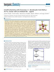
712 C.P. Raptopoulou et al. / Polyhedron 24 (2005) 711–721and phenolate groups as chelat<strong>in</strong>g and/or bridg<strong>in</strong>g units,along with their ability to act <strong>in</strong> their mono- or di-anionicform, make them <strong>in</strong>terest<strong>in</strong>g candidates for theexploration of their metal coord<strong>in</strong>ation <strong>chemistry</strong> [9].In their mono-anionic form, phenolic oximes usuallyact as chelat<strong>in</strong>g agents through their oximatenitrogen and phenolate oxygen atoms (mode A <strong>in</strong>the follow<strong>in</strong>g scheme) <strong>in</strong> a variety of mono- andpoly-nuclear metal complexes [10–14]. A more <strong>in</strong>terest<strong>in</strong>gcoord<strong>in</strong>ation mode, <strong>in</strong>cludes the use of the phenolateoxygen atom as both chelat<strong>in</strong>g and bridg<strong>in</strong>g agent(mode B) [10,15].In their di-anionic form, phenolic oximes usually usetheir phenolate oxygen and oximate nitrogen atoms tochelate to one metal ion and their oximate oxygen asbridg<strong>in</strong>g unit to b<strong>in</strong>d a second metal (mode C). A varietyof di- [16–18], tri- [14,19] and tetranuclear [20–22] metalcomplexes present<strong>in</strong>g the l 2 :g 1 :g 1 :g 1 coord<strong>in</strong>ationmode have been reported. The oximate oxygen atomcan also b<strong>in</strong>d two metal ions, present<strong>in</strong>g thel 3 :g 1 :g 2 :g 1 coord<strong>in</strong>ation mode (mode D) giv<strong>in</strong>g rise tohigher nuclearity complexes, such as hexanuclear[23,24], although this coord<strong>in</strong>ation mode is also observed<strong>in</strong> tr<strong>in</strong>uclear [25] and tetranuclear [11] complexes.There is also one example [10] where the dianion of phenolicoximes can act as chelat<strong>in</strong>g tridentate ligandaccord<strong>in</strong>g to mode E.ACH=N-OHOOCMCH=N-OMMCH=N-OHOMBODMCH=N-OMMM<strong>iron</strong>(<strong>III</strong>) salicylaldoximato complex is the tetranuclear[11] cluster [{Fe(Hsalox)(salox)} 4 ], <strong>in</strong> which the fourHsalox act as chelat<strong>in</strong>g agents through their phenolateoxygen and oximate nitrogen atoms (mode A), whilstthe four salox 2 further use their oximate oxygen atomto b<strong>in</strong>d to two additional metal centers (mode D). In therema<strong>in</strong><strong>in</strong>g examples, coligands have been used to completethe coord<strong>in</strong>ation spheres of <strong>iron</strong>(<strong>III</strong>) centers <strong>in</strong>polynuclear complexes. In the d<strong>in</strong>uclear compound[(Me 3 tacn)Fe(<strong>III</strong>)(salox) 3 Fe(<strong>III</strong>)] [16], as well as <strong>in</strong> thetetranuclear compound [(Me 3 tacn) 2 Fe 4 (salox) 2 (l 3 -O) 2(l 2 -CH 3 CO 2 ) 3 ](ClO 4 ) [20] (where Me 3 tacn = 1,4,7-trimethyl-1,4,7-triazacyclononane)the dianion ofsalicylaldoxime adopts the l 2 :g 1 :g 1 :g 1 coord<strong>in</strong>ationmode. The only tr<strong>in</strong>uclear <strong>iron</strong>(<strong>III</strong>) salicylaldoximatocomplex known so far is [(C 2 H 5 ) 3 NH][Fe 3 O(Hsalox)(salox)2 (salmp)] [19] (salmp = 2-(bis(salicylideneam<strong>in</strong>o)-methyl)phenol, a new ligand formed dur<strong>in</strong>g thereaction process), where the Hsalox ligands act as chelat<strong>in</strong>gunits and the salox 2 ligands adopt thel 2 :g 1 :g 1 :g 1 coord<strong>in</strong>ation mode.The aim of our project is to explore the use of salicylaldoxime<strong>in</strong> <strong>iron</strong>(<strong>III</strong>) <strong>carboxylate</strong> <strong>chemistry</strong>, encouragedby our previous results on manganese(<strong>III</strong>) [24]. Here<strong>in</strong>,we report the first results of our project, <strong>in</strong> particularthe synthesis, X-ray crystal structure, spectroscopic (solidstate and frozen solutions EPR, Mössbauer) characterizationand magnetic behavior of two neutraltr<strong>in</strong>uclear oxo-centered <strong>iron</strong>(<strong>III</strong>) complexes, [Fe 3 (l 3 -O)-(O 2 CPh) 5 (salox)L 1 L 2 ](L 1 =L 2 = MeOH (1), L 1 = EtOH,L 2 =H 2 O(2)). The reported complexes are the first examplesof tr<strong>in</strong>uclear carboxylato <strong>iron</strong>(<strong>III</strong>) complexes withsalicylaldoxime, and can be considered as precursorsfor the synthesis of higher nuclearity complexes, basedon the pr<strong>in</strong>ciple of us<strong>in</strong>g Ômetal oximatesÕ as Ôbuild<strong>in</strong>gblocksÕ for polynuclear clusters. In both compounds,the salox 2 ligands adopt the l 2 :g 1 :g 1 :g 1 coord<strong>in</strong>ationmode (mode C) thus, the possibility of further us<strong>in</strong>gthe oximate oxygen atom, under certa<strong>in</strong> reaction conditions,to b<strong>in</strong>d to a second metal is left open.2. ExperimentalCH=NO2.1. Compound preparationsOEMAll manipulations were performed under aerobic conditionsus<strong>in</strong>g materials as received (Aldrich Co). Allchemicals and solvents were of reagent grade.Further restrict<strong>in</strong>g our discussion to polynuclear<strong>iron</strong>(<strong>III</strong>) complexes with the simplest member of thephenolic oximes, 2-hydroxy-benzaldehyde (salicylaldoxime,H 2 salox), only a few examples have beenreported. As far as we know, the only example of2.1.1. [Fe 3 (l 3 -O)(O 2 CPh) 5 (salox)(MeOH) 2 ] Æ1.25MeOH Æ 1.05H 2 O(1)A methanolic solution of H 2 salox (0.069 g,0.50 mmol) was stirred under reflux, until sodium benzoate(0.216 g, 1.50 mmol) was added. The refluxcont<strong>in</strong>ued for 30 m<strong>in</strong> and then Fe(NO 3 ) 3 Æ 9H 2 O
C.P. Raptopoulou et al. / Polyhedron 24 (2005) 711–721 713(0.202 g, 0.50 mmol) was added. The color of the solutionimmediately turned to deep brown. After 24 h of reflux,small amounts of a brown-red solid were filtered,washed with MeOH and identified as compound 1 byFT-IR spectroscopy. The brown filtrate was sealed andafter a period of one week X-ray quality brownish-redcrystals of 1 were formed (Yield: 0.34 g, 70%). Thecrystals of 1 were collected by filtration, washed withcold MeOH and dried <strong>in</strong> vacuo. The result<strong>in</strong>g powderanalyzed as solvent-free. Anal. Calc. for (1) (C 44 H 38 -NO 15 Fe 3 ): C, 53.47; H, 3.88; N, 1.42. Found: C, 53.45;H, 3.86; N, 1.40%.2.1.2. [Fe 3 (l 3 -O)(O 2 CPh) 5 (salox)(EtOH)(H 2 O)] ÆEtOH (2)The procedure followed was the same as describedabove, but FeCl 3 Æ 6(H 2 O) and ethanol were used <strong>in</strong>stead(Yield: 0.35 g, 70%). The powder of 2 analyzed as solvent-free.Anal. Calc. for (2)(C 44 H 38 NO 15 Fe 3 ): C, 53.47;H, 3.88; N, 1.42. Found: C, 53.45; H, 3.87; N, 1.39%.Table 1Crystallographic data for complex 1 Æ 1.25MeOH Æ 1.05H 2 OFormula C 45.25 H 46.1 Fe 3 NO 17.3Formula weight 1048.30Space group P2 1 /cUnit cell dimensionsa (Å) 19.67(2)b (Å) 26.96(2)c (Å) 19.70(2)b (°) 98.84(3)V (Å 3 ) 10323(2)Z 8T (°C) 298RadiationMo Kaq calcd (g/cm 3 ) 1.348l (mm 1 ) 0.899aR 1 0.0775awR 2 0.1994R 1 = P (jF o j jF c j)/ P (jF o j)andwR 2 ¼ P ½wðF 2 o F 2 o Þ2 Š= P ½wðF 2 o Þ2 Šfor 5082 reflections with I >2r(I).a w ¼ 1=½r 2 ðF 2 o ÞþðaPÞ2 þ bPŠ and P ¼ððmax F 2 o ; 0Þþ2F 2 c Þ=3;a = 0.1046, b = 86.0886.1=22.2. General and physical measurementsElemental analysis for carbon, hydrogen, and nitrogenwas performed on a Perk<strong>in</strong> Elmer 2400/II automaticanalyzer. Infrared spectra were recorded as KBr pellets<strong>in</strong> the range 4000–500 cm 1 on a Bruker Equ<strong>in</strong>ox 55/SFT-IR spectrophotometer. EPR spectra were recordedon a Bruker ER 200D-SRC X-band spectrometerequipped with an Oxford ESR 9 cryostat <strong>in</strong> 4.2–300 Ktemperature range. Variable temperature magnetic susceptibilitymeasurements were carried out on polycrystall<strong>in</strong>esamples of 1 and 2 <strong>in</strong> the 2.0–300 K temperaturerange us<strong>in</strong>g a Quantum Design MPMS SQUID susceptometerunder a magnetic field of 1000 G. Magnetizationmeasurements were carried out at 2.5 K over the 0–5 Tmagnetic field range. Diamagnetic corrections for thecomplexes were estimated from PascalÕs constants.Mössbauer spectra were taken with a constant accelerationspectrometer us<strong>in</strong>g a 57 Co (Rh) source at RT and avariable temperature Oxford cryostat.2.2.1. X-ray crystallography and solution of structuresBrownish-red prismatic crystals of 1 (0.05 · 0.25 ·0.75 mm) and 2 (0.05 · 0.15 · 0.25 mm) were mounted<strong>in</strong> capillary with drops of mother liquid. Diffraction measurementswere made on a Crystal Logic Dual Goniometerdiffractometer us<strong>in</strong>g graphite monochromated Moradiation. Important crystal data and parameters fordata collection for 1 are reported <strong>in</strong> Table 1. Unit celldimensions were determ<strong>in</strong>ed and ref<strong>in</strong>ed by us<strong>in</strong>g theangular sett<strong>in</strong>gs of 25 automatically centered reflections<strong>in</strong> the range 11°