Posterhall Metabolomics 2012 eBook - Bruker
Posterhall Metabolomics 2012 eBook - Bruker
Posterhall Metabolomics 2012 eBook - Bruker

You also want an ePaper? Increase the reach of your titles
YUMPU automatically turns print PDFs into web optimized ePapers that Google loves.
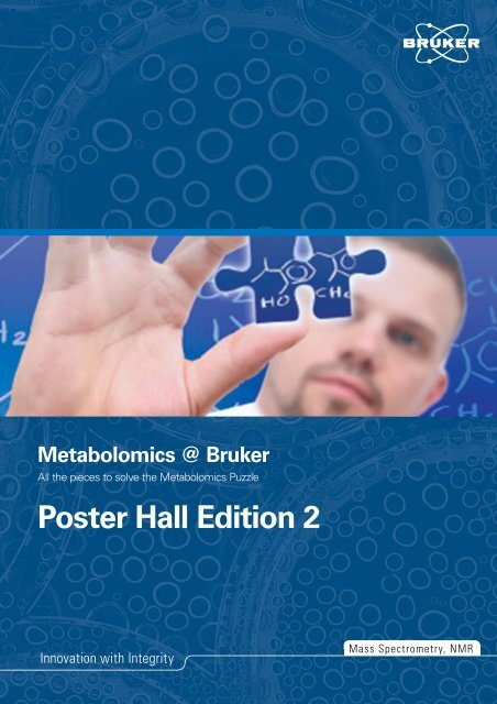
<strong>Metabolomics</strong> @ <strong>Bruker</strong><br />
All the pieces to solve the <strong>Metabolomics</strong> Puzzle<br />
Poster Hall Edition 2<br />
Innovation with Integrity<br />
Mass Spectrometry, NMR
-2-<br />
Table of content<br />
Food <strong>Metabolomics</strong><br />
Urinary excretion of phenolic compounds from Hibiscus sabdariffa<br />
in Humans by advanced analytical techniques 4<br />
RP-HPLC-DAD-ESI-TOF for the pharmacokinetic study of quercetin<br />
in adipocytes 5<br />
Metabolomic consequences of aronia juice intake by human<br />
volunteers 6<br />
Bacterial <strong>Metabolomics</strong><br />
Comprehensive workflow for Metabolite profiling and identification<br />
of secondary metabolites from myxobacteria 7<br />
The impact of high-resolution MS and NMR techniques on discovery<br />
of novel products from myxobacteria 8<br />
Discovery of novel myxobacterial secondary metabolites by<br />
mass spectrometry – based metabolome mining 9<br />
Metabolic profiling of a Corynebacterium glutamicum ∆prpD2 by<br />
GC-APCI high-resolution Q-TOF analysis 10<br />
GC-APCI-MS/MS based identification of metabolites in bacterial<br />
cell extracts 11<br />
Yeast <strong>Metabolomics</strong><br />
Untargeted metabolic profiling of yeast extracts by UHR-Q-TOF<br />
analysis to study arginine biosynthesis mutants 12<br />
Clinical <strong>Metabolomics</strong><br />
A workflow for metabolic profiling and determination of the<br />
elemental compositions using MALDI-FT-ICR MS 13<br />
NMR-based screening of neonate urines 14<br />
NMR-based metabolomics in clinical studies of human population:<br />
Quantification 15<br />
Plant <strong>Metabolomics</strong><br />
Structure elucidation and confirmation for plant metabolomics:<br />
Novel approaches 16<br />
MS-based dereplication and classification of diterpene glycoside<br />
profiles of 23 Solanaceous plant species 17<br />
GC-APCI-QTOF-MS: An innovative technique for metabolite<br />
profiling studies 18<br />
Improved method for characterization of phenolic compounds from<br />
rosemary extracts using ESI-Qq-TOF MS 19<br />
Sulfur-containing metabolite-targeted analysis using liquid<br />
chromatography – mass spectrometry 20<br />
Invertebrate <strong>Metabolomics</strong> –<br />
Drosophila melanogaster and Caenorhabditis elegans<br />
Metabolic characterization of Sod1 null mutant flies using liquid<br />
chromatography – mass spectrometry 21<br />
A mass spectrometry based metabolomic platform for the analysis<br />
of Caenorhabditis elegans 22<br />
Complementary GC-EI-MS and GC-APCI-TOF-MS analysis of a<br />
short-lived mitochondrial knockdown of C. elegans to study metabolic<br />
changes during aging 23<br />
Structure Elucidation<br />
The Fastest Way to a Molecular Formula: How fast can it be?<br />
TLC-UHPLC-ESI-QTOF MS Coupling for Molecular Formula<br />
Determination of New Natural Products 24<br />
MALDI Imaging<br />
Automated tissue state assignment for high-resolution<br />
MALDI-FTMS Imaging data 25<br />
References – Further Reading 26-27
„Putting Together the Pieces to Solve<br />
the <strong>Metabolomics</strong> Puzzle...“<br />
<strong>Metabolomics</strong> has emerged as an exciting new area of<br />
biological research and gained substantial attention in recent<br />
years. Previously, significant efforts have been made to probe<br />
and understand the genomes, transcriptomes and proteomes<br />
of a variety of organisms. Despite massive investments to<br />
sequence and identify cellular DNA, RNA and proteins, our<br />
understanding of many key biological functions remains<br />
rudimentary and incomplete. Many researchers believe that<br />
perhaps more complete answers can be gleaned from studying<br />
small molecules that are utilized for a variety of metabolic and<br />
cellular functions. Because of this belief, in many laboratories,<br />
<strong>Metabolomics</strong> is catching up with traditional Proteomics<br />
and Transcriptomics efforts and has been christened<br />
“Biochemistry’s new look” [1].<br />
Typically, during a <strong>Metabolomics</strong> study, a large number of<br />
cell or patient samples are analyzed and compared to identify<br />
potential biomarker compounds which correlate to a disease,<br />
drug toxicity, or genetic or environmental variation. Validated<br />
biomarkers can form the basis for new diagnostic assays, and<br />
lead the way to help establish truly personalized medicine,<br />
since changes in small molecule concentrations are closely<br />
related to the observed phenotype.<br />
Answering the Challenges of <strong>Metabolomics</strong><br />
<strong>Metabolomics</strong> researchers are regularly confronted with the<br />
daunting tasks of acquiring large sets of data and then analyzing<br />
them in minute detail. Due to the chemical diversity of small<br />
molecule metabolites, it is impossible to study the entire<br />
metabolome using a single analytical technique or technology.<br />
<strong>Bruker</strong>’s comprehensive solution for metabolomics is based on<br />
putting together “the pieces of the puzzle” to give a complete<br />
picture: The Metabolome’s chemical space can be fully<br />
explored by utilizing <strong>Bruker</strong>’s high performance LC-MS, GC-MS,<br />
CE-MS and NMR systems in conjunction with dedicated<br />
software for data evaluation.<br />
One of the most challenging tasks in the <strong>Metabolomics</strong> studies<br />
is the identification of unknown compounds. Exact mass<br />
and isotopic pattern information in MS and MS-MS spectra<br />
acquired by the solariX (FT-MS) or micrOTOF and maXis series<br />
(Q)-TOF-MS instruments can be used to definitively identify<br />
unknown compounds. The intrinsic capabilities of these<br />
instruments in conjunction with the SmartFormula3D software<br />
enables accurate and reliable molecular formula generation for<br />
a wide variety of unknowns. Subsequent database queries<br />
identify likely compound structures, which can be confirmed<br />
by complementary information derived from NMR spectra.<br />
In order to enhance separation and increase throughput,<br />
many laboratories have turned to utilization of UHPLC for<br />
<strong>Metabolomics</strong> studies. Since the mass accuracy, isotopic<br />
fidelity and resolution of maXis and micrOTOF-series ESI-(Q)-<br />
TOF instruments are independent of the acquisition rate, they<br />
are perfectly suited for a coupling to UHPLC separations.<br />
By utilizing a <strong>Bruker</strong> solution, their high-resolution and accurate<br />
mass LC-MS capabilities enable both targeted and untargeted<br />
<strong>Metabolomics</strong> workflows. In addition, the broad dynamic<br />
range characteristics of these systems allows both, high- and<br />
low-abundance compounds to be studied within the same<br />
dataset – even within the same mass spectrum.<br />
Finally, <strong>Bruker</strong> offers the capability to integrate both LC-MS<br />
and NMR techniques into a single analytical system for<br />
maximum metabolite coverage and molecular identification.<br />
The MetabolicProfilerTM is a powerful combination of<br />
techniques and is exclusively designed to address all analytical<br />
needs for metabolic profiling and structure elucidation.<br />
This poster hall is a “snapshot” of some of the current work<br />
utilizing a variety of <strong>Bruker</strong> solutions for <strong>Metabolomics</strong> studies.<br />
It nicely illustrates <strong>Bruker</strong>’s aim of providing and putting<br />
togehter the “puzzle pieces” to give a complete picture of<br />
the metabolome. We would like to thank all of our partners<br />
and customers for their constant input and feedback. This<br />
information has served as an invaluable guide for us in our<br />
solution development efforts. A special thanks are directed<br />
to everyone who has worked to generate the outstanding<br />
posters which we have assembled in this poster hall for your<br />
benefit.<br />
Sincerely yours,<br />
Dr. Aiko Barsch<br />
Market Manager <strong>Metabolomics</strong><br />
-3-
-4-<br />
<strong>Metabolomics</strong> Posterbook <strong>2012</strong><br />
Food <strong>Metabolomics</strong><br />
I. Borrás‐Linares (1, 2) , R. Beltrán‐Debón (3) , Gabriela Zurek (4) , Jorge Joven (3) , D. Arráez‐Román (1, 2) ,<br />
A. Segura‐Carretero (1, 2) , A. Fernández‐Gutiérrez (1, 2)<br />
(1) Department of Analytical Chemistry, Faculty of Sciences, University of Granada, Granada, Spain.<br />
(2) Functional Food Research and Development Center (CIDAF), Health Science Technological Park, Granada, Spain.<br />
(3) Centre de Recerca Biomèdica, Hospital Universitari de Sant Joan, IISPV, Universitat Rovira i Virgili, Reus, Spain.<br />
(4) <strong>Bruker</strong> Daltonik GmbH, Bremen, Germany.<br />
Hibiscus sabdariffa (HS) L. (family: Malvaceae), usually named bissap, karkade or roselle is a tropical plant commonly used as local soft drink. This plant contains organic<br />
and phenolic acids, anthocyanins, and flavonols as main compounds [1,2]. Traditionally the aqueous extracts of HS have been used in folk medicine to treat<br />
hypertension, fever, inflammation, liver disorders, and obesity.<br />
In recent years, this plant has been studied in depth because it exerts a great number of pharmacological activities. Previous studies have shown that HS possesses anti‐<br />
tumor and antioxidant properties [3, 4]. Recent data also indicate that aqueous extracts of HS might ameliorate metabolic disturbances [5, 6]. The antioxidant and<br />
antihyperlipemic properties of HS aqueous extract have been previously reported using a mouse model [7]. Furthermore, the hypolipemic properties of PEHS have been<br />
recently reported in a cell model for adipogenesis and insulin resistance [8]. However, to establish conclusive evidence for the effectiveness of phenolic compounds<br />
from this plant on health, it is very important to define the bioavailability and the excretion of these compounds in humans. The objective of this study was to identify<br />
the urinary excretion of phenolic compounds in humans after the ingestion of a phenolic‐enriched Hibiscus sabdariffa extract (PEHS).<br />
1<br />
Hibiscus sabdariffa<br />
flowers<br />
PEHS pills<br />
In this study a phenolic‐enriched Hibiscus sabdariffa extract<br />
(PEHS) has been obtained and encapsulated. Urine samples<br />
from a healthy volunteer were collected before and 2, 4 and 6<br />
hours after intake of 500 mg of the PEHS for the evaluation of<br />
the urinary excretion of the phenolic compounds present in this<br />
plant extract.<br />
2<br />
PEHS<br />
1000 μl AcOEt<br />
10 μl HCl 1M<br />
500 μl Sample<br />
Vortex<br />
Supernatant Dryness<br />
Centrifuge<br />
18800 g<br />
5 min<br />
250 μl<br />
Mobil phase A UHPLC‐ESI‐UHR‐Q‐TOF<br />
analysis<br />
UHPLC<br />
BPC OBTAINED BY<br />
UHPLC‐ESI‐UHR‐Q‐TOF<br />
[1] Salah, A. M., Gathaumbi, J., Vierling, W. (2002) Phytother. Res. 16:283‐285.<br />
[2] Tseng T. H., Hsu J., Dong L., Ming H., Chu C. Y. (1998) J. Agric. Food Chem. 46: 1101‐1105.<br />
[3] Chang Y. C., Huang H. P., Hsu J. D. (2005) Toxicol Appl Pharmacol. 205: 201‐207.<br />
[4] Ochani P. C., D´Mello P. (2009). Indian J. Exp. Biol. 47: 276‐282.<br />
[5] Carvajal‐Zarrabal O., Waliszewski S. M., Barradas‐Dermitz D. M. A. (2005) Plant Food Hum. Nutr. 60: 153‐159.<br />
[6] Alarcon‐Aguilar F. J., Zamilpa A., Pérez‐García M.D. (2007) J. Ethnopharmacol. 114: 66‐72.<br />
[7] Fernández‐Arroyo S., Rodríguez‐Medina I.C., Beltrán‐Debón R. (2011) Food Res. Int. 44: 1490‐1495.<br />
[8] Herranz‐López M., Fernández‐Arroyo S., Pérez‐Sánchez A. (<strong>2012</strong>) Phytomed. 19: 253‐261.<br />
3<br />
•Instrument: Dionex Ultimate 3000<br />
UHPLC<br />
•Column: Zorbax Eclipse Plus C18,<br />
3x250 mm, 5 μm<br />
•Flow: 0.3 mL/min<br />
•Temperature: 20 °C<br />
•Injection volume: 5 μl<br />
•Phase A: H 2O/AcN 90:10 + 0.5%<br />
Formic acid<br />
•Phase B: AcN<br />
•Gradient: :<br />
•from 0 to 20 min, from 5% B to 20%<br />
B,<br />
•from 20 to 25 min, from 20%B to<br />
40%B,<br />
•from 25 to 30 min, from 40% B to<br />
5% B,<br />
•from 30 to 35 min, to hold 5% B.<br />
UHR‐Q‐TOF<br />
•Instrument: <strong>Bruker</strong> Daltonics<br />
MaXis 4G<br />
UHR‐Q‐TOF<br />
•Mode: Negative<br />
•Mass range: 50‐1000 m/z<br />
•Nebulizing gas pressure: 2 bar<br />
•Drying gas flow: 8 L/min<br />
•Drying gas temp: 200 ºC<br />
•Calibrant: sodium formate 5mM<br />
•End Plate offset: ‐500 V<br />
•Capillary: ‐ 4500V<br />
•Funnel RF: 300,0 Vpp<br />
•Ion Energy: 4,0 eV<br />
•Collision Energy: 8,0 eV<br />
•Collision RF: 400,0 Vpp<br />
•Transfer time: 65 μs<br />
•PrePulseStorage time: 6 μs<br />
Compound Retention time (min) [M‐H ] ‐<br />
This powerful analytical technique<br />
revealed the presence of several<br />
phenolic compounds from the PEHS<br />
in the urine samples. The main<br />
compounds detected previously<br />
described in the extract were<br />
hibiscus acid, hibiscus acid<br />
dimethylesther, hydroxycitric acid,<br />
chlorogenic acid isomers, methyl<br />
digallate, cumaroylquinic acid and<br />
N‐feruloyltyramine. The maximum<br />
excretions of these compounds<br />
were found at 2 and 4 hours after<br />
the intake of PEHS.<br />
Molecular Formula<br />
Hydroxycitric acid 3,40 207,0146 C6H8O8<br />
Hibiscus acid 3,60 189,0040 C6H6O7<br />
Chlorogenic acid Isom I 5,30 353,0878 C16H18O9<br />
Hibiscus acid dimethylesther 6,00 217,0353 C9H10O7<br />
Chlorogenic acid 6,90 353,0878 C16H18O9<br />
Chlorogenic acid Isom II 7,10 353,0878 C16H18O9<br />
Methyldigallate 7,60 335,0408 C15H12O9<br />
Coumaroylquinic acid 9,60 337,0928 C16H18O8<br />
N‐Feruloyltyramine 25,60 312,1241 C18H20NO4<br />
The authors are grateful to the Spanish Ministry of Science and Innovation for the<br />
project AGL2011‐29857‐C03‐02, Andalusian Regional Government Council of<br />
Innovation and Science for the excellence project P10‐FQM‐6563 and P11‐CTS‐7625 and<br />
to GREIB.PT.2011.18 project. IBL acknowledges financial support from the Spanish<br />
Ministry of Education and Science (FPI grant, BES‐2009‐028128).
Food <strong>Metabolomics</strong><br />
1<br />
I. Borrás‐Linares (1, 2) , M. Herranz‐López (3) , E.Barrajón‐Catalán (3) , D. Arráez‐Román (1, 2) , V. Micol (3) ,<br />
A. Segura‐Carretero (1, 2) , A. Fernández‐Gutiérrez (1, 2)<br />
(1) Department of Analytical Chemistry, Faculty of Sciences, University of Granada, Granada, Spain.<br />
(2) Research and Development of Functional Food Centre (CIDAF), Health Science Technological Park, Granada, Spain.<br />
(3) Institute of Molecular & Cell Biology (IMCB), Miguel Hernández University, Elche, Spain.<br />
Hibiscus sabdariffa (HS) L. (family: Malvaceae), usually named bissap, karkade or roselle is a tropical plant commonly used as local<br />
soft drink. The aqueous extracts of HS have been used in folk medicine to treat hypertension, fever, inflammation, liver disorders,<br />
and obesity. This plant contains organic and phenolic acids, anthocyanins, and flavonols as main compounds. One of these major<br />
compounds found in this plant is quercetin, a flavonol commonly present in fruits and vegetables.<br />
In recent years, this plant has been studied in depth because it exerts a great number of pharmacological activities. Previous studies<br />
showed that HS possesses anti‐tumor and antioxidant properties [1]. In humans, HS elicits a significant decrease in MCP‐1 plasma<br />
concentration, suggesting that HS may be a valuable traditional herbal medicine for the treatment of chronic inflammatory diseases<br />
[2]. Recent data also indicate that HS aqueous extracts might ameliorate metabolic disturbances [3]. Furthermore, the antioxidant<br />
and antihyperlipemic properties of HS aqueous extract have been previously reported by our research group using a mouse model<br />
[4]. Moreover, the strong antioxidative and hypolipidemic properties of a HS polyphenolic extract have been recently reported in a<br />
cell model for adipogenesis and insulin resistance [5]. In this studies, we suggested that flavonol conjugated forms (such as<br />
quercetin) could be the responsible compounds for the observed antioxidant effects and also contribute to the salutary effects of<br />
HS. In this sense, a study of the molecular and cellular targets of these compounds in different cellular models deserves further<br />
attention. For this reason, a pharmacokinetic study of quercetin in a model of adipogenesis (3T3‐L1 cells) has been carried out.<br />
100 μl Cytoplasm<br />
2<br />
500 μl MeOH‐EtOH<br />
(50:50)<br />
2 hours<br />
Supernatant Dryness<br />
In this study 3T3‐L1 pre‐<br />
adipocytes were propagated<br />
and differentiated according<br />
to conventional procedures.<br />
Quercetin was added at 100<br />
µM to the media at the<br />
beginning of the study.<br />
Subsequently, samples of<br />
cytoplasm were taken at<br />
different time (3, 6, 12 and<br />
18 hours) after quercetin<br />
addition.<br />
Centrifuge<br />
18800 g<br />
5 min<br />
50 μl MeOH<br />
HPLC‐DAD‐ESI‐TOF<br />
analysis<br />
3<br />
HPLC<br />
BPC OBTAINED<br />
BY HPLC‐ESI‐TOF<br />
• Instrument: Agilent 1200 RRLC<br />
• Column: Zorbax Eclipse Plus C18,<br />
4.6x150 mm, 1.8 μm<br />
• Flow: 0.3 mL/min<br />
• Temperature: 25 °C<br />
• Injection volume: 10 μl<br />
• Phase A: H 2O/AcN 90:10 + 0.5% Formic acid<br />
• Phase B: AcN<br />
• Gradient: :<br />
• from 0 to 20 min, from 5% B to 20% B,<br />
• from 20 to 25 min, from 20%B to 40%B,<br />
• from 25 to 40 min, from 40% B to 5% B,<br />
• from 40 to 45 min, to hold 5% B.<br />
[1] Farombi EA, Fakoya A (2005) Free radical scavenging and antigenotoxic activities of natural phenolic compounds in dried flowers of Hibiscus sabdariffa L. Molecular Nutrition & Food Research 49:1120<br />
[2] Beltran‐Debon R, Alonso‐Villaverde C, Aragones G et al (2010) The aqueous extract of Hibiscus sabdariffa calices modulates the production of monocyte chemoattractant protein‐1 in humans. Phytomedicine<br />
17:186<br />
[3] Carvajal‐Zarrabal O, Waliszewski SM, Barradas‐Dermitz DMA et al (2005) The consumption of Hibiscus sabdariffa dried calyx ethanolic extract reduced lipid profile in rats. Plant Foods for Human Nutrition 60:153<br />
[4] Fernández‐Arroyo S, Rodríguez‐Medina IC, Beltrán‐Debón R et al (2011) Quantification of the polyphenolic fraction and in vitro antioxidant and in vivo anti‐hyperlipemic activities of Hibiscus sabdariffa aqueous<br />
extract. Food Res Int 44:1490<br />
[5] Herranz‐López M, Fernández‐Arroyo S, Pérez‐Sánchez A et al. Synergism of plant‐derived polyphenols in adipogenesis: perspectives and implications. Phytomedicine<br />
ESI‐TOF<br />
Quercetin‐3‐O‐Glucuronide<br />
• Instrument: <strong>Bruker</strong> Daltonics<br />
micrOTOF<br />
• Mode: Negative<br />
• Mass range: 50‐1000 m/z<br />
• Nebulizing gas pressure: 2 bar<br />
• Drying gas flow: 9 L/min<br />
• Drying gas temp: 200 ºC<br />
• Calibrant: sodium formate 5mM<br />
• Capillary Exit: ‐120 V<br />
• Skimmer 1: ‐40 V<br />
• Hexapole 1: ‐23 V<br />
• Hexapole RF: 100 Vpp<br />
• Skimmer 2: ‐22,5 V<br />
• Transfer time: 50 μs<br />
• PrePulseStorage time: 3 μs<br />
Quercetin<br />
Concentration (ppm)<br />
45<br />
40<br />
35<br />
30<br />
25<br />
20<br />
15<br />
10<br />
5<br />
0<br />
0 0,5 31 1,5 62 2,5 123 3,5 184<br />
4,5<br />
Time after incubation (h)<br />
Quercetin<br />
Quercetin was detected in all analyzed<br />
samples, except in the control. From the<br />
obtained data, the maximum concentration<br />
found in cytoplasm was at 3 hour after the<br />
quercetin addition. Nevertheless, it is<br />
important to note that at 6 hours time point<br />
the concentration of quercetin is still high. At<br />
the end of the assay, 18 hours after the<br />
addition, the level of quercetin is minimum. In<br />
all samples except in the control, quercetin‐3‐<br />
O‐glucuronide has been detected.<br />
The authors are grateful to the Spanish Ministry of<br />
Science and Innovation for the project AGL2011‐<br />
29857‐C03‐02, Andalusian Regional Government<br />
Council of Innovation and Science for the excellence<br />
project P10‐FQM‐6563 and P11‐CTS‐7625 and to<br />
GREIB.PT.2011.18 project. IBL acknowledges financial<br />
support from the Spanish Ministry of Education and<br />
Science (FPI grant, BES‐2009‐028128).<br />
-5-
-6-<br />
<strong>Metabolomics</strong> Posterbook <strong>2012</strong><br />
Food <strong>Metabolomics</strong><br />
<strong>Bruker</strong> Daltonics<br />
Metabolomic consequences of aronia juice intake by<br />
human volunteers<br />
Medina, S. 1 ; García-Viguera, C. 1 ; Ferreres, F. 1 ; Savirón, M. 2 ; Orduna,<br />
J. 2 ; Zurek, G. 3 ; Gil-Izquierdo, A. 1*<br />
1Research Group on Quality, Safety and Bioactivity of Plant Foods, CEBAS-<br />
CSIC, Murcia, Spain<br />
2Instituto de Ciencias de Materiales de Aragón. CSIC- Universidad de<br />
Zaragoza, Zaragoza, Spain<br />
3<strong>Bruker</strong> Daltonik GmbH, 28359 Bremen, Germany.<br />
*corresponding author: angelgil@cebas.csic.es<br />
Introduction<br />
<strong>Metabolomics</strong> is a powerful technique which focuses in highthroughput characterization of<br />
metabolome (the complete set of low-MW metabolites in biological samples) in order to obtain a<br />
molecular profile. This approach has shown a considerable potential in the field of nutrition, so, there<br />
is an increasing interest in the use of metabolomics as a tool for the understanding of the interaction<br />
between diet and metabolome and to be a promising technique for biomarker discovery related to<br />
food intake [1].<br />
Our study was conducted with the aronia fruit (Aronia melanocarpa) common name black<br />
chokeberry which belongs to the Rosaceae family. This berry stands out in high amount of flavanols,<br />
quercetin (71.13 mg Kg-1; 93.07%) and kaempferol (5.3 mg Kg-1; 6.9%). Aronia berries are one of<br />
richest plant sources of anthocyanins, mainly containing cyanidin glycosides [2].<br />
In a recent study, anthocyanins showed significant potency of antiobesity and ameliorate adipocyte<br />
function in vitro and in vivo systems, as well as, antioxidant, hypoglycemic and hypolipidomic<br />
activities and important implications for preventing metabolic syndrome of a chokeberry juice, indicate<br />
their contribution to the potential health benefits [3,4].<br />
The aim of this study was to observe the single and five days changes in the metabolic profile of<br />
healthy volunteers following the consumption of aronia juice and to identify the key metabolites.<br />
Urine samples<br />
(treatment and control)<br />
Assay design<br />
Number of patients: 8 (4 men and 4 women)<br />
Age: 28-47<br />
Race: Caucasian<br />
BMI means: 24.5 Kg/m 2<br />
Wash out<br />
2 days<br />
Day 1 Day 2 Day 3 Day 4<br />
200 mL 8:00 a.m<br />
200 mL 18:00 p.m<br />
Diet absent of fruit-vegetables and food derived products<br />
200 mL 8:00 a.m<br />
200 mL 18:00 p.m<br />
• Workflow for metabolomic analysis:<br />
Data adquisition<br />
LC-MS-q-TOF (<strong>Bruker</strong> Daltonics,<br />
Bremen, Germany)<br />
Column ACE 3 C18: 150 x 0.075<br />
mm<br />
Fase A: H2O/0.1% HCOOH<br />
Fase B: ACN/0.1% HCOOH<br />
Flow rate: 312.50 nL/min<br />
Injection volume: 6.25 nL<br />
Positive and negative mode<br />
Full scan: 50-900 m/z<br />
200 mL 8:00 a.m<br />
200 mL 18:00 p.m<br />
24 h urine collection and frozen at -80º C<br />
Data preprocessing<br />
Profileanalysis 2.0. (<strong>Bruker</strong><br />
Daltonics, Bremen, Germany)<br />
Software for profiling and<br />
statistical evaluation of LC/MS<br />
data set.<br />
200 mL 8:00 a.m<br />
200 mL 18:00 p.m<br />
Multivariate statistical<br />
analysis<br />
PCA (Principal Component<br />
Analysis) with Pareto scaling<br />
algorith and Student´s t-test (P-value<br />
Bacterial <strong>Metabolomics</strong><br />
COMPREHENSIVE WORKFLOW FOR METABOLIC<br />
PROFILING AND IDENTIFICATION OF SECONDARY<br />
METABOLITES FROM MYXOBACTERIA<br />
Thomas Hoffmann 1,2 , Daniel Krug 1, 2 , Aiko<br />
Barsch 3 , Gabriela Zurek 3 , Rolf Müller 1,2<br />
1 Pharmaceutical Biotechnology, Saarland<br />
University, Saarbruecken, Germany<br />
2 Helmholtz-Institute for<br />
Pharmaceutical Research Saarland (HIPS),<br />
Saarbruecken, Germany<br />
3 <strong>Bruker</strong> Daltonik GmbH, Bremen, Germany<br />
Introduction<br />
Extracting the relevant information from complex<br />
data sets is an important bottleneck in (microbial)<br />
metabolomics research. Compared to a focused<br />
targeted approach, this objective becomes even<br />
more challening in untargeted metabolomics, aiming<br />
for the identification of all compounds produced by a<br />
particular bacterial strain. Besides the identification<br />
of all known compounds it is important to assign the<br />
subset of interesting compounds that are “worth”<br />
further examination.<br />
Here, we present the combination of targeted and<br />
untargeted metabolomics utilizing statistical methods<br />
of data mining. ESI-UHR-Q-TOF based analysis of<br />
myxobacterial extracts allows for fast generation of<br />
high resolution MS as well as MS² spectra which<br />
support our screening workflow for the discovery of<br />
novel secondary metabolites.<br />
6 x extract<br />
6 x blank<br />
Fig. 1: Overall 24 runs were analyzed. The feature<br />
finding algorithm assigned ~ 3500 features per run.<br />
Sample Set<br />
• The sample set consisted of 6 biological replicates<br />
of a Sorangium cellulosum strain and 6 replicates of<br />
growth medium blank. Extracts were prepared by<br />
methanol extraction of an adsorber resin which was<br />
added to the cultivation broth (Amberlite XAD-16).<br />
Samples were diluted 5x with methanol prior to<br />
injection. For each sample two technical replicates<br />
were measured.<br />
Methods<br />
• Sorangium cellulosum extracts were separated using a Waters<br />
BEH C18 column (100 x 2.1 mm, 1.7 µm particle size) on an<br />
UltiMate 3000TM RSLC system (Dionex). Gradient elution was<br />
performed at 600 µl/min and 45 °C using H2O + 0.1 % FA (A) and<br />
ACN + 0.1 % FA (B). The gradient started at 5 % B for 0.5 min<br />
before increasing to 95 % B in another 19 min.<br />
• ESI-MS measurements were performed using positive ionization<br />
on a maXis UHR-Q-TOF-MS (<strong>Bruker</strong> Daltonik) m/z range: 150-<br />
1500, repetition rate: 2 Hz for MS, 3 Hz for MS².<br />
• Targeted screening was carried out using precise extracted ion<br />
chromatograms (EICs), retention time and isotope pattern<br />
evaluation via SigmaFitTM with TargetAnalysisTM (<strong>Bruker</strong> Daltonik).<br />
• Pre-processing of data and statistical interpretation using t-test<br />
was performed with ProfileAnalysisTM 2.0 (<strong>Bruker</strong> Daltonik).<br />
Scheduled precursor lists (SPL) for targeted MS/MS experiments<br />
were created by the same software tool. SPL derived MS² data<br />
was processed with MZmine 2.2 [1] . Features were extracted from<br />
raw data using the FindMolecularFeatures (FMF) algorithm in<br />
DataAnalysisTM 4.1.<br />
Feature Finding<br />
• Myxobacterial extracts represent a complex<br />
mixture of small molecules derived from the medium<br />
and bacterial secondary metabolites covering several<br />
magnitudes in dynamic range. The feature finding<br />
algorithm (FMF) assigned approximately 3500<br />
features for each measurement (Fig. 1).<br />
• Overall 24 samples gave ~80,000 features.<br />
• The FMF algorithm is able to automatically detect<br />
and combine adduct ions and common neutral losses<br />
like [M+NH4] + , [M+Na] + , or [2M+H] + to one feature.<br />
This approach lowered the total number of features<br />
by around 10 % to ~70,000 features.<br />
For research use only. Not for use in diagnostic procedures.<br />
80,000 features<br />
(24 x 3500)<br />
Untargeted Analysis<br />
• The result was evaluated by a t-test comparing<br />
extracts and blanks and value-count filtering for<br />
features which were present in at least 11 out of 12<br />
measurements in the extract.<br />
• A resulting subset of 176 features was exported as<br />
a scheduled precursor list (SPL) for directed<br />
precursor selection in a subsequent MS/MS<br />
experiment (Fig. 2).<br />
Fig. 2: Features that are only present within bacterial<br />
extracts are assigned by means of a t-test. An SPL of<br />
176 features was exported automatically for further<br />
CID analysis.<br />
A<br />
# rt [min] m/z Extract Blank<br />
1 1.1 383.2 12 0<br />
2 2.2 298.1 12 0<br />
3 2.3 293.1 11 0<br />
4 2.4 180.1 12 0<br />
… … … … …<br />
176 17.9 253.25 11 0<br />
Name Formula [M+X] + m/z theo. m/z meas. Error [ppm] mSigma<br />
Ambruticin A<br />
[M+H] +<br />
B<br />
C28H40O6 473.28977 473.28964 0.3 8.3<br />
Ambruticin A<br />
[M+NH +<br />
4]<br />
C28H43N1O6 490.31632 490.31572 1.2 4.5<br />
Ambruticin A<br />
[M-H +<br />
2O+H]<br />
C28H38O5 455.27920 455.27909 0.2 23.6<br />
Fig. 3: A: Result of target compound identification as<br />
presented in TargetAnalysis. B: Example: Assignment<br />
of several adducts for the same compound gives more<br />
reliable hits.<br />
References<br />
t-test result<br />
Targeted Analysis<br />
Important features<br />
assigned by t-test analysis<br />
Targeted search against<br />
in-house database<br />
• An in-house database (Myxobase) covering ~700<br />
myxobacterial secondary metabolites representing<br />
more than 100 distinct compound classes with<br />
retention time and sum formula information was<br />
used to identify the known compounds (Fig. 3A).<br />
• The 176 filtered features obtained by the<br />
untargeted workflow were then compared to the<br />
TargetAnalysis result to assign known metabolites.<br />
22 features are related to TA hits or fragments<br />
thereof.<br />
• To increase the confidence in compound<br />
identification by targeted analysis common adduct<br />
information was additionally taken into account. The<br />
Myxobase system enables the export of a “screening<br />
file” for TargetAnalysis which automatically creates<br />
common adduct ions for a given sum formula<br />
(Fig. 3B).<br />
[1] T. Pluskal, S. Castillo, A. Villar-Briones, M. Orešič,<br />
BMC Bioinformatics 11:395 (2010).<br />
Distribution of 176 SPL features<br />
Funding from BMB+F, DFG and DAAD is gratefully acknowledged.<br />
176 features<br />
(22 assigned)<br />
MS/MS<br />
Unknown identification<br />
Conclusions<br />
<strong>Bruker</strong> Daltonics & HIPS<br />
Parent ion selection for MS²<br />
• Automatic precursor selection during acquisition<br />
routinely allows for the fragmentation of dominant species.<br />
However, compounds of interest may be low abundant and<br />
coeluting with matrix compounds from cultivation media.<br />
In such cases, the preselection of parent ions can increase<br />
the number of MS² spectra for compounds of interest.<br />
• Manual definition of hundreds of precursors is time<br />
consuming. Therefore, the subset of 176 features was used<br />
for directed precursor selection via an SPL (Fig. 2).<br />
• 37 % of the precursors of interest were not<br />
fragmented previously during an auto-MS² run. Using SPL<br />
lists generated based on t-test results could improve the<br />
MS² coverage to 100 % for the target compounds.<br />
Putative ambruticin derivatives:<br />
Fragmentation proposal:<br />
Intens.<br />
2000<br />
1500<br />
1000<br />
500<br />
0<br />
1+<br />
224.1272<br />
1+<br />
250.1434<br />
O<br />
OH<br />
O N<br />
H<br />
High accurate m/z of CID<br />
peaks allow for structural<br />
identification<br />
271.2439 288.1587<br />
1+<br />
Peak 10<br />
C18H26NO3, [I+H], -2.1 ppm<br />
1+<br />
304.1914<br />
1+<br />
333.2231 352.2979<br />
• The number of features representing potentially novel<br />
metabolites was reduced further by matching specific<br />
neutral losses within coeluting features.<br />
Features in one MS file 3500<br />
Features after t-test 176<br />
- fragmented with SPL method 176<br />
- fragmented with auto-MS² 110<br />
Related to known compounds - 22<br />
Assigned by online database search - 0<br />
Identified as neutral loss - 26<br />
Remaining unknown features 128<br />
Assignment using MS/MS data<br />
Peptides - 4<br />
found due to similar fragmentation - 11<br />
- related to 3 Thuggacins<br />
- related to 8 Ambruticins<br />
New features for further investigation 113<br />
• High resolution accurate mass LC-MS data sets allow<br />
for both targeted and untargeted analysis.<br />
• Complexity is reduced by statistical data evaluation.<br />
• Parent ion selection is significantly improved by a<br />
scheduled precursor list (SPL)<br />
• 63 out of 176 filtered features from myxobacterial<br />
extracts could be identified based on MS 2 data, thereby<br />
effectively supporting de-replication during screening<br />
NH2<br />
C25H40NO, [I+H], +0.7 ppm<br />
1+<br />
370.3102<br />
O<br />
C22H42N3O3, [I+H], -3.9 ppm<br />
1+<br />
396.3236<br />
1+<br />
423.3188<br />
381.2747<br />
+MS2(Ambruticin new01), 29.4eV, 11.87min #1401<br />
O<br />
O<br />
OH<br />
NH2<br />
1+<br />
440.3134<br />
1+<br />
458.3263<br />
1+<br />
476.3357<br />
250 300 350 400 450 m/z<br />
Fig. 4: Unknown identification: Calculation of sum<br />
formulae suggests the presence of new ambruticin<br />
derivatives. Subsequent MS² spectra evaluation using<br />
Fragment Explorer allows for the annotation of highly<br />
accurate MS² peaks, supporting the identification.<br />
• Guided MS/MS<br />
acquisition allows for<br />
the assignment of<br />
interesting features<br />
and facilitates their<br />
identification (Fig. 4).<br />
• Searching in<br />
publicly accessible<br />
databases did not<br />
yield any hits.<br />
• 113 features remained<br />
for further<br />
evaluation by MS and<br />
NMR experiments.<br />
ESI-UHR-QTOF-MS<br />
C24H38N7O, [I+H], -0.4ppm<br />
C28H46NO5, [I+H], +2.8 ppm<br />
O<br />
-7-
-8-<br />
<strong>Metabolomics</strong> Posterbook <strong>2012</strong><br />
Bacterial <strong>Metabolomics</strong><br />
The impact of high-resolution MS and NMR<br />
techniques on the discovery of novel natural<br />
products from myxobacteria<br />
Daniel Krug 1,2 , Thomas Hoffmann 1 , Kirsten<br />
Harmrolfs 2 , Alberto Plaza 1 , Aiko Barsch 3 ,<br />
Gabriela Zurek 3 , Rolf Müller 1,2<br />
1 Helmholtz-Institute for Pharmaceutical Research<br />
Saarland (HIPS), 66123 Saarbrücken/Germany<br />
2 Saarland University, 66123 Saarbrücken/Germany<br />
3 <strong>Bruker</strong> Daltonik GmbH, 28359 Bremen, Germany<br />
Introduction<br />
Microbial natural products represent a rich and still<br />
largely untapped resource for novel chemical<br />
scaffolds. Their discovery enables characterization of<br />
the underlying biosynthetic pathways and at the<br />
same time chances are good to find compounds with<br />
potent biological activity. UHPLC-coupled UHR-TOF<br />
mass spectrometry and LC-NMR represent most<br />
valuable analytical tools for the discovery of novel<br />
natural products, efficient dereplication, comprehensive<br />
mining of metabolomes and rapid<br />
structure elucidation of novel metabolites.<br />
Chondromyces crocatus Sorangium cellulosum<br />
A<br />
B<br />
Name Formula [M+X] + m/z theo. m/z meas. Error [ppm] mSigma<br />
Ambruticin A<br />
[M+H] +<br />
Ambruticin A<br />
[M+NH +<br />
4]<br />
Ambruticin A<br />
[M-H +<br />
2O+H]<br />
C 28H43O6<br />
Dm/z = 0.9 ppm<br />
14.8 mSigma<br />
C 28H41O5<br />
Dm/z = 1.1 ppm<br />
6.8 mSigma<br />
C 28H39O4<br />
Dm/z = 0.9 ppm<br />
8.9 mSigma<br />
1+<br />
439.2847<br />
1+<br />
457.2953<br />
1+<br />
475.3059<br />
493.3160<br />
510.3428<br />
360 380 400 420 440 460 480 500 520 m/z<br />
+MS, 11.20min<br />
Fig. 1 Chromatogram:<br />
separation of a crude extract<br />
from a Sorangium strain. Right<br />
inset: comparison of measured<br />
and calculated spectra for<br />
Ambruticin, a myxobacterial<br />
compound that exhibits potent<br />
antifungal activity.<br />
475.3054<br />
5 475.3059<br />
x10<br />
1.0<br />
0.8<br />
Ambruticin S<br />
C28H42O6 [M+H]<br />
0.6<br />
476.3088<br />
0.4<br />
476.3091<br />
0.2<br />
477.3117<br />
calculated<br />
477.3111<br />
measured<br />
0.0<br />
475 476 477 478<br />
+ = 475.3054<br />
Dm/z = 0.9 ppm<br />
14.8 mSigma<br />
C 28H40O6 473.28977 473.28964 0.3 8.3<br />
C 28H43N1O6 490.31632 490.31572 1.2 4.5<br />
C 28H38O5 455.27920 455.27909 0.2 23.6<br />
Fig. 2: A: Result of target compound identification as presented in<br />
TargetAnalysis TM . B: Example: Assignment of several adducts for<br />
the same compound gives more reliable hits.<br />
Methods<br />
• Myxobacteria were cultivated in complex media and<br />
extracts were separated by RP chromatography<br />
using an U-HPLC system. MS measurements were<br />
performed in ESI positive mode (scan range 100-<br />
2000 m/z) using an ultra high resolution Q-TOF<br />
mass spectrometer (<strong>Bruker</strong> maXis 4G).<br />
• A specialized database system – MYXOBASE - was<br />
developed to support targeted screening. Identification<br />
of known compounds was accomplished<br />
using precise EICs and considering exact mass,<br />
retention time and isotope pattern for increased<br />
confidence (<strong>Bruker</strong> TargetAnalysis 1.3 software).<br />
• In addition, feature-extracted HR-MS data were<br />
used for untargeted profiling by PCA (Principle<br />
Component Analysis) using the <strong>Bruker</strong> Profile<br />
Analysis 2.1 software.<br />
• LC-SPE-NMR analysis was performed using a VWR<br />
2400 series solvent delivery system, with a<br />
Phenomenex Luna C-18 150 x 4.6 mm (2.5 μm)<br />
column, SPE unit and <strong>Bruker</strong> Biospin Avance 700<br />
NMR platform with cryoprobe.<br />
For research use only. Not for use in diagnostic procedures.<br />
LCMS profiling<br />
& dereplication<br />
M. xanthus<br />
wildtype<br />
5 10 15<br />
Scores & Loadings plot<br />
wt mutant<br />
1<br />
3<br />
2<br />
4<br />
BPC<br />
(500-600 m/z)<br />
506.2708 m/z<br />
2.97 min<br />
4 x<br />
Samples<br />
2 3 4 5 min<br />
#3<br />
#3<br />
1 H-NMR<br />
p3919gp, TD: 360, NS: 16<br />
5<br />
x10<br />
2.0<br />
1.5<br />
1.0<br />
Targeted Profiling<br />
• Full scan spectra enable the targeted screening<br />
for known myxobacterial compounds based on<br />
highly selective EIC traces, using MYXOBASE as<br />
analyte database (Fig. 1, 2).<br />
•Profiling reports are parsed into MYXOBASE with all<br />
relevant analytical performance qualifiers, enabling<br />
collaborative evaluation of results and crosslinking<br />
with bioactivity assays and strain-related data.<br />
• Our screening campaign enables the in-depth<br />
comparison of high numbers of complex myxobacterial<br />
metaboilte profiles and the identification<br />
of alternative producers across all myxobacterial<br />
strains from our world-wide collection.<br />
Genome-<br />
& metabolomemining<br />
2<br />
4<br />
3<br />
1<br />
M. xanthus<br />
mutant<br />
PCA<br />
Natural product<br />
discovery<br />
5 10 15<br />
4 x<br />
506.271 m/z<br />
2.97 min<br />
Molecular Features<br />
2+<br />
506.2708<br />
Identification<br />
& structure<br />
elucidation<br />
Myxochelin A New Myxochelin derivative<br />
405.167<br />
+MS, 7.7min<br />
#457<br />
0.5<br />
269.2<br />
212.1<br />
432.2<br />
374.3<br />
387.2<br />
200 300 400 500<br />
x10<br />
5<br />
1.50<br />
1.25<br />
1.00<br />
0.75<br />
0.50<br />
1 H-NMR<br />
p3919gp, TD: 360, NS: 16<br />
1+<br />
652.4018<br />
1+<br />
652.4039<br />
+MS, 2.97min #483<br />
500 600 700 m/z<br />
419.182<br />
0.25<br />
283.1<br />
334.7<br />
402.2 472.2<br />
205.1 223.1 267.2 343.7<br />
0.00 200 300 400 500<br />
+MS, 8.6min<br />
#512<br />
UV, l= 220nm<br />
Myxobase<br />
MS-guided collection of<br />
LC-SPE fractions<br />
+MS2(506.271), 25-30 eV<br />
Intens.<br />
8000<br />
6000<br />
4000<br />
2000<br />
Myxochelin A<br />
C 20H24N2O7<br />
[M+H] + 405.1656 m/z<br />
0<br />
Intens.<br />
x10 4<br />
2.5<br />
2.0<br />
1.5<br />
1.0<br />
0.5<br />
0.0<br />
1+<br />
215.1403<br />
N-MeSer-Leu + H +<br />
D = 1.1 ppm<br />
2+<br />
506.2708<br />
2+<br />
506.7725<br />
+MS, 2.97min #483<br />
2+<br />
507.2738<br />
2+<br />
507.7743<br />
2+<br />
506.2711<br />
506 507 508 m/z<br />
References<br />
<strong>Bruker</strong> Daltonics & HIPS<br />
Fig. 3 Statistical treatment of extracts derived from wild-type and geneknockout<br />
strains of Mycococcus xanthus DK1622. Principle component<br />
analysis (PCA, plot shows PC1 vs. PC2) facilitates the discovery of a novel<br />
compound class, the myxoprincomides [2], by using a combined genome-<br />
and metabolome-mining strategy (left). Full scan and MS2 spectra for<br />
myxoprincomide-c506 (right).<br />
- Myxobacteria strain collection<br />
- Chemical compounds database<br />
- Analytical observations library<br />
- Screening results archive<br />
- Metabolome data repository<br />
- MS2 reference spectra collection<br />
M+H + - N-MeSer-Leu<br />
D = 0.7 ppm<br />
Myxoprincomide c506<br />
C 45H 74O 16N 10<br />
[M+2H] 2+ 506.2714 m/z<br />
Summary<br />
1+<br />
797.4045<br />
M+H + - N-MeSer<br />
D = 1.2 ppm<br />
1+<br />
910.4891<br />
200 300 400 500 600 700 800 900 m/z<br />
HSQC<br />
hsqcetgpprsisp2.2 , TD: 128, NS: 16<br />
Fig. 4: Rapid structure elucidation of metabolites from myxobacterial crude extracts enabled by LC-<br />
SPE-NMR, following semi-preparative liquid chromatography coupled to MS- and UV/Vis detection.<br />
The NMR spectrum (right path) corresponds to a new derivative of Myxochelin.<br />
Metabolome Mining<br />
• The number of known compounds identified to<br />
date from many myxobacterial strains is often<br />
significantly lower than expected from genome<br />
sequence information [1, 2].<br />
• The comparison of feature-extracted secondary<br />
metabolomes using statistical tools thus has the<br />
potential to uncover previously "hidden" natural<br />
products (Fig. 3).<br />
• This comprehensive genome- and metabolomemining<br />
approach is exemplified by the recent<br />
discovery of myxoprincomides, an intriguing family<br />
of novel secondary metabolites from Myxococcus<br />
xanthus [2, 3].<br />
Myxochelin 419<br />
C 20H22N2O8<br />
[M+H] + 419.1454 m/z<br />
Identification<br />
• Accurate-mass MS 2 fragmentation<br />
is used for the identification of<br />
compounds differentiating bacterial<br />
strains, for evaluation of chemical<br />
novelty and as starting point for<br />
structure elucidation.<br />
• LC-SPE-NMR is an important key<br />
for the rapid identification of known<br />
and novel compound classes due to<br />
its ability to generate a partial or<br />
even complete structure based on<br />
NMR information early in the course<br />
of the discovery workflow (Fig. 4).<br />
[1] “The biosynthetic potential of myxobacteria and their impact<br />
on drug discovery”, Curr. Opin Drug Disc Dev., 2009, 12(2),<br />
p. 220-230.<br />
[2] “Myxoprincomide; a natural product from Myxococcus xanthus<br />
discovered by comprehensive analysis of the secondary meta<br />
bolome”, Angew. Chem. Int. Ed., <strong>2012</strong>, 51(3), p. 811-816.<br />
[3] “Discovering the hidden secondary metabolome of Myxococcus<br />
xanthus: a study of intraspecific diversity”, Appl. Environ.<br />
Microbiol., 2008, 74, p. 3058-3068.<br />
• LC-UHR-QTOF-MS and databaseassisted<br />
strategies enhance the natural<br />
products identification workflow<br />
• Metabolome-mining using statistical<br />
tools enable novel metabolite discovery<br />
• LC-SPE-NMR/MS for rapid dereplication<br />
and structure elucidation<br />
ESI-UHR-QTOF-MS<br />
& LC-SPE-NMR
Bacterial <strong>Metabolomics</strong><br />
Daniel Krug 1,2 , Niña S. Cortina 1,2 , Ole Revermann 2 , Rolf Müller 1,2<br />
1 Helmholtz Institute for Pharmaceutical Research Saarland, Saarbrücken, and Helmholtz Center<br />
for Infection Research, Braunschweig, Germany<br />
2 Saarland University, Dept. of Pharmaceutical Biotechnology, Saarbrücken, Germany<br />
Discovery of Novel Myxobacterial Secondary Metabolites<br />
by Mass Spetrometry-based Metabolome Mining<br />
Discovery of myxobacterial natural products supported by Myxobase<br />
Myxoprincomide biosynthesis: a molecular assembly line “under construction”<br />
INTRODUCTION<br />
M. xanthus DK1622 M. xanthus DK897<br />
3 mDa<br />
mxp 1622 gene (MXAN_3779, 42.8 kbp) mxp 897 gene (46.3 kbp)<br />
Myxobacteria represent an important source of biologically active<br />
natural products with considerable promise for human therapy.<br />
While the enormous genetic potential of many myxobacterial species<br />
for secondary metabolite biosynthesis is well recognized, [1]<br />
resolution mass spectrometry and statistical data evaluation, can<br />
significantly facilitate the process of uncovering these "hidden"<br />
secondary metabolites.<br />
the number of compound classes reported from individual strains<br />
clearly falls short of the genetic capabilities (A). Improved analytical<br />
methods, based on the combined use of LC-coupled high-<br />
[2-4] This metabolome-mining approach<br />
is exemplified here by the assignment of three novel compound<br />
classes produced in low quantities to NRPS/PKS gene clusters in<br />
the genome of Myxococcus xanthus DK1622 (B), including the intriguing<br />
myxoprincomides. [2]<br />
m/z<br />
C<br />
D<br />
A B<br />
497 s<br />
c506<br />
A B<br />
3.0<br />
506.2712<br />
506.7727<br />
507.2742<br />
1.5 506 508<br />
E<br />
MXAN_3779<br />
I x 10 6<br />
m/z<br />
DKxanthene<br />
[M+H] + = 416.316 m/z<br />
@ Ret. = 497 s<br />
Ret.<br />
HZI collection:<br />
~9000 myxobacteria<br />
c382<br />
c534<br />
c649<br />
c683<br />
c798<br />
O<br />
O<br />
H<br />
H<br />
H<br />
N<br />
N<br />
N<br />
N<br />
N<br />
Myxoprincomide<br />
Myxoprincomide<br />
H<br />
H<br />
O<br />
HO<br />
c506<br />
c580<br />
OH<br />
(1010 Da)<br />
(1158 Da)<br />
HO<br />
1 2 3 4 5 6 7 8 9 10 11 1 2 3 4 5 6 7 8 9 10 11 12<br />
predicted: Ser Phe Val Ser Cys mCoA Ser Phe β-Lys Ile Ser predicted: Ser Orn Val Val Ser Cys mCoA Ser Phe β-Lys Ile Ser<br />
observed: Ser Leu β-OHVal Ser Val mCoA Ser Tyr β-Lys - - - Ala observed: Ser β-OHVal Phe β-OHVal Ser Val mCoA Ser Tyr β-Lys - - - Ala<br />
or Met<br />
or Leu<br />
or Leu<br />
ACL ACP C A N-MTA PCP C A PCP C A PCP C A PCP C A PCP KS AT ACP C Ox C A PCP C A PCP C A PCP C A PCP C A PCP TE<br />
ACL ACP C A N-MTA PCP C A PCP C A PCP A PCP C A PCP C A PCP KS AT ACP<br />
C A PCP C A PCP C A PCP C A PCP C A PCP C<br />
C Ox<br />
TE<br />
S S S S<br />
S<br />
S<br />
S<br />
S<br />
S<br />
S<br />
S S S<br />
S S<br />
S<br />
S<br />
O O<br />
O O<br />
O<br />
S<br />
S<br />
S<br />
S<br />
O<br />
O O H2N O<br />
O<br />
O O<br />
O O<br />
HO O O<br />
O<br />
O<br />
O O NH O<br />
2<br />
HO<br />
OH<br />
OH<br />
HO NH<br />
HN<br />
NH<br />
HN OH<br />
NH<br />
O<br />
?<br />
HO<br />
HO NH<br />
HN<br />
NH<br />
HO NH<br />
HN<br />
NH<br />
HN<br />
NH<br />
HN<br />
O<br />
O<br />
O<br />
O<br />
O<br />
?<br />
? HN OH<br />
NH<br />
?<br />
HN<br />
O<br />
O<br />
HO<br />
OH NH<br />
H2N O<br />
O<br />
O<br />
O<br />
O<br />
HO O<br />
NH<br />
O HO O<br />
HN<br />
O NH2 O<br />
O<br />
OH<br />
OH<br />
HN<br />
HO NH<br />
HN<br />
NH<br />
HN OH<br />
O<br />
O<br />
HO<br />
O<br />
HO NH<br />
HO NH<br />
HN<br />
NH<br />
HN<br />
NH<br />
O O<br />
O<br />
HO O<br />
HN OH<br />
NH<br />
O<br />
HN<br />
NH<br />
O O<br />
HO<br />
O O<br />
HN OH<br />
HN<br />
O<br />
NH<br />
HN<br />
O<br />
O<br />
O<br />
O<br />
HO NH<br />
HN<br />
NH<br />
O<br />
OH<br />
OH<br />
OH NH<br />
HO O<br />
O<br />
O<br />
O<br />
HO NH<br />
HN<br />
O<br />
O<br />
NH<br />
HN<br />
O<br />
HO<br />
HN OH<br />
NH HO NH<br />
HN<br />
O<br />
NH<br />
O<br />
O<br />
OH<br />
NH<br />
HN<br />
HN OH<br />
NH<br />
O O<br />
O<br />
O<br />
HO NH<br />
HN<br />
OH<br />
O<br />
OH<br />
HN<br />
O<br />
HO O<br />
O<br />
O<br />
HO<br />
O<br />
O<br />
HO NH<br />
HN<br />
NH<br />
O<br />
OH<br />
O<br />
NH<br />
HN OH<br />
HO NH<br />
HN<br />
O O<br />
HO NH<br />
O<br />
O<br />
NH<br />
O<br />
HN<br />
NH<br />
HN OH<br />
OH<br />
O<br />
NH<br />
HO<br />
O<br />
O<br />
O<br />
O<br />
HN<br />
O<br />
HO NH<br />
HN<br />
O<br />
OH HO O<br />
O<br />
HN<br />
NH<br />
O<br />
HO NH<br />
O O<br />
HN OH<br />
NH<br />
OH<br />
NH<br />
HN<br />
O<br />
O<br />
HN<br />
O<br />
O<br />
NH<br />
HN<br />
HO NH<br />
HO NH<br />
HN<br />
O<br />
HO<br />
OH<br />
O<br />
HO O<br />
O<br />
NH<br />
HN OH<br />
HN<br />
NH<br />
OH<br />
O<br />
O<br />
HO NH<br />
O<br />
O<br />
HN<br />
NH<br />
OH<br />
HO NH<br />
HO<br />
O<br />
O<br />
HN<br />
NH<br />
HO NH<br />
HN<br />
OH<br />
O<br />
O<br />
O<br />
NH<br />
HN<br />
O<br />
O<br />
HO NH<br />
HN<br />
OH<br />
O<br />
HN<br />
NH<br />
O<br />
O<br />
HO NH<br />
OH<br />
HO NH<br />
HN<br />
O<br />
HO NH<br />
OH<br />
NH2 OH<br />
OH<br />
HO<br />
O O<br />
O<br />
O<br />
O<br />
O O<br />
O O<br />
H<br />
H H<br />
H<br />
H<br />
H<br />
H<br />
N<br />
N N<br />
N<br />
N<br />
N<br />
N<br />
OH<br />
N<br />
OH<br />
N<br />
N<br />
N N<br />
N N<br />
H<br />
H<br />
H<br />
H H<br />
H H<br />
O O<br />
O O<br />
O<br />
O<br />
O O<br />
O<br />
OH<br />
HO<br />
OH<br />
NH OH<br />
2<br />
0.0<br />
c844<br />
3.0<br />
O<br />
O<br />
N<br />
N<br />
O<br />
H<br />
HN<br />
O O<br />
NH2<br />
HN<br />
N<br />
O O<br />
H<br />
O<br />
N<br />
?<br />
O<br />
?<br />
Myxochromide<br />
O<br />
O<br />
O<br />
? ?<br />
HN<br />
O<br />
Myxovirescin<br />
OMe<br />
OH<br />
? OH<br />
?<br />
?<br />
OH<br />
Myxalamid<br />
HO<br />
NH<br />
?<br />
O<br />
c844<br />
?<br />
OH<br />
Myxochelin<br />
HN NH<br />
HO<br />
O O<br />
OH HO<br />
OH<br />
HO<br />
M. xanthus DK1622<br />
Genome ~ 9 Mbp<br />
A B<br />
844.3742<br />
845.3770<br />
846.3810<br />
MXAN_1608 - MXAN_1598<br />
5.0<br />
4.0<br />
3.0<br />
BPC<br />
EIC 535.26 m/z<br />
EIC 506.27 m/z<br />
2.0<br />
WT<br />
MXAN_3779-<br />
EIC 844.37 m/z<br />
WT<br />
1.0<br />
MXAN_1607-<br />
EIC 329.19 m/z<br />
WT<br />
0.0<br />
MXAN_3631-<br />
0 2 4 6 8 10<br />
t / min<br />
I x 10 8<br />
Myxalamid A<br />
E<br />
844 846<br />
1.5<br />
I x 10<br />
0.0<br />
6<br />
Strains and bioactivity<br />
profiles<br />
C<br />
LCMS library<br />
dereplication<br />
3.0<br />
D<br />
c329<br />
329.1861<br />
330.1892 c329<br />
331.1893<br />
MXAN_3633 - MXAN_3628<br />
329 331<br />
1.5<br />
I x 10<br />
0.0<br />
6<br />
c506<br />
O<br />
N<br />
Myxoprincomide<br />
COOH<br />
NH2<br />
?<br />
DKxanthene<br />
H<br />
N<br />
O<br />
HO<br />
H<br />
N<br />
0 2 4 6<br />
t / min<br />
O<br />
Myxobase<br />
506.271 m/z @ 2.9 min<br />
Genomemining<br />
1 2 3 4<br />
5 6<br />
Metabolome<br />
mining<br />
?<br />
?<br />
?<br />
?<br />
?<br />
.CGCGATCGAGATC<br />
CGCTAGATCCGTGG<br />
TGCGCGATAGCGA.<br />
Discovery of myxoprincomide by comprehensive analysis of the Myxococcus<br />
xanthus DK1622 secondary metabolome<br />
Starter unit<br />
ACP C A N-MTA PCP C A PCP C A PCP C A PCP KS AT ACP<br />
C A PCP C A PCP C A PCP C A PCP C A PCP C A PCP C Ox<br />
TE<br />
CL A<br />
Module 2 & 3 Module 6 Module 8 & 9 Module 10 Termination<br />
? ?<br />
Scores Loadings<br />
Mxp 1622 protein (~1.6 MDa)<br />
1.<br />
Mutant<br />
506.271<br />
[M+2H]<br />
Ret. = 2.97 min<br />
2+ 506.773<br />
507.274<br />
m/z<br />
= 506.271 min<br />
WT<br />
ACL ACP C A N-MTA PCP C A PCP C A PCP C A<br />
PCP C A PCP C<br />
8 9<br />
4<br />
• modules 8 and 9 in Mxp in- 1622<br />
corporate Tyr and β-Lys, respectively<br />
PCP C<br />
A N-MTA PCP C A PCP C A PCP C A<br />
ACL ACP C<br />
1<br />
• no myxoprincomides with<br />
acyl starter unit observed<br />
WT<br />
4 x<br />
M. xanthus<br />
DK1622<br />
O<br />
H<br />
N<br />
O<br />
N<br />
H<br />
HO<br />
H<br />
N<br />
O<br />
H<br />
N<br />
PCA<br />
N<br />
H<br />
506.2712<br />
?<br />
O<br />
O<br />
OH<br />
HO<br />
PCP C A PCP C A<br />
S<br />
O O NH2 HO<br />
HN<br />
O<br />
NH<br />
βLys<br />
Tyr<br />
Ser<br />
Val*<br />
Ser<br />
OHVal<br />
Leu<br />
S<br />
NH<br />
βLys<br />
Tyr<br />
Ser<br />
Val*<br />
Ser<br />
OHVal<br />
Leu<br />
NMe<br />
-Ser<br />
C A<br />
8 9<br />
C A PCP C A PCP C A<br />
S<br />
S<br />
O O NH2 HO<br />
NH<br />
HO<br />
HN<br />
Ser<br />
O<br />
NH<br />
Val*<br />
Ser<br />
Ser<br />
Val*<br />
OHVal<br />
Ser<br />
Leu<br />
OHVal<br />
Leu<br />
PC P<br />
SH<br />
A<br />
HO<br />
tn5<br />
• however, promotor insertion<br />
upstream of module 1 results<br />
in loss of production<br />
506.7727<br />
507.2742<br />
1.2<br />
0.8<br />
Mutant<br />
NMe<br />
-Ser<br />
38 x<br />
• these modules are sometimes<br />
used iteratively as a „mixed<br />
double“, a to date undescribed<br />
feature in NRPS biosynthesis<br />
A N-MTA PCP C A PCP C A PCP C A<br />
506 508<br />
m/z<br />
Molecular feature<br />
506.271 m/z at 3.0 min<br />
WT<br />
NMe<br />
-Ser<br />
PCP C<br />
0.4<br />
I x 10 6<br />
4 x<br />
?<br />
Mutant<br />
0.0<br />
600<br />
NMe<br />
-Ser<br />
Reference: DK1622<br />
strain A2<br />
500<br />
%<br />
400<br />
300<br />
2<br />
c382 (381 Da) NMeSer – OHVal – Phe<br />
• insertion of OHVal-activating<br />
A-domain 2 in Mxp and c683 (682 Da) NMeSer – OHVal – Phe – OHVal – Ser – Val<br />
897<br />
specificity change to Phe in<br />
O<br />
module 3<br />
c812 NMeSer – OHVal – Phe – OHVal – Ser – Leu<br />
Ser<br />
(811 Da)<br />
O<br />
• A-domain promiscuity in<br />
O<br />
Mxp module 2 (Leu, Met<br />
1622 c798 NMeSer – OHVal – Phe – OHVal – Ser – Val<br />
Ser<br />
& Mxp module 6 (Val, Leu)<br />
897 (797 Da)<br />
O<br />
O<br />
c515 (1028 Da) NMeSer – Met – OHVal – Ser – Val<br />
Ser –Tyr – βLys – Ala<br />
O<br />
2.6 2.8 3.0 3.2 3.4<br />
t / min<br />
H2N O<br />
OH<br />
• High-resolution LC-MS measurements of<br />
samples from knockout mutant strains (replicates)<br />
and Principal Component Analysis (PCA).<br />
O<br />
H<br />
N<br />
O<br />
H<br />
N<br />
H<br />
N<br />
O<br />
H<br />
N<br />
O<br />
H<br />
N<br />
O<br />
N<br />
H<br />
HO<br />
H<br />
N<br />
O<br />
H<br />
N<br />
c708 (1414 Da)<br />
OH<br />
N<br />
H<br />
N<br />
H<br />
N<br />
H<br />
N<br />
H<br />
O<br />
O<br />
O<br />
O<br />
O<br />
O<br />
OH<br />
OH<br />
HO<br />
MIC (M. hiemalis): 60 µg<br />
OH<br />
H 2 N<br />
• Relative production of myxoprincomide “c506” as determined<br />
from the secondary metabolomes of 98 M. xanthus<br />
strains, using data from HR LC-MS measurements.<br />
2.<br />
DK1622 wildtype<br />
Intens.<br />
5<br />
x10<br />
200<br />
5<br />
• A-domain 10 in Mxp occas-<br />
1622<br />
sionally incorporates Leu, but is<br />
also skipped frequently<br />
3<br />
• Identification of a lowabundance<br />
candidate<br />
compound (506.2712+ m/z)<br />
in LC-MS datasets.<br />
• Identification of alternative producers represents an important<br />
key for the isolation and structure elucidation of<br />
novel myxobacterial metabolites which are initially produced<br />
in low yields only.<br />
0.06 x<br />
2<br />
100<br />
10 11<br />
A PCP C A PCP C A PCP TE<br />
S S S<br />
βLys Leu Ala<br />
Tyr βLys Leu<br />
Ser Tyr βLys<br />
Val* Ser Tyr<br />
Ser Val* Ser<br />
Ser Val*<br />
Ser<br />
OHVal<br />
Leu<br />
BPC 100-2000 m/z<br />
1<br />
OHVal<br />
Leu<br />
strain no.<br />
OHVal<br />
Leu<br />
NMe<br />
-Ser<br />
10 11<br />
A PCP C A PCP C A PCP TE<br />
S S S<br />
βLys βLys Ala<br />
Tyr Tyr βLys<br />
Ser Ser Tyr<br />
Val* Val* Ser<br />
Ser Ser Val*<br />
OHVal OHVal Ser<br />
Leu Leu OHVal<br />
NMe NMe Leu<br />
-Ser -Ser<br />
NMe<br />
-Ser<br />
• skipping apparently always<br />
occurs in the corresponding<br />
module 11 of Mxp897 3<br />
EIC 506.2713 m/z<br />
Ret. (min)<br />
0.0<br />
NMe<br />
-Ser<br />
O<br />
2 4<br />
Wildtype<br />
NMe<br />
-Ser<br />
Ser<br />
– OHVal – Ser – Val<br />
NMeSer – Leu<br />
c649<br />
(648 Da)<br />
• module 6 is involved in formation<br />
of the central α-oxo<br />
moiety from Val and mCoA<br />
DK1622 mutant<br />
Intens.<br />
5<br />
x10<br />
O<br />
O<br />
SUMMARY REFERENCES & ACKNOWLEDGEMENT<br />
– Tyr – βLys – Leu – Ala<br />
Ser<br />
c562 (1122 Da)<br />
3<br />
NMeSer – Leu – OHVal – Ser – Val<br />
OH<br />
O<br />
Ser<br />
NMeSer – Leu – OHVal – Ser – Val<br />
• 13 c651<br />
C acetate feeding revealed<br />
(650 Da)<br />
that only C2 is maintained in<br />
myxoprincomides<br />
2<br />
[1] Wenzel SC, Müller R (2009). Curr.Opin.Drug Disc.Dev. 12(2), 220-230<br />
[2] Cortina NS, Krug D, Plaza A, Revermann O, Müller R (<strong>2012</strong>). Angew.<br />
Chem.Int.Ed. 51, 811-816.<br />
• Metabolome-mining led to the disovery of myxprincomides<br />
from M. xanthus DK1622 and subsequently<br />
enabled characterization of a new compound family<br />
widely distributed in Myxococcus strains.<br />
O<br />
O<br />
– Tyr – βLys – Ala<br />
Ser<br />
– OHVal – Ser – Leu<br />
NMeSer – OHVal – Phe<br />
c587 (1172 Da)<br />
BPC 100-2000 m/z<br />
EIC 506.2713 m/z<br />
Ret. (min)<br />
1<br />
Mutant<br />
O<br />
0<br />
2 4<br />
[3] Krug D, Zurek G, Schneider B, Garcia R, Müller R (2008). Anal.Chim.<br />
Acta 624, 97-106<br />
8 9 10 11<br />
C A PCP C A PCP C A PCP C A PCP TE<br />
OH<br />
3.<br />
[4] Krug D, Zurek G, Revermann O, Vos M, Velicer GJ, Müller R (2008).<br />
Appl.Environ.Microbiol. 74, 3058-3068<br />
6<br />
• biosynthetic intermediates<br />
are released from the assembly<br />
line (LC-MS and NMR data)<br />
C A PCP<br />
S<br />
O<br />
HO NH<br />
O<br />
OH<br />
NH<br />
O<br />
C A PCP<br />
S<br />
O<br />
H2N OH<br />
ACP<br />
S<br />
O<br />
O<br />
OH<br />
NH<br />
O<br />
• Analysis of the giant NRPS/PKS mxp genes from<br />
strains DK1622 and DK897 sheds light on the intriguing<br />
details of myxoprincomide biosynthesis.<br />
6 7<br />
KS AT ACP C Ox C A PCP C<br />
S<br />
S<br />
O<br />
O<br />
O<br />
[Ox]<br />
H2N OH<br />
NH<br />
O<br />
OH<br />
T7A1<br />
∗<br />
HO<br />
O<br />
H<br />
N<br />
H<br />
N<br />
O<br />
H<br />
N<br />
O<br />
H<br />
N<br />
O<br />
H<br />
N<br />
O<br />
H<br />
N<br />
We wish to thank <strong>Bruker</strong> Daltonik for their continued support and for providing their<br />
LC-MS software tools. Funding from DAAD is gratefully acknowledged (NSC).<br />
• active-site mutation of the TE<br />
domain does not alter the observed<br />
product profile<br />
OH<br />
N<br />
H<br />
N<br />
H<br />
N<br />
H<br />
M. xanthus A2<br />
MXAN_3779<br />
∗<br />
C A PCP<br />
S<br />
O<br />
H2N OH<br />
O<br />
O<br />
O<br />
O<br />
O<br />
OH<br />
OH<br />
HO<br />
• A number of myxoprincomides which could be isolated<br />
and structurally elucidated are actually biosynthetic intermediates,<br />
drawing the picture of a NRPS/PKS<br />
assembly line “under evolutionary construction”.<br />
C A PCP C A PCP C A PCP C A PCP TE<br />
C A PCP<br />
S<br />
O<br />
HO NH<br />
O<br />
O<br />
[Ox]<br />
NH<br />
O<br />
ACP<br />
S<br />
O<br />
O<br />
OH<br />
NH<br />
O<br />
ACP<br />
S<br />
OH<br />
O<br />
O<br />
NH<br />
O<br />
ACP<br />
S<br />
O<br />
HO<br />
O<br />
NH<br />
O<br />
ACP<br />
S<br />
O<br />
O<br />
OH<br />
NH<br />
O<br />
NH 2<br />
Ser 14090<br />
∗<br />
Myxoprincomide c506<br />
C H N O 45 74 10 16<br />
Ala<br />
• Selection of an alternative producer and yield improvement<br />
by promotor insertion prior to upscaling for fermentation, compound<br />
isolation and full structure elucidation.<br />
-9-
-10-<br />
<strong>Metabolomics</strong> Posterbook <strong>2012</strong><br />
Bacterial <strong>Metabolomics</strong><br />
Metabolic profiling of a Corynebacterium<br />
glutamicum ΔprpD2 by GC-APCI high<br />
resolution Q-TOF analysis<br />
Aiko Barsch 1 , Marcus Persicke 2 , Jens<br />
Plassmeier 2 , Karsten Niehaus 2 , Gabriela<br />
Zurek 1<br />
1 <strong>Bruker</strong> Daltonik GmbH, Bremen, Germany<br />
2 Centrum für Biotechnologie, Universität<br />
Bielefeld, Germany<br />
Introduction<br />
<strong>Metabolomics</strong> studies based on Gas<br />
chromatography – Mass spectrometry<br />
(GC-MS) are well established and<br />
typically employ electron impact (EI)<br />
ionisation. Currently, many possible<br />
biomarkers remain “unknowns” in<br />
these studies. This is due to the lack of<br />
reference spectra for a majority of<br />
biologically relevant compounds.<br />
Hyphenating GC with ESI-(Q)-TOF<br />
technology by atmospheric pressure<br />
chemical ionisation (APCI) permits the<br />
characterization of compounds which<br />
can not easily be identified by classical<br />
GC-EI-MS.<br />
Corynebacterium glutamicum, a gram<br />
positive bacterium, is used in the<br />
industrial production of amino acids<br />
and can be grown on different carbon<br />
sources. Glucose is metabolized via<br />
glycolysis and tricarboxylic acid cycle<br />
(TCA) whereas propionate is<br />
catabolized through the methylcitric<br />
acid pathway (see Fig. 1). An<br />
involvement of the prpD2 gene,<br />
encoding 2-methylcitrate dehydratase,<br />
in the degradation of propionate has<br />
previously been shown by Plassmeier<br />
et. al [1].<br />
Here, we used a GC-APCI-MS based<br />
metabolic profiling approach to analyse<br />
metabolite extracts of a prpD2 mutant<br />
C. glutamicum strain grown on glucose,<br />
with a propionate pulse in the<br />
exponential growth phase. The benefits<br />
of high resolution MS data in<br />
combination with GC separations to<br />
facilitate structure elucidation will be<br />
demonstrated.<br />
References<br />
[1] J. Plassmeier, A. Barsch, M. Persicke, K.<br />
Niehaus, J. Kalinowski, J. Biotechnol. 130<br />
(2007) 354-363<br />
Propionate<br />
Propionyl-P<br />
Propionyl-CoA<br />
2-Methylcitrate<br />
PrpD2<br />
2-Methyl-cis-aconitate<br />
2-Methylisocitrate<br />
Pyruvate<br />
Oxaloacetate<br />
Malate<br />
Fumarate<br />
Succinate<br />
Glucose<br />
PEP<br />
Pyruvate<br />
Acetyl-CoA<br />
TCA cycle<br />
Succinyl-CoA<br />
Citrate<br />
cis-Aconitate<br />
Isocitrate<br />
2-Oxo-glutarate<br />
Fig. 1: Metabolic pathways of the glycolysis,<br />
tricarboxylic acid (TCA) and methylcitric acid cycles<br />
highlighting the step catalyzed by PrpD2, which is<br />
not present in the C. glutamicum ΔprpD2 deletion<br />
mutant - adapted from [1].<br />
For research use only. Not for use in diagnostic procedures.<br />
Methods<br />
C. glutamicum ΔprpD2 strains were<br />
cultivated and harvested as described in<br />
[1].<br />
• two biological replicates were grown on<br />
glucose, with a propionate pulse in the<br />
exponential growth phase.<br />
• Two technical replicates of each culture<br />
were harvested by centrifugation before<br />
and 1 h after the propionate pulse.<br />
• Dried methanolic metabolite extracts<br />
and reference standards were derivatized<br />
by methoxymation and trimethylsilylation.<br />
• Gas chromatography conditions:<br />
Injection volume: 1µl; Column: HP-5MS<br />
(30 m x 0.25 mm i.d.; 0.25 µm); Injector<br />
temperature: 250°C; Helium carrier gas:<br />
1 ml / min; Temp. Gradient: 3 min @<br />
80°C, 5°C / min to 325°C.<br />
• MS: <strong>Bruker</strong> Daltonics micrOTOF-Q IITM interfaced via a <strong>Bruker</strong> GC-APCI source.<br />
Data was acquired in positive ionization<br />
mode from 85 – 750m/z at 4 spectra per<br />
second.<br />
• Relevant features were extracted using<br />
the Find Molecular Features algorithm.<br />
Feature intensities were normalized to the<br />
intensity of the internal standard ribitol.<br />
Principal Component Analysis (PCA) as<br />
well as sum formula generation by<br />
SmartFormulaTM were performed using<br />
ProfileAnalysisTM 2.0. MS/MS data was<br />
correlated with structural hypothesis using<br />
the FragmentExplorerTM in DataAnalysis<br />
4.1TM .<br />
Results<br />
• High resolution MS data acquisition<br />
by coupling of gas chromatography<br />
via an APCI source to a<br />
micrOTOF-Q II instrument enabled to<br />
use the same data (pre-)processing<br />
methods established for LC-TOF-MS<br />
data.<br />
• A PCA based on all extracted features<br />
revealed a clear separation of<br />
samples according to the carbon<br />
sources used for cultivating the C.<br />
glutamicum ΔprpD2 cells (Fig.2 A<br />
scores plot) .<br />
• Peak intensities for all samples of two<br />
selected compounds are displayed as<br />
bucket statistics in Fig. 2 B. Both<br />
show a higher abundance in cells<br />
metabolizing propionate.<br />
A<br />
B<br />
PC 2<br />
Scores<br />
1.0<br />
0.5<br />
0.0<br />
-0.5<br />
Intensity x 10 5<br />
1.5<br />
1.0<br />
0.5<br />
0.0<br />
-Prop<br />
+Prop<br />
-1.0 -0.5 0.0 0.5 PC 1<br />
Bucket: 27.2min : 495.21m/z<br />
2 4 6 Analysis<br />
• Utilizing exact mass and isotopic<br />
pattern information SmartFormula<br />
generated 18 possible elemental<br />
formulae for compound 27.2min :<br />
495.21m/z (Fig.3 A). C 19H 43O 7Si 4<br />
was ranked as first sum formulae<br />
according to the goodness of the<br />
isotopic fit - the SigmaRank.<br />
• Combining accurate mass and<br />
isotopic pattern information of MS<br />
and MS/MS spectra, sum formulae<br />
suggestions for this compound<br />
could be reduced to a single hit<br />
(Fig.3 B). The formula C 19H 43O 7Si 4,<br />
is in accordance to derivatized 2methylcitric<br />
acid (Fig. 3B inset) and<br />
could be confirmed in comparison<br />
to the reference standard.<br />
• An accumulation of 2-methylcitrate<br />
in C. glutamicum cells metabolizing<br />
propionate could be expected in<br />
cells lacking 2-methylcitrate<br />
dehydratase (see Fig.1). A higher<br />
concentration for this compound in<br />
prpD2 mutant C. glutamicum<br />
grown on propionate was also<br />
observed by Plassmeier et al.<br />
based on “classical low resolution”<br />
GC-EI-(Iontrap)-MS measurements<br />
[1].<br />
• The molecular formula of<br />
Compound 9.3min : 234.13m/z<br />
(Fig. 2 B) could be limited to<br />
C 9H 24NO 2Si 2 by SmartFormula 3D<br />
(not shown). A Sum formula query<br />
in public databases using the<br />
Compound Crawler TM returned TMS<br />
derivatized alanine as a likely<br />
structure (Fig.4 A). The top part of<br />
Fig. 4 B displays the MS/MS<br />
spectrum of derivatized alanine<br />
with assigned sum formulae for<br />
neutral losses. The automatically<br />
calculated SmartFormula 3D<br />
spectrum (Fig.4 B bottom) includes<br />
annotations for the observed<br />
fragment ions.<br />
• Correlating accurate mass MS/MS<br />
data with possible fragments of the<br />
likely precursor structure using the<br />
FragmentExplorer TM further<br />
substantiated this hypothesis (Fig.<br />
4 C), which could be confirmed by<br />
comparison to the reference<br />
standard.<br />
PC 2<br />
Loadings<br />
1.0<br />
-0.5<br />
-1.0 -0.5 0.0 0.5 PC 1<br />
Fig. 2: A: PCA scores and loading plot (Explained Variance PC1 vs.<br />
PC2: 44.4% + 29.9%) of C. glutamicum ΔprpD2 mutant strains grown<br />
on glucose before and after a propionate pulse. The scores plot<br />
revealed a clustering of samples according to the carbon sources used<br />
for cultivation. B: Bucket statistics plots for two selected loadings<br />
mainly contributing to the grouping of samples observed in A.<br />
0.5<br />
0.0<br />
Intensity x 10 5<br />
5<br />
4<br />
3<br />
2<br />
1<br />
Bucket: 9.3min : 234.13m/z<br />
2 4 6 Analysis<br />
A<br />
B<br />
TMS-O<br />
O<br />
O<br />
TMS-O<br />
H3C O-TMS<br />
TMSO -<br />
O<br />
Elemental formulae<br />
of precursor ion<br />
combining MS and<br />
MS/MS information<br />
Fragment and neutral loss<br />
elemental formulae<br />
Fig. 3: Identification of compound 27.2min:<br />
495.21m/z (see Fig.2 B): Molecular Formula<br />
generation by SmartFormula (A) and<br />
SmartFormula3D (B). Combining accurate mass<br />
and isotopic pattern information in MS and MS/MS<br />
spectra one candidate formula was generated<br />
which is in accordance to derivatized 2methycirate<br />
(inset).<br />
A<br />
B<br />
Intensity x 10 4<br />
Intensity x 10 4<br />
C<br />
+MS2(234.1327)<br />
C4 H10 2 Si1 + 0.83 mDa<br />
6<br />
C1 H4 + 0.30 mDa<br />
C3 H10 O1 Si1 - 0.22 mDa<br />
234.1336<br />
4<br />
C4 H14 O1 Si1 + 0.09 mDa<br />
C1 H4 - 0.32 mDa<br />
2<br />
144.0833<br />
116.0894<br />
128.0523<br />
0<br />
2.5<br />
218.1019<br />
SmartFormula3D spectrum: C 9 H 24 N 1 O 2 Si 2<br />
C9 H24 N1 O2 Si2,<br />
2.0<br />
C6 H14 N1 O1 Si1,<br />
0.66 mDa,<br />
0.38 mDa,<br />
234.1340<br />
1.5 C5 H14 N1 Si1,<br />
-0.39 mDa<br />
144.0839<br />
1.0 C5 H10 N1 O1 Si1,<br />
0.36 mDa,<br />
C 8 H 20 N 1 O 2 Si 2,<br />
0.5 116.0890<br />
0.76 mDa,<br />
0.0<br />
120 140 160 180 200 220 240 m/z<br />
Fig. 4: Compound 9.3min:234.13m/z (see Fig.2<br />
B) – Identification as alanine - 2TMS. A:<br />
Compound Crawler database query. B:<br />
SmartFormula 3D spectrum with annotated sum<br />
formulae for fragment ions and neutral losses. C:<br />
FragmentExplorer TM - correlation of fragments of<br />
likely precursor structure with MS/MS data.<br />
Conclusions<br />
• PCA analysis of GC-APCI-TOF-MS<br />
data revealed several compounds<br />
elevated in C. glutamicum extracts<br />
following an addition of propionate<br />
• 2-methylcitric acid and alanine<br />
were identified using accurate<br />
mass and isotopic pattern<br />
information in MS and MS/MS<br />
spectra - proof of concept for the<br />
identification of target compounds<br />
using high resolution MS<br />
technology<br />
• TOF instrument performance<br />
(mass accuracy, resolution and<br />
isotopic fidelity) are independent<br />
of the acquisition rate. Therefore<br />
these systems are perfectly suited<br />
for coupling to gas<br />
chromatography.<br />
GC-APCI-QTOF-MS
Bacterial <strong>Metabolomics</strong><br />
GC-APCI-MS/MS based identification of metabolites<br />
in bacterial cell extracts<br />
Christian Jäger, Maike Sest & Dietmar Schomburg<br />
Department of Bioinformatics & Biochemistry, TU Braunschweig, Germany<br />
ch.jaeger@tu-braunschweig.de<br />
Introduction<br />
In order to analyze and understand the metabolism of microorganisms, it is<br />
important to know all the components and their interaction. The goal of<br />
metabolomic research is to measure concentrations of as many metabolites as<br />
possible. It is estimated that about 800 different chemical entities, covering a wide<br />
range of polarities, reactive behavior and sizes, are involved in a microbial<br />
metabolism.<br />
Conventionally used ionization techniques in gas chromatography – mass<br />
spectrometry, like Electron Ionization, generate reliable and unique fragment<br />
patterns. Commercial and in-house libraries can be used to identify the separated<br />
compounds. For identification of unknown compounds additional information is<br />
needed.<br />
cell<br />
cultivation<br />
Conclusions<br />
harvesting<br />
washing<br />
cell lysis<br />
metabolite<br />
extraction<br />
Metabolic profiling with GC-APCI-MS<br />
Metabolome analysis<br />
derivatization<br />
(MeOX & MSTFA)<br />
Normally, 250-300 targets were detected in GC-EI-MS chromatograms of<br />
P. putida. Only 100 derivatives could be identified. With GC-APCI-MS<br />
measurements it is possible to detect up to 350 chemical species with accurate<br />
mass. We used the same chromatographic methods on every GC-MS system for<br />
a fast compound recovery.<br />
GC-APCI-MS chromatogram<br />
(deconvoluted) of a polar cell<br />
extract (P. putida)<br />
The presence/absence of a mass shift between the 12 C- and the co-eluting 13 C-<br />
peak indicates the biological / non-biological origin and the number of carbon<br />
atoms of the measured compound.<br />
C-source:<br />
labeled / unlabeled<br />
glucose<br />
12 C<br />
Almost all of the identified derivatives from EI - measurements could be confirmed<br />
by sum formula comparison. In addition, many compounds below matching factor<br />
(0,75) were identified as the first suggested possible hit.<br />
● deconvoluted targets GC-EI-MS : 265 → 96 identified derivatives (36%)<br />
● deconvoluted targets GC-APCI-MS : 343<br />
●biological origin: 220 (64%)<br />
●non-biological origin: 43 (13%)<br />
●no assignment: 80<br />
● GC-APCI-MS/MS data provides information of the molecular ion and enables the<br />
assignment of the elemental composition to the TMS derivatives and its fragments.<br />
Additionally, functional groups can be derived from specific neutral losses.<br />
● The developed identification strategy is a valuable supplementing method in<br />
combination with GC-EI-MS. It can be used for a fast, reliable and comprehensive<br />
evaluation of metabolic profiles on following terms:<br />
► sum formulae of the metabolite ► biological / non-biological origin<br />
► confirmation of ambiguous EI–spectra ► improvement of deconvolution<br />
13 C<br />
Identification strategy<br />
We screened GC-MS chromatograms of bacteria especially for unknown<br />
substances with biological relevance. The microorganisms were cultivated on<br />
glucose and stable isotope-labeled [U- 13 C]-glucose to allow unambiguous<br />
identification of biologically related compounds. Sum formulae were calculated<br />
with high resolution - mass spectrometry and structural information were gained<br />
by MS/MS fragmentation.<br />
We present the evaluation of GC-APCI-MS chromatograms using the example of<br />
Pseudomonas putida and the structural analysis of a compound from the polar<br />
cell extract of Yersinia pseudotuberculosis. This compound does not match to<br />
already known derivatives from our in-house library, the Golm Metabolome<br />
Database 1 (GMD) and the NIST08 database.<br />
GC-MS<br />
measurement<br />
References<br />
untargeted<br />
compound analysis 2<br />
identified derivates<br />
selected unknowns<br />
identification<br />
pipeline<br />
Mass spectrometry based structure elucidation<br />
The analysis of 70 eV fragmentation pattern of (derivatized) unknown compounds<br />
is difficult without specific information. For instance, the molecular ion is of low<br />
abundance or not detectable.<br />
TMS+H<br />
TMS-OH+H<br />
C6H4O3 C7H5O4 - 2 TMS-OH<br />
- CO<br />
[M-15] +<br />
- 2 TMS-OH<br />
C13H23O5Si2 MS/MS<br />
- TMS-OH<br />
[M+H] +<br />
C 16 H 33 O 6 Si 3<br />
Top GC-APCI-MS(/MS) – spectra. One metastable ion (m/z 315.1075) appears.<br />
Bottom MS/MS - fragment pattern of m/z 405.1579 plus possible neutral losses.<br />
● molecular ion [M+H] + → m/z 405.1579<br />
● molecular ion [M+H] + → m/z nominal 412 ( 13 C)<br />
● sum formula derivative → C 16 H 33 O 6 Si 3<br />
● sum formula metabolite → C 7 H 8 O 6 (- 3TMS groups)<br />
● rings and double bonds → 4<br />
Electron Ionization – mass spectrum of<br />
'unknown YPY / RI 1846'. Loss of one<br />
methyl group is a common reaction of<br />
derivatized metabolites with MSTFA.<br />
With GC-APCI-MS/MS measurements we could determine the molecular weight,<br />
sum formulae of almost all measured ions/fragments and the presence of<br />
functional groups.<br />
- TMS-OH<br />
- CO<br />
1 Golm Metabolome Database (GMD), http://gmd.mpimp-golm.mpg.de (download, 2011)<br />
Schauer, N., et al. (2005) 'GC-MS libraries for the rapid identification of metabolites in complex biological<br />
samples', FEBS letters, 579, pp 1332-1337<br />
2 Hiller, K., et al. (2009), 'MetaboliteDetector: Comprehensive Analysis Tool for Targeted and<br />
Nontargeted GC/MS Based Metabolome Analysis', Anal. Chem., 81, pp 3429–3439<br />
GC-APCI-MS system<br />
Agilent 7890A gas chromatograph equipped with a ZB-5MS column (Phenomenex), 29 min run<br />
<strong>Bruker</strong> micrOTOF-QII with APCI, in full scan mode m/z 50...1550, 0.4 Hz repetition rate<br />
- CH 4<br />
All library entries can gradually be<br />
updated with new information<br />
concerning the chemical nature of<br />
the unknown compounds.<br />
-11-
-12-<br />
<strong>Metabolomics</strong> Posterbook <strong>2012</strong><br />
Yeast <strong>Metabolomics</strong><br />
Untargeted metabolic profiling of yeast<br />
extracts by UHR-Q-TOF analysis to study<br />
arginine biosynthesis mutants<br />
Sandy Yates 1 ; Manhong Wu 2 , Aiko Barsch 3 ;<br />
Gabriela Zurek 3 ; Bob St. Onge²; Sundari Suresh²,<br />
Ron Davis², Gary Peltz²<br />
1 <strong>Bruker</strong> Daltonics, Fremont, CA<br />
2 Stanford University, CA<br />
3 <strong>Bruker</strong> Daltonik GmbH, Bremen, Germany<br />
Introduction<br />
Many primary polar metabolites, such as<br />
amino acids, are poorly retained by reversed<br />
phase LC column chemistries which are<br />
widely used for untargeted metabolic<br />
profiling studies. Dansylation of primary<br />
amines, secondary amines and phenolic<br />
hydroxyl functional groups in these<br />
compounds alters the chromatographic<br />
behaviour, thus enabling a standard RPLC<br />
separation with simultaneously<br />
enhancement of the signal intensities in<br />
electrospray ionization [1].<br />
We analysed dansylated metabolite extracts<br />
from yeast wild type and deletion mutants<br />
in the arginine biosynthetic pathway using a<br />
novel UHR-Q-TOF instrument. To prove the<br />
hypothesis that upstream or downstream<br />
metabolites are altered in abundance in<br />
mutants we applied an untargeted<br />
<strong>Metabolomics</strong> workflow. Target compounds<br />
could be identified based on accurate mass<br />
and isotopic pattern information as well as<br />
based on fragment ions of the dansylated<br />
target compounds.<br />
Fig. 1. Dansylation reaction schema<br />
Methods<br />
HPLC: U3000 RSLC, (Dionex).<br />
Column: Kinetex C18 2.1x100mm; 2.6µm<br />
(Phenomenex). Column temp. 30 °C.<br />
Flow rate: 0.5 mL/min. Injection volume: 5<br />
µL. Mobile phase: A = H 2O, B =AcN (each<br />
containing 0.1% HCOOH). Gradient: 0.5min 5%<br />
B; linear gradient 5 – 60% B in 20 min, linear<br />
gradient 60 – 95% B in 4.5 min hold 5 min. MS:<br />
maXis Impact (UHR-Q-TOF MS, <strong>Bruker</strong> Daltonik<br />
GmbH). Ionization: ESI(+). Scan range: m/z<br />
50-1000. Acquisition rate: 2.5 Hz<br />
Data processing: SmartFormula3D and the<br />
FragmentExplorer used for sum formula<br />
generation and assignment of fragment structures<br />
are part of DataAnalysis 4.1. ProfileAnalysis 2.1<br />
was used for statistical data evaluation (<strong>Bruker</strong><br />
Daltonik GmbH).<br />
Results<br />
150 200 250 300 350 400 m/z<br />
Sample: Yeast wild type as well as arg4 and arg7<br />
gene deletion mutant strains were grown on a<br />
synthetic growth medium. Six biological<br />
replicates were harvested by centrifugation at the<br />
same growth phase and washed with PBS buffer.<br />
Metabolites were extracted using the Folch<br />
method and dansylated as described in [1].<br />
21 amino acid standards (2.5 µmol/ml each<br />
peptide) were mixed and dansylated yielding a<br />
final concentration of 1 µmol amino acid / ml.<br />
C<br />
Intens. Fig. 3. Example - Dansylmethionine A: MS<br />
5<br />
x10<br />
2.0<br />
1.5<br />
1.0<br />
0.5<br />
Dns-Asparagine<br />
Dns-Arginine<br />
Dns-Glutamine<br />
Dns-Serine<br />
Dns-Aspartate<br />
Dns-Glutamate<br />
Dns-Glycine<br />
Dns-Threonine<br />
Dns-Alanine<br />
Dns-GABA<br />
Dns-Proline<br />
Dns-Valine<br />
Dns-Methionine<br />
Dns-Tryptophan<br />
Dns-Phenylalanine<br />
Dns-Isoleucine<br />
Dns-Leucine<br />
Dns-Norleucine<br />
0.0<br />
4 6 8 10 12 14 16 18 Time [min]<br />
Fig. 2. 2mDa trace width high resolution<br />
EICs for dansylated standards reveal a<br />
significant retention for derivatized amino<br />
acids on a reversed phase column.<br />
For research use only. Not for use in diagnostic procedures.<br />
Dns-Lysine<br />
Dns-Histidine<br />
Dns-Tyrosine<br />
Dansylation of purified amino acids<br />
– method development<br />
A mix of pure amino acids was derivatized<br />
by dansylation (see Fig. 1) and separated by<br />
C18 RP chromatography. The high mass<br />
accuracy of the acquired full scan MS data<br />
enabled to create 2mDa trace width high<br />
resolution Extracted Ion Chromatograms for<br />
the target compounds, providing a very high<br />
selectivity (Fig. 2).<br />
As shown for the example of Dansylmethionine<br />
(Fig. 3) the correct sum formula<br />
could be generated with a mass error of<br />
1ppm and a very good mSigma value which<br />
provided a measure for the goodness of the<br />
measured and theoretical isotopic fit. The<br />
MS/MS spectrum of dansylmethionine<br />
contained a characteristic fragment ion near<br />
m/z 171. The SmartFormula3D tool<br />
correctly assigned the sum formulae for the<br />
observed fragment ions. This information<br />
was linked to structural information using<br />
the FragmentExplorer, which also enabled a<br />
quick annotation of the MS/MS spectrum.<br />
Untargeted <strong>Metabolomics</strong> of Yeast<br />
Mutants in the Arginine Synthesis<br />
Pathway<br />
The established method was applied to<br />
study yeast mutants blocked in the<br />
arginine biosynthetic pathway. Fig. 4<br />
shows the biochemical pathway of<br />
arginine with the reactions catalysed by<br />
the proteins encoded by arg4 and arg7<br />
highlighted.<br />
A<br />
B<br />
Intens.<br />
4<br />
x10<br />
6<br />
4<br />
2<br />
0<br />
Intens.<br />
x10 4<br />
6<br />
4<br />
2<br />
0<br />
1+<br />
383.1090<br />
Dansyl-Methionine<br />
[M+H] + C 17H 23N 2O 4S 2<br />
Error 1.0 ppm<br />
mSigma 6.7<br />
Res. 23921<br />
382.5 383.0 383.5 384.0 384.5 385.0 385.5 386.0 386.5 387.0 m/z<br />
+MS2(383.1090)<br />
170.0966<br />
N<br />
234.0587<br />
MS/MS<br />
MS<br />
383.1096<br />
spectrum measured on a maXis Impact; B:<br />
MS/MS spectrum with annotated fragment<br />
structures, including the 170.0997 m/z<br />
significant reporter ion indicating a<br />
danylated compound; C: SmartFormula3D:<br />
sum formula generation based on precursor<br />
and fragment ion information. D:<br />
FragmentExplorer links molecular formulae<br />
from SmartFormula3D result with structural<br />
suggestions for fragment ions and enables<br />
a quick annotation of the MS/MS spectrum.<br />
References<br />
[1] Guo K., Li L. (2009). Anal. Chem. 81, 3919-3932<br />
D<br />
All features were extracted from the<br />
raw data using “Find Molecular<br />
Features”. This algorithm can combine<br />
isotopic peaks, charge states and<br />
adducts which belong to the same<br />
compound into one feature. Based on<br />
these extracted features an<br />
unsupervised clustering could clearly<br />
separate the derivatized metabolite<br />
extracts from wild type and both<br />
mutant strains (Fig. 5).<br />
Fig. 4. Arginine biosynthesis<br />
highlighting steps blocked in yeast arg7<br />
and arg4 deletion mutants.<br />
Arg4<br />
arg4<br />
arg7<br />
WT<br />
Fig. 5. Unsupervised clustering:<br />
Dendrogram reveals a class separation<br />
according to the analysed yeast wild type<br />
and mutants.<br />
Comparison of arg7 vs. Wild Type<br />
In order to determine statistically significant<br />
differences between sample groups, a<br />
student’s t-test was performed comparing<br />
wild type vs. arg 4 and wild type vs. arg7<br />
samples. Fig. 6 presents the Volcano plot of<br />
the t-test result, plotting the log2 of fold<br />
changes vs. -log10 of the p-Value, for the<br />
arg 7 vs. wild type comparison. The<br />
compound with the highest fold change of<br />
60 fold increase in arg7 samples and a p-<br />
Value of 0.0000026 had an accurate mass of<br />
408.1588 m/z. Fig. 6 B shows the overlaid<br />
measured spectrum and the simulated<br />
spectrum for C19H26N3O5S, which was<br />
ranked #1 with the best isotopic fit by<br />
SmartFormula. This sum formula fits to<br />
Dansylated N-acetylornithine which is the<br />
metabolite upstream of the reaction<br />
catalysed by the arg7 gene product. EIC<br />
traces for the target compound were plotted<br />
in ProfileAnalysis which could validate the<br />
accumulation of Dansyl N-acetylornithine in<br />
arg7 mutant samples (Fig. 6 B inset).<br />
Comparison of arg4 vs. Wild Type<br />
Table 1 presents several selected<br />
compounds which were significantly<br />
different (p-Value
Clinical <strong>Metabolomics</strong><br />
A workflow for metabolic profiling and<br />
determination of the elemental<br />
compositions using MALDI-FT-ICR MS<br />
Kazunori Saito 1 ; Tatsuhiko Nagao 2 ;<br />
Daisuke Miura 2 ; Takashi Nirasawa 1 ;<br />
Hiroyuki Wariishi 2<br />
1 <strong>Bruker</strong> Daltonics K.K., Yokohama, Japan;<br />
2 Kyushu University, Fukuoka, Japan;<br />
Introduction<br />
Metabolomic studies can lead to the<br />
enhanced understanding of<br />
mechanisms for drug or xenobiotic<br />
effects and an increased ability to<br />
predict individual variations in drug<br />
response phenotypes. Tandem mass<br />
spectrometry (MS) has been<br />
extensively used for this purpose,<br />
however, an identification of<br />
metabolites in this approach is<br />
dependent on spectral databases or<br />
comparisons with authentic reference<br />
standard spectra. This is hampered by<br />
the fact that only about 20% of all<br />
expected metabolites are commercially<br />
available. Since Fourier transform ion<br />
cyclotron resonance (FT-ICR) MS<br />
provides an ultrahigh mass resolution<br />
and accuracy which enables isotopic<br />
fine structure analysis it allows for<br />
highly confident chemical annotations.<br />
In addition, due to this high resolving<br />
power a separation of complex samples<br />
by liquid chromatography is often not<br />
essential. Here we present a metabolic<br />
profiling workflow based on matrix<br />
assisted laser desorption ionization<br />
(MALDI)-FT-ICR MS which offers rapid<br />
profiling capabilities for metabolomic<br />
analyses.<br />
Methods<br />
HepG2 cells, a human liver carcinoma cell,<br />
were treated for 24 h with the anticancer<br />
drug 5-fluorouracil (5FU) at IC50 (10 µM;<br />
5FU-Low) and excess (50 µM; 5FU-High)<br />
concentrations. Intracellular metabolites<br />
of five biological replicates (n=5) were<br />
extracted by a biphasic extraction method<br />
using methanol/water/chloroform=2/2/1.<br />
A solariX FT-ICR MS (<strong>Bruker</strong> Daltonik<br />
GmbH, Bremen, Germany) has been used<br />
for MS measurements. The MS system<br />
was equipped with a 9.4 tesla<br />
superconductive magnet and an<br />
ESI/MALDI dual ion source.<br />
t[2]<br />
7<br />
6<br />
5<br />
4<br />
3<br />
2<br />
1<br />
0<br />
-1<br />
-2<br />
-3<br />
-4<br />
-5<br />
-6<br />
-7<br />
(A) (B)<br />
5FU-Low<br />
5FU-High<br />
Control<br />
-8 -7 -6 -5 -4 -3 -2 -1 0<br />
t[1]<br />
1 2 3 4 5 6 7 8<br />
For research use only. Not for use in diagnostic procedures.<br />
Measurements were carried out in MALDI<br />
mode. 9-aminoacridine (9AA) was applied<br />
as matrix in negative ion mode. Data<br />
were acquired within 30 seconds per<br />
spectrum and external calibration was<br />
applied. Acquired data were evaluated<br />
using multivariate statistical analysis.<br />
Principal component analysis (PCA) and<br />
orthogonal partial least-squares<br />
discriminant analysis (OPLS-DA) were<br />
carried out using SIMCA-P+ ver. 12.0.1.<br />
software (Umetrics AB, Umeå, Sweden).<br />
Elemental compositions were calculated<br />
by SmartFormula software (<strong>Bruker</strong><br />
Daltonik GmbH). The derived formula<br />
candidates were queried in the public<br />
database Chemspider using the<br />
CompoundCrawler software tool (<strong>Bruker</strong><br />
Daltonik GmbH).<br />
Results and Discussion<br />
5FU acts as a pyrimidine analogue and<br />
is transformed inside the cell into<br />
different cytotoxic metabolites which<br />
can be incorporated into DNA and RNA,<br />
finally inducing cell cycle arrest and<br />
apoptosis by inhibition of cellular DNA<br />
synthesis. Although these drugs are<br />
well-known and well-established<br />
anticancer drugs that inhibit nucleotide<br />
biosynthesis, there is little information<br />
concerning the metabolic response of<br />
cancer cells against 5FU. Metabolic<br />
responses of HepG2 cells to this<br />
anticancer drug were studied by<br />
comparing extracts of treated and<br />
untreated cells by metabolic profiling.<br />
For comprehensive identification of<br />
metabolites, FT-ICR MS was selected<br />
as analytical platform.<br />
MALDI-FT-ICR MS analysis revealed<br />
thousands of peaks in the spectra. We<br />
evaluated the complex FT-ICR MS data<br />
by PCA and OPLS-DA to differentiate<br />
metabolic states of HepG2 cell treated<br />
with different concentrations of 5FU. As<br />
shown in Fig. 1A, metabolic profiles of<br />
5FU-high and control groups could be<br />
separated. The OPLS S-plot comparing<br />
5FU-High and control samples is shown<br />
in Fig. 1B. Several loadings were<br />
responsible for this separation.<br />
Elemental compositions for these<br />
compounds were calculated and<br />
queried in public databases to retrieve<br />
possible structures. As shown in Table<br />
1, those could be nucleotides and<br />
amino acids. Figure 2 reveals that the<br />
peak intensities of 328.045 and<br />
540.054Da were reduced in a dosedependent<br />
manner.<br />
Fig. 1. PCA scores plot between 5FU-High, 5FU-Low, and control groups (A) and OPLS S-plot of 5FU-High versus control (B).<br />
p(corr)[1]<br />
1.0<br />
1.0<br />
0.9<br />
0.8<br />
0.7<br />
0.6<br />
0.5<br />
0.5<br />
0.4<br />
0.3<br />
0.2<br />
0.1<br />
0.0<br />
0.0<br />
-0.1<br />
-0.2<br />
-0.3<br />
-0.4<br />
-0.5<br />
-0.5<br />
-0.6<br />
-0.7<br />
-0.8<br />
-0.9<br />
-1.0<br />
-1.0<br />
p(corr)[1]<br />
4<br />
9<br />
2<br />
11 6<br />
10<br />
7<br />
8<br />
1 3<br />
110513_OPLS2.M1 (OPLS/O2PLS-DA)<br />
p[Comp. 1]/p(corr)[Comp. 1]<br />
Colored according to Var ID (Primary)<br />
▲<br />
12<br />
5<br />
-0.22 -0.20 -0.20 -0.18 -0.16 -0.14 -0.12 -0.10 -0.10 -0.08 -0.06 -0.04 -0.02 0.00 -0.00 0.02 0.04 0.06 0.08 0.10 0.10 0.12 0.14 0.16 0.18 0.20 0.20 0.22 0.24 0.26<br />
p[1]<br />
p[1]<br />
R2X[1] = 0.40124 SIMCA-P+ 12.0.1 - 2011-05-18 11:44:06 (UTC+9)<br />
328.05860<br />
328.05860 Measured Spectrum<br />
329.06205<br />
(A) (B) (C)<br />
328.04524<br />
328.05847<br />
Δ13mDa<br />
328.04541<br />
328.040 328.060 m/z<br />
329.06205 330.05448<br />
329.04860<br />
400,000 FWHM<br />
Theoretical Spectrum<br />
for (C10H11N5O6P) -<br />
Theoretical Spectrum<br />
for (C10H11N5O6P) -<br />
Theoretical Spectrum<br />
for (C10H11N5O6P) -<br />
Theoretical Spectrum<br />
for (C10H15N3O6SNa) -<br />
Theoretical Spectrum<br />
for (C10H15N3O6SNa) -<br />
Theoretical Spectrum<br />
for (C10H15N3O6SNa) -<br />
Fig. 3. Measured FT-ICR mass spectrum and theoretical spectra of [C 10H 11N 5O 6P] - and<br />
[C 10H 15N 3O 6SNa] - . Resolving power was 400,000 FWHM. m/z 328-330 (A), 329 (B), 330<br />
(C) are indicated. Isotope fine structures are in accordance to the theoretical ones.<br />
To enhance the confidence of the<br />
formula assignments, isotopic fine<br />
structures were compared with the<br />
theoretical ones. Figure 3 shows<br />
that the ultrahigh resolution<br />
spectrum for two compounds is in<br />
accordance to the theoretical<br />
spectra of the corresponding sum<br />
formulae which were suggested by<br />
elemental composition analyses.<br />
The high spectral resolution of<br />
>400,000 FWHM was required not<br />
only for separation of both<br />
monoisotopic peaks close each<br />
other but also for the isotopic fine<br />
structure separation. Since<br />
metabolomics samples are typically<br />
highly complex mixtures, this level<br />
of ‘ultrahigh resolution’ is one of<br />
the key features for non-targeted<br />
metabolomics and biomarker<br />
discovery.<br />
No m/z Formula Compound<br />
1 328.045 C10H11N5O6P cAMP<br />
2 294.071 C10H13N3O6Na<br />
3 540.054 C15H20N5O13P2<br />
cyclic-ADPribose<br />
4 328.059 C10H15N3O6SNa Glutathione<br />
5 228.099 C9H14N3O4<br />
6 606.074 C17H26N3O17P2 UDP-GlcNAc<br />
7 448.004 C10H13N5O10P2Na ADP<br />
8 306.077 C10H16N3O6S Glutathione<br />
9 429.994 C10H11N5O9P2Na ADP-H2O<br />
10 408.012 C10H12N5O9P2 ADP-H2O<br />
11 272.089 C10H14N3O6<br />
12 146.046 C5H8NO4<br />
Glutamic<br />
acid<br />
329.04888<br />
329.05574<br />
13 C<br />
329.04860<br />
15 N<br />
(A)<br />
(B)<br />
13 C<br />
329.06183<br />
329.06183<br />
330.05427<br />
15 N 33S<br />
329.05551<br />
328.0 328.5 329.0 329.5 330.0 m/z 329.040 329.050 329.060 m/z<br />
Table 1. Summary of elemental<br />
composition analysis and database<br />
search.<br />
Summary<br />
330.04763<br />
No.1. 328.045<br />
5FU-High 5FU-Low Control<br />
No.3. 540.054<br />
330.05448<br />
18 O 13 13C2 C2 C2<br />
330.04949<br />
34 S<br />
330.05427<br />
5FU-High 5FU-Low Control<br />
MALDI-FT-ICR MS analysis was<br />
successfully employed for metabolic<br />
profiling of HepG2 cells treated with<br />
the drug 5FU. PCA and OPLS-DA of<br />
ultrahigh resolution MS data could<br />
differentiate sample groups and find<br />
several responsible loadings.<br />
Elemental composition analysis<br />
provided candidates which could be<br />
confirmed by the ultrahigh resolution<br />
data permitting a comparison of<br />
measured and theoretical isotopic<br />
fine structures. The presented<br />
method enables rapid and nontargeted<br />
metabolic profiling.<br />
Conclusions<br />
• High throughput metabolic<br />
profiling was achieved by MALDI-<br />
FT-ICR MS<br />
• Ultrahigh resolution mass spectra<br />
can provide more reliable<br />
elemental composition analysis by<br />
resolving isotopic fine structures<br />
• Candidate compounds were found<br />
through database search<br />
• Metabolic profiling results indicate<br />
that 5FU seems to inhibit the<br />
biosynthesis of the nucleotides and<br />
amino acids in human liver<br />
carcinoma cell.<br />
FT-ICR MS<br />
330.06283<br />
18 O<br />
330.06272<br />
13 C 15 N 13<br />
C2<br />
330.05886<br />
330.050 330.060 m/z<br />
Fig. 2. Peak intensities of the metabolites<br />
no. 1 and 3. Bars show standard deviation<br />
(n=5).<br />
-13-
-14-<br />
<strong>Metabolomics</strong> Posterbook <strong>2012</strong><br />
Clinical <strong>Metabolomics</strong><br />
<strong>Bruker</strong> BioSpin<br />
NMR-based screening of neonate urines<br />
C.Cannet 1 , D.Krings 1 , M. Spraul 1 ,H. Schäfer 1 , B. Schütz 1 , F.Fang 1 ,S.Aygen 2 ,U.Dürr 2 ,P.Tremmel 2<br />
1 <strong>Bruker</strong> BioSpin GmbH, Rheinstetten, Germany<br />
2 INFAI Institute for Biomedical Analysis and NMR Imaging GmbH, Köln, Germany<br />
Introduction<br />
NMR is a quantitative technique which can be used for identification of<br />
clinically relevant metabolites after a simple preparation of body fluids. A<br />
routine 1D proton spectrum contains hundreds to thousands of<br />
spectroscopic lines. (Fig. 1: typical 1H-NMR neonate urine spectrum).<br />
Hence from a single unspecific measurement it is possible to obtain<br />
information on a plethora of metabolites in a multi-marker approach.<br />
Figure 1: 1 H-NMR spectrum (~4 minutes) of a turkish neonate urine and two<br />
enlargements (magnification of factor 10 and 50) with some signal assignments.<br />
NMR can deliver targeted and nontargeted results from one measurement<br />
of neonate urine in short time, thus combining quantification of a large set<br />
of compounds indicative of inborn errors with the statistical analysis of<br />
deviations from normality.<br />
Study Design<br />
700 neonate urine samples from 8 centers in Turkey were collected and<br />
Spectra were recorded in full automation in two different laboratories<br />
(INFAI GmbH and <strong>Bruker</strong> BioSpin GmbH).<br />
Targeted Analysis: Quantification<br />
Typically, analysis of bodyfluids relies on direct quantification of preselected<br />
diagnostic biomarkers. The NMR signals of such biomarkers can be<br />
described in knowledgebases and quantified in urine spectra. Two<br />
examples of quantification are illustrated in Fig. 2. These<br />
2-Hydroxyisovaleric Acid<br />
Phenylalanine<br />
Figure 2: Quantification of 2-Hydroxyisovaleric Acid (top) and Phenylalanine<br />
(bottom). Left panel: Comparison of quantified values with real spiked<br />
concentration values. Right panel: Illustration of quantification of<br />
2-Hydroxyisovalecic Acid (quantified value: 52 mmol/mol creatinine, spiked value:<br />
50 mmol/mol creatinine) and Phenylanlanine (quantified value: 588 mmol/mol<br />
creatinine, spiked value: 500 mmol/mol creatinine) in NMR spectra.<br />
examples based on a real spiking study and, as a result, we can compare<br />
the quantified values with the real concentration values.<br />
Figure 3: Comparison of Glycine and Alanine concentrations by NMR to<br />
textbook ranges [1].<br />
The ability to quantify a large number of compounds in every sample<br />
allows valid concentration distributions to be accumulated. Some were<br />
found to be different from the typical value sets published in textbooks<br />
and literature about inborn error analysis, where the number of samples<br />
previously used has been typically limited to small cohorts. Fig. 3 shows<br />
distributions of two examples with the textbook ranges drawn in.<br />
Untargeted Analysis<br />
The idea of getting references ranges for relevant metabolites can be<br />
extended to the complete spectroscopic NMR-pattern of a urine. This leads<br />
to models which can even detect deviations of unknown or not yet<br />
recognized compounds, a so called “untargeted analysis”. Figure 4<br />
illustrates this methodology on the example of 4-Hydroxyphenyllactic Acid<br />
(marker for Tyrosinemia I). The coloured model shows the natural<br />
variance of more than 700 urine samples. The spectrum with the<br />
Figure 4: Automated detection of deviating spectroscopical regions. The model is<br />
plotted as a quantile-plot, showing the natural variance of each chemical shift.<br />
The overlaid black spectrum is detected as non-compliant since the signals of<br />
4-Hydroxyphenyllactic Acid (1052 mmol/mol creatinine) do not fit in the model.<br />
4-Hydroxyphenyllactic Acid does obviously not fit into this cohort. This<br />
approach allows an automated screening procedure for the detection of<br />
diseases without any previous knowledge.<br />
References<br />
[1] N. Blau, M. Duran, M. E. Blaskovics, K. M. Gibson, Physican’s Guide to the Laboratory Diagnosis of Metabolic<br />
Diseases, Springer 2005, p. 13.<br />
NMR
Clinical <strong>Metabolomics</strong><br />
<strong>Bruker</strong> BioSpin<br />
NMR Based <strong>Metabolomics</strong> in Clinical Studies of<br />
Human Population : Quantification<br />
D.Krings, C. Cannet, B. Schütz, H. Schäfer, M. Spraul , F. Fang<br />
<strong>Bruker</strong> BioSpin GmbH, Rheinstetten, Germany<br />
Introduction<br />
NMR is a quantitative technique which can be used for identification of<br />
clinically relevant metabolites after a simple preparation of body fluids.<br />
With the resulting NMR spectra it is possible to detect and quantify<br />
selected metabolites and thus creating a snapshot of the current metabolic<br />
phenotype.<br />
Preliminary Studies<br />
Taking into account the natural variability in urine NMR-profiles, we<br />
perform a virtual spiking study on the basis of 700 urine spectra of Turkish<br />
neonates. Consequently, we obtain ranges for reliable quantification of the<br />
respective metabolites. In Fig. 1 we show results of the detection of<br />
Methylmalonic Acid and 3-Phenyllactic Acid produced in 50000 spiking<br />
experiments for each metabolite.<br />
Figure 1: Detection rate of a spiking experiment in a cohort of newborns. The<br />
plot shows results of Methylmalonic Acid and 3-Phenyllactic Acid.<br />
Detection Limits<br />
In each spiking experiment we store the true and false detections. The<br />
curves in Fig. 1 are functions that represent the true and false detection<br />
rate in dependency on the spiked concentration value. The courses of such<br />
curves are determined by several characteristics of the searched signal<br />
like the chemical background of the search region and the complexity<br />
Methylmalonic Acid<br />
3-Phenyllactic Acid<br />
Figure 2: Two different metabolites spiked with 50 mmol/mol Creatinine virtually<br />
in three spectra. In addition to the concentration, the area of the signals depends<br />
on the number of protons (Methylmalonic Acid:3 protons, 3-Phenyllactic Acid:1<br />
proton).<br />
of the signal pattern. Fig. 2 shows a comparison of three spectra after<br />
spike experiments with Methylmalonic Acid and 3-Phenyllactic Acid.<br />
Quantification Accuracy<br />
Quantification accuracy of Methylmalonic Acid<br />
Figure 3: Measured concentration value and distribution of the true<br />
spiked concentration values.<br />
After successful detection of a metabolite we start a quantification<br />
procedure. Again the variability of the chemical background in different<br />
spectra can influence the results. A statistical analysis of such experiments<br />
delivers distributions of possible true concentrations values when<br />
measured a certain concentration value. Such distributions are exemplarily<br />
shown in Fig. 3 for Methylmalonic Acid.<br />
Detection and Quantification Example<br />
Finally, real spiking experiments can be used to verify the implementation<br />
that based on the virtual spiking studies. A process of a complete<br />
quantification procedure is illustrated in Fig. 4. After a successful detection<br />
we fit the curve of the concentration and obtain a quantification result. If<br />
the result reaches or exceeds the detection limit a distribution with<br />
Detection<br />
Quantification<br />
Result<br />
Distribution of<br />
possible Values<br />
527<br />
mmol/mol<br />
creatinine<br />
Detection<br />
Limit<br />
reached<br />
Figure 4: After successful detection of the desired pattern the<br />
quantification procedure starts and produces a distribution of possible<br />
concentration values (Example of L-Isoleucine)<br />
possible real concentration values can be generated. The distribution<br />
based again on the results of a virtual spiking study in 700 urine spectra.<br />
NMR<br />
-15-
-16-<br />
<strong>Metabolomics</strong> Posterbook <strong>2012</strong><br />
Plant <strong>Metabolomics</strong><br />
OR2<br />
Structure elucidation and confirmation<br />
for plant metabolomics: novel approaches<br />
Sven Heiling 1 , Gabriela Zurek 2 Emmanuel<br />
Gaquerel 1 , Matthias Schöttner 1 , Bernd<br />
Schneider 1 , Marina Behrens 2 , Aiko<br />
Barsch 2 , Ian Baldwin 1<br />
1 Max-Planck-Inst. for Chemical Ecology, Jena,<br />
Germany<br />
2 <strong>Bruker</strong> Daltonik GmbH, Bremen, Germany<br />
Introduction<br />
Presently, the key bottleneck of plant<br />
metabolomics is structural confirmation<br />
and elucidation of secondary<br />
metabolites.<br />
Nicotiana attenuata is a well-established<br />
model system to monitor plantherbivore<br />
interactions with<br />
metabolomics being a novel approach to<br />
investigate the underlying biology [1].<br />
17-Hydroxygeranyllinallool diterpene<br />
glycosides (HGL-DTGs) are abundant<br />
direct defense compounds with their<br />
mode of action being largely unknown<br />
[1-3]. New acyclic HGL-DTGs<br />
(attenoside, nicotianoside I, II, III)<br />
were characterized using MS and NMR<br />
(1H, 13C) after extraction of several<br />
hundred grams of raw plant material<br />
[2,3]. Such scale is not compatible to<br />
the analytical scope of metabolomics.<br />
Here, we present novel tools facilitating<br />
the identification process of natural<br />
products when mass spectral libraries<br />
are not yet available and the sample<br />
amount is limited.<br />
[1] Gaquerel, E., J. Agric. Food Chem.<br />
58 (2010), 9418-9427.<br />
[2] Jassbi, A., Z. Naturforsch. 61b<br />
(2006), 1138-1142.<br />
[3] Heiling, S., Plant Cell 22 (2010),<br />
273-292.<br />
Methods<br />
Especially enriched samples in HGL-DTGs<br />
from Nicotiana leaves were generated by<br />
extraction with 80% methanol, subsequent<br />
washing and preparative chromatography<br />
[2,3]. Samples were subjected to LC-MS<br />
analysis and detailed fragmentation studies<br />
by means of direct infusion measurements<br />
(3µL/min; dilution 1:50). Standard extracts<br />
of various Nicotiana species were prepared<br />
as described in [1].<br />
Chromatographic separation was carried<br />
out using an RSLC system (Dionex) with a<br />
100x2mm Acclaim RSLC 120 C18 column,<br />
flow rate 0.3mL/min, Solvent A: water +<br />
0.1% HCOOH, Solvent B: acetonitrile +<br />
0.1% HCOOH, linear gradient from 5%B to<br />
80% B in 10min.<br />
R1 R1 R2 R2<br />
Formula Mass<br />
Malonyl<br />
milli<br />
Sugar Sugar Sugar Sugar<br />
detected Accuracy<br />
-groups<br />
Sigma<br />
1 2 1 2<br />
NH4-Adduct [ppm]<br />
Lyciumoside I Glc Glc C32H58NO12 -0.10 3.8<br />
Lyciumoside II Glc Glc Glc C38H68NO17 -0.05 7.9<br />
Lyciumoside IV Glc Rh Glc C38H68NO16 -0.16 10.5<br />
Nicotianoside I Glc Rh Glc 1 C41H70NO19 0.27 3.4<br />
Nicotianoside III Glc Rh Glc Rh C44H78NO20 0.21 5.8<br />
Nicotianoside IV Glc Rh Glc Rh 1 C47H80NO23 0.13 6.3<br />
Attenoside Glc Rh Glc Glc C44H78NO21 -0.37 6.4<br />
Nicotianoside VI Glc Rh Glc Glc 1 C47H80NO24 -0.28 4.8<br />
Fig. 1 Core structure of HGL-DTGs (Glc= Glucose,<br />
Rh= Rhamnose) with the respective sugar and<br />
malonylgroups and identification from one<br />
selected Nicotiana extract.<br />
For research use only. Not for use in diagnostic procedures.<br />
OR1<br />
MS detection was performed using a<br />
maXis UHR-Qq-TOF mass spectrometer<br />
(<strong>Bruker</strong> Daltonik) operated in ESI<br />
positive and negative mode in a scan<br />
range from 50-1500m/z (MS fullscan &<br />
AutoMS/MS). Mass calibration was<br />
achieved with sodium formate clusters.<br />
Data was evaluated with DataAnalysis<br />
4.1 software (<strong>Bruker</strong> Daltonik) using the<br />
FindMolecularFeatures (FMF) and Dissect<br />
algorithms for deconvolution. Molecular<br />
formula determination was carried out by<br />
combined evaluation of mass accuracy,<br />
isotopic patterns, adduct and fragment<br />
information using SmartFormula3D and<br />
the FragmentExplorer. Library spectra<br />
were stored in the LibraryEditor.<br />
Results<br />
Deconvolution- FMF<br />
Intensity x10 6<br />
FMF: Positive Mode<br />
1.5<br />
M+H<br />
1.0<br />
M-H2O+H<br />
0.5 921.4691<br />
939.4798<br />
+MS, MolFeature, 6.40-6.65min<br />
FMF: Negative Mode<br />
M+NH4<br />
956.5065<br />
M+Na<br />
937.4654<br />
983.4713<br />
Meas. m/z Ion Formula Sum Formula Adduct m/z<br />
err<br />
[ppm] mSigma<br />
0.0<br />
0<br />
920 925 930 935 940 945 950 955 960 m/z 930 940 950 960 970 980 990 m/z<br />
921.4691 C44H73O20 C44 H74 O21 M-H2O+H 921.469 -0.14 6.7<br />
939.4798 C44H75O21 M+H 939.4795 -0.33 15.4<br />
956.5065 C44H78NO21 M+NH4 956.5061 -0.42 6.8<br />
961.4616 C44H74NaO21 M+Na 961.4615 -0.10 5.9<br />
937.4654 C44H73O21 C44 H74 O21 M-H 937.465 -0.39 10.6<br />
983.4713 C45H75O23 M+HCOOH-H 983.4705 -0.87 9.9<br />
4<br />
3<br />
2<br />
1<br />
Fig. 2 LC-MS Basepeak chromatogram of plant<br />
extract enriched in HGL-DTGs; FMF spectra &<br />
formula assignment of Attenoside (�) in<br />
positive and negative mode.<br />
M-H<br />
-MS, MolFeature, Line, 6.36-6.65min<br />
M+HCOOH-H<br />
HGL-DTGs often show a very distinct<br />
pattern of adduct formation in ESI<br />
positive mode. In many cases the<br />
ammonium adduct is the most prominent<br />
ion in a series of the [M+H] + , [M+NH4] +<br />
and [M+Na] + adducts, however the<br />
adduct ion ratios are easily changed by<br />
different salt concentrations. In ESI<br />
negative mode, only the [M-H] - and the<br />
formic acid adduct [M+HCOO] - are<br />
observed. Adduct information can be<br />
combined with isotopes and charge states<br />
by the FindMolecularFeatures (FMF) peak<br />
finding algorithm. The corroboration of a<br />
sum formula by evaluating multiple<br />
adducts is achieved in SmartFormula.<br />
The FMF spectra of Attenoside are<br />
presented in Fig. 2 in combination with<br />
the SmartFormula results. Peaks assigned<br />
with a formula are highlighted by a yellow<br />
circle. In metabolomics, the FMF peak<br />
finder is the first step of data preprocessing<br />
prior to statistical analysis.<br />
Deconvolution- Dissect<br />
HGL-DTGs measured in MS positive mode<br />
exhibit many in-source fragments due to<br />
cleavages of the glycosidic bonds even at<br />
very soft ion transfer conditions. In<br />
complex samples such as the Nicotiana<br />
extracts, a peak deconvolution algorithm<br />
combining the adduct and fragments to<br />
individual compounds is one possible<br />
processing strategy in the identification<br />
process.<br />
The Dissect algorithm for deconvolution<br />
combines all ions with a similar<br />
chromatographic elution profile to one<br />
compound.<br />
A set of Dissect chromatogram traces is<br />
shown in Fig. 3 with an example<br />
spectrum of Attenoside. Combining<br />
precursor ion information with in-source<br />
fragments enabled sum formula<br />
annotation for neutral losses. The<br />
neutral losses of glucose and rhamnose<br />
are observed. In general MS spectra<br />
with in-source fragments and MS/MS<br />
spectra from CID fragments (Fig. 4)<br />
have a very similar fingerprint.<br />
Intensity x10 7<br />
Intensity x 10 6<br />
1.0<br />
0.8<br />
0.6<br />
0.4<br />
0.2<br />
0.0<br />
1.0<br />
0.8<br />
0.6<br />
0.2<br />
0.0<br />
6.48 min<br />
6.2 6.4 6.6 6.8 7.0 7.2 7.4 Time [min]<br />
H2O - 0.0001<br />
0.4<br />
271.2420<br />
Gluc + 0.0001<br />
417.3000<br />
Rha<br />
471.1710<br />
597.3633<br />
Gluc + 0.0003<br />
+MS, Dissect, 6.33-6.65min #377-396<br />
956.5064<br />
939.4798<br />
759.4164<br />
300 400 500 600 700 800 900 m/z<br />
Gluc - 0.0002<br />
921.4691<br />
Fig. 3 Dissect chromatogram traces after<br />
deconvolution and Dissect MS spectrum<br />
grouping the in-source fragments of<br />
Attenoside.<br />
MS/MS Interpretation<br />
A<br />
B<br />
Intensity x10 5<br />
O<br />
HO<br />
O<br />
O<br />
H O<br />
H O<br />
HO<br />
O<br />
O H<br />
H O H O<br />
1.25<br />
H O H O<br />
O<br />
H<br />
O<br />
+<br />
HO<br />
O<br />
O H<br />
O<br />
H O<br />
O H<br />
Attenoside<br />
H O<br />
1.00<br />
H O<br />
C 20 H 31 , -0.21 mDa<br />
271.2420<br />
C 18 H 31 O 14 , -0.27 mDa,<br />
0.75<br />
471.1708<br />
0.50<br />
0.25<br />
0.00<br />
SmartFormula3Dspectrum:C44H75O21, 939.48, -0.44 mDa, -0.47 ppm, mSigma: 12.0<br />
C 20 H 33 O 1 , -0.25 mDa<br />
C 12 H 21 O 9 , -0.2 mDa<br />
C 12 H 21 O 10 , -0.25 mDa<br />
C 26 H 41 O 4 , -0.09 mDa<br />
417.2999<br />
C 18 H 33 O 15 , -0.29 mDa<br />
C 32 H 53 O 10 , -0.11 mDa<br />
C 24 H 41 O 19 , -0.47 mDa<br />
597.3633<br />
633.2237<br />
759.4161<br />
939.4795<br />
300 400 500 600 700 800 900 m/z<br />
H +<br />
O<br />
O<br />
H O<br />
O H<br />
O<br />
H O<br />
O<br />
Fig. 4 (A) SmartFormula 3D result for<br />
Attenoside showing the proposed sum<br />
formulae for precursor (left) and fragment<br />
(right) ions; (B) SmartFormula 3D MS/MS<br />
spectrum of Attenoside with formula<br />
assignments within the spectrum; (C)<br />
Fragment Editor for MS/MS interpretation<br />
linking molecular formulae, structural<br />
suggestions for fragments & the MS/MS<br />
spectrum.<br />
In a direct infusion experiment, the<br />
[M+H] + ion of Attenoside at<br />
939.4795m/z was submitted to an<br />
MS/MS experiment. As the glycosidic<br />
bonds fragment easily, very low collision<br />
energies of only 10-12eV are applied.<br />
The combined evaluation of mass<br />
accuracy and isotopic pattern in MS and<br />
MS/MS in SmartFormula 3D provides a<br />
fast way of safely assigning molecular<br />
formulae to the precursor and<br />
fragments as shown in Fig. 4. The<br />
SmartFormula 3D results can be<br />
combined with structural information<br />
using the FragmentExplorer. The<br />
structure of Attenoside and possible<br />
resulting fragment structures are linked<br />
to the MS/MS spectrum.<br />
O H<br />
C 38 H 63 O 15 , -0.47 mDa<br />
C 38 H 65 O 16 , -0.44 mDa<br />
C 44 H 73 O 20 , -0.74 mDa<br />
H +<br />
C 44 H 75 O 21 , -0.44 mDa<br />
H2O + 0.0002<br />
C<br />
Characteristic for the fragmentation of<br />
the HGL-DTGs are successive neutral<br />
losses of the sugar moieties glucose<br />
(Dm= 162.0528 =C6H10O6) and<br />
rhamnose (Dm= 146.0579 =C6H10O5).<br />
The fragment at 271.242 m/z with the<br />
formula C20H31 represents the<br />
hydroxygeranylinalool backbone after<br />
losing the hydroxyl groups.<br />
This fragment in combination with the<br />
neutral losses of sugars and malonyl<br />
groups is a very good indicator for<br />
presence of a HGL-DTG.<br />
The table in Fig. 1 lists all HGL-DTGs that<br />
were successfully identified in a selected<br />
enriched Nicotiana extract by means of<br />
accurate mass and isotopic pattern only.<br />
Identification<br />
Intensity x 10 6<br />
0.8<br />
0.6<br />
0.4<br />
0.2<br />
BPC of pooled Flower Extract<br />
EIC 271.2420±0.001 of pooled Flower Extract<br />
0.0<br />
3 4 5 6 7 8 9 Time [min]<br />
Library Search Nicotiana Nicotiana Nicotiana Nicotiana x<br />
MS/MS<br />
attenuata attenuata obtusifolia sanderae<br />
Effective Purity leaf flower leaf leaf<br />
Lyciumoside II 725(MS) - -<br />
Lyciumoside IV 984 977 - -<br />
Nicotianoside I 965 926 - 551<br />
Nicotianoside II 983 976 963 -<br />
Nicotianoside III 652 - - -<br />
Nicotianoside IV 802 823 -<br />
Nicotianoside V - 934(MS) 873 -<br />
Attenoside 807 899 - -<br />
Nicotianoside VI 863 - 848 -<br />
Nicotianoside VII 914 968 867 -<br />
Fig. 5 Chromatogram of pooled flower &<br />
reproductive organ extracts from Nicotiana<br />
attenuata in ESI positive mode, BPC and<br />
EIC for m/z 271; Table with effective purity<br />
scores from MS/MS library search for<br />
different Nicotiana samples.<br />
Based on the identified HGL-DTGs an<br />
MS/MS library was then created. The<br />
identification results of different HGL-<br />
DTGs in extracts (no special enrichment)<br />
from Nicotiana using the library search<br />
are summarized in Fig. 5.<br />
Conclusions<br />
HGL-DTGs in complex samples can<br />
be recognized by<br />
• distinct adduct pattern of [M+H] + ,<br />
[M+NH4] + and [M+Na] + in ESI<br />
positive mode<br />
• rich in-source fragmentation with<br />
neutral losses of rhamnose &<br />
glucose<br />
• backbone fragment 271.242m/z<br />
Deconvolution algorithms (FMF,<br />
Dissect) have been successfully<br />
applied in the early identifcation<br />
steps.<br />
Dedicated MS/MS interpretation<br />
incorporating structural<br />
assignments was performed<br />
leading to a well characterized<br />
MS/MS library.<br />
All presented tools for identification<br />
of HGL-DTGs will be applied now to<br />
other Solanum species.<br />
UHR-Qq-TOF-MS
Plant <strong>Metabolomics</strong><br />
MS-based dereplication and classification of diterpene<br />
glycoside profiles of 23 Solanaceous plant species<br />
Sven Heiling1 , Emmanuel Gaquerel1 , Aiko Barsch2 , Gabriela Zurek2 , Matthias Schöttner1 and Ian T. Baldwin1 1Max-Planck Institute for Chemical Ecology, Jena, Germany<br />
2<strong>Bruker</strong> Daltonics, Bremen, Germany<br />
HGL-DTG profile of 23 solanaceous species<br />
4. Results<br />
5. Conclusion<br />
3. Workflow<br />
1. Introduction<br />
HGL-DTGs found<br />
with malonyl-<br />
Species Varieties all together<br />
groups<br />
Capsicum annuum 12 11 6<br />
Capsicum baccatum 3 5 3<br />
Capsicum chinese 2 9 4<br />
Capsicum frutescens 2 9 5<br />
Datura wrightii 1 - -<br />
Lycium barbarum 1 15 15<br />
Lycium sp. 1 - -<br />
Lycium torreyi 1 - -<br />
Nicandra physaloides 1 - -<br />
Nicotiana alata 1 3 1<br />
Nicotiana attenuata 1 17 12<br />
Nicotiana glauca 1 - -<br />
Nicotiana obtusifolia 1 21 15<br />
Nicotiana sylvestris 1 - -<br />
Nicotiana tabacum 1 - -<br />
Nicotiana x sanderae 1 3 2<br />
Petunia exserta 1 - -<br />
Petunia graniflora 1 - -<br />
Petunia violacea 1 - -<br />
Physalis 1 - -<br />
Solanum burchelii 1 - -<br />
Solanum lycopersicum 1 - -<br />
Solanum tuberosum 1 - -<br />
+MS, Dissect, 20.2-20.6min #2404-2452<br />
A<br />
LC/MS-Chromatogram<br />
271.2417<br />
743.4218<br />
430.1561<br />
889.4797<br />
2.5<br />
1.5<br />
0.5<br />
Intens. ×10 5<br />
1172.5709<br />
597.3631<br />
5 10 15 20 25 30 35 40 [min]<br />
127.0411<br />
1035.5395<br />
1.2<br />
1.0<br />
0.8<br />
0.6<br />
0.4<br />
0.2<br />
0<br />
�We developed an efficient pipeline for the dereplication<br />
and class prediction of HGL-DTGs produced by different<br />
solanaceous species<br />
Dissect cluster<br />
[M+NH 4] +<br />
Intens.×10 4<br />
EIC trace 271.24<br />
17-hydroxygeranyllinalool diterpene glycosides<br />
(HGL-DTGs) are abundant secondary metabolites<br />
in aboveground tissue of the coyote tobacco,<br />
Nicotiana attenuata. These HGL-DTGs act as potent<br />
anti-herbivore compounds, whose biosynthetic<br />
steps are regulated by the defense phytohormones<br />
jasmonic acid. Although the strongest effect can be<br />
seen in malonylation of these compounds, nearly no<br />
malonylated HGL-DTGs are described so far.<br />
200 400 600 800 1000 1200 m/z<br />
�This pipeline is based on the extraction of high<br />
resolution IS-CID spectra containing the characteristic<br />
fragments for HGL-DTG aglycone and their classification<br />
based on diagnostic fragment analysis and alignment<br />
with known HGL-DTG tandem MS.<br />
4.0<br />
2.0<br />
0<br />
18 20 22 24 26 [min]<br />
Intens. ×10 4<br />
+MS2(1155.5429 [M+H] + ), 12.0eV, 20.4min #1207<br />
DHex<br />
MS<br />
DHex<br />
Hex<br />
Ma<br />
DHex<br />
DHex Ma<br />
DHex Hex<br />
DHex Hex+Ma DHex<br />
2 Spectrum<br />
[M+H] +<br />
1155.5429<br />
3<br />
7<br />
11<br />
15<br />
16<br />
1<br />
H 2O<br />
B<br />
H2O H2O 10<br />
Dissect clustering<br />
14<br />
13<br />
18<br />
20<br />
1 2 3 5 7 8<br />
3.0<br />
2.5<br />
2.0<br />
1.5<br />
1.0<br />
0.5<br />
0.0<br />
Intens. ×10 5<br />
R<br />
R1 R2 Malonyl-<br />
Class* Sugar 1 Sugar 2 Sugar 1 Sugar 2 groups<br />
Lyciumoside I 1 Glc Glc<br />
Lyciumoside II 1 Glc Glc Glc<br />
Lyciumoside IV 1 Glc Rh Glc<br />
Nicotianoside I 2 Glc Rh Glc 1<br />
Nicotianoside II 3 Glc Rh Glc 2<br />
Nicotianoside III 1 Glc Rh Glc Rh<br />
Nicotianoside IV 2 Glc Rh Glc Rh 1<br />
Nicotianoside V 3 Glc Rh Glc Rh 2<br />
Attenoside 1 Glc Rh Glc Glc<br />
Nicotianoside VI 2 Glc Rh Glc Glc 1<br />
Nicotianoside VII 3 Glc Rh Glc Glc 2<br />
* 1 Core 2 Malonylated 3 Dimalonylated<br />
2 OR<br />
O<br />
1<br />
17 18 19 20<br />
H 2O<br />
H 2O<br />
H 2O<br />
H 2O<br />
4.0<br />
Dissect<br />
Deconvolution<br />
H 2O<br />
3.0<br />
H 2O<br />
DHex<br />
Hex<br />
H 2O<br />
DHex<br />
2.0<br />
H 2O<br />
Intens.×10 4<br />
� Applying this annotation pipeline to the analysis of the<br />
metabolomics profile of 23 solanaceae identifies<br />
i. non-uniform HGL-DTG biosynthetic capacities in the<br />
four main genus analyzed and<br />
ii. malonylation as a highly conserved reaction.<br />
1.0<br />
0<br />
300 400 500 600 700 800 900 1000 1100 m/z<br />
� Deep annotation of tandem MS fragments supports<br />
that malonylation takes place on glucose moieties and<br />
future work will examine the biological relevance of this<br />
reaction.<br />
A) Dissect and B) MS/MS-Spectrum of Nicotiana alata. Compound<br />
1172 [M+NH4] + is a malonylated HGL-DTG. Structure prediction:<br />
2 hexose (Hex), 3 desoxyhexose (DHex) , 1 malonylgroup (Ma)<br />
18 20 22 24 26 [min]<br />
First Annotation<br />
We defined a set of rules deduced from typical<br />
neutral losses Dm (18.0106 = H2O, A) Ma = m/z<br />
86.0004 = C3H2O3, B) Rha = m/z 146.0579 =<br />
C6H10O4, C) Glc = 162.0528 = C6H10O5, D)<br />
266.0638 = C9H14O9) and characteristic adduct<br />
formation ([M+NH4] + and [M+Na] + ).<br />
Direct injection MS 2 of Nicotianoside II<br />
C H 2<br />
2. Methods<br />
m/z: 417.2999<br />
Accurate mass: 417.2999<br />
ppm: 0.00<br />
Formula: C +<br />
26H41O4 O +<br />
+MS2(863.4300), 10.0eV, 3.2-3.6min #(92-100)<br />
+MS, Dissect, 22.0-22.4min #2610-2659<br />
O H<br />
C<br />
H 3<br />
H 3C<br />
O<br />
H<br />
O<br />
H<br />
CH 2<br />
863.4278<br />
H 3C<br />
CH 2 CH3 CH3<br />
845.4169<br />
597.3635<br />
395.1184<br />
417.3000<br />
271.2419<br />
O<br />
B<br />
683.3640<br />
O H<br />
C<br />
H 3<br />
O<br />
H<br />
O<br />
O<br />
D<br />
O<br />
O<br />
O<br />
H<br />
827.4065<br />
6<br />
5<br />
4<br />
3<br />
2<br />
1<br />
0<br />
Dissect cluster<br />
(14)<br />
880.45<br />
3.0<br />
Intens. ×10 4<br />
Glc+H 2O Glc+Ma+NH 3<br />
Rha<br />
Fragmenter<br />
Diagnostic fragments<br />
MS2 library constructed with 11<br />
purified and semi-purified HGL-<br />
DTGs at different CID voltages (10<br />
– 30 eV) in positive electrospray<br />
ion mode. We annotated 988<br />
fragments and found 176 nonredundant<br />
fragments.<br />
2.0<br />
H +<br />
1.0<br />
H O H O<br />
O H<br />
Nicotiana attenuata samples<br />
were prepared as described<br />
previously [1]. Chromatographic<br />
separation of extracts was<br />
carried out using a UHPLC<br />
system combined with ultrahigh-resolution<br />
QTOF MS<br />
O<br />
A<br />
O<br />
0<br />
300 400 500 600 700 800 m/z<br />
271.24 417.31<br />
451.31<br />
395.13 597.37<br />
249.06<br />
557.18<br />
845.43<br />
200 300 400 500 600 700 800 m/z<br />
Intens. ×10 4<br />
O<br />
MS2 library / Library editor Editor<br />
O<br />
detection.<br />
O<br />
H<br />
C<br />
H O H O<br />
m/z: 451.3050<br />
Accurate mass: 451.3054<br />
ppm: 0.89<br />
Formula: C +<br />
26H43O6 O<br />
O<br />
H<br />
O H<br />
O H<br />
O<br />
H O H O<br />
Glc Rha<br />
H O H O<br />
MS 2 spectra<br />
H +<br />
13<br />
37<br />
20<br />
Annotation Smart Formula 3D<br />
O<br />
C H 2<br />
O<br />
O<br />
H<br />
O H<br />
60<br />
13 0<br />
Due to the presence of multiple labile glycosidic bonds,<br />
HGL-DTGs exhibit intensive fragmentation which<br />
provides information valuable for structure elucidation<br />
[2]. Deconvolution of IS-CID clusters was performed<br />
using the Dissect algorithm, molecular formulae were<br />
determined by SmartFormula 3D (<strong>Bruker</strong> Daltonics).<br />
m/z: 597.3631<br />
Accurate mass: 597.3633<br />
ppm: 0.33<br />
Formula: C +<br />
32H53O10 Sugar fragmentation<br />
O<br />
H O<br />
+MS2(863.4300), 10.0eV, 3.2-3.6min #(92-100)<br />
0<br />
O<br />
O<br />
O +<br />
O<br />
O<br />
H<br />
Glc<br />
Glc<br />
Ma<br />
863.4278<br />
Rha<br />
Ma<br />
Ma<br />
O H<br />
H 2O<br />
H 2O<br />
H 2O<br />
Fragmentation with aglycone<br />
+MS2(863.4300), 10.0eV, 3.2-3.6min #(92-100)<br />
Rha Glc<br />
Glc<br />
Glc Glc<br />
Glc<br />
Rha Ma<br />
Glc<br />
Ma<br />
C<br />
H 3<br />
O<br />
O<br />
H<br />
C H 2<br />
Z17,0 O H Y17,0 O<br />
O<br />
H<br />
Acknowlegdment and Reference<br />
O<br />
H O<br />
H 2O<br />
H 2O<br />
H 2O<br />
H 2O<br />
O<br />
O<br />
H<br />
Rha<br />
H 2O<br />
Thanks to Thomas Hahn for the technical support and<br />
the MPI and DAAD for funding.<br />
O<br />
H<br />
6<br />
5<br />
4<br />
3<br />
2<br />
1<br />
0<br />
H 2O<br />
H 2O<br />
H 2O<br />
H 2O<br />
6<br />
5<br />
4<br />
3<br />
2<br />
1<br />
0<br />
Z 3,0<br />
O<br />
H C<br />
C H 3<br />
3<br />
O C3,0 V3,6 O B3,0 O<br />
W0 H O<br />
H O<br />
O H<br />
O H<br />
H 2O<br />
Intens. ×10 4<br />
Intens. ×10 4<br />
Y 3,0<br />
C H 3<br />
H O<br />
O<br />
C3,1<br />
B3,1 O<br />
O<br />
C17,0 B17,0 H3C H O<br />
H O<br />
O<br />
H O<br />
W0 = random<br />
loss of water<br />
VX,6 = loss of<br />
malonyl-group<br />
O H<br />
[1] Heiling et al. (2010). The Plant Cell, 22, 273-292.<br />
[2] Gaquerel et al. (2010) Journal of Agricultural and<br />
Food Chemistry, 58, 9418-9427.<br />
O H<br />
300 400 500 600 700 800 m/z<br />
300 400 500 600 700 800 m/z<br />
Z 3,1<br />
Y 3,1<br />
Nicotianoside II<br />
Selected plant samples were<br />
fractionated by HPLC.<br />
Enriched fractions of individual<br />
HGL-DTGs were subjected to<br />
detailed fragmentation studies<br />
by means of direct infusion<br />
measurements on micrOTOF-<br />
Q II and Maxis UHR-Q-TOF-<br />
MS systems in electrospray<br />
positive ion mode. In-source<br />
collision induced dissociation<br />
(IS-CID) accurate mass full<br />
scan spectra as well as<br />
precursor and product ion<br />
spectra were recorded for<br />
structural assignment and<br />
confirmation.<br />
O H<br />
Venn diagram of all sugar<br />
containing fragments. Comparison<br />
shows that malonyl-moieties are<br />
just present, when glucose is part<br />
of the fragment.<br />
-17-
-18-<br />
<strong>Metabolomics</strong> Posterbook <strong>2012</strong><br />
Plant <strong>Metabolomics</strong><br />
1. Motivation<br />
2. Concept<br />
GC-APCI-QTOF-MS: An innovative Technique<br />
for Metabolite Profiling Studies<br />
Strehmel N, Boettcher C, Bernstein C, Scheel D<br />
Leibniz Institute of Plant Biochemistry, Department of Stress and Developmental Biology, Weinberg 3, 06120 Halle (Saale)<br />
GC-MS-based metabolite profiling of crude plant extracts reveals more than 100 compounds. Thereof, more than half can be identified using comprehensive mass spectral libraries [1,2] , the rest remains<br />
unknown. In order to identify the unknown compounds we applied GC-APCI-QTOF-MS in succession to our metabolite profiling studies since this technique allows for the determination of elemental<br />
compositions of quasi-molecular and fragment ions. For the mapping of spectral features of both techniques we used the retention index (based on Fatty Acid Methyl Ester) and the molecular ion as long as it<br />
could be derived from metabolite profiles. As proof of concept, we cocultivated Arabidopsis thaliana for two weeks with Piriformospra indica with the objective to study the metabolic phenotype of the growth<br />
promoting effect.<br />
1. Metabolite Profiling by GC-EI-QUAD-MS 2. Multivariate Statistics (ICA, ANOVA, t-test)<br />
Samples<br />
RI, m/z<br />
3. MST Annotation by Library Search<br />
3. Materials and Methods<br />
4. MST Annotation<br />
5. Mapping of unknown MSTs<br />
4. Structure Elucidation of unknown MSTs<br />
Electron Impact<br />
Ionization<br />
- Molecular Fingerprint<br />
Sugar Breakdown and Glycolysis Amino Acid Biosynthesis<br />
TCA<br />
Miscellaneous Metabolites<br />
Mass<br />
Spectral<br />
Tag<br />
(m/z; RI)<br />
Atmospheric Pressure<br />
Chemical Ionization<br />
- Elemental Composition<br />
- CID Mass Spectra<br />
GC-EI-QUAD-MS-based metabolite profiling of roots (At and AtPi) revealed 29 differential compounds (t-test:<br />
control vs. co-cultivated, p < 0.05), of which 15 could be identified. Thereof, 13 could be classified into<br />
metabolic pathways.<br />
Retention Index GC-APCI-QTOF-MS<br />
EXPERIMENTAL SPECTRUM<br />
LIBRARY SPECTRUM<br />
1. Plant Cultivation 2. Extraction of Roots 3. Derivatization 4. Measurement<br />
AtPi<br />
At<br />
Two week old<br />
hydroponically grown<br />
plantlets were<br />
inoculated with fungal<br />
mycelium (Piriformospora<br />
indica; Pi). After<br />
two weeks of cocultivation<br />
roots were<br />
harvested (control: At;<br />
cocultivated: AtPi) and<br />
analyzed for metabolic<br />
differences.<br />
- 30 mg fresh weight<br />
- 300 µL MeOH<br />
- 15 min at 70°C<br />
- 200 µL CHCl3 - 400 µL H2O - 40 µL MeOX<br />
- 60 µL BSTFA<br />
- 10 µL FAMES<br />
- 10 µL Alkanes<br />
1. Metabolite Profiling:<br />
GC-EI-QUAD-MS<br />
RI Transfer<br />
Molecular Ion<br />
2. Structure Elucidation:<br />
GC-APCI-QTOF-MS<br />
Retention Index GC-EI-QUAD-MS<br />
Roots of Arabidopsis thaliana (At)<br />
Piriformospora indica co-cultivated roots of Arabidopsis thaliana (AtPi)<br />
In order to identify the remaining MSTs we used the<br />
retention index, as this is a useful measure to map peaks<br />
from one chromatographic system to another [3] . Often<br />
alkanes are used as retention index markers. However,<br />
they do not ionize under atmospheric pressure chemical<br />
ionization conditions. Consequently, we had to switch to<br />
fatty acid methyl esters and estimated the retention<br />
index according to Fiehn [4] .<br />
Besides the retention index we consulted the molecular<br />
ion for the MST mapping as long as it was available.<br />
MST MAPPING<br />
Retention Index + Molecular Ion<br />
6. Elemental Composition<br />
Intens. 1. +MS2(594.0000), 20-20eV, 25.2-25.3min #(4469-4480)<br />
x105<br />
361.1755 APCI (+)-CID-MS (594 @20 eV)<br />
8<br />
6<br />
4<br />
2<br />
0<br />
Retention<br />
Index<br />
[FAMES]<br />
169.0732<br />
126.0424 191.0956<br />
144.0553<br />
217.1099<br />
150 200 250 300 350 400 450 500 550 m/z<br />
EI<br />
6. GC-APCI-QTOF-MS vs. GC-EI-QUAD-MS<br />
IAA (2TMS)<br />
ABA (1MeOX, 1TMS)<br />
JA (1MeOX, 1TMS)<br />
GA1 (3TMS)<br />
Fumaric acid (2TMS)<br />
Isocitric acid (4TMS)<br />
Tartaric acid (4TMS)<br />
7. Conclusions<br />
GC-APCI-QTOF-MS can complement GC-EI-MS-based metabolite profiling. Its soft<br />
ionisation manner, high mass accuracy and resolution make it feasible. In addition we<br />
could show that GC-APCI-QTOF-MS is much more sensitive for most substance<br />
classes relevant for metabolite profiling studies than GC-EI-MS and can not only be<br />
used for structure elucidation purposes of so-far unidentified compounds, but also for<br />
the profiling of low-abundant compounds.<br />
8. References<br />
Molecular<br />
Ion<br />
[m/z]<br />
243.1273<br />
[1] Hummel J, Strehmel N, Bölling C, Schmidt S, Walther D, Kopka J: Mass Spectral Search and<br />
Analysis using the Golm Metabolome Database (<strong>2012</strong>) <strong>Metabolomics</strong> and More, in press.<br />
[2] Horai H et al.: MassBank: a public repository for sharing mass spectral data for life sciences (2010)<br />
Journal of Mass Spectrometry 45(7) 703-714.<br />
[3] Strehmel N, Hummel J, Erban A, Strassburg K, Kopka J: Retention index thresholds for compound<br />
matching in GC-MS metabolite profiling (2008) Journal of Chromatography B 871(2) 182-90.<br />
[4] Kind T, Wohlgemuth G, Lee DY, Lu Y, Palazoglu M, Shahbaz S, Fiehn O: FiehnLib: Mass Spectral<br />
and Retention Index Libraries for <strong>Metabolomics</strong> Based on Quadrupole and Time-of-Flight Gas<br />
Chromatography/Mass Spectrometry (2009) Analytical Chemistry 81 (24) 10038-10048.<br />
9. Acknowledgement<br />
C 11H23O2Si2 + C 13H31O3Si3 + C 14H31O3Si3 +<br />
271.1234<br />
Differential<br />
Analytes<br />
319.1632<br />
331.1636<br />
Putrescine (4TMS)<br />
Spermidine (5TMS)<br />
Glucose (1MeOX, 5TMS)<br />
Ribitol (5TMS)<br />
Maltose (1MeOX, 8TMS)<br />
myo Inositol (6TMS)<br />
Analyte<br />
Composition<br />
Metabolite<br />
Composition<br />
1016361 530 At > AtPi C 28H 42O 6Si 2 C 22H 26O 6<br />
726948 374 At > AtPi C 13H 30N 4O 3Si 3 C 4H 6N 4O 3<br />
644769 324 At > AtPi C 15H 24O 4Si 2 C 9H 8O 4<br />
501849 329 At > AtPi C 13H 27N 3OSSi 2 C 7H 11N 3OS<br />
905598 593 At < AtPi C 24H 51NO 6SSi 4 C 12H 19NO 6S<br />
476838 232 At < AtPi C 9H 20O 3Si 2 C 3H 4O 3<br />
Thymine (2TMS)<br />
Kinetin (2TMS)<br />
100 µL standard stock solution<br />
(39 nM – 20 µM) were dried<br />
down and derivatized<br />
according to the routine<br />
protocol. GC-EI-QUAD-MS<br />
resulted in lower limits of<br />
detection (10 µM – 100 µM)<br />
than GC-APCI-QTOF-MS, i.e.<br />
GC-APCI providing the better<br />
sensitivity.<br />
The authors thank Jessica Thomas for the preparation of extracts and acknowledge<br />
Aiko Barsch as well as Thomas Arthen-Engeland from <strong>Bruker</strong> Daltonics for technical<br />
support during the setup of the system.
Plant <strong>Metabolomics</strong><br />
Improved method for characterization of<br />
phenolic compounds from rosemary extracts<br />
using ESI-Qq-TOF MS<br />
Isabel Borrás-Linares 1 , Gabriela Zurek 2 , David<br />
Arráez-Román 1 , Antonio Segura-Carretero 1 ,<br />
Alberto Fernández-Gutiérrez 1<br />
1 CIDAF, Granada, Spain<br />
2 <strong>Bruker</strong> Daltonik, Bremen, Germany<br />
Introduction<br />
Rosemary (Rosmarinus officinalis L.) is a shrub<br />
traditionally used as culinary spice, as well as in folk<br />
medicine, being a greatly valued medicinal herb. In<br />
fact, this plant exerts a great number of<br />
pharmacological activities: hepatoprotective,<br />
antibacterial, antithrombotic, antiulcerogenic, diuretic,<br />
antidiabetic, antinociceptive, anti-inflammatory,<br />
antitumor and antioxidant activities. Most of these<br />
observed effects are linked to its phenolic content. The<br />
characterization of phenolic compounds (Fig. 1) in the<br />
Rosemary extracts is an important step towards a<br />
better understanding of the pharmacological action.<br />
Recently there has been a growing interest in the use<br />
of natural antioxidants in the food industry as a<br />
preservation method. Due to its high antioxidant<br />
capacity, rosemary turned out being one of the most<br />
used natural antioxidant because of its value for<br />
preservation and its beneficial effects mentioned<br />
above.<br />
Carnosic<br />
acid<br />
Rosmanol<br />
Fig. 1 Structures of selected phenolic diterpenes from<br />
Rosemary.<br />
Methods<br />
Two rosemary extracts obtained using supercritical<br />
fluid extraction (SFE, CO2 at 150 bar) and pressurized<br />
liquid extraction (PLE, H2O at 200°C) were dissolved in<br />
ethanol at 10 mg/mL. Chromatographic separation was<br />
performed on an Ultimate 3000 UHPLC with a Kinetex<br />
C18 column (2x100mm, 2.6µm particles) using as<br />
mobile phase A 0.1% aqueous formic acid and as<br />
mobile phase B acetonitrile. A 30 min linear gradient<br />
from 5 to 100 % solvent B at a flow rate of 0.5 ml/min<br />
was used. Extracts were analyzed in ESI positive and<br />
negative mode using a maXis impact ESI-Qq-TOF<br />
system (<strong>Bruker</strong> Daltonik) in the mass range 50-<br />
1000m/z operated at 3Hz, in MS full scan and MS/MS<br />
mode.<br />
Compounds were characterized by taking into account<br />
retention time, elemental formulae derived from<br />
accurate mass and isotopic pattern information<br />
(calculated in the DataAnalysis software) as well as<br />
both in-house and public databases (SciFinder,<br />
ChemSpider, MassBank) and literature.<br />
Results<br />
Carnosol<br />
This work aims for the comprehensive characterization<br />
of bioactive secondary metabolites present in two<br />
rosemary extracts obtained by SFE and PLE. Both<br />
extracts were analyzed in positive and negative<br />
ionization modes in order to obtain a complete picture.<br />
Compared to a previous study [1], the<br />
chromatographic runtime was shortened to 30 min<br />
without significant loss of chromatographic resolution.<br />
The four resulting base peak chromatograms (BPCs)<br />
after spectral background subtraction are depicted in<br />
Fig. 2. The elution profiles indicate that the PLE<br />
extraction method yields compounds of higher polarity<br />
compared to the SFE extraction.<br />
Target compounds were tentatively identified taking<br />
into account retention time, accurate mass and<br />
isotopic pattern (yielding the molecular formula) and<br />
subsequent database and literature searches. In this<br />
particular work, the data was compared to the<br />
previous characterization of rosemary metabolite<br />
extracts by HPLC-DAD-ESI-TOF-MS [1].<br />
For research use only. Not for use in diagnostic procedures.<br />
Extract Polarity<br />
The presented methodology has proven to be a useful<br />
tool for the separation of a wide range of phenolic<br />
compounds in rosemary extracts: phenolic diterpenes<br />
(carnosol, carnosic acid, rosmanol and its isomers<br />
epirosmanol /epiisorosmanol), flavonoids (apigenin,<br />
genkwanin, ladanein, scutellarein, cirsiliol), phenolic<br />
acids (artepillin C), among others classes of phenolic<br />
compounds such as rosmarinic acid or<br />
glycoside/glucuronide conjugates of flavonoids<br />
(nepitrin, hesperidin, luteolin-7-glucuronide). Due to<br />
the higher sensitivity of the ESI-Qq-TOF system, we<br />
were able to find new compounds which were not<br />
detected previously in these extracts such as cirsiliol, 5methoxy-6,7-methylenedioxyflavone,12-Omethylcarnosol,<br />
12-O-methylcarnosic acid. Table 1<br />
presents a summary how many compounds have been<br />
detected in the different extract types as well as the<br />
mass accuracy and mSigma values obtained. mSigma<br />
values provide a measure for the goodness of the<br />
isotopic fit (ranging from 0-1000, the lower the value<br />
the better the fit), Fig. 3 shows the mass spectrum of<br />
scutellarein with its simulated spectrum as an example.<br />
In total, 51 phenolic compounds were tentatively<br />
identified. Table 2 presents the identification results in<br />
ESI negative mode as a representative excerpt for all<br />
characterized metabolites.<br />
Tab. 1 Summary of identification results.<br />
Number of identified<br />
phenolic compounds<br />
Average mass<br />
accuracy [ppm]<br />
Average<br />
mSigma<br />
SFE positive 13 -0.6 7.7<br />
SFE negative 15 -0.86 13.9<br />
PLE positive 15 -0.11 10.3<br />
PLE negative 34 -0.52 9.7<br />
Tab. 2 Excerpt of identification results from<br />
ESI negative mode.<br />
RT<br />
Ion Formula<br />
Meas. m/z<br />
[min] [M-H] -<br />
SFE negative mode<br />
SFE positive mode<br />
Fig. 2 Base peak chromatograms (BPC) of SFE and PLE<br />
Rosemary extracts in electrospray positive and negative<br />
mode.<br />
m/z<br />
err<br />
mSigma Extract Proposed compound<br />
[ppm]<br />
...<br />
8,1 269,046 C15H9O5 269,0455 -1,8 3,6 SFE /PLE Apigenin<br />
8,2 299,056 C16H11O6 299,0561 0,4 15,9 PLE Diosmetin / Hispidulin<br />
8,4 329,0671 C17H13O7 329,0667 -1,3 4,3 SFE Cirsiliol<br />
9,4 345,1711 C20H25O5 345,1707 -1 4,3 SFE/PLE Rosmanol<br />
9,5 299,0561 C16H11O6 299,0561 0,2 10,7 PLE Diosmetin / Hispidulin<br />
9,6 313,0715 C17H13O6 313,0718 0,7 8,2 SFE/PLE Cirsimaritin<br />
9,6 313,0721 C17H13O6 313,0718 -1,1 12,3 SFE/PLE Ladanein<br />
9,9 345,1705 C20H25O5 345,1707 0,7 9,5 SFE/PLE Epiisorosmanol<br />
10,4 345,171 C20H25O5 345,1707 -0,8 31,5 SFE/PLE Epirosmanol<br />
10,8 283,0619 C16H11O5 283,0612 -1,4 4,7 SFE/PLE Genkwanin<br />
12,7 319,192 C19H27O4 319,1915 -1,5 10,6 SFE Ubiquinol 10<br />
13 299,1654 C19H23O3 299,1653 -0,4 14,5 SFE Artepillin C<br />
13,3 329,1761 C20H25O4 329,1758 -0,7 2,9 SFE/PLE Carnosol<br />
14 343,1557 C20H23O5 343,1551 -1,8 10,8 SFE Rosmadial<br />
14,3 373,2015 C22H29O5 373,202 1,6 8,9 SFE 7-Ethoxyrosmanol<br />
14,9 331,1918 C20H27O4 331,1915 -1,1 22 SFE/PLE Carnosic Acid<br />
PLE negative mode<br />
PLE positive mode<br />
maXis<br />
Impact<br />
References:<br />
[1] Borrás-Linares et al, J.<br />
Chromatography A, 1218 (2011) 7682-<br />
7690.<br />
[2] http://msbi.ipb-halle.de/MetFrag/<br />
Intens.<br />
21. -MS, 7.0-7.3min #1242-1294, -Peak Bkgrnd<br />
5<br />
x10<br />
285.0407<br />
1.5<br />
Mass Accuracy -0.7ppm<br />
Isotopic Pattern Fit 7.1mSigma<br />
1.0<br />
Resolution 21926<br />
0.5<br />
286.0439<br />
0.0<br />
5<br />
x10<br />
21.<br />
1-<br />
C15H9O6, -285.0405<br />
285.0405<br />
1.5<br />
Scutellarein<br />
1.0<br />
0.5<br />
0.0<br />
286.0438<br />
284 285 286 287 288 289 290 m/z<br />
Fig. 3 Measured and simulated mass spectrum of<br />
Scutellarein in ESI negative mode.<br />
For further characterization MS/MS experiments were<br />
carried out with a special focus on the negative<br />
ionization mode. Some general observations could be<br />
made: at medium collision energies of approx. 20eV<br />
most fragmentation occurs in the periphery of the<br />
phenolic compounds. The phenolic diterpenes, such as<br />
rosmanol, primarily exhibit a neutral loss of CO 2 from<br />
the carboxylic acid group and H 2O. SmartFormula 3D<br />
allows for the combined evaluation of MS and MS/MS<br />
spectra and supports the direct link to the in-silico<br />
fragmentation in MetFrag [2]. Out of 815 hits in<br />
PubChem for the elemental composition C 20H 26O 5,<br />
rosmanol was returned as the 3. hit (Fig. 4) .<br />
Intens.<br />
5<br />
x10<br />
0.8<br />
0.6<br />
0.4<br />
7.<br />
0.2<br />
182.9888<br />
0.0 7.<br />
5<br />
x10<br />
0.8<br />
0.6<br />
0.4<br />
0.2<br />
0.0<br />
3. hit<br />
Rosmanol<br />
221.1191 248.9601<br />
283.1711<br />
283.1707<br />
Conclusions<br />
• The novelty of this work is the<br />
development of a faster and more<br />
sensitive method for characterization<br />
of rosemary phenolic extracts<br />
compared to previous studies.<br />
• The presented methodology has<br />
proven to be a useful tool for the<br />
separation of a wide range of<br />
phenolic compounds in rosemary<br />
extracts: phenolic diterpenes,<br />
flavonoids, phenolic acids, among<br />
other classes of polyphenols.<br />
• Due to the higher sensitivity of the<br />
novel ESI-Qq-TOF system used, we<br />
were able to find and tentatively<br />
identify new compounds which were<br />
not detected previously in these<br />
extracts.<br />
UHR-QqTOF<br />
313.0725<br />
-MS, 9.4-9.5min #1117-1135<br />
345.1712<br />
-MS2(345.1712), 22.4eV, 9.4-9.5min #1118-1137<br />
301.1814<br />
345.1708<br />
180 200 220 240 260 280 300 320 340 m/z<br />
815hits in PubChem<br />
Fig. 4 MS and MS/MS spectrum of Rosmanol in ESI<br />
negative mode with SmartFormula 3D and MetFrag<br />
result.<br />
-19-
-20-<br />
<strong>Metabolomics</strong> Posterbook <strong>2012</strong><br />
Plant <strong>Metabolomics</strong><br />
Sulfur-containing metabolite-targeted analysis using<br />
liquid chromatography-mass spectrometry<br />
Ryo Nakabayashi 1 , Yuji Sawada 1 , Makoto Suzuki 1 , Masami Yokota Hirai 1 ,<br />
Noriya Masamura 2 , Masayoshi Shigyo 3 and Kazuki Saito 1,4<br />
1 RIKEN Plant Science Center, 2 Somatech Center, House Foods Corporation, 3 Faculty of Agriculture,<br />
Yamaguchi University, 4 Graduate School of Pharmaceutical Sciences, Chiba University<br />
P<br />
lants accumulate human beneficial metabolites such as amino acids,<br />
flavonoids and sulfur-containing metabolites (S-metabolites) with a wide<br />
range of biological activities related to antioxidation, platelet aggregation<br />
inhibition, and anticancer benefit. Profiling methods of S-metabolites with<br />
high accuracy and throughput are being required for efficient<br />
phytochemical functional genomics and crop breeding. Here, we describe<br />
the S-metabolite-targeted analysis using liquid chromatography (LC)-fourier<br />
transform ion cyclotron resonance mass spectrometry (FT-ICR-MS) with U-<br />
13C-labeled onion bulb. Natural abundance of 32S (31.972072 Da) and its<br />
isotope 34S (33.967868 Da) is 95.02 % and 4.21 %, respectively. These facts<br />
are obviously reflected on molecular ion peaks of S-metabolites. Thus, the<br />
use of such information allows us to efficiently and accurately extract the<br />
peaks related to S-metabolites. By using this method, finally, Salk(en)ylcysteine<br />
sulfoxides together with their intermediates were<br />
chemically annotated in onion bulb. It means to realize S-omics using LC-<br />
FT-MS for screening of biomarkers in any other organisms.<br />
Signal intensity<br />
Monoisotopic ion M�1 ion M�2 ion<br />
12 Cv 1 Hw 14 Nx 16 Oy 32 Sz<br />
1<br />
2<br />
Theoretical ion peak pattern<br />
0.99703 Da<br />
15 N-substituted<br />
1.99579 Da<br />
m/z<br />
0.0063 Da<br />
13 C-<br />
0.0084 Da<br />
0.0024 Da<br />
34 S- 18O- 13C�2-<br />
Signal intensity<br />
BPI<br />
Extraction of S-peaks<br />
min<br />
Summary<br />
Health-promoting crops<br />
Amino acids<br />
Vitamines<br />
Lipids<br />
Terpenoids<br />
Phenylpropanoids<br />
Flavonoids<br />
Sulfur-metabolites<br />
Alkaloids<br />
Genomics<br />
Transcriptomics<br />
Next generation sequencer etc<br />
ATGC<br />
Integration<br />
<strong>Metabolomics</strong><br />
LC-MS etc<br />
High throughput analysis<br />
Highly accurate analysis<br />
FT-ICR-MS (Solarix 7.0 T)<br />
Dual ion funnel<br />
Phytochemical genomics<br />
Crop breeding<br />
Diverse population of<br />
plants/chromosomes<br />
Profiling of S-metabolites using LC-FT-MS in non-labeled and 13 C-labeled onion bulb<br />
EICs of 6 representative S-metabolites<br />
3<br />
0 2 4 6 8 10 12 14<br />
min<br />
S-1-propenyl mercaptoglutathione (5)<br />
m/z 380.09438; resolution 330,000<br />
S-peak picking<br />
C0-50H0-100N0-5O0-50S0-5 < 1 mDa<br />
5 elemental compositions<br />
13 C-labeled<br />
1 elemental composition<br />
C13H22N3O6S2<br />
Comparison of<br />
peak pattern<br />
Determination of<br />
elemental composition<br />
C13H21N3O6S2<br />
4<br />
5<br />
6<br />
Characterization<br />
MS/MS analysis<br />
S-trans-1-prenylcysteine sulfoxide<br />
(PRENCSO)<br />
1<br />
�-glutamyl-S-1-propenylcysteine<br />
4<br />
13 C-labeled Non-labeled<br />
Non-labeled<br />
Theoretical<br />
Image of picking S-metabolite peaks<br />
10.20<br />
10.20<br />
min<br />
380.09438<br />
380.09445<br />
380.09438<br />
381.09773<br />
382.09018<br />
M M+1<br />
Peak picking of S-metabolite<br />
EIC MS<br />
381.09132<br />
381.09149<br />
381.09773<br />
m/z<br />
C13<br />
m/z<br />
�-glutamyl-S-1-carboxy<br />
propylcysteine sulfoxide<br />
2<br />
S-1-propenyl mercaptoglutathione<br />
5<br />
392.13473<br />
392.13056<br />
M+2<br />
393.13805<br />
382.09018<br />
382.09849<br />
382.10100<br />
381.09781 382.09025<br />
13C 382.09870<br />
34S 382.10116<br />
15N 13<br />
18 C�2<br />
O<br />
13 Simulated Non-labeled<br />
C-labeled Non-labeled<br />
125.52647<br />
C4<br />
129.53986<br />
125.52647<br />
C4H2N1O2S1<br />
127.98008<br />
C5<br />
177.03274<br />
188.01982<br />
C7<br />
182.04983<br />
195.04333<br />
177.03274<br />
188.01982<br />
217.06474<br />
MS/MS<br />
234.02540<br />
251.05195<br />
C8<br />
C8<br />
242.05225<br />
259.07880<br />
m/z<br />
MS/MS<br />
234.02540<br />
251.05195<br />
Q<br />
S-2-carboxypropylglutathione<br />
3<br />
S-1-propyl mercaptoglutathione<br />
6<br />
287.05208<br />
305.06266<br />
C11 C11<br />
298.08899<br />
287.05208<br />
316.09941<br />
C5H9N2O3S1<br />
177.03282<br />
C7H10N1O1S2<br />
188.01983<br />
C8H13N2O3S1<br />
217.06414<br />
C8H12N1O3S2<br />
234.02531<br />
C8H15N2O3S2<br />
251.05186<br />
C11H15N2O3S2<br />
287.05186<br />
C11H17N2O4S2<br />
305.06242<br />
m/z<br />
380.09481<br />
C13<br />
C5H7N1O3<br />
C5H10N2O3<br />
C6H12N2O5<br />
C8H13N1O3S1<br />
C9H20N2O4S1<br />
C5H9N1O3S1<br />
C2H7N1O3<br />
C2H5N1O2<br />
305.06266<br />
393.13853<br />
380.09481<br />
C13H22N3O6S2<br />
380.09445<br />
Q<br />
mQTL<br />
Selection of potential inbred<br />
parents of higher-yielding hybrids<br />
Crossing of inbreds to make<br />
hybrids<br />
Selection and evaluation of<br />
heteroic hybrids for commerce<br />
Hexapol ion transfer<br />
Magnet<br />
Magnet<br />
ICR cell<br />
13C-labeled onion bulb was purchased from<br />
SI Science (http://www.si-science.co.jp/)<br />
Conclusion<br />
�LC-FT-MS is a strong tool for extraction<br />
and characterization of S-metabolites.<br />
�Onion bulb includes quite rare Smetabolites<br />
which have strong antiinflammatory<br />
activity.<br />
�Strategy of biomarker screening will be<br />
changed by this method with approaches<br />
of natural products chemistry<br />
Acknowledgements<br />
�Strategic International Collaborative<br />
Research Program (SICORP) for JP-NZ<br />
and JP-US, JST<br />
�Japan Advanced Plant Science Network
Invertebrate <strong>Metabolomics</strong>:<br />
Drosophila melanogaster and Caenorhabditis elegans<br />
Metabolic Characterization of Sod1 null mutant flies<br />
using Liquid Chromatography/Mass Spectrometry<br />
Jose M. Knee 1 , Teresa 1 Z.<br />
Rzezniczak 1 , Kevin Kun Guo 2 , Aiko<br />
Barsch 3 & Thomas J. S. Merritt 1,<br />
1 Department of Chemistry & Biochemistry<br />
Laurentian University Sudbury, Ontario, Canada<br />
tmerritt@laurentian.ca<br />
2 <strong>Bruker</strong> Daltonics Inc., Billerica, MA<br />
3 <strong>Bruker</strong> Daltonics Inc., Bremen, Germany<br />
Introduction<br />
We are characterizing the metabolomic impact of knocking out<br />
(KO) cytosolic Superoxide dismutase (Sod1) in Drosophila<br />
melanogaster using the Sod1 KO allele cSOD n108 . Sod1 is<br />
responsible for scavenging the O -. ion, a highly reactive chemical<br />
that contributes to oxidative stress (1) . cSOD n108 mutants have a<br />
variety of phenotypes including an accumulation of oxidative<br />
damage, decreased lifespan, male infertility and an overall<br />
enfeebled phenotype (2) . Here we describe our initial steps to<br />
use a liquid chromatography coupled mass spectrometry (LC-<br />
MS) based approach to characterize the broad metabolomic<br />
impact of this KO.<br />
Methods<br />
Adult Sod1 null (cSOD n108 ) and transgenic rescue control<br />
Drosophila melanogaster were homogenized in cold methanol<br />
and protein and debris were removed by centrifugation.<br />
Supernatant was brought to approximately 80% water before<br />
injection into a micrOTOF QII mass spectrometer (<strong>Bruker</strong><br />
Daltonics) with an ESI source coupled to a UltiMate 3000 rapid<br />
separation liquid chromatography system (Dionex). The LC was<br />
run with diamylammonium acetate at pH 4.95 using a watermethanol<br />
multi-step gradient on a Kinetex C18 RP 100x2.1mm,<br />
1.7µm column (Phenomenex). The MS was run under negative<br />
ionization mode. Chromatograms were analyzed using<br />
DataAnalysis software (<strong>Bruker</strong> Daltonics). Principal component<br />
analysis (PCA) and t-tests were performed with ProfileAnalysis<br />
software (<strong>Bruker</strong> Daltonics).<br />
Results<br />
• Polar molecules are notoriously difficult to separate with C-18<br />
reversed phase columns and an alternative, hydrophilic<br />
interaction chromatography (HILIC), generates less than<br />
optimal peak shapes and reproducibility (3) . We have recently<br />
focused on establishing a C-18 reversed phase column based<br />
separation with a basic ion pairing reagent to improve<br />
resolution. Fig 1 and Table 1 show a series of polar standards<br />
we are currently able to resolve. Polar basic compounds are<br />
still poorly retained.<br />
• LC-MS raw data from fly samples was subject to peak finding<br />
using the FindMolecularFeatures (FMF) algorithm which can<br />
efficiently differentiate real signals and background noise.<br />
FMF features from all samples were assigned to buckets, a<br />
combination of time and m/z, in a bucket table as basis for<br />
PCA or t-test analysis.<br />
• PCA loadings plot (Fig 2 A) and bucket statistics (Fig 2 D)<br />
reveal how the FMF compounds contribute to the variation<br />
between groups. Each point in the loadings plot represents a<br />
bucket and the bucket statistics displays the levels at which<br />
each sample expresses the bucket.<br />
• PCA scores plot (Fig 2 B: PC1 vs PC2) demonstrates separate<br />
grouping between the two fly sample types. As expected,<br />
genetic differences between the groups results in<br />
downstream alterations of the metabolic profile.<br />
A<br />
B<br />
Figure 1: LC-MS chromatograms of A) 0.05mM prepared standards B) Fly<br />
homogenate with extracted chromatograms of targeted compounds<br />
A<br />
B<br />
C<br />
ATP<br />
Control<br />
ADP<br />
Prepared Standards<br />
Fly Homogenate<br />
Sod1-null<br />
Control<br />
1.28 fold<br />
Change<br />
p=0.002<br />
Sod-null<br />
[M-H] - = C6H9N3O2 Histidine<br />
Figure 2: PCA analysis of cSODn108 flies and controls (A)<br />
Loadings plot (B) Scores plot (C) Chromatograms and spectrum<br />
of differentially expressed metabolite, Histidine (D) Bucket<br />
statistics of histidine across all samples (E) Histidine structure<br />
and elemental formula<br />
• Fig 2 C and D represent information of the loading<br />
highlighted in green in Fig 2 A. Fig 2 C shows the extracted<br />
ion chromatogram (EIC) of bucket (0.8min; 154.06m/z)<br />
and the mass spectrum. On average there is a 1.28 fold<br />
decrease of this compound in Sod1-null flies. Fig 2 E<br />
presents the identity of the bucket at 0.8min; 154.06m/z:<br />
histidine as revealed in comparison to the standard.<br />
• t-test results generated 30 FMF compounds differentially<br />
expressed with p≤0.05 between the cSODn108 flies and the<br />
controls and 15 differentially expressed with p≤0.01. There<br />
were a total of 336 detected compounds. The first 11 most<br />
varying features are represented in Table 2.<br />
• Analysis of an unknown bucket is demonstrated in Figure 3.<br />
An EIC and spectrum of bucket 0.8 min; 376.10 m/z is<br />
depicted in Fig 3 A and Fig 3 B, respectively. The<br />
SmartFormula feature uses accurate mass measurement,<br />
isotopic pattern, and, potentially, fragmentation pattern to<br />
generate a candidate formula. Fig 3 C reveals the<br />
SmartFormula results for the selected bucket ranked<br />
according to the best isotopic fit. Unknown metabolites can<br />
then be identified using the CompoundCrawler software,<br />
which searches online metabolite databases such as<br />
KEGG (4,5) Time (min)<br />
for possible structures.<br />
(m/z)<br />
D<br />
E<br />
D<br />
Control<br />
Loadings<br />
Scores<br />
Sod-null<br />
Table 1: Targeted Standards. ( * ) denotes standards not<br />
detected in our fly samples<br />
Bucket P-Value "Sodnull/Control”<br />
Fold Change<br />
“Sodnull /control”<br />
0.8min : 376.10m/z 0.00008 0.53 -1.88<br />
5.3min : 421.07m/z 0.00026 1.62 1.62<br />
23.4min : 354.22m/z 0.00047 2.18 2.18<br />
23.7min : 356.24m/z 0.00077 2.46 2.46<br />
0.8min : 152.08m/z 0.00085 0.64 -1.57<br />
0.8min : 718.21m/z 0.00123 0.58 -1.74<br />
1.2min : 168.04m/z 0.00127 0.71 -1.4<br />
23.6min : 362.24m/z 0.00158 1.81 1.81<br />
0.8min : 154.06m/z 0.00232 0.78 -1.28<br />
23.1min : 460.92m/z 0.00236 0.74 -1.35<br />
7.5min : 393.04m/z 0.00312 0.72 -1.39<br />
Table 2: MS t-Test results of Sod-null flies vs. controls of the<br />
first 11 significant metabolites. Blue highlighted metabolite<br />
was identified as Histidine. Red highlighted metabolite is<br />
shown as example in figure 3.<br />
A<br />
1 Arginine 13 valine 25 Fructose 6-phosphate 35 α-ketoglutaric acid<br />
2 Ornithine 14 methionine 25 Mannose 6-phosphate 36 Fumarate<br />
3 Lysine 15 tyrosine 25 Glucose 1-phosphate 37 6-phosphogluconoic acid *<br />
4 Dopamine* 16 leucine 26 Pyroglutamic acid 38 Fructose 1,6-biphosphate<br />
5 Trehalose 17 isoleucine 27 Glycerol 1-phosphate 39 Citric acid<br />
6 Asparagine 18 phenylalanine 28 Glutathione (red) 39<br />
B<br />
(m/z)<br />
C<br />
Figure 3: Analysis of Bucket 0.8 min; 376.10 m/z a) Extracted<br />
ion chromatogram B) Mass spectra of unknown compound C)<br />
SmartFormula result<br />
Conclusions<br />
Isocitric acid<br />
7 Glutamine 19 Glutamic acid 29 β-glycerophosphate * 40 Phosphoenolpyruvate *<br />
8 Histidine 20 Aspartic acid 30 NAD + 41 NADP + *<br />
9 Threonine 21 Uric acid 31 AMP 42 ADP<br />
10 Serine 22<br />
Tryptophan<br />
32 Succinic Acid 43 NADH<br />
11 Proline 23 Ascorbic Acid 33 Malic acid 44 NADPH<br />
12 Cysteine 24 Glucose 6-phosphate 34 Glutathione (ox) 45 ATP<br />
Control<br />
Sod-null<br />
These are preliminary results from LC-MS analysis aimed<br />
at characterizing the metabolic impact of Sod-null<br />
genotypes and oxidative stress in D. melanogaster .<br />
This poster also demonstrates the applicability of LC-MS<br />
analysis for D. melanogaster metabolomics.<br />
The future directions, outlined below, will allow us to<br />
more completely characterize the SOD1 metabolic<br />
fingerprint and to move into a broader suite of metabolites<br />
and genes of interest.<br />
• Acquire MS/MS on all known standards to confirm<br />
compound identity in fly samples<br />
• MS/MS on unknown compounds to identify structure<br />
and compare to metabolite databases<br />
• Refining metabolite extraction procedures to detect<br />
currently undetected compounds<br />
• Optimize resolution and analysis of basic analytes<br />
• Analysis of relatively non-polar compounds<br />
• Pathway reconstruction and biological significance<br />
References<br />
(1) Zelko, Mariani, & Folz. (2002). Free Radic Biol Med 33 (3), 337-349.<br />
(2) Phillips, Campbell, Michaud, et al. (1989). Proc. Natl. Acad. Sci. 86,<br />
2761-2765.<br />
(3) Luo, Groenke, Takors, et al. (2007). J. Chromatogr. A. 1147, 153- 164.<br />
(4) Kanehisa, Sato, Furumichi, & Tanabe. (<strong>2012</strong>). Nucleic Acids Res. 40, D109-<br />
D114.<br />
(5) Kanehisa & Goto. (2000). Nucleic Acids Res. 28, 27-30.<br />
-21-
-22-<br />
<strong>Metabolomics</strong> Posterbook <strong>2012</strong><br />
Invertebrate <strong>Metabolomics</strong>:<br />
Drosophila melanogaster and Caenorhabditis elegans<br />
introduction<br />
workflow instrumentation<br />
conclusion<br />
Analytical BioGeoChemistry<br />
A mass spectrometry based metabolomic platform<br />
for the analysis of Caenorhabditis elegans<br />
Witting Michael 1,2 , Ruf Alexander 1 , Garvis Steve 3 , Wägele Brigitte 2,4 , Voulhoux Romé 3 ,<br />
Schmitt-Kopplin Philippe 1,5<br />
michael.witting@helmholtz-muenchen.de<br />
schmitt-kopplin@helmholtz-muenchen.de<br />
Caenorhabditis elegans is a widely used model organism, that was introduced in the 1960s by<br />
Sydney Brenner. Its sequenced genome, the easy cultivation, the short reproduction cycle and<br />
transparency makes it an ideal model organism.<br />
It is routinely used for studying host-pathogen interactions, neurobiology, physiology, ecology<br />
and many more. Despite its popularity, surprisingly few metabolomics studies, mostly using NMR<br />
have been carried out using C. elegans [1]. Here we present the setup of a MS based<br />
metabolomic platform for C. elegans studies.<br />
UPLC-MS:<br />
Waters Acquity UPLC<br />
<strong>Bruker</strong> maXis UHR-ToF-MS<br />
Resolution > 50,000<br />
error < 2 ppm<br />
Advion NanoMate for Chip-ESI<br />
and fraction collection<br />
Sample preparation:<br />
Metabolite extraction from worm pellets (~1000 worms)<br />
with 50% MeOH at -80°C<br />
Non-targeted metabolomics using UPLC-MS:<br />
RP method for non- and mid-polar metabolites:<br />
Acquity BEH C8, 1.7 µm particles, 150 x 2.1 mm<br />
A: 10% MeOH + 0.1% formic acid<br />
B: 100 % MeOH + 0.1% formic acid<br />
HILIC method for polar metabolites:<br />
Acquity BEH Amide, 1.7 µm particles, 150 x 2.1 mm<br />
A: 95 % ACN + 10 mM ammonium acetate + 0.1 % formic acid<br />
B: 50 % ACN + 10 mM ammonium acetate + 0.1 % formic acid<br />
Measurements in positive and negative mode<br />
Calibration of individual UPLC-MS runs through injection of<br />
standard mix via 6-port valve at the beginning of each run and<br />
lock mass calibration<br />
Data pre-processing I:<br />
Automated internal recalibration using standard mix and<br />
lock mass and export to net-CDF format using <strong>Bruker</strong><br />
Data Analysis 4.0 and VB scripting<br />
ICR-FT-MS:<br />
<strong>Bruker</strong> solariX with 12 T magnet<br />
Resolution > 300,000<br />
error < 100 ppb<br />
Gilson 223 sample changer,<br />
6-port valve and Agilent 1100<br />
quad pump for automated<br />
sample infusion<br />
Non-targeted metabolomics using<br />
ICR-FT-MS:<br />
Dilution of sample, measurements in<br />
positive and negative mode<br />
1 Department of BioGeoChemistry and Analytics, Helmholtz Zentrum München, German Research Center for Environmental Health, Ingolstaedter Landstrasse 1, 85764 Neuherberg, Germany<br />
2 Department of Genome Oriented Bioinformatics, Life and Food Science Center Weihenstephan, Technische Universität München, 85354 Freising Weihenstephan, Germany<br />
3 Centre National de la Recherche Scientifique, 31 Chemin Joseph Aiguier, 13402 Marseille Cedex 20, France<br />
4 Institute of Bioinformatics and Systems biology, Helmholtz Zentrum München, German Research Center for Environmental Health, Ingolstaedter Landstrasse 1, 85764 Neuherberg, Germany<br />
5 Chair of Analytical Food Chemistry, Life and Food Science Center Weihenstephan, Technische Universität München, 85354 Freising Weihenstephan, Germany<br />
Intens.<br />
x108<br />
8<br />
6<br />
4<br />
2<br />
172.09437<br />
212.56868<br />
261.13090<br />
300.35351<br />
393.29757<br />
500.27451 589.47997<br />
442.30089<br />
630.87865<br />
674.40266<br />
745.46711<br />
Size: ~ 1 mm x 65 µm<br />
Cells: 959 (hermaphrodite)<br />
1031 (male)<br />
Lifespan: 2-3 weeks<br />
Food: bacteria<br />
803.09675<br />
834.54253<br />
960.78217<br />
0<br />
200 300 400 500 600 700 800 900 m/z<br />
Cel Ecoli OP50 fed 2M 300s pos 001_000001.d: +MS<br />
Data pre-processing II:<br />
Raw data import into MZmine 2.1 [4],<br />
chromatogram deconvolution, peak picking,<br />
identification of isotopes and adducts, alignment<br />
and database searching for metabolite annotation<br />
(All steps can be automated by the MZmine batch<br />
function)<br />
Further development:<br />
• Development of metabolite extraction method using 96-well plate format and “fast”<br />
UPLC-MS for high throughput screening and deep phenotyping<br />
• Identification of novel compounds using a LC-SPE-NMR-MS system with a 800 MHz<br />
NMR (<strong>Bruker</strong> Metabolic Profiler)<br />
Data analysis / Bioinformatics:<br />
Internal spectra recalibration, masslist<br />
export and automated metabolite<br />
annotation using MassTRIX server<br />
[2,3]<br />
Multivariate statistic / Data<br />
analysis<br />
Unsupervised methods (PCA and<br />
HCA) and supervised methods<br />
(PLS) for pattern recognition and<br />
identification of masses<br />
contributing to groups separation<br />
Comparison and Combination of<br />
ICR-FT-MS and UPLC-MS<br />
results<br />
Collection and identification of<br />
unknown peaks using NanoMate<br />
based fraction collection, exact<br />
mass from ICR-FT-MS, MS/MS<br />
experiments and NMR<br />
• Mass difference networking [5]<br />
Our presented MS based metabolomic platform allows the measurement of both, polar and non-polar, metabolites in one platform. The developed<br />
methods are currently applied to different projects using C. elegans as model organism in the fields of host-pathogen interactions, nutritional studies,<br />
probiotic research and the analysis of knock-out mutants.<br />
Ongoing developments in sample preparation and chromatographic methods, in combination with the implemented automated data pre-processing will<br />
allow us to use this platform for high throughput metabolome analysis of bigger sample amounts and studies.<br />
Literature:<br />
[1] Blaise BJ, Giacomotto J, Elena Bnd, Dumas M-E, Toulhoat P, et al. (2007) Metabotyping of Caenorhabditis elegans reveals latent phenotypes. Proceedings of the National Academy of Sciences 104: 19808-19812.<br />
[2] Suhre K, Schmitt-Kopplin P (2008) MassTRIX: mass translator into pathways. Nucleic Acids Research 36: W481-484.<br />
[3] Wägele B, Witting M, Schitt-Kopplin P, Suhre K (<strong>2012</strong>) MassTRIX Reloaded: Combinded Analysis and Visualization of Transcriptome and Metabolome Data. PLoS ONE 7(7). doi:10.1371/journal.pone.0039860<br />
[4] Pluskal T, Castillo S, Villar-Briones A, Oresic M (2010) MZmine 2: Modular framework for processing, visualizing, and analyzing mass spectrometry-based molecular profile data. BMC Bioinformatics 11: 395.<br />
[5] Tziotis D, Hertkorn N, Schmitt-Kopplin P (2011) Kendrick-analogous network visualisation of ion cyclotron resonance Fourier transform mass spectra: improved options for the assignment of elemental compositions and the classification of<br />
organic molecular complexity. European Journal of Mass Spectrometry 17(4):415-421
Invertebrate <strong>Metabolomics</strong>:<br />
Drosophila melanogaster and Caenorhabditis elegans<br />
Complementary GC-EI-MS and GC-APCI-TOF-MS<br />
analysis of a short-lived mitochondrial knockdown<br />
of C. elegans to study Metabolic changes during aging<br />
Verena Tellström 1 , Aiko Barsch 1 , Gabriela Zurek 1 ,<br />
Carsten Jaeger 1 , Bernd Kammerer 1<br />
1 <strong>Bruker</strong> Daltonik GmbH, Bremen, GERMANY<br />
2 Albert-Ludwigs-Universität Freiburg, Freiburg,<br />
GERMANY<br />
Introduction<br />
Non-targeted GC-EI-MS profiling is a core<br />
metabolomics technique for studying central<br />
metabolism in a wide range of organisms. However,<br />
many detected metabolites cannot be identified and<br />
unknown identification is mostly impossible due to<br />
lack of molecular ion information. By contrast, GC-<br />
APCI-TOF-MS readily yields pseudomolecular ions,<br />
facilitating generation of molecular formulae and<br />
thereby partial or complete compound identification.<br />
As a test case, we studied primary metabolism in the<br />
nematode worm Caenorhabditis elegans, a popular<br />
model organism for aging processes and mitochondrial<br />
dysfunction. Profiling with GC-EI-MS revealed<br />
biochemical differences between control and<br />
mitochondrially deficient animals, but many<br />
metabolites including important ones for phenotype<br />
differentiation could not be identified. We therefore<br />
compared GC-APCI-TOF-MS to GC-EI-MS profiles of<br />
C. elegans with respect to unknown identification and<br />
also phenotype differentiation.<br />
Methods<br />
For metabolite profiling, worms were homogenized in cold<br />
methanol/water by bead beating and extracted metabolites were<br />
derivatized with methoxyamine/pyridine and MSTFA (Fig. 1). 1-µL<br />
aliquots of the reaction mixture were injected into GC-EI-MS and<br />
GC-APCI-TOF-MS instruments (Fig. 4), respectively. Identical<br />
column type and gas chromatographic conditions were used to<br />
ensure the same compound elution order.<br />
Peak identification of EI data was achieved by mass spectral and<br />
retention index matching using AMDIS 2.68 and NIST05/Golm<br />
Metabolite Library (GMD: http://gmd.mpimpgolm.mpg.de/Default.aspx)<br />
as databases. High resolution TOF-MS<br />
data from micrOTOF-Q II (<strong>Bruker</strong> Daltonik) was processed with<br />
<strong>Bruker</strong> DataAnalysis 4.0 software. Molecular formulae were<br />
derived from accurate mass and isotopic pattern using the<br />
SmartFormula algorithm. Corresponding structures were searched<br />
in public databases (PubChem, KEGG, Chemspider) with <strong>Bruker</strong><br />
CompoundCrawler.<br />
C. elegans worms were maintained on NGM plates seeded with E.<br />
coli OP50 as food source. To study the mitochondrial phenotype,<br />
animals were subjected to RNAi-mediated knockdown of<br />
prohibitin, a 1 MDa protein complex acting as a molecular<br />
chaperone at the respiratory chain. Depletion of this protein<br />
affects mitochondrial function and reduces lifespan. Changes in<br />
metabolite abundances after treatment were compared against<br />
non-treated control animals.<br />
Results<br />
Initial profiling with GC-EI-MS yielded 212<br />
reconstructed mass spectra, of which 134 could be<br />
assigned structures at a match factor of ≥800. To<br />
evaluate GC-APCI-TOF-MS as identification platform,<br />
we tested if EI matched metabolites could also be<br />
found in TOF-MS chromatograms. Searching for the<br />
respective mass traces, successful molecular formula<br />
confirmation was possible for 57 out of 60 test<br />
candidates, with measured m/z values deviating in<br />
average 1.52 ppm from theoretical values (Fig. 1).<br />
GC-APCI-TOF-MS also allowed identification of<br />
unknown metabolites. By comparing peak elution<br />
order, EI-MS peaks without database match were<br />
located in the APCI-MS chromatogram. Molecular<br />
formulae were generated, in part allowing structural<br />
assignment (Fig. 2) after searching public databases.<br />
Differentiation of metabolic phenotypes was tested by<br />
principal component analysis (PCA) carried out on both<br />
EI-MS and APCI-MS datasets. Both technologies<br />
indicated different metabolic patterns between<br />
knockdown and control animals (Fig. 3).<br />
Summary<br />
In a test set of 60 metabolites identified by GC-EI-MS,<br />
95% of the compounds were confirmed via their<br />
molecular formula by GC-APCI-TOF-MS. Identification<br />
of unknown compounds from GC-EI-MS measurements<br />
is enabled by GC-APCI-TOF-MS. A GC-APCI interface<br />
preserves the precursor ion information and enables<br />
molecular formula generation based on accurate mass<br />
and isotopic pattern information.<br />
Both techniques yield similar clustering of treated and<br />
control animals in principal component analysis.<br />
For research use only. Not for use in diagnostic procedures.<br />
Fig. 1 Metabolite profiling by GC-EI-MS and GC-APCI-TOF-MS in C. elegans. (A) Metabolite extraction and derivatization.<br />
(B) EI-MS and APCI-TOF-MS profiles. (C) Identified metabolites with measured TOF-MS m/z values.<br />
Fig. 2 Unknown identification by GC-APCI-TOF-MS. (A) Peak #3 could not be identified by EI-MS but its location in the APCI-<br />
TOF-MS chromatogram could be derived from known neighboring peaks #1, 2 and 4. (B) Exact mass and isotopic pattern<br />
allowed for molecular formula generation and structure proposal.<br />
Fig. 3 Discrimination of biochemical phenotypes by GC-EI-MS and GC-APCI-<br />
TOF-MS. Metabolic features (GC-EI-MS: 212, GC-APCI-TOF-MS: 455) were<br />
extracted from profiling data and submitted to principal component analysis<br />
(PCA). Two treatment groups (P1, P2) cluster separately from the control (C)<br />
in the PCA scores plot.<br />
Fig. 4 micrOTOF QII<br />
(QTOF) coupled to 450-<br />
GC via APCI source<br />
Conclusions<br />
• GC-EI-MS and GC-APCI-TOF-MS<br />
were successfully applied as<br />
complementary platforms for<br />
metabolic profiling and<br />
identification in C. elegans.<br />
• The potential of GC-APCI-TOF-<br />
MS for the identification of<br />
unknown metabolites has been<br />
demonstrated with an<br />
unidentified compound from EI-<br />
MS measurements.<br />
• Further investigations with GC-<br />
APCI acquiring high resolution Q-<br />
TOF MS/MS data will extend the<br />
capabilities for structural<br />
elucidation.<br />
GC-APCI-TOF-MS<br />
-23-
-24-<br />
<strong>Metabolomics</strong> Posterbook <strong>2012</strong><br />
Structure Elucidation<br />
The Fastest Way to a Molecular Formula: How fast can it<br />
be? TLC-UHPLC-ESI-QTOF MS Coupling for Molecular<br />
Formula Determination of New Natural Products<br />
O<br />
OH<br />
O<br />
•Semi-automation with TLC-MS<br />
OH<br />
O<br />
O<br />
•Mass accuracy of TLC-MS as good<br />
as with (U)HPLC-MS: ~ 1 ppm<br />
O<br />
H<br />
O<br />
Emetine<br />
O<br />
H<br />
O<br />
O<br />
H<br />
COOH<br />
•Sum Formula determination as<br />
reliable as with (U)HPLC-MS<br />
Methods<br />
Mass Spectrometer: <strong>Bruker</strong><br />
Daltonik maXis 4G and maXis<br />
IMPACT<br />
HPLC: Dionex RSLC<br />
Column: Waters, BEHC18, 1.7 µm,<br />
2.1 x 50 mm<br />
A: H2O, B: ACN, both 0.1% HCOOH<br />
5 minutes gradient from 5 to 90%B<br />
TLC-MS: CAMAG TLC-MS Interface<br />
Plates: Merck, TLC Silica 60 F254 Software SmartFormula3D;<br />
Compound Crawler and<br />
Fragmentation Explorer, <strong>Bruker</strong><br />
Daltonik.<br />
Dirk Wunderlich1 , Sven Meyer1 ,<br />
Stephanie Grond2 , Torben Hofeditz2 , and<br />
Tilmann Weber 2<br />
1<strong>Bruker</strong> Daltonik GmbH, Fahrenheitstraße 4,<br />
28359 Bremen, Germany 2Faculty of Science,<br />
Eberhard-Karls-Universität Tübingen, Auf der<br />
Morgenstelle 18, 72076 Tübingen, Germany<br />
OH<br />
H<br />
Collinolacton<br />
Hexacyclinic Acid<br />
O<br />
•TLC-MS analysis requires high<br />
sample amounts (5-50 µg on plate)<br />
Introduction<br />
O<br />
H<br />
HO<br />
OH<br />
OH<br />
7<br />
OH<br />
1<br />
O<br />
15<br />
21<br />
O<br />
•High background from TLC plates:<br />
a) challenging to find compound<br />
peak<br />
9 O<br />
Bafilomycin A1<br />
b) compounds with no or only one<br />
nitrogen atom ionize as sodium<br />
adducts (most MS/MS spec not<br />
useful)<br />
The coupling of (U)HPLC with high<br />
resolution QTOF mass spectrometers<br />
allows fast determination of<br />
sum formulae for both known and<br />
unknown compounds in a Natural<br />
Product Screening.<br />
Fig. 1 Structural formulae of several analyzed metabolites for this study<br />
and instrumental setup of TLC-UHR MS<br />
+MS2(825.4396), 3.9-3.9min #886-902<br />
Intens.<br />
x105<br />
284.2221<br />
c) only main compound ionizes,<br />
difficulties in detection of side<br />
products<br />
Results<br />
Arylomycine A 2<br />
3<br />
•TLC-MS analysis well suited for<br />
pure compounds and samples of low<br />
complexity.<br />
This approach is often applied to<br />
crude extracts or enriched<br />
fractions. The combination of<br />
UHPLC systems with fast scanning<br />
QTOF mass spectrometers has the<br />
potential to perform the analysis in<br />
a few minutes.<br />
2<br />
1<br />
•Only few compounds found by TLC-<br />
MS of iromycine extract, but many<br />
by (U)HPLC-MS (separation, less ion<br />
supression)<br />
383.1238<br />
Several Natural Products were<br />
analyzed by a combination of<br />
(U)HPLC-MS and TLC-MS:<br />
Reserpine, curcurmine, emetine,<br />
bafilomycin, several iromycines,<br />
collinoketon, collinolacton,<br />
hexacyclinic acid, arylomycin, PKS<br />
products of LLp-series and three<br />
Streptomyces crude extracts.<br />
Here, we investigated if the<br />
analysis time could be further<br />
reduced by coupling TLC-MS with<br />
direct sum formula determination<br />
from TLC spots by high resolution<br />
MS analysis.<br />
160.1079<br />
0<br />
100 200 300 400 500 600 700 800 m/z<br />
•Software FragmentationExplorer<br />
helpful for MS/MS spectrum<br />
interpretation<br />
•TLC-MS analysis only 30 sec<br />
Fig. 2 Interpretation of arylomycine<br />
MS/MS spectrum by<br />
Fragmentation Explorer<br />
Iromycin<br />
+MS, 0.41-0.49min #24-29<br />
342.2040<br />
Intens.<br />
x105<br />
4<br />
b)<br />
a)<br />
SmartFormula₃Dspectrum:C₂ ₆H₁ ₅ O₁ ₀ , 487.07, -0.20 mDa, -0.42 ppm, mSigma: 16.3<br />
Intens.<br />
x105<br />
b)<br />
a)<br />
TLC-MS of spot 043<br />
C19H29NO3Na 0.1 ppm<br />
0.8<br />
2<br />
C 25 H 15 O 8 , -0.4 mDa,<br />
Outlook<br />
617.3047<br />
164.9207<br />
1<br />
443.0772<br />
1-<br />
0<br />
•Structure elucidation of<br />
compounds with similar sum<br />
formula as iromycine. Are they<br />
all derivatives of iromycins?<br />
200 400 600 m/z<br />
Iro Spot 4 pos MS_1-C,6_01_2502.d: TIC +All MS<br />
Intens.<br />
x107<br />
c)<br />
C 26 H 15 O 10 , -0.2 mDa,<br />
C 26 H 13 O 9 , 0.19 mDa,<br />
C 23 H 13 O 7 , -0.24 mDa,<br />
0.6<br />
0.4<br />
m/z 403<br />
C19H32NO5 C20H32NO4 C20H32NO3 C20H30NO4 C19H30NO3 C18H30NO4 C19H28NO3 1.25<br />
487.0671<br />
1-<br />
401.0667<br />
1-<br />
0.2<br />
1.00<br />
LC-MS of spot 04<br />
469.0565<br />
1-<br />
•Structure elucidation of<br />
additional arylomycines found by<br />
HR-MS/MS.<br />
0.75<br />
C25H18O9 M = 462<br />
C19H30NO8 C19H30NO6 0.0<br />
0.50<br />
400 420 440 460 480 500 m/z<br />
0.25<br />
0.00<br />
Engineered PKS product<br />
LLp-WR1<br />
•Addition of a short column<br />
between TLC and MS for salt<br />
removal.<br />
2.0 2.5 3.0 3.5 Time [min]<br />
Fig. 3 a) TLC of crude extract b) MS spectrum of TLC spot 04 (new oxidized iromycine)<br />
indicates only 1 compound as sodium adduct<br />
c) LC-MS Total Ion Chromatogram of manually extracted TLC spot 04 reveals a complex<br />
mixture measured as protonated species<br />
Fig. 4 a) Structure proposal for the new LLp-WR1 natural product of the engineered PKSII;<br />
gives only 1 fragment in MS/MS (m/z 403, C22H11O8, [M-H] - )<br />
b) MSMS spectrum for the second, purple colored compound LLp-WL1 in the same extract<br />
UHR-QqTOF<br />
For research use only. Not for use in diagnostic procedures.
MALDI Imaging<br />
Automated Tissue State Assignment for<br />
High Resolution MALDI-FTMS Imaging Data<br />
ASMS <strong>2012</strong>, #WP396<br />
Results<br />
Spleen<br />
Brain<br />
Kidney<br />
A<br />
Lung<br />
Stomach<br />
Heart<br />
Liver<br />
Intestines<br />
J. Paul Speir1 ; Soeren-Oliver<br />
Deininger2 ; Jens Fuchser2 ; Katherine A.<br />
Kellersberger1 ; D. Shannon Cornett1 ;<br />
Cristine Quiason3 ; Sheerin K. Shahidi-<br />
Latham3 ; Michael Becker2 ; Rainer<br />
Paape2 1<strong>Bruker</strong> Daltonics, Inc., Billerica, MA<br />
2<strong>Bruker</strong> Daltonik GmbH, Bremen, Germany<br />
3Genentech, San Francisco, CA<br />
Fig. 4) Segmentation map of normalized and spatially smoothed dataset (hierarchical clustering<br />
with euclidean distance and ward linkage of first 7 PCs. A total of 21 clusters is shown.<br />
Introduction<br />
Fig. 1 Optical image of the whole rat<br />
section<br />
The calculation of a PCA on the whole rat<br />
dataset with almost 20k pixels typically took<br />
around 20 minutes, the calculation of the<br />
clustering around 3 hours on a well equipped<br />
state-of-the-art workstation. Since these<br />
calculations can run unattended, these timeframes<br />
are compatible with a routine workflow.<br />
Multivariate feature extraction methods such as<br />
principal component analysis (PCA) and<br />
segmentation methods are routinely used in<br />
MALDI-TOF imaging. These methods provide a<br />
concise overview of the main structure in<br />
MALDI imaging datasets.<br />
Summary<br />
To assess the parameters for the PCA and the<br />
clustering, calculations were performed on a<br />
subset of data (each 64 spectra from selected<br />
organs). Figure 2 shows the scores plots for<br />
three parameter settings in the PCA. It is seen<br />
that PCA on the raw data leads to scattered<br />
and overlapping clusters (Fig. 2a) . After<br />
normalization, the clusters are better<br />
separated, but still overlap to some extent (Fig.<br />
2b). Spatial smoothing (all peak intensities are<br />
replaced with the average of a 3x3 pixel<br />
window) leads to well separated clusters (Fig.<br />
2c).<br />
B<br />
A<br />
High mass resolution imaging datasets<br />
generated from MALDI-FTMS imaging typically<br />
are much more complex than MALDI-TOF data,<br />
containing thousands of mass signals. Although<br />
FTMS imaging datasets should, therefore,<br />
benefit highly from concise representations, not<br />
much work has been done in this field so far.<br />
Principal component analysis and clustering<br />
enable efficient and concise interpretation of<br />
small molecule MALDI-FTMS data. Best results<br />
are obtained when a spatial smoothing step is<br />
incorporated.<br />
B<br />
This effect is also visible in the whole dataset.<br />
The scores of the first PC align well with the<br />
anatomy of the sample (Fig. 3a) as is expected<br />
when the main variance in the data comes from<br />
real features in the sample. However, a slight<br />
granularity (salt-and-pepper-noise) can be<br />
observed.<br />
C<br />
Here we investigated the use of PCA and<br />
hierarchical clustering for FTMS imaging data.<br />
Methods<br />
Conclusions<br />
C<br />
• Hierarchical clustering and PCA of<br />
FTMS-imaging data<br />
• Automated assignment of tissue<br />
states<br />
• Spatial smoothing clustering<br />
results<br />
• Detailed annotation of anatomical<br />
features<br />
This noise leads to the fact that the clusters of<br />
spectra are not perfectly separated. The<br />
clustering shows main anatomical features<br />
(such as liver, brain, cecum) but with a lot of<br />
granularity that prevents a perfect<br />
segmentation (Fig. 3b).<br />
Cecum Heart Kidney Liver<br />
Muscle Spleen Stomach<br />
Spatial smoothing can effectively remove the<br />
noise. The PC scores show a much smoother<br />
distribution after spatial smoothing of the<br />
dataset (Fig. 3c).<br />
Fig. 3<br />
A) 1st PC scores (on normalized data)<br />
B)) Segmentation map based on<br />
hierarchical clustering (ward linkage,<br />
euclidean distance on normalized data)<br />
C) 1st PC score on normalized and<br />
spatially smoothed data<br />
MALDI-FTMS-Imaging<br />
Hierarchical clustering of the dataset after<br />
normalization and smoothing leads to a very<br />
detailed and good segmentation, and all<br />
clusters can be attributed to actual anatomical<br />
features (Fig. 4). Various clusters were<br />
assigned a color to aid in visualization, and in<br />
general one cluster per organ or region of an<br />
organ (such as medulla and cortex of the<br />
kidney) is observed.<br />
Fig. 2 PCA-scores of a subset of spectra<br />
from 7 regions in the whole body dataset<br />
A) raw data, B) after normalization to<br />
RMS, C) after normalization to RMS and<br />
spatial smoothing (average of 3x3 pixel<br />
window)<br />
The MALDI imaging dataset of a whole body rat<br />
section (19275 pixels, 500µm spatial<br />
resolution) was acquired on a Solarix 9.4T<br />
mass spectrometer (<strong>Bruker</strong>). The sample was<br />
sectioned at 20 um thickness and mounted on<br />
a polished steel target. Matrix (DHB, 40 mg/mL<br />
in 50% methanol) was applied using the HTX<br />
Sprayer (HTX).<br />
The peak lists (mass range 400 Da to 900 Da)<br />
of the individual spectra were binned into one<br />
bucket table (bin width=0.02 Da). In all<br />
calculations Pareto scaling on the peak<br />
intensities was used. Data were normalized to<br />
RMS intensity. All calculations were done in R<br />
(www.r-project.orf). The results of the PCA and<br />
hierarchical clustering were saved in the<br />
ClinProTools format and imported into<br />
flexImaging 3.0 (<strong>Bruker</strong>) for visualization.<br />
For research use only. Not for use in diagnostic procedures.<br />
-25-
-26-<br />
References – Further Reading<br />
Original papers<br />
Plant <strong>Metabolomics</strong><br />
Development and validation of a liquid chromatography-electrospray ionizationtime-of-flight<br />
mass spectrometry method for induced changes in Nicotiana<br />
attenuata leaves during simulated herbivory.<br />
E. Gaquerel, S. Heiling, M. Schoettner, G. Zurek, I.T. Baldwin; J Agric Food Chem.<br />
2010 Sep 8;58(17):9418-27.<br />
17-Hydroxygeranyllinalool glycosides are major resistance traits of Nicotiana obtusifolia<br />
against attack from tobacco hornworm larvae.<br />
A.R. Jassbi, S. Zamanizadehnajari, I.T. Baldwin; J Chem Ecol. 2010; 36(12):1398-407.<br />
Jasmonate and ppHsystemin Regulate Key Malonylation Steps in the Biosynthesis<br />
of 17-Hydroxygeranyllinalool Diterpene Glycosides, an Abundant and Effective<br />
Direct Defense against Herbivores in Nicotiana attenuata<br />
S. Heiling, M. C. Schuman, M. Schoettner, P. Mukerjee, B. Berger, B. Schneider,<br />
A. R. Jassbi, I. T. Baldwin; The Plant Cell 2010; 22: 273–292<br />
The multifunctional enzyme CYP71B15 (PHYTOALEXIN DEFICIENT3) converts<br />
cysteine-indole-3-acetonitrile to camalexin in the indole-3-acetonitrile metabolic<br />
network of Arabidopsis thaliana.<br />
C. Böttcher, L. Westphal, C. Schmotz, E. Prade, D. Scheel and E. Glawischnig;<br />
The Plant Cell 2009 Jun 30; 21: 1830-1845<br />
LC-MSMS Profiling of Flavonoid Conjugates in Wild Mexican Lupine, Lupinus<br />
reflexus.<br />
M. Stobiecki, A. Staszkow, A. Piasecka, P. M. Garcia-Lopez, F. Zamora-Natera<br />
and P. Kachlicki; J. Nat. Prod. 2010; 73, 1254–1260<br />
Fragmentation Pathways of Acylated Flavonoid Diglucuronides from Leaves of<br />
Medicago truncatula.<br />
L. Marczak, M. Stobiecki, M. Jasin´ski, W. Oleszekb and P. Kachlickic;<br />
Phytochem. Anal. 2010; 21, 224–233<br />
Evaluation of glycosylation and malonylation patterns in flavonoid glycosides<br />
during LC/MS/MS metabolite profiling<br />
P. Kachlicki, J. Einhorn, D. Muth, L. Kerhoas, M. Stobiecki; J. Mass Spectrom.<br />
2008; 43: 572–586<br />
Differential metabolic response of narrow leafed lupine (Lupinus angustifolius)<br />
leaves to infection with Colletotrichum lupini<br />
D. Muth, P. Kachlicki, P. Krajewski, M. Przystalski, M. Stobiecki;<br />
<strong>Metabolomics</strong> 2009; 5:354–362<br />
LC/MS profiling of flavonoid glycoconjugates isolated from hairy roots, suspension<br />
root cell cultures and seedling roots of Medicago truncatula<br />
A. Staszków, B. Swarcewicz, J. Banasiak, D. Muth, M. Jasinski, M. Stobiecki;<br />
<strong>Metabolomics</strong> 2011; 7:604–613<br />
Combined NMR and LC-MS analysis reveals the metabonomic changes in Salvia<br />
miltiorrhiza Bunge induced by water depletion.<br />
H. Dai, C. Xiao, H. Liu, H. Tang; J Proteome Res. 2010 Mar 5; 9(3):1460-75.<br />
Overexpression of Sinapine Esterase BnSCE3 in Oilseed Rape Seeds Triggers<br />
Global Changes in Seed Metabolism<br />
K. Clauß, E. von Roepenack-Lahaye, C. Boettcher, M. R. Roth, R. Welti, A. Erban,<br />
J. Kopka, D. Scheel, C. Milkowski, D. Strack; Plant Phys.2011 March; 155: 1127–1145<br />
Combined NMR and flow injection ESI-MS for Brassicaceae metabolomics<br />
J.M. Baker, J.L. Ward, M.H. Beale; Methods Mol Biol. <strong>2012</strong>; 860:177-91.<br />
Chemical identification strategies using liquid chromatography-photodiode<br />
array-solid-phase extraction-nuclear magnetic resonance/mass spectrometry.<br />
S. Moco, J. Vervoort; Methods Mol Biol. <strong>2012</strong>; 860:287-316<br />
Bacterial / Sponge <strong>Metabolomics</strong><br />
Efficient mining of myxobacterial metabolite profiles enabled by liquid chromatography-electrospray<br />
ionisation-time-of-flight mass spectrometry and compound-based<br />
principal component analysis.<br />
D. Krug, G. Zurek, B. Schneider, R. Garcia, R. Müller; Anal Chim Acta. 2008 Aug 22;<br />
624(1):97-106.<br />
Discovering the Hidden Secondary Metabolome of Myxococcus xanthus: a Study<br />
of Intraspecific Diversity<br />
D. Krug, G. Zurek, O. Revermann, M. Vos, G.J. Velicer, R. Müller; Appl Environ<br />
Microbiol. 2008 May; 74(10):3058-68.<br />
Myxoprincomide, a novel natural product from Myxococcus xanthus discovered by<br />
a comprehensive secondary metabolome mining approach<br />
N.S. Cortina, D. Krug, A. Plaza, O. Revermann, R. Müller; Angewandte Chemie<br />
International Edition <strong>2012</strong>; 51(3): 811–816.<br />
Methods to optimize myxobacterial fermentations using off-gas analysis<br />
S. Hüttel, R. Müller; Microbial Cell Factories <strong>2012</strong>; 11:59<br />
Microbial Strain Prioritization Using <strong>Metabolomics</strong> Tools for the Discovery of<br />
Natural Products<br />
Y. Hou, D.R. Braun, C.R. Michel, J.L. Klassen, N. Adnani, T.P. Wyche, T.S. Bugni;<br />
Anal Chem. <strong>2012</strong> May 15; 84(10): 4277–4283.<br />
Metabolic fingerprinting as an indicator of biodiversity: towards understanding<br />
inter-specific relationships among Homoscleromorpha sponges<br />
J. Ivaniševi, O. P. Thomas, C. Lejeusne, P. Chevaldonné, T. Pérez;<br />
<strong>Metabolomics</strong> 2011; 7(2): 289-304<br />
Synthesis of 7-(15)N-Oroidin and evaluation of utility for biosynthetic studies<br />
of pyrrole-imidazole alkaloids by microscale (1)H-(15)N HSQC and FTMS.<br />
Y.G. Wang, B.I. Morinaka, J.C. Reyes, J.J. Wolff, D. Romo, T.F. Molinski.;<br />
J Nat Prod. 2010 Mar 26; 73(3):428-34.<br />
GC-APCI based <strong>Metabolomics</strong><br />
Gas chromatography/atmospheric pressure chemical ionization-time of flight<br />
mass spectrometry: analytical validation and applicability to metabolic profiling.<br />
A. Carrasco-Pancorbo , E. Nevedomskaya , T. Arthen-Engeland, T. Zey, G. Zurek,<br />
C. Baessmann, A.M. Deelder, O. Mayboroda; Anal Chem. 2009 Dec 15;<br />
81(24):10071-9.<br />
Evaluation of GC-APCI/MS and GC-FID As a Complementary Platform.<br />
T. Pacchiarotta, E. Nevedomskaya, A. Carrasco-Pancorbo, A.M. Deelder, and<br />
O.A. Mayboroda; J Biomol Tech. 2010 Dec; 21(4): 205–213.<br />
Performance Evaluation of Gas Chromatography Atmosphericc Pressure<br />
Chemical Ionization Time-of-Flight Mass Spectrometry for Metabolic<br />
Fingerprinting and Profiling<br />
C. J. Wachsmuth M. F. Almstetter, M. C. Waldhier, M. A. Gruber, N. Nuernberger,<br />
P. J. Oefner, K. Dettmer; Anal. Chem., 2011; 83 (19): 7514–7522<br />
Lipidomics<br />
Combined Reversed Phase HPLC, Mass Spectrometry, and NMR Spectroscopy<br />
for a Fast Separation and Efficient Identification of Phosphatidylcholines.<br />
J. Willmann, H. Thiele, and D. Leibfritz; J Biomed Biotechnol. 2011; 2011: 385786.<br />
Time-Dependent Oxidation during Nano-Assisted Laser Desorption Ionization<br />
Mass Spectrometry: A Useful Tool for Structure Determination or a Source of<br />
Possible Confusion?<br />
K. Pavlásková, M. Strnadová, M. Strohalm, V. Havlícek, M. Šulc, M. Volný;<br />
Anal. Chem., 2011, 83 (14): 5661–5665<br />
Food <strong>Metabolomics</strong><br />
Unraveling different chemical fingerprints between a champagne wine and<br />
its aerosols.<br />
G. Liger-Belair, C. Cilindre, R. D. Gougeon, M. Lucio, I. Gebefügi, P. Jeandet,<br />
P. Schmitt-Kopplin; PNAS. 2009 Sept. 29; 106(39):16545–16549<br />
Expressing forest origins in the chemical composition of cooperage oak woods<br />
and corresponding wines by using FTICR-MS.<br />
R.D. Gougeon, M. Lucio, A. De Boel, M. Frommberger, N. Hertkorn, D. Peyron,<br />
D. Chassagne, F. Feuillat, P. Cayot, A. Voilley, I. Gebefügi, P. Schmitt-Kopplin;<br />
Chemistry. 2009; 15(3):600-11.<br />
Exploratory analysis of human urine by LC-ESI-TOF MS after high intake of olive oil:<br />
understanding the metabolism of polyphenols.<br />
R. García-Villalba, A. Carrasco-Pancorbo, E. Nevedomskaya, O. Mayboroda,<br />
A. Deelder, A. Segura-Carretero, and A. Fernández-Gutiérrez;<br />
Analytical and bioanalytical chemistry. 2010 Sep; 398(1):463-75<br />
Automated identification of phenolics in plant-derived foods by using library<br />
search approach.<br />
M. Gómez-Romero, G. Zurek, B. Schneider, C. Baessmann, A. Segura-Carretero,<br />
and A. Fernández-Gutiérrez; Food Chemistry. 2011 Jan; 124(1): 379-386<br />
Scope and limitations of principal component analysis of high resolution LC-TOF-MS<br />
data: the analysis of the chlorogenic acid fraction in green coffee beans as a case<br />
study<br />
N. Kuhnert, R. Jaiswal, P. Eravuchira, R. M. El-Abassy, B. von der Kammer;<br />
Anal. Methods 2011; 3, 144-155<br />
Apple Peels, from Seven Cultivars, Have Lipase-Inhibitory Activity and Contain<br />
Numerous Ursenoic Acids As Identified by LC-ESI-QTOFHRMS<br />
T. K. McGhie, S. Hudault, R. C. M. Lunken, J. T. Christeller; J. Agric.Food Chem.<br />
<strong>2012</strong>; 60: 482−491
Ultra high performance liquid chromatography-time of flight mass spectrometry<br />
for analysis of avocado fruit metabolites: Method evaluation and applicability to the<br />
analysis of ripening degrees<br />
E. Hurtado-Fernández, T. Pacchiarotta, M. Gómez-Romero, B. Schoenmaker,<br />
R. Derks, A. M. Deelder, O. A. Mayboroda, A. Carrasco-Pancorbo, A. Fernández-<br />
Gutiérrez; Journal of Chromatography A 211; 1218(42): 7723–7738<br />
Human / Clinical <strong>Metabolomics</strong><br />
Amino acid profiling in urine by capillary zone electrophoresis - mass spectrometry.<br />
O.A. Mayboroda, C. Neusüss, M. Pelzing, G. Zurek, R. Derks, I. Meulenbelt,<br />
M. Kloppenburg, E. Slagboom, and A. Deelder; Journal of chromatography A. 2007<br />
Aug; 1159(1-2):149-53<br />
CE-MS for metabolic profiling of volume-limited urine samples: application to<br />
accelerated aging TTD mice.<br />
E. Nevedomskaya, R. Ramautar, R. Derks, I. Westbroek, G. Zondag, I. van der<br />
Pluijm, A. M. Deelder, O. A. Mayboroda; J. Proteome Res. 2010; 9 (9): 4869–4874<br />
Cross-platform analysis of longitudinal data in metabolomics<br />
E. Nevedomskaya, O. A. Mayboroda, A. M. Deelder; Mol. BioSyst., 2011; 7: 3214–<br />
3222<br />
Explorative Analysis of Urine by Capillary Electrophoresis-Mass Spectrometry in<br />
Chronic Patients with Complex Regional Pain Syndrome<br />
R. Ramautar, A. A. van der Plas, E. Nevedomskaya, R. J. E. Derks, G. W. Somsen,<br />
G. J. de Jong, J. J. van Hilten, A. M. Deelder, O. A. Mayboroda;<br />
Journal of Proteome Research 2009; 8: 5559–5567<br />
Metabolic profiling of human urine by CE-MS using a positively charged capillary<br />
coating and comparison with UPLC-MS<br />
R. Ramautar, E. Nevedomskaya, O. A. Mayboroda, A. M. Deelder, I. D. Wilson,<br />
H. G. Gika, G. A. Theodoridis, G. W. Somsena, G. J. de Jonga;<br />
Mol. BioSyst., 2011; 7: 194–199<br />
Metabolite profiling of blood plasma of patients with prostate cancer<br />
P. G. Lokhov, M. I. Dashtiev, S. A. Moshkovskii. A. I. Archakov;<br />
<strong>Metabolomics</strong> <strong>2012</strong>; 6(1): 156-163<br />
Potential Effect of Diaper and Cotton Ball Contamination on NMR- and LC/MS-<br />
Based Metabonomics Studies of Urine from Newborn Babies<br />
A. M. Goodpaster, E. H. Ramadas, M. A. Kennedy; Anal. Chem. 2011; 83: 896–902<br />
<strong>Metabolomics</strong> Reveals Metabolic Biomarkers of Crohn’s Disease<br />
J. Jansson, B. Willing, M. Lucio, A. Fekete, J. Dicksved, J. Halfvarson, C. Tysk,<br />
P. Schmitt-Kopplin; PLoS ONE 4(7): e6386<br />
Holistic metabonomic profiling of urine affords potential early diagnosis for bladder<br />
and kidney cancers<br />
Z. Huang, Y. Chen, W. Hang, Y. Gao, L. Lin, D. Y. Li, J. Xing, X. Yan;<br />
<strong>Metabolomics</strong> <strong>2012</strong>, DOI: 10.1007/s11306-012-0433-5<br />
Single Cell <strong>Metabolomics</strong><br />
Capillary electrophoresis with electrospray ionization mass spectrometric<br />
detection for single-cell metabolomics.<br />
T. Lapainis, S. S. Rubakhin, J. V. Sweedler; Anal Chem. 2009; 81(14): 5858-64.<br />
Metabolic Differentiation of Neuronal Phenotypes by Single-cell Capillary<br />
Electrophoresis Electrospray Ionization-Mass Spectrometry<br />
P. Nemes, A. M. Knolhoff, S. S. Rubakhin, J. V. Sweedler; Anal. Chem. 2011; 83:<br />
6810–6817<br />
Environmental <strong>Metabolomics</strong><br />
Metabolic evidence for biogeographic isolation of the extremophilic bacterium<br />
Salinibacter ruber<br />
R. Rossello´-Mora, M. Lucio, A. Pena, J. Brito-Echeverrıa, A. Lopez-Lopez,<br />
M. Valens-Vadell, M. Frommberger, J. Anto´n, P. Schmitt-Kopplin;<br />
The ISME Journal 2008; 2: 242–253<br />
High molecular diversity of extraterrestrial organic matter in Murchison meteorite<br />
revealed 40 years after its fall<br />
P. Schmitt-Kopplina, Z. Gabelicab, R. D. Gougeonc, A. Feketea, B. Kanawatia,<br />
M. Harira, I. Gebefuegia, G. Eckeld, N. Hertkorn; PNAS 2010; 107(7): 2763–2768<br />
Localization of secondary metabolites in marine invertebrates: contribution of<br />
MALDI MSI for the study of saponins in Cuvierian tubules of H. forskali.<br />
S. Van Dyck, P. Flammang, C. Meriaux, D. Bonnel, M. Salzet, . Fournier,<br />
M. Wisztorski; PLoS One. 2010 Nov 10; 5(11):e13923.<br />
Unknown Identification<br />
A strategy for the determination of the elemental composition by fourier<br />
transform ion cyclotron resonance mass spectrometry based on isotopic<br />
peak ratios.<br />
D. Miura, Y. Tsuji, K. Takahashi, H. Wariishi, K. Saito; Anal Chem. 2010 Jul 1; 8<br />
2(13): 5887-91<br />
<strong>Bruker</strong> Daltonics – Application Notes<br />
ET-19<br />
Metabolic profiling of tea extracts by high-resolution LC in combination<br />
with maXis UHR-TOF MS analysis<br />
ET-21<br />
Challenges in metabolomics addressed by targeted and untargeted<br />
UHR-Q-TOF analysis<br />
ET-22<br />
Metabolic profiling of a Corynebacterium glutamicum ∆prpD2 by GC-APCI<br />
high-resolution Q-TOF analysis<br />
ET-23<br />
Accurate molecular formula determination and identification of molecules<br />
with > 1100 m/z with UHR-TOF<br />
ET-26<br />
Novel approaches for small molecule identification in metabolomics research<br />
ET-29<br />
Screening for novel natural products from myxobacteria using LC-MS and<br />
LC-NMR<br />
ET-30<br />
Metabolic profiling of Arabidopsis thaliana secondary metabolites using<br />
a maXis impact<br />
TN-23<br />
Certainty in small molecule identification by applying SmartFormula3D on<br />
a UHR-TOF mass spectrometer<br />
CA #271518<br />
Using gas chromatography – tandem mass spectrometry to improve the<br />
reliability and accuracy of organic acid analyses in urine<br />
-27-
For research use only. Not for use in diagnostic procedures.<br />
<strong>Bruker</strong> Daltonik GmbH<br />
Bremen · Germany<br />
Phone +49 (0)421-2205-0<br />
Fax +49 (0)421-2205-103<br />
sales@bdal.de<br />
www.bruker.com/metabolomics<br />
<strong>Bruker</strong> Daltonics Inc.<br />
Billerica, MA · USA<br />
Phone +1 (978) 663-3660<br />
Fax +1 (978) 667-5993<br />
ms-sales@bdal.com<br />
Fremont, CA · USA<br />
Phone +1 (510) 683-4300<br />
Fax +1 (510) 490-6586<br />
ms-sales@bdal.com<br />
<strong>Bruker</strong> Daltonics is continually improving its products and reserves the right<br />
to change specifications without notice. © BDAL 09-<strong>2012</strong>, #703376