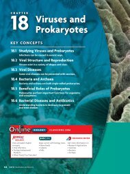
29.5Brain Function <strong>and</strong>ChemistryKEY CONCEPT Scientists study the functions <strong>and</strong> chemistry of the brain.MAIN IDEAS• New techniques improve our underst<strong>and</strong>ingof the brain.• Changes in brain chemistry can cause illness.• Drugs alter brain chemistry.VOCABULARYaddiction, p. 893desensitization, p. 893tolerance, p. 893sensitization, p. 893stimulant, p. 894depressant, p. 894Reviewneurotransmitter,action potentialConnect When you take medicine for a headache, the drug alters your brain’schemistry. Aspirin, for example, stops your brain from making certain chemicals.In small amounts, it is beneficial to your health. However, even nonprescriptiondrugs can cause permanent damage to your nervous system if taken incorrectly.CTMRIFIGURE 29.14 Modern technologiesuse computers <strong>and</strong> sensingdevices to observe the brain withoutthe need for surgery.MAIN IDEANew techniques improve our underst<strong>and</strong>ing ofthe brain.For many years, the only way scientists could study brain function was byobserving changes that occurred in people who had accidental brain injuriesor by dissecting the brains of people who had died. A live patient would haveto undergo surgery in order for scientists to learn about brain function.Today, scientists use imaging technologies such as CT, MRI, <strong>and</strong> PET scansto study the brain in living patients without the need for surgery. Thesemethods use x-rays, magnetic fields, or radioactive sensors <strong>and</strong> computerprograms to form images of the brain, as shown in FIGURE 29.14.PETComputerized tomography (CT) scans use x-rays to viewthe brain. Magnetic resonance imaging (MRI) uses magneticfields <strong>and</strong> radio waves. Both CT <strong>and</strong> MRI scans make imagesthat show the structure of the brain. These scans are used toexamine the brain’s physical condition.Positron emission tomography (PET) scans show whichareas of the brain are most active. During PET scans, a personis injected with radioactive glucose. Recall from Chapter 4 thatcells use glucose for energy. By measuring where the radioactive glucosecollects in the brain, scientists can see which areas of the brain are using themost energy. In the image shown above, the bright red <strong>and</strong> yellow areas are themost active, <strong>and</strong> the dark blue areas are the least active.Infer Why are modern technologies for studying the brain safer for the patientsbeing studied?Chapter 29: <strong>Nervous</strong> <strong>and</strong> <strong>Endocrine</strong> <strong>Systems</strong> 891
NormalConnectingCONCEPTSEnzymes In Chapter 2 you readthat enzymes are like locksbecause only certain shapedmolecules can fit into them.Neurotransmitters only affectcertain areas of the brain becausethey are like keys that can only fitinto certain neurons’ receptors.SchizophrenicFIGURE 29.15 These PET imagesshow the activity of a normal, aschizophrenic, <strong>and</strong> a depressedbrain. Blue <strong>and</strong> green areas havelow activity, <strong>and</strong> red areas are themost active.MAIN IDEAChanges in brain chemistry can cause illness.In Section 29.2, you learned that your nervous system cannot work withoutneurotransmitters. These chemicals regulate different functions in differentareas of the brain. Neurotransmitters are specific to some areas of the brainbecause, like hormones, they only affect cells that have specific receptors. Thefunction of neurotransmitters relates to the functions of the cells they stimulate.• Acetylcholine is involved in learning <strong>and</strong> memory.• Dopamine primarily influences your emotional behavior, but it also playssome role in stress <strong>and</strong> voluntary muscles.• Serotonin is mainly found in the hypothalamus <strong>and</strong> midbrain. It alsoinfluences mood, some muscle functions, <strong>and</strong> hunger.• Glutamate affects learning, memory, <strong>and</strong> brain development.• Gamma amino butyric acid (GABA) is found throughout the brain. Unlikeother neurotransmitters, when GABA binds to a neuron’s membrane, itprevents the neuron from generating an impulse.When your brain produces the correct amount of these neurotransmitters,homeostasis is maintained. If your body produces too much or too little of aneurotransmitter, the areas of your brainDepressedthat are targeted by that chemical will bemore or less active than normal. Becauseall of the areas of your brain worktogether, abnormal activity in one partof the brain can affect the whole brain<strong>and</strong> change the way you move, behave,<strong>and</strong> think. FIGURE 29.15 shows that thebrain activity of a healthy patient differsfrom that of patients with depression orschizophrenia, which are associated withchemical imbalances in the brain.Illnesses such as Parkinson’s disease <strong>and</strong> schizophrenia are linked toabnormal amounts of dopamine. Parkinson’s disease is caused by lowamounts of dopamine in certain areas of the brain. People with Parkinson’sdisease have difficulty controlling their movements, maintaining their balance,<strong>and</strong> starting movements. Many patients with Parkinson’s take drugs thatincrease the amount of dopamine in the brain. On the other h<strong>and</strong>, schizophreniasometimes occurs when a person has too much dopamine. Schizophreniais a mental disorder that causes hallucinations, irrational behavior,<strong>and</strong> illogical speech. It is treated with drugs that block dopamine receptors inthe brain.Depression is linked to low amounts of serotonin in parts of the brain.Depression causes extended periods of intense sadness, inability to sleep, <strong>and</strong>feelings of helplessness. One treatment for clinical depression uses drugs thatextend the time that serotonin remains active in nerve synapses.Summarize How do treatments for neurological illnesses alter brain chemistry?892 Unit 9: Human Biology