Amphipols: polymeric surfactants for membrane ... - Yale University
Amphipols: polymeric surfactants for membrane ... - Yale University
Amphipols: polymeric surfactants for membrane ... - Yale University

You also want an ePaper? Increase the reach of your titles
YUMPU automatically turns print PDFs into web optimized ePapers that Google loves.
CMLS, Cell. Mol. Life Sci. 60 (2003) 1559–15741420-682X/03/081559-16DOI 10.1007/s00018-003-3169-6© Birkhäuser Verlag, Basel, 2003CMLS Cellular and Molecular Life Sciences<strong>Amphipols</strong>: <strong>polymeric</strong> <strong>surfactants</strong> <strong>for</strong> <strong>membrane</strong>biology researchJ.-L. Popot a, *, E. A. Berry b ,D.Charvolin a ,C.Creuzenet c,† ,C.Ebel d ,D.M.Engelman e ,M.Flötenmeyer f ,F. Giusti a ,Y.Gohon a ,P.Hervé a ,Q.Hong g ,J.H.Lakey g ,K.Leonard f ,H.A.Shuman i ,P.Timmins i ,D. E. Warschawski a ,F.Zito a ,M.Zoonens a ,B.Pucci j and C. Tribet kaLaboratoire de Physico-Chimie Moléculaire des Membranes Biologiques, UMR 7099, CNRSand Université Paris-7, Institut de Biologie Physico-Chimique, CNRS IFR 550, 13 rue Pierre et Marie Curie,75005 Paris (France), Fax: +33 1 58 41 50 24, e-mail: jean-luc.popot@ibpc.frbLawrence Berkeley National Laboratory, 1 Cyclotron Road, Berkeley, Cali<strong>for</strong>nia 94720 (USA)cMassachusetts Institute of Technology, Department of Biology and Chemistry, 77 Massachusetts Ave., Cambridge,Massachusetts 02139 (USA)dLaboratoire de Biophysique Moléculaire, Institut de Biologie Structurale, UMR 5075 CEA-CNRS-UJF,41 rue Jules Horowitz, 38027 Grenoble Cedex 01 (France)eDepartment of Molecular Biophysics and Biochemistry, <strong>Yale</strong> <strong>University</strong>, 420 Bass Center, New Haven,Connecticut 06520-8114 (USA)fEuropean Molecular Biology Laboratory, Meyerhofstrasse 1, 69117 Heidelberg (Germany)gSchool of Cell and Molecular Biosciences, the Medical School, <strong>University</strong> of Newcastle upon Tyne,NE2 4HH (United Kingdom)hDepartment of Microbiology, Hammer Science Center, College of Physicians and Surgeons, Columbia <strong>University</strong>,701 West 168th Street, New York, New York 10032 (USA)iLarge Scale Structures Group, Institut Laue-Langevin, Avenue des Martyrs, B.P.156, 38042 Grenoble Cedex 9(France)jUniversité d’Avignon, Laboratoire de Chimie Bioorganique et des Systèmes Moléculaires Vectoriels,33 rue Louis Pasteur, 84000 Avignon (France)kLaboratoire de Physico-Chimie Macromoléculaire, CNRS UMR 7615, ESPCI, 10 rue Vauquelin,75005 Paris (France)Abstract. Membrane proteins classically are handled inaqueous solutions as complexes with detergents. The dissociatingcharacter of detergents, combined with the needto maintain an excess of them, frequently results in moreor less rapid inactivation of the protein under study. Overthe past few years, we have endeavored to develop a novelfamily of <strong>surfactants</strong>, dubbed amphipols (APs). APs areamphiphilic polymers that bind to the trans<strong>membrane</strong>surface of the protein in a noncovalent but, in the absenceof a competing surfactant, quasi-irreversible manner.Membrane proteins complexed by APs are in their nativestate, stable, and they remain water-soluble in the absenceof detergent or free APs. An update is presented of thecurrent knowledge about these compounds and theirdemonstrated or putative uses in <strong>membrane</strong> biology.Key words. Membrane proteins; <strong>surfactants</strong>; detergents; amphipols.* Corresponding author.†Present address: <strong>University</strong> of Western Ontario, Department ofMicrobiology and Immunology, Dental Sciences Building, London,Ontario, N6A 5C1 (Canada)
1560 J.-L. Popot et al. <strong>Amphipols</strong>Solubilization and inactivation of <strong>membrane</strong> proteinsby detergentsIntegral <strong>membrane</strong> proteins are involved in such essentialcell functions as bioenergy transduction, trans<strong>membrane</strong>transfer of nutrients and drugs, signal detectionand cell-to-cell communication, adhesion, tissue<strong>for</strong>mation and so on. They comprise about one-third ofthe proteins encoded in the genome of eukaryoticcells, and a majority of the targets of currently marketeddrugs. A detailed knowledge of their structure, functionand dysfunction is essential to a wide range of biomedicaland biotechnological applications. In vitro studiesof integral <strong>membrane</strong> proteins, however, are severelycomplicated by their insolubility in water, to the pointthat these proteins represent only ~ 0.2% of currentlyavailable high-resolution structures. The insolubility of<strong>membrane</strong> proteins is due to the highly hydrophobiccharacter of those protein surfaces that, in situ, are incontact with the <strong>membrane</strong> interior. Detergents classicallyare used to handle them in aqueous solutions. Detergentsare small amphiphilic molecules that mix wellwith lipids and, as a result, can partition into biological<strong>membrane</strong>s and, under favorable circumstances, solubilizethem. Thereby, <strong>membrane</strong> components becomedispersed into lipid/detergent mixed micelles and protein/detergentcomplexes, the latter usually retainingprotein-bound lipids. Detergent molecules adsorb cooperativelyonto the trans<strong>membrane</strong> surface of the proteinand <strong>for</strong>m a monolayer-like assembly, which is inrapid equilibrium with aqueous monomers and proteinfreemicelles [1, 2]. Membrane proteins aggregate and,in general, precipitate when the concentration of free detergentfalls under its critical micellar concentration(CMC), an indication that the detergent layer has comeapart and the protein trans<strong>membrane</strong> surface has becomeexposed to water. Physical stability of the solutions is ensuredby keeping the concentration of free detergentabove the CMC, but this frequently entails biochemicalinstability.The instability of detergent-solubilized proteins is a majorproblem in <strong>membrane</strong> biology. Its origin is seldomstudied in detail, but in most well-characterized instancesit results from the dissociating character of detergents:the very property that enables them to extractthe protein from its environment makes them prone to interferewith intra- or intermolecular interactions that stabilizeit. A frequent observation is that newly solubilizedproteins that are reasonably stable when kept in the solubilizationsupernatant inactivate when they are movedto a fresh detergent solution, as occurs in the course ofpurification. This phenomenon usually can be traced tostabilizing components, such as lipids or hydrophobiccofactors, partitioning into detergent micelles. Yet freemicelles need be present since detergents cannot be usedunder their CMC 1 . Some types of functional and/orstructural studies can be carried out after the protein hasbeen reinserted into artificial lipid vesicles, which veryoften stabilizes it. However, an isotropic solution is generallyneeded, be it <strong>for</strong> purification or <strong>for</strong> biophysicalstudies. Classical protective measures are to limit theamount of free micelles, to supplement them with lipidsor cofactors and/or to transfer the protein, once solubilized,to a less dissociating detergent. These countermeasures,however, are often imperfect, and they generatetheir own problems (see e.g. 3–5).This difficult situation has prompted the developmentboth of ‘protein-friendly’ detergents (see [1, 5]) and ofalternative media based, <strong>for</strong> instance, on the use of nondetergent<strong>surfactants</strong> and/or nonmicellar phases (reviewedin [5]). Over the past few years, we have beenexploring two original approaches. The first one relies ondiminishing the miscibility of the surfactant and lipids, sothat micelles will not act as sinks. The difficulty is <strong>for</strong> thesurfactant to remain able to prevent protein aggregation.It appears that these seemingly incompatible requirementscan be met by hemifluorinated <strong>surfactants</strong>, whichare designed to mix poorly with lipids while adsorbing efficientlyenough onto the trans<strong>membrane</strong> surface of theproteins [5–8]. The second concept is to do away altogetherwith free micelles. This implies such a high affinityof the surfactant <strong>for</strong> the surface of the protein that protein/surfactantcomplexes remain stable in the presenceof very low concentrations of free surfactant, whichmeans that the dissociation rate constant has to be vanishinglysmall. One way to achieve this result, as discussedin this review, is to engineer multi-point attachmentbetween a <strong>polymeric</strong> surfactant and the protein.Rationales <strong>for</strong> the design of amphipols<strong>Amphipols</strong> (APs) are amphipathic polymers specially designedto keep <strong>membrane</strong> proteins soluble [9]. The associationproperties of amphipathic polymers and theiradsorption onto hydrophobic surfaces have been extensivelystudied by physical chemists (see e.g. [10], andreferences therein). The molecules studied are often very1In principle, no micelles are present at the cmc of the detergent.Thus it is theoretically possible to work with <strong>membrane</strong> protein/detergentcomplexes in the absence of micelles by keepingthe free detergent concentration exactly at the cmc or slightly below(the chemical potential of detergent in a mixed detergent/protein/lipidcomplex has to be lower than in a pure detergent micelle).However, as the cmc is a function of temperature, ionicstrength and the presence of other solutes in the medium, the preciseconcentration to be used at each step would have to be determinedempirically. The practice has been rather to use detergentsat slightly above the cmc so that micelles are present and bufferthe detergent activity at the cmc, whatever that may be under aparticular set of experimental conditions.
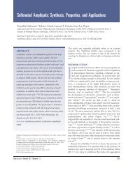
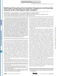
CMLS, Cell. Mol. Life Sci. Vol. 60, 2003 Multi-author Review Article 1561big, typically hundreds or thousands of kDa, with a longhydrophilic, highly charged backbone to which a fewvery hydrophobic (e.g. octadecyl) groups are attached.When bound to a mineral particle, or a droplet of oil, theymake it hydrophilic and water soluble. Because of thestrong electrostatic repulsion between polymer-coatedparticles, very high loads can be obtained withoutprecipitation. In industry, such polymers are used <strong>for</strong> instancein the manufacturing of inks or paints.Our rationales <strong>for</strong> the design of amphipathic polymersadapted to <strong>membrane</strong> protein studies were the following[9]:1) The basic idea was that they should bind to the surfaceof the protein by multiple attachment points, so as todiminish the dissociation rate and render the associationquasi-irreversible; this implied that the moleculesbe big enough and soluble enough to carry many hydrophobicchains.2) APs had to be much smaller than the amphipathic polymersordinarily used by physical chemists, with a sizecommensurate with that of protein trans<strong>membrane</strong> regions(typically a few nanometers).3) They ought to be highly flexible, so as to be able toadapt to the very small radius of curvature of proteinsurfaces and to their irregularities; in physical terms,their persistence length, which is the distance pastwhich a polymer chain keeps no memory of its initialdirection, had to be sufficiently short.4) To favor <strong>for</strong>mation of a thickly packed layer around thetrans<strong>membrane</strong> region of the protein, rather than afluffy one, we used closely spaced hydrophobicgroups, separated by short loops; <strong>for</strong> the polymer to remainsufficiently soluble, the hydrophobic groupswould be kept short.5) From the point of view of organic chemistry, it was desirableto keep the synthesis of APs relatively simple;applications to structural biology, in particular, whichmay consume hundreds if not thousands of milligramsof material, would be jeopardized by a complicatedand costly synthesis; amphipols of course also ought tobe chemically stable <strong>for</strong> extended periods (weeks ormonths) in biological buffers.Amphipol structurePolyacrylate-based APsThe concept of APs was initially implemented usinghydrophobically derivatized polyacrylates [9] (fig. 1, 1).At variance with, <strong>for</strong> instance, a polypeptide chain (whichotherwise would provide an attractive way to preparewell-defined APs), a polyacrylate backbone does notcontain any conjugated bonds, and it does not <strong>for</strong>m hydrogenbonds with itself. This makes it highly flexible.The extension and flexibility of polymer chains are oftencharacterized by two complementary parameters: (i) theaverage radius of gyration, ·R g Ò, and (ii) the persistencelength, L p (length of a segment along the chain that wouldrequire an energy of ~1 kT to be curved by a radius equalto its own length). The size and persistence length ofpolyelectrolytes such as polyacrylate-based APs dependFigure 1. Structure of amphipols. 1, polyacrylate-based, anionic AP; <strong>for</strong> amphipol A8-35, with which most of the results discussed in thepresent review have been obtained, x g 0.25, y g 0.40 and z g 0.35; <strong>for</strong> an average chain, this corresponds respectively to ~17, ~28 and ~25monomers, randomly distributed [9]. 2, PMAL-B-100, an AP that is zwitterionic at neutral and basic pH and cationic at acidic pH [15]. 3,a nonionic AP obtained by random cotelomerization of hydrophilic and hydrophobic monomers derived from THAM [19].
1562 J.-L. Popot et al. <strong>Amphipols</strong>on intrachain hydrophobic attraction, electrostatic repulsion,and there<strong>for</strong>e on charge density and ionic strength.A lower ionic strength is expected to result in significantswelling of the polymer coil, due both to electrostaticstiffening (longer persistence length due to the repulsionbetween successive monomers) and to increasing excludedvolume around monomers (repulsion betweenmonomers that can be distant along the chain but becomeclose in space in the coiled polymer).Experimental studies of the persistence length of highlycharged polyacrylates are based on viscometry or lightscatteringmeasurements of chain extension [11, 12]. Itappears difficult in practice to clearly distinguish betweenthe contributions of electrostatic stiffening and excludedvolume to the observed effects of ionic strength. Ifthese effects are entirely attributed to stiffening, an apparentvalue of the persistence length, L¢ p , can be determined.Because this analysis neglects excluded volume effects,L¢ p exceeds the true value of L p . L¢ p is usually writtenas:L¢ p = L¢ i + L e ¢where L¢ i is the apparent intrinsic persistence length (i.e.the asymptotic value at infinite ionic strength) and L e ¢ theelectrostatic component. L¢ i characterizes the rigidity ofthe chain under an imaginary uncharged <strong>for</strong>m and includesrotameric, steric and secondary structure features.For polyacrylates, estimates of L¢ i vary from 1.4 nm [11]to 2.7 nm [12], and L e ¢ has been found to vary as the reciprocalsquare root of ionic strength. For copolymers ofacrylamide and sodium acrylate, <strong>for</strong> instance, the followingempirical relation is obtained [12]:L¢ p (nm) = L¢ i + 35 x/C0.5swith C s the salt concentration (in mM) and x=l b /b (Manningcharge parameter), where b is the intercharge spacingalong the chain and l b the Bjerrum length (l b =q 2 /e · k B· T g 0.72 nm at 25°C in water). For highlycharged polyelectrolytes, b is taken equal to l b and x g 1[12]. For derivatized polyacrylates in which two out ofthree carboxylates have been neutralized (i.e., purelyhydrophilic polymers with the same charge density asamphipol A8-35), and in 100 mM salt, which is at thelower end of salt concentrations generally used in <strong>membrane</strong>biochemistry, L¢ e estimates are close to 3.5 nm [12]or 3.9 nm [11].Estimates of L¢ e obtained by the above approach are larger(by a factor of ~2) than the theoretical values of L e calculatedby widely accepted theories. In addition to the overestimationentailed, as mentioned above, by neglectingexcluded volume effects, one must note that small errorsin estimates of the actual charge density, length polydispersityand the ‘end-effects’ characteristic of short polymersall significantly affect the variations of apparentpersistence length, as shown by Monte Carlo simulations[13]. The above estimates there<strong>for</strong>e must be considered asgiving a (fair) order of magnitude of the persistencelength of polyacrylate chains and the way it depends onionic strength, rather than rigorous determination. Altogether,the persistence length of polyacrylates in biologicalsolutions appears likely to be small enough not to preventa close association between the derivatized polymerand <strong>membrane</strong> proteins, which is borne out by experiment(see below).In most of our own work, the average length of the chainhas been ~70 monomer units (see below), i.e. ~19 nmwhen fully extended. This corresponds to an average molecularmass of ~5 kDa <strong>for</strong> the underivatized polymer,~8 kDa after derivation [9]. Similar molecules have beenused (under the name OAPA-20) by C. R. Sanders and coworkers[14, 15]. Less extensive experiments have beencarried out with longer chains (~300 monomers [9]) andwith shorter ones (~25 monomers [16]). The solubility ofpolyacrylate-based APs can be improved by partial graftingwith sugar groups [C. Prata and C. T., unpublisheddata].It is difficult to synthesize polyacrylate molecules of perfectlydefined length. Our starting material usually has apolydispersity of ~2 (polydispersity is defined as the ratioof the weight average molecular weight over thenumber average moledular weight, i.e. (∑ n i M i2 /∑ n i M i )/(∑ n i M i /n i ), where n i stands <strong>for</strong> the numer of molecules ofmolecular mass M i in a given volume of solution). Uponsize exclusion chromatography (SEC) analysis, the widthof the peak at half-height covers the range from 2 to16 kDa. One way to improve APs certainly lies in limitingthis considerable polydispersity. This can be achievedeither upon synthesis, e.g. by resorting to living radicaltelomerization or to ionic polymerization, or a posterioriby fractionation. Our experience indicates that both fractionationand telomerization yield final products withtrapping properties similar to those of more polydispersepreparations, but a detailed comparison remains to bedone. An interesting question that remains open to thisday is the extent to which <strong>membrane</strong> proteins may select,from the mixture of AP molecules they are exposed to,those they have the highest affinity <strong>for</strong>, in which case itcould turn out to be counterproductive to aim <strong>for</strong> perfectlydefined APs.Amphipathy was conferred by derivation with fattyamines. In most of our experiments, this was done withoctylamine, but dodecylamine was also tested, less extensively,with satisfying results [17, and unpublished data].The degree of derivation is a critical parameter. APs thatsucceed in maintaining <strong>membrane</strong> proteins soluble haveto be highly derivatized. Our own experiments have generallyaimed <strong>for</strong> 25% derivation with octylamine. Toohigh a level of derivation yields insoluble polymers, whiletoo low levels lead to poor results when it comes to trapping<strong>membrane</strong> proteins. The charge density along the
CMLS, Cell. Mol. Life Sci. Vol. 60, 2003 Multi-author Review Article 1563polymer can be lowered by amidation of a further 40% ofthe carboxylates with isopropylamide. This does notseem to have much of an effect on the ability of the polymersto keep <strong>membrane</strong> proteins in solution [9], but thereare some indications that a lower charge density may favorthe biochemical stability of the trapped proteins [9,18] (see below). The two APs with which most experimentshave been carried out to date (fig. 1, 1) have finalmolecular masses around 8 kDa, 25% derivation withoctylamine, and, depending on whether they have beenfurther derivatized with isopropylamine, either 75 or35% free carboxylates. They are dubbed A8-75 andA8-35, respectively, where ‘A’ stands <strong>for</strong> ‘acrylate’, ‘8’<strong>for</strong> the average molecular mass, in kDa, and the last figure<strong>for</strong> the percentage of free carboxylates. On average, asingle molecule of A8-35 contains ~70 acrylate units,~17 of which carry an octyl chain, ~25 a free carboxylateand the rest an isopropyl. The various types of units arerandomly distributed. In this nomenclature, the OAPA-20molecules used by Sanders and co-workers [14] would becalled A8–80.Strong-acid, nonionic and zwitterionic APsPolyacrylate-based APs, which hitherto have been by farthe most extensively studied, have the two characteristicsof carrying a net charge and of being weak acids. The firstfeature prevents the use of <strong>membrane</strong> protein/AP complexesin some experimental situations, such as ion-exchangechromatography or isoelectrofocusing, and islikely to be a hindrance in others, such as crystallization.The second one limits their use to neutral or basic pH,since protonation of the carboxylates induces aggregationand precipitation. When monodispersity of the complexesis essential, as in crystallization attempts, it is even advisablenot to work below pH 7.5 (see below). This is a problemalso <strong>for</strong> nuclear magnetic resonance (NMR) applications,where it is useful to limit amide proton exchange bylowering the pH, or when working with proteins that aremore stable at acidic pH. To alleviate these constraints,other types of APs have been experimented with.The weak-acid character can be avoided by substitutingsulfonate groups <strong>for</strong> carboxylates. The properties of polysulfonatedAPs are currently under study [F. Giusti and M.Zoonens, unpublished data]. The development of overallneutral APs is in a more advanced stage. We have shownthat nonionic amphipathic polymers derived from tris(hydroxymethyl)-acrylamidomethane(THAM) (fig. 1, 3) canbe used to stabilize <strong>membrane</strong> proteins in aqueous solutionsin the same manner as polyacrylate-derived APs do[19]. The study of the complexes thus obtained is inprogress. Others have successfully used APs that are zwitterionicat neutral and basic pH (fig. 1, 2) [15]. Preliminaryexperiments using hydrophobically derivatized pullulane(a polysaccharide) have also been reported [20].AP synthesis and purification, and their importance<strong>for</strong> biochemical applicationsA very important point needs to be stressed. Membranebiochemists are used to detergents. Detergents have theirproblems (many of them), but they usually equilibraterapidly between phases and compartments. To quote afigure, the residency time of an octyl-b-D-glucopyranoside(C 8 -G) molecule in a micelle is in the microsecondtime range [21]. A biochemist having to deal with a<strong>membrane</strong> protein/AP preparation will instinctively thinkthat any slow process in the system has to do with the protein.This is not always true. Polymers, because they arelong molecules, do not necessarily rapidly find a path totheir free energy minimum (see e.g., <strong>for</strong> adsorption phenomena,[22]). They present a hysteresis of their own andcan become locked into undesirable states, e.g. aggregatedones. We have found out (the hard way) that twochemically identical preparations of APs, or differentsamples from the same batch after having been handleddifferently, can exhibit widely different aggregation properties,which in turn reflect on those of the complexesthey <strong>for</strong>m with <strong>membrane</strong> proteins [18]. This problem,which is briefly discussed in the next section and will befurther documented in <strong>for</strong>thcoming articles, should bekept in mind by anyone working with APs, and appropriatecontrols carried out (see next section).Solution properties of amphipolsDetailed studies of the solution properties of pure APs areavailable only <strong>for</strong> A8-35 [18]. A8-35 molecules auto-organizeinto small micelles, ~30 kDa in mass, with a relativelynarrow size distribution: upon SEC, ‘good’ (see below)preparations of A8-35 exhibit a half-height peakwidth only slightly larger than that of a soluble protein ofcomparable mass, horseradish peroxydase [18]. A8-35particles have been studied by SEC, small angle X-rayand neutron scattering (SAXS and SANS), quasi-elasticlight scattering (QELS) and analytical ultracentrifugation(AUC) [18, and unpublished data]. The data are compatiblewith the existence of more or less spherical objectscontaining ~1.5–2 g of bound water per gram of AP.SANS measurements suggest that the particles do notfeature a well-defined hydrophobic core. This may seemsurprising. It becomes much less so, however, if one reflectsthat the distance that separates two alkyl chainsalong the polymer (statistically, eight C–C bonds, i.e.~1 nm when the chain is fully extended) is similar to thelength of an octyl chain, and significantly shorter than theStokes radius of a particle (~3 nm according to SEC andSANS measurements). This prevents hydrophilic andhydrophobic groups from segregating completely. Becausethe size of the particles <strong>for</strong>med by pure A8-35 is notvery different from that of small proteins, and because the
1564 J.-L. Popot et al. <strong>Amphipols</strong>density of this polymer is relatively high (1.16 g · l –1 ; [C.Ebel, unpublished data) and not very different from thatof <strong>membrane</strong> proteins (typically ~1.33 g · l –1 ), it is notstraight<strong>for</strong>ward to totally separate protein/A8-35 complexesfrom excess A8-35 when trapping small proteinssuch as the trans<strong>membrane</strong> domain of outer <strong>membrane</strong>protein A (tOmpA) or bacteriorhodopsin (BR), unlessone resorts to affinity chromatography [18, and M. Zoonensand F. Zito, unpublished data].Polydispersity is a major concern when preparing APs. Itfalls, roughly, in two categories. First, A8-35 preparationscontain a few percents (in mass) of very large particles(MDa range). These are not a concern in most experimentsbecause they are a small minority and do not associatewith <strong>membrane</strong> proteins [18]. The trouble startswith particles that are only slightly larger than the minimalsize. These particles are found in many AP preparations,and upon protein trapping, they do associate withproteins and yield polydisperse protein/AP complexes.This may not be too much of a hindrance <strong>for</strong> some applications,such as when exploring the environment of <strong>membrane</strong>proteins or their spectroscopic or ligand-bindingproperties, but it is a major hurdle in others, in particularcrystallography or solution-state NMR. Extensive investigationshave led us to the conclusion that polydispersityis not an intrinsic property of some batches of APsthat would be chemically different. It actually depends onthe way the polymers have been purified and handled.Polydispersity, <strong>for</strong> instance, develops over time if anaqueous solution of A8-35 is kept <strong>for</strong> several days at pH≤ 7 [C. Prata and C. Tribet, unpublished data). More detailswill be given elsewhere. From a practical point ofview, suffice it to say that it is advisable, whenevermonodispersity is a concern, to check, e.g. by SEC orAUC, on the size and dispersity of the particles <strong>for</strong>med bythe pure polymer under the conditions to be used, and notto let the pH of the solutions fall below ~7.5.If the complexity of the solution behavior of A8-35 is anyindication, any rational development of APs will dependon rather extensive studies of the physicochemical propertiesof each new type of molecule. This is a harsh constraintbecause these studies are rather laborious and, tothe biochemist, unrewarding. Short-cutting them, however,will most likely lead to unsatisfying if not irreproducibleresults. As of now, much less in<strong>for</strong>mation is availableabout APs other than A8-35. Preliminary observationsindicate that upon SEC nonionic, THAM-derivedAPs behave as particles whose size is commensurate withA8-35 particles and PMAL-B-100 as somewhat largerones [D. Charvolin, Y. Gohon and F. Giusti, unpublisheddata].Effects of amphipols on biological and artificial<strong>membrane</strong>sAPs are not detergents, or, more exactly, they are onlyvery weak ones. Biological <strong>membrane</strong>s as a rule are notsolubilized by soaking them in a solution of APs [9, 17].This basic observation, however, must be qualified. First,APs do interact with <strong>membrane</strong>s. They permeabilize thesarcoplasmic <strong>membrane</strong> to calcium ions [17] and <strong>for</strong>mpores in black lipid <strong>membrane</strong>s [A. Ghazi, personal communication].Giant (tens of micrometers) egg phosphatidylcholineunilamellar vesicles exposed to A8-35 (called5-25C8-40C3 in that paper) first produce filaments andbuds be<strong>for</strong>e breaking up into small vesicles, whereas inthe presence of A8-75 (called 5-25C8), they take uppolyedral shapes and show evidence <strong>for</strong> lateral segregationof their components [23]. Upon extended incubation(hours), A8-35 is able to disperse large (tens of nanometers)unilamellar dipalmitoylphosphatidylcholine/dipalmitoylphosphatidicacid vesicles into large (~10 nm in radius)mixed micelles [24]. A8-35 also dissolves lipidmonolayers at the air-water interface [M. Flötenmeyerand K. Leonard, unpublished data), but not monolayersof fluorinated lipids [J. Dietrich and C. Vénien-Bryan,personal communication]. Certain proteins, such as themaltose transporter from Escherichia coli [M. Zoonensand H. A. Shuman, unpublished data] or the human insulinreceptor overexpressed in Chinese hamster ovary cells[G. Crémel, personal communication], can be directlyextracted by A8-35. Hydrophobically modified pullulanehas been reported to extract proteins from E. coli andPseudomonas fluorescens outer <strong>membrane</strong>s [20].Because APs are not strong detergents, they can potentiallybe used to deliver <strong>membrane</strong> proteins to pre<strong>for</strong>med<strong>membrane</strong>s. This has been best documented in a study ofthe delivery of amphipol-trapped diacylglycerol kinase(DAGK) to lipid vesicles [14]. Fibroblasts and mouse embryonicstem cells survive prolonged exposure (days) toconcentrations of APs up to 0.05–0.1 g · l –1 , i.e. higherthan those they would need be exposed to in a protein deliveryexperiment [J. Barra, personal communication].This is an interesting novel experimental situation, becauseunlike protein/detergent complexes, which are destabilizedby dilution below the CMC of the detergent, a protein/APcomplex injected into a cell culture medium orthe buffer that bathes a black film will remain physicallystable until it interacts with the target <strong>membrane</strong>. Uponinteraction, it is likely that the polymer associates withand diffuses over the surface of the <strong>membrane</strong>, favoringthe insertion of the surfactant-depleted protein. Needlessto say, the insertion process must subject proteins tostrong distorting <strong>for</strong>ces, and only the toughest of themcan be expected to survive this step and remain functional.Nevertheless, APs clearly open an interesting newavenue to reconstitution or cell culture experiments.
CMLS, Cell. Mol. Life Sci. Vol. 60, 2003 Multi-author Review Article 1565In keeping with the low toxicity of APs in cell culture,pure APs or protein/AP complexes can be injected intomice (to the level of 50 mg per injection) without any visiblepathogenic effect. According to ELISA tests and immunoreplicae,antibodies are raised against the trappedproteins, but not against APs [C. Leclerc, Y. Pierre and Y.Gohon, unpublished data).Keeping <strong>membrane</strong> proteins soluble with amphipolsIn a typical trapping experiment, a purified protein in detergentsolution is supplemented with an amount of AP(usually in the range of 1–10 g per gram of protein) thatlargely exceeds the binding capacity of the protein (typically0.2–1 g/g; see below, table 2). The concentration ofthe detergent is then lowered below its CMC. This can beachieved in a variety of ways. Often, the solution is firstdiluted below the CMC, after which detergent monomerscan be eliminated by adsorption onto Bio-Beads, by dialysisor by running the protein/AP complexes onto a sucrosegradient or a molecular sieve [9, 17–19, 25–27].Depending on the protocol chosen and the size of the protein,excess AP may or not be more or less efficiently removedat that stage, which may affect the behavior of theprotein/AP particles (see below). In some cases, Bio-Beads have been added directly to the ternary mixturewithout prior dilution below the CMC [17, 18, and D.Charvolin, Y. Gohon and M. Zoonens, unpublished data].It is also possible to attach protein/detergent complexesonto an affinity column [14, 15, and M. Zoonens and H.A. Shuman, unpublished data] or a BIAcore chip [28] andcarry out the exchange of surfactant on the immobilizedprotein (fig. 2). The dispersity of the final particles maydepend on the protocol chosen [17].An important observation is that APs bind to proteinseven in the presence of detergent micelles. The <strong>for</strong>mationof such ternary complexes is essential to the success oftrapping [25]. Although its understanding is likely to becritical to that of the properties of the final complexes, thesuccession of events that lead from a protein/detergentcomplex to a protein/AP one has not yet been studied indetail. It is not known, <strong>for</strong> instance, what the kinetics of<strong>for</strong>mation of the ternary complexes are, nor whether theircomposition evolves over extended incubation, nor has itbeen determined whether additional binding of APs takesplace upon lowering the detergent concentration below itsCMC nor during its final elimination.Once <strong>for</strong>med, and in the absence of competing <strong>surfactants</strong>,the association between APs and <strong>membrane</strong> proteins is extremelystable. Experiments in which complexes of<strong>membrane</strong> proteins and radioactive APs have been centrifuged<strong>for</strong> several hours onto gradients containing <strong>surfactants</strong>or not have shown: (i) the absence of a measurableloss of radioactive AP when the gradient contains neitherFigure 2. Dissociation rates of <strong>surfactants</strong> from immobilizedOmpF porin. An OmpF mutant (D183C) with a single cysteine in aperiplasmic turn was labeled with biotin maleimide and immobilized,at the level of 150 resonance units (RU), on the surface of aBIAcore SA chip bearing covalently attached streptavidin. Thebuffer contained 10 g · l –1 octylpolyoxyethylene (C 8 -POE), 10 mMHEPES, 150 mM NaCl and 3.4 mM EDTA, pH 7.4. The chip waswashed with a 10 g · l –1 solution (or, in the case of amphipols, a1g·l –1 solution) of either amphipol A8-35, dodecyl-b-D-glucopyranoside(C 12 -G), C 8 -G or C 8 -POE, and the exchange of surfactant followedby surface plasmon resonance (SPR) at a flow rate of5 ml·min –1 . Once a stable baseline had been achieved, i.e. after30–60 min (zero RU value on this graph), the solution was replacedwith surfactant-free buffer (t = 0) and the dissociation of surfactantmonitored by SPR. The signal from a blank surface bearing onlystreptavidin was subtracted from the rough data, so that the datashown represent the evolution over time of the amount of surfactantthat is actually bound to OmpF. (Q. Hong, J.-L. Popot and J. Lakey,unpublished data).AP nor detergent; (ii) partial AP exchange in the presenceof unlabeled AP and (iii) desorption of most of the boundAP if the gradient contains detergent above its CMC [25].Extensive washing of OmpF/AP complexes adsorbed ontoBIAcore chips with surfactant-free solutions revealed noAP desorption either over tens of minutes (fig. 2) (Q.Hong, J.-L. Popot and J. Lakey, unpublished data). Whethercomplexes that are washed <strong>for</strong> hours or days with surfactant-freebuffer release some of the bound APs remains,however, an open question. A limited amount of desorption,difficult to detect with the approaches used thus far,could indeed account <strong>for</strong> the observed tendency of cytochromebc1/A8-35, BR/A8-35 and tOmpA/A8-35 complexesto <strong>for</strong>m small, soluble aggregates following extensiveremoval of free AP [D. Charvolin, Y. Gohon and M.Zoonens, unpublished data, and 18]. Similar observationshave been made with the Ca 2+ -ATPase, some of the latterdata suggesting that this process may be modulated by thepresence or absence of bound lipids [17]. It is, however,difficult, in the case of the Ca 2+ -ATPase, to sort out aggregationphenomena that are due to the physical properties ofthe particles from those that may be subsequent to proteindenaturation. The extensive removal of APs from protein/APcomplexes upon exposure to detergents above theirCMC [25] offers an alternative route to reconstitution of
1566 J.-L. Popot et al. <strong>Amphipols</strong>TableI.A list of integral <strong>membrane</strong> proteins whose complexes with amphipols have been studied in some detail (others are mentioned inthe text).Protein name Source Function Subunits Overall Secondary APs Properties of complexes Referencesmass (kDa) structureSoluble Native FunctionalCytochrome b 6 f Chlamydomonas redox pump 2 ¥ 8 228 a A, N + + (+) 9,reinhardtii 19, 25, 26Bacterio- Halobacterium light-driven 1 27 a A, N + + 9, 18, 19rhodopsin salinarum H + pumpOmpF porin Escherichia pore 3 ¥ 1 102 b A + + + 9, 25, 28 acoliPhotosynthetic Rhodobacter light-driven 3 96 a A + + 9reaction center sphaeroides redox pumpRhodopsin Bos taurus G-protein- 1 39 a A + + ± UcoupledreceptortGpA [E.coli] ? 2¥ 1 ~8 a A + + 16, UCa 2+ -ATPase Oryctolagus calcium 1 110 a A + + ± 17cunniculus pumpComplex I Neurospora redox ~35 1120 (a) A + + 30 bcrassapumpAcetylcholine Torpedo gated 2 ¥ 5 535 (a) A + + + 27receptor marmorata channeltOmpA [E.coli] ? 1 19 b A, N + + 34, UMaltose E.coli ABC pump 4 150 (a) A + + ± UtransporterCytochrome bc 1 Bos taurus redox 2 ¥ 11 490 a A, N + + UpumpDiacylglycerol [E.coli] kinase 3 ¥ 1 40 (a) A, Z + + ±, + 14, 15kinasePhotosystem II Pisum sativum light-driven 5 103 a A + + + Ureaction centerredox pumpPhotosystem II Thermosyne- light-driven 2 ¥ 17 550 a A, N + + + Ureaction center chococcus redox pumpelongatusaQ. Hong, J.-L. Popot and J. Lakey, unpublished data.bM. Flötenmeyer et al., unpublished data.Source of the protein: brackets indicate heterologous or ectotopic expression; tGpA: glycophorin A trans<strong>membrane</strong> region ; tOmpA: outer<strong>membrane</strong> protein A trans<strong>membrane</strong> region. Secondary structure: a-helix bundles (a) or b-barrels (b); parentheses indicate that no highresolutionstructure is available. APs: ‘A’, anionic (most often A8-35); ‘N’, non-ionic, THAM-based; ‘Z’, zwitterionic. Native state: ‘+’ indicatesthat there are strong indications (spectroscopic or others) that the protein or complex is in its native state; Functional: ‘+’ means thatat least one important function, such as ligand binding, has been tested and found native-like; ‘±’ means that the AP-trapped protein wasonly partially functional (low activity, or not all tested functions present); ‘(+)’ refers to the fact that cytochrome b 6 f was tested in a mediumcontaining detergent above its CMC (see text). References: the names of the authors of unpublished work (‘U’) are given in the text.AP-trapped proteins that would not resist direct integrationinto pre<strong>for</strong>med vesicles, namely that of exchanging APs<strong>for</strong> detergent be<strong>for</strong>e per<strong>for</strong>ming a classical reconstitutionfrom detergent solution.No data are available concerning the dynamics of proteinboundAPs. It is reasonable to think that even though exchangewith the solution may be very slow, adsorbedmolecules are constantly moving and wriggling, with individualalkyl chains temporarily leaving the surface and rebindingto it, or crawling over the protein or protein-boundlipids. By analogy with the dynamics of detergent moleculesin micelles [21], one may speculate that the residencytime of most AP octyl chains at a given point of theprotein surface is likely to be in the sub-microsecond range.Because of the dense crisscrossing of the surface by thepolymers (statistically, anchoring points are about 1 nmapart), the question may be asked whether AP dynamicshas an influence on the kinetics of protein con<strong>for</strong>mationaltransitions that involve large-scale (nanometer) rearrangementsof the trans<strong>membrane</strong> surface (see below).The ability of APs to keep <strong>membrane</strong> proteins soluble inthe absence of detergent micelles does not depend on any
CMLS, Cell. Mol. Life Sci. Vol. 60, 2003 Multi-author Review Article 1567Table 2. Composition of <strong>membrane</strong> protein/amphipol complexes, and a comparison with that of <strong>membrane</strong> protein/dodecylmaltoside ones.Protein MW <strong>Amphipols</strong> DodecylmaltosidekDa Bound C 12 -MAmphipol type salt Bound amphipols Bound lipidsmM # of C 12 g/g# of C 8 kDa g/g(b 6 f ) 2 228 A8-75 20 103 (a) 46 0.22 + (b) 260 (c) 0.65RC 102 A8-75 18 83 (a) 41 0.41 ? 148 (d) 0.90(OmpF) 3 96 A8-75 30 83 (a) 41 0.43 ?(AChR) 2 535 A8-35 120 330 (e) 150 0.28 + (e)BR 27 A8-35 120 110–120 (f) 50–55 1.8–2 + (f) 208 (d) 4.1tOmpA 19 A8-35 120 31 (g) 14 0.75 – (g)(bc 1 ) 2 490 A8-35 120 110–142 (h) 49–63 0.10–0.13 ?Data refer to the dimeric cytochrome b 6 f complex from C. reinhardtii, the photosynthetic reaction center from R. sphaeroides, trimericOmpF from E. coli, dimeric acetylcholine receptor from T. marmorata, monomeric bacteriorhodopsin from H. salinarum and themonomeric trans<strong>membrane</strong> domain of OmpA from E. coli. The amount of bound AP per particle was determined using radioactive amphipols(all proteins) and by SANS and AUC (BR, tOmpA). There is evidence that the amount of bound AP may depend on the ionic strengthat which trapping was per<strong>for</strong>med [25] and on the way the particles were separated from free APs [18]. AP binding is expressed in kDa perparticle, as the mass of AP per mass of protein and as the number of AP octyl chains per particle. The presence or absence of lipids wasascertained using radioactive lipids (b 6 f ), chemical analysis (AChR, BR, tOmpA) and biophysical measurements (BR, tOmpA). The bindingof C 12 -M, which has a similar mass per alkyl chain, is shown <strong>for</strong> comparison. Data are from the following sources: a [25]; b [26]; c [3];d [29]; e [27]; f [18]; g [34 and unpublished data]; h [D. Charvolin, unpublished data].protein feature. As shown in table 1, proteins that havebeen shown to remain soluble under these conditions encompassthe whole gamut of size, complexity, secondarystructure, trans<strong>membrane</strong> mass distribution, subcellularlocalization and function.It appears that with due caution, APs can be used to exploreinteractions between <strong>membrane</strong> proteins. Trappingwith A8-35 of a mixture of BR monomers and oligomersin octylthioglucoside (C 8 -TG) solution yielded protein/APcomplexes whose polydispersity reflected that ofthe original mixture [18]. Trapping of a mixture ofmonomers and dimers of the nicotinic acetylcholine receptor(nAChR) in CHAPS resulted in a mixture ofmonomeric and dimeric nAChR/A8-35 complexes [27],while trapping of purified monomeric and dimeric <strong>for</strong>msof cytochrome b 6 f yielded AP-trapped monomers anddimers [26]. When carried out under appropriate conditions,trapping of <strong>membrane</strong> proteins with APs there<strong>for</strong>eseems to ‘freeze’the association <strong>for</strong>ms they have in detergentsolution. In keeping with this conclusion, trappingwith either of the four original APs [9] of a supernatant ofthylakoid <strong>membrane</strong>s from Chlamydomonas reinhardtiisolubilized with the detergent Hecameg yielded a mixtureof protein/AP complexes that could be resolved by sucrosegradient fractionation into the complexes classicallyobserved in Hecameg solution [C. Tribet and J.-L. Popot,unpublished data]. As discussed below, trapping of detergent-sensitivecomplexes is probably one of the most interestingapplications APs can be put to.Composition, structure and solution properties of<strong>membrane</strong> protein/amphipol complexesCurrently available quantitative data about the compositionof protein/AP complexes are summarized in table 2 and illustratedin figure 3 by models showing what a large (cytochromebc 1 ) and a small (tOmpA) protein trapped by APsmay look like. Two points are particularly worth noting:(i) a given protein always binds much less AP (in mass)than detergent, the difference being, in some cases, as highas a factor of 3; (ii) lipids, whenever associated with theprotein in detergent solution, become trapped, <strong>for</strong>ming aternary protein/lipid/AP complex. The lower mass ofbound AP as compared with detergent has an importantimplication, namely that it translates into a smaller numberof alkyl chains (table 2). Cytochrome b 6 f, <strong>for</strong> instance,binds ~260 molecules of dodecylmaltoside (C 12 -M) perdimer [3] and only ~100 A8-75 alkyl chains [25]; a photosyntheticreaction center, ~150 molecules of C 12 -M [29]vs. ~80 A8-75 octyl chains [25]; BR and its associatedlipids, ~280 molecules of C 8 -TG [18] against ~120 A8-35octyl chains [18]. This points to a different organization ofthe alkyl chains of the surfactant with respect to the hydrophobicsurface of the protein and, possibly, a higher accessibilityof the latter to water. As regards the trapping oflipids, it can have profound effects on the stability andfunctionality of the protein (see below). It provides also,potentially, a novel approach to investigating specific protein/lipidinteractions: ternary complexes <strong>for</strong>med soon aftersolubilization, either by trapping from a crude detergentsupernatant or, in favorable cases, upon direct solubilizationby amphipols, can be purified in the absence of detergentand their composition analyzed.
1568 J.-L. Popot et al. <strong>Amphipols</strong>The one series of experiments that has been carried outusing radioactive detergent indicated that following separationof detergent monomers and AP-trapped cytochromeb 6 f on surfactant-free sucrose gradients, detergentremoval was extensive: from ~260 in detergent solution[3], the amount of bound [ 14 C]C 12 -M fell below thedetection threshold of ~10 molecules per b 6 f dimer [25].On the other hand, there is every reason to think that,whenever present and even well below their CMC, detergentsassociate with the AP layer, which can affect thefunctionality and stability of the protein (see below).SANS analysis of BR/A8-35 particles indicates that asshould be anticipated, the polymer lies in a peripheral positionas compared with the protein [18]. Although themass of AP that binds to a given protein is less than thatof detergent, the polymer layer is likely to be about asthick as a conventional detergent layer because of itshigher degree of hydration. Indeed, BR-adsorbed A8-35still binds some 1.3–1.5 g of water per gram of polymer[18], which nearly makes up <strong>for</strong> the lower mass of boundsurfactant. Data obtained by surface <strong>for</strong>ce measurementson A8-35 layers adsorbed onto macroscopic hydrophobicsurfaces [A. Kumpelainen et al., unpublished data), electronmicroscopy of AP/Complex I complexes [30, M.Flötenmeyer et al., unpublished data] and the study bySEC, SANS and AUC of BR/A8-35 particles [18] all suggestthat in the presence of salt (100 mM NaCl) most ofthe AP mass lies in a compact, 1–2 nm-thick layer, whichis compatible with the volume occupied by hydrated AP(fig. 3). Surface <strong>for</strong>ce measurements indicate that as thesalt concentration is lowered, the AP layer swells and<strong>for</strong>ms a ‘pseudo-brush’ [A. Kumpelainen et al., unpublisheddata]. As a rule, protein/AP complexes featureslightly lower sedimentation coefficients and slightlylarger apparent Stokes radii upon SEC than the same proteinin detergent solution (see e.g. [17, 18, 27]).Protein/AP complexes repulse each other electrostaticallyat high concentration and low ionic strength (fig. 4,left). At higher ionic strength, the repulsion disappears, tobe replaced by attraction and precipitation if the salt concentrationis further increased. At least in the case of BR,the latter does not entail any denaturation of the protein,which can be resuspended by diluting the salt [D. Charvolinand Y. Gohon, unpublished data]. Ideal behavior(fig. 4, right) is obtained at lower protein concentrationsand/or intermediate ionic strength. Controlling the aggregationstate of protein/AP particles is a difficult problemwhich will be discussed in more detail elsewhere. Basicrules <strong>for</strong> improving monodispersity are to start from amonodisperse solution of AP particles and a monodispersesolution of protein/detergent complexes – otherwisepolydispersity is sure to result – and, at least in somecases, to avoid complete removal of free AP. In the caseof the Ca 2+ -ATPase, extensive removal of the detergentalso appeared deleterious, which may be a consequenceFigure 3. Molecular models of the cytochrome bc 1 complex (left)and tOmpA (right, in cross-section) in association with amphipolA8-35. Protein models are based on the X-ray structures of the twoproteins (PDB accession numbers 1BGY and 1BXW, respectively).The amount of amphipol bound to each protein was experimentallydetermined either by SANS, using deuterated A8-35, and/or withradio-labelled A8-35 (table 2). The height of the amphipol beltaround the trans<strong>membrane</strong> region of the proteins has been taken tobe 4.0 nm. Its volume and thickness were deduced from the mass ofamphipol bound per mass of protein [D. Charvolin and M. Zoonens,unpublished data] and the hydration of protein-bound APs as estimatedfrom SEC, AUC and SANS data [18]. Complexes of cytochromebc 1 and tOmpA with APs represent interesting models <strong>for</strong>crystallization attempts and <strong>for</strong> the development of solution-stateNMR, respectively. Models built by D. Chazvolin.of the limited biochemical stability of the protein whenassociated with pure AP [17]. In the cases of BR, cytochromebc 1 and tOmpA, it is clear that the detergent (butnot all of the AP) can be extensively removed withoutcausing any SEC-detectable aggregation [18, and D.Charvolin and M. Zoonens, unpublished data]. Protein/APcomplexes can, at least in the case of BR, be keptfrozen <strong>for</strong> future use: excellent reproducibility wasobtained, with no inactivation and no change in dispersity,when a preparation of BR/AP complexes was studiedagain after being stored <strong>for</strong> 3 years at –80°C in the absenceof cryoprotectant [18]. Cytochrome b 6 f/A8-35complexes can be frozen (once, but not repeatedly) withoutinactivating [Y. Gohon, unpublished data].AP-trapped proteins can be adsorbed onto solid supports,e.g. on immobilized metal columns, via a polyhistidinetag [18, and M. Zoonens and F. Zito, unpublished data],on glutathione columns via a glutathione S-transferase(GST) tag [H. A. Shuman and M. Zoonens, unpublisheddata], or on the surface of a BIAcore chip via an avidin/biotininteraction [28] (fig. 2). A8-35/AChR complexesdirectly adsorb onto ELISA plates between pH 7.4and 9.6 [J. Humbert and Y. Gohon, unpublished data].
CMLS, Cell. Mol. Life Sci. Vol. 60, 2003 Multi-author Review Article 1569Figure 4. Solution behavior of <strong>membrane</strong> protein/A8-35 complexesas probed by small-angle scattering. Top. Guinier plots ofSAXS data collected on a concentrated sample of cytochrome bc 1trapped in A8-35, at various concentrations of salt and protein. Deviationsfrom linearity in the small-angle region of the plot show thetransition from repulsive interactions at low ionic strength (downwarddeflection) to aggregation at high ionic strength (upwarddeflection) [D. Charvolin and E. A. Berry, unpublished data]. Bottom.Guinier plot of SANS data collected on a sample of BRtrapped in deuterated A8-35. The buffer contained 85% D 2 O, a concentrationat which the deuterated polymer does not contribute toneutron scattering. The linearity of the plot is consistent with thepresence of monodisperse BR/AP particles. A more detailed analysiscombining SANS and AUC data, however, revealed that thesample actually comprised 75 % monomeric BR, 20% dimers and5% trimers [18]. Ordinates: logarithm of the intensity measured asa function of the scattering angle 2q. Abscissae: Q 2 =(4pl –1 sinq) 2and s 2 =(2l –1 sinq) 2 , where l is the wavelength of the radiation(spectively X-rays nd neutrons).Stability and functionality of amphipol-trappedproteinsAs mentioned above, one of the incentives that led to thedesign of APs was to get rid of the destabilizing effect ofdetergents. Usually – but not always – AP-trapped proteinsindeed appear to be stabilized as compared withtheir detergent-solubilized counterparts [9, 17]. Thedifference can be spectacular: in the case of calcium-freeCa 2+ -ATPase, a particularly fragile protein, the enzymesolubilized in the presence of lipids has a half-life of ~1 hafter addition of A8-35 and dilution below the CMC ofC 12 E 8 , compared with ~1–2 min in detergent solution[17]. Cytochrome b 6 f, on the other hand, is slightly lessstable once complexed with either A8-35 or A8-75 than itis in a lipid/detergent mixture [9]. For both proteins, the<strong>for</strong>mation of ternary complexes with lipids (and/or, in thecase of the Ca 2+ -ATPase, detergent) enhances stability [9,17]. The underlying mechanism is not known. Part of theeffect could be due to lipids binding to specific sites, andpart to better screening of the trans<strong>membrane</strong> surfacefrom water. BR, at pH 8 and 4°C, was found to be muchmore stable after trapping with A8-35 or A8-75 than it isin C 8 -TG solution, but it definitely prefers the first, leastchargedAP [9, 18]. DAGK shows roughly the same stabilityin C 12 -M and after trapping by PMAL-B-100 (cf.fig. 1, 2) [15]. The biochemical stability of pea photosystemII (PSII) reaction centers supplemented with A8-35in detergent solution (either CHAPS or C 12 -M) and thendiluted under the CMC of the detergent depends both onthe temperature and the nature of the detergent. At 4°C,the stability is highest <strong>for</strong> CHAPS-solubilized or APtrappedcenters (independent of the nature of the detergentsolution they were trapped from) and lowest <strong>for</strong>C 12 -M. With the <strong>for</strong>mer three, there is practically nochange in activity over 15 h. At 20°C, all samples areconsiderably less stable, and the stability order changes toCHAPS > {A8-35 from CHAPS} > {A8-35 from C 12 -M}> C 12 -M. The latter observations confirm that even belowtheir CMC, detergents associate with the amphipol layer,which can affect the properties of the trapped protein [A.Zehetner and H. Scheer, personal communication] [31].Functional studies have yielded contrasting results. In thecase of Ca 2+ -ATPase, association with pure A8-35 has aninhibitory effect, which is partially relieved by lipidsand/or detergent [17]. In the case of nAChR, on the otherhand, addition of A8-35 and dilution below the CMC ofCHAPS relieves the solubilized protein from the perturbingeffect of the detergent [27]. Upon illumination,AP-trapped rhodopsin undergoes the transition to themeta-II state (fig. 5), but its interactions with transducinand rhodopsin kinase are affected (see below). DAGKtrapped by PMAL-B-100 has been shown to be fully activewithout addition of either lipids or detergent [15]. Afterbeing trapped with A8-35, the maltose transporter, aprotein of the ABC-cassette family, features an ATPase activitysimilar to that observed in proteoliposomes, but itsstimulation by the maltose binding protein (MBP) is muchreduced, if not absent [M. Zoonens and H. A. Shuman,unpublished data]; C 12 -M strongly stimulates basal (MBPindependent)activity, which is reminiscent of the observationsof Champeil and co-workers on Ca 2+ -ATPase [17].Synechocystis PCC 6803 PSI reaction centers trappedwith A8-35 and deposited on a gold electrode have beenshown to be electrochemically active, but no detailedstudy has been reported [32]. The photochemical activity
1570 J.-L. Popot et al. <strong>Amphipols</strong>Figure 5. Ultraviolet (UV)-visible spectra of resting-state andlight-activated rhodopsin trapped with amphipol A8-35. Rhodopsinwas solubilized and purified in 7 g · l –1 CHAPS. The preparationwas supplemented with 0.2 g · l –1 amphipol A8-35 (mass ratioAP/rhodopsin ~14:1) and diluted 20 ¥ below the CMC of CHAPS.If A8-35 was omitted, dilution led to the precipitation of rhodopsin(data not shown). In its presence, rhodopsin remained soluble, stableand exhibited a typical dark-state spectrum (1), with maximalabsorption of the chromophore at 480 nm. Light-induced activationof AP-trapped rhodopsin (30 s illumination at l > 495 nm) led to the<strong>for</strong>mation of the meta-II intermediate, with an absorption maximumat 380 nm, as typically observed in detergent solutions (2).Acidification of the bleached sample to pH 1.9 (in the presence of2 g·l –1 SDS in order to prevent precipitation of A8-35) led to the<strong>for</strong>mation of the protonated retinyl Schiff base, which absorbs maximallyat 440 nm (3). Similar results were obtained starting fromrhodopsin purified in C 12 -M. Spectra were recorded on a PerkinElmer 7 UV/visible spectrophotometer. Cell path 1 cm [C. Creuzenetand H. G. Khorana, unpublished data].of A8-35-trapped pea PSII reaction centers, measured atroom temperature by the accumulation of the pheophytinfree radical upon illumination, is intermediate betweenthat in CHAPS and that in C 12 -M solutions [A. Zehetnerand H. Scheer, personal communication] [31]. The oxygen-evolvingactivity of cyanobacterial PSII reaction centersisolated from Thermosynechococcus elongatus (asdetermined by dynamic luminescence quenching) increasesby ~15% after trapping with A8-35 as comparedwith PSII particles solubilized by the best detergent,C 12 -M [M. Nowaczyk and M. Rögner, personal communication].The activity of AP-trapped proteins may conceivably beaffected indirectly, e.g., in the case of polyacrylate-basedAPs, because the optimal pH, if acidic, cannot be reachedor because the concentration of divalent cations must bekept low. Champeil and co-workers measured the enzymaticactivity of the Ca 2+ -ATPase under the standard conditionsused <strong>for</strong> the detergent-solubilized protein (5 mMMg-ATP, 0.1 mM Ca 2+ , pH 7.5). From an analysis of thecompetition between A8-35 and murexide <strong>for</strong> calcium,they concluded that complexation of Ca 2+ by the AP didnot account <strong>for</strong> the observed inhibition of the enzymaticactivity [17] and estimated the K D <strong>for</strong> calcium binding byA8-35 to be 0.15–0.2 mM [unpublished data]. The pHdependence was similar in the presence and absence ofamphipols, suggesting that changes in local pH were notresponsible <strong>for</strong> the drop in activity either [17]. Sandersand co-workers observed that DAGK was much more activein zwitterionic than in anionic APs. They reportedprecipitation of Mg 2+ (which is used at 10 mM in theirassay) by the anionic APs PMAL-B-0 and PMAL-B-50 [15].It is clear from the above that the effects of APs on <strong>membrane</strong>protein function vary from protein to protein, fromone AP to the next and that they can be modulated by thepresence of lipids and/or detergent. It would be prematureto try and generalize on the basis of such scattered observations.One may, however, offer the following remarks:• First, detergents often perturb the function of <strong>membrane</strong>proteins, either in situ or in solution. In mostcases, this effect is likely to involve competition betweenthe detergent and lipids <strong>for</strong> the trans<strong>membrane</strong>surface of the protein. APs, being poor detergents, maybe expected to displace lipids less efficiently, whichcould account <strong>for</strong> some of their functional effects. Therelief by APs of a perturbing effect such as that ofCHAPS on the nAChR may there<strong>for</strong>e result either (i)from lipids rebinding to critical sites because APs donot as efficiently displace them as CHAPS does,and/or (ii) from the polymer replacing the detergent,but without its perturbing effect on allosteric equilibria[27].• Second, we have noted above that APs may not shieldthe trans<strong>membrane</strong> surface of the solubilized proteinfrom water as efficiently as a layer of detergent does.If this hypothesis is correct, one may expect the hydrophobiceffect to unbalance the equilibrium betweencon<strong>for</strong>mational states if those expose differentamounts of hydrophobic surface. A protein that undergoesimportant trans<strong>membrane</strong> con<strong>for</strong>mationalchanges, as may be the case of the Ca 2+ -ATPase or themaltose transporter, may then find itself pushed towardsthat state that exposes the minimum hydrophobicsurface. Lipids and detergents may moderate thiseffect by diminishing the access of water to the trans<strong>membrane</strong>surface.• Finally, and on an even more speculative note, it wouldbe interesting to compare the molecular dynamics ofdetergent-solubilized and AP-trapped proteins. Proteintranscon<strong>for</strong>mations typically occur on a millisecondtime scale, while wriggling and shifting movementsof APs at the trans<strong>membrane</strong> surface, one canspeculate, are likely to take place mostly in the sub-microsecondone. Despite these widely different timescales, one may wonder whether the dense crisscrossingof the protein surface by APs does slows down orlimits the amplitude of large-scale vibrational movements,which could affect the rate of con<strong>for</strong>mationaltransitions (‘Gulliver’ effect).
CMLS, Cell. Mol. Life Sci. Vol. 60, 2003 Multi-author Review Article 1571Binding of ligands to amphipol-trapped proteinsOne could expect, a priori, that the binding of ligands tosites located away from the trans<strong>membrane</strong> region shouldnot be directly affected by the presence of the AP layer.There may be cases, however, where the hydrophobicity ofthe site would make it energetically favorable <strong>for</strong> an APalkyl chain carried by a loop or tail to intrude into it, leadingto competitive inhibition. A somewhat fuzzy AP layercould sterically prevent interactions at sites close to thetrans<strong>membrane</strong> region. In early experiments with cytochromeb 6 f, we observed that the AP-trapped protein wasfunctional after injection into a C 12 -M-based reactionmedium [9]. This means that it was interacting with bothplastoquinol and plastocyanin, whose binding sites are locatedrespectively within and outside of the trans<strong>membrane</strong>region. The exact composition of the surfactant layerunder the conditions where electron transfer was beingmeasured is not known, but it is likely to have been aternary mixture of lipids, detergent and APs. AP-trappednAChR was found to bind a fluorescent analog of its solubleligand, acetylcholine, whose binding sites are carriedby extra<strong>membrane</strong> domains, with kinetics indistinguishablefrom those recorded with the <strong>membrane</strong>-bound protein[27]. The same receptor has recently been observed tobind monoclonal antibodies directed towards various regionsof its extra<strong>membrane</strong> surface with the same affinityas in detergent solution [J. Humbert, personal communication].Similarly, immobilized OmpF/A8-35 complexesbind both anti-OmpF antibodies and the water-solubleR domain of colicin N [28]. In the latter case, the affinitywas improved by more than one order of magnitudeas compared with OmpF/detergent complexes. Preliminarystudies with rhodopsin have yielded less satisfactoryresults: phosphorylation by rhodopsin kinase was observed,although at a reduced rate, but no activation oftransducin [C. Creuzenet and H. G. Khorana, unpublisheddata]. Since these attempts were carried out in the presenceof free AP, it is not clear whether AP binding to thehydrophobic moiety of the G protein may not have createdparticle-particle electrostatic repulsion, which could account<strong>for</strong> this inhibition. As noted above, the AP-trappedmaltose transporter is functional, but the regulation of itsATPase activity by MBP is impeded. This could be due eitherto direct interference of APs with MBP binding, or,perhaps more likely, to perturbation of allosteric equilibria[M. Zoonens and H. A. Shuman, unpublished data].As <strong>for</strong> functional measurements, it is clearly too early togeneralize from these few observations. Let us simply saythat, while the <strong>for</strong>mation of water-soluble <strong>membrane</strong> protein/APcomplexes is universal and biochemical stabilizationof the protein is frequent, the effects of complexationby APs on the protein’s functionality and on its interactionswith ligands cannot be safely predicted andmust be ascertained case by case.ApplicationsEven though data are still preliminary and many conclusionsremain tentative, the in<strong>for</strong>mation given above suggeststhat APs provide an interesting new way of handling<strong>membrane</strong> proteins in vitro. Mainly because methodologicaldevelopment is best carried out on already wellknownproteins, it is fair to say, however, that up till nowfew novel biological insights have been obtained specificallythanks to APs and that applications are still in theprocess of being worked out.Most of the methodological advantages of APs derivefrom the possibility of working in the absence of detergent.As indicated in the introduction, many of the problemsencountered by <strong>membrane</strong> biochemists when handling<strong>membrane</strong> proteins in solution can be traced to thedissociating effects of detergents. The fact that APtrappedproteins tend to be more stable than their detergent-associatedcounterparts is probably largely due tothe fact that APs are very weak detergents, which do not,by themselves, disperse <strong>membrane</strong> components very efficientlyand are expected to be poorly delipidating. Thisis compounded by the fact that no excess of free surfactantneed be present: free APs can be eliminated fromthe preparations more or less completely, driving protein/lipidequilibria towards association. As a result, APsmake it easier to handle fragile proteins, and they probablyprovide an extremely interesting novel way of exploringprotein/lipid associations and <strong>membrane</strong> protein supercomplexes,which are known to exist in situ (see e.g.[33]) but do not resist purification in detergent solution.The potential usefulness of APs in various analytical proceduresis certainly worth investigating. In cell biology,APs can probably be used, among other things, to introduce<strong>membrane</strong> proteins and other compounds into the<strong>membrane</strong> of living cells without killing them. In immunology,it seems that APs can be used to stabilize immunogenicpreparations as well as to detect, in detergentfreesystems, antibodies directed against <strong>membrane</strong> proteins.Most AP-trapped <strong>membrane</strong> proteins being stable in theabsence of detergent, it becomes possible to study theirinteractions with ligands and with other macromoleculesin the absence of this complicating factor. The first biologicallyrelevant new in<strong>for</strong>mation on a <strong>membrane</strong> proteinbrought by the use of APs was the distinction betweenmolecular and physical control of the allostericproperties of the nAChR by its environment: APs made itpossible to separate physical effects due to the disappearanceof the <strong>membrane</strong> upon solubilization from equilibriumdisplacement due to detergent binding [27]. Apromising area of development is the study of the interactionof soluble factors with immobilized AP-trapped<strong>membrane</strong> proteins, where the solution that is flown overthe chip or resin does not need to contain any free surfac-
1572 J.-L. Popot et al. <strong>Amphipols</strong>tant. One such example is the recent analysis of the interactionbetween OmpF and the R fragment of colicin N,which cannot be studied in the presence of detergent [Q.H., J.-L. P. & J. L., unpublished data].Applications to <strong>membrane</strong> structural biology <strong>for</strong>m a fascinatingfield, but definitely not the easiest one to breakinto. In principle, it is extremely attractive to be able tohandle an AP-trapped <strong>membrane</strong> protein as though itwere a soluble protein – and this was a major incentive atthe inception of this work – but difficulties are many. Single-particleelectron microscopy is probably a field wherethe use of APs ought to be easiest to implement 2 [26, 30](fig. 6). In solution-state NMR, where accelerating thetumbling rate of particles is one of the keys to obtainingwell-resolved spectra, the lower mass of AP bound to proteinsas compared with detergents may be a deceptive advantagebecause of the high hydration of the polymer and,possibly, a less compact structure. HSQC spectra of thetrans<strong>membrane</strong> regions of glycophorin A [Y. Gohon, K.MacKenzie, D. M. Engelman and D. E. Warschawski,unpublished data] and of OmpA [M. Zoonens, D. E.Warschawski, F. Ferrage and G. Bodenhausen, unpublisheddata] trapped with APs have been obtained, butmuch work is still needed to optimize them to the pointwhere they could be used to establish a structure. Asnoted above, a significant improvement could come fromthe use of APs that remain soluble at acidic pH. It may bedifficult – or, in any case, lengthy – to engineer moleculesand to devise conditions that will yield spectra of betterquality than those obtained using the best detergents.However, one should recall that the NMR conditions usedcurrently tend to be extremely drastic (high temperature,often high concentrations of detergent), and that the fewtrans<strong>membrane</strong> regions whose three-dimensional (3D)structures have been hitherto explored (those of glycophorinA, OmpA, OmpX and PagP) are unusually resistantto detergents (they are not denatured by SDS at roomtemperature). Being able to do away with the detergentmay open to investigation proteins that would not standsuch harsh conditions. Finally, 3D and 2D crystallizationof <strong>membrane</strong> protein/AP complexes is being vigorouslypursued, with little success so far. Crystallization attemptsare critically dependent on the homogeneity of theparticles, which, as we have seen, is not easily achieved,and on striking the right balance between reducing the repulsionbetween AP layers and preventing aggregation(fig. 4, top). Given the general difficulty of crystallizing<strong>membrane</strong> proteins, however, there is a strong need <strong>for</strong>innovative approaches, and this one certainly deserves tobe thoroughly investigated.Figure 6. 3-D reconstruction of single particles of mitochondrialComplex I solubilized in detergent (A) or trapped by amphipolA8-35 (B). (A)A 3D reconstruction of Complex I made by the conicaltilt method, from images of single particles solubilized in detergentand stained negatively with uranyl acetate. The horizontalarm is the <strong>membrane</strong> domain and includes detergent density. Thevertical arm is the cytoplasmic domain. Each arm is ~20 nm inlength [35]. (B) A preliminary 3D reconstruction made by themulti-reference alignment method <strong>for</strong> single particles stabilized (inthe absence of detergent) by amphipol A8-35 [30, M. Flötenmeyeret al.]. In this case, samples were unstained and imaged in thefrozen-hydrated state. Both reconstructions were filtered to a cutoffof 3 nm and, at this resolution, show comparable features.2The use of detergent <strong>for</strong> solubilizing <strong>membrane</strong> proteins is a wellestablishedtechnique <strong>for</strong> electron microscopic studies of negativelystained single particles. However, the presence of free detergent,which is needed to prevent the protein from aggregating,makes cryoelectron microscopy of unstained specimens difficultif not impossible: the free detergent lowers the surface tension ofthe protein solution, causing uneven distribution of the proteinmolecules in the ice film, with the result that most of the proteinstend to aggregate or to cluster around the edges of the ice-filledholes in the supporting carbon film. If amphipols are substituted<strong>for</strong> detergent and excess amphipol removed, there is very littlereduction in surface tension and rapidly frozen suspensions havean excellent distribution of protein particles.ConclusionIn this first survey of work done on or with APs, we havetried to offer a reasonably balanced view of the assets andproblems of this novel approach, and to point out somedirections that seem to deserve investigation. It should bekept in mind that our understanding of the physical chemistryof APs and protein/AP complexes remains extremelypatchy. The development of improved moleculesand procedures, which will be necessary <strong>for</strong> many applications,will be a lengthy process, whereby physical andbiochemical studies will guide the design of better ormore specialized APs. Close interdisciplinary collaborationsinvolving chemists, physical chemists and biologistswill remain essential to success. One difficulty, aspointed out above, will be to properly balance the ef<strong>for</strong>tsinvested into developing new molecules and gaining a detailedunderstanding of their behavior and those targetingtheir exploitation <strong>for</strong> biological investigations. As hasbeen the case up till now, there will necessarily be a trialand-errorprocess in which some applied projects will belaunched prematurely and will lead to disappointing results,followed by backtracking. All the same, the poten-
CMLS, Cell. Mol. Life Sci. Vol. 60, 2003 Multi-author Review Article 1573tial of this new methodology appears rich enough to warranttaking risks and sustaining long-term developmentef<strong>for</strong>ts.Acknowledgments. We are extremely grateful to J. Barra, E.Blomberg, G. Bodenhausen, C. Breyton, P. Champeil, P. Claesson,G. Crémel, J. Dietrich, F. Ferrage, A. Ghazi, J. Humbert, H. G. Khorana,A. Kumpelainen, C. Leclerc, M. le Maire, K. MacKenzie, M.Nowaczyk, Y. Pierre, C. Prata, M. Rögner, H. Scheer, C. Vénien-Bryan and A. Zehetner <strong>for</strong> permission to quote unpublished dataobtained in collaboration with them and/or <strong>for</strong> comments on themanuscript, and to C. R. Sanders <strong>for</strong> communication of manuscriptsprior to publication. We also thank F. Rouvière (UMR 7099) <strong>for</strong> expertsynthesis of polyacrylate-based and polysulfonated amphipols.SANS and SAXS data were collected at the Institut Laue-Langevin(Grenoble) and LURE (Orsay) facilities, respectively. This projectoriginated in the frame of the program launched in 1990 by the Départementde Chimie of the CNRS under the name Systèmes MoléculairesOrganisés and of the laboratory networks associatingchemists, physicists and biologists that were created in its wake(GDR 1082, GDR 692 and GDR 2478). The inception of this projectis a direct consequence of this interdisciplinary action. Ourwork has received support from these networks, from the CNRSinterdisciplinary programs Physique et Chimie du Vivant and Protéomiqueet Génie des Protéines, from EC grant BIO4-CT98-0269,from NEDO/RITE, from the Blaise Pascal Chairs of the Ile-de-France region and from a grant from the Human Frontier ResearchProgram Organization, without which very few of the most recentdevelopments would have been possible.1 le Maire M., Champeil P. and Møller J. V. (2000) Interaction of<strong>membrane</strong> proteins and lipids with solubilizing detergents.Biochim. Biophys. Acta 1508: 86–1112 Garavito R. M. and Ferguson-Miller S. (2001) Detergents astools in <strong>membrane</strong> biochemistry. J. Biol. Chem. 276:32403–324063 Breyton C., Tribet C., Olive J., Dubacq J.-P. and Popot J.-L.(1997) Dimer to monomer transition of the cytochrome b 6 fcomplex: causes and consequences. J. Biol. Chem. 272:21892–219004 Bowie J. U. (2001) Stabilizing <strong>membrane</strong> proteins. Curr. Opin.Struct. Biol. 11: 397–402.5 Gohon Y. and Popot J.-L. (2003) Membrane protein-surfactantcomplexes. Curr. Opin. Colloid Interface Sci. 8: 15–226 Barthélémy P., Améduri B., Chabaud E., Popot J.-L. and PucciB. (1999) Synthesis and preliminary assessment of ethyl-terminatedperfluoroalkyl non-ionic <strong>surfactants</strong> derived fromTris(hydroxymethyl)acrylamidomethane. Org. Lett. 1: 1689–16927 Chabaud E. (2001) Contribution à l’étude biochimique du complexecytochrome b 6 f de Chlamydomonas reinhardtii: manipulationin vitro et stabilisation, organisation des hèmes et spectroscopiede la chlorophylle a. Doctorat de l’Université Paris-VI, Paris8 Chaudier Y., Zito F., Barthélémy P., Stroebel D., Améduri B.,Popot J.-L. et al. (2002) Synthesis and preliminary biochemicalassessment of ethyl-terminated perfluoroalkylamine oxide <strong>surfactants</strong>.Bioorg. Med. Chem. Lett. 12: 1587–15909 Tribet C., Audebert R. and Popot J.-L. (1996) <strong>Amphipols</strong>: polymersthat keep <strong>membrane</strong> proteins soluble in aqueous solutions.Proc. Natl. Acad. Sci. USA 93: 15047–1505010 Poncet C., Tiberg F. and Audebert R. (1998) Ellipsometricstudy of the adsorption of hydrophobically modified polyacrylatesat hydrophobic surfaces. Langmuir 14: 1697–170411 Tricot M. (1984) Comparison of experimental and theoreticalpersistence length of some polyelectrolytes at various ionicstrengths. Macromolecules 17: 1689–170412 Reed W. F., Ghosh S., Medjahdi G. and François J. (1991) Dependenceof polyelectrolyte apparent persistence lengths, viscosityand diffusion on ionic strength and linear charge density.Macromolecules 24: 6189–619813 Nguyen T. T. and Shklovskii B. I. (2002) Persistence length of apolyelectrolyte in salty water: Monte Carlo study. Phys. Rev. E.66: 21801–2180714 Nagy J. K., Kuhn Hoffmann A., Keyes M. H., Gray D. N.,Oxenoid K. and Sanders C. R. (2001) Use of amphipathic polymersto deliver a <strong>membrane</strong> protein to lipid bilayers. FEBSLett. 501: 115–12015 Gorzelle B. M., Hoffman A. K., Keyes M. H., Gray D. N., RayD. G. and Sanders II C. R. (2002) <strong>Amphipols</strong> can support theactiity of a <strong>membrane</strong> enzyme. J. Am. Chem. Soc. 124:11594–1159516 Gohon Y. (1996) Etude des interactions entre un analogue dufragment transmembranaire de la glycophorine A et despolymères amphiphiles: les amphipols. Mémoire de stage deDEA de Biophysique Moléculaire, Université Paris VI, Paris17 Champeil P., Menguy T., Tribet C., Popot J.-L. and le Maire M.(2000) Interaction of amphipols with the sarcoplasmic Ca 2+ -ATPase. J. Biol. Chem. 275: 18623–1863718 Gohon Y. (2002) Etude structurale et fonctionnelle de deux protéinesmembranaires, la bactériorhodopsine et le récepteurnicotinique de l’acétylcholine, maintenues en solution aqueusenon détergente par des polymères amphiphiles. Thèse de Doctoratd’Université, Université Paris-VI, Paris19 Prata C., Giusti F., Gohon Y., Pucci B., Popot J.-L. and Tribet C.(2001) Non-ionic amphiphilic polymers derived from Tris(hydroxymethyl)-acrylamidomethanekeep <strong>membrane</strong> proteinssoluble and native in the absence of detergent. Biopolymers56: 77–8420 Duval C. (2002) Elaboration de dérivés du pullulane à amphiphiliecontrôlée: application à l’extraction de protéinesmembranaires intégrales. Thèse de Doctorat d’Université,Université de Rouen, Rouen21 Frindi M., Michels B. and Zana R. (1992) Ultrasonic absorptionstudies of surfactant exchange between micelles and bulkphase in aqueous micellar solutions of nonionic <strong>surfactants</strong>with a short alkyl chain. 3. Surfactants with a sugar head group.J. Phys. Chem. 96: 8137–814122 Cohen Stuart M. A. and Kleijn J. M. (2001) Kinetics of polyelectrolyteadsorption. In: Physical Chemistry of Polyelectrolytes,pp. 281–304, Radeva, T. (ed.), M. Dekker, New York23 Ladavière C., Tribet C. and Cribier S. (2002) Lateral organizationof lipid <strong>membrane</strong>s induced by amphiphilic polymer inclusions.Langmuir 18: 7320-732724 Ladavière C., Toustou M., Gulik-Krzywicki T. and Tribet C.(2001) Slow reorganization of small phosphatidylcholine vesiclesupon adsorption of amphiphilic polymers. J. Colloid InterfaceSci. 241: 178–18725 Tribet C., Audebert R. and Popot J.-L. (1997) Stabilisation ofhydrophobic colloidal dispersions in water with amphiphilicpolymers: application to integral <strong>membrane</strong> proteins. Langmuir13: 5570–557626 Tribet C., Mills D., Haider M. and Popot J.-L. (1998) Scanningtransmission electron microscopy study of the molecularmass of amphipol/cytochrome b 6 f complexes. Biochimie 80:475–48227 Martinez K. L., Gohon Y., Corringer P.-J., Tribet C., Mérola F.,Changeux J.-P. et al. (2002) Allosteric transitions of Torpedoacetylcholine receptor in lipids, detergent and amphipols: molecularinteractions vs. physical constraints. FEBS Lett. 528:251–25628 Reference removed in proof29 Møller J. V. and le Maire M. (1993) Detergent binding as a measureof hydrophobic surface area of integral <strong>membrane</strong> proteins.J. Biol. Chem. 268: 18659–1867230 Reference removed in proof
1574 J.-L. Popot et al. <strong>Amphipols</strong>31 Zehetner A. (2003) Modifikationen am Photosystem II-Reaktionszentrum:Untersuchungen zu Austausch und Rekonstitutionvon Chlorinkofaktoren. Dissertation, Universität München,München32 Kievit O. and Brudvig G. W. (2001) Direct electrochemistry ofphotosystem I. J. Electroanal. Chem. 497: 139–14933 Joliot P., Verméglio A. and Joliot A. (1993) Supramolecular<strong>membrane</strong> protein assemblies in photosynthesis and respiration.Biochim. Biophys. Acta 1141: 151–17434 Zoonens M. (2000) Préparation et étude physico-chimique descomplexes <strong>for</strong>més entre la région transmembranaire de laprtéine OmpA et les amphipols. Mémoire de stage de DEAStructure, Fonction et Ingénierie des Protéines, UniversitéParis-VI, Paris35 Guenebaut V., Vincentelli R., Mills D., Weiss H. and Leonard K.(1997) Three-dimensional structure of NADH-dehydrogenasefrom Neurospora crassa by electron microscopy and conical tiltreconstruction. J. Mol. Biol. 265: 409–418