Molecular Modeling & Simulation - Nano Mahidol - Mahidol University
Molecular Modeling & Simulation - Nano Mahidol - Mahidol University
Molecular Modeling & Simulation - Nano Mahidol - Mahidol University
You also want an ePaper? Increase the reach of your titles
YUMPU automatically turns print PDFs into web optimized ePapers that Google loves.
Atomistic Model• Quantum Mechanics Model- Use the First Principles (know only atomic number andcoordinates)- Include the presence of electrons• <strong>Molecular</strong> Mechanics Model- Use valency and bonding concepts (molecule made of ballsand springs)- Neglect electronsQuantum Mechanics<strong>Molecular</strong> <strong>Modeling</strong>Electronic Structure MethodsQuantum pioneers- nuclei and electrons are mixed up- bonding is only interpretation- require no empirical parametersMolecule is just “electronin a box” problem.
Energy<strong>Nano</strong>science <strong>Mahidol</strong> UnivEnergy is one of the most usefulinformation we get from solving thewave equations.EmptystatesFermi levelFilledstatesEnergy<strong>Nano</strong>science <strong>Mahidol</strong> UnivExamples: Hydrogen Fluoride (H-F)QM model gives “molecular orbitals”, theplace where electrons stay.
<strong>Molecular</strong> Mechanics<strong>Nano</strong>science <strong>Mahidol</strong> UnivMolecule as we know, is a soup of electrons and nuclei.Bonding is only interpretation or explanation of this soup.<strong>Molecular</strong> Mechanics<strong>Nano</strong>science <strong>Mahidol</strong> Univ<strong>Molecular</strong> Mechanics consider amolecule as system of rigid ballsconnected via springs- depend strongly on concepts of bonding- neglect the electronic degrees of freedom- follows the Newtonian laws
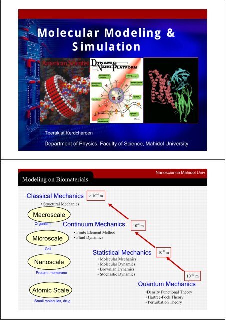
<strong>Molecular</strong> Mechanics<strong>Nano</strong>science <strong>Mahidol</strong> UnivA nanostructure can be model by ensemble of mass connected byensemble of springs.<strong>Molecular</strong> Mechanics<strong>Molecular</strong> <strong>Modeling</strong>Picture from: Peter J. Steinbach, “Introduction to Macromolecular <strong>Simulation</strong>”.
<strong>Molecular</strong> Mechanics<strong>Molecular</strong> <strong>Modeling</strong>Energy of a system is a sum of allinteractions within and between thesprings<strong>Molecular</strong> Mechanics<strong>Molecular</strong> <strong>Modeling</strong>Valence term is the relative energy of aspring.
<strong>Molecular</strong> Mechanics<strong>Molecular</strong> <strong>Modeling</strong>Valence terms are interactions within the springs.A spring wants to relax to its original shape.<strong>Molecular</strong> Mechanics<strong>Molecular</strong> <strong>Modeling</strong>Cross term is due to coupling between 2springs. It is a correction to independentspring model.
<strong>Molecular</strong> Mechanics<strong>Nano</strong>science <strong>Mahidol</strong> UnivCross terms are interactions between 2 or more springs.Cross terms are corrections to the independent spring model.<strong>Molecular</strong> Mechanics<strong>Molecular</strong> <strong>Modeling</strong>Non-Bonded term is interaction betweentwo balls.
<strong>Molecular</strong> Mechanics<strong>Molecular</strong> <strong>Modeling</strong>Non-bond terms are interactions between the balls.Non-bond terms are long-range interactions.RepulsionAttraction<strong>Molecular</strong> Mechanics<strong>Molecular</strong> <strong>Modeling</strong>Non-bond terms are interactions between the balls.Non-bond terms are long-range interactions.3V(r)0-3σεr, nm0.8 1.0 1.2 1.4 1.6 1.8 2.0RepulsionAttraction
<strong>Molecular</strong> Mechanics<strong>Molecular</strong> <strong>Modeling</strong>Summation of energy of a molecularsystem is very long equation.E =Stretching(C-H1)+Stretching(C-H2)+…...+ Bend(H1-C-H2) + Bend (H1-C-H3) + ……+ Bend(H1-C-C) + …… + Bend (C-C=O) + …….+ Torsion (H1-C-C=O) + ... ++ Torsion (O1=C-O2-H4)+ vdW(H1-H4) + ……+ Elec (H1-H4) + ……<strong>Molecular</strong> Mechanics<strong>Molecular</strong> <strong>Modeling</strong><strong>Molecular</strong> Mechanics needs to knowforce constants and equilibrium geometryof all springs. This set of parameters iscalled “Force Field parameters”.ATOM TYPEC(sp3): C1C(carbonyl): C2O(C=O): O1O(acid): O2H: H1, H2, H3H(acid): H4k str : C(sp3)-C(carbonyl), C(sp3)-H, ….k bnd : H-C(sp3)-C(carbonyl), ……k tor : H-C(sp3)-C(carbonyl)-O(acid), …R 0 : C(sp3)-C(carbonyl), …..And all equilibrium angles, torsion plusvan der Waals parameters and partial charges
Force Field<strong>Molecular</strong> <strong>Modeling</strong>http://www.ks.uiuc.edu/Force Field in Market<strong>Molecular</strong> <strong>Modeling</strong>
Wire Frame<strong>Molecular</strong> <strong>Modeling</strong>BPTIProteinStick<strong>Molecular</strong> <strong>Modeling</strong>BPTIProtein
Solvent Accessible Surface<strong>Molecular</strong> <strong>Modeling</strong>BPTIProteinCa Wire<strong>Molecular</strong> <strong>Modeling</strong>BPTIProtein
Line Ribbon<strong>Molecular</strong> <strong>Modeling</strong>BPTIProteinSolid Ribbon<strong>Molecular</strong> <strong>Modeling</strong>BPTIProtein
Cartoon<strong>Molecular</strong> <strong>Modeling</strong>BPTIProteinCombined<strong>Molecular</strong> <strong>Modeling</strong>BPTIProtein
Model & Method<strong>Molecular</strong> <strong>Modeling</strong>MODEL- Atomistic ModelQuantum Mechanical Model<strong>Molecular</strong> Mechanical ModelMETHOD- Electronic Structure Calculation- <strong>Molecular</strong> Visualization- Structural <strong>Modeling</strong>- <strong>Molecular</strong> <strong>Simulation</strong>Structural <strong>Modeling</strong><strong>Molecular</strong> <strong>Modeling</strong>DNA sequence Protein sequence 3D model (3’ structure)atgGGCAAACTGGTGCTGTATGGCGTGGAAGCGAGCCCGCCGGTGCGTGCGTGCAAACTGACCCTGGATGCGCTGGGCCTGCAGTATGAATATCGTCTGGTGAACCTGCTGGCGGGCGAACATAAAACCAAAGAATTTAGCCTGAAAAACCCGCAGCATACCGTGCCGGTGCTGGAAGATGATGGCAAATTTATTTGGGAAAGCCATGCGATTTGCGCGTATCTGGTGCGTCGTTATGCGAAAAGCGATGATCTGTATCCGAAAGATTATTTTAAACGTGCGCTGMGKLVLYGVEASPPVRACKLTLDALGLQYEYRLVNLLAGEHKTKEFSLKNPQHTVPVLEDDGKFIWESHAICAYLVRRYAKSDDLYPKDYFKRALVDQRLHFESGVLFQGCIRNIAIPLFYKNITEVPRSQIDAIYEAYDFLEAFIGNQAYLCGPVITIADYSVVSSVSSLVGLAAIDAKRYPKLNGWLDRMAAQPNYQSLNGNGAQMLIDMFSSKITKIV
Structure Prediction<strong>Molecular</strong> <strong>Modeling</strong>Picture: http://www.ks.uiuc.edu/Homology <strong>Modeling</strong>Homology<strong>Molecular</strong> <strong>Modeling</strong>Issara Sramala• Predicting the tertiary structure ofan unknown protein using a known3D structure of a homologousprotein(s) (i.e. same family).[Template]• Known structure is used as atemplate to model an unknown (butlikely similar) structure [Model]• Assumption that structure is moreconserved than sequenceTemplateAlpha ToxinClostridium perfringensModelPhospholipaseBacillus cereus• If sequences can be aligned but are not identical substitution• If sequences cannot be aligned deletion/insertion• In the protein-protein alignment del/ins are called GAPS. Theyare presented in the alignment as a consecutive dashes.
HomologyBasic Steps in Homology <strong>Modeling</strong>Issara Sramala1. Database searching forhomologous proteins in PDB2. Alignment (Pairwise/ MultipleAlignments)• Align query sequence with homologues• Find Structurally Conserved Regions (SCRs)• Identify Structurally Variable Regions (SVRs)3. Model Building• Generate coordinates for core region• Generate coordinates for loops• Add side chains (Check rotamer library)4. Model Refinement and Evaluation• Refine structure using energy minimization• Validate structure1234Homology modelling1. DB SearchIssara SramalaIdentify Homologues in PDBPRTEINSEQENCEPRTEINSEQUENCEPRTEINSEQNCEQWERYTRASDFHGTREWQIYPASDFGHKLMCNASQERWWPRETWQLKHGFDSADAMNCVCNQWERGFDHSDASFWERQWKQuery SequencePDB
1) A known protein in a known classSupported evidences from experimental or otherbioinformatics techniquesHomology modelling1. DB SearchIssara Sramala2) BLAST searchIn general, PSI-BLAST correctly aligns 40% of the residues whenthe sequence identity is larger than 15%.HomologuesE-Value < 10 -5Identity and length the aligned sequencesTemplate DatabasePDB Protein Data Bankwww.rcsb.org/pdb/PSC database (nrl_3d)tourney.psc.eduTemplate• Template environment• pH• Presence of Ligands ?• Resolution of the templates• Family of proteins• Phylogenetic tree• Multiple templates?• Biological role, relevant fold• Expected biological/biochemical functionHomology modelling1. DB SearchIssara SramalaPRTEINSEQENCEPRTEINSEQUENCEPRTEINSEQNCEQWERYTRASDFHGTREWQIYPASDFGHKLMCNASQERWWPRETWQLKHGFDSADAMNCVCNQWERGFDHSDASFWERQWK
2. AlignmentHomology modellingIssara SramalaStep 2: Align SequencesQueryHit #1Hit #2ACDEFGHIKLMNPQRST--FGHQWERT-----TYREWYEGASDEYAHLRILDPQRSTVAYAYE--KSFAPPGSFKWEYEAMCDEYAHIRLMNPERSTVAGGHQWERT----GSFKEWYAA• Key step in Homology Modelling• Small error in alignment can leadto big error in structural model• Multiple alignments are usuallybetter than pairwise alignmentsHit #1 Hit #2Homology modelling2. AlignmentIssara Sramala• Regions in the homologous proteins that arestructurally/functionally important are moreconserved (i.e. have little sequence variation) in the1º AA sequence than the regions, which are not asrelevant for protein folding and function.• Similar region often called SCR (structurallyconserver regions)• Variable regions called VRs or SVR (for variableregions)
Homology modelling2. AlignmentIssara SramalaSCR #1SCR’sSCR #2QueryHit #1Hit #2ACDEFGHIKLMNPQRST--FGHQWERT-----TYREWYEGASDEYAHLRILDPQRSTVAYAYE--KSFAPPGSFKWEYEAMCDEYAHIRLMNPERSTVAGGHQWERT----GSFKEWYAAHHHHHHHHHHHHHCCCCCCCCCCCCCCCCCCBBBBBBBBBHit #1 Hit #2• Corresponds to the most stablestructures or regions (usuallyinterior) of protein• Corresponds to sequence regionswith lowest level of gapping,highest level of sequenceconservation• Usually corresponds tosecondary structuresQueryHit #1Hit #2SVR’sACDEFGHIKLMNPQRST--FGHQWERT-----TYREWYEGASDEYAHLRILDPQRSTVAYAYE--KSFAPPGSFKWEYEAMCDEYAHIRLMNPERSTVAGGHQWERT----GSFKEWYAAHHHHHHHHHHHHHCCCCCCCCCCCCCCCCCCBBBBBBBBBHit #1 Hit #2SVR (loop)Homology modelling2. AlignmentIssara Sramala• Corresponds to the least stableor most flexible regions (usuallyexterior) of protein• Highest level of gapping, lowestlevel of sequence conservation• Usually corresponds to loopsand turns
Generate Core CoordinatesHomology modelling2. AlignmentIssara Sramala• For identical amino acids, transfer all atom coordinates(XYZ) to query protein• For similar amino acids, transfer backbone coordinates &replace side chain atoms while respecting c angles• For different amino acids, transfer only the backbonecoordinates (XYZ) to query sequence3. Model BuildingReplace SVRs (loops)Homology modellingIssara SramalaQueryYAYE--KSHit #1FGHQWERT• Loops extracted from PDB usinghigh resolution (
Homology modelling3. Model BuildingIssara SramalaReplace SVRs (loops)• Must match desired no. of residues• Must match Ca-Ca distance• Must not bump into other parts of protein• Preceding and following Ca’s (3 residues) from loopshould match well with corresponding Ca coordinatesin template structure• Loop placement and positioning is done usingsuperposition algorithm• Loop fits are evaluated using RMSD calculations andstandard “bump checking”• If no “good” loop is found, some algorithms createloops using randomly generated φ/ψ anglesAdd Side ChainsHomology modelling3. Model BuildingIssara Sramala• Done primarily for SVRs(not SCRs)• Rotamer placement andpositioning is done via asuperposition algorithmusing rotamers taken froma standardized library(Trial & Error)Relation Between χ and φ/ψ• Rotamer fits are evaluatedusing simple “bumpchecking” methods
4. Energy MinimizationBad modelsHomology modellinga) Side chain packingb) Distortions and shiftsc) no templated) Misalignmentse) incorrect templateModel Quality• A structure can (and often does) havemistakes• A poor structure will lead to poormodels of mechanism or relationship• Unusual parts of a structure mayindicate something important (or anerror)• Assess experimental fit (look at R factor or rmsd)X-ray structure R = 0.25 typical proteinNMR structure rmsd = 0.8 Å typical• Assess correctness of overall fold (look at disposition ofhydrophobes)• Assess structure quality (packing, stereochemistry, badcontacts, etc.)Homology modelling4. Energy MinimizationIssara Sramala
4. Energy MinimizationHomology modellingRadius & Radius of Gyration• Folded ~ 3.875 x NUMRES 0.333• Unfolded ~ 0.41 x (110 x NUMRES) 0.54. Energy MinimizationHomology modellingPacking VolumeLoose Packing Dense Packing Protein
ValidationHomology modellingStructure Validation Servers• WhatIf Web Server - http://www.cmbi.kun.nl:1100/WIWWWI/• Biotech Validation Suite - http://biotech.ebi.ac.uk:8400/cgi-bin/sendquery• Verify3D - http://www.doe-mbi.ucla.edu/Services/Verify_3D/• VADAR - http://redpoll.pharmacy.ualberta.caStructure Validation Programs• PROCHECK - http://www.biochem.ucl.ac.uk/~roman/procheck/procheck.html• PROSA II - http://lore.came.sbg.ac.at/People/mo/Prosa/prosa.html• VADAR - http://www.pence.ualberta.ca/ftp/vadar/• DSSP – http://www.embl-heidelberg.de/dssp/ValidationProcheckHomology modellingIssara Sramala
<strong>Molecular</strong> <strong>Simulation</strong><strong>Molecular</strong> <strong>Modeling</strong>Newton Mechanics of ParticlesNewton’s laws determine motion of particles.d rdt2i2=FmiiThe force on each particles can be calculated from derivative of thepotential energy.Fi=−∇riU ( r , r2,..., r1 NThe potential energy is usually approximated by pair summation.∑∑U r , r ,..., r ) = u(r − r )(1 2Nij>i)ij<strong>Molecular</strong> Dynamics<strong>Molecular</strong> <strong>Modeling</strong>Solve the equations of motion using “<strong>Molecular</strong>Mechanics”x 3F(x 2 )x 1F(xx 2 1 )x’ 3
<strong>Molecular</strong> Dynamics<strong>Molecular</strong> <strong>Modeling</strong>http://www.ks.uiuc.edu/<strong>Molecular</strong> Dynamics<strong>Molecular</strong> <strong>Modeling</strong>Slide: http://www.ks.uiuc.edu/
<strong>Molecular</strong> Dynamics<strong>Molecular</strong> <strong>Modeling</strong>• Time evolution ofsystems• Structural anddynamical properties• Thermodynamicproperties<strong>Molecular</strong> Dynamics<strong>Molecular</strong> <strong>Modeling</strong>A simulation can be dividedinto 4 phases:• Initialization Phase• Equilibration Process•Production Phase• Analysis of the ResultsPicture from Siegfried Fritzsche
<strong>Simulation</strong> in Water<strong>Molecular</strong> <strong>Modeling</strong>MD simulation of proteinin water reveals structuraldynamics of the moleculein condense phase.System Size<strong>Molecular</strong> <strong>Modeling</strong>http://www.ks.uiuc.edu/System size is larger everyday.
Bt Cry4B Pore Formation<strong>Molecular</strong> <strong>Modeling</strong>Collaboration with Dr. Chanan AngsuthanasombatCrystal Protein Inclusion IngestionSolubilizationProteolytic ActivationBinding with a SpecificReceptor+Gut Cell MembraneMembraneInsertionPore FormationBt Cry4B Structure<strong>Molecular</strong> <strong>Modeling</strong>Picture: Issara SramalaDomain I- Seven-helix bundle- Central helix : α5-Pore-forming domainα5Domain II- Three β sheets- β-prism motif- Insect specificityDomain III- β sandwich motif1) Pore-formation2) Insect specificity3) Protein stabilizingCrystal structure of Cry3A (Li J., 1991)
Homology Model of Bt Cry4B<strong>Molecular</strong> <strong>Modeling</strong>Collaboration with Dr. Chanan AngsuthanasombatHomology Model of Cry4B Toxinswas constructed by using Cry3Aa s a t e m p l a t eSeq. IdentityCry1AaCry3A~32%Cry4BCry3A~23%Cry3ACry4BHomology based model of Cry4B (Uawithya et al., 1999)Cry4B ModelHomology modellingCollaboration with Dr. Chanan AngsuthanasombatLi J1991Bacillus thuringiensis (Bt) toxinCry3A – Cry4B23% IdentityUawithya P1997RMSD5.71 ÅBoonserm P2003Cry3AXRayTemplateCry4BModelCry4BXRay
<strong>Simulation</strong> in Water<strong>Molecular</strong> <strong>Modeling</strong>Collaboration with Dr. Chanan Angsuthanasombat595 amino acids21,065 water<strong>Simulation</strong> time = 4nsTime step = 0.002 psBt Cry4B in Water<strong>Molecular</strong> <strong>Modeling</strong>Collaboration with Dr. Chanan AngsuthanasombatAtomic movement in Cry4B is larger than average proteins,indicating the flexibility in the structure.7.57.06.56.0RMSD (Angstrom)5.55.04.54.03.53.02.50 1 2 3 4Time (ns)Domain1Domain2Domain3Cry4B
Bt Cry4B in Water<strong>Molecular</strong> <strong>Modeling</strong>Collaboration with Dr. Chanan Angsuthanasombat<strong>Simulation</strong> has found movement of domain 1 from otherdomains, which could be the initial step for pore formation.3.63.53.4Distance (nm)3.33.23.13.0Domain1 and Domain2Domain1 and Domain3Domain2 and Domain32.92.80 1 2 3 4Time (ns)Bt Cry4B in Water<strong>Molecular</strong> <strong>Modeling</strong>Collaboration with Dr. Chanan AngsuthanasombatSolvent Accessible Surface Area (SASA) of hydrophilic partis decreasing, leading to total decrease of over all SASA.1000SASA of hydrophobic partSASA of hydrophilic partArea (nm 2 )700Area / nm 2950650Hydrophobic partHydrophilic part9001 2 3 4 1 2 3 4Time (ns)t / ns
Bt Cry4B in Water<strong>Molecular</strong> <strong>Modeling</strong>The protein is suspected to initiate molecular self assemblyprocess assisted by water.1080protein-water38720387101060H-bond Number10401020water-water387003869038680H-bond Number3867010001 2 3 4Time (ns)Acknowledgments<strong>Nano</strong>science <strong>Mahidol</strong>GRANT Faculty of Science, <strong>Mahidol</strong> <strong>University</strong> National Metal and Materials Center (MTEC) Thailand Research Fund (TRF) National Electronic and Computer Center (NECTEC)TEAMProf. Dr. I Ming TangAsst. Prof. Dr. Tanakorn OsotchanAsst. Prof. Dr. Teerakiat KerdcharoenAsst. Prof. Dr. Toemsak SrikhirinDr. Udom RobkobDr. Somsak DangtipDr. Wannapong TriampoProf. Dr. Michael KiselevChonticha SuwattanasophonThanyarat UdommaneethanakitAnurak UdomvechPirote JaidiawYingyote InfasaengSongwut SuramitrKriengsak SriwichitkamolSriprajak KrongsukEtc.


