28. McMillan SA, Haughton DJ, Biggart JD, Edgar JD, Porter KG, McNeill TA. Predictive valuefor coeliac disease of antibodies to gliadin, endomysium, and jejunum in patients attendingfor jejunal biopsy. BMJ. 1991;303:1163–1165.29. Dieterich W, Laag E, Schopper H, et al. Autoantibodies to tissue transglutaminase aspredictors of celiac disease. Gastroenterology. 1998;115:1584–1586.30. Leon F, Camerero C, R-Pena R, et al. Anti-transglutaminase IgA ELISA: clinical potentialand drawbacks in celiac disease diagnosis. Scand J Gastroenterol. 2001;36:849–853.31. Fabiani E, Catassi C. The serum IgA class anti-tissue transglutaminase antibodies in thediagnosis and follow up of coeliac disease. Results of an international multi-centre study.International Working Group on Eu-tTG. Eur J Gastroenterol Hepatol. 2001;13:659–665.32. Seissler J, Boms S, Wohlrab U, et al. Antibodies to human recombinant tissuetransglutaminase measured by radioligand assay: evidence for high diagnostic sensitivity forceliac disease. Horm Metab Res. 1999;31:375–379.33. Troncone R, Maurano F, Rossi M, et al. IgA antibodies to tissue transglutaminase: Aneffective diagnostic test for celiac disease. J Pediatr. 1999;134:166–171.34. Vitoria JC, Arrieta A, Arranz C, et al. Antibodies to gliadin, endomysium, and tissuetransglutaminase for the diagnosis of celiac disease. J Pediatr Gastroenterol Nutr.1999;29:571–574.35. Wong RC, Wilson RJ, Steele RH, Radford-Smith G, Adelstein S. A comparison of 13 guineapig and human anti-tissue transglutaminase antibody ELISA kits. J Clin Pathol.2002;55:488–494.36. Kolho KL, Savilahti E. IgA antiendomysium antibodies on human umbilical cord: Anexcellent diagnostic tool for celiac disease in childhood. J Pediatr Gastroenterol Nutr.1997;24:563–567.37. Mustalahti K, Sulkanen S, Holopainen P, et al. Coeliac disease among healthy members ofmultiple case coeliac disease families. Scand J Gastroenterol. 2002;37:161–165.38. Prince HE, Normal GL, Binder WL. Immunoglobulin A (IgA) deficiency and alternativedisease-associated antibodies in sera submitted to a reference laboratory for endomysial IgAtesting. Clin Diag Lab Immunol. 2000:7:192–196.39. Hansson T, Dahlbom I, Rogberg S, et al. Recombinant human tissue transglutaminase fordiagnosis and follow up of childhood coeliac disease. Pediatr Res. 2002;51:700–705.40. Picarelli A, di Tola M, Sabbatella L, et al. Identification of a new coeliac disease subgroup:antiendomysial and anti-transglutaminase antibodies of IgG class in absence of selective IgAdeficiency. J Intern Med. 2001;249:181–188.41. Picarelli A, Sabbatella L, Di Tola M, et al. <strong>Celiac</strong> disease diagnosis in misdiagnosedchildren. Pediatr Res. 2000;48:590–592.31
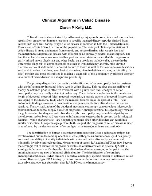
Clinical Algorithm in <strong>Celiac</strong> <strong>Disease</strong>Ciaran P. Kelly, M.D.<strong>Celiac</strong> disease is characterized by inflammatory injury to the small intestinal mucosa thatresults from an aberrant immune response to specific ingested dietary peptides derived fromcereals such as wheat, barley, or rye. <strong>Celiac</strong> disease is common in both the United States andEurope and affects 0.5 to 1 percent of the population. The variety of clinical presentations ofceliac disease is broad and ranges from chronic and severe diarrhea with weight loss andmalnutrition to symptomless disease with minimal or no clinically evident malabsorption. Thefact that celiac disease is common and has protean manifestations means that the diagnosis iseasily missed unless physicians and other health care providers include celiac disease in thedifferential diagnosis of common conditions such as iron deficiency anemia, mild chronicdiarrhea, recurrent abdominal discomfort, failure to thrive as well as less common manifestationssuch as skin rashes, hair loss, neurological disorders, vitamin deficiency states, or infertility. Inbrief, the first and most critical step in making a diagnosis of this commonly overlooked disorderis to think of celiac disease as a diagnostic possibility.The primary diagnostic criterion is the identification of an enteropathy that is consistentwith the inflammatory intestinal injury seen in celiac disease. This requires that a small bowelbiopsy be obtained prior to effective treatment with a gluten-free diet. Changes of celiacenteropathy may be visually evident at endoscopy. These include: a reduction in the number orheight of duodenal mucosal folds, mucosal nodularity, a mosaic pattern of mucosal fissures, andscalloping of the duodenal folds where the mucosal fissures cross the apex of a fold. Theseendoscopic findings, alone or in combination, are quite specific for celiac disease but are notsensitive. Thus, visualization of the duodenal mucosa at endoscopy cannot replace microscopicexamination of duodenal biopsy tissue for diagnosis. Although intestinal histopathology remainsthe gold standard for diagnosis of celiac disease, the enteropathy may be mild and patchy andtherefore missed on biopsy. Even when an inflammatory enteropathy is present, the histologicalfeatures—while characteristic—are not pathognomonic since other disorders can result in asimilar or identical histopathologic picture. In this regard, the diagnosis of celiac disease is aidedor reinforced by the demonstration of serum IgA tissue transglutaminase autoantibody.The identification of human tissue transglutaminase (htTG) as a celiac autoantigen hasrevolutionized our understanding of celiac disease pathogenesis. Simultaneously, it has greatlyenhanced our ability to identify individuals with untreated celiac disease by accurate andminimally invasive serologic testing. Measurement of serum IgA against htTG has now becomethe serologic test of choice for diagnosis or exclusion of untreated celiac disease. IgA htTGserology is far more specific than the older gliadin-based immunoassays, to the point that theantigliadin assays are now of minimal clinical utility. The identification of serum IgAendomycial antibodies (EMA) remains a sensitive and highly specific marker of untreated celiacdisease. However, IgA EMA testing by indirect immunofluoresence is more cumbersome,expensive, and operator dependent than IgA htTG enzyme immunoassay.33
- Page 1 and 2: NIH Consensus Development Conferenc
- Page 3 and 4: III. What Are the Manifestations an
- Page 5 and 6: • What is the management of celia
- Page 7 and 8: Monday, June 28, 2004 (continued)I.
- Page 9 and 10: Monday, June 28, 2004 (continued)II
- Page 11 and 12: Wednesday, June 30, 2004 (continued
- Page 13 and 14: Lisa H. RichardsonConsumer Represen
- Page 15 and 16: Ciaran P. Kelly, M.D.Director, Celi
- Page 17 and 18: Van S. Hubbard, M.D., Ph.D.Director
- Page 19 and 20: AbstractsThe following are abstract
- Page 21 and 22: susceptibility (e.g., DR17 homozygo
- Page 23 and 24: The Pathology of Celiac DiseaseDavi
- Page 25 and 26: In this regard, the transport pathw
- Page 27 and 28: for the IgG-based test, while speci
- Page 29: 15. de Lecea A, Ribes-Koninckx C, P
- Page 33 and 34: Considera diagnosisof celiac diseas
- Page 35 and 36: There are populations at particular
- Page 37 and 38: Serological Testing for Celiac Dise
- Page 39 and 40: Estimates of the sensitivity of the
- Page 41 and 42: the risk and severity of CD may als
- Page 43 and 44: What Are the Prevalence and Inciden
- Page 45 and 46: ascribed to excess menstrual loss.
- Page 47 and 48: ReferencesFamily History of Celiac
- Page 49 and 50: Carroccio A, Iannitto E, Cavataio F
- Page 51 and 52: identified by surveys or through so
- Page 53 and 54: Clinical Presentation of Celiac Dis
- Page 55 and 56: een widely reported. The question r
- Page 57 and 58: The Many Faces of Celiac Disease: C
- Page 59 and 60: References1. Green PH, Jabri B. Coe
- Page 61 and 62: Association of Celiac Disease and G
- Page 63 and 64: Does the Gluten-Free Diet Protect F
- Page 65 and 66: Skin Manifestations of Celiac Disea
- Page 67 and 68: allow a better understanding of the
- Page 69 and 70: 4. Henriksson KG, Hallert C, Walan
- Page 71 and 72: characterized, clinically-identifie
- Page 73 and 74: 10. Hoffenberg EJ, Emery LM, Barrig
- Page 75 and 76: ataxia, epilepsy with posterior cer
- Page 77 and 78: Consequences of Testing for Celiac
- Page 79 and 80: Osteoporosis/FracturesThere were 11
- Page 81 and 82:
Dietary Guidelines for Celiac Disea
- Page 85 and 86:
21. Thompson T. Thiamin, riboflavin
- Page 88 and 89:
In order to effectively counsel ind
- Page 90 and 91:
9. Hallert C, Granno C, Hulten S, M
- Page 92 and 93:
adhered to the GFD after more than
- Page 94 and 95:
Patient education, close supervisio
- Page 96:
26. Mustalahti K, Lohiniemi S, Laip


