PHARYNGEAL AIRWAY VOLUME FOLLOWING ...
PHARYNGEAL AIRWAY VOLUME FOLLOWING ...
PHARYNGEAL AIRWAY VOLUME FOLLOWING ...

Create successful ePaper yourself
Turn your PDF publications into a flip-book with our unique Google optimized e-Paper software.
T0 A T1 A<br />
T0 B<br />
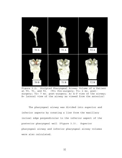
Figure 3.2: Sculpted Pharyngeal Airway Volume of a Patient<br />
at T0, T1, and T2. (T0= Pre-surgery; T1= 2 mo. postsurgery;<br />
T2= 7 mo. post-surgery; A= A-P view of the airway;<br />
B= lateral view of the airway as viewed from the anterior)<br />
The pharyngeal airway was divided into superior and<br />
inferior aspects by creating a line from the maxillary<br />
incisal edge perpendicular to the inferior aspect of the<br />
posterior pharyngeal wall (Figure 3.3). Superior<br />
pharyngeal airway and inferior pharyngeal airway volumes<br />
were also calculated.<br />
T1 B<br />
32<br />
T2 A<br />
T2 B