PHARYNGEAL AIRWAY VOLUME FOLLOWING ...
PHARYNGEAL AIRWAY VOLUME FOLLOWING ...
PHARYNGEAL AIRWAY VOLUME FOLLOWING ...
You also want an ePaper? Increase the reach of your titles
YUMPU automatically turns print PDFs into web optimized ePapers that Google loves.
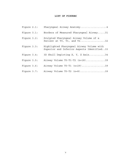
LIST OF FIGURES<br />
Figure 2.1: Pharyngeal Airway Anatomy..................4<br />
Figure 3.1: Borders of Measured Pharyngeal Airway.....31<br />
Figure 3.2: Sculpted Pharyngeal Airway Volume of a<br />
Patient at T0, T1, and T2.................32<br />
Figure 3.3: Highlighted Pharyngeal Airway Volume with<br />
Superior and Inferior Aspects Identified..33<br />
Figure 3.4: 3D Skull Depicting X, Y, Z Axis...........34<br />
Figure 3.5: Airway Volume T0-T1-T2 (n=18).............39<br />
Figure 3.6: Airway Volume T0-T1 (n=29)................39<br />
Figure 3.7: Airway Volume T0-T2 (n=8).................39<br />
v


