PHARYNGEAL AIRWAY VOLUME FOLLOWING ...
PHARYNGEAL AIRWAY VOLUME FOLLOWING ...
PHARYNGEAL AIRWAY VOLUME FOLLOWING ...

Create successful ePaper yourself
Turn your PDF publications into a flip-book with our unique Google optimized e-Paper software.
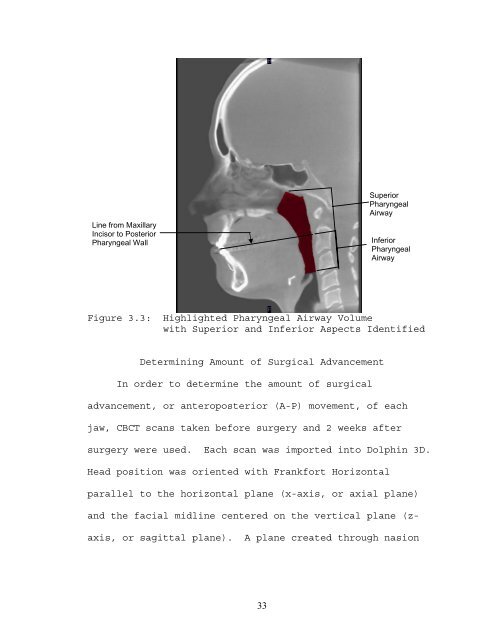
Line from Maxillary<br />
Incisor to Posterior<br />
Pharyngeal Wall<br />
Figure 3.3: Highlighted Pharyngeal Airway Volume<br />
with Superior and Inferior Aspects Identified<br />
Determining Amount of Surgical Advancement<br />
In order to determine the amount of surgical<br />
advancement, or anteroposterior (A-P) movement, of each<br />
jaw, CBCT scans taken before surgery and 2 weeks after<br />
surgery were used. Each scan was imported into Dolphin 3D.<br />
Head position was oriented with Frankfort Horizontal<br />
parallel to the horizontal plane (x-axis, or axial plane)<br />
and the facial midline centered on the vertical plane (z-<br />
axis, or sagittal plane). A plane created through nasion<br />
33<br />
Superior<br />
Pharyngeal<br />
Airway<br />
Inferior<br />
Pharyngeal<br />
Airway