Glaucoma-I Free Papers - aioseducation
Glaucoma-I Free Papers - aioseducation
Glaucoma-I Free Papers - aioseducation
You also want an ePaper? Increase the reach of your titles
YUMPU automatically turns print PDFs into web optimized ePapers that Google loves.
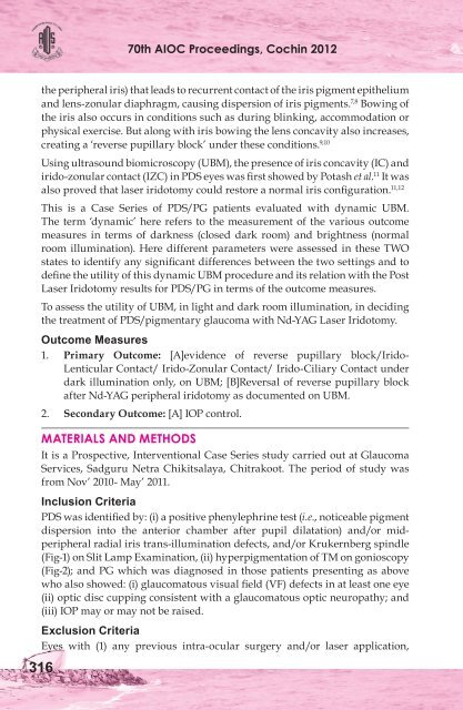
70th AIOC Proceedings, Cochin 2012the peripheral iris) that leads to recurrent contact of the iris pigment epitheliumand lens-zonular diaphragm, causing dispersion of iris pigments. 7,8 Bowing ofthe iris also occurs in conditions such as during blinking, accommodation orphysical exercise. But along with iris bowing the lens concavity also increases,creating a ‘reverse pupillary block’ under these conditions. 9,10Using ultrasound biomicroscopy (UBM), the presence of iris concavity (IC) andirido-zonular contact (IZC) in PDS eyes was first showed by Potash et al. 11 It wasalso proved that laser iridotomy could restore a normal iris configuration. 11,12This is a Case Series of PDS/PG patients evaluated with dynamic UBM.The term ‘dynamic’ here refers to the measurement of the various outcomemeasures in terms of darkness (closed dark room) and brightness (normalroom illumination). Here different parameters were assessed in these TWOstates to identify any significant differences between the two settings and todefine the utility of this dynamic UBM procedure and its relation with the PostLaser Iridotomy results for PDS/PG in terms of the outcome measures.To assess the utility of UBM, in light and dark room illumination, in decidingthe treatment of PDS/pigmentary glaucoma with Nd-YAG Laser Iridotomy.Outcome Measures1. Primary Outcome: [A]evidence of reverse pupillary block/Irido-Lenticular Contact/ Irido-Zonular Contact/ Irido-Ciliary Contact underdark illumination only, on UBM; [B]Reversal of reverse pupillary blockafter Nd-YAG peripheral iridotomy as documented on UBM.3162. Secondary Outcome: [A] IOP control.MATERIALS AND METHODSIt is a Prospective, Interventional Case Series study carried out at <strong>Glaucoma</strong>Services, Sadguru Netra Chikitsalaya, Chitrakoot. The period of study wasfrom Nov’ 2010- May’ 2011.Inclusion CriteriaPDS was identified by: (i) a positive phenylephrine test (i.e., noticeable pigmentdispersion into the anterior chamber after pupil dilatation) and/or midperipheralradial iris trans-illumination defects, and/or Krukernberg spindle(Fig-1) on Slit Lamp Examination, (ii) hyperpigmentation of TM on gonioscopy(Fig-2); and PG which was diagnosed in those patients presenting as abovewho also showed: (i) glaucomatous visual field (VF) defects in at least one eye(ii) optic disc cupping consistent with a glaucomatous optic neuropathy; and(iii) IOP may or may not be raised.Exclusion CriteriaEyes with (1) any previous intra-ocular surgery and/or laser application,


