

TEMPORALIS MUSCLE FLAP - Vula - University of Cape Town
TEMPORALIS MUSCLE FLAP - Vula - University of Cape Town
TEMPORALIS MUSCLE FLAP - Vula - University of Cape Town

Create successful ePaper yourself
Turn your PDF publications into a flip-book with our unique Google optimized e-Paper software.
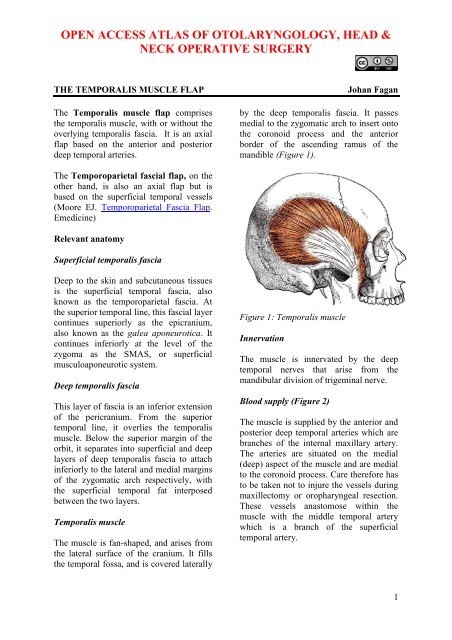
OPEN ACCESS ATLAS OF OTOLARYNGOLOGY, HEAD &NECK OPERATIVE SURGERYTHE <strong>TEMPORALIS</strong> <strong>MUSCLE</strong> <strong>FLAP</strong>The Temporalis muscle flap comprisesthe temporalis muscle, with or without theoverlying temporalis fascia. It is an axialflap based on the anterior and posteriordeep temporal arteries.Johan Faganby the deep temporalis fascia. It passesmedial to the zygomatic arch to insert ontothe coronoid process and the anteriorborder <strong>of</strong> the ascending ramus <strong>of</strong> themandible (Figure 1).The Temporoparietal fascial flap, on theother hand, is also an axial flap but isbased on the superficial temporal vessels(Moore EJ. Temporoparietal Fascia Flap.Emedicine)Relevant anatomySuperficial temporalis fasciaDeep to the skin and subcutaneous tissuesis the superficial temporal fascia, alsoknown as the temporoparietal fascia. Atthe superior temporal line, this fascial layercontinues superiorly as the epicranium,also known as the galea aponeurotica. Itcontinues inferiorly at the level <strong>of</strong> thezygoma as the SMAS, or superficialmusculoaponeurotic system.Deep temporalis fasciaThis layer <strong>of</strong> fascia is an inferior extension<strong>of</strong> the pericranium. From the superiortemporal line, it overlies the temporalismuscle. Below the superior margin <strong>of</strong> theorbit, it separates into superficial and deeplayers <strong>of</strong> deep temporalis fascia to attachinferiorly to the lateral and medial margins<strong>of</strong> the zygomatic arch respectively, withthe superficial temporal fat interposedbetween the two layers.Temporalis muscleThe muscle is fan-shaped, and arises fromthe lateral surface <strong>of</strong> the cranium. It fillsthe temporal fossa, and is covered laterallyFigure 1: Temporalis muscleInnervationThe muscle is innervated by the deeptemporal nerves that arise from themandibular division <strong>of</strong> trigeminal nerve.Blood supply (Figure 2)The muscle is supplied by the anterior andposterior deep temporal arteries which arebranches <strong>of</strong> the internal maxillary artery.The arteries are situated on the medial(deep) aspect <strong>of</strong> the muscle and are medialto the coronoid process. Care therefore hasto be taken not to injure the vessels duringmaxillectomy or oropharyngeal resection.These vessels anastomose within themuscle with the middle temporal arterywhich is a branch <strong>of</strong> the superficialtemporal artery.1
2Figure 4: Hemicoronoal incisionFigure 2: Blood supplyFacial nerve (Figure 3)Frontal/temporal branches <strong>of</strong> the nervecross the zygomatic arch, and run acrossthe superficial temporal fat pad which isdeep to the orbicularis oculi muscle, justlateral to the orbital rim.Superficialtemporal fat padFrontal branch<strong>of</strong> facial nerveFigure 3: Facial nerve crossing fat padRaising the flapThe flap is accessed via a hemicoronal skinincision commencing in a preauricular skincrease just below the level <strong>of</strong> the zygomaand placed behind the hairline for cosmeticreasons (Figure 4). The incision is extendedto the temporalis fascia. The skin andsubcutaneous tissue are elevated in theplane situated on the temporalis fascia(Figure 5).Figure 5: Exposed temporalis fasciaAnteriorly, elevation in this plane isstopped when the superficial temporal fatpad with the facial/temporal branches <strong>of</strong>the facial nerve is encountered. At thispoint the deep layer <strong>of</strong> deep temporalisfascia is incised in a vertical direction, andthe underlying temporalis muscle isexposed. Anteriorly, further dissection isdone in a subfascial plane, deep to the fatpad, up to the lateral orbital bony rim(anterior margin <strong>of</strong> temporal fossa).The temporalis fascia is now incised alongthe superior temporal line and the posteriormargins <strong>of</strong> the muscle, down onto thebone. If an extended flap is planned, thenthe superficial temporal vessels areidentified and preserved in the preauriculararea. The superior aspect <strong>of</strong> the zygomaticarch is identified along its full length. Thismight require quite forceful retraction <strong>of</strong>
3the s<strong>of</strong>t tissues with a Langenbeckretractor. The two layers <strong>of</strong> deep temporalfascia are incised along the superiormargin <strong>of</strong> the zygoma (Figure 6).Figure 6: Mobilisation <strong>of</strong> temporalismuscle and exposure <strong>of</strong> zygomatic archThe temporalis muscle is elevated from thebone <strong>of</strong> the temporal fossa using eitherdiathermy or a periosteal elevator. Thedissection remains hard on the bone, andextends medial to the coronoid process <strong>of</strong>the mandible that is now readily palpablemedial to the zygoma, especially when themouth is closed (Figure 7).muscle and its origin from the coronoidprocess. With a thin muscle the flap cannow be passed through this tunnel into themouth, taking care not to rotate the flapand strangulate its blood supply. With abulky muscle, the zygomatic arch and/orthe coronoid process <strong>of</strong> the mandible maybe osteotomised to permit passage <strong>of</strong> theflap. The zygomatic bone can be kept insaline and plated/wired back later in theprocedure. The coronoid osteotomy is doneeither from above via the temporal fossa,or via the mouth. Great care has to betaken not to injure the deep temporalvessels by staying close to the bonysurface <strong>of</strong> the coronoid.Additional length <strong>of</strong> flap may be obtainedby transecting or resecting the coronoidprocess and thus freeing the origin <strong>of</strong> themuscle from the bone, or by using anextended my<strong>of</strong>ascial flap that includesadditional temporalis fascia.Clinical ApplicationsThe temporalis muscle flap may be usedfor reconstruction <strong>of</strong> oral (floor <strong>of</strong> mouth,tongue, buccal, retromolar trigone, andpalate), oropharyngeal, nasopharyngeal,orbital, maxillectomy and facial s<strong>of</strong>t tissuedefects.Palatal and maxillary resectionsFigure 7: Flap completely elevated fromtemporal fossaShould the flap need to be passed into theoral cavity, then a finger can now beentunnelled into the mouth medial to theThe flap is ideally suited to reconstruction<strong>of</strong> the palate following Le Fort 1maxillectomy (Figure 8), but als<strong>of</strong>ollowing total maxillectomy. Thecoronoid process <strong>of</strong> the mandible on whichit is a pedicled is immediately adjacent tothe defect. It may reach across the midline.The muscle is left bare in the mouth, andmucosalises within a matter <strong>of</strong> weeks.Bilateral palatal resection defects may bereconstructed with bilateral temporalismuscle flaps that are sutured together inthe midline.
4Figure 8: Reconstruction <strong>of</strong> inferiormaxillectomy defectFigure 10: Temporalis muscle filling theorbitNote in the Figure 9 how the muscle hasbecome covered with mucosa in a patientwith bilateral temporalis muscle flaps forbilateral inferior maxillectomy.Figure 11: Outcome <strong>of</strong> primary closure <strong>of</strong>skin over temporalis muscle flapFigure 9: Mucosalised muscle followingpalatal reconstructionOrbital exenterationThe lateral wall <strong>of</strong> the orbit is removed toprovide space for passage <strong>of</strong> the musclepedicle. The orbit is filled with the TMF(Figure 10), and is covered with skin orsplit skin graft, or closed primarily if a lidhas been spared (Figure 11).Drawbacks <strong>of</strong> the temporalis muscleflapTemporal fossa concavity (Figure 12)The concavity <strong>of</strong> the temporal fossa fromwhich the muscle has been mobilized canbe quite pronounced. It may be filled witha fat graft or hydroxyappatite or mouldedprosthetic implants.
5Author & EditorJohan Fagan MBChB, FCORL, MMedPr<strong>of</strong>essor and ChairmanDivision <strong>of</strong> Otolaryngology<strong>University</strong> <strong>of</strong> <strong>Cape</strong> <strong>Town</strong><strong>Cape</strong> <strong>Town</strong>South Africajohannes.fagan@uct.ac.zaFigure 12: Temporal fossa concavityDental rehabilitationThe Open Access Atlas <strong>of</strong> Otolaryngology, Head &Neck Operative Surgery by Johan Fagan (Editor)johannes.fagan@uct.ac.za is licensed under a CreativeCommons Attribution - Non-Commercial 3.0 UnportedLicenseDue to the absence <strong>of</strong> alveolar bone, it isnot possible to insert dental implantsfollowing inferior maxillectomyreconstruction. With bilateral inferiormaxillectomy, it is not possible to fit andretain an upper denture.Some useful references1. Moore EJ. Temporoparietal FasciaFlap. emedicine2. Smith J, Ducic Y, Adelson R. Theutility <strong>of</strong> the temporalis muscle flap fororopharyngeal, base <strong>of</strong> tongue, andnasopharyngeal reconstruction.Otolaryngol Head Neck Surg2005;132:373-80


