

Meatoplasty - Vula - University of Cape Town
Meatoplasty - Vula - University of Cape Town
Meatoplasty - Vula - University of Cape Town

Create successful ePaper yourself
Turn your PDF publications into a flip-book with our unique Google optimized e-Paper software.
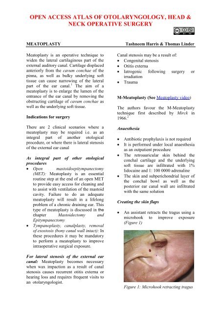
OPEN ACCESS ATLAS OF OTOLARYNGOLOGY, HEAD &NECK OPERATIVE SURGERYMEATOPLASTY<strong>Meatoplasty</strong> is an operative technique towiden the lateral cartilaginous part <strong>of</strong> theexternal auditory canal. Cartilage displacedanteriorly from the cavum conchae <strong>of</strong> thepinna, as well as bulky underlying s<strong>of</strong>ttissue can cause narrowing <strong>of</strong> the lateralpart <strong>of</strong> the ear canal. 1 The aim <strong>of</strong> ameatoplasty is to enlarge the lumen <strong>of</strong> theentrance <strong>of</strong> the ear canal by removing theobstructing cartilage <strong>of</strong> cavum conchae aswell as the underlying s<strong>of</strong>t tissue.Indications for surgeryThere are 2 clinical scenarios where ameatoplasty may be required i.e. as anintegral part <strong>of</strong> another otologicalprocedure, or where there is lateral stenosis<strong>of</strong> the external ear canalAs integral part <strong>of</strong> other otologicalproceduresOpen mastoidoepitympanectomy(MET): <strong>Meatoplasty</strong> is an essentialroutine step at the end <strong>of</strong> an open METto provide easy access for cleaning andto assist with ventilation <strong>of</strong> the mastoidcavity. Failure to do an adequatemeatoplasty will result in a lifelongproblem <strong>of</strong> a chronic draining ear. Thistype <strong>of</strong> meatoplasty is discussed in thechapter Mastoidectomy andEpitympanectomyTympanoplasty, canalplasty, removal<strong>of</strong> exostosis (bony canal wall intact): Inthese procedures it may be mandatoryto perform a meatoplasty to improveintraoperative surgical exposure.For lateral stenosis <strong>of</strong> the external earcanal: <strong>Meatoplasty</strong> becomes necessarywhen wax impaction as a result <strong>of</strong> canalstenosis causes recurrent otitis externa orhearing loss and requires frequent visits toan otolaryngologist.Tashneem Harris & Thomas LinderCanal stenosis may be a result <strong>of</strong>:Congenital stenosisOtitis externaIatrogenic following surgery orirradiationTraumaM-<strong>Meatoplasty</strong> (See <strong>Meatoplasty</strong> video)The authors favour the M-<strong>Meatoplasty</strong>technique first described by Mirck in1966. 2AnaesthesiaAntibiotic prophylaxis is not requiredIt is performed under local anaesthesiaas an outpatient procedureThe retroauricular skin behind theconchal cartilage and the underlyings<strong>of</strong>t tissue are infiltrated with 1%lidocaine and 1: 100 0000 adrenalineThe skin and subperichondrial layer <strong>of</strong>the conchal bowl as well as theposterior ear canal wall are infiltratedwith the same solutionCreating the skin flapsAn assistant retracts the tragus using amicrohook to improve exposure(Figure 1)Figure 1: Microhook retracting tragus
Excision <strong>of</strong> obstructing cavum cartilageand underlying s<strong>of</strong>t tissueA circle <strong>of</strong> about 1cm diameter is cutout <strong>of</strong> the cavum cartilage (Figure 7)Figure 9Figure 7: Circle cut out <strong>of</strong> cavum cartilageThe underlying subcutaneous tissue is<strong>of</strong>ten bulky and is also excised (Figure8)Figure 10Figure 8: Subcutaneous tissue is excised6/0 Nylon sutures are used to suture theskin flaps as follows:Sutures are placed between the pointedends <strong>of</strong> the two intrameatal skin flapsand either side <strong>of</strong> the base <strong>of</strong> thecentral triangular skin flap (Figures 11& 12)V-Y plastyA fourth 1cm transverse incision ismade in the posterior canal wall, thuscreating two intrameatal skin flaps(Figures 9 & 10)Figure 11: Sutures placed between ends <strong>of</strong>the intrameatal skin flaps3
Figure 12: Sutures placed between ends <strong>of</strong>the intrameatal skin flapsFigure 14A suture is placed at the beginning <strong>of</strong>the intrameatal skin incision and theapex <strong>of</strong> the middle triangular skin flap.This step widens the entrance <strong>of</strong> theexternal ear canal (Figure 13)Figure 15Figure 13: Suture placed at beginning <strong>of</strong>the intrameatal skin incision and apex <strong>of</strong>middle triangular skin flapThe two redundant triangular skin flapsare excised and sutures are placedbetween the remaining edges <strong>of</strong> skin <strong>of</strong>the cavum concha and the intrameatalskin flaps. (Figure 14,15,16)Figure 16This results in a scar shaped like an“M” (Figure 17)4
AuthorTashneem Harris MBChB, FCORL,MMED (Otol), Fisch InstrumentMicrosurgical FellowENT SpecialistDivision <strong>of</strong> Otolaryngology<strong>University</strong> <strong>of</strong> <strong>Cape</strong> <strong>Town</strong><strong>Cape</strong> <strong>Town</strong>, South Africaharristasneem@yahoo.comFigure 17A Terracortril (ointment containing asteroid and antiseptic agent) gauze isplaced in the external ear canal for 5daysSutures are removed after 1 weekReferences1. Fisch U, May J, Linder T. Tympanoplasty,Mastoidectomy, and StapesSurgery. New York: Thieme; 2008.2. Mirck PG. The M-meatoplasty <strong>of</strong> theexternal auditory canal. Laryngoscope.1996; 106(3):367-69.Senior AuthorPr<strong>of</strong> Thomas Linder, M.D.Chairman and Head <strong>of</strong> Department <strong>of</strong>Otorhinolaryngology,Head, Neck and Facial Plastic SurgeryLucerne Canton Hospital, Switzerlandthomas.linder@ksl.chEditorJohan Fagan MBChB, FCORL, MMedPr<strong>of</strong>essor and ChairmanDivision <strong>of</strong> Otolaryngology<strong>University</strong> <strong>of</strong> <strong>Cape</strong> <strong>Town</strong><strong>Cape</strong> <strong>Town</strong>South Africajohannes.fagan@uct.ac.zaDownload meatoplasty videohttps://vula.uct.ac.za/access/content/group/9c29ba04-b1ee-49b9-8c85-9a468b556ce2/Johan%20Fagan%20Surgery%20Atlas/M%20-%20<strong>Meatoplasty</strong>.aviTHE OPEN ACCESS ATLAS OFOTOLARYNGOLOGY, HEAD &NECK OPERATIVE SURGERYwww.entdev.uct.ac.zaThe Open Access Atlas <strong>of</strong> Otolaryngology,Head & Neck Operative Surgery by JohanFagan (Editor) johannes.fagan@uct.ac.zais licensed under a Creative CommonsAttribution - Non-Commercial 3.0Unported License5


