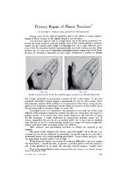
The Use of Paratenon, Polyethylene Film, or Silastic Sheeting to ...
The Use of Paratenon, Polyethylene Film, or Silastic Sheeting to ...
The Use of Paratenon, Polyethylene Film, or Silastic Sheeting to ...

You also want an ePaper? Increase the reach of your titles
YUMPU automatically turns print PDFs into web optimized ePapers that Google loves.
908 D. M. LICHTMAN, G. R. MACK, R. I. MACDONALD, S. F. GUNTHER, AND J. N. WILSON<br />
14. SHANDS, A. R.. JR., and RANEY, R. B., SR.: Handbook <strong>of</strong> Orthopaedic Surgery. Ed. 7. St. Louis, C. V. Mosby, 1967.<br />
15. STAHL, FOLKE: On Luna<strong>to</strong>malacia (Kienb#{246}ck’s Disease). A Clinical and Roentgenological Study, Especially on Its Pathogenesis and the Late<br />
Results <strong>of</strong> Immobilization Treatment. Acta Chir. Scandinavica, Supplementum 126, 1947.<br />
16. STEINDLER, A.: <strong>The</strong> Traumatic Def<strong>or</strong>mities and Disabilities <strong>of</strong> the Upper Extremity. Springfield. Illinois, Charles C Thomas, 1946.<br />
17. SWANSON, A. B.: Silicone Rubher Implants f<strong>or</strong> the Replacement <strong>of</strong>the Carpal Scaphoid and Lunate Bones. Orthop. Clin. N<strong>or</strong>th America. 1:<br />
299-309, 1970.<br />
18. SWANSON, A. B.: Flexible Implant Resection Arthroplasty in the Hand and Extremities. St. Louis, C. V. Mosby, 1973.<br />
19. TUREK, S. L.: Orthopaedics. Principles and <strong>The</strong>ir Application. Ed. 2. Philadelphia, J. B. Lippincott, 1967.<br />
<strong>The</strong> <strong>Use</strong> <strong>of</strong> <strong>Paratenon</strong>, <strong>Polyethylene</strong> <strong>Film</strong>, <strong>or</strong><br />
<strong>Silastic</strong> <strong>Sheeting</strong> <strong>to</strong> Prevent<br />
Restricting Adhesions <strong>to</strong> Tendons in the Hand*<br />
BY HERBERT H. STARK, M.D.t, JOSEPH H. BOYES, M.D.t, LOS ANGELES.<br />
LANNY JOHNSON, M.D.t, EAST LANSING, MICHIGAN,<br />
AND CHARLES R. ASHWORTH, M.D.t, LOS ANGELES, CALWORNIA<br />
ABSTRACT: We treated 132 patients by insertion <strong>of</strong><br />
paratenon, polyethylene, <strong>or</strong> <strong>Silastic</strong> between a digital<br />
tendon and a bone, ligament, <strong>or</strong> fixed fascial structure<br />
<strong>to</strong> prevent adhesions. From 1950 <strong>to</strong> 1974, au<strong>to</strong>genous<br />
paratenon was used in thirty patients; from 1956 <strong>to</strong><br />
1965, polyethylene film was used in sixty-three pa-<br />
tients; and from 1965 <strong>to</strong> 1974, <strong>Silastic</strong> sheeting was<br />
used in thirty-nine patients. By comparing the<br />
preoperative and pos<strong>to</strong>perative measurements <strong>of</strong> joint<br />
motion and the changes in the distance separating the<br />
pulp <strong>of</strong> a finger from the palm during fiexion, these pa-<br />
tients were classified as improved, unchanged, <strong>or</strong><br />
w<strong>or</strong>se. In some areas the material used appeared <strong>to</strong><br />
make little difference, but in other areas one <strong>or</strong> the<br />
other was superi<strong>or</strong>. <strong>Silastic</strong> sheeting (non-reinf<strong>or</strong>ced)<br />
proved <strong>to</strong> be the best material f<strong>or</strong> most conditions, but<br />
it should not be employed when the skin is <strong>of</strong> po<strong>or</strong><br />
quality <strong>or</strong> beneath a pedicle flap, and it should not be<br />
used adjacent <strong>to</strong> a tendon graft in an area that has re-<br />
covered from an infection Under those circumstances,<br />
paratenon is the preferred material.<br />
During the past twenty-four years, we have tried <strong>to</strong><br />
prevent the f<strong>or</strong>mation <strong>of</strong> restricting adhesions by interpos-<br />
ing one <strong>of</strong> three materials between a scarred tendon and<br />
bone, ligaments, <strong>or</strong> other fixed structure. We followed 132<br />
patients in whom one <strong>of</strong> these three materials was used.<br />
Twelve other patients were excluded from this study be-<br />
cause <strong>of</strong> inadequate follow-up rec<strong>or</strong>ds.<br />
* Read at the Sixth Combined Meeting <strong>of</strong> the Orthopaedic Associa-<br />
lions <strong>of</strong> the English-Speaking W<strong>or</strong>ld, London, England, September 15,<br />
I 976.<br />
1- 2300 South Flower Street, Los Angeles, Calif<strong>or</strong>nia 90007.<br />
t 4528 South Hagad<strong>or</strong>n Road, East Lansing. Michigan 48823.<br />
From 1950 <strong>to</strong> 1974, we used au<strong>to</strong>genous paratenon in<br />
thirty patients. <strong>Polyethylene</strong> film was used in sixty-three<br />
patients operated on from 1956 <strong>to</strong> 1965. <strong>Silastic</strong> sheeting<br />
became available in 1965, and since then we have used it<br />
in thirty-nine patients. In most cases, insertion <strong>of</strong> the in-<br />
terpositional material was just one part <strong>of</strong> a reconstructive<br />
operation; it was usually combined with other procedures,<br />
such as tenolysis, osteo<strong>to</strong>my, excision <strong>of</strong> a scar <strong>or</strong> bone,<br />
capsulec<strong>to</strong>my, neurolysis, nerve repair, <strong>or</strong> shifting <strong>of</strong> local<br />
skin flaps. Even so, by comparing the preoperative and<br />
pos<strong>to</strong>perative measurements <strong>of</strong> joint motion and the<br />
changes in the distance separating the pulp <strong>of</strong> the involved<br />
finger from the palm during flexion, we were able <strong>to</strong><br />
classify these patients as improved, unchanged, <strong>or</strong> w<strong>or</strong>se.<br />
Adhesions between a tendon and surrounding fixed<br />
tissue usually restrict voluntary motion. When such adhe-<br />
sions are long, loose, and elastic, and when they connect a<br />
tendon <strong>to</strong> mobile tissues such as those in the f<strong>or</strong>earm <strong>or</strong><br />
palm, a tendon may still glide a reasonable distance. How-<br />
ever, sh<strong>or</strong>t and inelastic adhesions that are attached <strong>to</strong><br />
fixed tissues tether the tendon and greatly restrict volun-<br />
tary motion <strong>of</strong> the digit.<br />
Pri<strong>or</strong> attempts <strong>to</strong> prevent such adhesions have been <strong>of</strong><br />
three types. Rods, tubes, <strong>or</strong> ribbons <strong>of</strong> inert material have<br />
been inserted <strong>to</strong> provoke the f<strong>or</strong>mation <strong>of</strong> a pseudosheath<br />
through which a tendon is passed later. Silver 17 tan-<br />
talum 22 stainless steel 15,1820.25.26 plastic, and Silas-<br />
tic 2.7,11.13.14 have been used in this manner. Of these,<br />
<strong>Silastic</strong> rods have been most successful, if used under the<br />
proper conditions. A second method has been <strong>to</strong> encircle<br />
the tendon with some substance l,2.912,16.17.21,2729 but re-<br />
gardless <strong>of</strong> the material used this method has failed, f<strong>or</strong><br />
scar f<strong>or</strong>ms at the ends <strong>of</strong> these materials and limits gliding<br />
THE JOURNAL OF BONE AND JOINT SURGERY
USE OF PARATENON TO PREVENT RESTRICTING ADHESIONS TO TENDONS IN THE HAND 909<br />
<strong>of</strong> the tendon 1,2.18 Even membranes with selective per-<br />
meability have failed <strong>to</strong> prevent restricting adhesions if<br />
wrapped about a tendon A third method has been <strong>to</strong> in-<br />
sert an interpositional material in a limited area where ten-<br />
don gliding is restricted because <strong>of</strong> its attachment <strong>to</strong> a fixed<br />
structure 3.6.24.28 In the past, many materials have been<br />
used in this procedure. F<strong>or</strong>eign substances have included<br />
vaseline bismuth paste “, cellophane 10.27 Millip<strong>or</strong>e ‘,<br />
<strong>Silastic</strong> 2.7.13 latex , gelatin foam 21.27 Oxycel cot<strong>to</strong>n 27<br />
celloidin 17 Ivalon 12 and polyethylene film 5,11,20,25<br />
Materials derived from animals that have been used in this<br />
manner include cargile membrane ‘, amniotic mem-<br />
brane , allan<strong>to</strong>in membrane , sheet catgut ‘, beef<br />
cecum , bovine fibrin film 27 and c<strong>or</strong>tisone acetate 531<br />
Au<strong>to</strong>genous materials such as fascia “‘, paratenon ,<br />
veins fibrin film27, and tunica vaginalis29 have also<br />
been used.<br />
VOL. 59-A, NO. 7, OCTOBER 1977<br />
<strong>Paratenon</strong><br />
<strong>Paratenon</strong> is a thin, transparent layer <strong>of</strong> loose, filmy<br />
tissue found covering the deep fascia overlying a muscle<br />
compartment. We obtained the material from the lateral<br />
aspect <strong>of</strong> the thigh in seventeen patients, from the wrist <strong>or</strong><br />
distal part <strong>of</strong> the f<strong>or</strong>earm in twelve, and from the posteri<strong>or</strong><br />
aspect <strong>of</strong> the arm (over the triceps tendon) in one patient.<br />
This delicate tissue must be handled carefully, f<strong>or</strong> it is fri-<br />
able and tears easily. It was fixed in position with fine cat-<br />
gut sutures which were brought out through the skin and<br />
tied (Fig. 1).<br />
<strong>The</strong> substance was used in thirty patients (Table I).<br />
Twenty had improved motion, five were unchanged, and<br />
five were w<strong>or</strong>se after operation. <strong>The</strong> minimum period <strong>of</strong><br />
FIG. I<br />
Interpositional material is kept in place between a tendon and bone by<br />
fine catgut sutures which are brought out through the skin and tied.<br />
tion.<br />
Site<br />
TABLE I<br />
PARATE NON (THIRTY PATIENTS)<br />
Result<br />
Improved Unchanged W<strong>or</strong>se Total<br />
Beneath extens<strong>or</strong> tendons<br />
Wrist<strong>or</strong>hand 4 1 1 6<br />
Metacarpophalangeal I I<br />
joint<br />
Proximal phalanx 4 2 3 9<br />
Total 9 3 4 16<br />
Beneath flex<strong>or</strong> tendons<br />
Carpal canal 3 3<br />
Palm 2 2<br />
Metacarpophalangeal<br />
joint 2 2<br />
Proximal phalanx 3 2 5<br />
Middle phalanx 1 1<br />
Total II 2 0 13<br />
Special situations<br />
Full-length <strong>of</strong> flex<strong>or</strong><br />
tendon <strong>of</strong> thumb I I<br />
Total 0 0 1 1<br />
Grand <strong>to</strong>tal 20 5 5 30<br />
observation was three months and the maximum was<br />
sixty-two months (average, fifteen months).<br />
<strong>Paratenon</strong> gave a satisfact<strong>or</strong>y result when used as a<br />
local patch between a flex<strong>or</strong> <strong>or</strong> extens<strong>or</strong> tendon and a<br />
phalanx after tenolysis. It was beneficial when used in the<br />
carpal canal, f<strong>or</strong> it helped prevent restricting adhesions be-<br />
tween the flex<strong>or</strong> tendons and the flo<strong>or</strong> <strong>or</strong> ro<strong>of</strong> <strong>of</strong> the canal.<br />
It was also successful when placed beneath a previously<br />
applied abdominal flap after tenolysis. On one occasion, it<br />
was used <strong>to</strong> encircle a flex<strong>or</strong>-tendon graft in a thumb<br />
completely. That patient had less motion after surgery,<br />
presumably because <strong>of</strong> adhesions.<br />
<strong>The</strong>re were no serious complications in the don<strong>or</strong><br />
area. One patient, however, had a collection <strong>of</strong> serum at<br />
the don<strong>or</strong> site in the thigh which cleared after one aspira-<br />
<strong>Polyethylene</strong> <strong>Film</strong><br />
<strong>Polyethylene</strong> film, 0.38 millimeter (0.015 inch) in<br />
thickness, was used in sixty-three patients. This material<br />
also was anch<strong>or</strong>ed in place with fine catgut sutures brought<br />
out through the skin. F<strong>or</strong>ty-five <strong>of</strong> the sixty-three patients<br />
were improved, thirteen were unchanged, and five had less<br />
motion after operation. <strong>The</strong> average follow-up period was<br />
fourteen months; the longest was sixty months and the<br />
sh<strong>or</strong>test, two months. Of the twenty-one patients in whom<br />
polyethylene was used between an extens<strong>or</strong> tendon and a<br />
proximal phalanx <strong>of</strong> a finger <strong>or</strong> thumb, eighteen were im-<br />
proved, two were unchanged, and one was w<strong>or</strong>se. In six<br />
other patients, polyethylene film was placed beneath the<br />
scarred extens<strong>or</strong> tendon <strong>of</strong> a finger after removal <strong>of</strong> a turret<br />
exos<strong>to</strong>sis 30 All six had had limited flexion preoperatively<br />
and regained excellent finger motion after operation. Of<br />
twelve patients in whom the film was used <strong>to</strong> separate a<br />
flex<strong>or</strong> tendon from a proximal phalanx, nine were im-
910 H. H. STARK, J. H. BOYES, LANNY JOHNSON, AND C. R. ASHWORTH<br />
Site<br />
TABLE II<br />
POLYETHYLENE (SIxTY-THREE PATIENTS)<br />
Result<br />
Improved Unchanged W<strong>or</strong>se Total<br />
Beneath extens<strong>or</strong> tendons<br />
Wrist<strong>or</strong>hand 2 1 1 4<br />
Metacarpophalangeal<br />
joint 1 I<br />
Proximal phalanx 18 2 1 21<br />
Proximal interphalan-<br />
geal joint 1 1 2<br />
Middle phalanx 3 1 4<br />
Total 25 5 2 32<br />
Beneath flex<strong>or</strong> tendons<br />
Carpalcanal 2 3 5<br />
Palm 2 1 3<br />
Metacarpophalangeal<br />
joint 1 1<br />
Proximal phalanx 9 3 12<br />
Proximal interphalan-<br />
geal joint 2 2<br />
Middle phalanx 1 1<br />
Total 16 5 3 24<br />
Special situations<br />
Interpositional arthroplasty<br />
<strong>of</strong> proximal<br />
interphalangeal joint 1 1<br />
Between flex<strong>or</strong> and cxtens<strong>or</strong><br />
tendons and<br />
proximal phalanx 1 1<br />
Pulley lining 2 2<br />
Entire flex<strong>or</strong> surface<br />
<strong>of</strong>finger 1 1<br />
Between flex<strong>or</strong>-tendon<br />
graft and proximal<br />
phalanx 2 2<br />
Total 4 3 0 7<br />
Grand <strong>to</strong>tal 45 13 5 63<br />
proved and three were unchanged. Seven <strong>of</strong> the ten in<br />
whom the film was used on the flex<strong>or</strong> <strong>or</strong> extens<strong>or</strong> surface<br />
<strong>of</strong> a thumb were improved. <strong>Polyethylene</strong> film was used on<br />
one occasion in primary treatment after open reduction and<br />
internal fixation <strong>of</strong> a fractured proximal phalanx. A small<br />
patch <strong>of</strong> film was placed between the badly damaged flex<strong>or</strong><br />
tendon and the bone, and the patient regained excellent<br />
finger motion. <strong>The</strong> results acc<strong>or</strong>ding <strong>to</strong> ana<strong>to</strong>mical site are<br />
compared in Table II.<br />
<strong>The</strong> film was not removed unless it caused a compli-<br />
cation (seven patients), <strong>or</strong> unless the part was re-expl<strong>or</strong>ed<br />
f<strong>or</strong> some other reason (twenty-five patients). A smooth,<br />
glistening surface was always found next <strong>to</strong> the film, and<br />
this area was free <strong>of</strong> adhesions. <strong>The</strong>re was minimum scar<br />
about the material if it remained fixed in position; if it was<br />
fragmented and loose, however, which was a common<br />
finding in the carpal canal, then the polyethylene was sur-<br />
rounded by dense scar tissue.<br />
<strong>Polyethylene</strong> film was also used in several special sit-<br />
uations. A patient with scarred flex<strong>or</strong> tendons and a dam-<br />
aged proximal interphalangeal joint was improved by re-<br />
section <strong>of</strong> the collateral ligaments and coverage <strong>of</strong> the ar-<br />
ticular surface <strong>of</strong> the proximal phalanx with polyethylene<br />
film. In another patient, after tenolysis <strong>of</strong> both the flex<strong>or</strong><br />
and the extens<strong>or</strong> tendons, the film was inserted between<br />
these tendons and the proximal phalanx. In two patients,<br />
pulleys were reconstructed and lined with polyethylene<br />
after excising the scarred and damaged flex<strong>or</strong> tendons. At<br />
a second operation, the film was removed and a flex<strong>or</strong>-<br />
tendon graft was inserted through the new pulleys, now<br />
lined with pseudosynovium. Both patients had a satisfac-<br />
t<strong>or</strong>y result. Two patients were unchanged when the film<br />
was placed between a flex<strong>or</strong>-tendon graft and a phalanx,<br />
and one was unchanged after it was used between a flex<strong>or</strong><br />
tendon and the phalanges along the entire length <strong>of</strong> the<br />
digit.<br />
<strong>The</strong>re were a few complications after the use <strong>of</strong><br />
polyethylene. In one patient, the material interfered with<br />
the blood supply <strong>of</strong> a local skin flap, which was already<br />
thin and <strong>of</strong>po<strong>or</strong> quality; this resulted in a small skin blister<br />
which eventually healed. In another patient, when the film<br />
was placed beneath the extens<strong>or</strong> tendon in the middle<br />
segment <strong>of</strong> a finger, a tiny area <strong>of</strong> skin became necrotic<br />
and the film was exposed. After removal <strong>of</strong> the film,<br />
the skin healed promptly. A third patient had local swell-<br />
ing which cleared spontaneously after three months.<br />
All four patients who had polyethylene film placed under<br />
a previous applied pedicle skin flap had some com-<br />
plication that seemed related <strong>to</strong> interference with the<br />
blood supply <strong>to</strong> the flap. In three patients, polyethylene<br />
film was used in an area that had recovered from a previous<br />
infection, and two <strong>of</strong> these three had less motion after<br />
surgery.<br />
Non-Reinf<strong>or</strong>ced <strong>Silastic</strong> <strong>Sheeting</strong><br />
<strong>Silastic</strong> is a flexible, translucent elas<strong>to</strong>mer <strong>of</strong><br />
medical-grade silicone. <strong>The</strong> physical and chemical prop-<br />
erties <strong>of</strong> this substance were described by Blocksma and<br />
Braley “. <strong>Silastic</strong> sheeting 0. 13 millimeter (0.005 inch)<br />
thick was used in thirty-nine patients. <strong>The</strong> average<br />
follow-up period was fifteen months; the sh<strong>or</strong>test was three<br />
months and the longest, thirty-nine months. <strong>The</strong> youngest<br />
patient was six years old and the oldest was fifty-eight; the<br />
aVerage age was thirty-three years. Twenty-six patients<br />
had improved motion, eleven were unchanged, and two<br />
were w<strong>or</strong>se after operation.<br />
Like polyethylene, <strong>Silastic</strong> was most commonly<br />
placed between an extens<strong>or</strong> tendon and a proximal<br />
phalanx. Of the fifteen patients so treated, ten were im-<br />
proved, four were unchanged, and one had less motion<br />
than bef<strong>or</strong>e operation. In eleven patients the sheeting was<br />
placed between a flex<strong>or</strong> tendon and a proximal phalanx. Of<br />
these, six were improved and five were unchanged. Con-<br />
sidering all thirty-nine patients, two had less motion after<br />
operation. In one <strong>of</strong>these the material was placed between<br />
the extens<strong>or</strong> tendon and the proximal phalanx, and in the<br />
other it was placed between the extens<strong>or</strong> tendon and a<br />
middle phalanx (Table III).<br />
Although patients seldom had a reaction <strong>to</strong> <strong>Silastic</strong>, it<br />
was removed at the time <strong>of</strong> a second operation on six oc-<br />
casions. A good gliding bed was observed in the area<br />
covered by the material, but there were adhesions between<br />
THE JOURNAL OF BONE AND JOINT SURGERY
Site<br />
USE OF PARATENON TO PREVENT RESTRICTING ADHESIONS TO TENDONS IN THE HAND<br />
TABLE III<br />
SILASTIC (THIRTY-NINE PATIENTS)<br />
H /1<br />
I<br />
I<br />
.. I<br />
H--<br />
FIG. 2-A<br />
.1<br />
Limited flexion <strong>of</strong> the long finger six months after primary repair <strong>of</strong><br />
the pr<strong>of</strong>undus tendon in the distal part <strong>of</strong> the palm.<br />
VOL. 59-A, NO. 7. OCTOBER 1977<br />
Result<br />
Improved Unchanged W<strong>or</strong>se Total<br />
Beneath extens<strong>or</strong> tendons<br />
F<strong>or</strong>earm 1 1<br />
Wrist <strong>or</strong> hand 2 2<br />
Proximal phalanx I 0 4 1 15<br />
Middle phalanx I 1 1 3<br />
Total 14 5 2 21<br />
Beneath flex<strong>or</strong> tendons<br />
F<strong>or</strong>earm 1 1<br />
Carpal canal I 1<br />
Palm I 1<br />
Proximal phalanx 6 5 11<br />
Proximal interphalangeal<br />
joint I I<br />
Total 10 5 0 15<br />
Special situations<br />
D<strong>or</strong>sum <strong>of</strong> hand (at time<br />
<strong>of</strong> primary treatment) 1 1<br />
Beneath extens<strong>or</strong> tendon<br />
after curettage and<br />
grafting <strong>of</strong> enchondroma<br />
<strong>of</strong> proximal phalanx<br />
<strong>of</strong> finger 1 1<br />
Arthroplasty <strong>of</strong> proximal<br />
interphalangeal joint 1 1<br />
Total 2 1 0 3<br />
Grand <strong>to</strong>tal 26 1 1 2 39<br />
the tendon and s<strong>of</strong>t tissues proximal and distal <strong>to</strong> the sheet-<br />
ing. However, these adhesions were usually long enough<br />
so the tendon could glide a reasonable distance. One pa-<br />
tient had a sinus tract and serous drainage from the palm.<br />
After removal <strong>of</strong> the <strong>Silastic</strong> four months after insertion,<br />
he had no further drainage <strong>or</strong> difficulty.<br />
<strong>Silastic</strong> was used in three special situations. In one<br />
patient, it was placed between an extens<strong>or</strong> tendon and a<br />
FIG. 2-B<br />
FIG. 2-C<br />
Figs. 2-B and 2-C: Flexion and extension two months after removal <strong>of</strong><br />
the flex<strong>or</strong> superficialis, tenolysis <strong>of</strong>the pr<strong>of</strong>undus tendon, and insertion<br />
<strong>of</strong> <strong>Silastic</strong> sheeting.<br />
proximal phalanx following curettage and bone-grafting <strong>of</strong><br />
an enchondroma. Motion <strong>of</strong>the finger was n<strong>or</strong>mal both be-<br />
f<strong>or</strong>e and after surgery. In another patient it was used at the<br />
time <strong>of</strong> injury , placed between the extens<strong>or</strong> tendons and<br />
the second and third metacarpals in a severely crushed<br />
hand with metacarpal fractures. This patient regained ex-<br />
cellent motion <strong>of</strong> all fingers. It was also used successfully<br />
in one patient as an interpositional material after arthro-<br />
plasty <strong>of</strong> the proximal interphalangeal joint <strong>of</strong> the ring<br />
finger.<br />
Discussion<br />
In a twenty-four-year period, we used an interposi-<br />
tional material 128 times after tenolysis (Figs. 2-A, 2-B,<br />
and 2-C). During the same period, we perf<strong>or</strong>med over 650<br />
tenolyses. <strong>The</strong>se materials, then, were used only when<br />
911
912 H. H. STARK, J. H. BOYES, LANNY JOHNSON, AND C. R. ASHWORTH<br />
FIG. 3-A<br />
Roentgenogram <strong>of</strong> the proximal phalanx six weeks after an untreated<br />
fracture.<br />
tendons were densely adherent <strong>to</strong> fixed tissues <strong>or</strong> bone, <strong>or</strong><br />
when we believed that the tendons would readhere if they<br />
were not protected from contacting these scarred areas<br />
(Figs. 3-A through 3-D). Although we cannot compare<br />
these 128 patients with a similar group treated by tenolysis<br />
alone, our experience with tenolysis leads us <strong>to</strong> believe<br />
that tendons <strong>or</strong>dinarily readhere <strong>to</strong> badly scarred areas un-<br />
less they are separated from the scarred bed by some<br />
method. Al<strong>to</strong>gether, ninety-one patients were improved by<br />
tenolysis and use <strong>of</strong> an interpositional material. We be-<br />
lieve that very few <strong>of</strong> them would have regained as good<br />
motion had they been treated by tenolysis alone.<br />
<strong>The</strong> interpositional material should be placed between<br />
the tendon and the scarred bed, and it should extend ap-<br />
proximately one centimeter beyond the scarred area. <strong>The</strong><br />
material should only contact one surface <strong>of</strong> the tendon; the<br />
tendon should not be wrapped <strong>or</strong> encircled within it, f<strong>or</strong><br />
this will prevent revascularization <strong>of</strong>the lysed tendon. Our<br />
FIG. 3-B<br />
pos<strong>to</strong>perative regimen was identical <strong>to</strong> that used f<strong>or</strong> pa-<br />
tients treated by tenolysis. Occasional active motion under<br />
supervision was commenced between the second and<br />
fourth days after operation, but splint protection was con-<br />
tinued f<strong>or</strong> at least three weeks.<br />
Over two-thirds <strong>of</strong> the patients had improved motion<br />
after surgery, regardless <strong>of</strong> the interpositional material<br />
used. However, a detailed study <strong>of</strong> the results allows a<br />
comparison <strong>of</strong> the merits <strong>of</strong> au<strong>to</strong>genous paratenon with<br />
those <strong>of</strong>the two synthetic materials, polyethylene film and<br />
<strong>Silastic</strong> sheeting. In some situations the material used<br />
made little difference, but in others one <strong>or</strong> the other was<br />
superi<strong>or</strong>. All three prevented adhesions between an exten-<br />
s<strong>or</strong> <strong>or</strong> flex<strong>or</strong> tendon and a proximal phalanx if interposed<br />
between the tendon and bone. <strong>Paratenon</strong> was satisfact<strong>or</strong>y<br />
in all locations, and it gave the best result when conditions<br />
were less than ideal. It was better than the other materials<br />
when used in the palm <strong>or</strong> carpal canal, <strong>or</strong> beneath abdomi-<br />
nal flaps <strong>or</strong> adjacent <strong>to</strong> flex<strong>or</strong>-tendon grafts. However, it<br />
was m<strong>or</strong>e difficult <strong>to</strong> use, and the operative time was in-<br />
creased since a second incision was required <strong>to</strong> obtain it.<br />
<strong>Polyethylene</strong> and <strong>Silastic</strong> w<strong>or</strong>ked equally well when used<br />
<strong>to</strong> cover a small scarred area next <strong>to</strong> a phalanx, <strong>or</strong> when<br />
inserted as a preliminary procedure <strong>to</strong> f<strong>or</strong>m a localized<br />
gliding bed. <strong>Polyethylene</strong> was not satisfact<strong>or</strong>y when<br />
placed in the carpal canal. <strong>Silastic</strong> was used in the carpal<br />
canal on only one occasion, and that patient was im-<br />
proved.<br />
Both synthetic materials failed when placed beneath a<br />
tendon graft <strong>or</strong> beneath a pedicle flap, <strong>or</strong> when used in an<br />
area that had been previously infected. Furtherm<strong>or</strong>e, this<br />
study indicates that the overlying skin must be <strong>of</strong> good<br />
quality if polyethylene is used beneath it.<br />
<strong>Silastic</strong> is the best material f<strong>or</strong> most conditions, f<strong>or</strong> it<br />
<strong>The</strong> fracture was exposed, aligned, and pinned. Both flex<strong>or</strong> tendons were badly scarred and fixed <strong>to</strong> the callus. After the fracture healed, the pins<br />
and the superficialis tendon were removed and <strong>Silastic</strong> sheeting was placed between the scarred pr<strong>of</strong>undus tendon and the proximal phalanx.<br />
THE JOURNAL OF BONE AND JOiNT SURGERY
VOL. 59-A, NO. 7, OCTOBER 1977<br />
USE OF PARATENON TO PREVENT RESTRICTING ADHESIONS TO TENDONS IN THE HAND 913<br />
FIG. 3-C<br />
FIG. 3-D<br />
Figs. 3-C and 3-D: Flexion and extension six months after insertion <strong>of</strong><br />
the interpositional material.<br />
is readily available in non-reinf<strong>or</strong>ced sheets 0. 13 millime- beneath a pedicle flap <strong>or</strong> a tendon graft, and it should not<br />
ter thick, it is easy <strong>to</strong> use, and it is usually effective in pre- be placed in an area that has recovered from an infection.<br />
venting the f<strong>or</strong>mation <strong>of</strong> restricting adhesions. It should Under such circumstances, paratenon is the preferred<br />
not be used when the overlying skin is <strong>of</strong> po<strong>or</strong> quality <strong>or</strong> material.<br />
References<br />
1. ASHLEY, F. L.; STONE, R. S.; EDWARDS, J. W.; and SLOAN, R. F.: Further Studies on the Application <strong>of</strong>MonomolecularCellulose Filter Tubes<br />
<strong>to</strong> Create Artificial Tendon Sheaths in the Hand and Wrist. Western J. Surg., Obstet. and Gynec. , 68: 156-161 . 1960.<br />
2. ASHLEY, F. L.; MCCONNELL, D. V.; POLAK, T.; STONE, R. S.; and MARMOR, L.: An Evaluation <strong>of</strong> the Healing Process in Avian Digital Flex<strong>or</strong><br />
Tendons and Grafts Following the Application <strong>of</strong> an Artificial Tendon Sheath. Plast. Reconstr. Surg., 33: 411-421 , 1964.<br />
3. BADER, F. F.; SETHI, G.; and CURTIN, J. W.: Silicone Pulleys and Underlays in Tendon Surgery. Plast. Reconstr. Surg., 41: 157-164, 1960.<br />
4. BLOCKSMA, R. , and BRALEY, S. , JR.: Implantation Materials. In Plastic Surgery, edited by W. C. Grabb and J. W. Smith. Bos<strong>to</strong>n, Little,<br />
Brown, 1966.<br />
5. BROWN, M . H .; GRINDLAY, J. H .; and CRAIG, W. M .: Preliminary Rep<strong>or</strong>t on Experimental and Clinical Studies with Polythene <strong>Film</strong>. Proc. Staff<br />
Meet. Mayo Clin. , 22: 453-456, 1947.<br />
6. BUNNELL’S SURGERY OF THE HAND: Ed. 4, pp. 424-429. Edited by J. H. Boyes. Philadelphia, J. B. Lippincott, 1964.<br />
7. CARROLL, R. E., and BASSETT, C. A. L.: F<strong>or</strong>mation <strong>of</strong>lendon Sheath by Silicone Rod Implant. Bull. Dow C<strong>or</strong>ning Ctr. f<strong>or</strong> Aid <strong>to</strong> Med. Res.,<br />
6: April 1964. -<br />
8. CARSTAM, NILS: <strong>The</strong> Effect <strong>of</strong> C<strong>or</strong>tisone on the F<strong>or</strong>mation <strong>of</strong> Tendon Adhesions and on Tendon Healing. An Experimental Investigation in the<br />
Rabbit. Acta Chir. Scandinavica, Supplementum 182, 1953.<br />
9. DAVIS, LOYAL, and ARiEs, L. J.: An Experimental Study upon the Prevention <strong>of</strong> Adhesions about Repaired Nerves and Tendons. Surgery. 2:<br />
877-888, 1937.<br />
10. FARMER, A. W.: Experiences in the <strong>Use</strong> <strong>of</strong> Cellophane as an Aid in Tendon Surgery. Plast. Reconstr. Surg., 2: 207-213, 1947.<br />
1 1 . GONZALEZ, RI.: Experimental Tendon Repair within the Flex<strong>or</strong> Tunnels: <strong>Use</strong> <strong>of</strong> <strong>Polyethylene</strong> Tubes f<strong>or</strong> Improvement <strong>of</strong> Functional Results in<br />
the Dog. Surgery, 26: 181-198, 1949.<br />
12. HOCHSTRASSER, A. E.: BROADBENT, T. R.; and WOOLF, ROBERT: Sheath Replacement in Tendon Repair. Experimental Study with Ivalon.<br />
Rocky Mt. Med. J., 57: 30-33, July 1960.<br />
13. HUNTER, JAMES: Artificial Tendons. Early Development and Application. Am. J. Surg. , 109: 325-338, 1955. . .<br />
14. HUNTER, J. M . , and SALISBURY, R. E.: Flex<strong>or</strong>-Tendon Reconstruction in Severely Damaged Hands. A Two-Stage Procedure Using a Silicone-<br />
Dacron Reinf<strong>or</strong>ced Gliding Prosthesis Pri<strong>or</strong> <strong>to</strong> Tendon Grafting. J. Bone and Joint Surg., 53-A: 829-858, July 1971.<br />
15. MCKEE, G. K.: Metal Anas<strong>to</strong>mosis Tubes in Tendon Suture. Lancet, 1: 659-660, 1945.<br />
16. MAYER, L.: Orthopedic Treatment <strong>of</strong>Gun Shot Injuries, p. 133. Philadelphia, W. B. Saunders, 1918.<br />
17. MAYER, LEO, and RANSOHOFF, NICHOLAS: Reconstruction <strong>of</strong> the Digital Tendon Sheath. A Contribution <strong>to</strong> the Physiological Method <strong>of</strong> Repair<br />
<strong>of</strong> Damaged Finger Tendons. J. Bone and Joint Surg. , 18: 607-616, July 1936.<br />
18. MILGRAM, J. E.: Creation <strong>of</strong> Artificial Tendon Sheaths f<strong>or</strong> Tendon Transfer. In Proceedings <strong>of</strong> the International Society <strong>of</strong> Orthopaedic Surgery<br />
and Trauma<strong>to</strong>logy. J. Bone and Joint Surg. , 40-B: 158, Feb. 1958.<br />
19. MILGRAM, J. E.: Transplantation <strong>of</strong> Tendons through Pref<strong>or</strong>med Gliding Channels. Bull. Hosp. Joint Dis. . 21: 250-295, 1960.<br />
20. MOBERG, ERIK: Provision f<strong>or</strong> Gliding Mechanism <strong>of</strong>Tendons. In Proceedings <strong>of</strong>the Scandinavian Club f<strong>or</strong> Surgery <strong>of</strong> the Hand. J. Bone and<br />
Joint Surg., 40-B: 835, Nov. 1958.<br />
21. NICHOLS, H. M.: A Discussion <strong>of</strong>Tendon Repair. With Clinical and Experimental Data on the <strong>Use</strong> <strong>of</strong>Gelatin Sponge. Ann. Surg., 129: 223-<br />
234, 1949.<br />
22. PEARLMAN, R. C.: <strong>The</strong> <strong>Use</strong> <strong>of</strong>Tantalum in Tendon Reconstruction <strong>of</strong> the Hand. U.S. Naval Med. Bull., 46: 1647-1650, 1946.<br />
23. POTENZA, A. D.: Critical Evaluation <strong>of</strong> Flex<strong>or</strong>-Tendon Healing and Adhesion F<strong>or</strong>mation within Artificial Digital Sheaths. An Experimental<br />
Study. J. Bone and Joint Surg., 45-A: 1217-1233, Sept. 1963.<br />
24. SCHNEIDER, C. F., andLEYVA, SOLoMON: Siliconized Dacron Interposition f<strong>or</strong>Traumatic Radio-Ulnar Synos<strong>to</strong>sis. Case Rep<strong>or</strong>t. J. Med. Assn.<br />
State <strong>of</strong> Alabama, 33: 185-188, 1964.<br />
25. SKOOG, lORD, and PERSSON, B. H.: An Experimental Study <strong>of</strong> the Early Healing <strong>of</strong> Tendons. Plast. Reconstr. Surg. . 13: 384-399, 1954.<br />
26. THATCHER, H.vH.: <strong>Use</strong> <strong>of</strong> Stainless Steel Rods <strong>to</strong> Canalize Flex<strong>or</strong> Tendon Sheaths. Southern Med. J., 32: 13-18, 1939.<br />
27. WECKESSER, E. C.; SHAW, B. W.; SPEARS, G. N.; and SHEA, P. C.: A Comparative Study <strong>of</strong> Various Substances f<strong>or</strong> the Prevention <strong>of</strong> Adhesions<br />
about Tendons. Surgery. 25: 361 -369, 1949.<br />
28. WILLIAMS, C. W.; DICKIE, W. R.; and COLVILLE, J.: <strong>Silastic</strong> <strong>Sheeting</strong> in Hand Surgery. Hand, 4: 273-276. 1972.<br />
29. WILMOTH, C. L.: Tendinoplasty <strong>of</strong>the Flex<strong>or</strong> Tendons <strong>of</strong>the Hand. <strong>Use</strong> <strong>of</strong>Tunica Vaginalis in Reconstructing Tendon Sheaths. J. Bone and<br />
Joint Surg., 19: 152-156, Jan. 1937.<br />
30. WISSINGER, H. A.; MCCLAIN, E. J.; and BOYES, J. H.: Turret Exos<strong>to</strong>sis. Ossifying Hema<strong>to</strong>ma <strong>of</strong>the Phalanges. J. Bone and Joint Surg.. 48-A:<br />
105-I 10, Jan. 1966.<br />
31 . WRENN, R. N.; GOLDNER, J. L.; and MARKEE, J. L.: An Experimental Study <strong>of</strong> the Effect <strong>of</strong> C<strong>or</strong>tisone on the Healing Process and Tensile<br />
Strength <strong>of</strong> Tendons. J. Bone and Joint Surg., 36-A: 588-601, June 1954.