Chapter 7: Mitochondria
Chapter 7: Mitochondria
Chapter 7: Mitochondria

You also want an ePaper? Increase the reach of your titles
YUMPU automatically turns print PDFs into web optimized ePapers that Google loves.
MITOCHONDRIA<br />
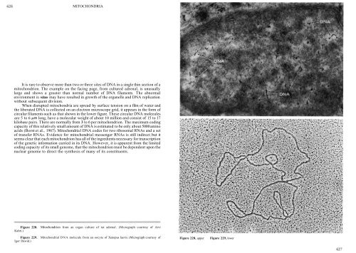
It is rare to observe more than two or three sites of DNA in a single thin section of a<br />
mitochondrion. The example on the facing page, from cultured adrenal, is unusually<br />
large and shows a greater than normal number of DNA filaments. The abnormal<br />
environment in vitro may have resulted in growth of the organelle and DNA replication<br />
without subsequent division.<br />
When disrupted mitochondria are spread by surface tension on a film of water and<br />
the liberated DNA is collected on an electron microscope grid, it appears in the form of<br />
circular filaments such as that shown in the lower figure. These circular DNA molecules<br />
are 5 to 6 pm long, have a molecular weight of about 10 million and consist of 15 to 17<br />
kilobase pairs. There are normally from 3 to 6 per mitochondrion. The maximum coding<br />
capacity of this relatively small amount of DNA is estimated to be only about 5000 amino<br />
acids (Borst et al., 1967). <strong>Mitochondria</strong>1 DNA codes for two ribosomal RNAs and a set<br />
of transfer RNAs. Evidence for mitochondria1 messenger RNAs is still indirect but it<br />
seems clear that each mitochondrion has all of the ingredients necessary for transcription<br />
of the genetic information carried in its DNA. However, it is apparent from the limited<br />
coding capacity of its small genome, that the mitochondrion must be dependent upon the<br />
nuclear genome to direct the synthesis of many of its constituents.<br />
Figure 228. Mitochondrion from an organ culture of rat adrenal. (Micrograph courtesy of Arvi<br />
Kahri.)<br />
Figure 229. <strong>Mitochondria</strong>l DNA molecule from an oocyte of Xenopus laevis. (Micrograph courtesy of<br />
Igor Dawid.)<br />
Figure 228, upper Figure 229, lower