Chapter 7: Mitochondria
Chapter 7: Mitochondria
Chapter 7: Mitochondria

Create successful ePaper yourself
Turn your PDF publications into a flip-book with our unique Google optimized e-Paper software.
NUMBERS AND DISTRIBUTION<br />
The numbers of mitochondria vary over a wide range depending upon the size of the<br />
cell and its energy requirements. Reliable estimates have been made for only a few cell<br />
types. Values for the liver are 800 to 2000; oocytes of echinoderms 150,000; and in the<br />
giant amoeba Chaos chaos as many as half a million.<br />
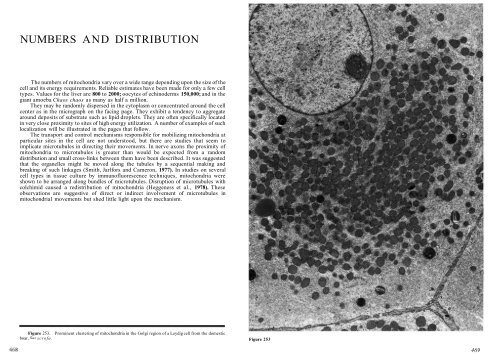
They may be randomly dispersed in the cytoplasm or concentrated around the cell<br />
center as in the micrograph on the facing page. They exhibit a tendency to aggregate<br />
around deposits of substrate such as lipid droplets. They are often specifically located<br />
in very close proximity to sites of high energy utilization. A number of examples of such<br />
localization will be illustrated in the pages that follow.<br />
The transport and control mechanisms responsible for mobilizing mitochondria at<br />
particular sites in the cell are not understood, but there are studies that seem to<br />
implicate microtubules in directing their movements. In nerve axons the proximity of<br />
mitochondria to microtubules is greater than would be expected from a random<br />
distribution and small cross-links between them have been described. It was suggested<br />
that the organelles might be moved along the tubules by a sequential making and<br />
breaking of such linkages (Smith, Jarlfors and Cameron, 1977). In studies on several<br />
cell types in tissue culture by immunofluorescence techniques, mitochondria were<br />
shown to be arranged along bundles of microtubules. Disruption of microtubules with<br />
colchimid caused a redistribution of mitochondria (Heggeness et al., 1978). These<br />
observations are suggestive of direct or indirect involvement of microtubules in<br />
mitochondria1 movements but shed little light upon the mechanism.<br />
Figure 253. Prominent clustering of mitochondria in the Golgi region of a Leydig cell from the domestic<br />
boar, Sus scrofa. Figure 253<br />
469