Chapter 7: Mitochondria
Chapter 7: Mitochondria
Chapter 7: Mitochondria

Create successful ePaper yourself
Turn your PDF publications into a flip-book with our unique Google optimized e-Paper software.
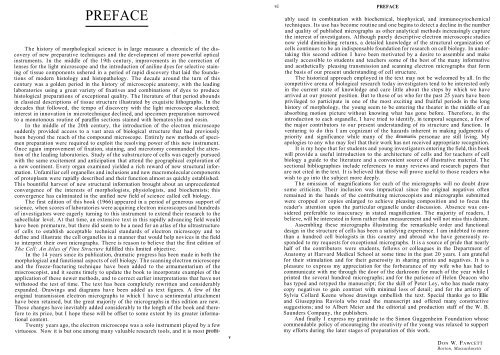
PREFACE<br />
The history of morphological science is in large measure a chronicle of the dis-<br />
covery of new preparative techniques and the development of more powerful optical<br />
instruments. In the middle of the 19th century, improvements in the correction of<br />
lenses for the light microscope and the introduction of aniline dyes for selective stain-<br />
ing of tissue components ushered in a period of rapid discovery that laid the founda-<br />
tions of modern histology and histopathology. The decade around the turn of this<br />
century was a golden period in the history of microscopic anatomy, with the leading<br />
laboratories using a great variety of fixatives and combinations of dyes to produce<br />
histological preparations of exceptional quality. The literature of that period abounds<br />
in classical descriptions of tissue structure illustrated by exquisite lithographs. In the<br />
decades that followed, the tempo of discovery with the light microscope slackened;<br />
interest in innovation in microtechnique declined, and specimen preparation narrowed<br />
to a monotonous routine of paraffin sections stained with hematoxylin and eosin.<br />
In the middle of the 20th century, the introduction of the electron microscope<br />
suddenly provided access to a vast area of biological structure that had previously<br />
been beyond the reach of the compound microscope. Entirely new methods of speci-<br />
men preparation were required to exploit the resolving power of this new instrument.<br />
Once again improvement of fixation, staining, and microtomy commanded the atten-<br />
tion of the leading laboratories. Study of the substructure of cells was eagerly pursued<br />
with the same excitement and anticipation that attend the geographical exploration of<br />
a new continent. Every organ examined yielded a rich reward of new structural infor-<br />
mation. Unfamiliar cell organelles and inclusions and new macromolecular components<br />
of protoplasm were rapidly described and their function almost as quickly established.<br />
This bountiful harvest of new structural information brought about an unprecedented<br />
convergence of the interests of morphologists, physiologists, and biochemists; this<br />
convergence has culminated in the unified new field of science called cell biology.<br />
The first edition of this book (1966) appeared in a period of generous support of<br />
science, when scores of laboratories were acquiring electron microscopes and hundreds<br />
of investigators were eagerly turning to this instrument to extend their research to the<br />
subcellular level. At that time, an extensive text in this rapidly advancing field would<br />
have been premature, but there did seem to be a need for an atlas of the ultrastructure<br />
of cells to establish acceptable technical standards of electron microscopy and to<br />
define and illustrate the cell organelles in a manner that would help novices in the field<br />
to interpret their own micrographs. There is reason to believe that the first edition of<br />
The Cell: An Atlas of Fine Structure fulfilled this limited objective.<br />
In the 14 years since its publication, dramatic progress has been made in both the<br />
morphological and functional aspects of cell biology. The scanning electron microscope<br />
and the freeze-fracturing technique have been added to the armamentarium of the<br />
miscroscopist, and it seems timely to update the book to incorporate examples of the<br />
application of these newer methods, and to correct earlier interpretations that have not<br />
withstood the test of time. The text has been completely rewritten and considerably<br />
expanded. Drawings and diagrams have been added as text figures. A few of the<br />
original transmission electron micrographs to which I have a sentimental attachment<br />
have been retained, but the great majority of the micrographs in this edition are new.<br />
These changes have inevitably added considerably to the length of the book and there-<br />
fore to its price, but I hope these will be offset to some extent by its greater informa-<br />
tional content.<br />
Twenty years ago, the electron microscope was a solo instrument played by a few<br />
virtuosos. Now it is but one among many valuable research tools, and it is most profit-<br />
v<br />
PREFACE<br />
ably used in combination with biochemical, biophysical, and immunocytochemical<br />
techniques. Its use has become routine and one begins to detect a decline in the number<br />
and quality of published micrographs as other analytical methods increasingly capture<br />
the interest of investigators. Although purely descriptive electron microscopic studies<br />
now yield diminishing returns, a detailed knowledge of the structural organization of<br />
cells continues to be an indispensable foundation for research on cell biology. In under-<br />
taking this second edition I have been motivated by a desire to assemble and make<br />
easily accessible to students and teachers some of the best of the many informative<br />
and aesthetically pleasing transmission and scanning electron micrographs that form<br />
the basis of our present understanding of cell structure.<br />
The historical approach employed in the text may not be welcomed by all. In the<br />
competitive arena of biological research today investigators tend to be interested only<br />
in the current state of knowledge and care little about the steps by which we have<br />
arrived at our present position. But to those of us who for the past 25 years have been<br />
privileged to participate in one of the most exciting and fruitful periods in the long<br />
history of morphology, the young seem to be entering the theater in the middle of an<br />
absorbing motion picture without knowing what has gone before. Therefore, in the<br />
introduction to each organelle, I have tried to identify, in temporal sequence, a few of<br />
the major contributors to our present understanding of its structure and function. In<br />
venturing to do this I am cognizant of the hazards inherent in making judgments of<br />
priority and significance while many of the dramatis personae are still living. My<br />
apologies to any who may feel that their work has not received appropriate recognition.<br />
It is my hope that for students and young investigators entering the field, this book<br />
will provide a useful introduction to the architecture of cells and for teachers of cell<br />
biology a guide to the literature and a convenient source of illustrative material. The<br />
sectional bibliographies include references to many reviews and research papers that<br />
are not cited in the text. It is believed that these will prove useful to those readers who<br />
wish to go into the subject more deeply.<br />
The omission of magnifications for each of the micrographs will no doubt draw<br />
some criticism. Their inclusion was impractical since the original negatives often<br />
remained in the hands of the contributing microscopists and micrographs submitted<br />
were cropped or copies enlarged to achieve pleasing composition and to focus the<br />
reader's attention upon the particular organelle under discussion. Absence was con-<br />
sidered preferable to inaccuracy in stated magnification. The majority of readers, I<br />
believe, will be interested in form rather than measurement and will not miss this datum.<br />
Assembling these micrographs illustrating the remarkable order and functional<br />
design in the structure of cells has been a satisfying experience. I am indebted to more<br />
than a hundred cell biologists in this country and abroad who have generously re-<br />
sponded to my requests for exceptional micrographs. It is a source of pride that nearly<br />
half of the contributors were students, fellows or colleagues in the Department of<br />
Anatomy at Harvard Medical School at some time in the past 20 years. I am grateful<br />
for their stimulation and for their generosity in sharing prints and negatives. It is a<br />
pleasure to express my appreciation for the forbearance of my wife who has had to<br />
communicate with me through the door of the darkroom for much of the year while I<br />
printed the several hundred micrographs; and for the patience of Helen Deacon who<br />
has typed and retyped the manuscript; for the skill of Peter Ley, who has made many<br />
copy negatives to gain contrast with minimal loss of detail; and for the artistry of<br />
Sylvia Collard Keene whose drawings embellish the text. Special thanks go to Elio<br />
and Giuseppina Raviola who read the manuscript and offered many constructive<br />
suggestions; and to Albert Meier and the editorial and production staff of the W. B.<br />
Saunders Company, the publishers.<br />
And finally I express my gratitude to the Simon Guggenheim Foundation whose<br />
commendable policy of encouraging the creativity of the young was relaxed to support<br />
my efforts during the later stages of preparation of this work.<br />
DON W. FAWCETT<br />
Boston, Massachusetts