NO - Besoin d'assistance
NO - Besoin d'assistance
NO - Besoin d'assistance

You also want an ePaper? Increase the reach of your titles
YUMPU automatically turns print PDFs into web optimized ePapers that Google loves.
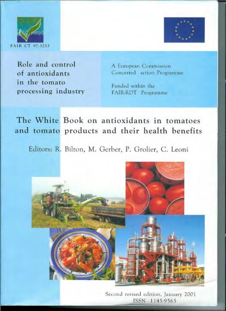
Introduction<br />
The European Commission Concerted Action FAIR CT97-3233 was set up in 1997 following<br />
the publication of several scientific papers showing a possible role of lycopene in the<br />
prevention of diseases such as cancer and cardiovascular diseases. Epidemiological studies<br />
showed that these risks increased with low tomato consumption, the main source of lycopene.<br />
A consortium of 15 tomato research institutes and tomato processors from all around Europe<br />
was formed, with funding from the European Commission within the framework of the FAIR<br />
RTD programme, to review the scientific literature available in order to produce a "white<br />
book" on the antioxidants in tomatoes and tomato products. This data would help the industry<br />
improve the nutritional quality of the tomato-based products and the scientists to understand<br />
better the relationship between nutrition and health.<br />
The researchers were organised into 4 working group to study different aspects:<br />
• Working Group 1 : Composition of tomatoes and tomato products in antioxidants<br />
(methods of analysis; breeding and biotechnology to increase the antioxidant content;<br />
evolution of the composition during ripening and storage), chaired by Dr. Pascal Grolier<br />
(INRA, Clermont-Ferrand, France)<br />
• Working Group 2 : Processing and bioavailability (effects of mechanical and thermal<br />
treatments and storage conditions on the antioxidants content and their bioavailability),<br />
chaired by Dr. Carlo Leoni (SSICA, Italy)<br />
• Working Group 3 : Epidemiological observational studies (protection offered by<br />
antioxidants on cardiovascular diseases, cancer and ageing), chaired by Dr. Mariette Gerber<br />
(INSERM-CRLC, Montpellier, France)<br />
• Working Group 4 : Mechanisms and biomarkers (mechanisms involved in the<br />
protection and biomarkers), chaired by Prof. Rod Bilton (Liverpool John Moores University,<br />
UK).<br />
Project management and co-ordination were provided by Prof. Gérard Bartholin (CTCPA,<br />
Avignon, France), Mr. Bernard Bièche & Mrs Sophie Colvine (AMITOM, Avignon, France).
Composition of tomatoes and tomato products in antioxidants<br />
Working group 1 - Composition of tomatoes and tomato products in antioxidants<br />
Chairman: P. Grolier, INRA, UMMM, Vitamin group, 63122 St Genes Champanelle,<br />
France.<br />
Authors:<br />
P. Grolier 1 , G. Bartholin 2 , L. Broers 3 , C. Caris-Veyrat 4 , M. Dadomo 5 , G. Di Lucca 6 , Y.<br />
Dumas 7 , F. Meddens 3 , L. Sandei 8 , W. Schuch 9 .<br />
1) INRA, UMMM, Vitamin group, 63122 St Genes Champanelle, France.<br />
2) CTCPA, Station Pierre Mainguy, ZA de l’aéroport, BP 1203, 84911 Avignon Cedex 09,<br />
France.<br />
3) Nunhems Zaden BV, PO Box 4005, 6080 AA Haalen, Netherlands.<br />
4) UMR INRA-UAPV "Sécurité et Qualité des Produits d'origine Végétale", Site Agroparc,<br />
Domaine Saint Paul, 84914 Avignon Cedex 09, France.<br />
5) Azienda Agraria Sperimentale Stuard, Str. Madonna dell’Aiuto, 7/A, 43016 S. Pancrazio,<br />
Italy.<br />
6) Sviluppo Produzioni Agricole, Tenuta La Fagianeria, 81015 Piana di Monte Verna, Italy.<br />
7) INRA, Station d’Ecophysiologie et Horticulture, Domaine Saint Paul, Site Agroparc,<br />
84914 Avignon Cedex 9, France.<br />
8) Stazione Sperimentale per l’Indusria delle Conserve Alimentari in Parma, Via Tanara<br />
31/A, 43100 Parma, Italy.<br />
9) Zeneca Plant Science, 3 Marston Lane, Eaton, NR4 6LZ, Norwich, UK.
Composition of tomatoes and tomato products in antioxidants (WG1) page 2<br />
__________________________________________________________________________________________<br />
Acknowledgements<br />
The authors wish to thank Peter Bramley from the University of London (UK) for it’s<br />
participation at the proceedings of the tomato and health seminar in Pamplona (1998). We<br />
also wish to thank Wilhelm Stahl from the University of Dusseldorf (Germany) and Marie-<br />
Josèphe Amiot from the INRA center of Avignon (France) for the reviewing of our<br />
manuscript.
Composition of tomatoes and tomato products in antioxidants (WG1) page 3<br />
__________________________________________________________________________________________<br />
Contents<br />
p2 Acknowlegdments<br />
p3 Contents<br />
p9 1. Introduction<br />
p10 2. Objectives<br />
p11 3. Composition of tomatoes in antioxydants<br />
p15 4. Breeding and biotechnology methods for high lycopene content<br />
p17 4.1. Breeding<br />
p18 4.2. Genetic modification approach<br />
p19 5. Influence of maturity stages and storage on the antioxidant composition of tomatoes<br />
p20 6. Influence of cultural practices and agronomic aspects on antioxidants content of<br />
tomato fruit<br />
p20 6.1. Lycopene, β-carotene and total carotene<br />
p20 6.1.1. Introduction<br />
p21 6.1.2. Influence of temperature<br />
p24 6.1.3. Influence of light<br />
p25 6.1.4. Influence of cultural techniques
Composition of tomatoes and tomato products in antioxidants (WG1) page 4<br />
__________________________________________________________________________________________<br />
p25 6.1.4.1. Variety<br />
p26 6.1.4.2. Water<br />
p27 6.1.4.3. Mineral nutrition<br />
p31 6.1.5. Ripening stage<br />
p35 6.1.6. Growth and development regulators<br />
p39 6.2. Vitamin C<br />
p39 6.2.1. Introduction<br />
p40 6.2.2. Influence of temperature<br />
p40 6.2.3. Influence of light<br />
p42 6.2.4. Influence of cultural techniques<br />
p42 6.2.4.1. Variety<br />
p42 6.2.4.2. Water<br />
p43 6.2.4.3. Mineral nutrition<br />
p48 6.2..4.4. Ripening stage<br />
p49 6.2.4.5. Growth and development regulators<br />
p50 6.3. Vitamin E<br />
p50 6.3.1. Introduction
Composition of tomatoes and tomato products in antioxidants (WG1) page 5<br />
__________________________________________________________________________________________<br />
p50 6.3.2. Influence of cultural techniques<br />
p50 6.3.2.1. Variety<br />
p50 6.3.2.2. Ripening stage<br />
p51 6.4. Phenolic compounds<br />
p51 6.4.1. Introduction<br />
p51 6.4.2. Influence of light<br />
p52 6.4.3. Influence of cultural techniques<br />
p52 6.4.3.1. Variety<br />
p52 6.4.3.2. Mineral nutrition<br />
p53 6.4.3.3. Fruit development and ripening stage<br />
p54 6.4.3.4. Influence of season<br />
p54 6.4.4. Conclusions<br />
p55 6.5. Conclusions on the influence of cultural practices and agronomic aspects<br />
p57 7. Physico-chemical characteristics and properties.<br />
p57 7.1. Lycopene<br />
p57 7.1.1. Chemical structure
Composition of tomatoes and tomato products in antioxidants (WG1) page 6<br />
__________________________________________________________________________________________<br />
p57 7.1.2. Physico-chemical characteristics and properties<br />
p57 7.1.2.1. Melting point<br />
p57 7.1.2.2. UV-vis spectrophotometry<br />
p59 7.1.2.3. IR Spectroscopy<br />
p59 7.1.2.4. Solubility<br />
p59 7.1.2.5. Sensitivity<br />
p61 7.1.2.6. Influence of the environment on physico-chemical<br />
properties.<br />
p61 7.1.3. Lycopene cis isomers.<br />
p61 7.1.3.1. Structures<br />
p62 7.1.3.2. Occurrence<br />
p62 7.1.2.3. Physico-chemical properties<br />
p64 7.1.4. Antioxidant properties<br />
p64 7.1.4.1. Quenching of singlet oxygen<br />
p65 7.1.4.2. Quenching of free radicals<br />
p67 7.2. Vitamin C<br />
p67 7.2.1. Chemical structure<br />
p67 7.2.2. Physico-chemical characteristics
Composition of tomatoes and tomato products in antioxidants (WG1) page 7<br />
__________________________________________________________________________________________<br />
p68 7.2.3. Antioxidant properties<br />
p69 7.3. Vitamin E<br />
p71 7.3.1. Physico-chemical characteristics<br />
p71 7.3.2. Occurrence<br />
p71 7.3.3. Antioxidant properties<br />
p72 8. Antioxidant analysis in foods and biological tissues<br />
p72 8.1. Samples<br />
p73 8.2. Storage of the samples<br />
p75 8.3. Standards<br />
p77 8.4. Extraction<br />
p81 8.5. Analysis<br />
p87 9. Description of specific methods for tomato antioxidant analyses<br />
p87 9.1. Lycopene analysis on tomato matrix<br />
p87 9.1.1. Fresh tomatoes<br />
p87 9.1.2. Tomato Puree (light) (Passata di pomodoro) and tomato paste<br />
p87 9.1.3. Peeled tomatoes and crushed tomatoes<br />
p88 9.1.4. Peels from peeled tomatoes.
Composition of tomatoes and tomato products in antioxidants (WG1) page 8<br />
__________________________________________________________________________________________<br />
p88 9.1.5. Extractions<br />
p88 9.1.6. Recommendations for working with lycopène<br />
p89 9.1.7. Differences verified with Hart and Scott method<br />
p89 9.2. Vitamin C in tomatoes and in tomato products.<br />
p89 9.2.1. Processing effects on Vitamin C<br />
p90 9.2.2. Determination of ascorbic acid in tomato products<br />
p90 9.2.2.1. Volumetric titration<br />
p91 9.2.2.2. Determination of ascorbic acid by HPLC<br />
p93 9.3. Polyphenols<br />
p93 9.3.1. Colorimetric method<br />
p93 9.3.2. HPLC determination<br />
p95 10. Conclusions about analysis data<br />
p96 References
Composition of tomatoes and tomato products in antioxidants (WG1) page 9<br />
__________________________________________________________________________________________<br />
1. Introduction<br />
Several epidemiological studies suggest that the consumption of fruit and vegetables<br />
would be beneficial to human health by reducing the risk of developing cancer and cardiovascular<br />
diseases (Koo 1997, Mayne 1996). However, this protective aspect is more difficult<br />
to estimate when one particular group of vegetables or one particular family of nutrients or<br />
micro-nutrients is considered (WCRF/AICR 1997). Fruit and vegetables are rich in<br />
constituents likely to contribute to the prevention mechanisms and the use of only one group<br />
of the constituents cannot totally demonstrate the potential interaction amongst them.<br />
Recently a great deal of attention has been focused on the tomato and its products.<br />
Studies from Giovannucci et al.(1995) and Franceschi et al.(1994) reported that the<br />
consumption of tomatoes, tomato sauce and pizza was associated with a reduced risk of<br />
developing digestive tract and prostate cancers. Tomatoes are also one of the main part of the<br />
Mediterranean diet which has been associated with a low mortality from cardio-vascular<br />
troubles. Because tomatoes constitute the almost exclusive source of lycopene, this pigment<br />
could be one of the active agents of this protection.<br />
Experimental studies reported that lycopene exhibited antioxidant activities (Di<br />
Mascio et al, 1989), suppressed cell proliferation (Levy et al, 1995) and interfered with the<br />
growth cancer cells (Clinton 1998). However, tomatoes are also rich sources of the essential<br />
nutrients vitamin C, potassium and folic acid, as well as beta-carotene, gamma-carotene,<br />
phytoene, selenium, flavonoids and phenolic acids which may exhibit antioxidant,<br />
immunostimulant, photoprotector or even chemopreventive activities on in vitro and animal<br />
models. In consequence, several of these constituents would contribute to the diseasepreventive<br />
properties.<br />
To date, limited information is available about specific composition of fruits and<br />
vegetables, including tomatoes. Mangels et al.(1993) published a food composition database<br />
for the 5 major dietary carotenoids, but no adequate table exists covering the composition in<br />
other antioxidant compounds.
Composition of tomatoes and tomato products in antioxidants (WG1) page 10<br />
__________________________________________________________________________________________<br />
The quality of a product can also be related to its composition in antioxidants. Thus,<br />
with tomatoes, the stability of the processed products, the nutritional value, the colour<br />
retention, the organoleptic qualities could be changed as a function of β-carotene, lycopene,<br />
flavonoids and vitamin C contents. Further insight into the antioxidant composition of<br />
tomatoes and the factors likely to modify this composition should help to better define the<br />
quality of tomatoes, to evaluate the intake of each antioxidant of humans and to assess the<br />
potential interactions among each of them during nutrition and public health studies. Such<br />
investigation would also contribute to improve information available to breeding and<br />
biotechnology companies, processing industries, health institutes and consumers.<br />
2. Objectives<br />
Our working group focused on the data collection of antioxidant composition in<br />
tomato and tomato products. A first database contains average values of all antioxidants we<br />
have found in literature. The antioxidant levels were reported for fresh tomatoes, but only few<br />
papers have reported values for specific varieties. So, information has been collected about<br />
the tomato varieties mainly used for the processing industry, and the concentrations of<br />
carotenoids and vitamin C have been collected in a second database.<br />
Beside fruit ripening, there are a number of factors that influence taste, colour and<br />
antioxidant levels in tomato products. The effects related to tomato processing are reported by<br />
the working group two.<br />
Different strategies can be applied in order to improve or to modulate the tomato<br />
quality and especially the carotenoid and ascorbic acid levels. Biosynthesis pathways of these<br />
compounds are described with a specific attention on the genes and enzymes involved in the<br />
metabolic steps. Breeding selection and biotechnology approach are presented.<br />
The fruit colour and the antioxidant levels can progress during ripening. Cultural<br />
practices can also modulate both parameters. Thus, influences of cultural practices and<br />
ripening are described in this report.
Composition of tomatoes and tomato products in antioxidants (WG1) page 11<br />
__________________________________________________________________________________________<br />
Through epidemiological studies, associations between diseases and antioxidant<br />
consumption or blood antioxidant levels have been examined by the working group three.<br />
Thus, discussion about epidemiological data largely depends on the measured antioxidant<br />
values and on the method used. The different current methods are listed and critically<br />
described. Physical and chemical properties of lycopene, beta-carotene and vitamin C are<br />
described.<br />
3. Composition of tomatoes in antioxidants<br />
In the United States, about 29% of daily lycopene intakes (0.5-5 mg/day) come from<br />
tomato sauces, 12% from ketchup, 8% from juice, 8% from pizzas (total from processing<br />
tomato products: 57%) and only 12% from fresh tomatoes (Chug-Ahuja et al.1993). Thus, it<br />
appears necessary to collect composition data not only for the market tomatoes but also for<br />
processing tomatoes.<br />
Composition tables show that the tomato (Lycopersicon esculentum) contains 93-95%<br />
water and low levels of solid matter. Tomatoes are relatively rich in antioxidants: vitamin C<br />
(160-240 mg/kg), provitaminic A carotenes (6-9 mg/kg) (Table 1) and phenolic compounds<br />
(Table 2): flavonoids (5-50 mg/kg) and phenolic acids (10-50 mg/kg). Also present in small<br />
quantities are vitamin E (5-20 mg/kg), flavonoids (5-50 mg/kg) and trace elements such as<br />
copper (0.1-0.9 mg/kg), manganese (1-1.5 mg/kg) and zinc (1-2.4 mg/kg) which are present in<br />
several antioxidant enzymes. Most often the tomato variety is not indicated and the reported<br />
values are a mean concentration of the constituents in tomatoes found in local markets. In<br />
addition, tables contain values for the whole fruit but heterogeneous distribution of the<br />
compounds may occur in the fruit. Thus, antioxidant intakes may be different if skin or seeds<br />
are discarded. Hence, the “Campbell 146” variety was shown to contain 4 and 59 mg/kg<br />
whole fruit of β-carotene and lycopene respectively. But the concentration of β-carotene was<br />
4-fold higher in the locular cavity than in the pericarp whereas the concentration of lycopene<br />
was 2-fold higher in the pericarp than in the locular cavity (Davies and Hobson 1981).<br />
Flavonoids and phenolic acids also seem more concentrated in the skin than in the flesh<br />
(Table 2) and vitamin E appears specifically located in seeds (Table 1).
Composition of tomatoes and tomato products in antioxidants (WG1) page 12<br />
__________________________________________________________________________________________<br />
in g<br />
in mg<br />
in µg<br />
Table 1. Basic analytical data for ripe tomato fruit<br />
Database<br />
Composition 1 2 3 4<br />
(/kg fresh matter)<br />
Water 931 942 933 ni<br />
Protein 7.0 9.5 9.0 9.6<br />
Fat 3.0 2.1 2.0 ni<br />
Carbohydrate 31.0 34.5 32.0 ni<br />
Fe 5.0 5.0 4.0 6.0<br />
Cu 0.1 0.9 0.6 0.9<br />
Zn 1.0 2.4 1.4 1.5<br />
Mn 1.0 1.4 1.1 0.9<br />
Vitamin C 170 242 180 160<br />
Vitamin E 12.2 8.0 10.0 ni<br />
Carotene 6.4 8.2 6.0* 7.6<br />
Folates 0.17 0.39 0.23 ni<br />
Se tr 6.0 tr ni<br />
ni : not indicated; * : beta-carotene Equivalent<br />
1 B. Holland, A.A. Welch, I.D. Unwin, D.H. Buss, A.A. Paul and D.A.T. Southgate. The composition of<br />
foods. Fifth revised and extended edition. McCance and Widdowson’s, UK, 1992<br />
(flesh, skin and seeds)<br />
2 H. Scherz and F. Senser. Food composition and nutritional tables 1989/90.5 th revised and completed<br />
edition. Wissenschaftliche Verlagsgesellschaft mbH Stuttgart, 1989.<br />
3 M. Feinberg, J.C. Favier and J. Ireland-Ripert. Répertoire général des aliments. Table de composition<br />
REGAL. Technique et Documentation, Lavoisier, 1991.<br />
4 H.C. Price et al.University of California. Veg. Crops. Series N°178, 171, 1976.<br />
Table 2. Phenolic compounds in fresh red tomato fruit
Composition of tomatoes and tomato products in antioxidants (WG1) page 13<br />
__________________________________________________________________________________________<br />
Concentration<br />
(mg/kg) Part of fruit<br />
__________________________________________________________________<br />
FLAVO<strong>NO</strong>IDS<br />
Quercetin glycosides a 3-7 whole<br />
Kaempferol glycosides a 0.2-0.8 whole<br />
Naringenin 8-42 whole<br />
0.8 flesh<br />
64 skin<br />
Rutin 10-15 whole<br />
PHE<strong>NO</strong>LIC ACIDS<br />
Chlorogenic acid 13–38 whole<br />
Caffeic acid a 29 flesh<br />
56 skin<br />
p-Coumaric acid a 16 whole<br />
Ferulic acid a 7 whole<br />
Sinapic acid a 2 whole<br />
Vanillic acid a<br />
1 whole<br />
Salicylic acid a 1 whole<br />
a : Calculated as the aglycone<br />
After the review made by J. N. Davis and G. E. Hobson, Critical Reviews in Food Science and Nutrition, 15,<br />
205-280, 1981.
Composition of tomatoes and tomato products in antioxidants (WG1) page 14<br />
__________________________________________________________________________________________<br />
Recent chromatographic methods have led to identify in tomatoes at least 17 different<br />
carotenoids of which the colouring and antioxidant properties may vary. Khachick et<br />
al.(1992) reported that tomatoes contain mainly lycopene and also β-carotene, ζ-carotene,<br />
phytofluene and phytoene and traces of lutein, α-carotene etc. The composition of carotenoids<br />
greatly depends on the variety. Such a variation is reported in Table 3 for fresh market<br />
tomato. Some red varieties, like Flavourtop and Beefsteak, contain up to 50 mg/kg of<br />
lycopene, of which 87% in the former and 56% in the latter of trans-isomers and very little β-<br />
carotene (Hart and Scott 1995). By contrast, Tigerella contains similar quantities of the 3<br />
pigments. In the yellow varieties β-carotene is the major pigment. Finally, it should be noted<br />
that the wild varieties can contain up to twice the lycopene and vitamin C quantity of the<br />
cultivated varieties (Stevens and Rick 1986)<br />
The composition of processing tomato vairetiee will be described in paragraph 6.1.4.1.<br />
Table 3. The content of lutein, lycopene and β-carotene in some tomato varieties<br />
(mg/kg)<br />
Lutein trans-Lycopene Total Lycopene trans-β-carotene<br />
Red varieties<br />
Cherry 1.0 26.9 37.8 4.7<br />
Flavourtop 0.5 49.6 56.5 4.3<br />
Tigerella 1.9 12.2 15.8 17.0<br />
Beefsteak 0.9 27.3 48.3 8.8<br />
Craig 1.5 29.5 39.1 10.9<br />
Yellow varieties<br />
Sungold 2.0 3.9 5.3 22.3<br />
Gold sunrise 1.1 0.2 0.2 0.9<br />
Adapted from Hart and Scott, Food Chem. 54: 101-111, 1995.
Composition of tomatoes and tomato products in antioxidants (WG1) page 15<br />
__________________________________________________________________________________________<br />
4. Breeding and biotechnology methods for high lycopene content<br />
The biochemical pathway involved in lycopene production has been largely<br />
elucidated. Until recently, the mevalonate (MVA) (Fig 1) pathway was believed to be the only<br />
route for the provision of the key isoprenoid intermediate, IPP (Britton 1990).<br />
Figure 1. Main stages of carotenoid synthesis in plants<br />
Mevalonate<br />
Isopentenyl diphosphate (IPP)<br />
Geranylgeranydiphosphate (GGDP)<br />
Phytoene synthase Phytoene<br />
Phytoene desaturase Lycopene<br />
Lycopene cyclase β- carotene<br />
Recently a non-MVA pathway has been identified (Schwender et al.1996). This is<br />
localised in the plastid in contrast to the MVA pathway which is localised in the cytoplasm of<br />
the cell. In this pathway, glyceraldehyde 3-phosphate (GAD) and pyruvate are the first<br />
precursors. Pyruvate is first decarboxylated and then condensated on the carbonyl of GAP<br />
yielding 1-deoxy-d-xylulose 5-phosphate and finally IPP. This novel mevalonate-independent<br />
biosynthesis pathway was first demonstrated in bacteria and then was shown to be involved in<br />
the carotenoid synthesis in higher plants.<br />
Then condensation of IPP molecules results in Geranylgeranyl diphosphate (GGDP)<br />
formation. The first C40 carotenoid phytoene is formed from two molecules of GGDP. The<br />
non-coloured phytoene undergoes a series of sequential desaturation reactions to produce<br />
phytofluene, ζ-carotene, neurosporene and finally the coloured lycopene. It was suggested<br />
that during these reactions cis-trans double bond isomerization occured. Finally, cyclization
Composition of tomatoes and tomato products in antioxidants (WG1) page 16<br />
__________________________________________________________________________________________<br />
of the two end-groups results in β-carotene formation. A collection of genes for the early<br />
isoprenoid/carotenoid specific pathway have been isolated from bacterial, yeast, and plant<br />
sources (Bird et al.1981, Sandman G 1994). Up to date details of this are being prepared as<br />
part of the “Neodiet” Concerted Action.<br />
Table 4. Genes that influence the biosynthesis of carotenoids in tomato fruits<br />
Genes Abbreviation Gene action<br />
t, t-v tangerine Structure (cis, trans) of the polyene chain of carotenoid<br />
molecules<br />
B Beta-carotene Cyclization of lycopene in β-carotene<br />
Del Delta-carotene Cyclization of lycopene in δ-carotene<br />
gh, gh-2 ghost Block in the dehydrogenation of phytoene<br />
vo virescent orange Block in dehydrogenation of ζ-carotene<br />
hp, (hp1, hp2,<br />
bs, dr)<br />
high pigment high total carotene content<br />
Ip Intense pigment Idem<br />
og (Crn, Cr,<br />
crn-2, cr2)<br />
old gold Idem<br />
gf green flesh Decrease in chlorophyll degradation with slightly<br />
increase in β-carotene<br />
r (r:3, r-2, r2) yellow flesh Low total carotene content<br />
at apricot Idem<br />
nor non-ripening Low total carotene content and fruit ripening greatly<br />
retarded<br />
rin ripening inhibitor Idem<br />
Nr, Nr-2 Never ripe Idem<br />
alc alcobaca Idem<br />
Gr Green ripe Idem<br />
u, u2 uniform ripening Uniform fruit colour<br />
The availability of the genes (table 4) now opens the way to enhance levels of<br />
lycopene in tomato fruit using the following general strategies:
Composition of tomatoes and tomato products in antioxidants (WG1) page 17<br />
__________________________________________________________________________________________<br />
4.1 Breeding<br />
In a breeding programme it is possible to accumulate the genes that increase the<br />
lycopene content and to eliminate those genes that decrease the lycopene content. There are<br />
several ways to accomplish this:<br />
First of all, it is very important to have a good genetic base to start with. All the<br />
desired agronomic treats need to be available in the breeding nursery. Incorporation of the<br />
genes that increase lycopene content and elimination of genes that decrease the lycopene<br />
content can be done by pedigree selection and backcross programme. That this approach can<br />
be successful has been proven by the Israelian company LycoRed. Together with the Volcani<br />
Institute they developed hybrids with 3 or 4 folds content of lycopene in tomato fruits.<br />
During natural or breeding selections, certain mutations can be produced on genes<br />
which regulate the synthesis of enzymes in the metabolism of carotenoids. For example, the<br />
mutants t accumulate the metabolic precursors of lycopene : phytoene, and ζ-carotene but<br />
contain no lycopene (Table 5) (Tomes 1963).<br />
Table 5- The carotenoid composition of tomato mutants (g/kg dry matter)<br />
Mutant Phytoene Lycopene β-carotene ζ-carotene δ-carotene<br />
normal 2.9 4.4 0.5 - -<br />
r 1.0 - trace - -<br />
t 5.1 - 0.1 4.5<br />
del 1.3 1.7 0.6 0.1 3.3<br />
hp 1.5 5.6 0.7 - -<br />
og 2.5 5.0 0.3 - -<br />
After Tomes 1963.<br />
In this case, the phytoene desaturase is apparently absent from the fruit. The mutant<br />
Del accumulates mainly the δ-carotene which is an intermediary in the synthesis of carotene.<br />
Certain types tested, for example the mutant hp, accumulate lycopene. This can either be the<br />
consequence of over-expression of phytoene desaturase (lycopene synthesis) or of repression<br />
of lycopene cyclase (β-carotene synthesis).
Composition of tomatoes and tomato products in antioxidants (WG1) page 18<br />
__________________________________________________________________________________________<br />
The variations reported for the composition of tomatoes might hinder interpretations<br />
of epidemiological studies in nutrition. At the opposite, such variations can be a welcome tool<br />
for breeding and biotechnology selections. Breeders can, by pedigree selection and backcross<br />
programmes, propose new tomato varieties. Marker assisted breeding (MAB) could be a<br />
welcome tool to fasten the breeding programme. Tomato is very well developed in this<br />
regard. There is a gene map available. Genes are well described and markers are available<br />
(Iso-enzyms, RAPD’s, RFLP’s, AFLP’s and micro-satellites).<br />
4.2 Genetic modification approach<br />
Another strategy to increase carotenoid levels may be to introduce into a plant one or<br />
several genes coding for carotenoid biosynthesis using bacteria vectors such as<br />
Agrobacterium tumefasciens. Such strategy produces new plants, genetically modified,<br />
enriched or depressed in lycopene content (Bird et al.1991, Fray and Grierson 1993) with:<br />
1. overexpression of IPP generating enzymes<br />
2. over-expression of late general isoprenoid enzymes<br />
3. over-expression of carotenoid biosynthesis specific enzymes<br />
4. inhibition of lycopene cyclase.<br />
The preferred strategy uses the following principles:<br />
1. Expression of new genes should be fruit/ripening specific<br />
2. Overexpression of rate-limiting genes is desirable.<br />
3. The use of enzymes which are not subject to metabolic control are desirable.<br />
4. Combination of genes likely to yield highest levels of lycopene deposition.<br />
Therefore, the use of DX Synthase, IPP Isomerase GGPP Synthase, Phytoene<br />
Synthase and the inhibition of lycopene cyclase offer the most promising strategy.<br />
However, there are also other considerations:
Composition of tomatoes and tomato products in antioxidants (WG1) page 19<br />
__________________________________________________________________________________________<br />
1. Tomato mutants exist which deliver high lycopene. One route chosen is to increase plastid<br />
number. Therefore an alternative approach could be the increase of chromoplast number<br />
within the tomato.<br />
2. Another approach could be to store lycopene in the vacuole.<br />
3. A further approach is to use carotenoid associated proteins to increase deposition of<br />
lycopene.<br />
These approaches are more technical challenging as the basic enabling research has<br />
not yet been carried out.<br />
5. Influence of maturity stages and storage on the antioxidant composition of<br />
tomatoes<br />
The colour and the composition of the fruit greatly vary during ripening. In the unripe<br />
fruits, the green colour is associated with chlorophylls and a low quantity of carotenoids. It<br />
should be noted that in these fruits the biosynthesis of carotenoids is active but the<br />
carotenoids are immediately broken down due to photoprotective and antioxidant properties<br />
(Giulano et al.1993, Fraser et al.1994). During the ripening period, the chlorophylls gradually<br />
disappear and become undetectable 7 days after the breaker stage. Simultaneously, even<br />
though the enzymatic activities of phytoene synthetase, phytoene desaturase and lycopene<br />
cyclase are reduced, both the expression of genes coding for phytoene synthetase and<br />
phytoene desaturase and the content in lycopene and β-carotene increase. At the turning stage,<br />
lycopene content considerably increases and can reach 80-100 mg/kg fresh matter at red stage<br />
(Giulano et al.1993, Fraser et al.1994).<br />
The content in chlorogenic acid is the highest in earlier stages of fruit development<br />
and rapidly decreases during fruit ripening reaching less than 20 mg/kg fresh matter at red<br />
stage (cv Ailsa Craig and Pik-Red) (Aziz et al.1973). This evolution is similar for numerous<br />
fruits (Buta and Spaulding 1997). While quinic conjugates of phenolics, like chlorogenic acid,<br />
are declining, glucoside conjugates (p-coumaric-glucose) become high in mature stages,<br />
especially in pulp (around 60 mg/kg fresh matter at red stage for the two cultivars previously<br />
cited). Glucose derivatives were suggested as biomarkers for maturation by Fleuriet and<br />
Macheix (1985). Rutin (or quercetin-3-rutinoside), specially located in tomato pericarp and
Composition of tomatoes and tomato products in antioxidants (WG1) page 20<br />
__________________________________________________________________________________________<br />
well known for its antioxidant properties, also decreases during ripening, especially between<br />
the immature green and mature green stages.<br />
The maximum concentration of ascorbic acid is estimated in tomato fruits at yellow<br />
turning stage (around 250 mg/kg fresh matter for numerous cultivars). This content decreases<br />
during ripening. This decrease is certainly due to its antioxidant function when the ripening<br />
cells absorb high amount of oxygen; this is the result of an increase in respiration,<br />
characteristic of climacteric fruits (with an autocatylitic synthesis of ethylene) (Macheix et<br />
al.1990).<br />
Changes in tocopherols are different among α, β and γ forms. There is an increase in α<br />
and β-tocopherol during ripening reaching 3 mg/kg fresh matter for α and 0.5 mg/kg fresh<br />
matter for β isomer at the red stage; γ-tocopherol reaches a maximum at colour break stage,<br />
between 1 and 1.5 mg/kg fresh matter. Its level declines in a similar manner than ascorbic<br />
acid (Abushita et al.1997).<br />
6. Influence of cultural practices and agronomic aspects on antioxidants content of<br />
tomato fruit<br />
6.1. Lycopene, β-carotene and total carotene<br />
6.1.1. Introduction<br />
The colour of red tomatoes is determined primarily by their lycopene content (Stevens<br />
and Rick, 1986). Johjima and Matsuzoe (1995) have shown that the fruit colour values (a/b)<br />
were highly correlated with lycopene content, including cis and trans forms. But according to<br />
Koskitalo and Ormrod (1972) surface colour could not be used to specify accurately the<br />
internal colour of fully ripened tomatoes. At lycopene concentrations between 32 and<br />
43 mg/kg fresh matter, fruit colour turns from orange to red and satisfactory red coloration<br />
required a total carotene concentration in excess of 55 mg/kg fresh matter of which lycopene<br />
accounted for 90% of the carotenes. Giovanelli et al.(1999) measured the colour changes at<br />
seven stages during vine and post-harvest ripening on two genotypes (Normal Red and
Composition of tomatoes and tomato products in antioxidants (WG1) page 21<br />
__________________________________________________________________________________________<br />
Crimson) of the tomato cv. Moneymaker grown in a greenhouse. Instrumental determination<br />
of red colour and evaluation of a*/b* index (Hunter’s ratio of red-to-green component of<br />
colour measured with a tristimulus Minolta Chromameter mod CR-210) made on the whole<br />
fruit skin were the most sensitive and significant indicators for fruit maturity, but they did not<br />
show a direct, unequivocal correlation with the lycopene content, since the same a*/b* value<br />
could correspond to lycopene contents differing by 100%.<br />
From various sources of data in the 70’s, tomato lycopene content may range from 43<br />
to 181 mg/kg fresh matter, with most frequent values between 55 and 80 (Davies and Hobson,<br />
1981). Sharma and Le Maguer (1996b) established that skins contained about five times more<br />
lycopene (540 mg/kg fresh matter; wet basis) than the whole tomato pulp (110 mg/kg). Most<br />
of the lycopene is probably attached to the insoluble and fibre portion of the tomatoes. The<br />
lycopene content in tomato pulp ranged from 64.6 to 107 mg/kg fresh matter (according to<br />
cultivars) whereas it ranged from 354 to 536 mg/kg in the wet insoluble fraction and from<br />
0.074 to 0.34 mg/kg in the soluble fraction. It would be expected that tomatoes with high<br />
insoluble solids would contain more lycopene. In addition, Moretti et al.(1998) have shown<br />
that fruit bruising at the breaker stage could significantly decrease (-37%) the total<br />
carotenoids content in the locular tissue of tomato fruit at the ripe stage.<br />
Many factors may influence the concentration of lycopene in tomato fruit:<br />
environmental factors (light, temperature) and cultural practices (variety, mineral nutrition,<br />
water alimentation, harvest date through ripening stage...).<br />
6.1.2. Influence of temperature<br />
McCollum (1954) had observed that tomato fruits exposed to direct sunlight in the<br />
field often developed poor colour due primarily to a low content of lycopene, because of the<br />
high temperatures of the exposed fruits. Lycopene synthesis in excised fruits from cultivars<br />
with various strains of pigments was drastically inhibited at 32°C in every strain which<br />
produced this pigment (Tomes, 1963).<br />
From the breaker point, Koskitalo and Ormrod (1972) distributed greenhouse-grown<br />
tomato plants (c.v. Early Red Chief) in controlled environment chambers at 4 night/day
Composition of tomatoes and tomato products in antioxidants (WG1) page 22<br />
__________________________________________________________________________________________<br />
temperature regimes respectively. Twenty-one days later, the fruit carotenoids contents were<br />
decreased for lower regimes, except for β-carotene (Table 6).<br />
Table 6. Effect of night/day temperature on carotenoid pattern in tomato (mg/kg fresh<br />
Temperature<br />
(°C)<br />
matter) (after Koskitalo and Ormrod, 1972)<br />
Phytoene Phytofluene β-Carotene Lycopene<br />
17.8/25.6 19.2 8.2 2.2 64.8<br />
7.2/18.3 19.1 7.2 3.4 51.5<br />
4.4/15.6 16.0 6.0 3.6 31.9<br />
2.8/13.9 13.2 4.6 3.7 24.2<br />
Baqar and Lee (1978) showed that 30°C temperature drastically reduced carotene<br />
synthesis in cultivar Rouge de Marmande, except for β-carotene (Table 7) and Grierson and<br />
Kader (1986) confirmed that the synthesis of lycopene, but not β-carotene, was inhibited by<br />
temperatures within the range 30-35°C.<br />
Table 7. Temperature effect on carotene synthesis (after Baqar and Lee, 1978)<br />
Carotene Carotene content (mg/kg fresh matter)<br />
20°C 30°C<br />
Phytoene 18.4 5.6<br />
Phytofluene 10.5 0.5<br />
β-Carotene 7.5 6.6<br />
ξ-Carotene 1.0 0.1<br />
γ-Carotene 0.5 0.1<br />
Neurosporene 0.4 0.1<br />
Lycopene 40.5 1.1<br />
Total 78.9 14.1
Composition of tomatoes and tomato products in antioxidants (WG1) page 23<br />
__________________________________________________________________________________________<br />
In fruit pericarp sections stored at various temperatures, Hamauzu et al.(1998) have<br />
observed that, at 20°C, the biosynthesis and accumulation of phytoene and lycopene were<br />
fast, and those of β-carotene was slow; by contrast, at 30°C, the biosynthesis and<br />
accumulation of lycopene and β-carotene were fast and phytoene accumulation was slow; at<br />
35°C the biosynthesis and accumulation of β-carotene was fast but slower that at 30°C and<br />
accumulation levels of phytoene and lycopene were very slow. Hamauzu et al.(1998) have<br />
postulated that high temperature (35°C) inhibits the accumulation of lycopene in tomato fruit<br />
because of the stimulation of the conversion of lycopene to β-carotene.<br />
In conclusion, the formation of lycopene depends on temperature range and seems to<br />
occur between 12°C and 32°C (Leoni, 1992). It is optimum in cell suspension culture of<br />
VFNT tomato for temperature between 18 and 26°C (Robertson et al, 1995). It is also<br />
optimum in the temperature range 16-26°C for tomato fruits (cv. Sunny) harvested at the<br />
pink-ripe stage and stored for ripening (Türk et al.1994) as presented in Table 8. The<br />
production of lycopene is hindered by excessive sunlight and the best conditions are<br />
temperature high enough together with dense foliage to protect fruit from direct exposure to<br />
the sun (Leoni, 1992). However in processing tomatoes, lycopene accumulation was enhanced<br />
by 5% at incubation temperatures of 30 and 34°C and by 33% at 37°C while lycopene<br />
accumulation declined in salad tomatoes above 30°C (Boothman et al, 1996). Indeed,<br />
processing tomatoes may often be exposed to temperatures of 35-40°C during the storage<br />
period prior to processing and over-ripening may occur accompanied with high lycopene<br />
content.<br />
Table 8. Total lycopene formation (mg/kg fresh matter) in tomato fruits (cv. Sunny)<br />
harvested at the pink-ripe stage and stored under the conditions of 0 and 18°C and 90%<br />
Relative Humidity (after Türk et al, 1994)<br />
Storage temperature Storage duration (weeks)<br />
(°C) 1 2 3 4 5 6<br />
Total lycopene 0 3.58 3.50 4.87 5.63 15.37 15.66<br />
18 3.58 4.79 7.75 37.83 - -
Composition of tomatoes and tomato products in antioxidants (WG1) page 24<br />
__________________________________________________________________________________________<br />
6.1.3. Influence of light<br />
At favourable temperatures (22-25°C) the rate of development of lycopene is<br />
increased by illumination. Light is effective until chlorophyll disappears. Carotene is<br />
increased by illumination of tomato fruits during ripening. If exposed to direct sunlight during<br />
development, the fruits will be higher in carotene when ripe than shaded fruits (McCollum,<br />
1954).<br />
Lipton (1970) found that the incidence of defective coloration of the shoulders or sides<br />
of fruits was higher in fruit exposed to the sun than in fruit shaded by foliage and seemed to<br />
be influenced by infrared and short-wave radiation. Thomas and Jen (1975) established that<br />
red light and the intensity of red light had a positive effect on carotenoid synthesis of detached<br />
mature-green tomato fruit and this effect was not a temperature effect. Below saturation but<br />
above a critical minimum, the phytochrome-mediated response to red light depended linearly<br />
on the log of the incident energy. The involvement of phytochrome in carotenoid biosynthesis<br />
was suggested. The far-red light suppressed carotenoid production, as compared to dark<br />
control.<br />
Cabibel and Ferry (1980) compared the content in carotenoids of tomato fruits<br />
produced under glass, plastic tunnel or in open field. β-Carotene content was equal or lower<br />
under glass or plastic tunnel than in open field. The lycopene content increased from glass to<br />
plastic tunnel to field. Carotenoid's contents were higher in open field than under glass or<br />
plastic tunnel. The level of intercepted light may influence carotenoid content. But<br />
interactions with high temperatures in protected cultivation may also have occurred.<br />
Adegoroye and Jolliffe (1987) observed that in tissues of fruit directly exposed to<br />
radiation (650 W m -2 from incandescent lamps inducing visible injury), lycopene<br />
accumulation was blocked. Lycopene is the carotenoid mainly affected by exposure to intense<br />
solar radiation. It has been suggested that radiation injury in tomato fruit might be due to<br />
generalised effects of overheating in irradiated tissues.
Composition of tomatoes and tomato products in antioxidants (WG1) page 25<br />
__________________________________________________________________________________________<br />
6.1.4. Influence of cultural techniques<br />
6.1.4.1. Variety<br />
The main tomato varieties cultivated for processing in the E.C. are : Perfect Peel (PS<br />
1296), Hypeel 244, Titano M, Rio Grande, Heinz 7151, Heinz 3044, Isola, Guadalete (PS<br />
121), Snob (EXH 98063), Soprano, Rio Fuego, Avalon, Cannery Row.<br />
Abushita et al.(1997) analysed tomato fruit from various poorly known cultivars<br />
cultivated in Hungary and found variations on more than 1 to 3-fold: β-carotene varied from<br />
1.15 to 3.7 mg/kg fresh matter and total carotenoids from 18.5 to 60.7 mg/kg fresh matter.<br />
Data from P. Spigno (Cirio Ricerche, Acerra, Naples; personal communication of<br />
unpublished data 1998, 1999) give fruit lycopene content for 24 varieties analysed from not<br />
replicated samples in 1998 and for 29 varieties (not all the same as the previous year)<br />
cultivated in the same conditions in 1999. The mean value was 86.0 mg/kg fresh matter in<br />
1998 and 87.0 mg/kg fresh matter in 1999. The maximum range of values was 34 to 150<br />
mg/kg fresh matter in 1998 and 45 to 163 mg/kg fresh matter in 1999, i.e. about 1- to 4-fold.<br />
Table 9 gives data extracted from the Degree Thesis ‘Carotenoid content in tomato<br />
and its processing products’ (Zanetti, 1996/97).<br />
Table 9. Carotenoid content in fresh tomatoes (mg/kg fresh matter) (after Zanetti, 1997)<br />
Cultivar Lycopene Neolycopene(*) β-Carotene Phytofluene(*)<br />
Isola 93.8 17.3 2.4 4.5<br />
EXH 98063 87.9 17.6 2.7 4.3<br />
PS 1296 94.9 18.1 2.5 4.1<br />
PS 121 90.9 17.9 2.6 4.7<br />
Calroma 89.1 17.4 2.4 4.2<br />
(*) as lycopene equivalent
Composition of tomatoes and tomato products in antioxidants (WG1) page 26<br />
__________________________________________________________________________________________<br />
Siviero et al.(2000) have measured fruit lycopene content in hybrids “HP” (high<br />
pigment) cultivated in open field in comparison with Perfectpeel a hybrid not “HP” as control<br />
(Table 10). They found variations ranging almost from 1- to 2-fold.<br />
Table 10. Lycopene content in tomato hybrids “HP” compared with Perfectpeel not<br />
“HP” (after Siviero et al, 2000)<br />
Hybrid Gardner colour a/b Lycopene (mg/kg<br />
fresh matter)<br />
H-01 2.39 118 2510<br />
H-04 2.35 111 2620<br />
DRD 8133 2.51 170 3750<br />
DR 10747 2.60 188 3460<br />
DR 10750 2.40 125 2570<br />
ES 85/95 2.25 113 2520<br />
Nun 1365 2.53 137 3020<br />
Nun 4994 2.40 129 2930<br />
PLX 1129 2.36 101 2440<br />
PS 1617 2.49 175 4210<br />
Forum 2.57 146 3540<br />
Perfectpeel (control) 2.34 116 2710<br />
6.1.4.2. Water<br />
Lycopene (mg/kg<br />
dry matter)<br />
In a study of the effect of 4 irrigation regimes (40, 50, 60 and 70% depletion of<br />
available soil moisture) on tomato cultivars Pusa Ruby, Pusa Early Dwarf and Sioux, the<br />
lycopene content in tomato fruit was reduced by moisture stress (Naphade, 1993). If this were<br />
verified, there could be an antagonism between improving some basic technological<br />
characteristics of the tomato fruit juice (dry matter content, °Brix, acidity) and improving<br />
lycopene content by means of water management at the field level.
Composition of tomatoes and tomato products in antioxidants (WG1) page 27<br />
__________________________________________________________________________________________<br />
On the contrary, in red and pink cherry tomato cultivars, Matsuzoe et al.(1998) found<br />
that total carotene of fully ripe fruits and the amount of lycopene were increased by soil water<br />
deficit. In red and pink large-fruited tomatoes, soil water deficit also tended to increase the<br />
amount of lycopene per fresh matter in the outer pericarp region but it had no effect on the<br />
amount and distribution of β-carotene and xanthophylls (Zushi and Matsuzoe, 1998).<br />
6.1.4.3. Mineral nutrition<br />
Effect of Nitrogen<br />
Aziz (1968) has cultivated tomato cv. Moneymaker in nutrient solution at three levels<br />
of nitrate nitrogen (1.0, 12.9 -considered as normal level- and 15.8 milli-equivalents). Fruit<br />
under the low N level were earlier and higher in gaining both total carotene and lycopene than<br />
those under the normal or the high N levels (1.0, 12.9 and 15.8 m.e. yielded 68, 44 and 38<br />
mg/kg fresh matter fruit of lycopene content respectively). On the contrary yield increased<br />
with N level. It was concluded that for good colour in tomato fruit, N supply should be as low<br />
as possible without reducing fruit yield.<br />
Montagu et al.(1990) cultivated tomatoes in pots filled with peaty loam soil. Various<br />
forms (nitrate, ammonium, compost, blood and bone) of nitrogen were applied at four rates<br />
(0, 150, 300, 600 kg N/ha). Fruit lycopene content tended to increase in average of 30% when<br />
N applied increased, except with compost.<br />
More generally, nitrogen fertilizers have been presented as able to increase the<br />
concentrations of carotenes in plants (Mozafar, 1993), but few specific information seems to<br />
be available.<br />
Effect of Phosphorus<br />
Saito and Kano (1970) found that increased P supply (from 0 to 100 mg/l of nutrient<br />
solution) in hydroponic culture greatly improved the fruit colour and lycopene content. Few<br />
specific information seems to be available.<br />
Effect of Potassium<br />
For tomato plants (var. Fireball) grown in sand culture in a growth chamber (16-h light<br />
period at 24°C and 8-h dark period at 18°C, 65% relative humidity) and at any particular stage
Composition of tomatoes and tomato products in antioxidants (WG1) page 28<br />
__________________________________________________________________________________________<br />
of maturity, the fruit carotenoid contents were increased by increasing levels of K (0 to 10<br />
meq per litre, applied when the first fruit were 1.0-1.5 cm diameter) in the nutrient solution,<br />
except β-carotene content (Trudel and Ozbun, 1970, 1971, Table 11). Lycopene content<br />
sharply increased when the K level in the nutrient solution was increased with a maximum at<br />
8 meq. Chlorophyll (a and b) content of tomato fruit regularly decreased during ripening,<br />
from 8 mg/kg fresh matter to 1.5 mg/kg fresh matter in average. The decline of the green<br />
pigment was more pronounced in high K fruit, and disrupted the grana to a greater extent than<br />
in K-deficient fruit where carotenoid synthesis was lower.<br />
Table 11. Tomato fruit carotenoid content (mg/kg fresh matter) in response to levels of<br />
K in the nutrient solution (after Trudel and Ozbun, 1970, 1971)<br />
K level<br />
(meq/l)<br />
Total<br />
carotenoids<br />
Phytoene Phytofluene β-carotene Lycopene<br />
0 72 11.8 4.1 3.5 36.8<br />
1 75 12.7 4.1 3.6 41.9<br />
2 91 16.2 5.4 3.1 53.6<br />
4 92 15.2 4.9 2.8 52.7<br />
6 110 14.7 5.0 2.8 59.3<br />
8 111 15.1 4.8 2.6 61.5<br />
10 104 16.3 5.3 2.4 52.4<br />
Winsor (1979) showed that increasing rates of potassium applied resulted in a striking<br />
decrease in the incidence of ripening disorders (mainly colour uniformity) as well as in<br />
increasing levels of titratable and total acidities (Table 12).
Composition of tomatoes and tomato products in antioxidants (WG1) page 29<br />
__________________________________________________________________________________________<br />
Table 12. Effect of potassium addition on ripening disorders (after Winsor, 1979)<br />
Cultivar Potassium applied (kg/ha)<br />
404 807 1614<br />
% unevenly ripened Moneymaker 38.9 14.9 2.1<br />
(all forms) J 168 26.1 8.6 1.4<br />
% unevenly ripened Moneymaker 29.5 9.2 1.9<br />
(severe forms only) J 168 18.0 5.0 1.7<br />
It clearly appears that K deficiency may lower the rate of carotenoid synthesis,<br />
particularly lycopene. However, the rates of K supply applied in the study presented above<br />
and which led to responses in ripening uniformity are very high. They should be considered as<br />
non adapted to cultural practices, particularly in modern agriculture which is supposed to<br />
reduce inputs, spare resources and respect environment.<br />
Effect of Calcium<br />
In a soil-pot culture experiment, Subbiah and Perumal (1990) tested the influence of<br />
sources of calcium (calcium oxide, calcium chloride, calcium sulphate), concentrations (0.0%,<br />
0.1%, 0.2% and 0.3% Ca), stages (S1 = 30th day after transplanting, S2 = 45th day after<br />
transplanting, S3 = 60th day after transplanting), and number of sprays (S1, S2, S3, S1+S2,<br />
S1+S3, S1+S2+S3, S2+S3) on physico-chemical properties of tomato fruits, cv. Co.3.<br />
Moreover, all the pots received also 120 kg N/ha, 100 kg P2O5/ha and 100 kg K2O/ha. They<br />
could significantly increase the lycopene content (from 85 to 263, 300 and 340 mg/kg fresh<br />
matter) of the tomato fruit by calcium sprays at 0.0, 0.1, 0.2 and 0.3% Ca concentration<br />
respectively, whatever the source of calcium. It would be interesting and necessary to verify<br />
these results.<br />
On the contrary, Paiva et al.(1998) cultivated tomato plants cv. Jumbo hydroponically<br />
in a greenhouse and imposed different calcium doses in the nutrient solutions. Total carotene<br />
and lycopene levels (expressed in fresh matter) in the tomato fruit decreased with increasing
Composition of tomatoes and tomato products in antioxidants (WG1) page 30<br />
__________________________________________________________________________________________<br />
Ca concentration, possibly due to the simultaneous reduction in potassium absorption because<br />
of competitive effect between the two cations.<br />
Effect of combined Nitrogen-Phosphorus-Potassium and others<br />
Some studies indicate an influence of combined fertilizers on fruit lycopene content.<br />
Bagal et al.(1989) obtained higher lycopene content (cultivar Pusa Ruby) with combined<br />
nitrogen, phosphorus and particularly potassium application (Table 8). Lycopene content<br />
increased even when expressed in relation to soluble solids. Increasing supply of phosphorus<br />
together with nitrogen might result in a decrease in lycopene content. But in this experiment<br />
the cultivar was ancient, it is surprising that °Brix largely varied while fruit moisture<br />
remained constant and, moreover, the values of lycopene content were so low that there might<br />
be a unit error or the data are not reliable. Lacatus et al.(1994) also found that increasing<br />
nitrogen supply (100, 200, 300 kg/ha) combined to various rates of phosphorus (75 and 150<br />
kg/ha) or of potassium (75 and 150 kg/ha) or of half-fermented organic manure (20, 40, 60<br />
t/ha) influenced positively the fruit lycopene content in average : from 61.8 mg/kg fresh<br />
matter for 100N and 200N to 66 mg/kg fresh matter for 300N. But the cultivar used is not<br />
well known.
Composition of tomatoes and tomato products in antioxidants (WG1) page 31<br />
__________________________________________________________________________________________<br />
Table 13. Effect of combined nitrogen, phosphoruss and potassium on tomato lycopene<br />
Fertilizer<br />
treatments (kg/ha)<br />
Fruit<br />
moisture<br />
levels (after Bagal et al, 1989)<br />
Soluble<br />
Solids<br />
Lycopene Lycopene<br />
N P2O5 K2O (%) SS (°Brix) (mg/kg fresh matter) (mg/kg SS) (1)<br />
0 0 0 94.8 4.2 2.0 47.6<br />
100 50 0 94.9 5.4 2.6 48.1<br />
200 100 0 94.8 5.3 2.5 47.2<br />
400 200 0 94.6 4.8 2.1 43.7<br />
100 0 50 94.4 5.6 3.0 53.6<br />
200 0 100 94.1 5.9 3.3 55.9<br />
400 0 200 94.8 6.1 3.4 55.7<br />
100 50 50 94.6 5.6 3.6 64.3<br />
200 100 50 94.7 6.0 3.9 65.0<br />
400 200 200 94.7 5.7 3.7 64.9<br />
(1) added calculation<br />
In conclusion, the available results are either rare (nitrogen, phosphorus) or not always<br />
reliable (calcium, combined fertilizers) or usuable (cultivar) or, when they are well<br />
established, they hardly correspond to current cultural practices.<br />
6.1.5. Ripening stage<br />
Laval-Martin et al.(1972) have reported that during cherry tomato growth and<br />
maturation, the metabolism of carotenoids and of chlorophylls is first preferentially situated in<br />
the center of the fruit and then in the external areas. The increase in foliar-type pigments is<br />
followed by a stagnation or even a reduction (chlorophylls, neoxanthin) while lycophyll and<br />
lycoxanthin continue to increase and new pigments appear which are characteristics of the<br />
tomato fruit (phytofluene and mainly lycopene). Several studies present pigment changes in<br />
tomato fruit during ripening.
Composition of tomatoes and tomato products in antioxidants (WG1) page 32<br />
__________________________________________________________________________________________<br />
Fruits of green Homestead variety were harvested and ripened at 22°C and carotenoids<br />
were analysed at 6 stages of maturity (Meredith and Purcell, 1966). Lycopene content<br />
increased from breaker stage ro red stage (Table 14).<br />
Table 14. Lycopene concentrations in tomato during ripening<br />
(after Meredith and Purcell, 1966)<br />
Stage of maturity Lycopene content (mg/kg fresh matter)<br />
Mature green -<br />
Breakers 0.41<br />
Turning 0.44<br />
Pink 9.01<br />
Light red 22.3<br />
Red 82.8<br />
Trudel and Ozbun (1970) grew tomato plants (var. Fireball) in sand culture in a<br />
growth chamber (16-h light period at 24°C and 8-h dark period at 18°C, 65% relative<br />
humidity), and observed that fruit total carotenoids increased constantly during ripening from<br />
0.1 to 70 mg/kg fresh matter.<br />
Tomato plants (c.v. Early Red Chief) grown in the greenhouse in soil containers were<br />
placed in a controlled environment at 17.8/25.6°C night/day temperature from the breaker<br />
point and fruit were harvested at 7, 14 and 21 days (Koskitalo and Ormrod, 1972); the<br />
concentrations of various carotenes were measured and are reported in Table 15.
Composition of tomatoes and tomato products in antioxidants (WG1) page 33<br />
__________________________________________________________________________________________<br />
Table 15. Time course of carotene levels (mg/kg fresh matter) in tomatoes at various<br />
harvest dates (after Koskitalo and Ormrod, 1972)<br />
Harvest time Phytoene Phytofluene β-Carotene Lycopene<br />
7 days 18.1 5.9 3.0 43.5<br />
14 days 23.0 7.9 2.2 57.8<br />
21 days 19.2 8.2 2.2 64.8<br />
(1) f. m. : fresh matter<br />
In tomato fruit cv. Moneymaker cultivated in greenhouse, Rabinowitch et al.(1975)<br />
observed a rapid fall in chlorophyll content with the onset of the mature-green stage and<br />
chlorophyll disappeared completely at the beginning of the pink stage. During the same<br />
period of time, the tissue content of β-carotene was doubled. Lycopene and its colourless<br />
precursors phytoene and phytofluene began to accumulate following the breaker stage. At the<br />
ripe-red stage, lycopene constituted 95% of the coloured carotenoids, or 73% of the total<br />
carotenoids including phytoene and phytofluene.<br />
Cabibel and Ferry (1980) considered 5 stages : 1 (mature green), 2 (white), 3 (yellow),<br />
4 (pink), 5 (red). The β-carotene content increased regularly in open field while lycopene<br />
increased sharply between the stage pink and the stage red. Lycopene content could be a good<br />
index of maturation degree (Table 16).
Composition of tomatoes and tomato products in antioxidants (WG1) page 34<br />
__________________________________________________________________________________________<br />
Stage of<br />
ripeness<br />
Table 16. Carotenoids content of tomato fruit in open field<br />
Dry matter<br />
content (%)<br />
(after Cabibel and Ferry, 1980)<br />
β-carotene<br />
(mg/kg f. m.)<br />
β-carotene<br />
(mg/kg d. m. (1) ) (2)<br />
Lycopene<br />
(mg/kg f. m.)<br />
1 6.79 6.75 99.41 -<br />
First 2 6.93 7.09 102.31 -<br />
truss 3 7.01 10.99 156.78 -<br />
Lycopene<br />
(mg/kg d. m.) (2)<br />
4 6.72 26.06 387.80 47.13 701.34<br />
5 6.86 30.12 439.07 106.55 1553.2<br />
1 8.34 9.69 116.19 -<br />
Sixth 2 7.75 10.42 134 46 -<br />
truss 3 8.08 15.07 186.51 -<br />
(1) d. m. : dry matter<br />
(2) added calculation<br />
4 8.10 23.91 295.18 28.46 351.36<br />
5 8.33 28.48 341.90 129.15 1550.42<br />
López-Andréu et al.(1986) considered 7 stages of ripeness : I (mature green), II (green<br />
yellow), III (yellow-orange), IV (orange-yellow), V (orange-red), VI (red), VII (deep red).<br />
Lycopene increased from 0.14 mg/kg fresh matter, slightly until stage IV then by a sharp rise<br />
up to 80.77mg/kg at VII. β-carotene increased from 1.05mg/kg (I) to 4.55mg/kg (III) then<br />
decreased to 0.42mg/kg (VII). The changes in lycopene percentage are presented in Table 17.<br />
Table 17. Relative content of lycopene during ripening (after López-Andréu et al, 1986)<br />
Stage I II III IV V VI VII<br />
Lycopene % total carotenoids 12 55 72 82 90 95 99.5
Composition of tomatoes and tomato products in antioxidants (WG1) page 35<br />
__________________________________________________________________________________________<br />
Giovanelli et al.(1999) evaluated the variation in the antioxidant content at seven<br />
stages during vine and post-harvest ripening on two genotypes (Normal Red and Crimson) of<br />
the tomato cv. Moneymaker grown in a greenhouse. In vine-ripened tomatoes lycopene and β-<br />
carotene concentrations progressively increased linearly during the ripening process.<br />
Conversely, in post-harvest-ripened fruit lycopene and β-carotene accumulation followed an<br />
exponential trend. At the end of the experiments, the lycopene and β-carotene concentration<br />
(roughly 125-130 and 12 mg/kg fresh matter respectively) in post-harvest-ripened tomatoes<br />
was almost twice the value reached in vine-ripened tomatoes (roughly 75-80 and 5-7 mg/kg<br />
fresh matter respectively) having the same colour (a*/b*) index. As a consequence, adequate<br />
post-harvest storage can result in increased fruit lycopene content. β-carotene synthesis was<br />
observed on both genotypes under both ripening conditions for a longer time than that usually<br />
considered in tomato ripening studies, particularly from Koskitalo and Ormrod (1972) who<br />
observed that β-carotene synthesis stopped after tomato colour had changed from orange into<br />
red.<br />
All the studies indicate the same trend of lycopene content to sharply increase during<br />
the last period of ripening (from pink stage). However it remains difficult to compare the<br />
results of the previous studies because the cultivars used were different and the stages<br />
considered were not the same and described with subjectivity. The data available in the papers<br />
do not generally allow to calculate temperature sums which could allow comparisons or<br />
modeling.<br />
6.1.6. Growth and development regulators<br />
The following growth and development regulators have been experimented on tomato<br />
plants in various conditions :<br />
DCPTA (2-(3,4-dichlorophenoxy)triethylamine) is a bioregulator of lipid and protein<br />
synthesis which has been shown to increase the harvestable yield of numerous crop plants by<br />
maintaining a balanced partitioning of photosynthetate between plant growth and crop yield.<br />
CPTA (2-(4-Chlorophenylthio)triethylamine hydrochlorid) is a bioregulator<br />
considered as an enhancer of protein synthesis and a carotenoid inducer, stimulator of the<br />
disappearance of chloroplasts and the accumulation of chromoplasts (Robertson et al, 1995).
Composition of tomatoes and tomato products in antioxidants (WG1) page 36<br />
__________________________________________________________________________________________<br />
Ethephon ((2-chloroethyl)phosphonic acid) is a plant growth and development<br />
regulator (flower induction, dwarfing effect) with systemic properties, progressively<br />
decomposed to ethylene in plant tissues. It is also known to enhance the ripening of green<br />
tomatoes by accelerating colour development.<br />
Gibberellins are plant growth promoters through stimulation of stem elongation,<br />
promotion of seed germination, inducement of flowering and fruit formation, changes of plant<br />
metabolism.<br />
CCC or Cycocel (2-chloroethyl trimethylammonium 3-chloride), Phosphon (2,4-<br />
dichlorobenzyl tributyl phosphonium chloride) and Alar (succinic acid 2,2-dimethyl<br />
hydrazide) are growth retardants generally inducing dwarfing effects.<br />
Duraset (N-metatolyl phtalamic acid) and the cytokinin Adenin (6-aminopurine) are<br />
growth substances exhibiting auxin.<br />
Rabinowich and Rudish (1972) dipped full sized green tomato fruits cv. Moneymaker<br />
during one minute into CPTA and/or ethephon solutions at various concentrations (0 to 4800<br />
mg/l) and observed colour development at room temperature or at 32°C. At room<br />
temperature, ethephon treatments enhanced fruit ripening and combining ethephon with<br />
CPTA resulted in faster and higher lycopene accumulation. At 32°C, only both CPTA and the<br />
combined ethephon and CPTA treatment resulted in an accumulation of red colour. CPTA<br />
appeared to prevent from the inhibition of lycopene synthesis by high temperature. The<br />
hypothesis of a pathway of lycopene synthesis different from the natural pathway in these<br />
conditions was proposed.<br />
Detached fruit at the breaker stage of normal red and high-beta tomato genotypes were<br />
dipped by Chang et al.(1977) for 1 min in an aqueous solution containing CPTA (4300<br />
µg/mL) and 1% Tween 80 as a surfactant, then were allowed to ripen at 21 or 32°C for 6 or<br />
12 days. Treatment of normal red fruit generally resulted in an increased synthesis of<br />
phytoene, phytofluene, ξ-carotene, lycopene (up to 550-580 mg/kg dry matter) and of γ-<br />
carotene with a concomitant decrease of the synthesis of β-carotene when ripened for 6 and<br />
12 days at 21°C. At 32°C, lycopene synthesis was much lower (up to 100 and 230 mg/kg dry<br />
matter for 6 and 12 days respectively) and not stimulated by CPTA treatment. The high-beta<br />
fruit produced relatively large amounts of β-carotene (about 1700 and 700 mg/kg dry matter<br />
at 21 and 32°C respectively) and small amounts of lycopene (1 and 120 mg/kg dry matter at<br />
21°C/6 and 12 days respectively). CPTA reduced high-beta fruit β-carotene content of 31 to
Composition of tomatoes and tomato products in antioxidants (WG1) page 37<br />
__________________________________________________________________________________________<br />
50% at 21°C and of 11 to 22% at 32°C and increased high-beta fruit lycopene content up to<br />
300 and 500 mg/kg dry matter at 21°C/6 and 12 days respectively.<br />
Baqar and Lee (1978) immersed fruit of the cultivar Rouge de Marmande at the<br />
mature green stage in 5000 mg/l CPTA solution. It increased carotene synthesis at 20°C<br />
except for β-carotene (Table 18). At 30°C CPTA appeared to be a direct antagonist of the<br />
high temperature effect of lowering the total carotene content of the fruit, except for β-<br />
carotene. It could partially remove the block imposed by a temperature of 30°C on lycopene<br />
synthesis in red tomato fruit.<br />
Table 18. Effect of CPTA exposure on carotene contents in tomato<br />
(after Baqar and Lee, 1978)<br />
Carotene content (mg/kg fresh matter)<br />
Carotene Control CPTA<br />
20°C 30°C 20°C 30°C<br />
Phytoene 18.4 5.6 35.8 8.6<br />
Phytofluene 10.5 0.5 18.6 1.0<br />
β-Carotene 7.5 6.6 4.3 4.1<br />
ξ-Carotene 1.0 0.1 2.1 0.2<br />
γ-Carotene 0.5 0.1 0.7 0.1<br />
Neurosporene 0.4 0.1 0.8 0.1<br />
Lycopene 40.5 1.1 88.2 11.5<br />
Total 78.9 14.1 150.5 25.7<br />
CPTA was tested by Fosket and Radin (1983) for its ability to bring about<br />
carotenogenesis in suspension cultures of tomato cells (Lycopersicon esculentum cv. EP-7).<br />
Untreated dark-grown cultured tomato cells (from callus tissues) contain low levels of<br />
carotenoids. The addition of CPTA to the culture medium brought about a 60-fold increase in<br />
their total carotenoids (from 0.26 to 16.8 mg/kg fresh matter) during a 14-day culture period<br />
(lycopene content increased from 0.21 to 15.8 mg/kg fresh matter).
Composition of tomatoes and tomato products in antioxidants (WG1) page 38<br />
__________________________________________________________________________________________<br />
Keithly et al.(1990) applied DCPTA to tomato (cv. Pixie) as a pre-germination seed<br />
treatment, at 5 rates ranging 0-3-15-30-150 µM (0-1-5-10-50 mg/kg) for 6h at 24°C with<br />
Tween 80 (0.1%, w/v). The seedling were then cultivated in soil pots in greenhouse at<br />
24/18°C day/night temperature. Ripe fruit weight per plant was doubled from 0 to 10 mg/kg<br />
DCPTA, while the total soluble solids increased from 3.75 to 4.68 % fresh matter. Lycopene<br />
and β-carotene contents increased like 58.5-81-98-112-118 mg/kg and 2.2-3.2-4.3-5.2-5.7<br />
mg/kg fresh matter respectively when DCPTA increased from 0 to 50 mg/kg. So 10 mg/kg<br />
was most efficient.<br />
Hsu and Yokohama (1991) cultivated tomatoes (cv. UCD-82) in soil pots under<br />
greenhouse. Plants were treated either by seed imbibition or by foliar application at the threeleaf<br />
stage with 0, 5, 10, 20 and 50 mg/kg of DCPTA solutions plus 0.01% Tween 80 only<br />
once. DCPTA at 10 mg/kg almost doubled fruit yield and gave a 28% increase in lycopene<br />
(from 1454 to 1857 mg/kg dry matter).<br />
CPTA induced an increase in lycopene synthesis of in vitro cultured VFNT Cherry<br />
tomato fruit which already naturally contained about 10-fold more lycopene than field-grown<br />
tomatoes (Ishida et al, 1998, Table 19).<br />
Table 19. Effect of CPTA on lycopene levels in Cherry tomatoes (after Ishida et al, 1998)<br />
Lycopene<br />
(mg/kg f. m.)<br />
Fruit at 26°C greenhouse<br />
-CPTA +CPTA green fruit red fruit<br />
340<br />
640<br />
In field grown tomatoes cultivated by Graham and Ballestero (1980), Gibberellic acid<br />
1.1<br />
and above all Cycocel increased fruit β-carotene content.<br />
Field tomato plants were sprayed twice (25 and 40 days after transplanting) with<br />
chlormequat (CCC) or alar separately at 250, 500 or 1000 mg/kg by Gabr et al.(1985).<br />
Chlorophylls a and b and carotenoids content (dry matter basis) of tomato plants were<br />
200
Composition of tomatoes and tomato products in antioxidants (WG1) page 39<br />
__________________________________________________________________________________________<br />
increased (the highest i.e. +10% after 15 days, +9% after 30 days and +14% after 45 days in<br />
carotenoids for 500 mg/kg) by the application of CCC or alar. Yields were increased by 250<br />
mg/kg CCC (+7%) and by all doses of alar (+28% for 250 mg/kg, then with decreasing<br />
influence). Mean fruit weight was reduced by 5% by CCC and increased by 9% by alar. Most<br />
levels of CCC and alar seemed to increase total soluble solids in fruits, mainly alar (+10% at<br />
135 days and +5% at 150 days after transplanting). No data about fruit carotenoids content<br />
were provided.<br />
In conclusion, the results about DCPTA, CPTA or CCC influence on carotenoid<br />
concentration in tomato fruit seem to be interesting. They probably could hardly be directly<br />
applied to field processing tomatoes. Studies of feasibility in open field and of the various<br />
consequences of applications on the crop response should be undertaken.<br />
6.2. Vitamin C<br />
6.2.1. Introduction<br />
Vitamin C concentration in tomato fruit may be influenced by many factors. Various<br />
studies performed by Hamner et al.(1945) have demonstrated that very large variations in<br />
ascorbic acid content of tomatoes may be associated with growing conditions, more than with<br />
varietal differences, and have indicated that a factor of primary importance in determining the<br />
ascorbic acid level may be the light intensity a few days previous to harvest.<br />
From various sources of data collected in the 70’s, Davies and Hobson (1981)<br />
mentioned a main influence of season, nutrition and environment on ascorbic acid content of<br />
tomatoes and they indicated that current mean values ranged from 150 to 230 mg/kg raw<br />
edible part of tomato fruit with a normal range from 84 to 590 mg/kg. Shi et al.(1999)<br />
demonstrated that for plum tomatoes (var. Heinz 9478) the ascorbic acid content in the juice<br />
in the locule section was 10% and 33% higher than in the mesocarp and pole sections<br />
respectively at full maturity. According to Stevens and Rick (1986), there is large variations<br />
in vitamin C levels among tomato cultivars and species (from 80 mg/kg in cultivated varieties<br />
to 1190 mg/kg in the wild spieces L. peruvianum). In spite of considerable effort to develop<br />
cultivars with high vitamin C levels, little success has been achieved. There appears to be a<br />
relationship between high vitamin C levels and relatively poor yields. Indeed Murneek et<br />
al.(1954) found that in the cultivar Marglobe and Stokesdale the larger the fruit the lower the
Composition of tomatoes and tomato products in antioxidants (WG1) page 40<br />
__________________________________________________________________________________________<br />
vitamin C content (20 mg for mean fruit weight above 85g and 27 mg for mean fruit weight<br />
25-85 g). Thus, cultural practices may have a great impact on tomato fruit vitamin C content.<br />
Moreover, Grantz et al.(1995) demonstrated that total ascorbate contents were greatest in<br />
leaves, with decreasing amounts in stems and roots and relatively constant levels in all stages<br />
of fruit. Ascorbic acid content in developing fruit peaked at the light-red stage before full<br />
colour was reached. Ascorbate Free Radical (AFR) reductase activity was inversely correlated<br />
with enzyme activity in tissues examined. AFR reductase mRNA abundance increased<br />
dramatically in response to wounding. AFR reductase may contribute to maintaining levels of<br />
ascorbic acid for protection against wound-induced free radical-mediated damage. Indeed,<br />
Moretti et al.(1998) have demonstrated that vitamin C content of fruit bruised at the breaker<br />
stage was decreased of at least 15% in locular tissue and in pericarp tissue at the ripe stage in<br />
comparison to unbruised fruit. Citing the U.S. Department of Agriculture (1997), Beecher<br />
(1998) reported a Vitamin C content of cooked tomatoes of 230 mg/kg.<br />
An investigation of literature knowledge of the influence of the main environment<br />
factors and of the main cultural factors is presented below.<br />
6.2.2. Influence of temperature<br />
Little information was found about the influence of temperature on vitamin C content.<br />
However, from various studies performed in control chambers by Hamner et al.(1945)<br />
ascorbic acid content was only slightly, if at all, influenced by storage of the fruit for 10 to 14<br />
days at temperatures from 19.5 to 32°C, at various relative air humidity. Relatively high<br />
temperature of maturing or ripening fruit, whether on or off the plant, coupled with low light<br />
intensity, may probably lead to reduction in ascorbic acid due to oxidation (Murneek et<br />
al.1954). However, in greenhouse, Liptay et al.(1986) observed seasonal variations of cv.<br />
Jumbo fruit vitamin C content from 70 to 230 mg/kg fresh matter at the mature-green stage, in<br />
direct relation with temperature variations.<br />
6.2.3. Influence of light<br />
In experiences conducted by Hamner et al.(1945) on tomato cv. Bonny Best, a great<br />
influence on fruit ascorbic acid content was produced by variations in light intensity previous<br />
to harvest. Increases in the ripe fruit of 66% in ascorbic acid (from about 160-170 to 240-270
Composition of tomatoes and tomato products in antioxidants (WG1) page 41<br />
__________________________________________________________________________________________<br />
mg/kg fresh matter) resulted when plants were transferred from shade to sunshine at the time<br />
the fruit was mature green.<br />
For tomatoes grown in sand culture (cv. Bonny Best) Somers et al.(1951) found that<br />
the ascorbic acid content of the fruits was associated with the degree to which they were<br />
shaded inside the plants: vitamin C content increased significantly from 298 mg/kg fresh<br />
matter in “full shade” fruit to 344 mg/kg fresh matter in “fully exposed” fruit. This was<br />
confirmed by Murneek et al.(1954): greenhouse-grown tomatoes (cvs. Marglobe and<br />
Stokesdale) were usually lower in vitamin C than those grown outdoors, due chiefly to lower<br />
light intensity and shorter days during late fall, winter and early spring. There seemed to be a<br />
seasonal increase in concentration in vitamin C in field-grown fruit from early summer to late<br />
summer (250 to 350 mg/kg fresh fruit). There was a strong positive correlation between<br />
vitamin C concentration and light intensity. In shade situation (by foliage) might reduce it by<br />
15-20% compared to in light situation. The side of a tomato that was directly exposed to light<br />
was invariably higher in vitamin C that the shaded side. Brown (1954) also reported that fruit<br />
receiving direct sunlight were higher in ascorbic acid than fruits shaded by leaves or artificial<br />
cover, showing that, like many previous studies, light is the predominant factor in ascorbic<br />
acid production and accumulation in plant material. Venter (1977) demonstrated also that<br />
vitamin C content of tomato fruit (cv. Sieger) increased (from 250 to 400 mg/kg fresh matter)<br />
with the length of the radiation period, with differences between shaded or unshaded fruits on<br />
the same plant or between shaded or unshaded fruit sections. López-Andréu et al.(1986) also<br />
found lower fruit vitamin C values in greenhouse with less direct sunlight than for field<br />
cultivated fruit. Another field study on the effects of shading with netting (0, 35, 51 or 63%<br />
shade) conducted in Egypt on two tomato cultivars showed that ascorbic acid content<br />
decreased with increasing shading while the best yield components were obtained from plants<br />
grown under 35% shading (El-Gizawy et al, 1993). Adegoroye and Jolliffe (1987) found that<br />
in tissues of fruit directly exposed to radiation (650 W m -2 from incandescent lamps inducing<br />
visible injury), ascorbic acid content was decreased, although treated fruit exhibited some<br />
capacity for ascorbic accumulation during subsequent storage. It has been suggested that<br />
radiation injury in tomato fruit might be due to generalised effects of overheating in irradiated<br />
tissues.<br />
Light exposure seems to be favourable to vitamin C accumulation in the tomato fruit,<br />
somewhat like for carotene synthesis in fruit. Thus fruit vitamin C and β-carotene contents<br />
might be affected positively by not too close plant spacing to provide radiation and the use of
Composition of tomatoes and tomato products in antioxidants (WG1) page 42<br />
__________________________________________________________________________________________<br />
cultivars naturally presenting a somewhat poor foliage as well as the use of moderate nitrogen<br />
rates to avoid excess of vegetative growth.<br />
6.2.4. Influence of cultural techniques<br />
6.2.4.1. Variety<br />
Hamner et al.(1945) estimated that variations of tomato fruit vitamin C content due to<br />
variety were small in comparison with variations caused by growing conditions.<br />
Abushita et al.(1997) analysed tomato fruit from various poorly known cultivars<br />
cultivated in Hungary and found variations on about 1 to 2-fold : from 250 to 480 mg/kg fresh<br />
matter.<br />
No data seem presently available for varieties currently cultivated in the European<br />
Community. Genetic improvement might be possible as the wild spieces Lycopersicon<br />
peruvianum reaches a fruit vitamin C content of 1190 mg/kg fresh matter.<br />
6.2.4.2. Water<br />
In tomatoes (var. Pusa Ruby) obtained from field experiments in winter and summer<br />
on a sandy loam medium fertility soil, Dastane et al.(1963) observed that fruit vitamin C<br />
content was increased by soil moisture depletion (40% and 50%) from 21 to 25.5 mg/100ml<br />
of juice mainly in winter. Rudich et al.(1977) confirmed that low water tensions maintained in<br />
soil by daily drip irrigation during the period of fruit development decreased fruit vitamin C<br />
concentration together with soluble solids content while yield increased. Sanchez Conde<br />
(1984) grew two cultivars of tomato (Marglobe and Moneymaker) in nutrient solution at three<br />
levels of osmotic pressure (0.65 -normal-, 3.5 and 5.0 atmospheres) obtained by increasing<br />
NaCl concentration. Fruit pulp vitamin C content decreased (231, 225 and 195 mg/kg fresh<br />
matter for Marglobe and 196, 181 and 154 mg/kg fresh matter for Moneymaker respectively)<br />
when osmotic pressure increased. But the total solids contents increased simultaneously (5.5,<br />
9.5 and 9.6% for Marglobe; 5.4, 6.5 and 7.8% for Moneymaker respectively). An opposite<br />
trend was reported by Albu-Yaron et al.(1993): in tomatoes (cv. VF M82-1-8) grown in an<br />
aerohydroponic system in a greenhouse, the vitamin C levels were slightly positively affected<br />
(variations between 8.5 and 12 mg/100ml juice) by the increase of the osmotic potential
Composition of tomatoes and tomato products in antioxidants (WG1) page 43<br />
__________________________________________________________________________________________<br />
(between 0.06 and 0.45 Mpa) of the nutrient solutions depending on either Cl - or/and <strong>NO</strong>3 - . In<br />
a study of the effect of 4 irrigation regimes (40, 50, 60 and 70% depletion of available soil<br />
moisture) on tomato cultivars Pusa Ruby, Pusa Early Dwarf and Sioux, Naphade (1993)<br />
observed that ascorbic acid content in tomato fruit increased as moisture stress increased,<br />
together with total soluble solids, acidity, sugars, sugar:acid ratio, and percentage of cracked<br />
fruits. The 60% depletion regime produced also fruits of good size and the highest water use<br />
efficiency. However Zushi et Matsuzoe (1998) reported that the effects of soil water deficit on<br />
vitamin C content (fresh matter basis) varied, depending on the cultivar; in some cultivars<br />
vitamin C content increased, whereas in others it was unaffected. In tomato plants of two<br />
cultivars (Matador and Elin) grown in rockwool slabs under greenhouse, Petersen et al.(1998)<br />
showed that increased salinity (3 to 9 mS/cm) in the root zone increased the concentration of<br />
vitamin C in the tomato fruit.<br />
Even if the varieties used in the previous studies are poorly known, it appears that<br />
water shortage tends to increase fruit vitamin C content, as well as dry matter and soluble<br />
solids content (°Brix).<br />
6.2.4.3. Mineral nutrition<br />
Effect of Nitrogen<br />
A number of studies over more than the last 50 years are consistent to demonstrate that<br />
increasing rates of nitrogen fertilizer tend to decrease the fruit vitamin C content.<br />
According to Hamner et al.(1945) the influence of mineral nutrition on ascorbic acid<br />
content has been found to be of relatively minor importance for different varieties, although<br />
heavy application of nitrogen might cause some decrease; this decrease might be an indirect<br />
effect since it might be associated with the influence of the nitrogen application in producing<br />
greater foliage and resultant shading of the fruit on not pruned plants grown in the field in<br />
rows 3 feet apart; in previous studies, no influence of nitrogen supply was found in plants<br />
cultivated outside in sand pots 4 feet apart, with vines supported on poles, axillary growth<br />
removed and thus evenly illuminated by direct sunshine.<br />
For tomatoes grown in sand culture (cv. Bonny Best), nitrate supply for the month<br />
prior the onset of ripening influenced fruit production and the ascorbic acid content of the
Composition of tomatoes and tomato products in antioxidants (WG1) page 44<br />
__________________________________________________________________________________________<br />
fruits (-10% out of 300 mg/kg fresh matter); high nitrate supply was associated with high fruit<br />
production, but with low ascorbic acid content; on the contrary, the nitrate supply after the<br />
onset of ripening influenced neither fruit production nor acid ascorbic content (Somers et al,<br />
1951). Working on cvs. Marglobe and Stokesdale, Murneek et al.(1954) observed that with<br />
increasing nitrogen supply there was a decrease in vitamin C concentration in the fruit and<br />
that differences due to nitrogen supply may be slight when the tomato crop is grown under a<br />
prevailing low light intensity but considerable in the presence of bright sunlight (250 to 320<br />
mg/kg in ripe fruit). In field and pot trials established by Neubert (1959), high N-applications<br />
(160 or 320 kg N/ha) were followed by a decrease of vitamin C content in ripe fruits (up to -<br />
25 or -30% according to the seasons), bound to the existence of monosaccharides which were<br />
also decreased, due to a concurrency coming from the increased synthesis of N-compounds<br />
and organic acids, in spite of an enlarged surface of assimilation. Dastane et al.(1963)<br />
obtained tomatoes (var. Pusa Ruby) from field experiments in winter and summer on a sandy<br />
loam medium fertility soil; fruit vitamin C content was not influenced by increasing nitrogen<br />
supply (45 to 135 kg N/ha) with a tendency to decreasing in winter (21.5 mg/100ml juice<br />
against 24 in summer). Kaniszewski et al.(1987) demonstrated on cv. New Yorker cultivated<br />
in field that increasing nitrogen fertilization (preplant and/or top dressing at the rates 37.5, 75,<br />
75 + 75, 150, 150 + 75, 225 and 150 + 150 kg N ha -1 ) reduced fruit vitamin C concentration<br />
(3136, 2723, 2494, 2218, 2169, 2068, 1786 mg/kg dry matter respectively) while yield<br />
increased. According to Müller and Hippe (1987), in tomato fruits the vitamin C content<br />
(variations from 300 to 450mg/kg fresh matter) was inversely correlated with the N supply<br />
(pot culture in a mixture of sand and soil) and there was only very small amounts of<br />
dehydroascorbic acid present. Montagu et al.(1990) cultivated tomatoes in pots filled with<br />
peaty loam soil. Various forms (nitrate, ammonium, compost, blood and bone) of nitrogen<br />
were applied at four rates (0, 150, 300, 600 kg N/ha). Fruit vitamin C content decreased<br />
almost linearly from about 320 mg/kg fresh matter to about 230-250 mg/kg fresh matter when<br />
N applied increased from 0 to 600 kg/ha. A general review from Mozafar (1993) reports that<br />
increased application of nitrogen fertilizers has been shown to decrease the content of<br />
ascorbic acid in many plants; there are also reports indicating that nitrogen fertilizer may have<br />
no effect or may even increase the content of vitamin C; there are both types for tomato but<br />
nitrogen fertilizers, especially at high rates, often seem to decrease the concentration of<br />
vitamin C in tomato fruit. However, urea sprays (6 applications on foliage at fortnight<br />
intervals) on field grown tomato “Marglobe” increased vitamin C content from 200 to 240<br />
mg/kg (Varma, 1970) and in sandy soils, fresh fruit vitamin C content was increased with the
Composition of tomatoes and tomato products in antioxidants (WG1) page 45<br />
__________________________________________________________________________________________<br />
higher level of N fertilization, like yield and yield components, but in the same time the<br />
percentage of soluble solids decreased (Merghany, 1997).<br />
In conclusion it generally seems that nitrogen supply, especially at heavy rate, tends to<br />
decrease the vitamin C content of tomato fruit and that it could be an indirect effect of<br />
increased shading caused by a greater development of plant foliage favoured by high nitrogen<br />
availability. The present tendency to reduce N supply in agriculture as much as possible for<br />
objectives of food and environment quality and cost lowering would thus be favourable to<br />
keeping high vitamin C content in tomato fruit.<br />
Effect of Phosphorus<br />
Increased P supply in hydroponics did not influence the vitamin C content of fruit<br />
which yielded variations from 150 to 220 mg/kg (Saito and Kano, 1970). The effect of<br />
phosphorus cannot be discussed because of the lack of studies. Further research would be<br />
necessary.<br />
Effect of Potassium<br />
Very little was found about the specific effect of potassium on the tomato fruit vitamin<br />
C content. Further research would be necessary.<br />
Effect of Calcium<br />
In a soil-pot culture experiment, Subbiah and Perumal (1990) tested the influence of<br />
sources of calcium (calcium oxide, calcium chloride, calcium sulphate), concentrations (0.0%,<br />
0.1%, 0.2% and 0.3% Ca), stages (S1 = 30th day after transplanting, S2 = 45th day after<br />
transplanting, S3 = 60th day after transplanting), and number of sprays S1, S2, S3, S1+S2,<br />
S1+S3, S1+S2+S3, S2+S3) on physico-chemical properties of tomato fruits, cv. Co.3. All the<br />
pots received also 120 kg N/ha, 100 kg P2O5/ha and 100 kg K2O/ha. They could significantly<br />
increase the ascorbic acid content (from 180 to 258, 268 and 265 mg/kg fresh matter) of the<br />
tomato fruit by calcium sprays at 0.0, 0.1, 0.2 and 0.3% Ca concentration respectively. This is<br />
in agreement with Bangerth (1976) and with a suggested hypothesis of a positive correlation<br />
of Ca and vitamin C levels in tomato fruit presented in Table 20 (Premuzic et al, 1998).
Composition of tomatoes and tomato products in antioxidants (WG1) page 46<br />
__________________________________________________________________________________________<br />
Table 20. Vitamin C concentration (mg/kg) and Calcium concentration (mg/kg) in<br />
tomato fruit obtained in two different growing environments (rough values estimated<br />
from a graph) (after Premuzic et al, 1998)<br />
Growing environment Vitamin C concentration Calcium concentration<br />
Hydroponic 60 100<br />
Organic 150 500<br />
Effect of combined Nitrogen-Phosphorus-Potassium...<br />
In several papers it has been reported that fruit vitamin C content could increase with<br />
the supply of combined fertilizers. Sharma and Mann (1971) obtained about 10% significant<br />
increase in ascorbic acid content of tomato juice, from about 20 to 22 or from about 21 to 23<br />
mg/100 ml juice according to the season (not significant in tomato fruit) for N fertilization<br />
increase (50 to 150 kg/ha) combined with phosphorus or P2O5 fertilization increase (30 to 90<br />
kg/ha) combined with nitrogen respectively in open field (Table 21). However, when the<br />
ascorbic acid content was expressed in relation to total soluble solids, P effect seemed to<br />
result in an increase but N effect did not.
Composition of tomatoes and tomato products in antioxidants (WG1) page 47<br />
__________________________________________________________________________________________<br />
Table 21. Main effect of increasing rate of phosphate fertilizer combined with nitrogen<br />
and of increasing rate of nitrogen fertilizer combined with phosphorus on ascorbic acid<br />
Fertilizer<br />
treatment<br />
concentration in tomato fruit juice (after Sharma and Mann, 1971)<br />
Total soluble solids (%) Ascorbic acid (mg/100<br />
ml juice)<br />
Ascorbic acid (mg/kg<br />
total soluble solids) (1)<br />
Winter Summer Winter Summer Winter Summer<br />
1963-64 1964 1965 1963-64 1964 1965 1963-64 1964 1965<br />
30 P2O5 5.86 6.17 5.93 20.15 21.06 21.20 3438 3413 3575<br />
60 P2O5 5.91 6.09 5.81 21.78 22.14 21.99 3685 3635 3784<br />
90 P2O5 5.99 6.02 6.02 22.19 22.76 22.42 3704 3780 3724<br />
P effect NS NS NS ** ** **<br />
50 N 5.41 5.83 5.72 20.21 21.14 20.83 3735 3626 3641<br />
100 N 6.14 6.13 5.94 21.60 21.67 21.86 3517 3535 3680<br />
150 N 6.20 6.31 6.10 22.31 23.15 22.92 3598 3668 3757<br />
N effect ** ** ** ** ** **<br />
(1) added calculation<br />
NS : non significant<br />
** : significant at p = 0.01%<br />
In tomato cultivated in sand pots, vitamin C content was reduced to one third in the<br />
less sulphur (65 mg/kg) and nitrogen (70 mg/kg), and to half in -Mg (110 mg/kg), -K (114<br />
mg/kg), -P (135 mg/kg) and -Ca (140 mg/kg) compared to control (210 mg/kg) (Dube and<br />
Misra, 1975). In field experiments tomato fruit vitamin C content was significantly increased<br />
by increasing levels of both nitrogen (up to 150 kg/ha) and phosphorus (up to 140 kg/ha), the<br />
variation range being from 24 to 29mg/100ml juice (Patil and Bojappa, 1984).<br />
Audisio et al.(1993) have cultivated tomato cv. Arletta in non heated greenhouse on<br />
sandy-loam soil poor in organic matter. Organic fertilizing yielded a low production of fruit<br />
rich in vitamin C (414 mg/kg fresh matter) while fertilizing with mineral or mineral + organic<br />
fertilizer yielded a high production of fruit poorer in vitamin C (280 mg/kg fresh matter). In<br />
glasshouse, Premuzic et al.(1998) have grown tomatoes cv. Platense on two substrates (pure<br />
sand and peat-perlite) hydroponically irrigated with a complete nutrient solution and on two<br />
organic substrates (100% vermicompost and 50% vermicompost:50% soil) irrigated with
Composition of tomatoes and tomato products in antioxidants (WG1) page 48<br />
__________________________________________________________________________________________<br />
water. Vitamin C concentration was higher in fruit grown on vermicompost (about 150<br />
mg/kg) than in fruit grown on the hydroponic substrates (about 60 mg/kg). This could be<br />
related to a shading effect, as high rates of fertilizers on the hydroponic substrates promoted<br />
foliage growth and shading of the developping fruit.<br />
6.2.4.4. Ripening stage<br />
In cv. Marglobe and Stokesdale, Murneek et al.(1954) showed that vitamin C content<br />
of greenhouse tomatoes picked “green” and allowed to ripen at room temperature (22-<br />
24.5°C.) increased from mature-green stage (110 mg/kg) to ripe (red) stage (200 to 260 mg/kg<br />
according to fruit size). López-Andréu et al.(1986) graded the stages of ripeness of the<br />
cultivar Vemone as follows : I (mature green), II (green yellow), III (yellow-orange), IV<br />
(orange-yellow), V (orange-red), VI (red), VII (deep red). From I to VII, vitamin C content<br />
increased from 84 to 180 mg/kg of fresh matter. The values were lower than those given in<br />
the literature because in greenhouse with less direct sunlight the fruit contains less vitamin C<br />
than field cultivated fruit. Normally the rise is sudden from the stage II. With plum tomatoes<br />
(var. Heinz 9478) Shi et al.(1999) found that ascorbic acid content of whole fresh fruit<br />
increased from the stage mature-light pink (175 mg/kg) to the stage mature-pink (209 mg/kg)<br />
and to the stage mature-red stage (256 mg/kg). However, Abushita et al.(1997) found that for<br />
Floriset cultivar, vitamin C content increased from green stage (130 mg/kg) to yellow stage<br />
(240 mg/kg) then decreased until red stage (120mg/kg), most likely due to its antioxidant<br />
function when the ripening cells absorb high amounts of oxygen as a result of increasing rate<br />
of cell respiration (characteristic physiological change in climacteric fruits).<br />
Giovanelli et al.(1999) evaluated the variation in the vitamin C content at seven stages<br />
during vine and post-harvest ripening on the tomato cv. Moneymaker grown in a greenhouse.<br />
Ascorbic acid content presented different patterns of variation with the ripening conditions :<br />
in post-harvest-ripened fruit it decreased from about 200 mg/kg fresh matter to 150 then<br />
increased to about 200 mg/kg fresh matter. In vine ripened fruit it first increased from about<br />
200 mg/kg to 250 then decreased to roughly 150-200 mg/kg fresh matter.
Composition of tomatoes and tomato products in antioxidants (WG1) page 49<br />
__________________________________________________________________________________________<br />
6.2.4.5. Growth and development regulators<br />
DCPTA (2-(3,4-dichlorophenoxy)triethylamine) is a bioregulator of lipid and protein<br />
synthesis which has been shown to increase the harvestable yield of numerous crop plants by<br />
maintaining a balanced partitioning of photosynthetate between plant growth and crop yield.<br />
Gibberellins are plant growth promoters through stimulation of stem elongation,<br />
promotion of seed germination, inducement of flowering and fruit formation, changes of plant<br />
metabolism.<br />
CCC or Cycocel (2-chloroethyltrimethylammonium chloride), Phosphon (2,4-<br />
dichlorobenzyl tributyl phosphonium chloride) and Alar (succinic acid 2,2-dimethyl<br />
hydrazide) are growth retardants generally inducing dwarfing effects.<br />
Duraset (N-metatolyl phtalamic acid) and the cytokinin Adenin (6-aminopurine) are<br />
growth substances exhibiting auxin.<br />
Sprays of plant growth-regulating substances (naphtalene acetamide, naphtoxyacetic<br />
acid, indolbutyric acid, P-Chlorophenoxyacetic acid) used to improve fruit set and fruit size<br />
under subnormal conditions of light and temperature, did not seem to have an appreciable<br />
direct influence on fruit vitamin C concentration which varied between 190 to 250 mg/kg,<br />
according to the season for the cultivars Marglobe and Stokesdale (Murneek et al, 1954).<br />
Irulapp an and Muthukrishnan (1974) increased fruit ascorbic acid content of 25%<br />
together with total soluble solids content of field grown tomatoes by Alar treatment at 1500<br />
and 3000 mg/kg. In field grown tomatoes cultivated by Graham and Ballestero (1980),<br />
gibberellic acid, cycocel and phosphon increased fruit ascorbic acid content.<br />
Gabr et al.(1985) sprayed field tomato plants twice (25 and 40 days after<br />
transplanting) with chlormequat (CCC) or alar separately at 250, 500 or 1000 mg/kg. Yields<br />
were increased by 250 mg/kg CCC and by all doses of alar, with decreasing influence. Most<br />
levels of CCC and alar seemed to increase slightly fruit vitamin C content (+5%; values<br />
ranging from 200 to 230 mg/kg according to successive harvests).<br />
Majeed et al.(1991) found that the stage and number of sprayings of DCPTA did not<br />
influence fruit yield and vitamin C content. Increased concentration of DCPTA from 0 to 80<br />
mg/kg doubled the fruit yield per plant and increased fruit vitamin C content from 386 to 549<br />
mg/kg fresh matter and total soluble solids content from 4.53 to 5.48 %.
Composition of tomatoes and tomato products in antioxidants (WG1) page 50<br />
__________________________________________________________________________________________<br />
6.3. Vitamin E<br />
6.3.1. Introduction<br />
Booth and Bradford (1963) reported that, in not peeled tomato fruit, the mean vitamin<br />
E content was 60 mg/kg dry matter or 3 mg/kg fresh matter, with 35-100 mg/kg (dry matter<br />
basis) as 95% confidence limits. From various sources of data in the 70’s Davies and Hobson<br />
(1981) indicated the following values of vitamin E concentration : 0.3, 2.7, 3.0, 12 mg/kg raw<br />
edible part of tomato fruit. They mentioned the influence of season, nutrition and<br />
environment. The tocopherol content of tomato fruit was found by Piironen et al.(1986) to be<br />
moderately high 6.6 mg/kg fresh matter at 95.4 % moisture. Citing the U.S. Department of<br />
Agriculture (1997), Beecher (1998) reported a vitamin E content of tomatoes of 3.2 mg/kg<br />
fresh matter.<br />
But little has been found in the literature about the influence of temperature, light and<br />
some aspects of cultural techniques (water, mineral nutrition, growth regulators and other<br />
molecules).<br />
6.3.2. Influence of cultural techniques<br />
6.3.2.1. Variety<br />
Abushita et al.(1997) analysed tomato fruit from various poorly known cultivars<br />
cultivated in Hungary and found variations of vitamin E content on about 1 to 3-fold : from<br />
0.96 to 3.15 mg/kg fresh matter for α-tocopherol and from 1.2 to 4.0 mg/kg fresh matter for<br />
total tocopherol.<br />
6.3.2.2. Ripening stage<br />
Abushita et al.(1997) analysed tomato fruit from various poorly known cultivars<br />
cultivated in Hungary and found an influence of the ripening stage on the concentration of<br />
various forms of tocopherol : for Floriset cultivar, α-tocopherol increased from 1.5 (green<br />
stage) to 3.2 mg/kg fresh matter (red stage); β-tocopherol increased from nearly 0 (green<br />
stage) to 0.3 mg/kg fresh matter (red stage); γ-tocopherol increased from 0.7 (green stage) to
Composition of tomatoes and tomato products in antioxidants (WG1) page 51<br />
__________________________________________________________________________________________<br />
1.4 mg/kg fresh matter (yellow stage) then decreased to 1 mg/kg fresh matter (red stage) due<br />
to a high antioxidative activity in the fruit and thus for food protection, but it is biologically<br />
inactive as vitamin E in the human body.<br />
6.4. Phenolic compounds<br />
6.4.1. Introduction<br />
Citing the U.S. Department of Agriculture (1997), Beecher (1998) gave estimates of<br />
tomato content in quercetin (8 mg/kg fresh matter), one of the flavonoids that has very high<br />
antioxidant activity relative to alpha-tocopherol.<br />
Senter et al.(1988) have shown that in tomato fruit the quantities of total phenols in the<br />
epidermial tissue, the placental tissue, the radial and inner wall of the pericarp and the outer<br />
wall of the pericarp were 30.2*10 3 , 25.2*10 3 , 20.8*10 3 and 19.4*10 3 mg/kg dry tissue<br />
respectively. Since the greater quantities of these compounds are concentrated predominantly<br />
in the epidermial tissue of the tomato, they might function as natural barriers to microbial<br />
invasion.<br />
6.4.2. Influence of light<br />
In a review, Herrmann (1976) reported that nearly the total flavonol content was in the<br />
outer parts of the tissues, which were 4-5% of the fruit. Since the formation of flavonol (e.g.<br />
kaempferol and quercetin) glycosides depends on light, it is not surprising that they are found<br />
mainly in the skins of fruit. For the tomato cv. “Ronald V” cultivated in open air and in<br />
glasshouse, values of kaempferol and quercetin glycoside (estimated and calculated as mg<br />
aglycon per kg of fresh matter) fruit contents are given respectively : 0.2 and 0.2 mg/kg<br />
kaempferol, 7 and 2.5 mg/kg quercetin; leaf contents are respectively : 20 and 4 mg/kg<br />
kaempferol, 420 and 155 mg/kg quercetin.<br />
Hunt and Baker (1980) have identified chalconaringenin, naringenin, naringenin-7glucoside,<br />
and m- and p-coumaric acids as phenolic constituents of tomato fruit cuticles of<br />
three cultivars (Aisa Craig, Alicante and Grower’s Pride). The composition of the flavonoid<br />
fraction was controlled by the spectral quality of incident radiation, red light favouring the<br />
formation of chalconaringerin.
Composition of tomatoes and tomato products in antioxidants (WG1) page 52<br />
__________________________________________________________________________________________<br />
Wilkens et al.(1996) investigated the influence of light resources on the accumulation<br />
of the soluble phenolics rutin and chlorogenic acid by Cherry tomato plants in greenhouse,<br />
together with the influence on growth. Plants grown under high light had approximately a<br />
two-fold higher concentration of soluble phenolics than low-light plants.<br />
6.4.3. Influence of cultural techniques<br />
6.4.3.1. Variety<br />
Senter et al.(1988) have shown that in tomato fruit the quantities of total phenols in the<br />
epidermial tissue, the placental tissue, the radial and inner wall of the pericarp and the outer<br />
wall of the pericarp (presented in § 6.4.1.) did not vary significantly among three tested<br />
cultivars (Patriot, Floridade, Walter).<br />
6.4.3.2. Mineral nutrition<br />
Effect of Nitrogen<br />
The pigment absorption spectrum of leaf ethanol extracts was analysed on tomato<br />
seedlings (cv. UC82) which were cultivated until fruit-setting by Dumas et al.(1993) in sand<br />
pots in glasshouse to study the influence of N availability (0, 2, 6 and 18 meq N L -1 nutrient<br />
solution) in the root environment on growth and development. Leaf polyphenol content giving<br />
a yellowish colour (absorbency measured between 300 and 350 nm) was considerably<br />
increased by low N availability (2 meq compared to 6 and 18), which also resulted in a slower<br />
rate of appearance of new trusses.<br />
Wilkens et al.(1996) investigated the influence of nitrogen resources on the<br />
accumulation of the soluble phenolics rutin and chlorogenic acid by Cherry tomato plants in<br />
greenhouse, together with the influence on growth. Plants grown with low N resources<br />
showed low levels of soluble phenolics and low plant mass. Plants grown with intermediate<br />
levels of N resources showed high phenolic concentrations but inhibited growth. Plants grown<br />
with high N resources had high growth but no increases in phenolic concentrations. The<br />
phenolic concentrations were higher in younger leaves. The differences in phenolic
Composition of tomatoes and tomato products in antioxidants (WG1) page 53<br />
__________________________________________________________________________________________<br />
concentrations were large enough to have potential consequences for insect herbivores<br />
feeding on tomato plants.<br />
Nitrogen availability was also found by Stout et al.(1998) to have a strong effect on<br />
constitutive levels of phenolics in tomato seedlings grown in sand in a greenhouse. Leaflets<br />
from a given position on 3- to 5-leaf stage plants in the low-N treatment ([N]≤2 mM) had<br />
levels of phenolics more than twice those found in corresponding leaflets from high-N plants<br />
([N]≤8 mM), with plants in the intermediate-N treatment ([N]≤4 mM) having intermediate<br />
levels.<br />
Effect of Phosphorus<br />
In tomato plants, cv. Marglobe, cultivated by Peñalosa et al.(1989) in sand pots in<br />
glasshouse, deficient P levels (0.2 and 0.5 mmol P/l) in the nutrient solution resulted in a<br />
flavonol glycosides decrease in leaves in comparison with adequate P supply (2.0 mmol P/l).<br />
High P level (4.0 mmol P/l) in nutrient solution also led to a decrease in flavonol glycosides<br />
content of leaves at flowering.<br />
6.4.3.3. Fruit development and ripening stage<br />
Hunt and Baker (1980) have identified chalconaringenin, naringenin, naringenin-7glucoside,<br />
and m- and p-coumaric acids as phenolic constituents of tomato fruit cuticles of<br />
three cultivars (Aisa Craig, Alicante and Grower’s Pride). The phenolic content of the cuticles<br />
increased substantially during fruit development, those from immature green and mature ripe<br />
fruit of cv. Ailsa Craig yielding respectively 2.8 and 61 µg/cm² (1.4 and 6% of the total<br />
membrane wt). Phenolics were minor constituents (
Composition of tomatoes and tomato products in antioxidants (WG1) page 54<br />
__________________________________________________________________________________________<br />
(3.7-5.7 µg/cm²). Coumaric acids, present only in the ‘cutin-bound’ phenolics, increased from<br />
2 to 24 µg/cm² during fruit development.<br />
Senter et al.(1988) have shown that for the whole tomato fruit the quantities of total<br />
phenols tended to increase from green stage to midripe stage, then to decrease to the green<br />
stage level at the ripe stage.<br />
Buta and Spaulding (1997) have related changes in phenolics in the pulp and pericarp<br />
of tomato fruit cv. Ailsa Craig and Pik-Red according to the stage of development. The<br />
highest quantities of chlorogenic acid were found in the pulp and pericarp at the earliest stage<br />
of fruit development, and they declined during fruit ripening. It was the same for rutin found<br />
only in the pericarp. The p-coumaric acid was always low. High levels of p-coumaric acid<br />
glucoside were detected in the pulp continuously during fruit maturation and ripening.<br />
Giovanelli et al.(1999) evaluated the variation in the total phenolics content at seven<br />
stages during vine and post-harvest ripening on two genotypes (Normal Red and Crimson) of<br />
the tomato cv. Moneymaker grown in a greenhouse. Total phenolic content was higher in<br />
post-harvest-ripened samples (about 100 to 200 mg/kg fresh matter, with a mean moisture<br />
content of 92.5% in all conditions) than in vine-ripened fruit (about 70 to 110 mg/kg fresh<br />
matter). There were no significant differences in phenolics in the two genotypes.<br />
6.4.3.4. Influence of the season<br />
In fresh market tomatoes, Hertog et al.(1992) found only quercetin in noticeable<br />
quantity. Fruit quercetin content was 4.6, 11.0, 8.2 and 4.9 mg/kg of fresh edible part of fruit<br />
in April 1991, August 1991, December 1991 and April 1992 respectively. Kaempferol content<br />
was always
Composition of tomatoes and tomato products in antioxidants (WG1) page 55<br />
__________________________________________________________________________________________<br />
temperature, light, water availability, fertilizer resources (except nitrogen) and variety.<br />
Another question is what could also be the influence of plant growth and development<br />
regulators on phenolic concentration since there could exist important interactions : some<br />
phenolics (chlorogenic acid and rutin) have been suggested as regulants of auxin (like indole-<br />
3-acetic acid) metabolism (Buta and Spaulding, 1997).<br />
6.5. Conclusions on the influence of cultural practices and agronomic aspects<br />
Many studies and results are old and can hardly apply to the current cultivars used<br />
now in processing tomato cropping. Recent studies apply to cultivars poorly known and not<br />
utilized in the European area. Moreover, in many cases the experiment conditions (e.g. soil<br />
characteristics) were not precisely detailed and it is difficult to consider the results as general<br />
enough.<br />
The contents in antioxidants were generally calculated relative to the fresh matter and<br />
the papers rarely give indications about the fruit dry matter content or the juice solids content.<br />
However it should be more accurate and more reliable to express the antioxidant<br />
concentrations relative to the dry matter in order to really understand the influence of the main<br />
involved factors because these factors often modify also the dry matter content of tomato<br />
fruit.<br />
Some factors seem antagonist with regard to the development of different antioxidants.<br />
For example, water shortage during cropping could be favourable to tomato fruit vitamin C<br />
content and possibly unfavourable to tomato fruit lycopene content. It seems that direct<br />
sunlight is favourable to vitamin C and phenols accumulation in fruit while lycopene would<br />
develop better in fruit protected by crop foliage. Few data have been found about the<br />
influence of some cultural factors, for example nitrogen and phosphorus/lycopene,<br />
water/lycopene, temperature, phosphorus and potassium/vitamin C, temperature, water and P-<br />
K nutrition/phenols, temperature, light, water and mineral nutrition/vitamin E.<br />
It is important to get a good understanding of the influence of water availability on<br />
antioxidant accumulation in tomato fruit because water is a major agronomic factor of great<br />
influence on the quantitative and qualitative characteristics of processing tomato production.<br />
Maybe fertilization could not be the most efficient way to influence the level of antioxydants.<br />
It is surely more difficult to explore the influence of fertilizers. Knowledge of soil nutrient<br />
availability by soil analysis after chemical extractions remains unsufficient because it gives an
Composition of tomatoes and tomato products in antioxidants (WG1) page 56<br />
__________________________________________________________________________________________<br />
idea of the nutrient stock, total or "available" but not of the soil feeding capacity (Dumas,<br />
1990). Indeed for nitrate there is a great variability within time and space according to<br />
environmental conditions (mineralisation, reorganisation, leaching). For P and K it has been<br />
shown that tomato roots could have a very high extraction capacity : 80% of P or K absorbed<br />
by plants can come from the soil P or K part which was not given as "available" by chemical<br />
analysis (Maertens and Bosc, 1988). Interactions exist between ions (antagonism between<br />
potassium and calcium-magnesium, or synergism nitrogen-phosphorus). Buffering properties<br />
of soils often moderate nutrient relations. Fertilization must be related to the whole crop<br />
management . It is an evidence that fertilization practice and efficiency are most dependent on<br />
irrigation mode for instance. But it also depends on soil structure and rooting conditions.<br />
Indeed P uptake has been found to be proportional to root surface area and K is above all<br />
absorbed by young parts of growing roots. Fertilization has to be reasoned according to the<br />
situations.<br />
Various growth and development regulators seem to positively influence the tomato<br />
fruit content in carotenes and vitamin C. But before planning to apply such substances to<br />
processing tomato fields, studies should be undertaken about the feasibility of such<br />
interventions in open field, about possible consequences of applications on the crop response<br />
and about possible modifications of fruit quality (other properties, residues?).<br />
As a result of the present literature review it appears very difficult and hazardous to<br />
define optimal growing conditions which would enable farmers to maximise the biosynthesis<br />
and the storage of lycopene, vitamin C, vitamin E and phenolic compounds in the tomato fruit<br />
during ripening. These issues have not been much addressed. It might be of great interest to<br />
understand better the effects of light and temperature -influenced themselves by water<br />
relations, mineral nutrition and cultural techniques like the date of crop installation and plant<br />
spatial distribution, which alter the crop canopy structure- on the concentrations of these<br />
compounds in different cultivars or genotypes during the fruiting period.
Composition of tomatoes and tomato products in antioxidants (WG1) page 57<br />
__________________________________________________________________________________________<br />
7. Physico-chemical characteristics and properties<br />
7.1. Lycopene<br />
7.1.1 Chemical structure<br />
Lycopene is the common name of ψ−ψ-carotene. It is a carotenoid which belongs to<br />
the subgroup of carotenes consisting only of hydrogen and carbon atoms, its molecular<br />
formula is C40H56 (MW 536.85 g/mol).<br />
Lycopene is characterized by a symmetrical and acyclic structure containing 11<br />
linearly arranged all-trans form conjugated double bonds in its central part.<br />
Figure 2 : Structural formula of lycopene<br />
The structure of a carotenoid is a key determinant of the physical properties, chemical<br />
reactivity and biological functions or actions observed.<br />
7.1.2 Physico-chemical characteristics and properties<br />
7.1.2.1. Melting point<br />
Lycopene in pure form is a red crystal with a melting point of 172-173 °C.<br />
7.1.2.2. UV-vis spectrophotometrie<br />
The system of 11 conjugated double bonds of lycopene constitutes a chromophore. It<br />
is responsible for bright red colour and determines the visible absorbing parameters. The UVvisible<br />
spectra of lycopene is characterized by a vibrational fine structure with 3 maximums<br />
of absorption in the visible region (see figure 3). Table 22 gives the values of these 3 peaks in<br />
different solvents. The molar extinction coefficient ε can be determined for each wavelength;<br />
the most intense is usually used (see values in Table 22).
Composition of tomatoes and tomato products in antioxidants (WG1) page 58<br />
__________________________________________________________________________________________<br />
The fine structure is characterized by a comparison between the size of the 2 bigger<br />
peaks of the absorption spectra (figure 4). The numerical notation is %III/II. In this notation,<br />
the baseline or zero value is taken as the minimum between the 2 peaks. The peak height of<br />
the longest-wavelength absorption band is designated as III, that of the middle absorption<br />
band as II. Spectra fine structure is then expressed as the ratio of the peak heights III/II as a<br />
percentage. The table 22 gives the value of % III/II for lycopene in ethanol and light<br />
petroleum.<br />
Figure 3: UV-visible absorption spectra of lycopene<br />
λ max (nm) % III/II εmol<br />
solvent<br />
448 474 505 acetone<br />
455 487 522 180600 (at 487 nm) benzene<br />
458 484 518 chloroform<br />
446 472 503 65 ethanol<br />
444 470 502 65 184900 (at 470 nm) light petroleum<br />
absorbance<br />
Table 22 : UV/vis spectroscopic data of lycopene.<br />
O<br />
II<br />
II<br />
wavelength<br />
Figure 4 : spectral fine structure. Calculation of %III/II for a carotenoid<br />
III II<br />
O
Composition of tomatoes and tomato products in antioxidants (WG1) page 59<br />
__________________________________________________________________________________________<br />
7.1.2.3 IR Spectroscopy<br />
The polyene structure of lycopene gives rise to 2 weak bands at 1629 and 1559 cm -1 ,<br />
(C=C stretching), and a quite strong band at 960 cm -1 (CH out-of-plane deformation).<br />
7.1.2.4 Solubility<br />
Figure 5 : IR spectra of lycopene<br />
Due to its hydrocarbon structure, lycopene is very lipophilic. It is then soluble in<br />
apolar solvents : hexane, light petroleum, benzene, dichloromethane, chloroform and THF. It<br />
is slightly soluble in acetone, ethanol. It is completely insoluble in water.<br />
Its partition coefficient (log P in octanol/water) has been calculated (Cooper et al.<br />
1997): the value is 17.64. Among the carotenoids, it is one of the most apolar compounds, the<br />
xanthophylls have a clalculated log P between 5.82 and 16.<br />
7.1.2.5 Sensitivity<br />
As a highly conjugated polyene, lycopene is particularly susceptible to oxidative<br />
degradation. Physico-chemical factors known to degrade carotenoids in general including<br />
lycopene are : - exposure to oxygen<br />
- elevated temperature<br />
- exposure to light<br />
- extreme pH
Composition of tomatoes and tomato products in antioxidants (WG1) page 60<br />
__________________________________________________________________________________________<br />
As an example of sensitivity to oxygen, Black (1998) has measured the decrease in<br />
absorbance of a solution of carotenoid in hexane when bubbled with oxygen. In these<br />
experimental conditions, lycopene is shown to be more sensitive to degradation than β-<br />
carotene (figure 6).<br />
Absorbance<br />
1,1<br />
1,0<br />
0,9<br />
0,8<br />
0,7<br />
0,6<br />
0 10 20 30 40 50 60<br />
Figure 6 : Decrease in absorbance of a solution of carotenoid in hexane<br />
when bubbled with oxygen with ■ : β-carotene ; ○ : lycopene<br />
Sharma and Le Maguer (1996a) have followed the time-dependent loss of lycopene in<br />
tomato pulp when heated at 100°C ( figure 7)<br />
Lycopene (mg/100g<br />
total solids)<br />
200<br />
190<br />
180<br />
170<br />
160<br />
150<br />
Time (min)<br />
0 20 40 60 80 100 120 140<br />
Time (min)<br />
Figure 7 : Kinetics of lycopene degradation in tomato pulp heated at 100°C
Composition of tomatoes and tomato products in antioxidants (WG1) page 61<br />
__________________________________________________________________________________________<br />
7.1.2.6 Influence of the environment on physico-chemical properties.<br />
The chemical and physical properties of lycopene and carotenoids in general are<br />
strongly influenced by other molecules within the microenvironment, particularly associated<br />
proteins and membrane lipids.<br />
These interactions usually improve stability of lycopene, which is then less susceptible<br />
to degradative oxidations. Indeed lycopene appears to be relatively stable during food<br />
processing and cooking (Tonucci et al. 1995); the food matrix might protect lycopene. Also<br />
lycopene appears to be relatively stable in blood samples collected using standard clinical<br />
techniques and stored at –70°C over several years (Comstock et al. 1995).<br />
7.1.3 Lycopene cis isomers.<br />
7.1.3.1 Structures<br />
Lycopene contains 13 carbon-carbon double bonds, among which 11 are conjugated.<br />
The most thermodynamically stable form of lycopene is the all-trans (or all-E) form where all<br />
the double bonds are in E configuration. With very few exceptions, lycopene from natural<br />
plant sources exist predominantly in the all-trans configuration.<br />
Theoretically, each one of the 11 carbon-carbon conjugated double bonds can undergo<br />
isomerization to produce an array of mono- or poly-cis isomers of lycopene, all together<br />
2 11 =2048 isomers are theoretically possible. Practically, because of steric hindrance, only<br />
certain ethylenic groups can participate in cis-trans isomerization. In fact, about 72 lycopene<br />
cis isomers are structurally favourable and only some have been observed (figure 8).
Composition of tomatoes and tomato products in antioxidants (WG1) page 62<br />
__________________________________________________________________________________________<br />
all-trans lycopene<br />
15-cis lycopene<br />
13-cis lycopene<br />
11-cis lycopene<br />
9-cis lycopene<br />
7-cis lycopene<br />
5-cis lycopene<br />
7.1.3.2 Occurrence<br />
7,9,7',9'-cis-lycopene<br />
Figure 8 : Natural lycopene geometrical isomers.<br />
All-trans lycopene is the predominant isomer in tomatoes and tomato products (~95%)<br />
(Clinton et al. 1996), 15-cis, 13-cis, 9-cis and 5-cis-lycopene have been found in human<br />
serum, making up approximatively half the total lycopene content, sometimes even more. The<br />
predominant cis isomer is the 5-cis-lycopene. An even greater proportion, approximatively
Composition of tomatoes and tomato products in antioxidants (WG1) page 63<br />
__________________________________________________________________________________________<br />
80% of total lycopene is found as cis isomers in prostate tissue. 7,9,7’,9’- cis-lycopene is the<br />
naturally occuring form of lycopene in fresh tangerine-type tomatoes.<br />
The trans-cis interconversion is thought to occur with the absorption of light, exposure<br />
to heat, or by participation in specific chemical reactions, for instance during metabolisation<br />
in vivo. Very little is known about formation and specific roles of cis isomers in the vertebrate<br />
biology.<br />
7.1.3.3 Physico-chemical properties<br />
Cis isomers of lycopene have distinct physical characteristics and chemical behaviours<br />
from their all-trans counterpart. Some differences observed as a result of a trans to cis<br />
isomerization reaction include :<br />
- lower melting points<br />
- decreased color intensity<br />
- lower extinction coefficient (hypochromic effect)<br />
- a displacement of λmax to shorter wavelength (hypsochromic effect)<br />
- a reduction in vibrational fine structure<br />
- the appearance of a new absorption band in the ultraviolet region so-called<br />
the « cis-peak ». It is at a characteristic position about 142 nm below the<br />
longest-wavelength absorption maximum (III) in the spectrum of the alltrans<br />
compound, when measured in hexane.<br />
For di-cis and poly-cis carotenoids, a large hypsochromic shift in the main absorption<br />
band may be seen. Prolycopene (7,9,7’,9’-cis-lycopene, see fig 8), for example, has λmax at<br />
414, 436, 463 nm, in contrast to 444, 470, 502 nm for all-trans-lycopene, and the spectral fine<br />
structure is also greatly reduced.<br />
Among the observed cis-isomers, some have been characterized<br />
spectrophotometrically ( Table 23).
Composition of tomatoes and tomato products in antioxidants (WG1) page 64<br />
__________________________________________________________________________________________<br />
Carotenoid λmax (nm) Ε 1%<br />
5-cis-lycopene 362 443 470 502 3466<br />
7-cis-lycopene 362 443 470 502 2901<br />
15-cis-lycopene 362 443 470 502 2072<br />
13-cis lycopene 360 437 463 494 -<br />
9-cis-lycopene 360 438 464 495 -<br />
7,9,7’,9’-cis-lycopene - 414 436 463<br />
Table 23 Spectrophotometric parameters of cis isomers of lycopene<br />
(in hexane containing 2% (v/v) dichloromethane or toluene), main maxima are underlined<br />
From Hengartner et al. 1992; Aebisher et al. 1999; Giger 1994; Schierle et al. 1997<br />
Cis isomers tend to cristallize or aggregate much less easily than all-trans isomers, this<br />
improves their solubility in lipophilic organic solvants.<br />
7.1.4 Antioxidant properties<br />
7.1.4.1 Quenching of singlet oxygen<br />
Because of its highly unsaturated structure, lycopene has the capacity of accepting<br />
energy from various electronically excited species in particular singlet oxygen : 1 O2, thus<br />
acting as an antioxidant molecule.<br />
The mechanism of quenching of singlet oxygen by carotenoids (figure 9) is mainly a<br />
physical process involving the transfer of excitation energy from singlet oxygen to the<br />
carotenoid, resulting in ground state oxygen and excited carotenoid. In turn, the energy is<br />
dissipated through physical interaction (rotation and vibration) between the excited carotenoid<br />
and its surroundings, yielding ground state carotenoid and heat.<br />
1<br />
O2 + Carotenoid ➔ 3 O2 + 3 Carotenoid<br />
3<br />
Carotenoid ➔ Carotenoid + heat<br />
Figure 9 : Physical quenching of singlet oxygen by a carotenoid
Composition of tomatoes and tomato products in antioxidants (WG1) page 65<br />
__________________________________________________________________________________________<br />
Among a series of carotenoids tested, lycopene was shown to be the most efficient<br />
quencher of singlet oxygen (Table 24). The explanation for the somewhat enhanced reactivity<br />
of lycopene towards singlet oxygen is not established.<br />
Carotenoid<br />
Lycopene<br />
Kq (x 10 9 M -1 s -1 )<br />
17<br />
Source<br />
Tomato, watermelon<br />
All-trans β-carotene 13 Carrots<br />
Astaxanthin 14 Salmon<br />
Zeaxanthin 12 Maize<br />
Lutein 2.9 Green vegetable<br />
Violaxanthin 1 Green vegetable<br />
Bixin 9.2 Annatto seeds<br />
Table 24 : Singlet oxygen quenching constants, from Conn et al. (1991)<br />
7.1.4.2 Quenching of free radicals<br />
Radicals are present in vivo in various forms, they are activated species of oxygen like<br />
hydroxyl radical (HO . .- .<br />
), superoxide (O2 ) or peroxyl radical (ROO ). The unpaired electron,<br />
characterizing the radical, gives them their instability. As molecules rich in electrons,<br />
carotenoids can react with a radical in order to fill its gap of electron, thus acting as a radicalquencher.<br />
Two mechanisms of radical quenching can occur. It is either a transfer of electron<br />
from the carotenoid to the radical (1), or an addition reaction (2). In both cases, the radical<br />
formed is delocalised along the polyunsaturated chain of the carotenoid, making it more stable<br />
and then less reactive. It should be noted that upon physical quenching of 1 O2 the carotenoid<br />
molecule stays intact. It is destroyed by chemical quenching of 1 O2 and by radical interactions<br />
(figure 10).<br />
R +. + CAR ➔ R + CAR +.<br />
(1)<br />
R +. + CAR ➔ [ R – CAR ] +. (2)<br />
Figure 10 : mechanisms of radical quenching by a carotenoid
Composition of tomatoes and tomato products in antioxidants (WG1) page 66<br />
__________________________________________________________________________________________<br />
In an in vitro experiment the relative antioxidant activities of a range of carotenoids<br />
were assessed by the extent of their ability to scavenge the ABTS radical cation (Miller et al.<br />
1996). The results were expressed as Trolox Equivalent Antioxidant Capacity (TEAC) values.<br />
In the ability to scavenge the radical cation under the conditions of this experiment, lycopene<br />
was the most effective (Table 25).<br />
Carotenoid TEAC (mM)<br />
lycopene 2.9<br />
β-carotene 1.9<br />
Lutein 1.5<br />
Canthaxanthin 0.02<br />
Table 25 : TEAC values of some carotenoids (from Miller et al. 1995)<br />
Mortensen and Skibsted (1997) have shown that lycopene is among the most efficient<br />
carotenoid free radical scavengers, because the lycopene radical cation is more stable than<br />
many other carotenoid radical cations.<br />
Conclusion for lycopene.<br />
In summary, lycopene is the main carotenoid in tomato, it is the characteristic pigment<br />
of this fruit confering its red colour. It is a hydrocarbon molecule containing 11 conjugated<br />
double bonds, this particular chemical structure is responsible for the physico-chemical<br />
properties of the molecule. It is a lipophilic molecule, completely insoluble in water. When<br />
isolated, lycopene is sensitive to oxygen and light and can isomerize easily. Like other<br />
carotenoids, it presents antioxidant properties demonstrated in vitro: inhibition of singlet<br />
oxygen, quenching of free radicals.
Composition of tomatoes and tomato products in antioxidants (WG1) page 67<br />
__________________________________________________________________________________________<br />
7.2 Vitamin C<br />
Vitamin C is used as the generic descriptor for all compounds exhibiting qualitatively<br />
the biological activity of ascorbic acid (AA). Therefore, this term refers to either or both of<br />
the common biologically active forms : ascorbic acid (AA) and dehydroascorbic acid<br />
(DHAA) (figure 11).<br />
7.2.1 Chemical structure<br />
Ascorbic acid (AA) is the enolic form of an α-ketolactone. The molecular structure<br />
contains 2 ionizable enolic hydrogen atoms that give the compound its acidic character (pKa1<br />
at carbon 3 = 4.17 ; pKa2 at carbon 2 = 11.57). The asymmetric carbon atom 5 allows two<br />
enantiomeric forms, of which the L form is naturally occuring.<br />
HO 5<br />
CH2OH CH O O<br />
4 1<br />
3 2<br />
HO OH<br />
ascorbic acid<br />
Figure 11 : Chemical structure of ascorbic acid and dehydroascorbic acid<br />
7.2.2 Physico-chemical characteristics<br />
- Molecular formula C6H8O6, Molecular weight 176.12 g/mol<br />
- White crystals (usually plates, sometimes needles), mp 190-192°C, d 1.65<br />
- λ max 245 nm (acid solution), 265 nm (neutral solution)<br />
CH2OH<br />
CH O O<br />
O O<br />
dehydroascorbic acid<br />
- Solubility : soluble in water (1g in 3 ml), absolute ethanol (1g in 50 ml). Insoluble in<br />
diethyl ether, chloroform, benzene, petroleum ether, oils, apolar solvents.<br />
HO
Composition of tomatoes and tomato products in antioxidants (WG1) page 68<br />
__________________________________________________________________________________________<br />
- E 0 (pH7) = 280 mV for ascorbate .- + H + + e - ➔ ascorbate -<br />
HO<br />
E 0 (pH7) = -174 mV for dehydroascorbic acid + e - ➔ ascorbate .-<br />
CH2OH CH O O<br />
- O OH<br />
Ascorbate -<br />
- Stability :<br />
- Stable to air when dry<br />
- It is extremely unstable in aqueous solution. Indeed it is easily oxidized by air<br />
to dehydroascorbic acid (DHAA), and further compounds. The oxidation of AA to DHAA is<br />
reversible, but oxidation beyond DHAA is irreversible and is enhanced by alkaline pH and<br />
metals, especially copper and iron. The degradation processes of ascorbic acid are very<br />
complex and contain a number of oxidation/reduction and intermolecular rearrangement<br />
reactions (Deutsch 1998; Yuan and Chen 1998). Hence, procedures for stabilizing the vitamin<br />
in biologic samples involve acidification with the addition of a reducing agent and a metal<br />
chelator.<br />
7.2.3 Antioxidant properties<br />
- e-, - H+<br />
+ e-, + H+<br />
HO<br />
CH2OH CH O<br />
-.<br />
O<br />
O O<br />
Ascorbate .-<br />
Ascorbic acid is a well known reducing agent both in vivo and in vitro, thus being an<br />
antioxidant compound. In contrast to this role, AA also appears to act as a prooxidant under<br />
certain conditions in vitro, in particular in presence of ferric iron (Fe 3+ ). Indeed ascorbate can<br />
reduce ferric iron to ferrous iron (Fe 2+ ) which, in turn, can enter the Fenton reaction (figure<br />
12) to produce the very toxic hydroxyl radical.<br />
Fe 3+ + ascorbate - ➔ Fe 2+ + ascorbate -.<br />
Fe 2+ + H2O2 ➔ Fe 3+ + HO - + HO . (Fenton Reaction)<br />
Figure 12 : role of ascorbic acid in the production of hydroxyl radical<br />
- e-<br />
+ e-<br />
HO<br />
CH2OH CH O O<br />
O O<br />
dehydroascorbic acid
Composition of tomatoes and tomato products in antioxidants (WG1) page 69<br />
__________________________________________________________________________________________<br />
The pro-oxidant activity of vitamin C in vivo is still controversial (Halliwell 1996; Podmore et<br />
al. 1998).<br />
Conclusion for vitamin C<br />
In summary, ascorbic acid is a water soluble vitamin, but unstable in aqueous solutions. It is a<br />
reducing agent, property which confers the molecule its antioxidant character. However, the<br />
pro-oxidant property of the molecule has also to be considered under certain conditions<br />
(presence of iron salt…).<br />
7.3 Vitamin E<br />
Vitamin E is a generic name for 8 naturally occuring compounds with a characteristic<br />
biological activity. Four vitamers are members of the tocopherol family and four are<br />
tocotrienols. Moreover, the stereochemistry of commercially synthesized tocopherols further<br />
complicates structural considerations because there are three asymmetric carbon atoms and<br />
therefore numerous stereoisomers.<br />
The most abundant and active isomer is RRR- α-tocopherol (figure 13). Natural α-<br />
tocopherol as found in foods is RRR- α-tocopherol, whereas chemical synthesis produces a<br />
mixture of eight epimers.<br />
In addition to the α-vitamer, three other tocopherols with biological activity are<br />
present in foods : β-tocopherol, γ-tocopherol, and δ-tocopherol. They differ from α-<br />
tocopherol only in regard to methyl substitutions on the benzene ring. The tocotrienols consist<br />
of four compounds similar to the corresponding tocopherols but with unsaturated side chains.
Composition of tomatoes and tomato products in antioxidants (WG1) page 70<br />
__________________________________________________________________________________________<br />
RRR- α-tocopherol<br />
β-tocopherol<br />
γ-tocopherol<br />
δ-tocopherol<br />
α-tocotrienol<br />
HO<br />
H 3C<br />
HO<br />
HO<br />
H3C<br />
HO<br />
HO<br />
H 3C<br />
Figure 13 : Main naturally occuring vitamin E compounds<br />
7.3.1 Physico-chemical characteristics<br />
- Molecular formula C29H50O2, Molecular weight 430.9 g/mol<br />
- Slightly viscous, pale yellow oil.<br />
- Natural α-tocopherol has been crystallized, its physical characteristics are :<br />
mp 2.5-3.5°C, d 25 4 0.950 ; bp0.1 200-220°C ; n 25 d 1.5045<br />
- λ max = 294 nm (E 1% 1cm = 71)<br />
CH3<br />
CH 3<br />
CH3<br />
CH 3<br />
CH 3<br />
CH 3<br />
CH 3<br />
CH 3<br />
O<br />
O<br />
O<br />
O<br />
O<br />
CH 3<br />
CH 3<br />
CH 3<br />
CH 3<br />
CH 3<br />
H CH3 H CH3<br />
CH3 - Solubility : practically insoluble in water. Highly soluble in oils, fats, acetone, alcohol,<br />
chloroform, ether, other lipophilic solvents.<br />
- E 0 (pH7) = 500 mV<br />
CH 3<br />
CH 3<br />
CH 3<br />
CH 3<br />
CH 3<br />
CH 3<br />
CH 3<br />
CH 3<br />
CH 3<br />
CH 3<br />
CH 3<br />
CH 3<br />
CH 3<br />
CH 3<br />
CH3<br />
CH 3<br />
CH 3
Composition of tomatoes and tomato products in antioxidants (WG1) page 71<br />
__________________________________________________________________________________________<br />
α-tocopheroxyl . + H + + e - ➔ α-tocopherol<br />
- Stability : - stable to heat and alkalies in the absence of oxygen<br />
- not affected by acids up to 100°C<br />
- slowly oxidized by atmospheric oxygen, rapidly by ferric and<br />
silver salts<br />
- gradually darkens on exposure to light<br />
- The International Unit (IU) of vitamin E is equal to one mg of standard dl-α-<br />
tocopheryl acetate<br />
7.3.2 Occurrence<br />
Found largely in plant materials, present in highest concentrations (0.1-0.3 %) in<br />
wheat germ, corn, sunflower seed, rapeseed, soybean oils, alfalfa and lettuce. Natural α-<br />
tocopherol is usually found together with β-tocopherol and γ-tocopherol.<br />
7.3.3 Antioxidant properties<br />
Vitamin E is the major chain-breaking antioxidant in body tissues and is considered<br />
the first line of defense against lipid peroxidation, protecting cell membranes and lipoproteins<br />
at an early stage of free radical attack through its free radical-quenching activity (Ingold et al.<br />
1987). Upon the chemical repair of, for example, a chain carrying lipid peroxyl radical, α-<br />
tocopherol undergoes a one-electron oxidation to form the α-tocopheroxyl radical (Burton<br />
and Ingold 1986). Although the α-tocopheroxyl radical is generally considered to be poorly<br />
reactive and to serve as a radical sink, under certain in vitro conditions the radical can slowly<br />
abstract a hydrogen atom from the bisallylic methylene groups of polyunsaturated fatty acids<br />
(Ingold et al. 1993). Consequently, as well as being an antioxidant, α-tocopherol can also be<br />
considered to possess pro-oxidant properties, which has been confirmed experimentally<br />
(Yamashita et al. 1998).
Composition of tomatoes and tomato products in antioxidants (WG1) page 72<br />
__________________________________________________________________________________________<br />
Conclusion for vitamin E<br />
In conclusion, vitamin E is a rather stable lipophilic molecule. It is the main chain-breaking<br />
antioxidant in the process of lipid peroxidation.<br />
8 Antioxidant analysis in foods and biological tissues<br />
Numerous analysis parameters have been shown to influence the extraction yield and<br />
the quantification of tocopherol and carotenoid levels from tomato products and human<br />
biological samples. Such variations could lead to an imprecise estimate of tomato quality or<br />
estimate of association between tomato product consumption and biological responses. So it<br />
is important to choose the most reliable method and this method will depend on the research<br />
objectives.<br />
8.1 Samples<br />
It has been reported that when parallel serum and plasma samples of blood were<br />
collected from one subject, the plasma extract showed 70% and 50% lower retinol and<br />
carotenoid peak heights respectively, as compared with the serum extract (Nierenberg 1984;<br />
Stacewicz-Sapuntzakis et al.1987), suggesting problems with plasma extraction. Because<br />
similar effect was observed for the internal standards, the corrected concentration of these<br />
micronutrients in plasma and serum were in the same range. Additionally, Aebischer et al.<br />
(1999) in a complete quality assurance study have reported that although no differences could<br />
be observed statistically for vitamins and carotenoids, the plasma levels for all analytes were<br />
slightly, but systematically, 2% lower than serum levels.<br />
No degradation of tocopherol or carotenoids was observed during plasma processing<br />
(Gross et al.1995) confirming that plasma samples collected by “typical” clinical blood<br />
collection and processing procedures can be used for carotenoid and tocopherol analysis.<br />
Moreover, the concentrations of carotenoids and tocopherol were similar in samples of<br />
plasma frozen immediately and those that were maintained at room temperature for up to 24 h<br />
collected, then frozen (Craft et al.1988).
Composition of tomatoes and tomato products in antioxidants (WG1) page 73<br />
__________________________________________________________________________________________<br />
When tissues are analyzed, depending on the lipid and the collagen levels, digestive<br />
enzymes such as lipase and collagenase must be added to improve further homogenisation<br />
and extraction (Nierenberg and Nann 1992).<br />
In tomato, the antioxidants are not distributed homogeneously. Seeds contained most<br />
of the vitamin E in the fruit and skin and pericarp contained about five times more lycopene<br />
than the whole tomato pulp (Le Maguer et al.1998) indicating that probably most of the<br />
lycopene is bound to the insoluble fraction of tomatoes. Thus it seems necessary to<br />
homogenize all the fruit and to use accurate seed and skin treatments to extract efficiently<br />
both vitamin E and lycopene.<br />
8.2 Storage of the samples<br />
Stablity studies showed that at –20°C plasma β-carotene was only stable for 6 months<br />
whereas tocopherol and vitamin A levels remained unchanged for 2 years, (Fig 14)<br />
(Aebischer et al. 1999). However all carotenoids and vitamin E were shown to be stable in<br />
plasma stored at –70°C for 28 months (Craft et al. 1988). At –80°C plasma β-carotene and α-<br />
tocopherol levels were not affected up to 5 years (Aebisher et al. 1999). When plasma were<br />
protected from light during processing and stored under nitrogen, vitamins A and E, and<br />
carotenoids did not undergo any significant degradation after repeated freezing and thawing<br />
cycles (Hsing et al. 1989, Zaman et al. 1993).
Composition of tomatoes and tomato products in antioxidants (WG1) page 74<br />
__________________________________________________________________________________________<br />
Normalized concentrations of β-carotene<br />
1.1<br />
(a)<br />
0.9<br />
0.7<br />
0.5<br />
0.3<br />
1,1<br />
1<br />
0,9<br />
0,8<br />
0,7<br />
0,6<br />
0,5<br />
0 3 6 12 24 36 60<br />
(b)<br />
Storage time at -20°C (month)<br />
0 12 24 36 48 60 72 84 96 108 120<br />
Fig. 14 Stability of retinol, α-tocopherol and β-carotene at –20°C (a) and –80°C (b) in<br />
human plasma. (Adapted from Aebisher et al.1999.)<br />
Storage time at -80°C (month)
Composition of tomatoes and tomato products in antioxidants (WG1) page 75<br />
__________________________________________________________________________________________<br />
8.3 Standards<br />
When High Performance Liquid Cromatography (HPLC) is used, identification, then<br />
quantification of the different analytes needs external and internal standards. These standards<br />
must be as pure as possible and must remain stable during standard solution preparation and<br />
storage, during extraction and analysis. Impurities can affect the standard calibration and the<br />
final quantification of the analytes becomes imprecise. There are actually no 100% pure<br />
standards that are commercially available. In consequence when purity is less than 97%, it is<br />
suggested to purify the compounds on open columns or specific cartridges just before their<br />
use. Osberg et al. (1999) have suggested that lycopene from Sigma Chemical was not always<br />
of sufficient quality and that lycopene from Indofine appeared to be better even if more<br />
expensive. It is generally accepted that the purity of standards would be calculated through<br />
HPLC from the specific area of the standard/sum of the areas before preparing calibration<br />
curves.<br />
Unlike tocopherol, carotenoids were shown to be weakly soluble in alcohols and<br />
acetonitrile at room temperature. At –20°C, α-carotene, β-carotene and lycopene in ethanol<br />
precipitated out at concentrations above 1 µg/ml and did not redissolve when returned to room<br />
temperature (Zaman et al. 1993). The more diluted standards were shown to be very stable,<br />
except for lycopene, which had to be diluted to about 0.2 µg/ml. More apolar solvents such as<br />
methylene chloride, hexane, toluene, benzene, or tetrahydrofuran (THF) were thus required to<br />
prepare stock solutions. However, chlorinated solvents were shown to degrade trans-lycopene<br />
and to form several cis-isomers. Hence, Scott (1992) observed that over a 20 day period and<br />
at –20°C, the concentration of lycopene in chloroform declined by 46-48% (Fig. 15-16).
Composition of tomatoes and tomato products in antioxidants (WG1) page 76<br />
__________________________________________________________________________________________<br />
Fig. 15 HPLC profile of lycopene after storage of stock solution in chloroform with and<br />
without Butyl hydroxy Toluene (BHT) at –20°C:<br />
a-d, Chloroform solution with (d) and without (a) 0.1% BHT, day 0<br />
b-e, Chloroform solution with (e) and without (b) 0.1% BHT, day 20<br />
c-f, Evaporated chloroform solution which was (f) or not (c) containing 0.1% BHT, day 20<br />
Fig 16. HPLC profile of lycopene after storage in mobile phase.<br />
a-b, working solution at day 0 (a) and 20 (b)<br />
c-d, working solution with BHT at day 0 (c) and 20 (d).<br />
In parentheses, the areas of peaks as % of total area.<br />
(From Scott, 1992)
Composition of tomatoes and tomato products in antioxidants (WG1) page 77<br />
__________________________________________________________________________________________<br />
When 0.1% of the antioxidant BHT was added in the stock solution, the % of lycopene<br />
degradation was limited to 7-11%. When diluted in the mobile phase (working solution),<br />
lycopene concentration remained unchanged over 20 days. At 27°C, lycopene isomerized<br />
readily in organic solvent and the content of cis-lycopene reached 50% after 3h of incubation.<br />
Lowering the temperature to 4°C or addition of BHT limited the isomerization rate but did not<br />
modify the final equilibrium distribution of lycopene isomers (Nguyen and Schwartz 1998).<br />
The BHT effect suggested that free radicals might be involved in the isomerization process of<br />
lycopene (Gao et al. 1996). Because of the lycopene instability, it has been proposed to<br />
evaporate lycopene solution under nitrogen before storing at –20°C , to prepare daily working<br />
solution and to inject it separately on the chromatographic system (Hart and Scott 1995,<br />
Porrini et al. 1998).<br />
8.4 Extraction<br />
In a comparative study of extraction procedure on 2 g tomato juice, Taungbodhitham<br />
et al. (1998) reported that the methods validated by the Association of Official Analytical<br />
Chemists (AOAC) were 2 fold more efficient for lycopene than the other methods based on<br />
chlorinated solvents, which were usually applied to extract total lipids (Table 26, Fig 17).<br />
Methods<br />
Table 26. Extraction methods for comparative study using canned tomato juice.<br />
1 Association of Official 2-5 g extracted 5 min with<br />
Analytical Chemists,<br />
1984, Section 43.015<br />
100 ml acetone:hexane (4:6)<br />
2 Folch et al.., 1957 2 g sample extracted with 34ml<br />
chloroform:methanol (2:1)<br />
3 Hara and Radin, 1978 2 g sample extracted with 36 ml<br />
hexane:isopropanol (3:2)<br />
4 Hsieh and Karel, 1983 2-5 g sample extracted with<br />
acetone:petroleum ether (50:50)<br />
(Solvent ratios in the parentheses are by volume ratio)
Composition of tomatoes and tomato products in antioxidants (WG1) page 78<br />
__________________________________________________________________________________________<br />
2,5<br />
1,5<br />
0,5<br />
Peak area ratio/<br />
b-apo-8 ’-carotenal<br />
2<br />
1<br />
0<br />
1 2 3 4<br />
Method number<br />
b-Carotene<br />
Lycopne<br />
Fig 17. Recovery of tomato carotenoids using different extraction phases.<br />
(method numbers in the figure correspond to numbers in table 26)<br />
However β-carotene was extracted with the same yield whatever methods was used.<br />
The effectiveness of different solvents in the extraction of carotenoids was tested on tomato<br />
soup (Scott 1992). With the dried material, alcohols were more effective than THF, acetone<br />
and hexane. When the tomato soup was dispersed in water, the combination of THF and<br />
methanol was found to result in efficient extraction. This mixture was proposed as a first step<br />
of extraction of a reference material (vegetable powder) during a certification study financed<br />
by the European community (MAT-CT 93-0022) undertaken by J.Scott, R.Seale and<br />
P.M.Finglas. The extraction was achieved by addition of petroleum ether and methylene<br />
chloride.
Composition of tomatoes and tomato products in antioxidants (WG1) page 79<br />
__________________________________________________________________________________________<br />
For plasma and serum, lipid soluble vitamins and carotenoids were usually extracted<br />
after protein precipitation by ethanol, by an organic solvent such as hexane. The extraction<br />
was improved if the plasma had been previously diluted by a same volume of water or<br />
physiologic buffer (Fig. 18) (Aebisher et al.1999).<br />
1<br />
0,8<br />
0,6<br />
0,4<br />
0,2<br />
0<br />
Fig 18. Effect of water addition in plasma on retinol, α-tocopherol and carotenoid<br />
(From Aebisher et al; 1999)<br />
extraction efficiency.<br />
Normalized amount of extracted lycopene<br />
0 1 2 3 4 5<br />
Ratioofwateraddedtoplasma<br />
In several studies, magnesium carbonate was initially added combined with the<br />
biological material (Scott 1992, Taungbodhitham et al. 1998, Tonucci et al. 1995). The best<br />
extraction yields were obtained when carbonate/solvent ratios were between 1/1 and 1/2
Composition of tomatoes and tomato products in antioxidants (WG1) page 80<br />
__________________________________________________________________________________________<br />
Higher proportions of solvents resulted in a 19% reduction of lycopene extraction<br />
(Taungbodhitham et al. 1998).<br />
Extractions of vitamin E and carotenoids in plasma were shown to be efficient (93-<br />
100%) after only one extraction (Table 27) (Aebisher et al. 1999). A second extraction leads<br />
to a total extraction of all tested compounds. Similar results have been reported for carotenoid<br />
extraction from tomato juice, carrots, and spinach (Taungbodhitham et al. 1998). In contrast,<br />
Zaman et al. (1993) found that in contrast to vitamins A and E, β-carotene and lycopene from<br />
plasma were only extracted around 80% after a single extraction and they extracted a second<br />
time to achieve a yield of 100%. Moreover, Aebischer et al. (1999) reported that mixing the<br />
plasma/water/ethanol/n-hexane emulsion on a vortex was not sufficient to extract all the<br />
vitamin A and E and carotenoids quantitatively and they suggested to horizontaly mix 4 ml<br />
tubes in order to have at least a void volume of 2 ml.<br />
Table 27. Effect of the number of extraction procedure on the vitamin A and E,<br />
and carotenoid recovery.<br />
Extraction 1 Extraction 2 Extraction 3<br />
Lycopene 97.2 2.8 0<br />
all-trans β-Carotene 97.2 2.8 0<br />
cis-β-Carotene 100 0 0<br />
α−Tocopherol 95.7 4.3 0<br />
To assess the extraction efficiency of the procedure the same plasma has been extracted three times with nhexane.<br />
(Adapted from Aebisher et al. 1999)<br />
When chlorophylls or high lipid levels were present in the biological material it would<br />
be necessary to use alcaline treatment (saponification). The objective of this procedure is to<br />
remove chlorophylls and to hydolyze triglycerides in water-soluble fatty acid salts. However<br />
incubation with 30% methanolic KOH for 3 h at room temperature under nitrogen resulted in
Composition of tomatoes and tomato products in antioxidants (WG1) page 81<br />
__________________________________________________________________________________________<br />
significant losses of xanthophylls (37% for lutein) but not carotenes (Khachick et al. 1986,<br />
Scott 1992). Granado et al. (1992) reported that these losses could be partially corrected if the<br />
sample was quantified on the basis of a calibration curve, which was also subjected to a<br />
process of saponification. Similarly, tocopherol from biological tissues and tocol (internal<br />
standard) were shown to be largely degraded during saponification (Sommerburg et al. 1997).<br />
However, when mild conditions of saponification were used, no marked losses of lutein levels<br />
from brocoli were observed (Heinonen et al. 1989). Recently, an enzymatic digestion<br />
alternative was proposed (Lietz and Henry 1997, Sommerburg et al. 1997). Hence, plasma<br />
samples were saponified by ethanolic KOH (30%) at 60°C for 30 min or were incubated with<br />
lipase and cholesterol esterase at room temperature in the dark for 1h. Without saponification<br />
or lipid digestion, lutein and α-tocopherol were well extracted whereas lycopene, β-carotene<br />
and γ-tocopherol were only moderately extracted by hexane. The enzymatic digestion of<br />
lipids resulted in higher recoveries for all analytes than the KOH saponification (Table 28).<br />
Because tocol and BHT were degraded during the chemical hydrolysis, the enzyme digestion<br />
was preferred.<br />
Table 28. Effect of saponification and lipolysis enzymes on the recovery of plasma<br />
vitamin E and carotenoids.<br />
Direct KOH Enzymes<br />
n=5 n=6 n=6<br />
Lycopene 80.7+ 0.3 91.7+ 10.8 96.7+ 4.2<br />
β- Carotene 66.1+ 1.8 92.7+ 3.9 99.1+ 4.5<br />
α- Tocopherol 99.9+ 0.9 96.4+ 7.1 104.7+ 4.2<br />
___________________________________________________________________________<br />
(Adapted from Sommerburg et al. 1997)
Composition of tomatoes and tomato products in antioxidants (WG1) page 82<br />
__________________________________________________________________________________________<br />
8.5 Analysis<br />
Total carotenoid levels can be estimated through the red color determination. By using the<br />
L*a*b colorimetric system, the red (a) and yellow (b) colors are mesured and the lycopene<br />
level is evaluated.<br />
Total carotenoid levels could be measured by spectrophotometric assessment of a hexane<br />
extract at 450 nm. However such simple methods cannot provide information on the<br />
carotenoid profile since most of these compounds absorb between 440 and 505 nm. Thus<br />
other methods must be used for analysis of plasma and fruits which contains a mixture of<br />
carotenoids. However in red tomatoes in wich lycopene is the predominant carotenoid and β-<br />
carotene and xanthophyll levels are very low, this method could represent a rapid, cheap and<br />
simple alternative to high performance liquid chromatography (HPLC) methods to determine<br />
the pigment concentration. Moreover the maximum absorption of lycopene occurs around 472<br />
nm and that of β-carotene occurrs around 450 nm. Thus shifting the absorbance measurement<br />
to the red radiations would result in sufficient response of lycopene and a very low if not<br />
undetectable response of β-carotene. Hence lycopene from tomato puree was extracted with<br />
hexane containing 0.08% BHT. Optical density of the hexane extract was measured on<br />
spectrophotometer at 502 nm against a hexane blank. For validation purposes, hexane extract<br />
of lycopene was analyzed by HPLC. HPLC data confirmed that quantitative determination of<br />
lycopene by direct absorbance measurement could be an accurate method (Fraile et al. 1998).<br />
However this method should not be recommended for tomatoes which contained both<br />
β−carotene and lycopene.<br />
When a mixture of tocopherol isomers and carotenoids are present in the sample,<br />
chromatographic analyses allowed for progress. The oldest methods described separation of<br />
these analytes on paper, thin-layer and open column (AOAC method) chromatographies.<br />
They were generally time consuming and resulted in low sensitivity. There are numerous<br />
HPLC methods published in literature using normal (Columns with polar solid phase)<br />
(Khachick et al. 1992, Schmitz et al. 1994), reverse (columns with non-polar solid phase)<br />
(Olmedilla et al. 1990, Schierle et al. 1997), and supercritical conditions (Lesellier et al.<br />
1993). Similarly isocratic mobile phase as well as gradient of mobile phase and flow rate were<br />
used. Some of these methods used single UV/Visible, diode array, fluorescence and<br />
electrochemical detectors. It should be noted that the detection limits for β-carotene by
Composition of tomatoes and tomato products in antioxidants (WG1) page 83<br />
__________________________________________________________________________________________<br />
electrochemical detection were measured as low as 10 fmol representing a 100- to 1000-fold<br />
increase over conventional UV-Visible detections (Ferruzzi et al. 1998). However because<br />
electrochemical response was shown to be dependent and sensitive to operational conditions,<br />
proper manipulation of the different variables is required. Different carotenoids including cis<br />
and trans isomers can be detected by this method.<br />
We will not here describe specifically all these methods but rather we will try to<br />
present several of the main factors that could influence the separation and the quantification<br />
of the analytes.<br />
Reverse phase analyses with isocratic conditions lead to fast separation of main<br />
plasma carotenoids but they did not allow efficient separation of lutein and zeaxanthin and<br />
trans and cis isomers of lycopene (Aebisher et al. 1999). When normal phases were used, 12<br />
cis isomers of lycopene were resolved from the same plasma sample and they consisted<br />
around 60% of the total lycopene area. These isomers exhibit different coefficient of<br />
absorption (Ε1%/cm) (Table 29). Thus in plasma, quantification as a single peak (reverse<br />
phase) of lycopene would result in an overestimation of its concentration in the sample as<br />
compared with the quantification of both trans-lycopene and cis-lycopene (normal phase)<br />
using their respective Ε1%/cm. However, very few cis-lycopene was detected in tomato and<br />
tomato products suggesting that quantification of one peak of lycopene would be an adequate<br />
measurement.<br />
Table 29. Absorption coefficients of lycopene isomers<br />
Analyte λ max (nm) E(1%/cm) Solvent<br />
all-trans-Lycopene 472 3450 n-Hexane<br />
5-cis-Lycopene 470 3466 n-Hexane<br />
7-cis-Lycopene 470 2901 n-Hexane<br />
15-cis-Lycopene 470 2072 n-Hexane<br />
(Adapted from Aebisher et al.1999)
Composition of tomatoes and tomato products in antioxidants (WG1) page 84<br />
__________________________________________________________________________________________<br />
Some specific columns were shown to separate efficiently and in a short time isomers<br />
of lycopene and β-carotene in reverse HPLC conditions. Five cis-lycopene peaks were<br />
detected in human serum using a 5-µm Suplex pKb 100 (4.6x250 mm) column (Fig 19) (Stahl<br />
and Sies 1992). When monomeric were changed for polymeric C18 columns (Vydac<br />
201TP54) lutein was separated from zeaxanthin and some cis-isomers of β−carotene could be<br />
detected (Fig 20) (Scott 1992).<br />
Fig 19. Plasma carotenoids eluted on a Suplex pKb100 column<br />
(From Stahl ans Sies 1992)
Composition of tomatoes and tomato products in antioxidants (WG1) page 85<br />
__________________________________________________________________________________________<br />
(From Scott 1992)<br />
Fig 20. plasma carotenoids eluted on a Vydac TP 54 column.<br />
a: column 2, metal frits<br />
b: column 2, metal free frits<br />
c: column 3, metal frits<br />
d:column 3, metal free frits<br />
When a new C30 polymeric column was used, a large number of carotenoids including<br />
cis isomers were detected in human plasma and vegetable samples (Emenhiser et al. 1996).<br />
However when a complex mixture of carotenoids was present in the samples, it was<br />
neccessary to previously elute different fractions on Sep-Pak cartridges. It must be noted that<br />
the recovery of α- and β-carotene from the column were less than 65% probably due to 1) the<br />
higher interaction of these pigments with the C30 stationary phase as compared with C18<br />
polymeric and 2) the lack of encapping of the C30 column that gives unreacted silanol groups<br />
which are thought to degrade partially carotenoids on the column. Addition of triethylamine<br />
(TEA), increased the carotenoid recovery by 21-55 % The mechanism is not known but<br />
several hypotheses, including buffering of acid groups of silanols by the basic properties of<br />
TEA, were proposed. The same positive effect on the carotenoid recovery was reported for<br />
ammonium acetate and a mixture of TEA/ammonium acetate (Scott 1992, Emenhiser et al.
Composition of tomatoes and tomato products in antioxidants (WG1) page 86<br />
__________________________________________________________________________________________<br />
1996). However TEA concentration must not exceed 0.1 % in the mobile phase because<br />
tailing process occured on peaks leading to misquantification.<br />
Of course, the mobile phase was shown to influence the separation efficiency of the<br />
different peaks but it can also modify the carotenoid recovery. Hence methanol or buffered<br />
acetonitrile mobile phase produced the higher recoveries (Epler et al. 1993). Switching from<br />
an unbuffered acetonitrile-based mobile phase to a methanol-based phase gave the most<br />
striking increase in recovery (Fig 21).<br />
Fig 21. Total recovery of a mixture of plasma carotenoids from 64 commercially<br />
available reverse phase columns using methanol and acetonitrile based mobile phases.<br />
(From Epler et al; 1993)<br />
The metal frits of HPLC columns were thought to increase recrystallization or to<br />
increase oxydative degradation of carotenoids during analysis (Scott 1992). Replacing metal<br />
frits by metal free biocompatible (teflon or ceramic) frits resulted in a reduction of minor<br />
contaminant peaks and an increase of the peak response (Fig 20).<br />
Finally, Scott (1992) reported that interactions between carotenoids, injection solvents<br />
and mobile phase can result in peak distortion and the production of artefacts which can lead<br />
to misinterpretation of the chromatogramm. Depending on the nature of the injection solvent,<br />
single or multiple peaks can be produced from a single compounds (Khachick et al. 1988).<br />
Hence we observed that THF solubilized very well the dry residue of the extraction organic
Composition of tomatoes and tomato products in antioxidants (WG1) page 87<br />
__________________________________________________________________________________________<br />
phase but if the injection volume exceeds 10-15 µl, then the individual peaks tended to<br />
double. The best solution is probably to dissolve the dry residue with a mixture of methylene<br />
chloride or THF with ethanol or methanol, and add mobile phase.<br />
9. Description of specific methods for tomato antioxidant analysis<br />
9.1 Lycopene analysis on tomato matrix<br />
9.1.1. Fresh tomatoes<br />
Ripe fruits (Quantity 3.5-5 Kg) are well washed and dried, crushed and cold<br />
homogenized in an industrial cutter with variable speed.<br />
All the system is maintained under vacumm; this is important to avoid dangerous air<br />
soaks with subsequent oxidation.<br />
The following phase is the analysis of the sample obtained right after homogenization<br />
with pigment extraction. If it is impossible to analyze immediately, it is necessary to stock the<br />
homogenized sample in under vacuum plastic bags hermetically sealed and placed in a light<br />
air safe freezer as soon as possible.<br />
9.1.2 Tomato Puree (light) (Passata di pomodoro) and tomato paste<br />
Industrial tomato puree and tomato pastes have usually a high degree of homogeneity<br />
and for this reason the product is analyzed without procedure.<br />
9.1.3 Peeled tomatoes and crushed tomatoes<br />
Before the extraction of pigments, canned peeled tomatoes and crushed tomatoes<br />
(whole and pulp) are finely crushed with a hand crusher (homogenizer) and immediately<br />
analyzed.
Composition of tomatoes and tomato products in antioxidants (WG1) page 88<br />
__________________________________________________________________________________________<br />
9.1.4 Peels from peeled tomatoes<br />
Peels discarded during processing are taken directly from skin eliminator .<br />
Peels are appropriately diluted with distilled water and treated in a Waring Blender<br />
homogenizer maintained under vacuum to obtain very small pieces. Right after, peels are<br />
broken using colloid grinding mill.<br />
Peels are suddenly disaerated and analyzed or stocked under proper conditions.<br />
The amounts of fresh tomatoes or preserved tomatoes to weight for the extraction are<br />
considered on the basis of the theoretical concentrations of pigments. In the previous<br />
examples we have :<br />
2 – 5 g for tomato peels; 5 g for tomato puree; 10 g for crushed tomatoes and for<br />
peeled tomatoes; in the case of tomato paste we must dilute the sample to reach the same<br />
soluble solids concentration of tomato puree (8°Bx), and to weight the same amount (5g).<br />
9.1.5 Extractions<br />
The extraction method considered is similar to the D.J.Hart and K.J. Scott’s method<br />
(1995) with some modifications devised to obtain better separations of pigments in tomato<br />
matrix and is well described in the work published in Industria Conserve, 74, 1999, (341-357)<br />
by R. Tamburini, L. Sandei et al.<br />
9.1.6 Recommandations for working with lycopene<br />
a) Lycopene and its derivatives are sensitive to sunlight, artificial light and<br />
oxidation. Therefore, it is necessary to avoid exposure to light and oxygen as<br />
much as possible while preparing sample for testing;<br />
b) it is recommendable to work with amber glassware;<br />
c) during all stages of determination in the laboratory, all the solutions will<br />
contain the antioxidant BHT in a similar concentration to that of lycopene;
Composition of tomatoes and tomato products in antioxidants (WG1) page 89<br />
__________________________________________________________________________________________<br />
d) the solutions diluted are not stable overnight even in the refrigerator (should be<br />
kept in the freezer).<br />
9.1.7 Differences Between our Analytical Method and One of Hart & Scott.<br />
- Quantity of tomato derivatives to weigh . Particular extraction for peels.<br />
- Use of THF instead of CH2Cl2 to redilute samples after evaporation with rotavapor:<br />
THF seems to be the best solvent to extract lycopene in tomato matrix and secondary CH2Cl2<br />
is responsible of decreasing in lycopene concentration after storage (even just 1 day).<br />
- Temperature of columns: not thermostated at 22.5 (from 20 to 24°C air conditioned<br />
ambiant temperature.<br />
9.2 Vitamin C in tomatoes and in tomato products.<br />
Vitamin C, the antiscorbutic vitamin necessary for normal body functions, is correctly<br />
associated by consumers with tomato juice and other tomato products. Whole red-ripe<br />
tomatoes contain nearly all the vitamin C activity in the reduced ascorbic acid form.<br />
Dehydroascorbic acid has been reported to be from 1 to 5% of the total ascorbic acid in<br />
tomatoes.<br />
The ascorbic acid concentration in fresh ripe tomatoes is about 25 mg per 100g.<br />
9.2.1 Processing effects on Vitamin C.<br />
The maintenance of high levels of ascorbic acid products during processing has<br />
received considerable emphasis by food technologists. In the manufacture of tomato juice,<br />
ascorbic acid is destroyed, mainly by oxidation. Ascorbic acid is oxidized to dehydroascorbic<br />
acid, wich is further oxidized to degradation products with no vitamin C activity.<br />
The oxidation may be enzymatic or non enzymatic, and is catalized by copper ions.<br />
The rate of oxidation is dependent on the dissolved oxygen, enzyme content, dissolved
Composition of tomatoes and tomato products in antioxidants (WG1) page 90<br />
__________________________________________________________________________________________<br />
copper, and temperature of the juice. The longer the tomato juice is held at optimum<br />
conditions for oxidation the lower will be the retention of ascorbic acid after processing.<br />
The temperature to which tomato products such as tomato juice are heated in the<br />
presence of air is the most important factor in the rate of ascorbic acid destruction; it has been<br />
found that the rate of ascorbic acid destruction increases with increased temperature in the<br />
presence of air. It is therefore important that juice be brought to the desiderated temperature<br />
as quickly as possible and held for only a short period at high temperature. Concentrated<br />
products present a further problem in retaining vitamin C.<br />
Tomato paste has been reported to have ascorbic acid content of 49 mg per 100 g on a<br />
solid percentage equal to that of tomato juice; however, concentrated products usually contain<br />
less ascorbic acid than do whole tomatoes or tomato juice (Lamb et al. 1951; Hummel and<br />
Okey 1950).<br />
Bolcato (1936) reported less loss of ascorbic acid in the preparation of tomato<br />
concentrates by heating at 60°C to 70 °C than at temperatures either higher or lower.<br />
It is evident from literature on canned tomato products that a good deal of ascorbic<br />
acid may be lost during the processing unless care is taken, but that most of the vitamins<br />
potency may be preserved if suitable precautions are taken . Tomatoes lend themselves to the<br />
production of juice high in ascorbic acid more readily than many other fruits, but the<br />
importance of proper processing methods is obvious. It is also clear that an important factor in<br />
improving the vitamin C content of commercial products is the utilization of fruits in the<br />
canning process, which are initially high in ascorbic acid.<br />
9.2.2 Determination of ascorbic acid in tomato products<br />
9.2.2.1 Volumetric titration<br />
Reagents: 2,6 dichlorphenolindophenol 0.05%<br />
Ascorbic acid (pure)<br />
Solution 3% of metaphosphoric acid in acetic acid at 8%
Composition of tomatoes and tomato products in antioxidants (WG1) page 91<br />
__________________________________________________________________________________________<br />
Apparatus: Flasks 200ml<br />
Pipettes 2 ml<br />
Burette 50 ml<br />
Burette 25ml precision 1/20<br />
Preparation of titrated solution of 2,6 dichlorphenolindophenol :<br />
0.1 g of 2,6 dichlorphenolindophenol are diluted in distilled water, then filtered and<br />
diluted to 200 ml. 0.1 g of pure L-ascorbic acid weighed precisely and dissolved with 100 ml<br />
of solution made of metaphosphoric acid 3% and acetic acid 8% (already prepared).<br />
2 ml of standard solution are transferred in a 50 ml flask and suddenly titrated with the<br />
2,6 dichlorphenolindophenol solution until a mild pink color remain for 5 seconds.<br />
From the media (A) of 3 of these titrations it is possible to calculate the concentration<br />
of 2,6 dichlorphenolindophenol solution :<br />
2<br />
x = --------; x = Amount of ascorbic acid titrated by 1 ml of 2,6 dichlorphenolindophenol<br />
A<br />
Extraction of tomato samples and determination:<br />
50 ml of fresh tomatoes are diluted to 100ml with solution made of 3% of<br />
metaphosphoric acid in acetic acid at 8%. Mix and filter, then 10 ml of filtered solution are<br />
titrated with the 2,6 dichlorphenolindophenol solution until a mild pink color remain for 5<br />
seconds.<br />
Ascorbic acid concentration is expressed in mg/L<br />
9.2.2.2 Determination of ascorbic acid by HPLC<br />
Samples are analyzed with HPLC after dilution with metaphosphoric acid to have the<br />
complete solubilization of ascorbic acid present .
Composition of tomatoes and tomato products in antioxidants (WG1) page 92<br />
__________________________________________________________________________________________<br />
Reagents: Solution of metaphosphoric acid (33.5-36.5) 6%.L+Ascorbic acid<br />
Apparatus HPLC with spectrophotometric detector UV-Vis wavelength: 254nm<br />
HPLC Column Merck Lichrosorb RP18 (10 µm) 250x4 mm<br />
Paper filter and 0,45 µm Millex (Millipore)<br />
Standard preparation:<br />
0,1 g of L+ascorbic acid are weighed and directly diluted with 100 ml of redistilled<br />
water (sol.1000 ppm). Then prepare 2 solutions with note concentrations 20 and 50 ppm for<br />
the calibration curve.<br />
Sample (tomato derivatives) preparation<br />
Fresh tomatoes are diluted with metaphosphoric acid (6%) to obtain the final<br />
concentration of ascorbic acid not over the standard max concentration. Filter with paper filter<br />
and with Millipore 0.45 µm.<br />
Dilutions: Tomato juice : x5<br />
Tomato puree : x10<br />
Tomato paste : x20<br />
HPLC Conditions :<br />
Detector UV 254 nm<br />
Column Merck Lichrosorb RP18 10 µm 250x4 mm<br />
Temperature Ambient<br />
Flow 1 ml/min<br />
Injection volume 20 µl<br />
Retention time 10 min<br />
Mobile phase metaphosphoric acid 0,3% (Isocratic)
Composition of tomatoes and tomato products in antioxidants (WG1) page 93<br />
__________________________________________________________________________________________<br />
area related.<br />
The concentration of the ascorbic acid is calculated with external standard and peak<br />
9.3 polyphenols<br />
All information about polyphenol analysis was taken from the very good review of K.<br />
Robards and M. Antolovich, Analyst, vol 122, 11-34, 1997.<br />
Many procedures have been developed for flavonoid compounds. The design of the<br />
analytical procedure will depend very much on the analysis.<br />
9.3.1 Colorimetric method<br />
Total phenolics were most conviently assessed by spectrophotometric measurement on<br />
a simple extract of plant. Many difficulties are associated with this system. First exhaustive<br />
extraction with alcoholic and aqueous alcoholic solvents is likely to leave behind much tannin<br />
and other bound at the cell wall. Second, the diversity of phenolics means that the selection of<br />
a reagent and absorbing wavelength will be a compromise. Colorimetric methods rely on the<br />
reaction of the flavonoid with one of a number of reagent of varying selectivity. Folin-<br />
Ciocalteu and vanillin are the classic reagents however Folin reagent react with coumpounds<br />
other than the target phenols and interfering reductants must be removed prior the assay.<br />
9.3.2 HPLC Determination<br />
Sample preparation<br />
Flavonoids are generally stable compounds and may be extracted from the dried,<br />
ground plant material with cold or hot solvents. Suitable solvents for this purpose are aqueous<br />
mixtures with ethanol, methanol, acetone and dimethylformamide. This procedure is<br />
unsuitable for anthocyanins and the less polar aglycones such as flavanones, isoflavones and<br />
flavonols. The latter are more soluble in chloroform, ethoxyethane and ethyl-acetatemethanol.<br />
The need of a clean-up step depends on the sample type and method of extraction.<br />
Carotenoid and chlorophyll pigments can be removed by liquid-liquid extraction with hexane
Composition of tomatoes and tomato products in antioxidants (WG1) page 94<br />
__________________________________________________________________________________________<br />
of the aqeous extract after removal of the organic solvents. Fractionation of phenolics can also<br />
be applied on silica and polyamide minicolumns.<br />
Hydrolysis of glycosides may be applied prior the analysis. Three types of hydrolytic<br />
treatments are used for this purpose, acidic, enzymatic and alkaline. The rate of acid-base<br />
hydrolysis of glycosides depends on acid-base strength, the nature of the sugar and the<br />
position of attachement to the flavonoid nucleus.<br />
HPLC analysis<br />
HPLC combines the advantages of simultaneous separation and quantification without<br />
the need for preliminary derivatization in most cases. Normal-phase chromatography has been<br />
used for the quantification of flavonoids in skin of ripe tomatoes. Non polar components were<br />
removed from the plant material by extraction, following which the aqueous phase was<br />
suitable to clean-up on a polyamide column. Flavonoids were eluted with methanol prior to<br />
acetylation. The recovered acetates were separated isocraticallyd on Lichrosorb Si60 using<br />
benzene-cetonitrile, benzene-ethanol or octane-ethanol-acetonitrile solvent systems and<br />
detection either 312 or 270nm. As an alternative to HPLC bare adsorbent, supercritical fluid<br />
chromatography allows excellent separation of polymethoxylated flavones. Carbon dioxide<br />
modified with methanol gave rapid elution of these compounds as sharp, well resolved peaks.<br />
For these normal-phase systems, there is the concern of highly polar materials that may be<br />
retained irreversibly on column, with the result that the separation characteristics may be<br />
gradually altered. Thus reversed-phase chromatography has invariably been the method of<br />
choice.<br />
The separation of flavonoids usually eluted on C8 or C18 columns with aqueous<br />
mobile phase with methanol, acetonitrile or tetrahydrofuran as organic modifiers. Under the<br />
usual reversed-pase conditions, diglycosides precede monoglycosides which precede<br />
aglycones. The elution pattern for flavonoids containing equivalent substitution patterns is<br />
flavanone followed by flavonol and flavone. Hence, Hertog et al. (1993) published a method<br />
to quantify flavonols (quercetin, kaempferol, myricetin) and flavones (apigenin, luteolin) on<br />
tomato juice. Glycosides were hydrolyzed under acidic conditions and aglycones were<br />
extracted. This products were isocratically eluted on a Nova-Pak C18 with acetonitrilephosphate<br />
buffer as mobile phase and detected at 370 nm.
Composition of tomatoes and tomato products in antioxidants (WG1) page 95<br />
__________________________________________________________________________________________<br />
Phenolics can be detected under UV wavelengths, 280 nm for flavanone glycosides<br />
and 313 nm for the polymethoxylated flavones. Fluorescence detection is an obvious mean of<br />
improving sensitivity and selectivity in flavonoid analysis. Electrochemical detection of<br />
phenolics has been described. Eluted species were characterized by retention data and<br />
voltammetric responses.<br />
10. Conclusions about analysis data<br />
Many methods were described to measure lycopene, polyphenols and vitamin C in tomatoes.<br />
For lycopene, the measurement of red colour by a chromatometer is quite related to the<br />
lycopene content, however this method is not sensible enough and cannot determine all<br />
carotenoids.<br />
Determination of lycopene at 502 nm using a spectrophotometer is more specific than the first<br />
one but less than high performance liquid chromatography (HPLC). Numerous HPLC<br />
analysis were described including normal, reverse and supercritical phase. Reverse phase<br />
methods appear more reproducible and more adequate for routine analysis.<br />
Total polyphenols can be determined by the classical method of Folin but HPLC methods are<br />
again more specific.<br />
Total vitamin C can be measured by a colorimetric method but both ascorbic and<br />
dehydroascorbic acids can be measured by HPLC.
Composition of tomatoes and tomato products in antioxidants (WG1) page 96<br />
__________________________________________________________________________________________<br />
References<br />
Abushita, A.A., Hebshi, E.A., Daood, H.G. and Biacs, P.A., 1997. Determination of<br />
antioxidant vitamins in tomatoes. Food Chemistry 60 (2): 207-212.<br />
Adegoroye, A.S. and Jolliffe, P.A., 1987. Some Inhibitory Effects of Radiation Stress on<br />
Tomato Fruit Ripening. J. Sci. Food Agric. 39: 297-302.<br />
Aebisher C.P., Shierle J., and Schüep W., 1999. Simultaneous determination of retinol,<br />
tocopherols, carotene, lycopene, and xanthophylls in plasma by means of reversed-phase<br />
high-performance liquid chromatography. Methods in Enzymology, 299: 348-362.<br />
Albu-Yaron A., Feigin A., and Rylski I., 1993. The quality of tomato for canning as affected<br />
by combined chloride, nitrate and osmotic potential of the nutrient solution. Plant Foods<br />
Hum. Nutr. 43 (3): 201-10.<br />
Association of Official Analysis Chem. (AOAC), 1980. Official methods of analysis. ed.<br />
Horwitz W., pp 734-740. Washington DC.<br />
Audisio, M., Dante, D., De Cicco, A. and Suraci, C., 1993. Il contenuto di vitamina C nei<br />
pomodori in relazione ai metodi di coltivazione. La Revista di Scienzi dell’Alimentazione,<br />
anno 22 (4): 513-518.<br />
Aziz, A.B.A., 1968. Seasonal changes in the physical and chemical composition of tomato<br />
fruits as affected by nitrogen levels. Meded. Landbouwhogeschool Wageningen 68-7, 6p.<br />
Bagal, S.D., Shaikh, G.A. and Adsule, R.N., 1989. Influence of different levels of N, P and K<br />
fertilizers on the yield and quality of tomato. Journal of Maharashtra Agricultural<br />
Universities vol. 14 (2): 158-160.<br />
Bangerth, F., 1976. Relationship between the Ca content and Ca treatment and the ascorbic<br />
acid content of apple, pear and tomato fruits. Qualitas Plantarum 26 (4): 341-348.<br />
Baqar, M.R. and Lee, T.H., 1978. Interaction of CPTA and High Temperature on Carotenoid<br />
Synthesis in Tomato Fruit. Z. Pflanzenphysiol. Bd. 88. S.: 431-435.<br />
Beecher, G.R., 1998. Nutrient content of Tomatoes and Tomato Products. Proceedings of the<br />
Soc. for Exper. Biology and Medecine, 218 (2): 98-100.<br />
Bird C.R., Ray J.A., Fletcher J.D., Boniwell J.M., Bird A.S., Teulieres C., Blain I., Bramley<br />
P.M. and Schuch W., 1991. Using antisense RNA to study gene function: inhibition of<br />
carotenoid biosynthesis in transgenic tomatoes. Biotechnology 9: 635-639.<br />
Black H.S., 1998. Nutrition and Cancer 31: 212-217.<br />
Booth, V.H. and Bradford, M.P., 1963. Tocopherol contents of vegetable and fruits. Brit. J.<br />
Nutrit. 17: 575-581.<br />
Boothman, S., Britton, G., Collin, H.A. and Giadevoni, P., 1996. Effects of temperature on<br />
lycopene accumulation in tomato. Journal of Experimental Botany 47 (Suppl.) Annual<br />
Meeting of the Society for Experimental Biology, Plant Biology Abstracts, Lancaster,<br />
March 24-29, 1996.<br />
Britton G., 1990. Carotenoid synthesis- An overview. In Carotenoids: Chemistry and Biology<br />
Ed Krinsky N.I., Mathews-Roth M., Taylor R.F. Plenum Press New York, 167-184.<br />
Brown, G.B., 1954. The Ascorbic Acid Content of Tomatoes as Related to Illumination. J.<br />
Am. Soc. Hort. Sci. 65: 342-348.<br />
Burton G.W., Ingold K.U., 1986. Vitamin E : application of the principles of physical organic<br />
chemistry to the exploration of its structure and function. Acc Chem Res 19: 194-201.<br />
Buta, J.G. and Spaulding, D.W., 1997. Endogenous Levels of Phenolics in Tomato Fruit<br />
during Growth and Maturation. J. Plant Growth Regul. 16: 43-46.
Composition of tomatoes and tomato products in antioxidants (WG1) page 97<br />
__________________________________________________________________________________________<br />
Cabibel, M. and Ferry, P., 1980. Evolution de la teneur en caroténoides de la tomate en<br />
fonction de la maturation et des conditions culturales. Annales de Technologie Agricole<br />
(Paris), 29 (1): 27-46.<br />
Chang, Y.H., Raymundo, L.C., Glass, R.W. and Simpson, K.L., 1977. Effect of High<br />
Temperature on CPTA-Induced Carotenoid Biosynthesis in Ripening Tomato Fruits. J.<br />
Agric. Food Chem. 25 (6): 1249-1251.<br />
Chug-Ahuja J.K., Holden J.M., Forman M.R., Mangels A.R., Beecher G.R., Lanza E, 1993.<br />
The development and application of a carotenoid data base for fruits, vegetables, and<br />
multicomponent foods containing fruits and vegetables. J. Am. Diet. Assoc. 93: 318-323.<br />
Clinton S.K., 1998. Lycopene : chemistry, biology and implications for human health and<br />
desease. Nutr Rev 56: 35-51.<br />
Clinton S.K., Emenhiser C., Schwartz S.J., Bostwick D.G., Williams A.W., Moore B.J.,<br />
Erdman J.W., 1996. Cis-trans lycopene isomers, carotenoids, and retinol in the human<br />
prostate. Cancer Epidemiol Biomarkers Prev 5: 823-33.<br />
Comstock G.W., Norkus E.P., Hoffman S.C., 1995. Stability of ascorbic acid, carotenoids,<br />
retinol and tocopherols in plasma stored at –70°C for 4 years. Cancer Epidemiol Biomark<br />
Prev 4: 505-507.<br />
Conn P.F., Schalch W., Truscott T.G., 1991. The singlet oxygen and carotenoid interaction, J.<br />
Photochem. Photobiol. B Biol : 11: 41-47.<br />
Cooper D.A., Webb R.D., Peters J.C., 1997. Evaluation of the Potential for Olestra to Affect<br />
the Availability of Dietary Phytochemicals, J. Nutr., 127: 1699S-1709S.<br />
Craft N.E., Brown E.D., Smith J.C., 1988. Effects of storage and handling conditions on<br />
concentrations of individual carotenoids, retinol, and tocopherol in plasma. Clin. Chem.<br />
34: 44-48.<br />
Dastane, N.G., Kulkarni, G.N. and Cherian, E.C., 1963. Effects of different moisture regimes<br />
and nitrogen levels on quality of tomato. Indian J. Agron. 8: 405-408.<br />
Davies, J.N. and Hobson, G.E., 1981. The constituents of tomato fruit; the influence of<br />
environment, nutrition and genotype. Crit. Rev. Food Sci. Nutr. 15 (3): 205-80.<br />
Deutsch J.C., 1998. Spontaneous hydrolysis and dehydration of dehydroascorbic acid in<br />
aqueous solution. Anal Biochem 260: 223-229.<br />
Di Mascio P., Kaiser S., Sies H. ,1989. Lycopene as the most efficient biological carotenoid<br />
singlet oxygene quencher. Arch Biochem Biophys 274: 532-538.<br />
Dube, S.D. and Misra, P.H., 1975. The effect of macronutrient deficiencies on the ascorbic<br />
acid content of some vegetables. Sci. Cult. 41 (10): 485-487.<br />
Dumas, Y., 1990. Tomatoes for processing in 90’s : nutrition and crop fertilization. Acta Hort.<br />
277: 155-164.<br />
Dumas, Y., Suniaga Quijada, J. and Bonafous, M., 1993. Influence of nitrogen availability on<br />
growth and development of tomato plants until fruit-setting. In: Optimization of Plant<br />
Nutrition. Kluwer Academic Publishers, the Netherlands, M.A.C. Fragoso and M.L. van<br />
Beusichem (eds.) : 235-241.<br />
El-Gizawy, A.M., Abdallah, M.M.F., Gomaa, H.M. and Mohamed, S.S., 1993. Effect of<br />
different shading levels on tomato plants. 2. Yield and fruit quality. Acta Hort. 323: 349-<br />
354.<br />
Emenhiser C., Simunovic N., Sander L.C., Schwartz SJ, 1996. Separation of geometrical<br />
carotenoid isomers in biological extracts using a polymeric C30 column in reversed-phase<br />
liquid chromatography. J. Agric. Food Chem. 44: 3887-3893.<br />
Epler K.S., Ziegler R.G., Craft N.E., 1993. Liquid chromatographic method for the<br />
determination of carotenoids, retinoids and tocopherols in human serum and in food. J.<br />
Chromat. 619: 37-48.
Composition of tomatoes and tomato products in antioxidants (WG1) page 98<br />
__________________________________________________________________________________________<br />
Ferruzzi M.G., Sander L.C., Rock C.L., Schwartz S.J., 1998. Carotenoid determination in<br />
biological microsamples using liquid chromatography with a coulometric electrochemical<br />
array detector. Analyt. Biochem. 256: 74-81.<br />
Fleuriet, A. and Macheix, J.J., 1985. Tissue compartmentation of phenylpropanoid<br />
metabolism in tomatoes during growth and maturation. Phytochem. 24: 929-932.<br />
Fosket, D.E. and Rabin, D.N., 1983. Induction of carotenogenesis in cultured cells of<br />
Lycopersicon esculentum. Plant Science Letters 30: 165-175.<br />
Fraile P., Alonso C, Jimenez A., Guerrero D., 1998. Lycopene levels in tomato samples<br />
proceeding from Navarra., Proc. Tomato and Health Seminar, Pamplona, 105-108.<br />
Franceschi S., Bidoli E., La Vecchia C., Talamini R., D’Avanzo B. and Negri E., 1994.<br />
Tomatoes and risk of digestive-tract cancers. Int. J. Cancer. 59: 181-184.<br />
Fraser P.D., Truesdale M.R., Bird C.R., Schuch W. and Bramley P.M., 1994. Carotenoid<br />
biosynthesis during tomato fruit development. Plant Physiol. 105: 405-413.<br />
Fray R.G. and Grierson D., 1993. Molecular genetics of tomato fruit ripening. TIG. 9: 438-<br />
443.<br />
Gabr, S., Sharaf, A. and El-Saadany, S., 1985. Effect of chlormequat and alar on some<br />
biochemical constituents in tomato plants and fruits. Die Nahrung 29 (3): 219-228.<br />
Gao G., Wei C.C., Jeevarajan A.S., Kispert L.D., 1996. Geometrical isomerization of<br />
carotenoids mediated by radical/cation formation. J. Phys. Chem. 100: 5362-5366.<br />
Giger, unpublished data, 1994.<br />
Giovanelli, G., Lavelli, V., Peri, C. and Nobili, S., 1999. Variation in antioxydant components<br />
of tomato during vine and post-harvest ripening. J. Sci. Food Agric. 79: 1583-1588.<br />
Giovannucci E., Ascherio A., Rimm E.B., Stampfer M.J., Colditz G.A. and Willett W.C.,<br />
1995. Intake of carotenoids and retinol in relation to risk of prostate cancer. J. Nat. Cancer<br />
Inst. 87: 1767-1776.<br />
Giulano G., Barley G.E. and Scolnik P.A., 1993. Regulation of carotenoid biosynthesis during<br />
tomato development. Plant Cell 5: 379-387.<br />
Graham, H.D. and Ballesteros, M., 1980. Effect of plant growth regulators on plant nutrients.<br />
Journal of Food Science 45: 502-508.<br />
Granado F., Olmedilla B., Blanco I., Rojas-Hidalgo E., 1992. Carotenoid composition in raw<br />
and cooked spanish vegetables. J. Agric. Food Chem. 40: 2135-2140.<br />
Grantz, A.A., Brummel, D.A. and Bennett, A.B., 1995. Ascorbate Free Radical Reductase<br />
mRNA Levels Are Induced by Wounding. Plant Physiol. 108: 411-418.<br />
Grierson, D. and Kader, A.A., 1986. Fruit ripening and quality; physiology and biochemistry<br />
of ripening p. 241. In: The Tomato Crop, a scientific basis for improvement. Edited by J.G.<br />
Atherton and J. Rudich, Chapman and HAL Ltd, London. pp. 661.<br />
Gross M.D., Prouty C.B., Jacobs D.R., 1995. Stability of carotenoids and a-tocopherol during<br />
blood collection and processing procedures. Clin. Chem. 41: 943-944.<br />
Halliwell B., 1996. Vitamin C : antioxidant or pro-oxidant in vivo ? Free Rad Res 25: 439-<br />
454.<br />
Hamauzu, Y., Chachin, K. and Ueda, Y., 1998. Effect of Postharvest Temperature on the<br />
Conversion of 14 C-Mevalonic Acid to Carotenes in Tomato Fruit. J. Japan Soc. Hort. Sci.<br />
67(4): 549-555.<br />
Hamner, K.C., Bernstein, L. and Maynard, L.A., 1945. Effects of light intensity, day length,<br />
temperature, and other environment factors on the ascorbic acid content of tomatoes. J.<br />
Nutrition 29: 85-97.<br />
Harris W.M. and Spurr A.R., 1969. Chromoplasts of tomato fruits. II. The red tomato. Am. J.<br />
Bot. 56: 369-379.
Composition of tomatoes and tomato products in antioxidants (WG1) page 99<br />
__________________________________________________________________________________________<br />
Hart D.J. and Scott K.J., 1995. Development and evaluation of an HPLC method for the<br />
analysis of carotenoids in foods, and the measurement of the carotenoid content of<br />
vegetables and fruits commonly consumed in the UK. Food Chem. 54: 101-111.<br />
Heinonen M.I., Ollilainen V., Linkola E.K., Varo P.T., Koivistomen P.E., 1989. Carotenoids<br />
in finnish foods : vegetables, fruits, and berries. J. Agric. Food Chem. 37: 655-659.<br />
Hengartner U., Bernhard K., Mayer K., Englert G., Glinz E., 1992. Synthesis, isolation and<br />
NMR-spectroscopic characterization of fourteen (Z)-isomers of lycopene and some<br />
acetylenic didehydro-and tetrahydrolycopenes. Helv. Chim.Acta 75: 1848.<br />
Herrmann, K., 1976. Flavonols and flavones in food plants: a review. J. Food Technol. 11:<br />
433-448.<br />
Hertog, M.G.L., Hollman, P.C.H. and Katan, M.B., 1992. Content of Potentially<br />
Anticarcinogenic Flavonoids of 28 Vegetables and 9 Fruits Commonly Consumed in The<br />
Netherlands. J. Agric. Food Chem. 40: 2379-2383.<br />
Hsing A.W., Comstock G.W., Polk B.F., 1989. Effect of repeated freezing and thawing on<br />
vitamins and hormones in serum. Clin. Chem. 35: 2145.<br />
Hsu, W.J. and Yokohama, H., 1991. Effect of 2-(3,4-Dichlorophenoxy)triethylamine on<br />
Tomato Lycopersicon esculentum Cv. UCD-82. Agric. Food Chem. 39: 96-98.<br />
Hunt, G.M. and Baker, E.A., 1980. Phenolic constituents of tomato fruit cuticles.<br />
Phytochemistry 19: 1415-1419.<br />
Ingold K.U., Webb A.C., Witter D., Burton G.W., Metcalfe T.A., Muller D.P.R., 1987.<br />
Vitamin E remains the major lipid-soluble, chain-breaking antioxidant in human plasma<br />
even in individuals suffering severe vitamin E deficiency. Arch Biochem Biophys 259:<br />
224-225.<br />
Ingold K.U., Bowry V.W., Stocker R., Walling C., 1993. Autoxidation of lipids and<br />
antioxidation by α-tocopherol and ubiquinol in homogeneous solution and in aqueous<br />
dispersions of lipids : unrecognised consequences of lipid particle size as exemplified by<br />
oxidation of human low-density lipoprotein. Proc Natl Acad Sci USA 90: 45-49.<br />
Irulappan, I. and Muthukrishnan, C.R., 1974. Effect of growth regulants on seed germination,<br />
growth, yield and quality of “Co. 1” tomato. South Indian Horticulture 22 (1/2): 54-56.<br />
Ishida, B.K., Mahoney, N.E. and Ling, L.C., 1998. Increased Lycopene and Flavor Volatile<br />
Production in Tomato Calyces and Fruit Cultured in Vitro and the Effect of 2-(4-<br />
Chlorophenylthio)triethylamine. J. Agric. Food Chem. 46: 4577-4582.<br />
Johannessen G.A. and Chan B.G., 1997. Probing biochemical patterns for tomato colour<br />
development. California Tomato Grower 4-7.<br />
Johjima, T. and Matsuzoe, N., 1995. Relationship between colour value (a/b) and coloured<br />
carotene content in fruit of various cultivars and breeding lines. Acta Hort. 412: 152-159.<br />
Kaniszewski, S., Elkner, K. and Rumpel, J., 1987. Effect of nitrogen fertilization and<br />
irrigation on yield, nitrogen status in plants and quality of fruits of direct seeded tomatoes.<br />
Acta Hort. 200: 195-202.<br />
Keithly, J.H., Yokohama, H. and Gausman, H., 1990. Enhanced yield of tomato in response to<br />
2-(3,4-dichlorophenoxy)triethylamine (DCPTA). Plant Growth Regulation 9: 127-136.<br />
Khachick F., Beecher G.R., Whittaker N.F., 1986. Separation, identification, and<br />
quantification of the major carotenoid and chlorrophyll constituents in extracts of several<br />
green vegetables by liquid chromatography. J. Agric. Food Chem. 34: 603-616.<br />
Khachick F., Beecher G.R., Vanderslice J.T., Furrow G., 1988. Liquid chromatographic<br />
artifacts and peak distortion. Sample-solvent interactions in the separation of carotenoids.<br />
Anal. Chem. 60: 807-815.<br />
Khachick F., Goli M.B., Beecher G.R., Holden J., Lusby W.R., Tenorio M.D. and Barrera<br />
M.R., 1992. Effect of food preparation on qualitative and quantitative distribution of major
Composition of tomatoes and tomato products in antioxidants (WG1) page<br />
100<br />
__________________________________________________________________________________________<br />
carotenoid constituents of tomatoes and several green vegetables. J. Agric. Food Chem. 40:<br />
390-398.<br />
Koo L.C., 1997. Diet and lung cancer 20+ years later/more questions than answers? Int. J.<br />
Cancer. S10: 22-29.<br />
Koskitalo, L.H. and Ormrod, D.P., 1972. Effects of sub-optimal ripening temperatures on the<br />
colour quality and pigment composition of tomato fruit. J. Food Sci. 37: 56-59.<br />
Lacatus, V., Botez, C., Chelu, M., Mirghis, R. and Voican, V., 1994. The influence of organic<br />
and mineral fertilizers on tomato quality for processing. Acta Horticulturae 376: 329-332.<br />
Laval-Martin, D., Quennemet, J. and Moneger, R., 1972. Sur l’évolution des caroténoïdes et<br />
des chlorophylles du fruit de Tomate « Cerise » pendant sa croissance et sa maturation. C.<br />
R. Acad. Sci. Paris, Ser D 274: 2879-2882.<br />
Le Maguer M., Shi J., Wang S., Kakuda Y., Liptay A., 1998. Lycopene in tomatoes and<br />
tomato pulp fractions. Proc. Tomato and Health Seminar, Pamplona : 117-122.<br />
Leoni C., 1992. Industrial quality as influenced by crop management. Acta Hort. 301: 177-<br />
184.<br />
Lesellier E., Tchapla A., Marty C., Lebert A., 1993. Analysis of carotenoids by highperformance<br />
liquid chromatography and supercritical fluid chromatography. J. Chromat.<br />
633: 9-23.<br />
Levy J., Bosin E., Feldman B., Giat Y., Miinster A., Danilenko M., Sharoni Y., 1995.<br />
Lycopene is more potent inhibitor of human cancer cell proliferation than either a- or bcarotene.<br />
Nutr Cancer 24: 257-267.<br />
Lietz G., Henry C.J.K., 1997. A modified method to minimise losses of carotenoids and<br />
tocopherols during HPLC analysis of red palm oil. Food Chem. 60: 109-117.<br />
Liptay, A., Papadopoulos, A.P., Bryan, H.H. and Gull, D., 1986. Ascorbic Acid Levels in<br />
Tomato (Lycopersicon esculentum Mill.) at Low Temperatures. Agric. Biol. Chem. 50<br />
(12): 3185-3187.<br />
Lipton, W.J., 1970. Effects of High Humidity and Solar Radiation on Temperature and Color<br />
of Tomato Fruits. J. Am. Soc. Hortic. Sci. 95: 680-684.<br />
Lopez-Andreu, F.J., Lamela, A., Esteban, R.M. and Collado, J.G., 1986. Evolution of quality<br />
parameters in the maturation stage of tomato fruit. Acta Horticulturae 191: 387-394.<br />
Macheix, J.J., Fleuriet, A. and Billot, J., 1990. Fruit phenolics. CRC Boca Raton.<br />
Maertens, C. and Bosc, M., 1988. Détermination des possibilités d'extraction du potassium et<br />
du phosphore du sol par les racines de tomates. In: Phosphore et potassium dans les<br />
relations sol-plante : conséquences sur la fertilisation. INRA, Paris : 225-238.<br />
Majeed, A., Madalageri, B.B., Dundi, K.B. and Dharmatti, P.R., 1991. Response of tomato to<br />
stage and concentration of DCPTA application. South Indian Horticulture 39 (5): 292-295.<br />
Mangels A.R., Holden J.M., Beecher G.R., Forman M.R. and Lanza E., 1993. Carotenoids in<br />
fruits and vegetables: An evaluation of analytic data. J. Am. Diet. Assoc. 93: 284-296.<br />
Matsuzoe N., Zushi K. and Johjima T., 1998. Effect of soil water deficit on coloring and<br />
carotene formation in fruits of red, pink and yellow type cherry tomatoes. J. Jap. Soc. Hort.<br />
Sci. 67(4): 600-606.<br />
Mayne S.T., 1996. Beta-carotène, caroténoids, and disease prevention in humans. Faseb J. 10:<br />
690-701.<br />
McCollum, J.P., 1954. Effects of light on the formation of carotenoids in tomato fruits. Food<br />
Res. 19: 182-189.<br />
Meredith, F.I. and Purcell, A.E., 1966. Changes in the concentration of carotenes of ripening<br />
Homestead tomatoes. Proc. Am. Soc. Hort. Sci., 89: 544.<br />
Merghany, M.M., 1997. Effect of irrigation system and nitrogen levels on vegetative growth,<br />
yield, yield components and some chemical composition of tomato plants grown in newly<br />
reclaimed sandy soils. Annals of Agric. Sc. Moshtohor 35 (2): 965-981.
Composition of tomatoes and tomato products in antioxidants (WG1) page<br />
101<br />
__________________________________________________________________________________________<br />
Miller N.J., Sampson J., Candeias L.P., Bramley P.M., Rice-Evans C.A., 1996. Antioxidant<br />
activites of carotenoids and xanthophylls. FEBS Letters 384: 240-242.<br />
Montagu, K.D. and Goh, K.M., 1990. Effects of forms and rates of organic and inorganic<br />
nitrogen fertilizers on the yield and some quality indices of tomatoes (Lycopersicon<br />
esculentum Miller). New Zealand Journal of Crop and Horticultural Science 18: 31-37.<br />
Moretti, C.L., Sargent, S.A., Huber, D.J., Calbo, A.G. and Puschmann, R., 1998. Chemichal<br />
Composition and Physical Properties of Pericarp, Locule, and Placental Tissues of<br />
Tomatoes with Internal Bruising. J. Amer. Soc. Hort. Sci. 123 (4): 656-660.<br />
Mortensen A, Skibsted LH., 1997. Free radical transients in photobleaching of xanthophylls<br />
and carotenes. Free Radical Research 26: 549-563.<br />
Mozafar A., 1993. Nitrogen fertilizers and the amount of vitamins in plants: a review. Journal<br />
of Plant Nutrition V. 16 (12): 2479-2506.<br />
Müller K. and David J.J., 1987. Influence of differences in nutrition on important quality<br />
characteristics of some agricultural crops. Plant and Soil 100: 35-45.<br />
Murneek A.E., Maharg L. and Wittwer S.H., 1954. Ascorbic acid (vitamin C) content of<br />
tomatoes and apples. Univ. Missouri, Agric. Exp. Stn. Res. Bull. 568: 3-24.<br />
Naphade A.S., 1993. Effect of water regime on the quality of tomato. Maharashtra Journal of<br />
Horticulture vol. 7 (2): 55-60.<br />
Neubert, P., 1959. Untersuchungen über den Einfluss der Stickstoffdüngung auf Reifung,<br />
Ertrag und Qualität der Tomatenfrucht. Arch. Gartenbau 7: 29-51.<br />
Nguyen M.L., Schwartz S.J., 1998. Lycopene stability during food processing. Proc. Experim.<br />
Biol. Med. 218: 101-105.<br />
Nierenberg D.W., 1984. Determination of serum and plasma concentrations of retinol using<br />
high performance liquid chromatography. J. Chromatogr. 311: 239-248.<br />
Nierenberg D.W., Nann S.L., 1992. A method for determining concentrations of retinol,<br />
tocopherol, and five carotenoids in human plasma and tissue samples. Am. J. Clin. Nutr.<br />
56: 417-426.<br />
Olmedilla B., Granado F., Blanco I. and Rojas-Hidalgo E., 1992. Determination of nine<br />
carotenoids, retinol, retinyl esters and α-tocopherol in control human serum using two<br />
internal standards. Food Chem. 45: 205-213.<br />
Osberg I., Craft N., Furr H., Sowell A., 1999. Method refinement and separation. NIST<br />
Micronutrients Measurement Quality Assurance Workshop, Washington April 16.<br />
Paiva E.A.S., Arruda Sampaio R. and Prieto Martinez H.E., 1998. Composition and Quality<br />
of Tomato Fruit Cultivated in Nutrient Solutions Containing Different Calcium<br />
Concentrations. J. Plant Nutr. 21 (12): 2653-2661.<br />
Patil A.A. and Bojappa K.M., 1984. Effects of cultivars and graded levels of nitrogen and<br />
phosphorus on certain quality attributes of tomato (Lycopersicon esculentum Mill.). I. TSS,<br />
acidity, ascorbic acid and puffiness. Mysore J. Agric. Sci. 18 (1): 35-38.<br />
Peñalosa, J.M., Sarro, M.J., Revilla, E., Carpena, R. and Cadahía, C., 1989. Influence of<br />
phosphorus supply on tomato plant nutrition. J. Pl. Nutr. 12 (5): 647-657.<br />
Petersen K.K., Willumsen J. and Kaack K., 1998. Composition and taste of tomatoes as<br />
affected by increased salinity and different salinity sources. J. Hort. Sci. Biotech. 73 (2):<br />
205-215.<br />
Piironen, V., Syväoja, E.L., Varo, P., Salminen, K. and Koivistoinen, P., 1986. Tocopherols<br />
and Tocotrienols in Finnish Foods : Vegetables, Fruits, and Berries. J. Agric. Food Chem.<br />
34: 742-746.<br />
Podmore I.D., Griffiths H.R., Herbert K.E., Mistry N., Mistry P., Lunec J., 1998. Vitamin C<br />
exhibits pro-oxidant properties . Nature : 392: 559.<br />
Porrini, M., Riso, P., Testolin, G., 1998. Absorption of lycopene from single or daily portions<br />
of raw and processed tomato. Brit. J. Nutr., 80, 353-361.
Composition of tomatoes and tomato products in antioxidants (WG1) page<br />
102<br />
__________________________________________________________________________________________<br />
Premuzic Z., Bargiela M., Garcia A., Rendina A. and Iorio A., 1998. Calcium, Iron,<br />
Potassium, Phosphorus, and vitamin C Content of Organic and Hydroponic Tomatoes.<br />
Hortscience 32(2): 255-257.<br />
Rabinowitch, H.D., Budowski, P. and Kedar, N., 1975. Carotenoids and Epoxide Cycles in<br />
Mature-green Tomatoes. Planta 122: 91-97.<br />
Rabinowitch, H.D. and Rudish, J., 1972. Effects of Ethephon and CPTA on Colour<br />
Development of Tomato Fruits at High Temperatures. Hortscience 7 (1): 76-77.<br />
Robards K., Antolovitch M., 1997. Analytical chemistry of fruit bioflavonoids. A review.<br />
Analyst 122: 11-34.<br />
Robertson G.H., Mahoney N.E., Goodman N. and Pavlath A.E., 1995. Regulation of lycopene<br />
formation in cell suspension culture of VFNT tomato (Lycopersicon esculentum) by<br />
CPTA, growth regulators, sucrose, and temperature. J. Exp. Bot. 46 (287): 667-673.<br />
Rudich, J., Kalmar, D., Geizenberg, C. and Harel, S., 1977. Low water tensions in defined<br />
growth stages of processing tomato plants and their effects on yield and quality. J. Hort.<br />
Sci. 52: 391-399.<br />
Saito S. and Kano F.J., 1970. Influence of Nutrients on Growth of Solanaceous Vegetable<br />
Plants, Quality and Chemical Composition in their Fruit. (Part 1) On the Effect of Different<br />
Phosphate Levels on the Lycopene Content of Tomatoes. J. Agric. Sci. Tokyo 14: 233-238.<br />
Sanchez Conde, M.P., 1984. Estudio del effecto de la presion osmotica del medio nutritivo<br />
sobre la composicion del fruto de tomate (Lycopersicon esculentum). Anales de Edafologia<br />
y agrobiologia 42 (11-12): 2217-2225.<br />
Sandman G.,1994. Carotenoid biosynthesis in microorganisms and plants. Eur. J. Biochem.<br />
223:7-24.<br />
Schierle J., Bretzel W., Buehler I., Faccin N., Hess D., Steiner K., Schüep., 1997. Content and<br />
isomeric ratio of lycopene in food and human blood plasma. Food Chemistry 59: 459-465.<br />
Schmitz H.H., Schwartz S.J., Catignani G.L., 1994. Resolution and quantitation of the<br />
predominant geometric β-carotene isomers present in human serum using normal-phase<br />
HPLC. J. Agric. Food Chem. 42: 2746-2750.<br />
Schwender J., Seeman M., Lichtenthaler H.K., Rohmers M.,1996. Biosynthesis of isoprenoids<br />
(carotenoids, sterols, prenyl side-chains of chlorophylls and plastoquinone) via a novel<br />
pyruvate/glyceraldehyde 3-phosphate non-mevalonate pathway in the green alga<br />
Scenedesmus obliquus. Biochem J. 316: 73-80.<br />
Scott K.J., 1992. Observations on some of the problems associated with the analysis of<br />
carotenoids in foods by HPLC. Food Chem. 45: 357-364.<br />
Senter, S.D., Horvat, R.J. and Forbus, W.R., 1988. Quantitative Variation of Total Phenols in<br />
Fresh Market Tomatoes at Three Stages of Maturity. Journal of Food Science 53 (2): 639-<br />
640.<br />
Sharma S.K. and Le Maguer M., 1996a. Kinetics of lycopene degradation in tomato pulp<br />
solids under different processing and storage conditions. Food Res. Internat. 29: 309-315.<br />
Sharma S.K. and. le Maguer M., 1996b. Lycopene in tomatoes and tomato pulp fractions.<br />
Italian J. Food Sci. 8: 107-113.<br />
Sharma C.B. and Mann H.S., 1971. Effect of phosphatic fertilizers at varying levels of<br />
nitrogen and phosphate on the quality of tomato fruits. Indian J. Hort. 28: 228-233.<br />
Shi, J.X., Le Maguer, M., Liptay, A. and Wang, S.L., 1999. Chemical composition of<br />
tomatoes as affected by maturity and fertigation practices. J. Food Qual. 22: 147-156.<br />
Siviero, P., Sandei, L. and Zanotti, G., 2000. Valutazione del contenuto di licopene in ibridi di<br />
pomodoro da industria “HP” (high pigment). L’Informatore Agrario 12: 83-87.<br />
Somers G.F., Kelly W.C. and Hamner K.C., 1951. Influence of nitrate supply upon the<br />
ascorbic acid content of tomatoes. Amer. J. Bot. 38: 472-475.
Composition of tomatoes and tomato products in antioxidants (WG1) page<br />
103<br />
__________________________________________________________________________________________<br />
Sommerburg O., Zang L.Y., Van Kuijk F.J.G.M., 1997. Simultaneous detection of<br />
carotenoids and vitamin E in human plasma. J. Chromat. B. 695: 209-215.<br />
Spigno P., 1998, 1999. Unpublished data, personal communication.<br />
Stacewicz-Sapuntzakis M., Bowen P., Kikendall J.W., Burgess M., 1987. Simultaneous<br />
determination of serum retinol and various carotenoids : their distribution in middle-age<br />
men and women. J. Micronutr. Analys. 3: 27-45.<br />
Stahl W. and Sies H., 1992. Uptake of lycopene and its geometrical isomers is greater from<br />
heat-processed than from unprocessed tomato juice in humans. J. Nutr. 122: 2161-2166.<br />
Stevens M.A. and Rick C.M., 1986. Genetics and Breeding; fruit quality p. 84. In: The<br />
Tomato Crop, a scientific basis for improvement. Edited by J.G. Atherton and J. Rudich,<br />
Chapman and HAL Ltd, London. pp. 661.<br />
Stout, M.J., Brovont, R.A. and Duffey, S.S., 1998. Effect of Nitrogen Availability on<br />
Expression of Constitutive and Inducible Chemical Defenses in Tomato, Lycopersicon<br />
esculentum. J. Chem. Ecol. 24 (6): 945-963.<br />
Subbiah K. and Perumal R., 1990. Effect of calcium sources, concentrations, stages and<br />
number of sprays on physico-chemical properties of tomato fruits. South Indian<br />
Horticulture vol. 38 (1): 20-27.<br />
Tamburini R., Sandei L., Aldini A., De Sio F., Leoni C. Effect of storage conditions on<br />
lycopene content in tomato purees obtained with different processing techniques. Industria<br />
Conserve, 74, 1999 (4): 341-357.<br />
Taungbodhitham A.K., Jones G.P., Wahlqvist M.L., Briggs D.R., 1998. Evaluation of<br />
extraction method for the analysis of carotenoids in fruits and vegetables. Food Chem. 63:<br />
577-584.<br />
Thomas R.L. and Jen J.J., 1975. Red light intensity and carotenoid biosynthesis in ripening<br />
tomatoes. J. Food Sci. 40: 566-568.<br />
Tomes M.L., 1963. Temperature inhibition of carotene biosynthesis in tomato. Bot. Gaz. 124:<br />
180-185.<br />
Tonucci L.H., Holden J.M., Beecher G.R., Khachick F., Davis C.S. and Mulokozi G., 1995.<br />
Carotenoid content of thermally processed tomato-based food products. J. Agric. Food<br />
Chem. 43: 579-586.<br />
Trudel M.J. and Ozbun J.L., 1970. Relationship between chlorophyll and carotenoids of<br />
ripening tomato fruits as influenced by K nutrition. J. Exp. Bot. 21 (69): 881-886.<br />
Trudel M.J. and Ozbun J.L., 1971. Influence of Potassium on Carotenoid Content of Tomato<br />
fruit. J. Am Soc Hort Sci 96: 763-765.<br />
Türk R., Seniz, V., Ozdemir N. and Suzen M.A., 1994. Changes in the chlorophyll carotenoid<br />
and lycopene contents of tomatoes in relation to temperature. Acta Horticulturae 368: 856-<br />
862.<br />
U.S. Department of Agriculture, 1997. Agriculture Research Service, USDA nutrient database<br />
for standard reference. http://www.nal.usda.gov/fnic/foodcomp/ Release 11-1.<br />
Varma A.N., Srivastava D.C. and Sharma R.K., 1970. Effect of urea spray on growth, yield<br />
and quality of tomato (Lycopersicon esculentum Mill.). Maysore J. Agric. Sci. 4: 107-109.<br />
Venter F., 1977. Solar radiation and vitamin C content of tomato fruits. Acta Hort. 58: 121-<br />
127.<br />
Wilkens, R.T., Spoerke, J.M. and Stamp, N.E., 1996. Differential Responses of Growth and<br />
Two Soluble Phenolics of Tomato to Resource Availability. Ecology 77 (1): 247-258.<br />
Winsor G.W., 1979. Some factors affecting the quality and composition of tomatoes. Acta<br />
Hort. 93: 335.<br />
WCRF/AICR, 1997. Food, nutrition and the prevention of cancer: a global perspective. World<br />
Cancer Research Fund / American Institute for Cancer Research.
Composition of tomatoes and tomato products in antioxidants (WG1) page<br />
104<br />
__________________________________________________________________________________________<br />
Yamashita N., Murata M., Inoue S., Burkitt M.J., Milne L., Kawanishi S.,1998. α-tocopherol<br />
induces oxidative damage to DNA in the presence of copper(II) ions. Chem Res Toxicol.<br />
11: 855-862.<br />
Yuan J.P., Chen F., 1998. Degradation of ascorbic acid in aqueous solutions. J Agric Food<br />
Chem 46: 5078-5082.<br />
Zaman Z., Fielden P., Frost P.G., 1993. Simultaneous determination of vitamins A and E and<br />
carotenoids in plasma by reversed-phase HPLC in elderly and younger subjects. Clin<br />
Chem 39 : 2229-2234.<br />
Zanetti M., 1997. Carotenoid content in tomato and its processing products. Degree Thesis.<br />
Zushi K. and Matsuzoe N., 1998. Effect of soil water deficit on vitamin C, sugar, organic<br />
acid, amino acid and carotene contents of large-fruited tomatoes. J. Jap. Soc. Hort. Sci.<br />
67(6): 927-933.
Effects of mechanical and thermal treatments<br />
and storage conditions on antioxidants content<br />
and their bioavailability in processed tomatoes<br />
Working Group 2 - Processing and bioavailability<br />
Chairman: C. Leoni - SSICA - Stazione Sperimentale per l'Industria delle Conserve<br />
Alimentari - v. Tanara 31/a 43100 Parma - Italy<br />
Members:<br />
♦ Sub Group 1 - Processing:<br />
Carlo Leoni - SSICA - Stazione Sperimentale per l'Industria delle Conserve<br />
Alimentari - v. Tanara 31/a 43100 Parma - Italy<br />
Gérard Bartholin - CTCPA - Station Pierre Mainguy ZA de l'aéroport - BP 1203<br />
84019 Avignon Cedex- France<br />
Gabriella Giovanelli - Distam - University of Milan - Via Celoria 2 - 20133 Milano -<br />
Italy<br />
Tiny van Boekel - Department of Food Science - Wageningen University<br />
PO Box 8129 6700 EV Wageningen - The Netherlands<br />
♦ Sub Group 2 - Bioavailability:<br />
Marisa Porrini - Distam - University of Milan - Via Celoria 2- 20133 Milano - Italy<br />
Richard Faulks - Department of Nutrition, Diet and Health - IFR Institute Food<br />
Research - Colney NR4 7UA Norwick – UK<br />
Susan Southon - Department of Nutrition, Diet and Health - IFR Institute Food<br />
Research - Colney NR4 7UA Norwick – UK<br />
Volker Böhm - Institute of Nutrition Friedrich Schiller University Jena - Dornburger<br />
Strasse, 29 - Jena - Germany
Processing and Bioavailability (WG2) page 2<br />
__________________________________________________________________________________________<br />
Introduction<br />
The lycopene in tomato products has always been considered very important, although<br />
until the '80s the interest in it was confined to its characteristic as a red pigment and thus<br />
responsible for the red colour of tomato products. The colour has been recognised by<br />
regulatory authorities in establishing standards of quality. A bright red colour, typical of the<br />
mature raw tomato, is taken as evidence of quality by processor and consumer alike.<br />
Millardet in 1875 obtained a crude mixture containing a red pigment, referring to it as<br />
solanorubin. The name was given by Schunck in 1903 after he had shown that the pigment<br />
from tomatoes (Solanum Lycopersicum L.) had a different absorption spectrum than carotenes<br />
from carrots (Nguyen and Schwartz, 1999).<br />
A considerable body of information is available regarding pigment types and levels in a<br />
wide range of tomato genotypes (Zscheile et al., 1996; Davis, 1948; Goodwin, 1952; Liu and<br />
Luh, 1977; Yamaghuci et al., 1960).<br />
During ripening, tomatoes change in colour from green, typical of chlorophylls, through<br />
pink-orange to bright red, due to the development of carotenoids. These are polyenes, in<br />
particular tetraterpenes, which originate from a head-to-tail condensation (with 1,4-bonds) of<br />
several isoprenic units; they are divided into xanthophylls, which are oxygen-containing<br />
carotenoids, and carotenes, consisting solely of hydrogen and carbon atoms. They present a<br />
long chain of double bonds, most of which are conjugated. This chain is responsible for their<br />
typical absorption of light in the visible region.<br />
Of the carotenoids occurring in ripe tomatoes, lycopene, red in colour, is the last to form<br />
and its formation increases especially after the breaker stage (colour change from green to<br />
pink) of the berry. Earlier literature reported that lycopene was found only in the red coloured<br />
strains (Zscheile, 1947). So far, little is known as to the effect of agricultural practices and<br />
soil/climate factors on the oxidant content of tomatoes. It is plain, however, that factors such<br />
as water, fertilisation, temperature and light have a bearing on carotenoid level in tomatoes, as<br />
have variety, degree of maturity, harvest date, fruit growth and post-harvest storage (Sies et<br />
al., 1998b).<br />
Lycopene formation, for example, is inhibited at temperatures above 30°-32°C, whereas<br />
it is favoured at temperatures from 16° to 21°C. The other carotenes occur in concentrations<br />
lower than that of lycopene which, in ripe tomatoes, accounts for 85% of total carotenoids<br />
(Leoni). Within the berry, lycopene level is higher in the outermost part of the mesocarp's
Processing and Bioavailability (WG2) page 3<br />
__________________________________________________________________________________________<br />
cells; here it builds up in vesicles (aged chloroplasts) which originate from chloroplast<br />
transformation-degeneration and which form, with the carotenoid molecules, the so-called<br />
LHC (light-heaping complexes). These complexes consist of sequences of hydrophobic<br />
membrane-linked proteins containing several pigment molecules coagulated in the form of<br />
elongated needle-shaped crystals (Voet et al., 1991). Laval-Martin (1974) categorized tomato<br />
chromoplasts into two types: globulous chromoplasts, containing mainly β-carotene found in<br />
the jelly part of the pericarp and other chromoplasts found in the outer part of the pericarp<br />
containing voluminous sheets of lycopene. The development and ultrastructure of these sheets<br />
of lycopene were studied by Ben-Shaul et al., and named crystalloids (or coagula, or clots).<br />
In the last decade, the red pigment has also been seen as a possible natural antioxidant<br />
compound, with promising implications for human nutrition and health.<br />
Vegetable products, including tomatoes, contain many substances which may have<br />
beneficial effects on the health, providing protection from certain pathologies correlated to<br />
oxidative processes. These substances have differing functions, such as free radical<br />
scavengers, singlet oxygen quenchers, metal chelants and inhibitors of enzymes involved in<br />
the formation of the active species of oxygen (Di Mascio et al., 1989). Several antioxidants<br />
are present in tomatoes (carotenoids, vitamin C, polyphenols, vitamin E), although, thanks to<br />
its specificity, lycopene has been the main centre of attention. Tomato is the main dietary<br />
source of lycopene, the typically red-coloured carotenoid. Other carotenoids, such as β-,γ- and<br />
ζ-carotene, lutein, phytoene and phytofluene, are present, though in much lower<br />
concentrations. Tomato is an important source of ascorbic acid, which exerts a well-known<br />
antioxidant and nutritional effect. Like other vegetables, the tomato contains a number of<br />
polyphenolic compounds, which can exert antioxidant activity. Finally, tocopherols are also<br />
found in tomatoes, though in low concentrations. The antioxidant composition of the tomato<br />
is complex and rich, and optimisation criteria of processing and storage technologies should<br />
take into account the preservation of the whole antioxidant pool and of its functional<br />
properties.<br />
In most Western diets the majority of carotenoids are derived from green vegetables,<br />
carrots, tomatoes and tomato products (Foreman et al., 1993, Bolton-Smith et al., 1991),<br />
although in some parts of the world other food items may be a major source, e.g. sweet<br />
potatoes. Lycopene concentration in plasma normally ranges from around 50-800 ng/ml (0.1-<br />
1.5 µmol/l) depending upon intake. This lycopene is almost exclusively derived from
Processing and Bioavailability (WG2) page 4<br />
__________________________________________________________________________________________<br />
tomatoes and tomato products. Large acute doses of lycopene from tomato juice (12 and<br />
80 mg lycopene) failed to cause any measurable change in plasma lycopene concentration<br />
(Stahl and Sies, 1992b), while acute doses from tomato puree (16.5 or 7 mg lycopene)<br />
resulted in significant increase in plasma (Porrini et al., 1998, Riso et al., 1999) and in<br />
chylomicrons (Gärtner et al., 1997); chronic dosing always elevates plasma (Sakamoto et al.,<br />
1994, Porrini et al., 1998, Riso et al., 1999) as well as lymphocyte (Porrini and Riso, 2000)<br />
concentrations.<br />
However, it is frequently seen that large acute doses of lycopene fail to cause any<br />
measurable change in plasma lycopene concentration, whereas chronic dosing does elevate<br />
plasma levels (Sies and Stahl, 1998). It is estimated that the intake of lycopene from fresh<br />
tomatoes is around 0.92 mg/day in older UK women (Scott et al., 1996) and that in the United<br />
States adult population is 3.7 mg/d (Foreman et al., 1993).<br />
Epidemiological studies have demonstrated that tomato consumption provides a<br />
protective effect against some types of cancers and ischaemic heart disease; this protective<br />
effect has mainly been ascribed to the antioxidant activity of some tomato components. These<br />
findings, which are extensively reported in other sections of this document, introduce novel<br />
optimisation criteria and goals for processed tomato products. If it is clear that the starting<br />
point for the optimisation of tomato nutritional properties is raw material, great attention must<br />
also be paid to avoid or minimise the detrimental effects induced by technological processing<br />
and by the storage of processed products.<br />
Contents:<br />
- sub-group 1: Processing pages 5 to 46<br />
- sub-group 2: Bioavailability: pages 47 to 73<br />
- references pages 74 to 80
Processing and Bioavailability (WG2) page 5<br />
__________________________________________________________________________________________<br />
Sub Group 1 : Processing<br />
Effects of mechanical and thermal treatments and storage conditions on the<br />
lycopene content<br />
Contents:<br />
C. Leoni, G. Bartholin, G. Giovanelli and T. Van Boekel<br />
p6 Lycopene from processed tomato products<br />
p8 The WG 2 - Subgroup Processing activity<br />
p13 Literature review and critical analysis<br />
A.Production (see WG1)<br />
p13 B. First processing<br />
p13 B.1 Operations common to all types of products<br />
p13 Handling and transport<br />
p14 Raw material storage<br />
p15 Thermal stabilisation treatments<br />
p18 Packaging<br />
p19 Storage of finished products<br />
p25 B.2 Operations specific for each type of product<br />
p25 Crushing and breaking of tomatoes (juice, puree, paste)<br />
p26 Refining (juice, puree, paste)<br />
p28 Homogenisation (juice, puree)<br />
p28 Evaporation (puree, paste)<br />
p30 Peeling (whole and non-whole peeled tomatoes - non-whole<br />
includes chopped, diced, crushed, strained, etc.)<br />
p31 Drying (powder , flakes)<br />
p33 C. Second processing<br />
p33 Ingredients (lipophilic or hydrophilic)<br />
p33 Cooking (industrial and domestic)<br />
p38 Freezing<br />
p39 D. New processes<br />
p39 High pressure<br />
p39 Microwave heating<br />
p39 Ohmic heating and other non-conventional heating<br />
p41 Other aspects<br />
p41 1. Lycopene and colour<br />
p42 2. Lycopene and antioxidant properties<br />
p43 Conclusions
Processing and Bioavailability (WG2) page 6<br />
__________________________________________________________________________________________<br />
Lycopene from processed tomato products<br />
Of the more than 50 dietary carotenoids, lycopene is the most prevalent in the Western<br />
diet and the tomato is the primary, and usually the only, source of dietary lycopene.<br />
It is, however, important to note that a large proportion of tomatoes are eaten in the form<br />
of industrially-processed products. Yearly, between 25 and 30 million tonnes of tomatoes are<br />
processed, more than a third of the 70 millions produced. The average per capita consumption<br />
is nearly 3.5 kg (on a fresh tomato basis), with variations from zero for some populations, to<br />
14-15 kg in the EU, and to more than 30 kg in Italy and the USA.<br />
The tomato is now the most important vegetable product used in the making of industrial<br />
preserves. The "traditional" tomato-growing nations are the USA, Italy, Greece, Spain,<br />
Portugal and France, to which were gradually added Turkey, the countries of North-Africa,<br />
Israel, Canada, Mexico, Chile and Brazil and, more recently, China, the Southern republics of<br />
the former USSR, Australia, Thailand, India and South-Africa. Table 1 shows the production<br />
data for the most recent production campaigns, from which it can be seen that, although this is<br />
an industry which produces products of relatively low added value, the USA and the EU alone<br />
process 70% of the world's entire tomato production..<br />
Table 1 - Quantity of processed tomatoes in the more important countries (000 tons) in the last 25 years (*)<br />
Country 1975 1980 1985 1990 1995 1999<br />
USA 7715 5646 6525 9307 10235 11724<br />
Italy 1575 3083 3785 3850 3535 4900<br />
Turkey 520 600 1100 1500 1920 1800<br />
Spain 827 499 819 1134 916 1480<br />
Greece 979 1500 1180 1150 1178 1200<br />
Brazil * * * 600 930 1100<br />
Portugal 800 454 716 760 831 996<br />
China * * * 420 550 900<br />
Chile * 275 * 609 822 900<br />
Tunisia * * - 100 435 720<br />
Canada 350 379 476 580 524 480<br />
France 280 416 392 340 281 370<br />
Argentina * * - 267 190 330<br />
Mexico 210 220 230 365 275 310<br />
Israel 163 166 257 300 315 287<br />
total * * * 21507 22937 27497<br />
World * * * 22821 24959 29592<br />
(*) some data have been excluded because of uncertainty
Processing and Bioavailability (WG2) page 7<br />
__________________________________________________________________________________________<br />
Tomatoes can be processed into a number of different products, such as:<br />
- canned tomato preserves (such as whole peeled tomatoes, tomato juice, tomato pulp,<br />
tomato sauce, tomato paste). Whatever the technological flow-sheet, these products are<br />
finally canned and stabilised by heat treatment. Only salt, sugar and citric acid are usually<br />
added to standardise the final products;<br />
- tomato-based foods (such as tomato soup, tomato ketchup, etc). In this case many other<br />
ingredients can be added to make up the final product, which is canned and stabilised by<br />
heat treatment;<br />
- dried tomatoes (tomato powder, tomato flakes, whole, halved and sliced dried tomatoes).<br />
These products are dehydrated by different techniques, and low moisture content<br />
represents the stabilising factor;<br />
- tomato-containing foods. Tomato is a major ingredient of many typical foods (such as<br />
pizza, bolognaise sauce and chili, to name but a few). The variety of tomato-containing<br />
foods makes it impossible to identify a general processing flow-sheet. Products can be<br />
heat-treated, refrigerated, frozen or dehydrated, and can be stored in different conditions,<br />
depending on their stability.<br />
In addition, many of the above-mentioned products require further home processing<br />
before consumption, such as cooking, baking or rehydration.<br />
Finally, some new processing and preservation technologies have been experimented on<br />
in recent years in order to minimise the technological damage and improve the sensory<br />
properties of final products. These aspects are treated elsewhere in this document.<br />
It is thus important to evaluate clearly the behaviour of lycopene during industrial<br />
processing and the other stages prior to consumption (storage, cooking processes and<br />
preparations). In particular, consideration must be made of those processes which may<br />
accelerate the degradation process (rises in temperature, contact with oxidatives) and of those<br />
which destroy the tissue structure, thus rendering lycopene less protected and therefore more<br />
open to attack, but at the same time probably more readily assimilated by man.<br />
The latter factor is of vital importance in the evaluation of whether the potential<br />
antioxidant property of the lycopene contained in the tomato and its industrial derivatives can<br />
actually be assimilated by man and to what extent it can thus effectively exert its protective<br />
role against the considerable number of serious degenerative diseases correlated to the<br />
presence of free radicals.
Processing and Bioavailability (WG2) page 8<br />
__________________________________________________________________________________________<br />
The activity of the WG 2 - Subgroup Processing<br />
Analysis of scientific literature on the matter<br />
The aim of Working Group 2 of the Concerted Action was to examine the scientific<br />
literature regarding the lycopene content of tomato products, lycopene degradation during<br />
processing and lycopene bioavailability for humans; a critical examination of what is reported<br />
in the scientific literature on the matter has been carried out.<br />
We thought to use a critical analysis of the literature collected, similar to the one used in<br />
HACCP. In other words, to analyse those factors which may give rise to lycopene<br />
modification during the stages from harvesting to consumption, in terms both of degradation<br />
and/or loss, and of other modifications (in particular isomerisations) which may influence its<br />
reactivity and bio-availability.<br />
In this way, the various factors analysed are viewed as possible causes of degradation,<br />
loss or isomerisation; an evaluation is obtained of the potential effect of the preventive<br />
measures already in use to optimise the technological processes, and of further corrective<br />
measures to adopt for the assignation to tomato products of a new role, that of a "nutraceutical<br />
food".<br />
The HACCP Plan can be summarized as follows:<br />
1. Develop and describe the whole flow sheet of the process<br />
2. Describe the product<br />
3. Prepare a list of steps where significant hazards occur<br />
4. Describe the preventive measures<br />
5. Identify the CCPs<br />
6. Establish critical limits for preventive measures with each identified CCP<br />
7. Establish CCP monitoring requirements<br />
8. Establish procedures for using the results of monitoring to adjust the process<br />
9. Establish corrective actions to be taken<br />
10. Establish procedures for verification that the system is working correctly.<br />
Obviously, since we did not analyse a process and did not have the aims of the HACCP (which are<br />
fundamentally those of hygiene and health), in our case the system was considerably simplified:<br />
1. Develop Flow Diagram<br />
Describe the whole chain from field to the consumer table<br />
2. Hazard Analysis<br />
Prepare a list of steps where significant lycopene modification hazards occur<br />
Describe the preventive measures normally used, when they may have an effect on lycopene content<br />
Identification of the Relevant Control Points for lycopene content reduction.<br />
Instead of HACCP, we introduced the HA-RCPL Hazard Analysis - Relevant Control<br />
Point for Lycopene content reduction.
Processing and Bioavailability (WG2) page 9<br />
__________________________________________________________________________________________<br />
A Relevant Control Point for Lycopene content reduction (RCPL) can be defined as a<br />
point, processing step or technological operation at which control can be allied and a<br />
reduction, degradation or loss of lycopene hazard can be prevented, eliminated or reduced to<br />
acceptable levels. The information developed during the hazard analysis should enable us to<br />
identify which steps in the process are RCPLs.<br />
1. Develop Flow Diagram - Describe the whole chain from field to the consumer table.<br />
Summarising, we can consider these points in the whole chain from field to the consumer table<br />
A. Agricultural production (see WG1)<br />
Climate, soil and geography<br />
Varieties<br />
Cultural practices<br />
Fertilising<br />
Harvesting<br />
B. First processing<br />
B.1 Operations common to all types of products<br />
Handling and transport<br />
Raw material storage<br />
Thermal stabilisation treatments<br />
Packaging<br />
Storage of finished products<br />
B.2 Operations specific for each type of product<br />
Crushing and breaking of tomatoes (juice, puree, paste)<br />
Refining (juice, puree, paste)<br />
Homogenisation (juice, puree)<br />
Evaporation ( puree, paste)<br />
Peeling (whole and non-whole peeled tomatoes - non-whole includes chopped,<br />
diced, crushed, strained, etc.)<br />
Drying (powder , flakes)<br />
C. Second processing<br />
Ingredients (lipophilic or hydrophilic)<br />
Cooking (industrial and domestic)<br />
Freezing<br />
Packaging<br />
Storage of finished products<br />
D. New processes<br />
High pressure<br />
Microwave heating<br />
Ohmic heating<br />
Pulsed electric fields
Processing and Bioavailability (WG2) page 10<br />
__________________________________________________________________________________________<br />
2. Hazard Analysis<br />
2.1 - Prepare a list of steps where significant hazards occur and describe the preventive<br />
measures<br />
The Hazards for Lycopene are identified as L, D, O, I<br />
L means Quantitative Loss<br />
D means Thermal Degradation<br />
O means Chemical Oxidation<br />
I means Isomerisation<br />
L occurs when a considerable quantity of tomatoes or portion of fruit rich in lycopene is lost.<br />
D occurs when the thermal treatment causes a lycopene degradation<br />
O occurs when lycopene reacts with oxygen. Oxidation is an irreversible step, since volatile<br />
hydroxylic and ketonic groups are formed.<br />
I occurs when all-trans-lycopene isomerises to cis-lycopene. Lycopene may alter in one of<br />
two ways: the first involves the transformation (reversible) from the all-trans to the cis form,<br />
which is less coloured and more readily oxidisable; whereas the second is practically the<br />
oxidation (irreversible) of both all-trans and cis-lycopene to a series of volatile compounds<br />
with a consequent discolouring of the product. Both mechanisms are described in the section<br />
relating to the activity of WG1.<br />
Referring to Section WG1 for the analysis of the themes sub A, for the Technological<br />
aspects (Themes B and C), we have identified the hazards for lycopene maintenance in<br />
processed tomatoes; these are reported in Table 2 below.<br />
In the same Table 2 we have also identified the preventive measures normally used to<br />
reduce damage of quality characteristics. Generally, the same preventive measures allow for a<br />
reduction in lycopene content degradation.
Processing and Bioavailability (WG2) page 11<br />
__________________________________________________________________________________________<br />
B1<br />
Step Hazard Preventive measures<br />
1 Handling and transport L Harvesting in good conditions -<br />
Avoid tomato height in truck higher than 120 cm.<br />
2 Raw material storage L Avoid storage in sunny hours and areas<br />
3 Thermal stabilisation treatments D Avoid fouling in heath exchangers<br />
Reduce holding time if not necessary<br />
I Use temperature as low as possible<br />
4 Packaging O Supply with oxygen barrier material<br />
5 storage of finished products O O Use barrier packaging<br />
I I use storage temperature as low as possible<br />
6 Crushing and Breaking of tomatoes<br />
(Juice, puree, paste)<br />
D<br />
4 Refining (juice, puree, paste) L<br />
D<br />
Avoid breaking at high temperatures if not strictly necessary<br />
for consistency characreristics<br />
B2<br />
L Avoid too high refining pressures<br />
D Avoid storage at high temperatures if not strictly necessary<br />
O Reduce the holding time before the evaporation (air<br />
O<br />
presence in juice)<br />
5 Other mechanical actions<br />
(homogenisation, pumping, etc.) for<br />
juice, puree, paste)<br />
O The operation must be carried out under vacuum<br />
6 Evaporation ( puree, paste) D Avoid fouling<br />
7 Peeling (whole and unwhole peeled L L Reduce the loss of under-peel pulp<br />
tomatoes)<br />
D D Use steam temperatures as low as possible<br />
8 Drying (powder , flakes) D D Use air temperatures as low as possible<br />
O O Reduce the time of contact with warm air<br />
9 Addition of ingredients (lipophilic or D D, 0 Avoid the solubilisation caused by lipid because in<br />
hydrophilic)<br />
O<br />
soluble forms the reactions are easier<br />
C 10 Cooking (industrial and domestic) D D , O, I Avoid too long cooking time, expecially in precsence<br />
O<br />
I<br />
of lipids.<br />
11 Freezing O Protect products from oxigen exposures<br />
14 High pressures unknown<br />
D 15 Microwave heating D Use temperatures as low as possible<br />
16 Electric heating D Use temperatures as low as possible<br />
2.2 - Identification of the Relevant Control Points for Lycopene.<br />
The identification of each RCPL can be facilitated by the use of an RCPL decision tree.
Processing and Bioavailability (WG2) page 12<br />
__________________________________________________________________________________________<br />
Lycopene degradation Tree<br />
Apply at each step of processing to determine if a particular step is important in order to prevent<br />
lycopene degradation (RCPL): answer in the indicated order.<br />
Q1 : Do preventive measures exist for the identified hazard?<br />
<strong>NO</strong><br />
Is control at this step necessary to reduce<br />
the hazard of lycopene degradation?<br />
<strong>NO</strong><br />
Stop*<br />
Modify step or operation<br />
Q2 : Does this step eliminate or reduce the likely occurrence<br />
of the hazard of lycopene degradation to an acceptable level?<br />
<strong>NO</strong><br />
Q3 : Could lycopene degradation occur<br />
in excess of acceptable level?<br />
YES<br />
YES<br />
YES<br />
<strong>NO</strong><br />
Stop*<br />
YES<br />
Q4 : Will a subsequent step eliminate identified lycopene<br />
degradation hazard?<br />
YES<br />
Stop*<br />
<strong>NO</strong><br />
RCPL<br />
Relevant Control<br />
Point for a<br />
* This step (or operation) is not relevant for lycopene degradation,<br />
proceed to next step in the described process<br />
possible lycopene<br />
reduction
Processing and Bioavailability (WG2) page 13<br />
__________________________________________________________________________________________<br />
Literature review and critical analysis<br />
The aim of this Concerted Action is to examine the scientific literature on antioxidant<br />
content and their degradation. Several antioxidants are present in tomatoes (carotenoids,<br />
vitamin C, polyphenols, vitamin E), although, thanks to its specificity, lycopene has been the<br />
main centre of attention. The HA-RCPL will be carried out with a critical examination of<br />
what is reported in the scientific literature on the matter.<br />
B. First processing<br />
B.1 Operations common to all types of products<br />
1 Handling and transport L Harvesting in good conditions -<br />
Avoid tomato height in truck higher than 120 cm<br />
Survey of references:<br />
Any<br />
Comments and Topics<br />
It can be assumed that there is no degradation or oxidation of lycopene if normal<br />
conditions of handling and transport are used. In the case of fruit crushing, there is obviously<br />
a loss of tomatoes and a quantitative loss of lycopene. All other causes of lycopene reduction<br />
are precluded if the preventive measures indicated are respected.<br />
Is this step an RCPL ?<br />
Q1 Q2 Q3 Q4 Result<br />
Yes No No No
Processing and Bioavailability (WG2) page 14<br />
__________________________________________________________________________________________<br />
2 Raw material storage L Avoid storage in sunny hours and areas<br />
In pools, storage longer than 6-8 hours must be<br />
avoided<br />
Survey of references:<br />
Any.<br />
Comments and topics<br />
Also in this case only a loss of raw material can be presumed, and consequently a<br />
quantitative loss of lycopene. All other causes of lycopene reduction are precluded if the<br />
preventive measures indicated are respected.<br />
Is this step an RCPL ?<br />
Q1 Q2 Q3 Q4 result<br />
Yes No No No
Processing and Bioavailability (WG2) page 15<br />
__________________________________________________________________________________________<br />
3 thermal stabilisation treatments D<br />
I<br />
Survey of references:<br />
Avoid fouling in heath exchangers<br />
Reduce holding time if not necessary<br />
Use temperature as low as possible<br />
The frequently cited studies by Cole and Kapur do not refer to industrially processed<br />
products, since they study degradation with the lycopene dissolved in an organic solution.<br />
Results show the importance of temperature in the breakdown of lycopene by oxygen, but in<br />
experimental conditions, different from those used in industrial processing. Important losses<br />
were observed, but with lycopene in organic solutions. In their pioneer study on the stability<br />
of lycopene, Cole and Kapur (1957a, 1957b) investigated the degradation of lycopene<br />
extracted from tomatoes by heating hexane and light petroleum ether lycopene solutions at<br />
65° and 100°C under a slow current of oxygen. The effects of the presence of copper (copper<br />
stearate 1 mg/L) were also investigated. Lycopene was determined by spectrophotometric<br />
analysis, and oxidation products were separated by paper chromatography.<br />
In pure solution, apparent lycopene losses of 26% and 35% were recorded after 3 h at<br />
65'°C and 100'°C respectively. Lycopene degradation was at an almost constant rate at<br />
100'°C, while an induction period was observed at 65'°C. The presence of copper increased<br />
lycopene loss in the above-mentioned conditions to 54% and 88.5% respectively. In the same<br />
study, the authors studied lycopene degradation due to heating of serum-free tomato pulp.<br />
Products were heated at 100'°C, under oxygen or CO2 flux for up to 3 h, both in the dark and<br />
in the light. Lycopene apparent loss was slightly higher in the daylight, and equal to 11.35 and<br />
33.1%, under CO2 and oxygen respectively. The effects of increasing temperature and<br />
increasing light intensity were also investigated. Both light intensity and temperature<br />
influenced the rate of lycopene loss, and the effect of increasing temperature was particularly<br />
marked. Lycopene loss after 3 hours' heating under oxygen was 18.9%, 34.9% and 53.9% at<br />
60°, 100 and 110'°C (all trials with 100 ft. candle light intensity). Some lycopene degradation<br />
products were detected, such as acetone, methyl-heptenone, laevulinic aldehyde and a α-<br />
carbonyl, probably glyoxal. From these first studies, it was concluded that lycopene is quite<br />
sensitive to heat treatments in the presence of oxygen. It also appeared that in these conditions<br />
(soluble form) the food matrix did not provide a protective effect against lycopene oxidative<br />
degradation.
Processing and Bioavailability (WG2) page 16<br />
__________________________________________________________________________________________<br />
During the heating of tomato pulp at 100°C for 120 min at atmospheric pressure, Sharma<br />
and Le Maguer (1996a) found that the lycopene content in pulp decreased from 185.5 to<br />
141.4 mg/100µg of d.w. (equation of pseudo first order, with an apparent reaction rate<br />
constant K of 0.0023 min -1 ). The experimental values suitably followed the fitted data (Figure<br />
1) with residual mean sum of squares (RMSS) less than 0.1%<br />
The experimental conditions were very severe and far from the ones used in a usual<br />
industrial process.<br />
Fig. 1 - Decrease in lycopene content in pulp during heating.<br />
L = Lo x [exp( -Kt)]<br />
L = amount of.lycopene at time t, mg/100 g TS<br />
Lo = initial amount of Iycopene, mg/ 100 g TS<br />
K = apparent reaction rate constant, min -1<br />
t = time of heating, min<br />
Nicoli et al. (1997) studied the effect of heat treatment on the complex of global<br />
antioxidant properties of a tomato juice. The information is aggregate and non-specific for<br />
lycopene. After 50 hours at 95°C, the antioxidant property of the juice was practically<br />
unaltered, and the authors hypothesize the formation of a compound with novel antioxidant<br />
properties (MRP). The lack of data relating to carotenoid concentrations renders the study<br />
invalid for the evaluation of the effect of heat treatment on the degradation of lycopene<br />
Similar results were obtained regarding the Lerici et al. (1997) study<br />
Zanetti studied the global effect of the industrial process. For the cold break production of<br />
concentrate manufactured under aseptic conditions and thus using medium-intensity heat<br />
treatments in the initial stages (extraction of the juice and concentration), becoming more<br />
intensive in the final stage, that of stabilisation, a 28% reduction of lycopene was obtained<br />
(with reference to the t.s. content.) as against a 38% β-carotene loss and a 49% phytoene loss.
Processing and Bioavailability (WG2) page 17<br />
__________________________________________________________________________________________<br />
In the case of diced tomato, no decrease was observed in the case of a filling under<br />
aseptic conditions (heating to a high temperature, but for relatively short times and with<br />
immediate cooling), or in a hot filling into 500 g cans (bulk heating and quite rapid cooling),<br />
whereas there was a decrease of around 10% in the case of a filling into 5 kg cans (slow and<br />
prolonged heating of the already canned product and slower cooling).<br />
Comments and topics<br />
The kinetics of lycopene degradation in an aqueous matrix is extremely slow and thus the<br />
rise in temperature which occurs during pasteurisation treatments does not cause significant<br />
reductions in lycopene content. The decline in quality (colour) is greater than the lycopene<br />
decrease, and this is probably due to the fact that the part which degrades is the outer surface<br />
of the coagula , i.e. that which is most exposed to the action of the oxidative agents, but also<br />
that which is responsible for the "visible" effect of pigmentation.<br />
Is this step an RCPL ?<br />
Q1 Q2 Q3 Q4 result<br />
Yes Yes Yes
Processing and Bioavailability (WG2) page 18<br />
__________________________________________________________________________________________<br />
4 Packaging O Supply with oxygen barrier material<br />
Survey of references:<br />
A thesis by Zanotti showed that in the case of a tomato puree, the best packaging material<br />
would seem to be unlacquered tinplate; in any case, the tests carried out reveal the practical<br />
suitability of all the packaging materials utilised (glass, lacquered tinplate, polypropylene<br />
with Al foil), and the high chemical inertia of lycopene in this type of product, substantially<br />
devoid of lipidic components, allowing for good maintenance in time also under improper<br />
storage conditions.<br />
Comments and topics<br />
Also on account of the substantial chemical inertia of lycopene in an aqueous matrix,<br />
oxidation (easier with the use of packaging materials which are partially permeable to<br />
oxygen) is rather slow, and of scant significance in quantitative terms. The reductions in<br />
colour are more evident. It would be interesting to observe what happens in the presence of<br />
oils or fats, which solubilise lycopene and make it more reactive and thus more susceptible to<br />
attack<br />
Is this step an RCPL ?<br />
Q1 Q2 Q3 Q4 Result<br />
Yes Yes Yes
Processing and Bioavailability (WG2) page 19<br />
__________________________________________________________________________________________<br />
5 Storage of finished products O<br />
D<br />
Survey of references:<br />
O use of barrier packaging<br />
D avoid high storage temperature<br />
Some research works deal with the effects of storage on the stability of tomato<br />
antioxidant components: most of these studies concern dried tomatoes. This is principally due<br />
to the fact that dried tomato products are very sensitive to colour fading, and this effect is<br />
mostly due to lycopene isomerisation and oxidation and to non-enzymatic browning.<br />
The tests carried out by Zanetti (1997) on industrially produced diced tomato lead to<br />
conclusions which are not always easy to explain: They yield a sizeable lycopene reduction<br />
only in the product packaged in raw tinplate cans (- 27% after 400 days), and a less noticeable<br />
reduction in the product packaged in glass bottles (- 13%)<br />
In their study on foam-mat-dried tomato powder, Lovric et al. (1970) made some<br />
fundamental observations on the mechanism of lycopene degradation during the storage of<br />
dehydrated products. The percent retention of lycopene at various temperatures at high<br />
temperatures and in the presence of oxygen and/or nitrogen are shown in Table 3. The main<br />
conclusions of this study were the following. With regard to colour stability, a much faster<br />
colour loss occurred in the presence of air. The predominating mechanism of colour fading is<br />
autoxidation of lycopene. The extreme desiccation achieved with in-package desiccant (
Processing and Bioavailability (WG2) page 20<br />
__________________________________________________________________________________________<br />
in nitrogen-packed samples. The authors concluded that while lycopene reversion and<br />
consequent colour deepening were favoured by increased storage temperatures (up to 20°C),<br />
oxidation was accelerated by higher temperatures. Carotenoid stability in dehydrated<br />
tomatoes thus depended upon a number of favourable and unfavourable factors, which had to<br />
be taken into account when selecting storage conditions.<br />
A study on vacuum-dried tomato powder showed that in-package desiccation and<br />
packaging in an inert atmosphere (e.g. nitrogen) favoured colour retention, while the presence<br />
of air caused a loss of lycopene and colour fading by oxidation (Kaufman et al., 1957).<br />
Analyses of the storage-study samples for lycopene (Wong and Bohart, 1957) showed that<br />
air-packed samples retained the lowest lycopene levels, and all air-packed samples showed a<br />
progressive loss of lycopene throughout the storage period. The most important factor<br />
contributing to degradation is availability of oxygen during storage. With careful selection of<br />
storage conditions to protect the products from such factors as air by storing in an inert<br />
atmosphere or under vacuum, it is possible to retain initial color levels during storage.<br />
Table 3 - Total lycopene retention in tomato powder stored in different atmospheres and<br />
temperatures for different time lengths.<br />
Storage period (days) Storage conditions Total lycopene retention. (%)<br />
Fresh tomato<br />
powder<br />
30<br />
80<br />
160<br />
210<br />
385<br />
from Lovric et al. (1970)<br />
100<br />
N2, 2°C 85.5<br />
N2, 20°C 90.0<br />
air, 2°C 37.0<br />
air, 20°C 46.3<br />
N2, 2°C 66.3<br />
N2, 20°C 78.5<br />
air, 2°C 11.3<br />
air, 20°C 28.7<br />
N2, 2°C 54.2<br />
N2, 20°C 76.5<br />
air, 2°C 9.35<br />
air, 20°C 25.5<br />
N2, 2°C 53.3<br />
N2, 20°C 69.8<br />
air, 2°C 8.55<br />
air, 20°C 23.0<br />
N2, 2°C 53.0<br />
N2, 20°C 65.8<br />
air, 2°C 8.2<br />
air, 20°C 21.8<br />
65
Processing and Bioavailability (WG2) page 21<br />
__________________________________________________________________________________________<br />
Also for Zanoni et al. (1998) lycopene appeared to be stable during drying, but not during<br />
the storage of the dried product, and thus storage conditions also require optimisation for<br />
product moisture, time and temp. of storage, and exposure of product to air and light. The<br />
authors observed a marked total lycopene loss (>50% and >70% after 90 days) during storage<br />
of powdered air-dried tomatoes (≅10% moisture) at 37°C in the dark in the presence of air.<br />
In a recent work, Anguelova and Whartesen (2000) affirmed that in tomato powders<br />
lycopene was degraded 30% and 60%, during 6 weeks of storage at 6 °C 45 °C respectively<br />
(Fig.2).<br />
Reviewing available data on the<br />
fate of lycopene during the<br />
production and storage of tomato<br />
powders, Boskovic (1979) proposed<br />
a pathway for all-trans lycopene<br />
degradation. This pathway involves<br />
an initial reversible stage, when alltrans<br />
lycopene isomerises into monocis<br />
and/or poly-cis lycopene. These<br />
isomers are energy-richer and<br />
relatively more reactive than the alltrans<br />
form, which is a fully stretched,<br />
planar molecule. The reversibility of<br />
the isomerisation reaction is<br />
explained by the return from an<br />
unstable, energy-rich state to the<br />
more favoured and stable ground<br />
state. Both cis isomers and all-trans<br />
lycopene can proceed to the reaction<br />
with an irreversible autoxidation, which Fig.2 - (a) All-trans lycopene and total lycopene<br />
Degradation during storage of two tomato powders.<br />
is favoured by low aw and low<br />
temperature. Since cis isomers are more<br />
Anguelova and Whartesen (2000)<br />
readily oxidised than the all-trans forms; the overall extent of oxidative degradation is related<br />
to the isomerisation rate. Even if this pathway was proposed for dehydrated tomato products,<br />
it could be assumed that the same reactions could occur during the processing and storage of<br />
other tomato products, favoured by oxygen availability and by temperature.
Processing and Bioavailability (WG2) page 22<br />
__________________________________________________________________________________________<br />
According to Rao and Agarwal ((1999), lycopene contents in canned tomato juice stored<br />
at room temperature for 2 months did not change significantly. At 3 different storage<br />
temperatures (4, 25 and 37 °C), lycopene levels did not change after 1 month of storage<br />
Sharma and Le Maguer (1996a) studied the kinetics of lycopene degradation during the<br />
storage of tomato pulp solids. Samples of fibre-rich tomato pulp, obtained by decanter<br />
centrifugation, were stored under different conditions (vacuum and dark, dark and air, and air<br />
and light) at –20°, +5° and + 25°C for 60 days. Lycopene loss was maximum (77.6%) at 25°C<br />
in the presence of air and light. Lycopene degradation followed a pseudo-first-order kinetics.<br />
The apparent reaction rate constant increased with the increase in storage temperature from<br />
20° to 25°C under different storage conditions, and were the lowest under vacuum and dark,<br />
confirming the potential detrimental effect of oxygen and light on lycopene loss. The authors<br />
calculated the activation energy (Ea) values for lycopene degradation under the three different<br />
storage conditions, finding non-statistically different Ea values ranging from 20 to 28'kJ/mol.<br />
Freeze-drying and oven drying (25, 50 and 75'°C) of fibre-rich tomato pulp did not cause any<br />
loss in lycopene content. However, after storage of dried samples at room temperature in the<br />
dark, lycopene loss reached 97% in freeze-dried samples and 73-79% in oven-dried ones.<br />
Although the moisture content of the differently dehydrated samples was not reported, it can<br />
be assumed that freeze-dried samples were more readily oxidised because of the higher<br />
surface exposure to oxygen and the lower moisture content.<br />
Tamburini et al. (1998) found that the hot-break juice extraction technique lowers, but<br />
less than 2%, the initial lycopene content because of the severe heat effect, although allowing<br />
the tomato cell structure to remain almost unchanged preserves lycopene more efficiently<br />
over time against the reactions that cause its destruction; in contrast, with other juice<br />
extraction techniques (cold-break); owing to the lack of this protective effect on account of<br />
the triggering of pectolytic reactions, an 8-15% decrease in lycopene content already occurs<br />
after 4-6 months of storage.<br />
A thesis study by Zanotti (1999) reveals that although lycopene remains substantially<br />
unaltered in time, even under rather drastic storage conditions, a marked degradation of colour<br />
occurs (see Fig. 3)<br />
Time Lycopene ± std dev.. Lycopene ± .std dev.<br />
(days) (ppm at 9°Bx) (mg/Kg of d.w.)<br />
0 187.4 ab ±9.3 2082 ab ±103<br />
30 183.8 b ±10.4 2042 b ±115<br />
90 178.5 c ±15.3 1983 c ±170<br />
165 180.5 c ±9.9 2006 c ±110
Processing and Bioavailability (WG2) page 23<br />
__________________________________________________________________________________________<br />
Lycopene<br />
(mg/Kg d.w.)<br />
Comments and topics -<br />
2100<br />
2050<br />
2000<br />
1950<br />
o days 30 days 90 days 165 days<br />
Figure 3: Variation in the a/b ratio with time<br />
As long as lycopene remains within the original hydrophilic matrix and, most of all,<br />
within a whole cell, it is considerably stable. The hot break juice extraction technique does not<br />
lessen the initial lycopene, but allows the lycopene concentration to remain almost unchanged<br />
during the storage of finished products, probably because of the more intact tomato cell<br />
structure.<br />
Very few studies concern the fate of tomato antioxidant constituents other than<br />
carotenoids during the storage of tomato products.<br />
Lee et al. (1977) studied ascorbic acid stability in tomato as a function of temperature<br />
(from 10° to 38°C), pH (from 3.53 to 4.36) and copper concentration under anaerobic<br />
conditions. The degradation reaction followed a pseudo-first-order kinetics, with activation<br />
energy of 13.8 kJ/mol at pH 4.06. The rate of ascorbic acid destruction was significantly<br />
influenced by pH, reaching a maximum near the pKa of ascorbic acid (pKa = 4.08). The<br />
presence of copper also increased the rate of ascorbic acid degradation, with a linear<br />
correlation between copper concentration (from 2 to 10 mg/L) and the rate constant.<br />
Mesic et al. (1993) studied the variation in redox potential, ascorbic acid and HMF<br />
concentrations during storage of tomato paste in aluminium tubes. Ascorbic acid degradation<br />
was followed in sample stored in various materials at 4°, 20° and 40°C. In all cases, residual<br />
ascorbic acid after 10 months of storage was approximately 20%-35% of the initial content,
Processing and Bioavailability (WG2) page 24<br />
__________________________________________________________________________________________<br />
with the lowest values in samples stored at 40°C, and with higher retention in lacquered tubes<br />
than in unlacquered ones.<br />
Giovanelli et al. (2000) studied the variations in heat damage indexes (HMF and colour)<br />
and oxidative damage indexes (lycopene, β-carotene, ascorbic acid, total phenolics,<br />
antioxidant activity of the hydrophilic and lipophilic fractions) during 3 months' storage of<br />
commercial tomato pulp, tomato puree and tomato paste. Variations in heat damage indexes<br />
were significantly higher in tomato paste than in puree and pulp. Colour changes, measured as<br />
∆E values, followed a zero-order kinetics, although darkening was only visually perceptible in<br />
tomato paste. Ascorbic acid decreased following pseudo-first-order kinetics, with higher rate<br />
constants in tomato paste and puree than in tomato pulp. Ascorbic acid total loss was 40% in<br />
tomato pulp (which had the highest initial concentration), 55% in tomato puree, and 60% in<br />
tomato paste (which had the lowest initial concentration). Total phenolics content increased<br />
slightly in tomato pulp and puree and was stable in tomato paste, which showed a higher<br />
initial concentration (about 6000 mg/kg d.w.). Lycopene was almost stable in all products,<br />
whereas β-carotene decreased to a final 30-40% reduction. The antioxidant activity, measured<br />
as the inhibition of oxidative reactions in model systems, had a similar decreasing trend in all<br />
tomato products: the final reduction was about 35%-40% for the hydrophilic fractions and<br />
30% for the lipophilic fractions.<br />
Is this step an RCPL ?<br />
Q1 Q2 Q3 Q4 result<br />
Yes Yes Yes
Processing and Bioavailability (WG2) page 25<br />
__________________________________________________________________________________________<br />
B.2 Operations specific for each type of product<br />
6 Crushing and breaking of tomatoes<br />
(juice, puree, paste)<br />
Survey of references:<br />
D Avoid high temperatures if not strictly<br />
necessary (HB)<br />
Rao and Agarwal (1999) detected a 15.8% lycopene depletion on a production line of<br />
tomato juice, although they expressed doubts as to the effective representativity of the sample.<br />
They found no further lycopene loss in any subsequent processing operations.<br />
Reference has already been made to the study by Tamburini et al. (1999), according to<br />
which the hot-break juice extraction technique lessens the initial lycopene content because of<br />
the severe heat effect, although allowing the tomato cell structure to remain almost unchanged<br />
preserves lycopene more efficiently over time.<br />
Comments and topics<br />
The operation would not appear to give rise to lycopene reductions; in the case of hotbreak<br />
products, the high heating temperature may cause lycopene reductions, but this is offset<br />
by the greater integrity of the cells and thus by the better resistance of the lycopene granules<br />
to oxidation.<br />
Is this step an RCPL ?<br />
Q1 Q2 Q3 Q4 result<br />
Yes No No No
Processing and Bioavailability (WG2) page 26<br />
__________________________________________________________________________________________<br />
7 Refining (Juice, puree, paste) L<br />
O<br />
Survey of references:<br />
L Use high refining pressures<br />
O Reduce the holding time before the<br />
evaporation (air presence in juice)<br />
For Tamburini et al. (1999) lycopene concentration in puree obtained with a pulper<br />
having 1.3 mm diameter holes was higher than in puree obtained with one having 0.8'mm<br />
diameter holes; however, the difference, albeit statistically significant, was of negligible<br />
practical interest (1%).<br />
Shi and Le Maguer (2000) hypothesise a possible lycopene reduction related to<br />
mechanical cell disruption.<br />
Comments and topics-<br />
In the field of refinement currently in use (0.6 - 2 mm), the operation would not appear to<br />
be capable of causing lycopene reductions.<br />
Is this step an RCPL ?<br />
Q1 Q2 Q3 Q4 result<br />
Yes No No No
Processing and Bioavailability (WG2) page 27<br />
__________________________________________________________________________________________<br />
8 Other mechanical actions<br />
(homogenisation, pumping, etc.)<br />
for juice, puree, paste)<br />
Survey of references:<br />
O<br />
Operate under vacuum, if possible<br />
The composition and structure of food may affect the release of lycopene from the tomato<br />
tissue matrix, with an impact on the bioavailability (absorption) of lycopene. Fine grinding of<br />
foods could increase the bioavailability of lycopene by disrupting or softening plant cell walls<br />
and disrupting lycopene-protein complexes. Giovannucci et al. (1995) compared the<br />
differences in lycopene bioavailability from fresh tomatoes and processed tomato products,<br />
and found that the lycopene serum concentration was greater with the consumption of heatprocessed<br />
tomato-based foods than with that of unprocessed tomatoes.<br />
Comments and topics<br />
These operations are very delicate, since many factors suspected of reducing the lycopene<br />
content (cell fragmentation, air, temperature) are involved. It would be preferable to avoid<br />
homogenisation (otherwise favourable to prevent synaeresis) if the product is intended for the<br />
manufacture of sauces with oil/lipid as ingredients.<br />
Is this step an RCPL ?<br />
Q1 Q2 Q3 Q4 result<br />
Yes No No No
Processing and Bioavailability (WG2) page 28<br />
__________________________________________________________________________________________<br />
9 Evaporation ( puree, paste) D Avoid fouling<br />
Survey of references<br />
In general, data are reported which reveal the values of lycopene in fresh tomato and in<br />
concentrates. Unfortunately, the products are hardly ever comparable, either because the fresh<br />
tomato is not the same as that utilised for the concentrate or because the concentration ratios<br />
are not fixed, or else because measurements are taken of all the processes and not only at the<br />
concentration stage. It must, however, be borne in mind that the concentration by evaporation<br />
occurs under vacuum and at temperatures lower than those utilised at the breaking and<br />
stabilisation stages, albeit for greater times.<br />
Abushita et al. (2000) evaluated the changes in carotenoids and antioxidant vitamins<br />
(ascorbic acid and tocopherols) in tomatoes during industrial tomato paste production.<br />
Products were sampled and analysed (solvent extraction followed by HPLC determination) at<br />
3 processing stages: raw tomato, crushed-sieved puree, and pasteurised paste (28°Bx). The<br />
authors observed an increase in the all-trans lycopene and total carotenoid content (on a dry<br />
weight basis) during processing, which was ascribed to the removal of seeds and other by-<br />
products. All-trans β-carotene concentration decreased from 37.2 in raw tomatoes to 26.3<br />
mg/kg d.w. in tomato paste, while cis β-carotene increased (from
Processing and Bioavailability (WG2) page 29<br />
__________________________________________________________________________________________<br />
(1995) who reported the aggregate result of processing. Again, it was observed that fresh and<br />
differently processed tomatoes contained the same carotenoid compounds with the exception<br />
of lutein, which was not detected in ketchup and tomato sauce. Since the data were expressed<br />
on a fresh weight basis and solid content was not given, it is only possible to compare<br />
quantitative ratios of the individual components in the various products. These ratios did not<br />
vary significantly for processed products and whole fresh tomatoes (with the exception of<br />
lutein, as previously mentioned), indicating that processing did not cause important<br />
degradation of carotenoids. Isomerisation of carotenoids was not investigated in this work.<br />
The lycopene content ranges from 93 ppm in fresh tomatoes to 167 in puree and 555 in paste,<br />
although the d.w. values are not reported. Normally, the paste/fresh concentration ratio is 6<br />
(the same as that between the content of paste and fresh tomatoes).<br />
The thesis by Zanetti (1997) yields a datum that can be utilised in part. In the production<br />
process of HB double concentrate, he observed a progressive lycopene reduction (with<br />
reference to 1 kg of dry weight) from 3.7 g (juice at 7 °Brix prior to concentration) to 2.75<br />
(concentrate 24'°Brix), which he attributes to the concentration phase by evaporation, but<br />
which is also influenced by the heat stabilisation stage (aseptic packaging)<br />
Comments and topics<br />
Evaporation would not appear capable of causing reductions in the lycopene content<br />
higher than those due to heat treatments in general. The prevention of fouling allows for<br />
quicker operating times and thus reduces the possibility of heat damage to the lycopene<br />
Is this step an RCPL ?<br />
Q1 Q2 Q3 Q4 result<br />
No No No No
Processing and Bioavailability (WG2) page 30<br />
__________________________________________________________________________________________<br />
10 Peeling (whole and non-whole<br />
peeled tomatoes)<br />
Survey of references:<br />
L<br />
D<br />
L Reduce the loss of under-peel pulp<br />
D Use steam temperatures as low as possible<br />
Sharma and Le Maguer (1996b) observed that in the fresh tomato lycopene is located<br />
above all in the insoluble fraction and that the concentration in the outermost layer (pulp thick<br />
fraction, rather vaguely defined) is 5 times greater than in the pulp, although the ratio goes<br />
down to 1:2 if referring to dry weight.<br />
Nguyen and Schwartz (1998) reported that the lye peeling of tomatoes (treatment in 18%<br />
NaOH at 82 °C for 15 sec) did not cause significant lycopene loss or isomerisation (results<br />
were expressed on a d.w. basis). The lycopene content and isomeric distribution found in<br />
tomato products produced at the University centre showed that lye peeling of tomato,<br />
maceration prior to juicing, pasteurisation of the juice (82°C for 15 sec), canning and heating<br />
at 104°C for 50 min of whole peeled tomatoes did not significantly affect either the formation<br />
of lycopene isomers or their relative abundance.<br />
Comments and topics.<br />
It is rather difficult to isolate the various tissues (esocarp and mesocarp) completely,<br />
hence the analytical data remain uncertain. The peeling technique now almost universally<br />
adopted (only in the USA are there a few NaOH peeling lines still in operation) involves<br />
scalding the tomatoes with high pressure steam (2-3 bar) for a few tens of seconds to heat the<br />
water content of the cells under the peel, followed by a sudden reduction in pressure, which<br />
causes the heated water to evaporate and the cells to break, so that the peel comes away from<br />
the pulp. The pressure can be lowered by the addition of cold water, or else by connection to a<br />
vacuum system; in the first case, the surface of the tomato is washed away, with a consequent<br />
loss of substance and thus also of lycopene.<br />
Is this step an RCPL ?<br />
Q1 Q2 Q3 Q4 result<br />
Yes Yes Yes
Processing and Bioavailability (WG2) page 31<br />
__________________________________________________________________________________________<br />
11 Drying (powder , flakes, halves) D<br />
O<br />
Survey of references<br />
D Use air temperatures as low as possible<br />
O Reduce the time of contact with warm air<br />
A few studies deal with the effects of drying technologies on the antioxidant components<br />
of tomatoes. Tomatoes are usually dried by drum- or spray-drying for the production of<br />
tomato powder, and by sun- or air-drying for the production of tomato pieces (tomato halves,<br />
quarters and slices). Drying is a critical technology in terms of oxidative damage because the<br />
product is in most cases exposed to high temperatures and high oxygen levels for long<br />
periods. In addition, solid concentration increases during drying, thus increasing the rate of<br />
many oxidative and degradation reactions.<br />
Lovric et al. (1970) produced foam-mat tomato powder (starting from tomato paste) to<br />
study the cis-trans isomerisation of lycopene and colour stability during storage. Although<br />
drying conditions were not specified, the authors observed a retention of soluble colour in<br />
freshly dehydrated powder from nitrogen- and air-foamed puree of 94% and 82%<br />
respectively. Data on carotenoid composition (determined by Al2O3 column separation<br />
followed by spectrophotometric evaluation) of the fresh foam-mat-dried powder showed that<br />
nearly 17% lycopene was represented by cis isomers.<br />
More recently, Zanoni et al. (1999) evaluated oxidative and heat damage induced by airdrying<br />
of tomato halves, which were dried using air at 80°C and 110'°C to approximately<br />
10% final moisture content. Lycopene, ascorbic acid, 5-hydroxymethyl-2-furfural (HMF)<br />
contents and colour were measured during drying. A marked loss of ascorbic acid occurred<br />
during drying: 40% and 80% ascorbic acid losses were already observed at 80%, residual<br />
moisture at 80°C and 110'°C respectively. No ascorbic acid remained in 50% moisture<br />
samples at 110'°C, whereas a 10% residual ascorbic acid content was detected in final<br />
products dried at 80'°C. In contrast, total lycopene had a high stability under the drying<br />
conditions applied: no significant lycopene loss occurred in tomato dried at 80°C, and a<br />
maximum 12% loss was observed in samples dried at 110'°C. Colour changes became evident<br />
when the HMF content had reached 20 mg/kg d.w., that is, when the temperature of tomato<br />
halves had reached 80°C.<br />
Shi et al. (1999) compared the effects of different dehydration techniques on lycopene<br />
degradation and isomerisation. In this study, whole mature tomatoes were perforated with a<br />
set of fine needles to create pier holes on the surface (20 holes/cm 2 ); the fruits were then
Processing and Bioavailability (WG2) page 32<br />
__________________________________________________________________________________________<br />
dehydrated by: a) conventional air-drying at 95'°C for 6-10 h; b) vacuum-drying at 55'°C for<br />
4-8 h; dehydration by an osmotic treatment at 25°C in a 65'°Bx sucrose solution for 4 h<br />
followed by vacuum-drying at 55'°C for 4-8 h. HPLC analysis of lycopene isomers on the<br />
dehydrated samples showed that no cis isomers were detected in raw tomatoes, while 6.5% cis<br />
isomers were found in osmo-vacuum-dried samples, 10.1% in vacuum-dried samples and<br />
16.6% in air-dried samples. Maximum (3.9%) and minimum (2.4%) lycopene losses were<br />
detected in the air-dried and osmo-vacuum-dried samples respectively. The authors concluded<br />
that the osmotic treatment could reduce lycopene losses in comparison with other dehydration<br />
methods: the protective effect was ascribed to the fact that sugar permeation into the tomato<br />
tissues strengthened the bonding force of lycopene in the tomato matrix.<br />
Data from other studies indicated that lycopene cis isomers accounted for 5.23% and<br />
6.25% in commercial spray-dried and drum-dried tomato powders respectively (Nguyen and<br />
Schwartz, 1998); in the above-mentioned study by Lovric et al. (1970), cis lycopene<br />
represented 17% of total lycopene in foam-mat-dried tomato powder.<br />
Comments and topics<br />
The data confirm the considerable stability of lycopene during industrial transformation,<br />
even in the case of extreme conditions (prolonged contact with hot air). Besides, the drying<br />
process did not significantly affect the carotenoid content.<br />
Is this step an RCPL ?<br />
Q1 Q2 Q3 Q4 result<br />
No Yes No
Processing and Bioavailability (WG2) page 33<br />
__________________________________________________________________________________________<br />
C. Second transformation<br />
12 Addition of ingredients (lipophilic<br />
or hydrophilic)<br />
D<br />
O<br />
13 Cooking (industrial and domestic) D<br />
O<br />
D The lipid ingredients solubilize the lycopene<br />
O In soluble forms the reactions are easier<br />
D The lipid ingredients solubilize the lycopene<br />
O In soluble forms the reactions are easier and<br />
the high temperatures activate the cinetics<br />
The two points are treated together, because the domestic or industrial preparations of<br />
sauces forecast the cooking with fatty ingredients and the references do not distinguish the<br />
two possibilities (with or without).<br />
Survey of references<br />
Most of the scientific literature examined shows that industrially processed products,<br />
above all those which are formulated (i.e. those in which the tomato is not the only ingredient)<br />
contain more lycopene, and in a form more readily assimilated by man.<br />
In particular, the studies by Rao et al. (1998), Gärtner et al. (1997), and Stahl and Sies<br />
(1992a) underline the greater assimilation properties of lycopene as a result of a diet based on<br />
concentrate rather than on an equivalent quantity of fresh tomatoes.<br />
The work of Böhm (1999) is more complex, and is linked to a complete diet, whereas that<br />
of Lerici et al. (1997), refers in general to the antioxidant property. The study by Lindley<br />
(1998) is also of a general nature.<br />
Schierle et al. (1997) evaluated the amount of lycopene isomers (all-trans lycopene, 5-cis<br />
lycopene, 9-cis lycopene, 13 and 15-cis lycopene) in various tomato-based commercial<br />
products. Samples were solvent extracted, the extracts were purified by aluminium oxide<br />
column chromatography and then injected in the HPLC system, which allowed for the<br />
separation and quantification of the lycopene and β-carotene isomers. The effects of the<br />
analytical procedure on lycopene isomerisation were also evaluated, showing that the<br />
percentage of all-trans lycopene increased or decreased by approximately 2-7%, depending<br />
on the initial concentration. Food analysis showed that the proportion of all-trans lycopene<br />
varied from 96% of total lycopene in preserved tomato paste down to 77% in tomato ketchup,<br />
67% in bolognaise sauce and 35% in a long-term cooked spaghetti sauce prepared from<br />
canned tomatoes. In order to study the effects of the presence of oil on lycopene
Processing and Bioavailability (WG2) page 34<br />
__________________________________________________________________________________________<br />
isomerisation, tomato paste dispersed both in water and olive oil was heated for more than 3 h<br />
at about 75°C. Heating increased the percentage of cis isomers from 7.4% to 16.6% in the<br />
aqueous dispersion and from 12.6 to 23.3% in the oily dispersion. The higher initial cis<br />
isomer proportion in the oily sample (12.6%) was ascribed to a greater isomerisation<br />
occurring during sample extraction in the presence of oil. It was concluded from these data<br />
that lycopene isomerisation can occur to a significant extent during tomato-based food<br />
preparation.<br />
It was found that 20% to 30% of total lycopene consisted of cis- isomers when tomatoes<br />
were heated at 100'°C for 1 h (Stahl and Sies, 1992a). Grtner et al. (1997) found that<br />
lycopene bioavailability from paste and processed tomato juice was significantly higher than<br />
from unprocessed fresh tomatoes.<br />
Khachik et al. (1992a) examined the effect of various methods of cooking (microwaving,<br />
boiling, steaming, stewing) on the carotenoid content of green vegetables, but only of stewing<br />
in ripe tomatoes. Major carotenoids isolated from raw and processed tomatoes were lycopene,<br />
ζ- and β-carotene, lycopene 5,6-epoxide, phytoene, phytofluene, neurosporene and lutein.<br />
Most of these components were present both in the all-trans and cis forms. The authors’ main<br />
conclusions were that tomato processing (8 minutes' stewing and industrial production of<br />
tomato paste) did not modify the carotenoid composition of the products to a great extent,<br />
since the same compounds were detected in the three samples. However, analysis of<br />
individual data shows that percentages of minor carotenoids (such as β- and ζ-carotene,<br />
lutein, phytoene and phytofluene) with respect to lycopene were much lower in tomato paste<br />
than in raw and stewed tomatoes. This could be due both to the different carotenoid<br />
composition of raw materials and to the effects of processing.<br />
Labrador et al. (1998) found that tomato sauces prepared in an open stirred kettle (15 min<br />
at 95 °C, in the open air) had significantly better colour attributes and lycopene content than<br />
that obtained in a tubular pasteuriser (105 °C for a non-specified time sufficient to reach an Fo<br />
value ensuring product stability), but showed a faster change in lycopene content during<br />
storage, indicating lower stability. Also in this case, cooking in an open stirred kettle may<br />
have given rise to greater cell destruction, and consequently to a more intense colour, greater<br />
aeration of the mass, and a better possibility of extraction (and thus the highest value of<br />
lycopene, not otherwise accountable for), although loss of stability during the subsequent<br />
storage phase would also occur .<br />
In a recent study, Nguyen and Schwartz (1998) investigated the effects of various<br />
processing technologies on the isomerisation rate of tomato carotenoids. The analytical
Processing and Bioavailability (WG2) page 35<br />
__________________________________________________________________________________________<br />
method (HPLC analysis using a specially developed C30 column) allowed for the separation<br />
and identification of 8 different lycopene isomers. In this research study, several commercial<br />
products were analysed for their carotenoid profile. In addition, a number of tomato products<br />
were produced at the University production centre to further investigate the formation of cis<br />
isomers as a result of thermal processing. Data collected both on commercial products and on<br />
home-cooked tomato products showed that cis lycopene levels were not significantly<br />
different, despite the different technological processes applied. For commercial tomato<br />
products, such as tomato juice, tomato sauce, tomato soup, tomato paste, pizza sauce, spraydried<br />
and drum-dried tomato powder, and sun-dried tomatoes in oil, the total cis lycopene<br />
percentage varied between 2.5% and 10.1%, with no relationship with the intensity of heat<br />
treatment applied to the products (Table 4).<br />
Table 4 - Relative abundance of lycopene isomers in various thermally-processed tomato<br />
products prepared at the Food Industry Centre<br />
lycopene,<br />
mg /100 g d.w.<br />
lycopene<br />
cis-isomer, %<br />
Tomato (fresh) 152.98 4.16 21.77<br />
Peeled tomato 148.89 5.37 23.83<br />
Tomato juice (hot-break) 161.23 5.98 57.55<br />
Tomato juice (retorted) 161.23 3.56 78.28<br />
Tomato (whole, retorted) 183.49 3.67 62.03<br />
Tomato paste (concentrated) 174.79 5.07 57.82<br />
Tomato paste (retorted) 189.26 4.07 85.85<br />
Tomato soup (retorted) 136.76 4.34 55.57<br />
Tomato sauce (retorted) 73.33 5.13 56.14<br />
From Nguyen and Schwartz, 1998.<br />
β-carotene<br />
cis-isomer, %<br />
Even the tomato soup and sauce, which were made from tomato paste and processed at<br />
104°C for 50 min, and the presence of olive oil at 2% and 15% in the tomato soup and sauce,<br />
did not affect the isomer content. Although lycopene did not isomerise under any of the<br />
processing conditions, significant levels of β-carotene cis isomers were formed. The authors<br />
found that the formation of cis lycopene was induced by severe heat treatments, such as<br />
heating a thin film of tomato puree at 200°C for a few seconds to remove water (cis lycopene<br />
level increased from 4.2 to 19.1%), as well as incubation in organic solvents. For example, 3<br />
hours' incubation of pure lycopene in 1:1 methanol and methyl-t-butyl ether in the dark at<br />
27°C resulted in a cis isomer content of 50% compared to 4.5% in the initial solution.<br />
Comments and topics
Processing and Bioavailability (WG2) page 36<br />
__________________________________________________________________________________________<br />
It is quite clear that any industrial transformation will exert a demolitive action on the<br />
cells of the fruit; since lycopene is in the state of lipophilic coagulation, it is more readily<br />
solubilised by the lipids taken up in a varied diet and thus is in a condition to be more easily<br />
assimilated. Lycopene availability from tomato-based food is significantly higher than from<br />
fresh tomatoes when co-ingested with oil. Ingestion of tomato juice cooked in an oil medium<br />
resulted in a two- to threefold increase in lycopene serum concentrations 1 day after ingestion,<br />
but an equivalent consumption of unprocessed tomato juice caused no rise in plasma<br />
concentration (Stahl and Sies, 1992b). This indicated that heat treatment and an oil medium<br />
are required to extract lycopene into the lipophilic phase. It was assumed that heating tomato<br />
juice in the presence of corn oil for 1 h converts lycopene from the trans to the cis form,<br />
thereby increasing its absorption by the body (Stahl and Sies, 1992a).<br />
The food matrix (i.e., the lipid and other constituents of chromoplasts as well as the fibre<br />
contained within the tomato fruits) may contribute greatly to the stability of the all-trans form<br />
of lycopene in the fruits. This is supported by the observation that when whole tomatoes are<br />
heat processed, no isomerization is noted. For example, tomato sauce and tomato paste<br />
contain about 90% trans-isomers (Nguyen and Schwartz, 1998). The food matrix that<br />
surrounds lycopene when it is present within the tomato seems to prevent this isomeric<br />
equilibrium from occurring.<br />
Lycopene is more easely extracted from cooked meals than raw material, where it is<br />
bound to the matrix. Thermal processing such as cooking and mechanical cell disruption such<br />
as chopping are convenient ways to enhance availability by breaking down sturdy cell wall<br />
structures, disrupting chromoplast membranes, and reducing cellular integrity, thus making<br />
lycopene more accessible (Fig. 4).<br />
Is this step an RCPL ?<br />
This paragraph includes aspects which vary widely and which cannot be grouped<br />
together, hence a hazard analysis does not appear to be applicable.
Processing and Bioavailability (WG2) page 37<br />
__________________________________________________________________________________________<br />
Lycopene<br />
Lycopene<br />
Fig. 4 - Microscope-photographs of a fresh tomato sample (a) and tomato juice sample (b) -<br />
Courtesy Friedrich-Schiller University Jena<br />
(a)<br />
(b)
Processing and Bioavailability (WG2) page 38<br />
__________________________________________________________________________________________<br />
14 Freezing O<br />
Survey of references<br />
Urbany and Horty (1989) evaluated colour and carotenoid changes during frozen storage of<br />
quick-frozen tomato cubes. Total carotenoids (determined by solvent extraction, column<br />
chromatography on magnesium oxide and spectrophotometric evaluation) decreased by<br />
approximately 40% during 6 months' storage at -20°C. Lycopene degradation was almost<br />
linear in time. Residual lycopene concentrations in 3 tomato varieties ranged from 75% to<br />
12% after 1 year's storage. Colour intensity was linearly correlated to residual carotenoid<br />
concentration in the products.<br />
Labrador et al. (1998) studied the effect of processing technique on the colour and<br />
lycopene content of tomato sauces for pizza during frozen storage. As stated above, sauces<br />
prepared in an open stirred kettle (o.s.k.) (15 min at 95 °C, in the open air) had significantly<br />
better colour attributes and lycopene content than those obtained in a tubular pasteuriser (105<br />
°C for an unspecified time adequate to reach an Fo value ensuring product stability) but<br />
showed a faster change in lycopene content during storage; this indicates lower stability (in<br />
the product obtained in o.s.k., the lycopene content diminishes from 110 to 16 ppm in 6<br />
months of storage at -10 °C, while for sauces obtained with the pasteuriser, the lycopene<br />
reduction is from 95 to 30 mg/kg).<br />
Comments and topics<br />
The scant bibliography on the subject makes it impossible to make any affirmations or to<br />
carry out an analysis of the hazards.<br />
Is this step an RCPL ?<br />
This aspect includes extremely varying aspects which cannot be grouped together, hence<br />
a hazard analysis would not appear to be applicable.<br />
.
Processing and Bioavailability (WG2) page 39<br />
__________________________________________________________________________________________<br />
D. New processes<br />
15 High pressures None<br />
16 Microwave heating D Use temperatures as low as possible<br />
17 Electric heating D Use temperatures as low as possible<br />
References:<br />
No data are reported on the effect of non-traditional heating processes such as microwave<br />
and ohmic treatments and non-thermal pasteurisation processes by high-pressure technology<br />
on the lycopene and more in general on the antioxidant content.<br />
The analysis of microwave processed juice made by Charanjit-Kaur et al. (1999) revealed<br />
that the juice obtained had low electrolytes, high viscosity and high retention of ascorbic acid,<br />
total carotenoid and lycopene content compared to conventionally processed juice.<br />
Comments and topics<br />
It is important to distinguish between microwave heating at the production stage, for<br />
enzyme inactivation in the tomato prior to chopping, and that used in home cooking. In the<br />
first case, maintaining optimal cell integrity, optimal quantitative and qualitative lycopene<br />
conservation is achieved but the lycopene is less readily available (see above comments<br />
regarding hot-break).<br />
In the case of microwave heating of industrial products in the home, the shorter cooking<br />
time compared to traditional heating methods (cooking or stewing) should reduce the<br />
possibility of isomerisation and oxidation and thus allow for greater lycopene retention with<br />
the same bioavailability found in the industrially processed product.<br />
Is this step an RCPL ?<br />
This paragraph includes extremely varying aspects which cannot be grouped together,<br />
and thus a hazard analysis would appear to be inapplicable.
Processing and Bioavailability (WG2) page 40<br />
__________________________________________________________________________________________<br />
We can now summarise the result of the RCPL analysis (Table 5).<br />
Table 5. - Hazard Analysis for RCPL (Relevant Control Point for Lycopene content<br />
reduction)<br />
Step Hazard RCPL<br />
Handling and transport<br />
Operations<br />
Raw material storage<br />
common<br />
to all types Heat stabilisation treatments<br />
of<br />
L<br />
L<br />
D<br />
No<br />
No<br />
Yes<br />
products<br />
I<br />
Packaging O Yes<br />
Storage of finished products O<br />
I<br />
Yes<br />
Crushing and Breaking of tomatoes (juice, puree,<br />
paste)<br />
D No<br />
Refining (juice, puree, paste) L No<br />
Operations<br />
D<br />
specific<br />
O<br />
for each<br />
type of<br />
product<br />
Other mechanical actions (homogenisation, pumping,<br />
etc.) for juice, puree, paste)<br />
Evaporation ( puree, paste)<br />
O<br />
D<br />
No<br />
No<br />
Peeling (whole and non-whole peeled tomatoes) L<br />
D<br />
Yes<br />
drying (powder, flakes) D<br />
O<br />
No<br />
Ingredients (lipophilic or hydrophilic) D n.d.<br />
Sec<br />
O<br />
Proc<br />
I<br />
Cooking (industrial and domestic) D<br />
O<br />
I<br />
n.d.<br />
Freezing O n.d.<br />
High pressures<br />
New<br />
Microwave heating<br />
processes<br />
Electric heating<br />
?<br />
D<br />
D<br />
n.d.<br />
n.d.<br />
n.d.
Processing and Bioavailability (WG2) page 41<br />
__________________________________________________________________________________________<br />
Other aspects<br />
At this point, in order to provide a more detailed view various topics must be addressed<br />
which are of importance for quality (ratio between lycopene content and colour of tomato<br />
conserves) and for the tomato/health aspect, which is the specific aim of this CA.<br />
1. Lycopene and colour<br />
Various studies have been carried out on the relationship between chemical-physical and<br />
sensory parameters of processed tomato, and several methods have been set up to evaluate the<br />
quality of products. Despite this, data on the relationship between lycopene content and<br />
sensory attributes, in particular colour, are poor, although this relationship may be useful to<br />
select products and to set up rapid methods of measuring the lycopene content in raw and<br />
processed tomatoes.<br />
Since lycopene is responsible for the red colour of tomatoes (95% of the carotenoids in the<br />
ripe fruit), it might seem natural, at first sight, to presume that a correlation exists between<br />
lycopene content and the colour of the tomato and its products; colour is normally measured<br />
by means of reflectance with tristimulus colorimeters (Gardner, Hunterlab, Minolta) which<br />
provide measurements with three parameters: L (visual lightness), a (red colour) and b<br />
(yellow colour) and combinations of these (a/b, L*b/a, (∆L 2 + ∆a 2 + ∆b 2 ) 1/2 with respect to a<br />
reference value, etc). In effect, this does not occur (Koskitalo, Zanotti), the attempts made<br />
yielding correlation coefficients lower than 70%. This is probably because the comparison<br />
made is between an objective measurement (absolute lycopene content) and an "artificial"<br />
instrumental one. Furthermore, an analytical measurement of lycopene yields the value of all<br />
the lycopene present, and not that which is responsible for the colour of the tomato or tomato<br />
product, on account of its physical state (solid coagula, of varying dimensions depending on<br />
the degree of crushing, in a liquid matrix). Stated in simple terms, the same red colour may be<br />
produced by a cumulus of small red granules, either empty or full of pigment.<br />
The same problem occurs with tomato powders, in which the same powder reduced to a<br />
different physical state (granulometry) yields, even to the naked eye, completely discordant<br />
results, despite starting from the same product and hence the same lycopene content.
Processing and Bioavailability (WG2) page 42<br />
__________________________________________________________________________________________<br />
2. Heat damage and antioxidant power<br />
When evaluating the effects of heat treatments on the antioxidant activity of tomato<br />
products, the formation of Maillard reaction products must also be considered. HMF and<br />
furosine (ε-N-(2-furoyl-methyl-L-lysine) formation in heat-treated tomato products has been<br />
studied by Hydalgo et al. (1999). In this study, HMF and furosine reaction kinetics were<br />
examined in four tomato products with differing dry matter content (from 10.2 to 34.5%) over<br />
a temperature range of 80-120°C. The reactions followed a pseudo-zero order kinetics; Ea<br />
values were 139.9 kJ/mol for HMF and 93.9 kJ/mol for furosine formation. Furosine<br />
concentration was also determined in many commercial products (detecting values from 43 to<br />
140 for tomato pulp, from 93 to 132 for tomato sauce, and from 220 to 468mg/100 g protein<br />
for tomato paste) and during the industrial production of tomato pulp and paste (Hydalgo et<br />
al., 1998). This study led to the conclusion that furosine can represent a sensitive heat damage<br />
index in tomato products. The interest in Maillard reaction products is due to their activity as<br />
pro-oxidant and antioxidant compounds.<br />
Anese et al. (1999) studied the antioxidant properties of tomato juice as affected by<br />
heating (70° or 95°C for up to 50 h). The antioxidant activity was measured as peroxyl radical<br />
quenching and oxygen scavenging activity. A decrease in the antioxidant potential was found<br />
for short heat treatments; this decrease was ascribed to both degradation of natural antioxidant<br />
components (such as ascorbic acid) and the formation of early Maillard reaction products with<br />
pro-oxidant properties. However, prolonged heating caused a recovery of the initial<br />
antioxidant activity and then an increase in the overall antioxidant activity. This was ascribed<br />
to the formation of melanoidins, which exert antioxidant activity during the advanced steps of<br />
the Maillard reaction.
Processing and Bioavailability (WG2) page 43<br />
__________________________________________________________________________________________<br />
Conclusions<br />
An important aspect of this Concerted Action was to verify the contribution of the<br />
processed tomato to the human diet; the tomato being the primary, and usually the only,<br />
source of dietary lycopene. An attempt has been made to establish accurately the content, type<br />
and bioavailability of lycopene intake through industrial derivates, also taking into account<br />
the events occurring during the transformation processes and/or resulting from subsequent<br />
storage, distribution and final handling on the part of the consumer.<br />
An initial observation is that very few data are available on the antioxidant components<br />
of tomato other than lycopene and carotenoids. The interest in lycopene is justified by its high<br />
concentration in tomatoes, which represent its main dietary source, and by its demonstrated<br />
antioxidant activity both in vitro and in vivo. Nevertheless, it is well known that the<br />
antioxidant activity of food is usually due to the combined action of many food constituents,<br />
which often act with synergistic mechanisms. A more profound knowledge of the other<br />
antioxidant components of the tomato and of their fate during processing and storage is<br />
needed to understand and optimise the nutritional properties of tomato products.<br />
Data and information supplied by scientific literature on lycopene degradation during<br />
common tomato processing such as heat sterilisation, concentration by evaporation and<br />
dehydration and also for storage of processed tomato products, though sometimes inconsistent<br />
or not completely clear, allow for some general conclusions and comments. Since the<br />
operating conditions applied to the tests are either not well defined or do not correspond to<br />
those used for industrial treatments, the results should be considered as being often unreliable.<br />
Also, the above mentioned studies are incomplete because they only aimed to measure the<br />
lycopene content, not its bioavailability, which is most important for the nutritional quality of<br />
the product.<br />
The data seem to suggest that lycopene is stable to heat treatments for tomato<br />
concentration and cooking but less stable during processed tomato storage even at low<br />
temperatures and in the absence of oxygen, particularly for powders and for products<br />
submitted to treatments which have damaged the cell walls and which have consequently<br />
reduced the protective effect concerning lycopene coagula. Exposure to oxygen, high<br />
temperature and low water activity may cause lycopene degradation. With regard to lycopene,<br />
researchers substantially agree in considering this compound stable to commercial production<br />
processes, in terms of both degradation and isomerisation rate. Even air-drying, which is a
Processing and Bioavailability (WG2) page 44<br />
__________________________________________________________________________________________<br />
really severe treatment in terms of oxidative stress, does not cause serious lycopene losses.<br />
Some of the studies reviewed witnessed a relatively high lycopene loss and isomerisation in<br />
heat-treated tomato products; a possible reason for these results, which are in contrast with<br />
other data, could be the differing analytical methods and procedures were applied. Various<br />
authors have demonstrated that lycopene is much less stable towards isomerisation and<br />
oxidation when it is solubilised in organic solvent, and that degradation is promoted by<br />
various factors, such as oxygen and light exposure, high temperature, or acidic pH. In order to<br />
compare data and results from different studies, standardised analytical methods should be<br />
defined and applied, and the use of antioxidant preservative agents (for example BHT) should<br />
be recommended.<br />
Other carotenoids, particularly β-carotene, are much more sensitive to oxidative and heat<br />
damage than lycopene and are partly isomerised and lost during tomato processing. This is<br />
probably due to the fact that different carotenoids have different bond energies and kinetics<br />
for isomerisation and oxidation reactions. With regard to other tomato antioxidants, research<br />
studies have shown that ascorbic acid is lost to a great extent during tomato processing, and<br />
the degradation rate is correlated to the intensity of heat treatments. Changes in polyphenolic<br />
compounds and tocopherols have not been sufficiently investigated; consequently, no<br />
indications can be given about these components.<br />
Storage of tomato products can represent a critical condition for antioxidant stability.<br />
This is particularly true in the case of dehydrated products. Many studies have demonstrated<br />
that lycopene is substantially stable to dehydration technologies, while other carotenoids and<br />
ascorbic acid are degraded to a considerable extent. However, serious lycopene losses have<br />
been observed during the storage of dried tomato products in differing conditions. Exposure<br />
to oxygen or light, increasing temperature and very low moisture content increase the rate of<br />
lycopene degradation. High total carotenoid and lycopene losses have also been observed<br />
during frozen storage of quick-frozen tomato cubes; these products can be considered as lowaw<br />
samples. The effects of oxygen, light and temperature can be easily explained considering<br />
the mechanism of lycopene autoxidation; the effect of low moisture has not been clearly<br />
explained. It is known that small amounts of water exhibit an important protective action and<br />
prevent lipid oxidation in general.<br />
However, many studies led to contradictory conclusions, relating to a presumed marked<br />
degradatory effect of storage, probably because measurements were taken on secondtransformation<br />
industrially processed products with high percentages of oil and/or fats
Processing and Bioavailability (WG2) page 45<br />
__________________________________________________________________________________________<br />
(sauces) involving a partial solubilisation of the lycopene and consequently greater reactivity<br />
and degradation.<br />
With regard to other tomato antioxidants, it has been shown that ascorbic acid<br />
concentration decreases during storage in various conditions.<br />
As a final consideration, it seems that more research work is needed to understand the<br />
behaviour of the whole antioxidant pool in processed tomato products. Relationships between<br />
individual components should be studied, in order to reveal mutual protective or pro-oxidative<br />
effects. Researchers usually evaluate the antioxidant power of products in terms of<br />
concentration of the various antioxidant constituents: this kind of determination is useful but<br />
not exhaustive in explaining the antioxidant effectiveness of food. The antioxidant activity of<br />
tomato products should be measured through “functional methods”, that is, methods allowing<br />
for the evaluation of the protective effects against oxidative reactions. These methods should<br />
use model oxidative reactions similar to those occurring in vivo and in food products.<br />
The most stimulating objective of the Concerted Action was to evaluate the presence and<br />
the actual bioavailability of lycopene and other antioxidants (in the forms in which they are<br />
present in processed tomato products) for humans.<br />
Although there are a number of comparative studies on the bioavailability of lycopene<br />
from tomato products, there are no validated methods for the quantitative assessment of<br />
bioavailability of the carotenoids; not even for β-carotene, which is the most well studied.<br />
There are only few studies on the bioavailability of lycopene in man, one indicating that<br />
absorption of lycopene is greater from heat treated tomato juice than from untreated juice and<br />
the other indicating that absorption from tomato paste is greater than from fresh tomatoes.<br />
What is known, however is that the physical state and processing history of food has a very<br />
marked effect on the availability of these compounds for absorption, hence, disruption of the<br />
food matrix and thermal history via processing technique could be the most important factor<br />
determining bioavailability. It is also known that bioavailability of carotenoids is markedly<br />
affected by the fat content of the diet, since the presence of lipid is essential for the extraction<br />
of the carotenoid from the aqueous bulk of the food and the formation of mixed micelles via<br />
which the carotenoids are absorbed by enterocytes and transfer to the tissues (via plasma<br />
lipoproteins). Carotenoids are passively absorbed lipophilic compounds and their<br />
bioavailability is therefore affected by those factors that influence the mass transfer of the<br />
carotenoids from the food into the mixed micelles, which can be absorbed by the gut.<br />
Interestingly, absorption can be improved by cooking and homogenising the food to break<br />
down the cell structure, and by cooking in the presence of oil or fat.
Processing and Bioavailability (WG2) page 46<br />
__________________________________________________________________________________________<br />
The level of absorption determined in these studies is hugely different, and so we have<br />
little reliable information at present to pass on to the industry and to the consumer. However,<br />
the involvement of scientists undertaking E.U. funded research in this area will facilitate<br />
access to new results which can be disseminated to the relevant industries via the activities of<br />
this Concerted Action.<br />
The currently available experimental data are not sufficient to recommend either the form<br />
of the food, processing strategy or dietary composition which will result in defined<br />
bioavailability.<br />
The results of the review prompt us to state that, so long as lycopene remains within the<br />
original hydrophilic matrix and, most of all, within a whole cell, it is considerably stable but<br />
that, precisely because of the subsequent scarce reactivity, it is probably less bioavailable and<br />
therefore it may be practically ineffective in exerting its potential antioxidant activity.<br />
Conversely, when lycopene solubilizes in a lipophilic matrix, it is considerably reactive and<br />
more available; therefore, it could efficiently perform its antioxidant activity. However, it is<br />
obvious that this higher reactivity renders it much more unprotected against the degradation<br />
activity of environmental conditions (air, biological matrix components, temperature).<br />
In conclusion, we can affirm tomato products represent an important, indeed the only,<br />
source of lycopene consumption both quantitatively (in amounts higher than those of fresh<br />
fruits which, in addition, are often eaten at an incomplete stage of ripeness, when the lycopene<br />
is present at lower levels) and qualitatively (i.e., in a form easier to attack and therefore more<br />
available). The preparation of sauces in the presence of oil or fat allows lycopene to be<br />
solubilized (particularly if the clot has no physical suitable defences, i.e. whole cell walls) and<br />
to be more available for humans.<br />
But it is also important to remember that, while lycopene in an aqueous matrix is very<br />
stable and therefore able to keep its potential for a long time, in a lipid matrix it is much more<br />
reactive and thus available, but also much more rapidly degradable.
Processing and Bioavailability (WG2) page 47<br />
__________________________________________________________________________________________<br />
Contents:<br />
Sub-Group 2 : Bioavailability<br />
The bioavailability of carotenoids, with special reference to lycopene<br />
p48 Introduction<br />
p50 Processing and storage<br />
p52 Intake<br />
p54 Digestion<br />
R.Faulks, S.Southon, V. Böhm and M. Porrini<br />
p 57 Absorption: methods of measurement<br />
p57 Metabolic balance techniques<br />
p58 Ileostomy mass balance<br />
p58 Gastrointestinal Lavage Technique (TGWM)<br />
p59 Plasma and plasma fraction concentration methods<br />
p59 Acute doses<br />
p61 Chronic dosing<br />
p64 Plasma Triglyceride Rich Lipoprotein (TRL) Fraction<br />
p65 Isotope Methods<br />
p67 Interactions<br />
p68 Isomerisation and cis-isomers<br />
p70 Transport<br />
p72 State-of-the-art: Summary<br />
p74 References
Processing and Bioavailability (WG2) page 48<br />
__________________________________________________________________________________________<br />
Introduction<br />
Most dietary carotenoids are derived from food plants where they serve as photoprotectants<br />
(quenchers of singlet oxygen), accessory light harvesting pigments and colourants. In most<br />
cases, the carotenoid (normally present as the all trans form) is associated intimately with the<br />
light harvesting complex in the thylakoid membranes of the chloroplast where it is found as<br />
an ordered structure in association with binding protein (Cogdell, 1988). In the case of the<br />
carrot root and tomato fruit the β-carotene and lycopene, respectively, occur as membrane<br />
bounded semi-crystalline structures derived from chromoplast, or chloroplast structures. It<br />
might be expected that in plants the lipophilic carotenoids would be found in association with<br />
sub-cellular lipid structures but it is also known that there are associated binding proteins<br />
(Cogdell, 1988). Such a complex environment has implications for their extraction, analysis<br />
and behaviour during digestion.<br />
The hypothesis, based on epidemiological evidence, that health benefits arising from the<br />
consumption of fruits and vegetables are at least in part due to their carotenoid content, has<br />
led to considerable interest in these compounds. However, at present, there are no quantifiable<br />
biochemical or physiological markers of carotenoid ‘status’ (other than in relation to the<br />
vitamin A activity). Carotenoid ‘deficiency’ is not recognised (Mathews-Roth, 1988) but,<br />
increasingly, plasma carotenoid concentration is used as an indicator of those ‘at risk’ of<br />
chronic disease based on the direct association between the intake of carotenoid containing<br />
vegetables and fruits, plasma and tissue concentrations of carotenoids (Foreman et al., 1993,<br />
Bolton-Smith et al., 1991) and the development of chronic disease states, particularly<br />
cardiovascular disease (Kohlmeier et al., 1997) and cancer of various sites (Block et al.,<br />
1992).<br />
Most recently, a prospective cohort study (Giovannucci et al., 1995) indicated a strong<br />
negative correlation between the intake of tomato and tomato products, but not tomato juice,<br />
and the incidence of prostate cancer. A similar association was found with plasma lycopene<br />
concentration. Thus, it is concluded that lycopene is the effective agent. But why should<br />
different tomato products exhibit different strengths of association between intake and<br />
efficacy? A currently favoured hypothesis is that the bioavailability of lycopene varies<br />
between products, such that lycopene is better absorbed from tomato sauce than from tomato<br />
juice. [Bioavailability is defined as the proportion of a nutrient (or other food component)<br />
which is absorbed into the body and used in normal metabolism].
Processing and Bioavailability (WG2) page 49<br />
__________________________________________________________________________________________<br />
Carotenoids are passively absorbed along with lipids. The efficiency of absorption of the<br />
carotenoids is, therefore, dependent on releasing the lipophilic molecules and crystals of<br />
carotenoids from the food microstructure and dissolution into dietary lipids during processing<br />
or domestic preparation, and during the digestive processes. It is now recognised that release<br />
and dissolution are the most important factors governing carotenoid bioavailability; so it is<br />
not surprising to find greater lycopene bioavailability from heat-treated tomato products that<br />
have been comminuted or co-processed with oils (Grtner et al., 1997, Porrini et al., 1998,<br />
Stahl and Sies, 1992a).<br />
Why is the study of carotenoid bioavailability so important ?<br />
• Knowledge of the total amount of a carotenoid in a food does not predict the amount of<br />
carotenoid available to the body; this will depend intimately on food microstructure and diet<br />
composition.<br />
• Epidemiologic associations between dietary carotenoids and disease incidence cannot<br />
be seen as causal; demonstration that the carotenoid is absorbed in sufficient and dose<br />
responsive amounts is required.
Processing and Bioavailability (WG2) page 50<br />
__________________________________________________________________________________________<br />
Processing and storage<br />
Lycopene like the other carotenoids is normally present in fruits and vegetables as the all-<br />
trans (E) form. During processing there are a number of physical and chemical changes that<br />
need to be considered for their possible impact on bioavailability.<br />
Giovanelli et al. (2000a) compared various tomato products (tomato pulp, tomato puree,<br />
tomato paste and air-dried tomato halves) in terms of their antioxidant content (lycopene, β-<br />
carotene, ascorbic acid and total phenolics) and antioxidant activity of the hydrophilic and<br />
lipophilic fractions. Significant differences were observed in ascorbic acid content, according<br />
to the severity of the technological process applied (ascorbic acid concentration was 2000,<br />
621 and 400 mg/kg d.w. in tomato pulp, tomato paste and dried tomatoes respectively).<br />
Conversely, total phenolics showed an opposite trend, with higher concentrations in tomato<br />
paste (6720 mg/kg d.w.) and dried tomatoes (5600 mg/kg d.w.) than in tomato pulp and puree<br />
(≅ 4000 mg/kg d.w.). Total lycopene and β-carotene concentrations were similar in tomato<br />
pulp, puree and paste, indicating that the different processing technologies did not affect these<br />
components.<br />
The main conclusions of Nguyen and Schwartz work are that heat and shear stress during<br />
typical industrial food processing operations did not produce lycopene isomerisation or loss,<br />
whereas β-carotene readily isomerised during the processing of tomato products. This<br />
conclusion agrees with a previous study carried out using the same analytical methods by<br />
Lessin et al. (1995), who observed an increase in β-carotene cis isomers from 15% (raw<br />
tomatoes) to 45% (canned tomatoes) and 48% (tomato juice).<br />
Thermal processing is normally undertaken to render the product edible, to eliminate any<br />
spoilage/pathogenic organisms and to inactivate enzymes. Cooking therefore softens the cell<br />
walls so that they are easily separated or broken mechanically, all cellular membranes are<br />
destroyed and proteins denatured. The carotenoid, stable within the original structure<br />
(Schierle et al., 1997, Nguyen and Schwartz, 1998), is then exposed to the external<br />
environment where it may be subject to light, atmospheric oxygen and reactive products of<br />
other components. Lycopene appears to be quite stable in the fresh tomato and during<br />
processing, however, once extracted it is readily oxidised. It has been shown that excessive<br />
thermal processing may also create cis (Z) isomers, particularly 5(Z)-lycopene although<br />
isomerisation can also occur at positions 9, 13 and 15 (Schierle et al., 1997, Nguyen and<br />
Schwartz, 1998). Cis isomers with their kinked structure tend to be more soluble in organic
Processing and Bioavailability (WG2) page 51<br />
__________________________________________________________________________________________<br />
solvents and this change in physical properties may have an influence on the ease with which<br />
they are absorbed by the gut and on their partitioning between the various lipoprotein carriers.<br />
Any losses of lycopene that occur after thermal processing may be a result of light<br />
exposure, or oxidation by compounds formed enzymatically or thermally during processing.<br />
Processing however, increases the availability of lycopene for absorption, particularly if<br />
processed in the presence of lipid. (Grtner et al., 1997, Porrini et al., 1998, Stahl and Sies,<br />
1992b). It is also recognised that a small amount of dietary fat (10 g) itself improves<br />
carotenoid bioavailability (Jayarajan et al., 1980, Dimitrov et al., 1988, Prince and Frisoli,<br />
1993, Reddy et al., 1995).
Processing and Bioavailability (WG2) page 52<br />
__________________________________________________________________________________________<br />
Intake<br />
In most Western diets the majority of carotenoids are derived from green vegetables,<br />
carrots, tomato and tomato products (Foreman et al., 1993, Bolton-Smith et al., 1991),<br />
although in some parts of the world other food items may be a major source e.g. sweet<br />
potatoes. Lycopene concentration in plasma normally ranges from around 50-800 ng/ml (0.1-<br />
1.5 µmol/l) depending upon intake. This lycopene is almost exclusively derived from<br />
tomatoes and tomato products. Large acute doses of lycopene from tomato juice (12 and 80<br />
mg lycopene) failed to cause any measurable change in plasma lycopene concentration (Stahl<br />
and Sies 1992b), while acute doses from tomato puree (16.5 or 7 mg lycopene) determined a<br />
significant increase in plasma (Porrini et al., 1998, Riso et al., 1999) and in chylomicrons<br />
(Gärtner et al., 1997); chronic dosing always elevates plasma (Sakamoto et al., 1994, Porrini<br />
et al., 1998, Riso et al., 1999) as well as lymphocyte (Porrini and Riso, 2000) concentrations.<br />
Additionally, apricots, guavas, watermelons, papayas, and pink grapefruits contain lycopene.<br />
Table 6 shows the lycopene contents of some foodstuffs rich in lycopene.<br />
Table 6. Contents of lycopene in selected foodstuffs (modified from Nguyen et al, 1999)<br />
Food Type Amount (mg/100 g)<br />
Apricots fresh 0.005<br />
Apricots dried 0.86<br />
Chili processed 1.08 - 2.62<br />
Grapefruit pink, fresh 3.36<br />
Guava pink, fresh 5.40<br />
Guava juice pink, processed 3.34<br />
Papaya red, fresh 2.00 - 5.30<br />
Pizza sauce canned 12.71<br />
Pizza sauce from pizza 32.89<br />
Spaghetti sauce processed 17.50<br />
Tomato juice processed 7.83<br />
Tomato paste canned 30.07<br />
Tomato soup canned 3.99<br />
Tomatoes red, fresh 3.10 - 7.74<br />
Tomatoes whole, peeled, processed 11.21<br />
Vegetable juice processed 7.29<br />
Watermelon red, fresh 4.10<br />
Recently, some capsulated products became commercially available, containing tomato<br />
extract or lycopene as single compound as well as mixed with other antioxidants.
Processing and Bioavailability (WG2) page 53<br />
__________________________________________________________________________________________<br />
However, it is frequently seen that large acute doses of lycopene fail to cause any<br />
measurable change in plasma lycopene concentration, whereas chronic dosing does elevate<br />
plasma levels (Sies and Stahl 1998). It is estimated that the intake of lycopene from fresh<br />
tomatoes is around 0.92 mg/day in older UK women (Scott et al., 1996) and that in the United<br />
States adult population is 3.7 mg/d (Foreman et al., 1993).
Processing and Bioavailability (WG2) page 54<br />
__________________________________________________________________________________________<br />
Digestion<br />
The carotenoids are insoluble within the aqueous environment of the GI tract and therefore<br />
to be absorbed by the body they must be released from the food matrix and solubilised within<br />
a lipophilic pool. Within the gastric environment the principal pool is the lipid ingested within<br />
the meal. The lipid forms droplets (an emulsion) within the chyme and thus provides a large<br />
surface area of dissolution of the carotenoids. The droplets are emptied into the duodenum<br />
where the pancreatic enzymes, principally lipase, hydrolyse the lipid into free fatty acids and<br />
monoglycerides. These lipolysis products form a complex phase at the droplet surface that<br />
dissociates to form a micellar (molecular aggregate) phase with bile constituents such as the<br />
phospholipids and bile salts. It is the micellar phase that is the diffusion vehicle of the<br />
carotenoids to the point of absorption at the mucosal surface.<br />
The carotenoids released from the food matrix can be solubilised within the emulsion<br />
droplets in the stomach or directly into the micellar phase within the duodenum. The ease of<br />
incorporation of lycopene within the micellar phase is determined not only by its ease of<br />
release from the food matrix itself but also by the ease of inclusion into the micellar phase<br />
either directly or via the emulsion droplets. Release from the food matrix is enhanced by<br />
efficient particle breakup of the food material (Rich et al., 1998) either by separation of the<br />
plant cells or by their complete disruption. Heating of foods prior to ingestion can also<br />
improve carotenoid availability as a result of the dissociation of the protein-carotenoid<br />
complexes or dispersion of the crystalline aggregates. (Grtner et al., 1997, Porrini et al.,<br />
1998).<br />
Similarly, if the solubilisation of the carotenoid within a lipid phase is allowed to occur<br />
during processing, for example by heating tomato juice with supplemental lipid then the<br />
measured absorption of lycopene is enhanced (Stahl et al., 1992b). It should be noted that the<br />
solubility of the carotenoids is still very limited within bulk lipid (0.112-0.141% for<br />
hydrocarbon carotenoids and 0.022-0.088% for hydroxyl carotenoids). Transfer of the<br />
carotenoid from the emulsion to the micellar phase may also be influenced by the<br />
hydrophobicity of the carotenoid. The hydroxy-carotenoids (eg. lutein) are located<br />
preferentially at the surface of the lipid droplets hence exchange with the micellar phase will<br />
occur. For the hydrocarbon carotenoids, (eg. lycopene) which are located within the random<br />
core of the lipid droplets, exchange with the micellar phase does not occur and lipolysis is
Processing and Bioavailability (WG2) page 55<br />
__________________________________________________________________________________________<br />
necessary for transfer to the micellar phase (Borel et al., 1996). Little information is currently<br />
available as to the solubilisation capacity of mixed micelles for lycopene. The structures of<br />
the micellar phases are generally closed packed and hence inclusion of a substrate is<br />
geometrically constrained. Thus the solubility of hydroxy carotenoids will differ to those of<br />
the hydrocarbon carotenoids. Initial results from in vitro digestive systems (Garrett et al.,<br />
1999) suggest the ease of solubilisation of selected carotenoids can be ranked in the following<br />
manner: lutein greater than β-carotene greater than lycopene. From this short discussion it can<br />
be recognised that the digestion and absorption of the carotenoids is a multistage and<br />
multiphase process. The relative ease of dissolution in each phase will be determined by the<br />
structure and physicochemical properties of the carotenoid in question, the food matrix it is<br />
incorporated within and the other components of the meal, principally the composition and<br />
level of lipid. These differences in physical properties and their preferred lipid domains will<br />
also control the possible transfer of carotenoids between lipid structures both in the gut lumen<br />
and post absorption.<br />
Carotenoids are passively absorbed from the micellar phase (Parker, 1997). However, it is<br />
not known if all the carotenoids present in a mixed micelle is absorbed, or whether some is<br />
left behind in association with unabsorbed bile salts and cholesterol, perhaps to be absorbed<br />
more distally or lost to the large intestine. Factors that increase the thickness of the unstirred<br />
layer on the surface of the gut, for example soluble dietary fibre, act as a barrier to the<br />
absorption of dietary fats and may, therefore, also inhibit the absorption of carotenoids (Gee<br />
et al., 1983, Rock and Swendseid, 1992). Disease states which impair lipid absorption, for<br />
example cystic fibrosis and coeliac disease, also lead to low plasma carotenoid levels,<br />
although in some cases persistent inflammation may be a significant factor in reducing plasma<br />
levels of carotenoids (Homnick et al., 1993). The mass transfer of the carotenoids from<br />
digesta to absorbable species is clearly a limiting step in the bioavailability of carotenoids on<br />
the basis that free carotenoids given orally either as supplements, or as oil solution, or<br />
suspension are much better absorbed (Faulks et al., 1997) than those from foods and the<br />
evidence that homogenisation of the food and heat treatment enhance absorption.<br />
Various types of dietary fibre were found to reduce the bioavailability of carotenoids in<br />
foods (Erdman et al., 1986). Matrix effects were proposed as an explanation for the lack of<br />
improvement in vitamin A status in Indonesian women fed green leaf vegetables compared<br />
with a manufactured wafer containing a similar amount of carotene in oil solution (De Pee et<br />
al., 1995). Rock and Swendseid (1992) tested the inhibitory effect of pectin, a typical dietary<br />
fibre, and their results showed that this type of dietary fibre affected the absorption of dietary
Processing and Bioavailability (WG2) page 56<br />
__________________________________________________________________________________________<br />
carotenoids in humans. High-methoxyl pectin is especially associated with the hypo-<br />
cholesterolemic effect of dietary fibres and the low absorption of lycopene because of<br />
promoting high-viscosity conditions
Processing and Bioavailability (WG2) page 57<br />
__________________________________________________________________________________________<br />
Absorption: methods of measurement<br />
Whilst the emphasis in the concerted action ‘Role and control of anti-oxidants in the<br />
tomato processing industry, FAIR CT 97-3233’ is on the ‘tomato’ carotenoid lycopene, data<br />
on the bioavailability of this carotenoid are very limited. This chapter is therefore dependant<br />
on examples related to beta-carotene, the most widely studied carotenoid. The points raised<br />
with regard to the importance of understanding factors affecting bioavailability, and methods<br />
for estimating carotenoid absorption are, however, applicable to the study of lycopene.<br />
Various approaches have been used to assess carotenoid absorption and in each case<br />
assumptions are made in order to estimate values, either absolute or comparative. The sections<br />
below describe the most widely used approaches for assessing carotenoid absorption. It<br />
should be remembered, however, that whilst absorption is a major element of carotenoid<br />
bioavailability, it is only part of the story; it take no account of their metabolic fate.<br />
Metabolic balance techniques<br />
Metabolic balance studies are commonly used to measure the absorption of minerals<br />
because they are still measurable as minerals even if they do undergo changes in oxidation<br />
state, or are associated with other molecular species during processing or passage through the<br />
GI tract. Such studies are normally carried out over a period (5-8 days) known as the balance<br />
period.<br />
Balance studies measure the difference between intake of a compound and its excretion.<br />
This may involve several meals given over several days (chronic dosing) or a single meal<br />
given after an overnight fast (acute dose).<br />
The ‘chronic’ method is dependent on establishing an equilibrium between intake and<br />
excretion and requires faecal markers at the beginning and end of the balance period to ensure<br />
that all relevant faecal samples are collected. Because absorption is the difference between<br />
intake and excretion, both values need to be determined with great accuracy. The use of food<br />
tables is insufficient and foods and or supplements need to be assayed accurately.<br />
If the method is applied to measuring absorption of a carotenoid from a single acute dose,<br />
the diet needs to be carotenoid free for 5 days before and during the test period. Faecal<br />
collections need to be continued until no further carotenoid from the test dose is lost in the<br />
faeces. The collection period is usually 3-5 days giving a total study period of 6-10 days
Processing and Bioavailability (WG2) page 58<br />
__________________________________________________________________________________________<br />
during which there will almost certainly be changes in the ‘normal’ plasma concentration of<br />
carotenoids due to the modification of the diet. As with the ‘chronic’ approach, intake and<br />
excretion values need to be assayed accurately.<br />
If these methods (acute or chronic) are used for the measurement of absorption of<br />
carotenoids, then it must be assumed that: faeces are the only significant excretory mechanism<br />
of unabsorbed carotenoids; there is no enterohepatic recycling; the carotenoids recovered<br />
from the faeces are of dietary origin; and none of the unabsorbed carotenoid has undergone<br />
biotransformation, or otherwise been lost, due to the activity of the colonic microflora.<br />
The latter assumption gives cause for concern. The carotenoids are likely to be susceptible<br />
both to microbial degradation in the large bowel and to oxidative degradation. Thus, it is<br />
likely that unabsorbed carotenoids are not quantitatively recovered from the faeces and that<br />
this approach does not give meaningful values for amounts absorbed.<br />
Unfortunately, much of the carotenoid absorption data from foods and isolates are based on<br />
either acute or chronic faecal mass balance methods (Klaui and Bauernfeind, 1981) and show<br />
great variability.<br />
Ileostomy mass balance<br />
In individuals who have undergone ileostomy, the colon has been surgically removed and<br />
the terminal ileum brought to a stoma on the abdominal wall. Ingested food passes through<br />
the stomach and ileum in around 6 h as it would in the intact individual. The digesta (ileal<br />
effluent) can be recovered at regular intervals (2 h) and all the residue from a test breakfast<br />
can be recovered in 12 h if the volunteers are given carotenoid free midday and evening<br />
meals. Test meals of either an isolated carotenoid or food can, therefore, be given to an<br />
overnight fasted volunteer at breakfast (without dietary modification) and the unabsorbed<br />
carotenoid recovered from the ileal effluent in real time without the delay of the colon and<br />
rectum, or the confounding influence of the colonic microflora. The model has the added<br />
advantage that an excretion profile can be obtained, the timing of which gives a time span for<br />
the absorption, which can in turn be compared to changes in plasma concentration over the<br />
12 h test period. This approach is described in Faulks et al. (1997).<br />
Gastrointestinal Lavage Technique(TGWM)<br />
In this technique the entire gastrointestinal track is washed out by consuming a large<br />
volume (1 gallon/4.5 litres) of ‘Colyte’ containing polyethylene glycol (PEG) and electrolyte<br />
salts. Washout is complete with the production of clear rectal effluent (2.5-3.5 h) and the
Processing and Bioavailability (WG2) page 59<br />
__________________________________________________________________________________________<br />
volunteers then consume the test meal and are permitted only water or ‘diet’ soft drinks (noncaloric)<br />
for the next 24 h. All the effluent is collected and pooled with the effluent collected<br />
on the following day when another dose of ‘Colyte’ is given to wash out the remainder of the<br />
test meal. The carotenoid recovered in the stool is subtracted from that fed to obtain an<br />
absorption figure. Absorption values for isolated β-carotene of 17 % were obtained in the<br />
absence of a meal and 52-29 % with a meal (Shiau et al., 1994). Difficulties associated with<br />
the method is that it is relatively time consuming, can only be applied to healthy individuals,<br />
and may give an underestimation of absorption if absorption is compromised or normal transit<br />
time is reduced due to the use of Colyte. In addition, as with the faecal mass balance, the<br />
method depends upon there being no degradation or loss of unabsorbed carotenoids. On the<br />
other hand, it has the advantage of standardising the residence time of carotenoids in the GI<br />
tract.<br />
Plasma and plasma fraction concentration methods<br />
Measurements of absorption are usually carried out by the administration of an acute or<br />
chronic oral dose of isolated carotenoid, or carotenoid containing food, and following the<br />
changes in plasma concentration of the carotenoid of interest. Changes in plasma<br />
concentration are then interpreted as measures of absorption. In assessing absolute absorption<br />
from plasma responses, however, it is essential to understand that the plasma response curve<br />
is composed of two, possibly three, elements. These are (i) newly absorbed carotenoids<br />
appearing in the blood (absorption); (ii) disappearance of carotenoids from the blood to<br />
tissues (clearance); and (iii) carotenoids appearing in the blood from the tissues (reexportation).<br />
Acute doses<br />
Such tests are usually carried out in fasted individuals who have restricted their dietary<br />
intake of the carotenoid of interest (and other carotenoids) for several days before the test day<br />
and for a period of days following. Blood samples are drawn at various time intervals after the<br />
test meal and the plasma/serum analysed for the carotenoid(s) of interest. Plasma<br />
concentration is then plotted as a function of time to produce a response curve. The Area<br />
Under the Curve (AUC) is calculated as concentration x time (C.t)<br />
This method cannot determine absolute absorption but it is possible to compare different<br />
doses and foods and derive some information as to the relative absorption by comparison to a
Processing and Bioavailability (WG2) page 60<br />
__________________________________________________________________________________________<br />
standard dose, normally the isolated carotenoid. Such studies cannot normally be carried out<br />
‘blind’ because of the problem of disguising the treatment. A crossover design, with an<br />
adequate period of washout between treatments, is the most suitable approach so that each<br />
individual can act as their own control (Brown et al., 1989) and data can be compared using a<br />
paired t-test. Each volunteer acting as their own control is essential, since the AUC for the<br />
same dose in different individuals will be very variable. Such variability does not only depend<br />
upon the amount absorbed but on the absorption and clearance kinetics which may vary<br />
widely between individuals.<br />
The measurement of absolute absorption of a carotenoid, calculated from the changes in<br />
plasma concentration following a single acute dose, is difficult and frequently misunderstood.<br />
The first point to deal with is the form and duration of the plasma response curve. Peak<br />
plasma concentration occurs at between 6 h and 48 h, depending upon the dose and the<br />
frequency of making the measurements. Since it is evident that the dose passes through the<br />
ileum in about 6 h, the advent of plasma peaks found beyond this time can only result from<br />
delayed passage of carotenoid into the blood, or rapid absorption of carotenoid into the body,<br />
followed by rapid sequestration from the circulation, and then re-exportation to the plasma.<br />
Evidence cited for the first case is a frequently found second plasma peak occurring<br />
following a meal. However, this is countered by lack of evidence for temporary storage in the<br />
enterocyte. There is no known storage mechanism, no ’tailing’ of ileal loss in ileostomy<br />
patients (Faulks et al., 1997) and radio labelled β-carotene absorption appears complete in<br />
less than 12 h (Goodman et al., 1966; Blomstrand and Werner, 1967). The second peak could<br />
simply result from an increase in the plasma lipids following a fat-containing meal. This<br />
would provide the lipoprotein and triglyceride needed to transport the carotenoid in the<br />
plasma. Alternatively, and most probably, the first peak in plasma concentration is due to the<br />
newly absorbed carotenoid present in chylomicrons and the second peak, or prolonged<br />
duration of the first peak, results from carotenoids exported from the liver in lipoproteins<br />
(very low density lipoprotein, VLDL and low density lipoprotein, LDL).<br />
The transfer of carotenoids from the short lived chylomicrons to the longer lived LDL and<br />
high density lipoprotein (HDL), which carry most of the carotenoids in the blood of fasting<br />
individuals, would explain why the plasma concentration remains elevated for up to and<br />
beyond 10 days post dose (Dimitrov et al., 1986; Brown et al., 1989).<br />
Under such circumstances, the plasma AUC approach is not appropriate for the calculation<br />
of absolute absorption because changes in the carotenoid concentration in the different plasma<br />
fractions (the chylomicrons, VLDL, LDL, HDL) are different and cannot be interpreted from
Processing and Bioavailability (WG2) page 61<br />
__________________________________________________________________________________________<br />
a ‘gross’ plasma response. Even comparative studies of two sources of the same carotenoid<br />
within a single individual may not be valid. This is because the AUC after a carotenoid dose<br />
may not be linear (i.e. a dose twice the size of another dose may not produce an AUC twice as<br />
large). The equation of the dose response curve needs to be obtained. In addition, it is not<br />
known if attenuated delivery or absorption caused by different physical characteristics of the<br />
meal can effect the AUC.<br />
Table 7 summarises recent studies on the intestinal absorption of lycopene using a single<br />
dose.<br />
Table 7. Human studies on the intestinal absorption of lycopene using a single dose<br />
Dosage Matrix Result Reference<br />
12 mg, single dose · Tomato juice (180 ml) No effect Brown et al.,<br />
1989<br />
16.5 mg, single dose · Tomato purée (60 g/d)<br />
· Tomatoes (300 g/d)<br />
23 mg, single dose · Tomato paste (40 g)<br />
· Tomatoes (400 g)<br />
Lycopene better<br />
available from purée<br />
Lycopene in chylomicrons<br />
after tomato<br />
paste ↑<br />
Porrini et al.,<br />
1998<br />
Gärtner et al.,<br />
1997<br />
Chronic dosing<br />
Chronic dosing with supplements or foods (Micozzi et al., 1992) needs to be carried out<br />
until the plasma concentration reaches a plateau. This normally takes a period of weeks when<br />
supplementing with dietary achievable amounts (15 mg/d) and may increase the plasma<br />
concentration of β-carotene up to 10-fold, with other common carotenoids, particularly<br />
lycopene, showing smaller increases. Again, absolute absorption cannot be measured but the<br />
data may allow comparisons between isolated compounds and foods, and between different<br />
foods. Dose response should also be considered (see acute dose AUC). Decay curves of<br />
falling plasma concentration of carotenoids, when supplementation is discontinued, may also<br />
provide some data on the half life of the body carotenoid pool (Dimitrov et al., 1988, Micozzi<br />
et al., 1992).<br />
Table 8 summarises recent studies on the intestinal absorption of lycopene using multiple<br />
doses.
Processing and Bioavailability (WG2) page 62<br />
__________________________________________________________________________________________<br />
Table 8. Human studies on the intestinal absorption of lycopene using multiple doses<br />
Dosage Matrix Result Reference<br />
5 mg/d, 6 w · Tomatoes (80 - 230 g/d)<br />
· Tomato juice (59 g/d)<br />
· Capsules<br />
Lycopene better<br />
available from tomato<br />
juice and capsules<br />
Böhm et al.,<br />
1999<br />
12 mg/d, 6 w · Tomato juice (180 ml/d) No effect Micozzi et al.,<br />
1992<br />
16,5 mg/d, 7 d · Tomato purée (60 g/d)<br />
· Tomatoes (300 g/d)<br />
39 - 75 mg/d, 1 w · Spaghetti sauce (126 g/d)<br />
· Tomato juice (540 ml/d)<br />
· Lycopene capsules<br />
Lycopene better<br />
available from purée<br />
Porrini et al.,<br />
1998<br />
Lycopene in serum ↑ Agarwal et al,<br />
1998<br />
40 mg/d, 2 w · Tomato juice (330 ml/d) Lycopene in plasma ↑ Müller et al.,<br />
1999<br />
70 - 75 mg/d, 4 w · Tomato juice (476 g/d)<br />
· Capsules<br />
Lycopene in plasma ↑ Paetau et al.,<br />
1998<br />
Figure 5 shows the plasma response after multiple doses of 5 mg/d lycopene comprised in<br />
capsules, tomatoes or tomato juice. Lycopene is significantly better absorbed from tomato<br />
juice and oleoresin capsules compared to raw tomatoes.
Processing and Bioavailability (WG2) page 63<br />
__________________________________________________________________________________________<br />
Plasma lycopene [µmol/l]<br />
Plasma lycopene [µmol/l]<br />
Plasma lycopene [µmol/l]<br />
0,40<br />
0,35<br />
0,30<br />
0,25<br />
0,20<br />
0,15<br />
0,10<br />
0,05<br />
0,00<br />
* -2<br />
* 0 *0<br />
* 0<br />
"tomato juice"<br />
-3 -2 -1 0 1 2 3 4 5 6 7<br />
0,40<br />
0,35<br />
0,30<br />
0,25<br />
0,20<br />
0,15<br />
0,10<br />
0,05<br />
0,00<br />
* -2<br />
Blood withdrawal day<br />
* 0<br />
"tomatoes"<br />
-3 -2 -1 0 1 2 3 4 5 6 7<br />
0,40<br />
0,35<br />
0,30<br />
0,25<br />
0,20<br />
0,15<br />
0,10<br />
0,05<br />
"tomato oleoresin capsules"<br />
0,00<br />
-3 -2 -1 0 1 2 3 4 5 6 7<br />
* 0<br />
Figure 5. Plasma lycopene contents (mean ± s) of all groups, * -2 significantly (p < 0.05)<br />
lower than the basal value (T-2), *0 significantly higher than the value before supplementation<br />
(TO)<br />
* 0<br />
* 0<br />
* 0<br />
* 0
Processing and Bioavailability (WG2) page 64<br />
__________________________________________________________________________________________<br />
Plasma Triglyceride Rich Lipoprotein (TRL) Fraction<br />
Newly absorbed carotenoids are initially present in plasma chylomicrons before they are<br />
sequestered by body tissues and then re-exported in, or transferred to, other lipoprotein<br />
fractions. Thus, measurement of carotenoids in this fraction, and a knowledge of the rate of<br />
clearance of the chylomicrons, should permit the calculation of rates of absorption, disposal<br />
and overall absorption based on AUC measurement.<br />
This method has the advantage that chylomicrons present in fasting plasma are few and<br />
they are almost devoid of carotenoids. The disadvantage is that the plasma has to be<br />
ultracentrifuged to separate the lipoprotein classes. Ultracentrifugation, however, does not<br />
normally permit the separation of the chylomicron fraction free of other low density<br />
lipoproteins, particularly the VLDL, which may be the primary vehicle for the hepatic reexport<br />
of absorbed carotenoids. Selective or sequential centrifugation may be used to obtain<br />
‘cleaner’ fractions (Dubois et al., 1998) but this may also lead to incomplete collections.<br />
In addition, absorption data based on oral AUC in TRL, and the theoretical AUC that<br />
would be obtained if the dose had been administered intravenously (using plasma volume and<br />
chylomicron clearance half life), give results that differ. For β-carotene, van Vliet et al.<br />
(1995) calculated an absorption figure of 11 % (central cleavage) or 17 % (eccentric<br />
cleavage), whereas O’Neill and Thurnham (1998) calculated 3.9 % and 2.5 % absorption in<br />
males and females, respectively, assuming only central cleavage. Both authors assume a cited<br />
chylomicron remnant half life of 11.5 min., however, a true clearance rate of carotenoid in the<br />
TRL fraction can also be obtained from the graph of TRL carotenoid concentration vs time<br />
and this could also be used to provide a true carotenoid half life term which would be<br />
independent of assumptions based on lipid kinetics.<br />
It is also worth noting that much shorter half lives (2.5-7.9 min) have been reported for the<br />
clearance of chylomicron triglyceride (Grundy and Mok, 1976) and use of these values<br />
(Faulks et al., 1997), rather than those of chylomicron remnant clearance (Cortner et al.,<br />
1987), would have the effect of proportionally increasing the apparent absorption (%<br />
absorption doubles every time the half life is halved). The plasma chylomicron concentration<br />
will depend both on the lipid load and the ability of the individual to clear the chylomicrons<br />
from the plasma and unless this is known it will introduce errors by the use of inappropriate<br />
half life values.<br />
The calculation of absorption, using a theoretical plasma concentration excursion based on<br />
plasma pool size and a theoretical intravenous dose (van Vliet et al., 1995, O’Neill and
Processing and Bioavailability (WG2) page 65<br />
__________________________________________________________________________________________<br />
Thurnham, 1998), must therefore be treated with caution unless exact clearance kinetics of the<br />
carotenoids are known.<br />
Some difficulties in explaining carotenoid kinetics may arise from: the observations that<br />
the triglyceride in the TRL peaks at 2 h whereas the β-carotene peaks at 5-6 h (van Vliet et<br />
al., 1995); individuals are highly variable in their plasma and TRL response to oral β-carotene<br />
(Dimitrov et al., 1986; Johnson andRussell, 1992; Borel et al., 1998a); and individuals with a<br />
high concentration of plasma β-carotene appear to be those that show the greatest increment<br />
in plasma carotenoid concentration on supplementation (Faulks et al., 1998).<br />
Isotope Methods<br />
The use of radioactive tracers in human volunteers, to determine the bioavailability of the<br />
carotenoids, is not now possible because of ethical constraints. There are, however, 2 studies<br />
(Goodman et al., 1966, Blomstrand and Werner, 1967) in males using 14 C and 3 H. These<br />
studies provide useful information on the duration and extent of absorption of β-carotene and<br />
the degree of conversion to retinol. Absorption of radio-labelled β-carotene was found to be in<br />
the range 8.7-16.8 % but most of this was recovered as the retinyl esters. This indicated that<br />
β-carotene absorbed by this route was largely converted to retinol. Peak absorption was found<br />
to be at 3-4 h and 6-7 h for each of 2 volunteers, respectively, and that this time coincided<br />
with maximum lactescence in the lymph as assessed visually. In both cases, despite the<br />
relatively low absorption, no further radio-label was found in the lymph after 12 h. Transitory<br />
storage in the enterocytes, prior to transfer to the serosal side, would probably have been<br />
detected as a tailing of the absorption curve and the high level of conversion may explain why<br />
elevation of plasma β-carotene is not always seen in volunteers given small acute doses.<br />
The use of stable isotopes is more ethically acceptable. Highly labelled β-[ 13 C] carotene<br />
has been used to study the metabolism of β-carotene in man (Parker et al., 1993; Parker<br />
1997). The single acute oral dose used in these studies was 1-2 mg of purified labelled (>95%<br />
13<br />
C), dissolved in tricaprylin or safflower oil and given with a standard meal. Blood samples<br />
were drawn at intervals and the β-carotene, retinol and retinyl esters separated, quantified and<br />
purified by HPLC. The β-carotene (converted to the perhydro derivative by hydrogenation<br />
over platinum oxide), and the retinol and retinol derived from retinol esters, were subjected to<br />
gas chromatography-combustion-isotope ratio mass spectrometry (GCC-IRMS). The method<br />
was sufficiently sensitive to track the 13 C in retinol esters up to 2 days and β-carotene and<br />
retinol up to 25 days.
Processing and Bioavailability (WG2) page 66<br />
__________________________________________________________________________________________<br />
Potentially, the use of [ 13 C] carotenoids, either as an isolate or within a food, should permit<br />
the measurement of absolute absorption and the kinetics of disposal and conversion to other<br />
metabolites.<br />
An alternative to the use of β-[ 13 C] carotene is octadeuterated ß-carotene (ß-carotene-d8),<br />
an isotopomer that can be separated from natural abundance β-carotene by HPLC, thus,<br />
avoiding the use of mass spectrometry (Dueker et al., 1994). The retinol-d4 derived from β-<br />
carotene-d8, has to be separated from the plasma using a solid phase (Dueker et al., 1993) and<br />
derivatised to the tert-butyldimethylsilyl ether (Handleman et al., 1993) before measurement<br />
by gas-chromatography-mass spectrometry. The method has been applied successfully to the<br />
tracking of both β-carotene-d8 and retinol-d4 in human volunteers for up to 24 days after an<br />
oral dose of 73 uM (40 mg) (Dueker et al., 1994). Application of a compartmental model<br />
(Novotny et al., 1995) indicated that 22 % of the carotenoid dose was absorbed; 17.8 % as<br />
carotenoid and 4.2% as retinoid. This result is close to the 11 % absorption of β-carotene<br />
found by van Vliet (1995) but indicates much lower percentage conversion to retinol than that<br />
found using very small oral doses of b-[ 13 C] carotene (Parker, 1997).
Processing and Bioavailability (WG2) page 67<br />
__________________________________________________________________________________________<br />
Interactions<br />
Single acute oral doses of lutein and ß-carotene have shown that the two carotenoids<br />
interact to reduce the apparent absorption of lutein as measured by the plasma area under the<br />
curve (AUC), and in some instances lutein has been shown to reduce the AUC for β-carotene<br />
( Kostic et al., 1995). A combined dose of β-carotene and lycopene does not appear to affect<br />
the absorption of β-carotene but enhances the absorption of lycopene (Johnson et al., 1997).<br />
Short term supplementation of volunteers with b-carotene, either as a pure compound or as<br />
the major constituent of a natural β-carotene source, has also been found to reduce the plasma<br />
concentration of lutein (Micozzi et al., 1992; Hughes et al., 1997; Faulks et al., 1998).<br />
However, in a long term study (4 years) of β-carotene supplementation, although there was a<br />
trend, the reduction of plasma lutein was not significant ( Nierenberg et al., 1997). The other<br />
carotenoids do not appear to affect the apparent absorption of lycopene, possibly because only<br />
β-carotene and lycopene are the two main hydrocarbon carotenoids. Although they use the<br />
same carriers, both in the gut lumen and in vivo, carrier capacity (LDL) is probably not<br />
limiting (Romanchik, 1997).<br />
With respect to other carotenoids and vitamin E, β-carotene supplementation of volunteers<br />
with colorectal adenomas for 2 y resulted in highly significant increases in plasma lycopene<br />
and α-carotene in both men and women (Wahlqvist et al., 1994). However, short-term<br />
supplementation (15 mg/d β-carotene for 35d) of apparently healthy volunteers had no effect<br />
on the plasma concentration of lycopene, or on plasma Vitamin E (Faulks et al., 1998).
Processing and Bioavailability (WG2) page 68<br />
__________________________________________________________________________________________<br />
Isomerisation and cis-isomers<br />
Human blood plasma contains mainly the all trans forms of the common dietary<br />
carotenoids but 5(Z)-lycopene (up to 50% of the total plasma lycopene) and 9(Z) and 9’(Z)<br />
lutein and 9(Z)- β-carotene (Khachik et al., 1992b) are also commonly found in human blood<br />
plasma. In some cases 5(Z)-lycopene appears in plasma in a much greater proportion than in<br />
the food (Schierle et al., 1997). This could suggest that the 5(Z)-lycopene is preferentially<br />
absorbed, or less rapidly cleared from the plasma, or that all trans lycopene undergoes<br />
isomerisation as a result of some biochemical interaction since simulated digestion in vitro<br />
does not cause significant acid catalysed isomerisation.<br />
It has been reported that 9(Z)-β-carotene preferentially accumulates in the lipoprotein<br />
carriers (Stahl et al., 1995) but Gaziano et al. (1995) found that there was a marked<br />
preferential absorption of the all trans-β-carotene in man. Supplementation of female<br />
volunteers with 15βmg/d of palm oil carotenoids (a mixture of trans and cis β-carotenes)<br />
elicited a plasma response where the ratio of cis:trans forms was much lower than in the<br />
supplement (Faulks et al., 1998). This indicates that the trans form is better absorbed than the<br />
cis form, or that the cis form is cleared from the plasma more rapidly. However, in<br />
ileostomists given an acute oral dose of all-trans-ß-carotene and 9(Z)-β-carotene, both<br />
isomers appeared to be equally well absorbed from the gut and cis-trans isomerisation did not<br />
occur during passage of the β-carotene through the GI tract (Faulks et al., 1997). Currently it<br />
is unclear at what stage of absorption (mass transfer from food, dissolution in the lumenal<br />
lipid structures, absorption from the micelle, transport within the enterocyte and incorporation<br />
into chylomicrons) these effects occur.<br />
In humans, concurrent feeding of single acute doses of β-carotene and canthaxanthin was<br />
found to inhibit the plasma appearance of canthaxanthin but there was no converse interaction<br />
(White et al., 1994).<br />
The observation that the carotenoid profile in the triglyceride rich (chylomicron) fraction is<br />
not the same as in a supplement (Grtner et al., 1996) clearly indicates that in order to assess<br />
bioavailability of any one carotenoid the carotenoid profile of the supplement or food needs to<br />
be defined, as does the amount and type of fat in the test meal (Borel et al., 1998b). The use<br />
of TRL is, currently, a useful option for the calculation of β-carotene bioavailability, but it’s<br />
use for the other carotenoids has yet to be tested.
Processing and Bioavailability (WG2) page 69<br />
__________________________________________________________________________________________<br />
These findings suggests that β-carotene has a sparing effect on lycopene and that if this is<br />
the case other ‘interactions’ in vivo may also occur which will need to be considered when<br />
looking at plasma carotenoid profiles and dietary habits.<br />
A general problem investigating the isomer pattern of carotenoids is the lack of reference<br />
substances. Besides the (E)-isomers of the main carotenoids, which are commercially<br />
available, there is no (Z)-isomer available. To study the isomerisation in food and plasma, a<br />
source of highly purified carotenoid isomers is needed.
Processing and Bioavailability (WG2) page 70<br />
__________________________________________________________________________________________<br />
Transport<br />
As has already been stated, the carotenoids are passively absorbed from mixed micelles at<br />
the brush border along with dietary lipids, lipid hydrolysis products, sterols and bile salts. The<br />
absorbed carotenoids are transported through the enterocyte from the lumenal side to the<br />
serosal side, where they are re-excreted in chylomicrons into the thoracic duct and hence find<br />
their way into the circulating blood. The cleavage of some of the retinol precursor carotenoids<br />
by 15, 15’ dioxygenase occurs in the enterocyte. The resulting retinal is reduced to retinol and<br />
subsequently esterified to the retinyl ester (mainly palmitate) which also enters the mesenteric<br />
lymph with the chylomicrons (Krinsky et al., 1993). It is probable that some of the retinol<br />
produced in the enterocyte is excreted into the portal blood in association with retinol binding<br />
protein. It is not known if any of the non-provitamin A carotenoids are competitive inhibitors<br />
of 15,15’ dioxygenase or if the other carotenoids can give rise to bioactive retinol analogues.<br />
The chylomicrons have a biologically controlled composition with regard to protein (2%,<br />
consisting of apoproteins A-1, A-2, A-4 and B-48) and lipid (dietary derived), and appear to<br />
carry the carotenoids (and other lipid soluble components) passively in the mesenteric lymph.<br />
The chylomicrons are acted upon by endothelial lipoprotein lipase in the extrahepatic<br />
capillary bed. Some of the lipid is absorbed as free fatty acids and the glycerol is metabolised.<br />
It is not known if any of the carotenoid is also absorbed at this point, although clearance<br />
kinetics (Faulks et al., 1997) would imply that some does get absorbed, perhaps by<br />
adipocytes. The chylomicron remnants, including residual carotenoids, are then cleared from<br />
the circulation by passage through the liver. However, lycopene in the rat and monkey model<br />
does appear to concentrate in the liver and gut (Mathews-Roth et al., 1990) and in the human<br />
to a range of tissues (Schmitz et al., 1991, Kaplan et al., 1990, Stahl et al., 1992b) indicating<br />
that it disperses to tissues differently to β-carotene and this may help to explain why it is so<br />
difficult to increase plasma concentration of lycopene with chronic supplementation of<br />
15mg/d (Hughes et al., 1997)<br />
The liver re-exports lipids in the form of very low density lipoproteins (VLDL) and these<br />
contain carotenoids dissolved in the lipid portion. No carrier proteins have been identified.<br />
The VLDL is acted upon by endothelial lipoprotein lipases in the extrahepatic capillary bed<br />
and this removes lipid and glycerol to produce intermediate density lipoproteins (IDL) and<br />
low density lipoproteins (LDL). The IDL is converted to LDL by receptor mediated
Processing and Bioavailability (WG2) page 71<br />
__________________________________________________________________________________________<br />
endocytosis in the liver and the LDL is cleared from the plasma by receptor mediated<br />
endocytosis in both liver and other tissues. The hydrocarbon carotenoids appear to remain<br />
associated with these lipoproteins, such that around 80 % of β-carotene and lycopene found in<br />
fasting plasma are carried by LDL (Krinsky et al., 1958; Vogel et al., 1996; Lepage et al.,<br />
1994). How much of the carotenoid (and how quickly it) is distributed to the tissues by these<br />
processes is unknown but endocytosis of LDL particles by adipose tissues is likely to be a<br />
major carotenoid ‘sink’.<br />
The hydroxy carotenoids (lutein, zeaxanthin) are found almost equally distributed between<br />
the LDL and the high density lipoproteins (HDL) in fasting subjects (Vogel et al., 1996).<br />
HDL particles are produced in the liver and intestine and are secreted into the blood and<br />
mesenteric lymph, respectively. Other HDL particles may arise from chylomicrons as the<br />
triacylglycerol is removed by lipoprotein lipase. The carotenoids associated with HDL may,<br />
therefore, be derived from the liver, or directly from the gut, or by exchange between<br />
lipoproteins. Unlike LDL, where the bulk of the particle is lipid, the bulk of HDL consists of<br />
cholesterol esters. However, both particles have a hydrophobic core in which the hydrocarbon<br />
carotenoids may be carried. It is unclear, therefore, why the polar hydroxy carotenoids<br />
partition almost equally between LDL and HDL, unless both particles have similar interfacial<br />
characteristics.<br />
If the currently accepted model of carotenoid absorption and transport is correct, then<br />
dietary carotenoids should first appear in the chylomicrons and then VLDL, LDL and HDL,<br />
depending upon the amounts of each lipoprotein and their residence time (half life) in the<br />
plasma. It would be expected that the chylomicrons would show a peak at around 4-6 h post<br />
ingestion, as the meal passes through the ileum, and would be clear at about 12 h. Studies of<br />
β-carotene absorption, using thoracic duct cannulation, found this to be the case (Goodman et<br />
al., 1966; Blomstrand and Werner, 1967). Studies of the absorption of α-carotene, lycopene<br />
and lutein in the triglyceride rich lipoprotein (TRL) fraction of plasma also show a peak<br />
plasma concentration around 4-6 h which returns to baseline by 12 h post dose (van den Berg<br />
and van Vliet, 1998). This would be followed by peaks in VLDL then LDL as the hepatically<br />
absorbed carotenoid is re-excreted into the circulation.
Processing and Bioavailability (WG2) page 72<br />
__________________________________________________________________________________________<br />
State-of-the-art: Summary<br />
• For mass balance measurements, the ileostomy model is prefered. It offers<br />
measurement of the absorption of any of the carotenoids (by difference between intake and<br />
ileal loss), not confounded by microbial degradation of carotenoids which may occur in the<br />
lower intestine. If performed with parallel blood sampling, ileal loss can be related directly to<br />
the appearance of carotenoids in blood fractions. This provides data related to both carotenoid<br />
absorption and metabolism. Such studies have been performed for β-carotene and are in<br />
progress for lycopene (from food sources).<br />
• Plasma Area Under the Curve may not be an appropriate method of measuring<br />
carotenoid bioavailability, because of the probable re-export of carotenoid from the liver into<br />
the plasma while absorption from the gut is still active.<br />
For the plasma response to hydrocarbon carotenoids like β-carotene and lycopene, the use<br />
of the triclyceride-rich lipoprotein (chylomocron) fraction offers the best prospect of<br />
quantifying absorption, especially if the chylomicron fraction can be isolated from<br />
contaminating VLDL. For the more polar carotenoids (xanthophylls) the chylomicron fraction<br />
may not be appropriate, especially if they are not exclusively carried by the chylomicrons<br />
when freshly absorbed.<br />
Application of metabolic modelling techniques to plasma, and plasma fraction, response<br />
curves provides a powerful tool for quantifying the kinetics of carotenoid absorption and<br />
clearance to other tissue pools.<br />
• Newer methods using stable isotopes indicate that they have the potential to permit<br />
measurements of absolute absorption and subsequent metabolism in human subjects in the<br />
presence of both dietary and endogenous carotenoids. Ideally, the use of stable isotope<br />
labelled food materials coupled with the measurement of the isotopomers of the native<br />
carotenoid and retinol in plasma fractions should provide the best possible measure of<br />
bioavailability.
Processing and Bioavailability (WG2) page 73<br />
__________________________________________________________________________________________<br />
• Although this review draws heavily on work with β-carotene, this is because as yet<br />
there is little work on lycopene. However, the methods are applicable to the carotenoids in<br />
general and are currently being used to measure lycopene bioavailability from raw and<br />
processed tomatoes (Modem, FAIR CT97-3100)<br />
• Future studies will have to look at the isomerisation and metabolism of carotenoids to<br />
get further insight in possible protection mechanisms. As recently observed with regard to the<br />
antioxidant activity, there are differences between the isomers.
Processing and Bioavailability (WG2) page 74<br />
__________________________________________________________________________________________<br />
References<br />
Abushita A.A., Daood H.G., Biacs P.A. (1999) Change in carotenoids and antioxidant<br />
vitamins in tomato as a function of varietal and technological factors. J. Agric. Food<br />
Chem. 48, 2075-2081.<br />
Agarwal S. and Rao A.V. (1998) - Tomato lycopene and low density lipoproteinoxidation: A<br />
human dietary intervention study. Lipids, 33, 981-984.<br />
Anese M., Manzocco L., Nicoli M.C., Lerici C.R. (1999) Antioxidant properties of tomato<br />
juice as effected by heating. J. Sci. Food Agric. 79, 750-754.<br />
Anguelova T., Warthesen J. (2000) - Lycopene Stability in Tomato Powders. - J. Food. Sci,<br />
65, 67-70<br />
Ben-Shaul Y., Naftali Y. (1969) The development and ultrastructure of lycopene bodies in<br />
chromoplasts ol Lycopersicon esculentum. Protoplasma, 67, 333-44<br />
Block G., Paterson B. and Subar A. (1992) - Fruit, vegetables and cancer prevention: A<br />
review of the epidemiological evidence. Nutr. Cancer 18, 1-29.<br />
Blomstrand R. and Werner B. (1967) - Studies on the intestinal absorption of radioactive βcarotene<br />
and vitamin A in man. Scand. J. Clin. Lab. Invest., 37, 250-261.<br />
Böhm V. and Bitsch R. (1999) - Intestinal absorption of lycopene from different matrices and<br />
interactions to other carotenoids, the lipid status, and the antioxidant capacity of human<br />
plasma. Eur. J. Nutr.,38, 188-125.<br />
Bolton-Smith C., Casey C.E., Gey K.F., Smith W.C. and Tunstall-Pedoe H. - (1991)<br />
Antioxidant vitamin intakes assessed using a food-frequency questionnaire: correlation<br />
with biochemical status in smokers and non-smokers. Brit. J. Nutr. 65, 337-346.<br />
Borel P., Grolier P., Armand M., Partier A., Lafont H., Lairon D. and Azais-Braesco V.<br />
(1996) - Carotenoids in biological emulsions: solubility, surface-to-core distribution, and<br />
release from lipid droplets. J. Lipid. Res. 37, 250-261.<br />
Borel P., Tyssandier V., Mekki N., Grolier P., Rochette Y., Alexandre-Gouabau C., Lairon D.<br />
and Azais-Braesco V. (1998b) - Chylomicron ß-carotene and retinyl palmitate responses<br />
are dramatically diminished when men ingest ß-carotene with medium-chain rather than<br />
long-chain triglycerides. J. Nutr., 128,1361-1367.<br />
Borel P., Grolier P., Mekki N., Boirie Y., Rochette Y., Le Roy B., Alexandre-Gouabau M.C.,<br />
Lairon D. and Azais-Braesco V. (1998a) - Low and high responders to pharmacological<br />
doses of b-carotene: proportion in the population, mechanisms involved and<br />
consequences on ß-carotene metabolism. J. Lipid Res. 39, 2250-2260..<br />
Boskovic M.A. (1979) Fate of lycopene in dehydrated tomato products: carotenoid<br />
isomerization in food system. J. Food Sci., 44, 84-86.<br />
Brown E.D., Micozzi M.S., Craft N.E., Bieri J.G., Beecher G., Edwards B.K., Rose A.,<br />
Taylor P.R. and Smith J.C. (1989) - Plasma carotenoids in normal men after a single<br />
ingestion of vegetables or purified β-carotene. Am. J. Clin. Nutr.,49, 1258-1265.<br />
Charanjit-Kaur, Khurdiya D.S., Pal P. K., Kapoor H.C. (1999) - Effect of microwave heating<br />
and conventional processing on the nutritional qualities of tomato juice. J. Food Sci. and<br />
Techn. (India), 36, 331<br />
Cogdell R. (1988) - The function of pigments in chloroplasts. In: Plant Pigments. Ed. T.W.<br />
Goodwin. Academic Press. London.<br />
Cole E.R., Kapur N.S. (1957a) The stability of lycopene I. J. Sci. Food Agric., 8, 360-365.<br />
Cole E.R., Kapur N.S. (1957b) The stability of lycopene II, J. Sci. Food Agric., 8, 366-368.
Processing and Bioavailability (WG2) page 75<br />
__________________________________________________________________________________________<br />
Cortner J.A., Coates P.M., Le N-H., Cryer D.R., Ragni M.C., Faulkner A. and Langer T.<br />
(1987) - Kinetics of chylomicron remnant clearance in normal and hyperlipoproteinemic<br />
subjects. J. Lipid. Res., 28, 195-206.<br />
Dietz J.M., Gould W.A. (1986) Effects of process stage and storage on retention of β-carotene<br />
in tomato juice. J. Food Sci. 51, 847-848.Edwards R.A., Reuter F.H. (1967) Pigment<br />
changes during the maturation of tomato fruits. Food Technology in Australia, 19, 352-<br />
57.<br />
Davis W.B. (1948) Preparation of lycopene from tomato paste for use as a spectrophotometric<br />
standard. Anal. Chem. 21, 1226-28.<br />
Di Mascio P., Kaiser S., Sies H. (1989) Lycopene as the most efficient biological carotenoid<br />
singlet oxygen quencher. Arch. Biochem. Biophys., 274, 532-538<br />
Dimitrov N.V., Meyer C., Ullrey D.E., Chenoweth W., Michelakis A., Malone W., Boone C.<br />
and Fink G. (1988) - Bioavailability of β-carotene in humans. Am. J. Clin. Nutr., 48, 298-<br />
304.<br />
Dubois C., Beaumier G., Juhel C., Armand M., Portugal H., Pauli A.M., Borel P., Latge C.<br />
and Larion D. (1998) - Effects of graded amounts (0-50g) of dietary fat on postprandial<br />
lipemia and lipoproteins in normolipidemic adults. Am. J. Clin. Nutr., 67, 31-38<br />
Dueker S.R., Jones A.D., Smith G.M. and Clifford A.J.(1994) - Stable isotope methods for the<br />
study of β-carotene-d8 metabolism in humans utilising tandem mass spectrometry and<br />
high performance liquid chromatography. Anal. Chem., 66, 4177-4158.<br />
Dueker S.R., Lunetta J.M., Jones A.D. and Clifford A.J. (1993) - Solid phase extraction<br />
protocol for isolating retinol-d4 and retinol from plasma for parallel processing for<br />
epidemiological studies. Clin. Chem., 39, 2318-2322.<br />
Faulks R.M., Hart D.J., Scott K.J. and Southon S. (1998) - Changes in plasma carotenoid and<br />
vitamin E profile during supplementation with oil palm fruit carotenoids. J. Lab. Clin.<br />
Med., 132, 507-511.<br />
Faulks R.M., Hart D.J., Wilson P.D.G., Scott K.J. and Southon S. (1997) - Absorption of all<br />
trans and 9-cis β-carotene in human ileostomy volunteers. Clin. Sci., 93, 585-591.<br />
Foreman M.R., Lanza E., Yong L.C., Holden J.M., Graubard B.I., Beecher G.R., Melitz M.,<br />
Brown E.D. and Smith J.C. (1993) - The correlation between two dietary assessments of<br />
carotenoid intake and plasma carotenoid concentrations: application of a carotenoid foodcomposition<br />
database. Am. J. Clin. Nutr. 58, 519-524.<br />
Garrett D, Failla M. and Sarama R.J. (1999) - Development of an in vitro digestion method to<br />
assess carotenoid bioavailability from meals. J. Agric. Food Chem, 47, 4301-4309.<br />
Gärtner C., Stahl W. and Sies H. (1996) - Preferential increase in chylomicron levels of the<br />
xanthophylls lutein and zeaxanthin compared to β-carotene in the human. Int. J. Vitam.<br />
Nutr. Res., 66, 119-125.<br />
Gärtner C., Stahl W. and Sies H. (1997) - Lycopene is more bioavailable from tomato paste<br />
that from fresh tomatoes. Am. J. Clin. Nutr., 66, 116-122.<br />
Gaziano J.M., Johnson E.J., Russell R.M., Manson J.A., Stampfer M.J., Ridker P.M., Frei B.,<br />
Hennekens C.H. and Krinsky N.I. (1995) - Discrimination in absorption or transport of βcarotene<br />
isomers after oral supplementation with either all-trans- or 9-cis-β-carotene. Am.<br />
J. Clin. Nutr., 6, 1248-1252.<br />
Gee J.M., Blackburn N.A. and Johnson I.T. (1983) - The influence of guar gum on intestinal<br />
cholesterol transport in the rat. Brit. J. Nutr., 50, 215-224.<br />
Giovanelli G., Lavelli V., Peri C., Pagliarini E., Zanoni B., Spigno P. (2000a) The antioxidant<br />
activity of tomato – III. Effects of processing technologies on oxidative and heat damage.<br />
Poster presented at the IV World Congress on the Processing Tomato, Sacramento, June<br />
2000.
Processing and Bioavailability (WG2) page 76<br />
__________________________________________________________________________________________<br />
Giovanelli G., Lavelli V., Peri C., Pagliarini E., Zanoni B., Petrone P. (2000b) The<br />
antioxidant activity of tomato – IV. Effects of storage on oxidative and heat damage.<br />
Poster presented at the IV World Congress on the Processing Tomato, Sacramento, June<br />
2000<br />
Giovannucci E., Ascherio A., Rimm E.B., Stampfer M.J., Colditz G.A. and Willet W.C.<br />
(1995) - Intake of carotenoids and retinol in relation to risk of prostate cancer. J. Natl.<br />
Cancer Inst., 87, 1767-1776.<br />
Goodman D.S., Blomstrand R., Werner B., Huang H.S. and Shiratori T. (1966) - The<br />
intestinal absorption and metabolism of vitamin A and β-carotene in man. J. Clin. Invest.,<br />
45, 1615-1623.<br />
Goodwin T.W. (1952). The comparative biochemistry of the carotenoids. Chapman and Hall ,<br />
London<br />
Grundy S.M. and Mok H.Y.I. (1976) - Chylomicron clearance in normal and hyperlipidemic<br />
man. Metabolism., 25, 1225-1239.<br />
Handleman G.J., Haskell M.J., Jones A.D. and Clifford A.J. (1993) - An improved protocol<br />
for determining ratios of retinol-d4 to retinol isolated from human plasma. Anal. Chem.,<br />
65, 2024-2028.<br />
Hidalgo A., Pompei C., Zambuto R. (1998) Heat damage evaluation during tomato products<br />
processing. J. Agric. Food Chem., 46, 4387-4390.<br />
Hidalgo A., Pompei C. (1999) Hydroxymethylfurfural and furosine reaction kinetics in<br />
tomato products. J. Agric. Food Chem., 48, 78-82.<br />
Homnick D.N., Cox J.H ., De Loof M.J. and Ringer T.V. (1993) - Carotenoid levels in normal<br />
children and in children with cystic fibrosis. J. Pediatrics.,122, 703-707.<br />
Hughes D.A., Wright A.J.A., Finglas P.M., Peerless A.C.J., Bailey A.L., Astley S.B. Pinder<br />
A.C. and Southon S. (1997) - The effect of ß-carotene supplementation on the immune<br />
function of blood monocytes from healthy male non-smokers. J. Lab. Clin. Med., 129,<br />
309-317.<br />
Jayarajan P., Reddy V. and Mohanram M. (1980) - Effect of dietary fat on absorption of βcarotene<br />
from green leafy vegetables. Indian J. Med. Res.,71, 53-56 .<br />
Johnson E.J. and Russell R.M. (1992) - Distribution of orally administered β-carotene among<br />
lipoproteins in healthy men. Am. J. Clin. Nutr., 56, 128-135.<br />
Johnson E.J., Qin J., Krinsky N.I. and Russell R.M. (1997) - Ingestion by men of a combined<br />
dose of beta-carotene and lycopene does not effect the absorption beta-carotene but<br />
improves that of lycopene. J. Nutr. 127, 1833-1837.<br />
Kaufman V.F., Wong F.F., Taylor D.H., Talburt W.F. (1957) Problems in the production of<br />
tomato juice powder by vacuum. Food Technol., 9, 120-123<br />
Kaplan L.A., Lua J.M. and Stein E.A. (1990) - Carotenoid composition, concentration, and<br />
relationship in various human organs. Clin. Physiol. Biochem., 8, 1-10.<br />
Khachik F., Beecher G.R., Goli M.b., Lusby W.R. and Smith J.C. (1992a) – Separation and<br />
identification of carotenoids and their oxidation products in extracts of human plasma.<br />
Anal.Chem. 64, 2111-2122.<br />
Khachik F., Goli M.B., Beecher G.R., Holden J., Lusby W.R., Tenorio M.D., Barrera M.R.<br />
(1992b) - Effect of food preparation on qualitative and quantitative distribution of major<br />
carotenoid constituent of tomatoes and several green vegetables - J. Agric. Food Chem.,<br />
40, 390-398<br />
Klaui H. and Bauernfeind J.C. (1981) - Carotenoids as food colors in Carotenoids as<br />
colorants and vitamin A precursors. Ed. J.C. Baurenfeind, Academic Press.<br />
Kohlmeier L., Kark J.D., Gomez-Garcia E. and Martin B.C. (1997) - Lycopene and<br />
mycocardial infarction. Am. J. Epidemiol., 146, 618-626..
Processing and Bioavailability (WG2) page 77<br />
__________________________________________________________________________________________<br />
Koskitalo L.N., Ormrod D.P. (1972) - Effects of sub-optimal ripening temperatures on the<br />
quality and pigment composition of tomato fruit. J. Food Sci., 37, 56-59<br />
Kostic D. White W.S. and Olsen J.A. (1995) - Intestinal absorption, serum clearance, and<br />
interactions between lutein and β-carotene when administered to human adults in separate<br />
or combined doses. Am. J. Clin. Nutr, 62, 604-610.<br />
Krinsky N.I., Cornwell D.G. and Oncley J.L. (1958) - The transport of vitamin A and<br />
carotenoids in human plasma. Arch. Biochem. Biophys.,73, 233-246.<br />
Krinsky N.I., Wang X-D., Tang G. and Russell R.M. (1993) Mechanisms of carotenoid<br />
clevage to retinoids. In. Carotenoids in Human Health. L.Canfield, N.Krinsky and<br />
J.Olson. Eds. Annals N.Y. Acad. Sci. 691. 167-176.<br />
Labrador A., Ruiz R.M., Latorre A. (1998) - Proceedings of the Tomato and Health Seminar.<br />
Pamplona, 157-160 – FAIR CT 97-3233 Ed. B. Bièche, Avignon<br />
Laval-Martin D. (1974). La maturation du fruit de tomate "cerise". Mise en évidence, par<br />
cryodecapage de l'évolution des chloroplastes en deux types de chromoplastes.<br />
Protoplasma, 82, 33-59<br />
Lee Y.C, Kirk J.R., Bedford C.L., Heldman D.R. (1977) Kinetics and computer simulation of<br />
ascorbic acid stability of tomato juice as functions of temperature, pH and metal catalyst,<br />
J. Food Sci., 42, 640-648.<br />
Le Maguer M., Shi J., Wang S., Kakuda Y., Liptay A. (1998) Lycopene in tomatoes and<br />
tomato pulp fraction - Proceedings of the tomato health seminar, 117-122 (Pamplona)<br />
Leoni C. (1993) I derivati industriali del tomato, Ed. SSICA, Parma<br />
Lepage S., Bonnefont-Rousselot D., Assogba U. and Delattre J. (1994) - Distribution of βcarotene<br />
in serum lipoprotein fractions separated by selective precipitation. Ann. Biol.<br />
Clin. Paris., 52, 139.<br />
Lerici C.R., Manzocco L., Nicoli M.C. (1997) - Cambiamenti del potere antiossidante in<br />
alimenti sottoposti a processi di trasformazione e conservazione - Industrie Alimentari,<br />
36, 977-982<br />
Lessin W.J., Catigani G.L., Schwartz S.J. (1997) Quantification of cis-trans isomers of<br />
provitamin A carotenoids in fresh and processed fruits and vegetables. J. Agric. Food<br />
Chem., 45, 3728-3732.<br />
Lindley M.G. (1998)- The impact of food processing on antioxidant in vegetable oils, fruits<br />
and vegetables - Trends in Food Science and Technology, 9, 336-340<br />
Liu Y.K., Luh B.S. (1977) Effect of harvest maturity on carotenoids in pastes made from VF-<br />
145-7879 tomatoes. J. Food Sci., 42, 216-20.<br />
Lovric T., Sablek Z., Boskovic M. (1970) Cis-trans isomerisation of lycopene and colour<br />
stability of foam-mat dried tomato powder during storage. J. Sci. Food Agric., 21, 641-<br />
647<br />
Mathews-Roth M.M., Welankiwar S., Sehgal P.K., Lausen N.C.G., Russet M. and Krinsky<br />
N.I. (1990) - Distribution of [ 14 C] Canthaxanthin and [ 14 C] Lycopene in rats and<br />
monkeys. J. Nutr., 120, 1205-1213.<br />
Mathews-Roth.M..M. (1988) - Lack of genotoxicity with beta-carotene. Toxicology Letters.,<br />
41, 185-191.<br />
Mesic Z., Galic K., Cikovic N. (1993). Redox-potential and browning of tomato paste in<br />
aluminium tubes during storage at different temperatures. Z. Lebensm. Unters. Forsch.,<br />
197, 525-530.<br />
Micozzi M.S., Brown E.D., Edwards B.K., Bieri J.G., Taylor P.R., Khachik F. Beecher G.R.<br />
and Smith J.C. (1992) - Plasma carotenoid responses to chronic intake of selected foods<br />
and β-carotene supplements in men. Am. J. Clin.Nutr., 55, 1120-1125.
Processing and Bioavailability (WG2) page 78<br />
__________________________________________________________________________________________<br />
Müller H., Bub A., Watzl B. and Rechkemmer G. (1999) - Plasma concentrations of<br />
carotenoids in healthy volunteers after intervention with carotenoid-rich foods. Eur. J.<br />
Nutr., 38, 35-44.<br />
Nguyen M.L. and Schwartz S.J. (1998) - Lycopene stability during processing. Proc. Soc.<br />
Expt. Biol. Med., 218, 101-105.<br />
Nguyen M., Schwartz S.J. (1999) Lycopene: Chemical and biological properties. Food Tech.,<br />
53 (2), 38-45<br />
Nicoli M.C., Anese M., Parpinel M.T., Franceschi S., Lerici, C.R. (1997) Loss and/or<br />
formation of antioxidants during food processing and storage. Cancer Letters 114, 71-74.<br />
Nierenberg D.W., Dain B.J., Mott L.A., Baron J.A. and Greenberg E.R. (1997) - Effects of 4y<br />
of oral supplementation with β-carotene on serum concentrations of retinol, tocopherol<br />
and five carotenoids. Am. J. Clin. Nutr.,66, 315-319.<br />
Novotny J.A., Dueker S.R., Zech L.A. and Clifford A.J. (1995) - Compartmental analysis of<br />
the dynamics of β-carotene metabolism in an adult volunteer. J. Lipid. Res., 36, 1825-<br />
1838.<br />
O’Neill M. and Thurnham D.I. (1998) - Intestinal absorption of β-carotyene, lycopene and<br />
lutein in men and women following a standard meal: response curves in the<br />
triacylglycerol-rich lipoprotein fraction. Brit. J. Nutr., 79, 149-159.<br />
Paetau I., Khachik F., Brown E.D., Beecher G.R., Kramer T.R., Chittams J. and Clevidence<br />
B.A. (1998) - Chronic ingestion of lycopene-rich tomato juice or lycopene supplements<br />
significantly increases plasma concentrations of lycopene and related tomato carotenoids<br />
in humans. Am. J. Clin. Nutr., 68, 1187-1195.<br />
Parker,R.S. (1997) - Bioavailability of carotenoids. Eur. J. Clin. Nutr., 51-Suppl 1. S86-S90.<br />
Parker R.S., Swanson J.E., Marmor B., Goodman K.J., Spielman A.B., Brenna J.T., Viereck<br />
S.M. and Canfield W.K. (1993) - Study of β-carotene metabolism in humans using 13 C- β<br />
-carotene and high precision isotope ratio mass spectrometry. Annals N.Y. Acad. Sci.,<br />
691, 86-95.<br />
Porrini M. and Riso P. (2000) - Lymphocyte lycopene concentration and DNA protection<br />
from oxidative damage is increased in women after a short period of tomato<br />
consumption. J. Nutr., 130, 189-192.<br />
Porrini M., Riso P., and Testolin G. (1998) - Absorption of lycopene from single or daily<br />
portions of raw and processed tomato. Brit. J. Nutr., 80, 353-361.<br />
Prince M.R. and Frisoli J. K. (1993) - β-carotene accumulation in serum and skin. Am. J. Clin.<br />
Nutr., 57, 175-181.<br />
Rao A.V., Waseem Z., Agarwal S. (1998) - Lycopene content of tomatoes and tomato<br />
products and their contribution to dietary lycopene - Food Research International, 31,<br />
737-741<br />
Rao A.V., Agarwal S. (1999) - Effect of processing and storage on the bioavailability in vivo<br />
antioxidant properties and biological effects of lycopene from tomato juice. Research<br />
report - private communication<br />
Reddy V., Underwood B.A., and Pee S.D. (1995) - Vitamin A status and dark green leafy<br />
vegetables. Lancet, 346, 1634-1636.<br />
Rich G.T. , Fillery-Travis A., Parker M.L. (1998) - Low pH enhances the transfer of carotene<br />
from carrot juice to olive oil. Lipids, 33, 985-992.<br />
Riso P., Pinder A., Santangelo A., and Porrini M. (1999) - Does tomato consumption<br />
effectively increase the resistance of lymphocyte DNA to oxidative damage? Am. J. Clin.<br />
Nutr., 69, 712-718.<br />
Rock C.L. and Swendseid M.E. (1992) - Plasma β-carotene response in humans after meals<br />
supplemented with dietary pectin. Am. J. Clin. Nutr. 55, 96-99.
Processing and Bioavailability (WG2) page 79<br />
__________________________________________________________________________________________<br />
Romanchik J. E. (1997) - Addition of lutein, lycopene or β-carotene to LDL or serum in vitro:<br />
effects on carotenoid distribution, LDL composition and LDL oxidation. J. Nutritional<br />
Biochem. 8, 681-688.<br />
Sakamoto H., Mori H., Ojima F., Ishiguro Y., Arimoto S., Imae Y., Nanba T., Ogawa M., and<br />
Fukuba H. (1994) - Elevation of serum carotenoids after continual ingestion of tomato<br />
juice. J. Jpn. Soc. Nutr. Food Sci., 47, 93-99.<br />
Schierle J., Bretzel W., Buhler I., Faccin N., Hess N., Steiner K., Schuep W. (1997) - Content<br />
and isomeric ratio of lycopene in food and human blood plasma. Food Chem., 59, 459-<br />
465.<br />
Schmitz H.H., Poor C.L., Wellman R.B. and Erdman J.W. (1991) - Concentrations of selected<br />
carotenoids and vitamin A in human liver, kidney and lung tissue. J. Nutr. 121, 1613-<br />
1621.<br />
Scott K.J., Thurnham D.I., Hart D.J., Bingham S.A. and Day K. (1996) - The correlation<br />
between the intake of lutein, lycopene and β-carotene from vegetables and fruits, and<br />
blood plasma concentrations in a group of women aged 50-65 years in the UK. .BrJNutr.,<br />
75, 409-418.<br />
Sharma S.K., Le Maguer M. (1996a) - Kinetic of lycopene degradation in tomato pulp solids<br />
under different processing and storage conditions - Food Research International, 29,<br />
309-315<br />
Sharma S.K., Le Maguer M. (1996b) - Lycopene in tomatoes and tomato pulp fraction - Ital.<br />
J. Food Sci., 2, 107-113<br />
Shi J., Le Maguer M. Kakuda Y., Liptay A., Niekamp F. (1999) - Lycopene degradation and<br />
isomerization in tomato dehydration - Food Research International, 32, 15-21<br />
Shi J., Le Maguer M. (2000) - Lycopene in tomatoes. Chemical and physical properties<br />
affected by food processing. CRC Crit. Reviews Food Sci. and Nutr., 110 (1) 1-42<br />
Shiau A., Mobarhan S., Stacewicz-Sapontzakis.M., Benya.R., Liao.Y., Ford. C., Bowen.P.,<br />
Friedman.H. and Frommel.T.O. (1994) - Assessment of the intestinal retention of βcarotene<br />
in humans. J. Am. Coll. Nutr.;13, 369-375.<br />
Sies H., Stahl W. (1998a) Bioavailability of lycopene in Proceedings of the Tomato and<br />
Health Seminar. Pamplona – FAIR CT 97-3233 Ed. B. Bièche, Avignon, 139-44.<br />
Sies H. and Stahl W. (1998b) - Lycopene: Antioxidant and biological effects and its<br />
bioavailability in the human. Proc. Soc. Expr. Biol. Med., 218, 121-124.<br />
Stahl W., Schwartz W., Sundquist A.R. and Sies H. (1992) - Cis-trans isomers of lycopene<br />
and β-carotene in human serum and tissues. Arch. Biochem.Biophys, 294, 173-177.<br />
Stahl W. and Sies H. (1992) - Uptake of lycopene and its geometrical isomers is greater from<br />
heat processed than from unprocessed tomato juice in humans. J. Nutr., 122, 2161-2166.<br />
Stahl W., Schwarz W.,von Laar J. and Sies H. (1995) - All trans-β-carotene preferentially<br />
accumulates in human chylomicrons and very low density lipoproteins compared with the<br />
9-cis geometrical isomer., J. Nutr., 125, 2128-2133.<br />
Tamburini R., Sandei L., Aldini A., De Sio F., Leoni C. (1999) - Effect of storage conditions<br />
on lycopene content in tomato purees obtained with different processing techniques Ind.<br />
Conserve, 74, 341-357<br />
Tamburini R. - (1998) - Aspetti varietali, tecnologici e condizioni di magazzinaggio che<br />
influiscono sul lycopene content nei derivati industriali del tomato. Dissertation Thesis -<br />
Facoltà di Agraria - Università di Parma<br />
Tavares C. A., Rodriguez-Amaya D.B. (1994) - Carotenoid composition of Brazilian<br />
tomatoes and tomato products - Lebensm. -Wiss. U.-Technol., 22, 219-224<br />
Tonucci L., Holden J.M., Beecher G.R., Khachik F., Davis C.S., Mulokozi G. (1995)<br />
Carotenoid content of thermally processed Tomato-based food products. - J. Agric. Food<br />
Chem., 43, 579-586.
Processing and Bioavailability (WG2) page 80<br />
__________________________________________________________________________________________<br />
Urbanyi G., Horti K. (1989) Colour and carotenoid content of quick-frozen tomato cubes<br />
during frozen storage. Acta Alim. 18, 247-267.<br />
Van den Berg. H. and van Vliet T. (1998) - Effect of simultaneous, single oral doses of ßcarotene<br />
with lutein or lycopene on the β-carotene and retinyl ester responses in the<br />
triacylglycerol-rich lipoprotein fraction of men. Am. J. Clin. Nutr., 68, 82-89.<br />
Van Vliet T., Schreurs W.H.P. and van den Berg H. (1995) - Intestinal β-carotene absorption<br />
and cleavage in men: response of ß-carotene and retinyl esters in the triglyceride-rich<br />
lipoprotein fraction after a single oral dose of b-carotene. Am. J. Clin. Nutr.,62, 110-116.<br />
Voet D., Voet J. G.(1991), Biochimica, ed. Zanichelli, Bologna.<br />
Vogel S., Contois J.H., Couch S.C. and Lammi-Keefe C.J. (1996) - A rapid method for<br />
separation of plasma low and high density lipoproteins for tocopherol and carotenoid<br />
analysis. Lipids., 31, 421-426.<br />
Wahlqvist M.L., Wattanapenpaiboon N., MacRea F.A., Lambert J.R., MacLennan R. and<br />
Hsu-Hage B.H. (1994) - Changes in serum carotenoids in subjects with colorectal<br />
adenomas after 24 mo. of β-carotene supplementation. Am. J. Clin. Nutr., 60, 936-943.<br />
White W.S., Stacewicz-Sapuntzakis M., Erdman J.W and Bowen P.E. (1994) -<br />
Pharmacokinetics of β-carotene and canthaxanthin after ingestion of individual and<br />
combined doses by human subjects. J. Am. Coll. Nutr., 13, 665-671.<br />
Wong F.F., Bohart G.S. (1957) Observation on the color of vacuum dried tomato juice<br />
powder during storage. Food Technol., 5 293-296<br />
Yamaguchi M., Howard F.D., Luh B.S., Leonard S.J. (1960) Effect of ripeness and harvest<br />
dates on the quality and composition of fresh canning tomatoes. Proc. Amer. Soc. Hort.<br />
Sci., 76, 560.<br />
Zanetti M. - (1997) Contenuto di carotenoidi nel tomato e nei suoi prodotti di trasformazione -<br />
Dissertation Thesis Università Cattolica del Sacro Cuore di Piacenza<br />
Zanoni B., Peri C., Nani R., Lavelli V. (1998) - Oxidative heat damage of tomato halves as<br />
affected by drying. Food Res. Int., 31, 395-401<br />
Zanotti G. (1999) - Valutazione del lycopene content e del colore in varietà di tomato<br />
dichiarate “high pigment” e studio della variazione nel tempo in confezioni di un derivato<br />
industriale preparate con diversi materiali da imballaggio. Dissertation Thesis - Facoltà di<br />
Agraria - Università di Parma<br />
Zscheile F.P., Porter J.W. (1947) Analytical methods for carotenes of Lycopersicon species<br />
and strains. Anal. Chem., 19, 147<br />
Zscheile F.P. Jr. (1966) Correlation of spectroscopy observation on polyenes with ripening of<br />
tomato fruits. Arch. Biochem. Biophys., 113, 554-563
The relationship between tomatoes and their constituents,<br />
and diseases<br />
Working Group 3: Observational epidemiological surveys<br />
Coordinator : Mariette GERBER 1<br />
Authors: Marie-Josèphe AMIOT 2 (cardio-vascular diseases); Mariette GERBER 1 (cancers,<br />
lung function, ageing, other tomato microconstituents); Elizabeth OFFORD 3 (cardiovascular<br />
diseases); Edmond ROCK 4 (lens opacities and age-related macula degeneration)<br />
1<br />
INSERM-CRLC - Rue des Apothicaires - Parc Euromédecine - 34298 Montpellier Cedex 5 -<br />
France<br />
2<br />
UMR INRA-UAPV "Sécurité et Qualité des Produits d'origine Végétale" - Site Agroparc -<br />
Domaine St Paul - 84914 Avignon Cedex 09 - France<br />
3<br />
Department of Nutrition - Nestlé Research Center - P.O. Box 44 - Vers-chez-les-Blanc -<br />
1000 Lausanne - Switzerland<br />
4<br />
INRA UMMM - Groupe Minéraux et Stress Antioxydant - 63122 Saint-Genes-<br />
Champanelle - France<br />
Consulting experts: Silvia FRANCESCHI, Centro de Riferimento, Aviano, Italy (cancers)<br />
and Franz KOK, Wageningen University, The Netherlands (cardio-vascular diseases).<br />
Discussion and review: Rod BILTON, Marisa PORRINI, Sue SOUTHON<br />
Editing: Mariette GERBER
Observational epidemiological surveys (WG 3) page 2<br />
_________________________________________________________________________________________<br />
Introduction, Objectives, Methodology p 4<br />
Part 1: Tomatoes and tomato products, lycopene, other carotenoids,<br />
vitamins C and E and cancers p 7<br />
1.1 Summary of the data provided by the three reference books p 7<br />
1.1.1 Tomatoes and tomato products p 7<br />
1.1.2 Other fruit and vegetables p 8<br />
1.1.3 Lycopene p 10<br />
1.1.3.1 studies based on dietary intake<br />
1.1.3.2 Studies based on plasma levels<br />
1.1.4 β-Carotene p 12<br />
1.1.5 Vitamin C p 13<br />
1.1.6 Vitamin E p 14<br />
1.1.7 Concluding statements p.15<br />
1.2 Recent studies p 17<br />
1.2.1 Tomatoes p 18<br />
1.2.2 Fruit and vegetables p 18<br />
1.2.3 Lycopene p 24<br />
1.2.4 Carotenoids p 28<br />
1.2.5 Vitamin C p 34<br />
1.2.6 Vitamin E p 38<br />
1.3 Major intervention trials p 43<br />
1.3.1 The Linxian studies p 43<br />
1.3.2 The ATBC study p 43<br />
1.3.3 The CARET study p 43<br />
1.3.4 The Physician Health Study p 44<br />
1.3.5 The Skin Prevention Study p 44<br />
1.3.6 The Polyp Prevention Study p 44<br />
1.3.7 The Australian Polyp Prevention Study p 44<br />
1.4 Synthetic conclusion p 47<br />
Part 2: Tomatoes and tomato products, lycopene, other carotenoids,<br />
vitamins C and E and CVD p 48<br />
2.1 Fruit and vegetables, tomatoes p 48<br />
2.2 Lycopene and other carotenoids p 49<br />
2.3 Antioxidant vitamins p 55<br />
2.3.1 Vitamin C p 55<br />
2.3.2 Vitamin E p 59<br />
2.4 Conclusion p 62
Observational epidemiological surveys (WG 3) page 3<br />
_________________________________________________________________________________________<br />
Part 3: Report on the relationship between tomatoes and their constituents<br />
(carotenoids, vitamin C and vitamin E), and lung function p 62<br />
Part 4: Tomatoes and tomato products, lycopene, other carotenoids, vitamins C and E<br />
and lens opacities and age related macular degenerative disease p 64<br />
4.1 Tomatoes, Fruit and other vegetables and cataract p 64<br />
4.2 Lycopene p 65<br />
4.3 Carotenoids p 68<br />
4.3.1 Cataract p 68<br />
4.3.2 AMD p 68<br />
4.4 Vitamin C p 71<br />
4.4.1 Cataract p 71<br />
4.4 2 AMD p 71<br />
4.5 Vitamin E p 73<br />
4 5 1 Cataract p 73<br />
4.5.2 AMD p 73<br />
4.6 Conclusions p 75<br />
Part 5: Tomatoes and tomato products, lycopene, other carotenoids,<br />
vitamins C and E and ageing p 75<br />
Part 6:Other microconstituents of tomatoes p 77<br />
6.1 Cancers p 78<br />
6.1.1 phenolics p 78<br />
6.1.2 folates p 78<br />
6.2 cardio-vascular diseases p 79<br />
6.2.1 phenolics p 79<br />
6.2.2 folates p 79<br />
6.2.3 potassium p 79<br />
6.3 Conclusion p 79<br />
Part 7: General conclusion p 80<br />
References p 83
Observational epidemiological surveys (WG 3) page 4<br />
_________________________________________________________________________________________<br />
Introduction<br />
Although not originally found in the Mediterranean area, tomato is one of the most used<br />
vegetables of the Mediterranean diet. This diet is known to be beneficial for health especially<br />
with regard to the development of chronic degenerative diseases (Corpet and Gerber, 1997.<br />
Gerber and Corbet, 1997). Therefore, tomatoes appear especially important in terms of public<br />
health since they are consumed in large quantities and rich in several compounds believed to<br />
protect or reduce the risk for chronic degenerative diseases. Carotenoids are among the first<br />
compounds to have attracted the attention of scientists on the effect of fruit and vegetables,<br />
and tomato is especially rich in one of them: lycopene. β-carotene is also present together<br />
with vitamin C, and vitamin E in the seeds. Moreover, there is a growing interest for other<br />
compounds present in tomatoes like folates and phenolics though not enough human studies<br />
are available to really estimate the effect of phenolics particularly on human health. Finally,<br />
tomatoes can be easily processed in several products which are consumed in large amounts,<br />
and during this processing, the main components of tomatoes (carotenoids) are preserved and<br />
even concentrated. Because of this widespread and large consumption, tomatoes appear as<br />
one of the most interesting food in terms of health.<br />
Objectives<br />
The objective of Working Group 3 was to summarise the state of the art with regards to the<br />
potential protective (or risk-reducing) effect of tomatoes and their constituents with regard to<br />
chronic degenerative disease, especially cancers, cardio-vascular disease, lung function, lens<br />
opacities and ageing.<br />
It was decided:<br />
1) to focus on studies reporting on tomatoes (fresh, cooked, processed whenever specified)<br />
intake<br />
2) to summarise all studies related to fruit and vegetables because most studies do not<br />
distinguish between the various vegetables<br />
3) to cover the studies related to the specific nutriments and microcompounds found in<br />
tomatoes<br />
4) to give the most important place to the antioxidants carotenoids, vitamins C and E because,<br />
so far most of the previous studies report on these compounds, but also to give the main<br />
results -yet limited- on folates and phenolics which are certainly very important with regard to
Observational epidemiological surveys (WG 3) page 5<br />
_________________________________________________________________________________________<br />
prevention of disease. Beside, a study on the contribution of tomatoes in the microcompounds<br />
of interest to the intake of a Mediterranean population will be presented.<br />
Methodology<br />
A large body of data concerning cancers and cardio-vascular disease in particular has been<br />
reviewed and summarised. For cancers, we decided to use four books as a basis (CNERNA,<br />
1996; World Cancer Research Fund-WCRF-, 1997; COMA Food and Nutrition Policy, 1998;<br />
Carotenoids, IARC, 1998) which were published in three different countries. For cardio-<br />
vascular disease Ness and Powles review (1997) up to 1996. We updated these reference<br />
books and reviews with studies published since 1996. When no background reviews existed<br />
for the remaining diseases and for the microcompounds we had to cover, all the reported<br />
studies were analysed as long as they satisfied methodological criteria. We did not make any<br />
attempt to obtain unpublished data.<br />
The criteria of selection of analysis were as follows :<br />
• Design of the study: case-control and prospective studies were reviewed.<br />
• Assessment of the exposure: dietary intake, plasma or tissue nutrient concentration.<br />
Quality of nutritional data was estimated and based on number of items of the<br />
questionnaire, type of quantitative assessment, and validation of the questionnaire.<br />
Quality of biological measurements was estimated by conditions of storage (-70°C)<br />
and of the method used (results based on total anti-oxidant capacity of the plasma<br />
were excluded).<br />
• Size of the sample: At least 100 cases for prospective studies and 400 cases in case-<br />
control studies for high incidence cancers (lung, breast, colorectal cancers) were<br />
analysed; 50 and 100 cases respectively for less frequent cancers. The studies based<br />
on plasma or tissue nutrient concentration were limited to 50 cases.<br />
• Statistical analysis had to include potential dietary and non-dietary confounders, and<br />
energy had to be included in the logistic regression model.<br />
• Significance of the results (indicated with 95% confidence interval) and presence of<br />
a dose-response effect.<br />
Rejection of paper has been justified.<br />
All the analysis of data must be based on the actual published report which is available for the<br />
reader.
Observational epidemiological surveys (WG 3) page 6<br />
_________________________________________________________________________________________<br />
Evaluation of epidemiological data will be based on the causal inference criteria as described<br />
first by Hill (1965) as follows:<br />
• Consistency of the results<br />
• Strength of the association<br />
• Dose- response<br />
• Biological plausibility<br />
• Analogy<br />
We did not use either experimental data, because animal models are not necessarily relevant<br />
to the study of nutritional risk factors in humans, or temporality relationship because of the<br />
difficulty in assessing retrospective nutritional intake. However, we included large<br />
intervention trials, in which epidemiological observations could be made, and they represent<br />
some types of experimental data.<br />
Vocabulary<br />
To characterise the relationship between cancer and food or nutriment we used the vocabulary<br />
of the three books mentioned above:<br />
• no data reported: the authors ignored any data related to this relationship<br />
• no relationship: no effect of the food or nutriment was considered on cancer<br />
• insufficient: not enough data to draw a conclusion (less than three studies)<br />
• consistent: good agreement of data<br />
• inconsistent: heterogeneity of data, no major trends in the results.<br />
• convincing: the evidence of a causal relationship is conclusive and sufficient for<br />
being the basis for dietary recommendations<br />
• probable: the causal relationship is likely and sufficient for being the basis for<br />
dietary recommendations<br />
• possible: a causal relationship may exist, but the evidence is not strong enough to<br />
be a basis for recommendations. This qualification covers inconsistent results, but<br />
with a tendency of more results showing an effect.<br />
Part 1: Tomatoes and tomato products, lycopene, other tomato<br />
microcompouds and cancers
Observational epidemiological surveys (WG 3) page 7<br />
_________________________________________________________________________________________<br />
This part consisted of:<br />
1. Summary of the data provided by the four reference books (CNERNA, 1996; World<br />
Cancer Res Fund, 1997; COMA Food and Nutrition Policy, 1998; Carotenoids, IARC,<br />
1998) accompanied with comments and comparison and conclusion.<br />
2. Up-dating of these data with studies reported since 1996.<br />
3. Major intervention trials<br />
4. Synthetic conclusion.<br />
1-1 SUMMARY OF THE DATA PROVIDED BY THE THREE REFERENCE BOOKS<br />
1-1-1 Tomatoes and tomato products (Table 1)<br />
Comments on table 1<br />
The specific effect of tomatoes was never considered in the COMA and was once considered<br />
in the CNERNA (a multicentric study conducted in Latin America showed a protective effect<br />
of tomatoes and other fruit-vegetables). Tomatoes were specifically quoted for 11 cancer sites<br />
in the WCRF book. However, there were less than three studies for most cancer sites and the<br />
conclusion was insufficient. For mouth and pharynx, there were three studies mentioning<br />
tomatoes; of these, two showed an inverse relationship, and one study showed a positive<br />
relationship. Altogether, the three studies for oesophagus and the four studies for lung and<br />
respiratory tract showed a reduced risk associated with a high tomato intake. Among the 11<br />
studies considering the relationship between a high tomato intake and stomach cancer, 9<br />
studies demonstrated a reduced risk, one study showed no relationship, and one study showed<br />
a positive association. Thus for these three localisations, results were said to be convincing.<br />
Only one of these studies (Giovanucci et al., 1995) stated whether the tomatoes were raw,<br />
cooked or processed.<br />
Table 1: Level of evidence of protection provided by studies on the relationship between<br />
tomatoes, tomato products and cancers<br />
cancer sites CNERNA<br />
World Cancer Res<br />
(France, 1996) Fund (USA, 1997)<br />
mouth and pharynx NM possible<br />
larynx insufficient insufficient<br />
lung and resp. tract NM convincing
Observational epidemiological surveys (WG 3) page 8<br />
_________________________________________________________________________________________<br />
oesophagus NM convincing<br />
stomach NM convincing<br />
rectum NM inconsistent<br />
pancreas NM insufficient<br />
breast NM insufficient<br />
endometrium NM insufficient<br />
cervix NM insufficient<br />
prostate insufficient insufficient<br />
NM: not mentioned<br />
1-1-2 Other fruit and vegetables (Table 2)<br />
We report below the summary of the three reference books with regard to « fruit and<br />
vegetables » because tomatoes are most often included in this foods group.<br />
Comments on table 2<br />
The level of agreement among the reference books was generally good:<br />
perfect(3/3 or 2/2) : • For oesophagus and pancreas cancers for which a high intake of fruit<br />
and vegetables is recommended. For cervix and bladder cancers, results are also consistent<br />
but the data are limited.<br />
moderate (2/3): • For the tobacco-related cancers, mouth and pharynx, larynx, lung<br />
and respiratory tract, the disagreement comes from COMA. The members of this group<br />
noted that the alteration of the diet might be tobacco-induced, and hence, that tobacco could<br />
be the only cause for these cancers.<br />
• For stomach cancers the disagreement comes from COMA, because<br />
only half of the cohort showed a decreased risk which was due to a potential confounding<br />
factor: helicobacter pylori.
Observational epidemiological surveys (WG 3) page 9<br />
_________________________________________________________________________________________<br />
Table 2: Level of evidence of protection provided by studies on fruit and<br />
vegetables and cancers<br />
cancer sites CNERNA<br />
World Cancer Res COMA Food and Nutrition<br />
(France, 1996) Fund (USA, 1997) Policy (UK, 1998)<br />
mouth and consistent convincing fruit: weakly consistent;<br />
pharynx<br />
vegetables: inconsistent<br />
larynx consistent probable moderately consistent<br />
oesophagus consistent convincing strongly consistent<br />
lung and<br />
respiratory tract<br />
consistent convincing fruit: moderately consistent<br />
vegetables: weakly<br />
consistent<br />
stomach consistent convincing moderately consistent<br />
colon-rectum vegetables:<br />
vegetables: vegetables: moderately to<br />
moderately consistent convincing weakly consistent<br />
pancreas consistent probable consistent but limited<br />
liver ND vegetables only:<br />
possible<br />
ND<br />
breast inconsistent green vegetables: green/yellow vegetables:<br />
probable moderately consistent<br />
ovary inconsistent possible inconsistent<br />
endometrium inconsistent insufficient inconsistent<br />
cervix ND possible consistent but limited<br />
prostate inconsistent vegetables: vegetables: moderately<br />
possible<br />
consistent but limited<br />
kidney ND vegetables only:<br />
possible<br />
ND<br />
bladder ND probable moderately consistent but<br />
limited<br />
thyroid<br />
ND: Not Done<br />
ND possible ND<br />
• For colon and rectum cancers, the three sources agreed that fruit had no<br />
relationship with these cancers. However, there was a disagreement on the level of risk<br />
reduction provided by vegetables. The reading of the original articles showed that WRCF<br />
proposed convincing effect for vegetables although half of the cohort studies did not show<br />
reduction of risk with a high intake of vegetables.<br />
• For all of the hormone-dependent cancers, breast, ovary, endometrium and<br />
prostate, CNERNA found no relationship with intake of vegetables. This opinion was shared<br />
by COMA for ovary and endometrium cancers. The relationship is judged possible by WCRF<br />
for ovary cancer although they quoted that not all studies were significant. For endometrium<br />
cancer, results are more inconsistent than insufficient. For breast cancer, there was a total<br />
agreement on the absence of effect from fruit. For vegetables, the results of a few cohort<br />
studies overwhelmed those of case-control studies in WCRF and COMA. However, certain<br />
studies indicated that compounds other than carotenoids are the likely agents of reduction of<br />
risk. For prostate cancer, the findings mainly differed by type of vegetables, therefore they
Observational epidemiological surveys (WG 3) page 10<br />
_________________________________________________________________________________________<br />
may appear inconsistent or weakly consistent if all types of vegetables are considered. It has<br />
to be noted that several studies attributed the risk reduction effect of vegetables specifically to<br />
vegetable fibre.<br />
1-1-3 Lycopene (Table 3)<br />
1-1-3-1 studies based on dietary intake<br />
The specific effect of lycopene was never considered in the COMA. There are very few data<br />
on dietary lycopene and cancers in the two other reference books.<br />
Table3: Level of evidence of protection provided by studies on<br />
dietary intake of lycopene and cancers*<br />
cancer sites CNERNA<br />
WCRF<br />
Carotenoids<br />
(France, 1996) (USA, 1997) (I A R C, 1998)<br />
lung and<br />
NM NM inconsistent<br />
respiratory tract<br />
1:-; 2: (-); 1: 0<br />
pancreas insufficient<br />
1: -<br />
NM NM<br />
breast NM NM insufficient<br />
1: 0<br />
prostate insufficient** insufficient inconsistent<br />
1: (+); 1-<br />
1:(+)1:-<br />
1:-; 3: 0.<br />
*the figures indicate the number of studies, -: a significant risk reduction; (-): a non<br />
significant risk reduction; 0: no relationship; + an increased risk.<br />
**the study showing an increased risk of prostate cancer with high intake of lycopene,<br />
was significant only when papaya was the source of carotenoids.<br />
NM: Not Mentioned<br />
The data were scarce and inconclusive. However, studies based on lycopene intake are<br />
hampered by lack of availability of complete and reliable food composition tables, which<br />
might explain the unexpected inconsistency of the relationship between lycopene intake and<br />
lung and respiratory tract cancers. Alternatively, it might be that compounds other than<br />
lycopene are responsible for a protective effect of tomatoes.<br />
1-1-3-2 Studies based on plasma levels (Table 4)<br />
Neither CNERNA nor COMA reported on plasma levels of lycopene and cancers. They were<br />
summarized in the IARC book and were shown to be too few to draw any conclusions except<br />
for breast (inconsistent) and skin (no relationship) cancers (Table 4). However, it is known<br />
that lycopene is characterised by unstability and fast turnover in plasma (see WG2), and this<br />
might explain these inconclusive results.<br />
Table 4: Level of evidence of protection provided by<br />
studies on plasma lycopene and cancers*
Observational epidemiological surveys (WG 3) page 11<br />
_________________________________________________________________________________________<br />
cancer sites World Cancer Res Carotenoids<br />
Fund (USA, 1997) (I A R C, 1998)<br />
mouth and<br />
NM insufficient<br />
pharynx<br />
1: - ; 1: 0<br />
larynx NM insufficient<br />
1: -<br />
oesophagus NM insufficient<br />
1: (-)<br />
lung NM insufficient<br />
1: 0<br />
colon-rectum NM insufficient<br />
1: -<br />
pancreas NM insufficient<br />
1: -<br />
breast NM inconsistent<br />
2: (-); 1: 0<br />
ovary NM insufficient<br />
1: (+)<br />
cervix NM inconsistent<br />
1: (-); 1: 0<br />
prostate insufficient<br />
insufficient<br />
1:+<br />
1:(-)<br />
bladder NM insufficient<br />
1:+<br />
skin NM no relationship<br />
4: 0<br />
*the figures indicate the number of studies, -: a significant risk<br />
reduction; (-): a non significant risk reduction; 0: no relationship;<br />
+: a significant increased risk; (+): a non significant increased risk.<br />
NM: Not Mentioned<br />
Comparison between tables 1, 3 and 4<br />
The evidence for a protective effect of tomato intake was stronger than for lycopene,<br />
especially for oesophagus, stomach and lung cancers, where this effect is judged<br />
« convincing ». Studies with lycopene have been less frequent, and the comment was mostly<br />
« insufficient ». But, when there were three studies (breast cancer) or more (skin cancer), the<br />
results were inconsistent. Therefore it is possible that other compounds present in tomatoes<br />
also play a protective role. These other compounds are essentially β-carotene, vitamins C and<br />
E, folates and phenolics. We will describe the relationship of cancers with β-carotene,<br />
vitamins C and E as reported in epidemiological studies.
Observational epidemiological surveys (WG 3) page 12<br />
_________________________________________________________________________________________<br />
1-1-4 β-Carotene (Table 5)<br />
Only the IARC book systematically differentiated between studies on dietary intake and<br />
plasma levels, therefore both methods are presented in table 5.<br />
Table 5: Level of evidence of protection provided by studies on dietary intake and<br />
plasma levels of β-carotene and cancers<br />
cancer sites CNERNA World Cancer Res COMA Food and Carotenoids<br />
(France, 1996) Fund (USA, 1997) Nutrition Policy<br />
(UK, 1998)<br />
(I A R C, 1998)*<br />
mouth and insufficient insufficient insufficient 1-inconsistent<br />
pharynx<br />
2-consistent but<br />
limited<br />
larynx consistent but insufficient moderately 1- consistent but<br />
limited<br />
consistent but limited<br />
limited<br />
2-insufficient<br />
oesophagus moderately possible consistent 1- consistent<br />
consistent<br />
2-insufficient<br />
lung and strongly probable 1-consistent 1-strongly consistent<br />
respiratory tract consistent<br />
2-strongly consistent 2-strongly consistent<br />
stomach consistent possible 1-moderately 1-consistent<br />
consistent<br />
2-consistent but<br />
2-strongly consistent limited<br />
colon-rectum inconsistent possible inconsistent 1-inconsistent<br />
2-inconsistent<br />
pancreas insufficient inconsistent insufficient 1-insufficient<br />
2-insufficient<br />
liver ND inconsistent ND 1-insufficient<br />
2-insufficient<br />
breast inconsistent possible weakly consistent 1- inconsistent<br />
2-inconsistent<br />
ovary insufficient insufficient insufficient 1-inconsistent<br />
2-insufficient<br />
endometrium insufficient insufficient insufficient 1-insufficient<br />
2-insufficient<br />
cervix ND possible weakly consistent 1-inconsistent<br />
2-consistent but<br />
limited<br />
prostate inconsistent inconsistent for all and inconsistent 1-inconsistent<br />
each carotenoids .<br />
2-inconsistent<br />
kidney ND insufficient ND 1-inconsistent<br />
2-insufficient<br />
bladder ND insufficient inconsistent 1-inconsistent<br />
2- inconsistent<br />
thyroid ND no data ND ND<br />
skin ND ND inconsistent 1-inconsistent<br />
2-inconsistent<br />
*1: conclusion from food intake; 2: conclusion from plasma level<br />
ND: Not Done<br />
Comments on table 5<br />
The level of agreement was generally good. All of the four sources agreed on the relationship<br />
between carotenoids and lung cancer. The WCRF said « probable » and not « convincing »
Observational epidemiological surveys (WG 3) page 13<br />
_________________________________________________________________________________________<br />
because of the results of intervention trials and the possibility that it is another fruit and<br />
vegetable compound which is responsible for the risk reduction.<br />
For stomach cancer, CNERNA, COMA and IARC agreed on the reduction of risk by<br />
carotenoids. WRCF only said « possible » because there was no certitude that it is not another<br />
fruit and vegetable compound which is responsible for the risk reduction. For larynx and<br />
oesophagus cancers, results consistently showed a reduction of risk but there are few studies<br />
which explain the « possible » of WCRF instead of the expected « probable ». For cervix<br />
cancer, there are few studies, but they tend to agree with a reduction of risk. For colon and<br />
rectum cancers, CNERNA, COMA and IARC agreed on the inconsistency of the results. The<br />
WCRF sources indeed are very heterogeneous, in spite of the statement « possible ».<br />
CNERNA and IARC found results on pancreas cancer more insufficient than inconsistent,<br />
which was chosen by WCRF and COMA. CNERNA and IARC agreed on the inconsistency<br />
of findings related to breast cancer. The « weakly consistent » of COMA was due to the<br />
including carotenoids with vitamin A activity. This point demonstrates the difficulty of<br />
assessing carotenoids in nutritional data base, since many of them evaluate carotenoids in<br />
vitamin A equivalent. WCRF noted that one cannot ascertain that carotenoids are the marker<br />
of other bioactive compounds. All of the four sources agreed to say that studies on prostate<br />
cancer are too heterogeneous to bring any type of conclusion and that studies are insufficient<br />
on ovary and endometrium cancers. Data are insufficient and/or inconsistent for all of the<br />
other cancer sites.<br />
Comparison between tables 2 and 5<br />
The evidence that β-carotene is inversely associated to the risk of larynx, oesophagus, lung<br />
and respiratory tract and stomach cancers was consistent. Thus it is possible that the reduction<br />
of risk which is attributed to fruit and vegetables is mediated through carotenoids for these<br />
cancers or alternatively that carotenoids are the markers of fruit and vegetables intake. For<br />
bladder and pancreas cancers, data were insufficient. Colon, rectum, breast, ovary and<br />
endometrium cancers appeared not related to carotenoids. Thus, the possible effect of<br />
vegetables on the risk of these cancers might be not related to carotenoids.<br />
1-1-5 Vitamin C (Table 6)<br />
The conclusions of the three reference books, which are summarised in this part, do not<br />
differentiate between dietary intake and plasma levels, and are presented as such in Table 6.
Observational epidemiological surveys (WG 3) page 14<br />
_________________________________________________________________________________________<br />
Table 6: Level of evidence of protection provided by studies on vitamin C<br />
and cancers<br />
cancer sites CNERNA<br />
(France, 1996)<br />
World Cancer Res<br />
Fund (USA, 1997)<br />
mouth and pharynx consistent possible no data reported<br />
larynx consistent but limited insufficient no data reported<br />
COMA Food and Nutrition<br />
Policy (UK, 1998)<br />
oesophagus consistent possible moderately consistent<br />
lung and respiratory<br />
tract<br />
moderately consistent possible inconsistent<br />
stomach consistent probable strongly consistent<br />
colon-rectum inconsistent insufficient inconsistent<br />
pancreas consistent possible moderately consistent but<br />
limited<br />
liver ND no data reported ND<br />
breast inconsistent insufficient insufficient<br />
ovary inconsistent insufficient no data reported<br />
endometrium no data reported insufficient no data reported<br />
cervix moderately consistent possible moderately consistent<br />
prostate no data reported no relationship inconsistent<br />
kidney ND insufficient ND<br />
bladder ND insufficient insufficient<br />
thyroid<br />
ND: Not Done<br />
ND no data reported ND<br />
Comments on table 6<br />
There was a high level of agreement either to show consistent reduction of risk for stomach<br />
cancer, and, moderately consistent, for oesophagus, mouth, pharynx, larynx, pancreas, and<br />
cervix cancers, or to acknowledge the inconsistency or insufficiency of findings on colon,<br />
rectum and hormone-dependent cancers.<br />
One disagreement was found for lung cancer, COMA denied the possible risk reduction<br />
because of the few studies showing an increase in risk. However, the OR are all non<br />
significant.<br />
Comparison between tables 2 and 6<br />
It appears that vitamin C can play a role in the reduction of risk of larynx, oesophagus, lung<br />
and respiratory tract, stomach, pancreas and cervix cancers; or alternatively that vitamin C is<br />
a marker of fruit and vegetables intake.<br />
1-1-6 Vitamin E (Table 7)<br />
The conclusions of the three reference books, which are summarised in this part, did not<br />
differentiate between dietary intake and plasma levels, and are presented as such in Table 7.
Observational epidemiological surveys (WG 3) page 15<br />
_________________________________________________________________________________________<br />
Table: 7 Level of evidence of protection provided by studies on vitamin E<br />
and cancers<br />
cancer sites CNERNA<br />
World Cancer Res COMA Food and Nutrition<br />
(France, 1996) Fund (USA, 1997) Policy (UK, 1998)<br />
mouth and pharynx insufficient insufficient no data reported<br />
larynx no data reported no data reported no data reported<br />
oesophagus insufficient insufficient moderately consistent<br />
lung and respiratory<br />
tract<br />
moderately consistent possible insufficient<br />
stomach insufficient no relationship inconsistent<br />
colon-rectum insufficient insufficient inconsistent<br />
pancreas insufficient insufficient insufficient<br />
liver no data reported no data reported ND<br />
breast inconsistent no relationship insufficient<br />
ovary no data reported no data reported no data reported<br />
endometrium no data reported no data reported no data reported<br />
cervix insufficient possible moderately consistent but<br />
limited<br />
prostate no data reported insufficient inconsistent<br />
kidney ND no data reported ND<br />
bladder ND insufficient insufficient<br />
thyroid<br />
ND: Not Done<br />
ND no data reported ND<br />
Comments on table 7<br />
None of the reference books had data on larynx, liver, ovary, endometrium, kidney and<br />
thyroid cancers. They agreed that data are insufficient for mouth and pharynx, oesophagus,<br />
colon, rectum, pancreas and prostate cancers. For stomach and breast cancers, the<br />
prevalent opinion is that there is no relationship. The only possible reduction of risk provided<br />
by vitamin E could be for lung and respiratory tract cancers (COMA said it was insufficient<br />
because they took into account intervention trials), and for cervix cancers, although data were<br />
limited.<br />
Comparison between tables 2 and 7<br />
There was little overlapping between Table 2 and Table 7, the only exception being the<br />
possible effect of vitamin E on lung and respiratory tract cancers. This discrepancy is largely<br />
explained by the food source of vitamin E which was mainly seed oils and not fruit and<br />
vegetables.<br />
1.1.7 Concluding Statements<br />
• Tomatoes:<br />
⇒ convincing for oesophagus, stomach and lung cancers; possible for mouth<br />
and pharynx cancers. For most of the other cancers, the results were
Observational epidemiological surveys (WG 3) page 16<br />
_________________________________________________________________________________________<br />
insufficient because the studies did not differentiate among the various<br />
vegetables and fruit. To be noted, the beneficial effect of processed tomato<br />
in Giovanucci et al. study on prostate cancer.<br />
• Fruit and vegetables:<br />
⇒ convincing evidence of a reduction of risk for all cancers of the upper aero-<br />
digestive tract including lung and stomach cancers, as well as pancreas,<br />
cervix and bladder cancers;<br />
⇒ possible reduction of risk by vegetables only for hormone-dependent<br />
cancers, colon, rectum liver, kidney and thyroid cancers.<br />
• Lycopene (dietary and plasma levels):<br />
⇒ one might wonder whether it is possible to bring a conclusion on lycopene<br />
at this point of research because of the paucity of studies and the difficulties<br />
in assessing lycopene levels by whatever method. Limited results on<br />
pancreatic and prostatic cancers.<br />
• β-carotene (dietary and plasma levels):<br />
• Vitamin C<br />
• Vitamin E<br />
⇒ convincing evidence of a reduction of risk for oesophagus, lung and<br />
stomach cancers; data are consistent but limited for larynx and cervix<br />
cancers; insufficient for pancreas, mouth and pharynx cancers;<br />
⇒ data were inconsistent or insufficient for all of the other cancers.<br />
⇒ convincing for stomach cancer, probable for oesophagus, mouth, pharynx,<br />
larynx, lung and respiratory tract, pancreas, and cervix cancers;<br />
⇒ data were inconsistent or insufficient for all of the other cancers.<br />
⇒ possible reduction was found for lung and respiratory tract and cervix<br />
cancers;<br />
⇒ no relationship demonstrated, inconsistent or insufficient data for all other<br />
cancers.<br />
1-2- RECENT STUDIES<br />
About 57 studies have been reported on the relationship between fruit and vegetables or<br />
carotenoids, vitamin C and E since the reference books were published: 40 case-control
Observational epidemiological surveys (WG 3) page 17<br />
_________________________________________________________________________________________<br />
studies and 17 prospective studies, most of them based on dietary intake, one study was based<br />
on blood concentration and three studies were based on tissue concentrations. We excluded<br />
the study by Harrisson et al., (1997) a case-control study on gastric adenocarcinomas with<br />
only 91 cases subdivided in intestinal (60) and diffuse type (31). This study concluded to the<br />
protective effect of fruit, and, only vitamin C for the diffuse type after energy adjustment. We<br />
also excluded the study by Huang et al. (2000) because the dietary assessment was scanty<br />
(frequent/less frequent). The results showed a significant but moderate risk reduction of<br />
gastric cancer risk by fruit and fresh vegetables. The study by Vlajinac et al. (1997) with 101<br />
cases with prostate cancer was excluded because the adjustment for potential confounders<br />
appeared unclear and insufficient. This study concluded on the protective effect of vitamin E.<br />
One study (Baldwin et al., 1997) quoted in the review by Giovanucci (1999) was not used<br />
because it was referenced as an abstract and was not yet published. Two case-control studies<br />
reported on cervical dysplasia. One reported in 1996, (Palan et al., 1996) was based on<br />
plasma concentration of lycopene. This study was not considered in the reference books<br />
because the samples were small and there is no estimation of the relative risk but the<br />
calculation of an inverse trend between the plasma lycopene levels of normal subjects (82),<br />
subjects with increasing severity of cervical intraepithelial neoplasia (52 grade 1; 37 grade 2<br />
and 25 grade 3) and subjects with actual cervical cancer (14). The trend was significant (P=<br />
0.0023) from 530 ± 24.5 µg/liter to 33.7 ± 12.0 µg/liter. However the limitations of this study<br />
precluded any conclusion. Kanetsky et al. (1998) reported also on lycopene blood levels and<br />
intake in relation to cervical dysplasia. It was excluded because of the small number of cases<br />
(32) and the non-significance of the results after adjustment. We excluded the study by<br />
Farrow et al. (1998) on naso-pharynx carcinoma because we were more interested in<br />
epithelial cancers, and the OR were only borderline significant for β-carotene. We made<br />
exception for the case-control studies by Negri et al., (1996-b) and d’Avanzo et al. (1997) in<br />
which the dietary questionnaire was limited to 29 food groups, because they report on less<br />
frequent cancers, i.e. endometrium and thyroid cancers; and for the study by Zhang et al.<br />
(1997) which reports on adipous breast tissue from 46 cases only because of the well known<br />
quality of the authors. Also, the study by Galanis et al. (1998) on gastric cancer using a short<br />
food freqency questionnaire of 13 food groups was retained because it is a prospective study.<br />
We present tables as summaries of these recent results on fruit and vegetables, carotenoids,<br />
vitamin C and vitamin E.
Observational epidemiological surveys (WG 3) page 18<br />
_________________________________________________________________________________________<br />
1- 2-1 Tomatoes (Table 8)<br />
Comments on table 8:<br />
The recent studies:<br />
• confirm the beneficial effect of tomato intake against larynx, lung and stomach<br />
cancers and indicate an absence of relationship between raw tomatoes and colo-rectal and<br />
breast cancers,<br />
• indicate a possible beneficial effect of cooked tomatoes against prostate cancer,<br />
• do not bring any new data on mouth and pharynx, oesophagus, endometrium and cervix<br />
cancers.<br />
The data reported by Giovanucci in his recent review (1999) were in agreement with these<br />
observations, although his conclusion (anticancer properties of tomatoes) tends to generalize<br />
data which, by now, appear to be restricted to some cancers.<br />
1-2-2 Fruit and vegetables (Tables 9 and 10)<br />
Comments on tables 9 and 10<br />
The recent case-control and prospective studies totally agreed with the previous results on the<br />
risk reduction of stomach and lung cancers conferred by fruit and vegetables intake with the<br />
exception of the Netherlands cohort study (Botterweck et al., 1998). However, in this study,<br />
adjustment on previous stomach disorders might represent an overadjustment and decrease<br />
the estimation of the effect. With regards to cancers for which the effect of fruit and<br />
vegetables appeared less pronounced (colon, rectum and hormone-related cancers) the recent<br />
case control studies tend to reinforce the inverse association with vegetables especially<br />
carotenoids-(but not lycopene) rich vegetables. It has to be noted that the studies showing this<br />
trend were based on a wide range of intake with high upper quantile (about two servings per<br />
day versus less than one) and that for these cancers, the prospective studies are only<br />
borderline significant and only for categories of plant food (e.g. only fruit, green leafy<br />
vegetables) or of patients (premenopausal breast cancers found in two studies). For prostate,<br />
cruciferous vegetables appear to reduce the risk in one study (Cohen et al., 2000) but not in<br />
another one (Norrish et al., 2000).
Observational epidemiological surveys (WG 3) page 19<br />
_________________________________________________________________________________________<br />
Table 8: Characteristics and results of recent case-control studies on tomatoes and cancers<br />
Cancer sites Authors and year Country<br />
Design<br />
OR (CI) Trend Remarks<br />
Larynx De Stefani et al.,<br />
Uruguay<br />
continuous OR for increment of -<br />
2000<br />
cases : 148 controls :444 17.3g/day: 0.32 (0.17-0.58)<br />
Lung Agudo et al.,1997 Spain<br />
H vs L (not defined) 0.026 women<br />
cases:103 controls: 206<br />
0,45 (0,22-0,91)<br />
Nyberg et al., 1998 Sweden<br />
H(daily) vs L(2-4 /week) 0.04 consumption expressed in frequency.<br />
cases: 124 controls: 235<br />
0,79 (0,43-1,46)<br />
De Stefani et al.,<br />
Uruguay<br />
H(>2/week)vs L109.6/day) vs L1/day)vsL(
Observational epidemiological surveys (WG 3) page 20<br />
_________________________________________________________________________________________<br />
Table 9: Characteristics and results of recent case-control studies studies on fruit and vegetables and cancers<br />
Cancer sites Authors and year Country<br />
Design<br />
OR (CI) Trend Remarks<br />
Mouth and Franceschi et al.,<br />
Italy<br />
raw vegetables: H (>31.1 g/day) 4.5 /week) vs L (1.5<br />
/week) : 0.5 (0.3-0.7)
Colon,<br />
Rectum<br />
Franceschi et al.,<br />
1997<br />
Breast Longnecker<br />
et al., 1997<br />
Favero et al.,<br />
1998<br />
Ronco et al.,<br />
1999<br />
Observational epidemiological surveys (WG 3) page 21<br />
_________________________________________________________________________________________<br />
controls: 556 0.47 (0.29-0.76) significant OR 18.1 /week)<br />
vs L (19 /week)<br />
vs L (2/ week) vs L (12.5 /week) vs L (4.9)<br />
0.73 (0.6-0.9)<br />
H(
Observational epidemiological surveys (WG 3) page 22<br />
_________________________________________________________________________________________<br />
Table 10: Characteristics and results of recent prospective studies studies on fruit and vegetables and cancers<br />
Cancer sites Authors and year Country<br />
Design<br />
Stomach Galanis et al., 1998 Hawai Japanese<br />
108/11907<br />
Terry et al.,<br />
Sweden,<br />
1998<br />
116/11500<br />
Botterweck et al.,<br />
Netherlands<br />
1998<br />
310/3500 (subcohort)<br />
Lung Ocké et al., 1997 Netherlands<br />
19years; 54/561<br />
Knekt et al.,<br />
1999<br />
Voorips et al.<br />
2000<br />
Breast Verhoeven et al.,<br />
1997<br />
Zhang et al.,<br />
1999<br />
Ovary Kushi et al.<br />
1999<br />
Bladder Michaud et al.,<br />
1999<br />
Nagano et al.,<br />
2000<br />
Finland<br />
25years; 138/4545<br />
Netherlands<br />
6.3years;<br />
1010/2953 (subcohort)<br />
Netherlands<br />
4years<br />
650/1812 (subcohort)<br />
USA<br />
14 years, 784/83234<br />
USA<br />
9years; 139/29083<br />
USA<br />
10 years; 252/47909<br />
Japan<br />
20 years; 114/38540<br />
OR (CI) Trend Remarks<br />
H (
Observational epidemiological surveys (WG 3) page 23<br />
_________________________________________________________________________________________<br />
1-2-3 Lycopene (Tables 11-14)<br />
Comments on tables 11-14<br />
All the studies (case-control, prospective, based on dietary intake or tissue concentration<br />
assessment) showed no relationship between lycopene and various cancer sites with the<br />
noticeable exception of the prospective Physicians Health Study which showed a decreased<br />
OR in prostate cancer patients with a high plasma level of lycopene, significant only in<br />
agressive tumors. This effect was found in non β-carotene supplemented subjects, although<br />
the OR was decreased in β-carotene supplemented subjects with low lycopene plasma levels.<br />
Because of this peculiar finding the authors remained very cautious in their conclusions and<br />
called for more studies. The study on adipose tissue from breast cancer patients (Zhang et al.,<br />
1997) showed a decreased risk with higher than median lycopene concentrations. This was<br />
also shown for β-carotene (Table 16). However, this study is limited by the small sample size<br />
(46) and by data collection in 2 one-year apart batches. One study from Japan (Nagata et al.,<br />
1999) based on plasma lycopene concentration suggest an inverse relationship between<br />
lycopene levels and cervical dysplasia, however the authors acknowledge the fact that low<br />
levels of carotenoids might be a consequense and not a cause of the cervical dysplasia.<br />
Altogether, these results are disappointing and there is no evidence that improved food<br />
composition tables allowed to unravel a protective effect of lycopene on cancers. Because<br />
antioxidants and especially carotenoids appeared related to cervical cancer, it might be<br />
interesting to look at lycopene relationship with this cancer in larger and better studies than<br />
the two reported so far (Palan et al., 1996; Kanetsky et al.,1998).<br />
Comparison between tables 8 and 11-14<br />
This comparison suggests that lycopene is not the micronutrient responsible for the<br />
relationship between tomato and lung cancer.
Observational epidemiological surveys (WG 3) page 24<br />
_________________________________________________________________________________________<br />
Table 11: Lycopene and cancers. Recent case-control studies base on dietary intake (µg/day)<br />
Cancer sites Authors and<br />
year<br />
Stomach Garcia-Closas<br />
et al., 1999<br />
Lung De Stefani<br />
et al., 1999<br />
Colon, La Vecchia et<br />
Rectum al., 1997<br />
Slattery et al.,<br />
2000<br />
Breast La Vecchia et<br />
al., 1998<br />
Ronco et al.,<br />
1999<br />
Prostate Deneo-<br />
Pelligrini et<br />
al., 1998<br />
Jain et al.,<br />
1999<br />
Cohen et al.,<br />
2000<br />
Norrish et a.l,<br />
2000<br />
Bladder Garcia et al.,<br />
1999<br />
Kidney Yuan et al.,.<br />
1998<br />
Country<br />
Design<br />
Spain<br />
cases: 354; controls: 354<br />
Uruguay<br />
cases : 541 controls : 540<br />
Italy<br />
cases: 1953; controls: 4154<br />
USA<br />
cases: 1993 controls: 2410<br />
Italy<br />
cases: 2569; controls: 2588<br />
Uruguay<br />
cases: 400 controls: 405<br />
Uruguay<br />
cases: 175 controls: 233<br />
Canada<br />
cases: 617 controls: 636<br />
USA<br />
cases : 628 controls: 602<br />
New Zealand<br />
cases: 317 controls: 480<br />
Spain<br />
cases: 497 P-controls: 547<br />
H-controls 566<br />
USA<br />
cases: 1204 controls 1204<br />
OR(CI) Trend Remarks<br />
H (≥1209)vs L (≤6)<br />
OR: 1.55 (0.91-2.64)<br />
H (≥2056)vs L (
Observational epidemiological surveys (WG 3) page 25<br />
_________________________________________________________________________________________<br />
Table 12: Lycopene and cancers. Recent case-control studies base on plasma level (µmol/l)<br />
Cancer sites Authors and<br />
year<br />
Colorectal polyps Shikany et al.,<br />
1997<br />
Breast Zhang et al.,<br />
1997<br />
Cervical dysplasia Nagata<br />
et al.,1999<br />
Country<br />
Design<br />
USA<br />
cases: 472; controls: 502<br />
USA<br />
cases: 46 controls: 63<br />
Japan<br />
cases: 156 controls: 156<br />
OR (CI) Trend Remarks<br />
H (>0.906) vs L (median. not defined)<br />
vs L (< median):<br />
OR: 0.32 (0.11-0.94)<br />
H (>0.9736) vs L<br />
(
Observational epidemiological surveys (WG 3) page 26<br />
_________________________________________________________________________________________<br />
Table 13: Lycopene and cancers. Recent prospective studies based on dietary intake (µg/day)<br />
Cancer sites Authors and<br />
year<br />
Lung Knekt et al.,<br />
1999<br />
Breast Zhang et al.,<br />
1999<br />
Country<br />
Design<br />
Finland<br />
138/4545 (25)<br />
USA<br />
2697/83234 (18 years)<br />
OR (CI) Trend Remarks<br />
H (not defined) vs L (not defined)<br />
OR: 1.00 (0.67-1.50)<br />
H (12688) vs L (1520)<br />
pre-menop OR 1.10 (0.87-1.38)<br />
post-menop OR 1.02 (0.88-1.18)<br />
Table 14: Lycopene and cancers. Recent prospective studies based on blood levels (µM/l)<br />
Cancer sites Authors and<br />
year<br />
Lung Comstock et<br />
al., 1997<br />
Breast Dorgan et al.,<br />
1998<br />
Prostate Gann et al.,<br />
1999<br />
Country<br />
Design<br />
USA<br />
cohort of blood donnors (8)<br />
cases 258; selection of<br />
matched controls 515<br />
USA<br />
cohort of blood donnors (9,5)<br />
cases 105; selection of<br />
matched controls 203<br />
USA<br />
Physician’s Health Study<br />
cases 578; matched controls<br />
1294<br />
0.77 smokers (90) mean intake (734): OR:<br />
1.26 (0.81-1.96)<br />
non-smokers (28) mean intake 701;<br />
OR: 0.46 (0.16-1.33)<br />
0.34<br />
0.97<br />
OR (CI) Trend Remarks<br />
(H vs L, not defined) 1.01<br />
H (0.51-1.75) vs L (≤0.22)<br />
0.5 (0.2-1.2)<br />
H (cut off point:1.08) vs L<br />
(0.487)<br />
0.75 (0.54-1.06)<br />
0.99<br />
0.97 non smokers: (86) 0.4 (0.2-1.1) trend:<br />
0.06<br />
restricted to 60 cases appearing after 2<br />
years:0.4 (0.1-1.1) trend 0.02<br />
0.12 agressive cases only (259)<br />
0.56 (0.34-0.92) trend: 0.05
Observational epidemiological surveys (WG 3) page 27<br />
_________________________________________________________________________________________<br />
1-2-4 Carotenoids (Table 15-17)<br />
Comments on Table 15-17<br />
The recent case-control and prospective studies totally agreed with the previous results on the<br />
risk reduction of stomach and lung cancers confered by fruit and vegetables intake (to which<br />
we can add the effect on mouth and pharynx cancer Bosetti et al., 2000). A possible<br />
interaction (or confusion) with vitamin C should be noted. For colon-rectum, the new results<br />
were inconsistent as stated in the reference books with only one case-control study based on<br />
dietary intake from Italy showing a significant risk reduction. Ten new studies were reported<br />
for breast cancers: three case-control studies conducted in Mediterranean countries showed<br />
significant protection, one in Uruguay showed a protection by α-carotene, whereas the<br />
prospective studies conducted in the USA and Netherlands were non significant, although the<br />
level of consumption appears similar between Italy and USA. For studies based on tissue<br />
concentration, only a small case-control study suggested that β-carotene concentration was<br />
lower in breast cancer patient’s fat than in fat from normal control. A large transversal<br />
multicentric study (Germany, Netherlands, Switzerland, Northern Ireland and Spain) and a<br />
prospective one showed no relationship between breast cancer and β-carotene tissue<br />
concentration (van’t Veer et al., 1996). Thus, positive results solely come from Mediterranean<br />
countries Italy and Greece. Reports of a protective effect of β-carotene for endometrium and<br />
thyroid cancers also came from Italy. Several questions arise from this observation:<br />
A methodological bias is not likely since methods were not the same in Greece and<br />
Italy, and not the same for breast, endometrium and thyroid cancers.<br />
Are these results an indication of a protective common factor in the Mediterranean<br />
diet, which is not necessarily carotenoids, but present together with carotenoids in the food<br />
habits?<br />
Comparison between tables 8, 9 and 15-18<br />
The main observation of this comparison is that β-carotene can in part mediate the protective<br />
effect of tomatoes and other fruit and vegetables for lung and stomach cancers. However a<br />
synergic or additive interaction with other compounds, among them vitamin C is strongly<br />
suggested.
Observational epidemiological surveys (WG 3) page 28<br />
_________________________________________________________________________________________<br />
Table 15: Carotenoids and cancers. Recent case-control studies based on dietary intake<br />
Cancer sites Authors and<br />
Country<br />
OR (CI) Trend Remarks<br />
year<br />
Design<br />
Salivary gland Horn-Ross et<br />
USA<br />
carotene H (>3.9mg/day) vs L (≤2.1 0.05 becomes NS after adjustment for<br />
al., 1997 cases: 133; controls: 191 mg/day) 0.54 (0.29-0.99)<br />
vitamin C<br />
Mouth and pharynx Bosetti et al., Italy and Switzerland β-carotene H vs L (not defined) 5mg/day)<br />
0.05 vitamin C+E+b-carotene: squamous<br />
2000 cases:185 adenocarc.; vs L (≤0.7mg/day) 0,6 (0.4-1.0) for<br />
id; oesophagus adenocarc.: OR: 0.5<br />
165:squamous cell carc.;. squamous cell carc., 0.5 (0.3-0.8) for<br />
(0.3-0.9) trend: 0.009 for<br />
controls: 815<br />
adenocarc.<br />
0.0005<br />
Stomach Ji et al., 1998 China<br />
M:H (≥1.5 mg/day) vs L (≤0.74) 0.4 M adjustment for alcohol and tobacco<br />
cases: 1124 controls: 1451 (0.3-0.6); F: H (1.3mg/day) vs L
Negri et al.,<br />
1996-a<br />
La Vecchia et<br />
al., 1998<br />
Breast Ronco et al.,<br />
1999<br />
Bohlke et al.,<br />
1999<br />
Endometrium Negri et al.,<br />
1996-b<br />
Prostate Key et al.,<br />
1997<br />
Prostate Deneo-<br />
Pelligrini et<br />
al., 1998<br />
Jain et al.<br />
1999<br />
Cohen et al.,<br />
2000<br />
Norrish et al.,<br />
2000<br />
Bladder Garcia et al.,<br />
1999<br />
Kidney Yuan et al.,<br />
1998<br />
Thyroid d’Avanzo et<br />
al., 1997<br />
Observational epidemiological surveys (WG 3) page 29<br />
_________________________________________________________________________________________<br />
Italy<br />
cases: 2569<br />
controls: 2588<br />
Italy<br />
cases: 2569 controls: 2588<br />
Uruguay<br />
cases: 400 controls 405<br />
Greece<br />
cases:820<br />
controls: 1548<br />
Italy<br />
cases: 368; controls: 713<br />
UK<br />
cases: 328; controls; 328<br />
Uruguay<br />
cases: 175 controls: 233<br />
Canada<br />
cases 617 controls 636<br />
USA<br />
cases : 628 controls: 602<br />
New Zealand<br />
cases: 317 controls: 480<br />
Spain<br />
cases: 497 P-controls: 547<br />
H-controls 566<br />
USA<br />
cases: 1204 controls 1204<br />
Italy<br />
cases: 399; controls: 617<br />
β-carotene H (>5.8mg/day) vs L<br />
(≤2.8mg/day)<br />
0.74 (0.6-0.9)<br />
β-carotene H (≥6.6mg/day) vs L<br />
(8.3mg/day) vs L<br />
(3.8mg/day)<br />
0.67 (0.49-0.91)<br />
β-carotene H (>5.5mg day) vs L<br />
(2.9mg/day) 0.5<br />
carotene H (≥ 3.48mg/day) vs L<br />
(
Observational epidemiological surveys (WG 3) page 30<br />
_________________________________________________________________________________________<br />
Table 16: Carotenoids and cancers. Recent case-control studies based on tissue levels<br />
Cancer sites Authors and<br />
year<br />
Colorectal polyps Shikany et al.,<br />
1997<br />
Breast van’t Veer et<br />
al., 1996<br />
Zhang et al.,<br />
1997<br />
Cervix dysplasia Nagata et al.,<br />
1999<br />
Country<br />
Design<br />
USA<br />
cases: 472<br />
controls: 502<br />
5 European countries<br />
post menopause<br />
cases: 347<br />
controls: 374<br />
USA<br />
cases: 46 controls: 63<br />
Japan<br />
cases : 156 controls : 156<br />
OR (CI) Trend Remarks<br />
β-carotene in plasma (µmol/L)<br />
H(>0.486) vs L (median, not defined)<br />
vs L (< median):<br />
OR: 0,30 (0,11-0,85)<br />
α−carotene in plasma (µmol/L)<br />
H (0.256) vs L(0.095)<br />
0.16 (0.04-0.62)<br />
0.89 all other carotenoids NS after<br />
adjustment on fruit and vegetables<br />
0.31 Euramic study<br />
no interaction<br />
no effect of composite antioxidant<br />
score<br />
- different batches collected 1 year<br />
apart<br />
no correlation with intake<br />
0.01 β-carotene (2.1vs 0.6) OR 0.65<br />
(0.22-1.92) trend NS
Observational epidemiological surveys (WG 3) page 31<br />
_________________________________________________________________________________________<br />
Table 17: Carotenoids and cancers. Recent prospective studies based on dietary intake<br />
Cancer sites Authors and<br />
year<br />
Lung Ocké et al.,<br />
1997<br />
Yong et al.,<br />
1997<br />
Knekt et al.,<br />
1999<br />
Breast Kushi et al.,<br />
1996<br />
Verhoeven et<br />
al., 1997<br />
Zhang et al.,<br />
1999-a<br />
Jumaan et al.,<br />
1999<br />
Country<br />
Design<br />
Netherlands<br />
prospective (19)<br />
54/561 men<br />
USA<br />
Prospective (19)<br />
248/10068<br />
Finland<br />
prospective(25)<br />
138/4545<br />
USA, postM<br />
prospective (6)<br />
879/34387<br />
Netherlands<br />
prospective (4,3)<br />
650/1812 (subcohort)<br />
USA<br />
2697/83234 (18 years)<br />
OR (CI) Trend Remarks<br />
≤33rd vs>33rd percentile of stable<br />
β-carotene intake 2,11 (1,02-4,38)<br />
Carotenoids intake<br />
H (2 290 IU)<br />
vs L (
Observational epidemiological surveys (WG 3) page 32<br />
_________________________________________________________________________________________<br />
Table 18: Carotenoids and cancers. Recent prospective studies based on blood levels<br />
Cancer sites Authors and<br />
year<br />
Stomach Eichholzer et<br />
al., 1996<br />
Lung Eichholzer et<br />
al., 1996<br />
Colon,<br />
Rectum<br />
Comstock et<br />
al., 1997<br />
Eichholzer et<br />
al., 1996<br />
Prostate Eichholzer et<br />
al., 1996<br />
Breast Dorgan<br />
et al.,<br />
1998<br />
Country<br />
Design<br />
Switzerland<br />
prospective (17)<br />
24/2974<br />
Switzerland<br />
prospective(17)<br />
87/2974<br />
USA<br />
cohort study on blood donnors<br />
(8 year)<br />
cases:258. selection of<br />
matched controls: 515<br />
Switzerland<br />
prospective (17)<br />
21/2974<br />
Switzerland<br />
prospective<br />
(17)<br />
29/2974<br />
USA<br />
prospective (9,5)<br />
105/7224<br />
OR Trend Remarks<br />
carotene plasma conc.<br />
L (
Observational epidemiological surveys (WG 3) page 33<br />
_________________________________________________________________________________________<br />
1-2-5 Vitamin C (Tables 19-21)<br />
Comments on Table 19-21<br />
Except for the case-control study on lung cancer in Uruguay (De Stefani et al., 1999) and the<br />
prospective study of Eicholzer et al. (1996), only 24 cases with stomach cancer, recent case-<br />
control prospective studies confirmed the conclusion of the three reference books on the risk<br />
reduction conferred by vitamin C for cancers of the upper aero-digestive tract including<br />
stomach, lung and respiratory tract cancers. But association for a greater effect with other<br />
antioxidants was often reported, emphasising again the importance of association of<br />
antioxidants. For colon cancer, we remain with insufficient and/or inconsistent data. For<br />
breast cancer, results were borderline in two Mediteranean case-control studies and non-<br />
significant in two prospective North-American and North-European studies. Again the<br />
Uruguayan results Ronco et al., 1999; Deneo-Pelligrini et al., 1999 are at odds with other<br />
studies on breast cancer and prostate cancer. It might be that vitamin C is coufounded by<br />
another microconstituant of fruit and vegetables.<br />
Comparison between tables 9 and 19-21 (Same comments as for tables 9 and 15-18).
Observational epidemiological surveys (WG 3) page 34<br />
_________________________________________________________________________________________<br />
Table 19: Vitamin C and cancers. Recent case control studies on dietary intake<br />
Cancer sites Authors and<br />
Country<br />
year<br />
Design<br />
Salivary gland Horn-Ross et al.,<br />
USA<br />
1997<br />
cases: 133<br />
controls: 184<br />
Oesophagus Terry et al.,<br />
Sweden<br />
2000 cases:185 adenocarc.;<br />
165:squamous cell carc.;.<br />
controls: 815<br />
Stomach Ji et al., 1998 China<br />
cases: 1124<br />
controls: 1451<br />
Kaaks et al., 1998 Belgium<br />
cases: 301<br />
Ekström et al.,<br />
2000<br />
Lung De Stefani<br />
et al., 1999<br />
Colon,<br />
Rectum<br />
La Vecchia et al.,<br />
1997<br />
Breast Negri et al.,<br />
1996-a<br />
Ronco et al.,<br />
1999<br />
Bohlke et al.,<br />
1999<br />
control: 2851<br />
Sweden<br />
cases: 567 controls: 542<br />
Uruguay<br />
cases : 541 controls :<br />
540<br />
Italy<br />
cases: 1953<br />
controls: 4154<br />
Italy<br />
cases: 2569<br />
controls: 2588<br />
Uruguay<br />
cases: 400 controls 405<br />
Greece<br />
cases:820 controls: 1548<br />
OR Trend Remarks<br />
H (>200mg/day)<br />
vs L (≤100mg/day)<br />
0.40 (0.22-0.70)<br />
H (>88mg/day)<br />
vs L (≤29mg/day)<br />
0.6 (0.4-1.0) for squamous cell<br />
carc., NS others<br />
M: (H≥ 86.1) vs L ≤48.7)<br />
0.5 (0.3-0.7)<br />
F: (H≥77.3 vs L ≤46.8):<br />
0.7 (0.5-1.1)<br />
H vs L (not defined)<br />
0.43<br />
P
Observational epidemiological surveys (WG 3) page 35<br />
_________________________________________________________________________________________<br />
et al., 1998 cases: 175 controls: 233 0.4 (0.2-0.8)<br />
Jain et al.,<br />
Canada H (243.71mg/day vs L(
Observational epidemiological surveys (WG 3) page 36<br />
_________________________________________________________________________________________<br />
Table 20: Vitamin C and cancers. Recent prospective studies on dietary intake<br />
Cancer sites Authors and Country<br />
OR Trend Remarks<br />
year<br />
Design<br />
Lung Ocké et al., Netherlands<br />
L (102mg/day)<br />
(19) 54/561<br />
2.16 (1.14-4.09)<br />
Yong et al., USA<br />
H (>113.055mg/day) vs 0.01 +vit E +carot: 0.32 (0.14-0.74)<br />
1997 Prospective (19) L
Observational epidemiological surveys (WG 3) page 37<br />
_________________________________________________________________________________________<br />
1-2-6 Vitamin E (Tables 22-25)<br />
Comments on tables 22-25<br />
For stomach cancer the results are inconsistent, with the Chinese case-control study based on<br />
dietary intake showing a strong reduction in risk and the European prospective study based on<br />
plasma levels (but only 28 cases) showing a non-significant increase in risk. This result raise<br />
the attention on the different behaviours in food intake among countries and on the difficult<br />
interpretation of studies with different designs. The recent studies tend to support a possible<br />
role of vitamin E in risk reduction of lung cancer, only in association with vitamin C and/or<br />
carotenoids (Berry et al., 1999). For colon cancer, the case-control studies showed a tendency<br />
towards a reduction of risk, which was non significant in the prospective study. In the<br />
Canadian study the intake appeared more protective than the supplements. For breast cancer,<br />
there was a non significant increase of risk in prospective studies conducted in Northern<br />
countries but a tendency to a risk reduction in the case-control studies from the Mediterranean<br />
countries and Uruguay. This effect decreased or became NS with mutual adjustment for other<br />
antioxidants. A decreased risk was also found in the Italian study for high levels of seed oil<br />
intake, which is the main source of vitamin E. Therefore, it is difficult to attribute a specific<br />
effect to one of these nutrients. Thus, the results from Mediteranean studies were intriguing<br />
but underlined the difficulty of inferring the effect of a nutrient from food intake, and even<br />
from food habits as a whole. Two case-control studys of the 3 recently reported on dietary<br />
vitamin E intake and the prospective studies in Switzerland (Eicholzer et al., 1996) and USA<br />
(the Physicians Health Study, Gann et al., 1999) based on plasma concentration indicated an<br />
effect on prostate cancer incidence, to be mentioned in spite of the small number of cases and<br />
the wide CI, in Switzerland and the borderline significant results in the USA and in the<br />
Uruguayan case-control study, because of the results of ATBC study (See below).
Observational epidemiological surveys (WG 3) page 38<br />
_________________________________________________________________________________________<br />
Table 22: Vitamin E and cancers. Recent case control studies on dietary intake<br />
Cancer sites Authors and<br />
year<br />
Salivary gland Horn-Ross et<br />
al., 1997<br />
Oesophagus Terry et al.,<br />
2000<br />
Stomach Ji et al.,<br />
1998<br />
Ekström et al.,<br />
2000<br />
Lung De Stefani<br />
et al., 1999<br />
Colon,<br />
Rectum<br />
La Vecchia et<br />
al., 1997<br />
Ghadirian et<br />
al., 1997<br />
Breast Negri et al.,<br />
1996-a<br />
Ronco et al.,<br />
1999<br />
Country<br />
Design<br />
USA<br />
cases: 133<br />
controls: 184<br />
Sweden<br />
cases:185 adenocarc.;<br />
165:squamous cell<br />
carc.;.<br />
controls: 815<br />
China<br />
cases: 1124<br />
controls: 1451<br />
Sweden<br />
cases: 567<br />
controls: 542<br />
Uruguay<br />
cases : 541 controls :<br />
540<br />
Italy<br />
cases: 1953<br />
controls: 4154<br />
Canada<br />
cases: 402<br />
controls: 668<br />
Italy<br />
cases: 2569<br />
controls: 2588<br />
Uruguay<br />
cases: 400<br />
controls: 405<br />
OR Trend Remarks<br />
H (>14mg/day) vs L (≤7mg/day)<br />
0.69 (0.38-1.2)<br />
H (>6.8mg/day)<br />
vs L (≤5.5mg/day)<br />
0.5 (0.2-1.0) for squamous cell<br />
carc.; NS others<br />
M: H (≥31.7mg/day) vs L<br />
(≤19.7)<br />
0.5 (0.3-0.7)<br />
F:H (≥28.8mg/day)<br />
vsL (≤17.2mg/day)<br />
0.5 (0.3-0.8)<br />
per an increment of 8mg/day 0.5<br />
(0.3-0.8) in non cardia smokers<br />
and h pylori+<br />
H (≥8.5mg/day)<br />
vs L (13.43mg/day)<br />
vs L (≤7.21mg/day)<br />
0.69 (0.6-0.8)<br />
H (>9.1 mg /day)<br />
vs L (
Observational epidemiological surveys (WG 3) page 39<br />
_________________________________________________________________________________________<br />
1999 cases: 820<br />
controls: 1548<br />
Prostate Tzonou et al., Greece<br />
1999<br />
cases: 320<br />
controls: 246<br />
Deneo-<br />
Uruguay<br />
Pelligrini et al., cases: 175<br />
1998 controls: 233<br />
Jain et al.,<br />
Canada<br />
1999<br />
cases: 617<br />
controls: 636<br />
Thyroid d’Avanzo et<br />
Italy<br />
al., 1997 cases: 399<br />
controls: 617<br />
vs L (≤5.2 IU/day)<br />
0.71 (0.48-1.05)<br />
increment of 1SD (not given)<br />
0.53 (0.30-0.94)<br />
H (≥7.9mg/day)<br />
vs L (59mg/day)<br />
0.6-(0.3-1.1)<br />
H (>37.25mg/day)<br />
vs L (
Observational epidemiological surveys (WG 3) page 40<br />
_________________________________________________________________________________________<br />
Table 24: Vitamin E and cancers. Recent prospective studies on dietary intakes<br />
Cancer sites Authors and<br />
year<br />
Lung Ocké et al.,<br />
1997<br />
Yong et al.,<br />
1997<br />
Woodson et al.,<br />
1999<br />
Breast Kushi et al.,<br />
1996<br />
Verhoeven et<br />
al.,<br />
1997<br />
Country<br />
Design<br />
Netherlands<br />
prospective(19)<br />
54/561 men<br />
USA<br />
prospective (19)<br />
248/10068<br />
Finland<br />
(7.7)<br />
1144/29102<br />
USA post-M<br />
prospective (6)<br />
879/34387<br />
Netherlands<br />
prospective (4)<br />
650/1812 (subc.)<br />
OR Trend Remarks<br />
L (19.8mg/day)<br />
1.47 (0.66-3.17)<br />
H (>6.71mg/day)<br />
vs L (14.2mg/day)<br />
vs L (
Observational epidemiological surveys (WG 3) page 41<br />
_________________________________________________________________________________________<br />
Table 25: Vitamin E and cancers. Recent prospective studies on tissue concentrations<br />
Cancer sites Authors and<br />
year<br />
Stomach Eichholzer et<br />
al.,<br />
1996<br />
Lung Eichholzer et<br />
al.,<br />
1996<br />
Woodson et al.,<br />
1999<br />
Colon Eichholzer et<br />
al.,<br />
1996<br />
Prostate Eichholzer et<br />
al.,<br />
1996<br />
Gann et al.,<br />
1999<br />
Country<br />
Design<br />
Switzerland<br />
prospective(17)<br />
24/2974<br />
Switzerland<br />
prospective(17)<br />
83/2974<br />
Finland<br />
(7.7)<br />
1144/29102<br />
Switzerland<br />
prospective(17)<br />
21/2974<br />
Switzerland<br />
prospective<br />
(17)<br />
29/2974<br />
USA<br />
Physician’s<br />
Health Study<br />
cases: 578;<br />
matched<br />
controls: 1294<br />
OR Trend Remarks<br />
plasma conc.<br />
L (30.02µm/l)<br />
0.52 (0.20-1.34)<br />
plasma conc.<br />
L (30.02µm/l)<br />
1.03 (0.60-1.78)<br />
plasma conc.<br />
H (10mg/l)<br />
0.81 (0.67-0.97)<br />
plasma conc.<br />
L (30.02µm/l)<br />
1.33 (0.47-3.80)<br />
plasma conc.<br />
L (30.02µm/l) +<br />
smokers:19.89 (3.6-109-8)<br />
non smokers: 5.66 (0.88-36.34)<br />
H(cut off point:33.5) vs L (19.9)<br />
1.06 (0.76-1.48)<br />
continuous covariates, first 2<br />
years of follow up excluded<br />
associated with low vitamin C:<br />
3.76 (1.63-8.71)<br />
NS continuous variables<br />
0.09 adj for serum cholesterol<br />
continuous covariates, first 2<br />
years of follow up excluded<br />
continuous covariates, first 2<br />
years of follow up excluded:<br />
8.34 (1.01-68.7)<br />
3.07 (0.55-17.3)<br />
NS agressive cases only (259)<br />
0.64 (0.38-1.07) trend: 0.11<br />
agressive cases current/exsmokers<br />
0.51 (0.26-0.98)
Observational epidemiological surveys (WG 3) page 42<br />
_________________________________________________________________________________________<br />
1-3 -MAJOR INTERVENTION TRIALS<br />
We are considering in this paragraph the intervention trials with an epidemiological<br />
dimension. Most of them used cancer incidence or mortality as an end-point, a few used a<br />
precancerous state (oesophagal dysplasia or colo-rectum adenomas). The characteristics of<br />
these intervention trials are given in table 25, but the results will be given in the text for each<br />
study as follows.<br />
1-3-1 The Linxian studies<br />
This study, conducted in a region of China where vitamin and mineral deficiencies are<br />
frequent, was characterized by a high incidence of gastric and oesophagus cancers. Four<br />
treatments (A, B, C, D,) were included in the design of the study. Only C and D regimens are<br />
relevant to our interest in this report. Only D treatment in Linxian 1 study, the main study,<br />
resulted in a signficant but weak reduction of total mortality (0.91; CI:0.84-0.99), mainly due<br />
to lower cancer rates and especially stomach cancer, both cardia and non cardia tumors (0.79;<br />
CI:0.64-0.99). The patterns of cancer incidence are comparable but are only borderline<br />
significant. No effect of the other treatments was noted. The Linxian dysplasia trial (Linxian 2<br />
and 3) had a more complex supplementation and adressed a subsample of the general study,<br />
subjects with various grades of oesophageal dysplasia. The all causes- and<br />
oesophageal/gastric cardia cancers- mortality was not significantly lower in the treated group.<br />
Cumulative cancer incidence rates were nearly the same in the treated as in the placebo group.<br />
However, in the Linxian 3, the treated group showed a significant tendency to dysplasia<br />
reversion as the OR of not having dysplasia during the follow-up was 1.23 (1.08-1.37) in the<br />
treated group compared to the placebo group.<br />
1-3-2 The ATBC study.<br />
At the end of the follow-up the relative risk for lung cancer was 1.2 (1.0-1.3) among the<br />
people receiving β-carotene and 0.99 (0.87-1.1) among the people receiving α-tocopherol.<br />
After 7.5 years the group receiving β-carotene had a 16% higher cumulative incidence of lung<br />
cancer than those not given β-carotene. With regard to prostate cancer, there was also a 35%<br />
higher cumulative incidence in β-carotene supplemented group, significant for the clinical<br />
tumors (stage II to IV), whereas, vitamin E decreased significantly the incidence by 40%<br />
(Heinonen et al., 1998). Neither agent had a statistically significant effect on latent cancers<br />
(stage 0-I).
Observational epidemiological surveys (WG 3) page 43<br />
_________________________________________________________________________________________<br />
1-3-3 The CARET study<br />
The relative risk for cancer incidence in the treated group was 1.3 (1.0-1.6), and the mortality<br />
for the whole cohort was 1.5 (1.1-2.0). A subsequent analysis showed that the risk was<br />
concentrated in the asbestos exposed group (1.8; CI:1.2-1.8), current smokers group (1.4; CI:<br />
1.0-1.9), and former smokers exposed to asbestos (2.3; CI:1.2-4.7), whereas former smokers<br />
at base-line not exposed to asbestos showed no excess risk.<br />
1-3-4 The Physician Health Study<br />
There were virtually no differences in the overall incidence of malignant neoplasms (0.98;<br />
CI:0.91-1.06), or in the overall mortality. There were also no difference according to the type<br />
of cancer or tobacco use. It is to be noted that there was a lower OR for prostate cancer in<br />
cases supplemented with β-carotene who displayed a low level of lycopene than in the<br />
placebo group with the same lycopene plasma concentration (Gann et al., 1999).<br />
1-3-5.The Women’s Health Study<br />
There were virtually no differences in the overall incidence of malignant neoplasms (1.03; CI:<br />
0.82-1.28), or in the overall mortality (1.11; CI: 0.67-1.85). There were too few smokers to<br />
conduct meaningful analysis for site specific cancers but the OR was non significant for all-<br />
cancer sites in smokers (Lee et al., 1999)<br />
1-3-6 The Polyp Prevention Study<br />
There was no evidence that either β-carotene (1.01; CI:0.85-1.2) or vitamins C and E (1.08;<br />
CI:0.91-1.29) reduced the incidence of adenomas. For one or more adenomas developed in<br />
the left colorectum, the OR was 1.16 (0.88-1.52) for β-carotene supplementation and 0.88<br />
(0.67-1.15) for vitamins C and E supplementation.<br />
1-3-7 The Australian Polyp Prevention Study<br />
There was no evidence of a reduction in incidence in the group receiving β-carotene (1.3;<br />
CI:0.8-2.2). In the supplemented group the adenomas tended to be larger than 1cm, but this<br />
increase in risk was also not significant.<br />
1-3-8 The Skin Prevention Study<br />
There was no effect of β-carotene supplementation on the development of new skin cancer in<br />
any subgroup (sex, age, smoking, skin type, number of previous skin cancers). The relative<br />
risk for all deaths from cancers was 0.83 (0.54-1.3) in the treated group and the OR for skin<br />
cancer was 1.4 (0.99-2.1) among the smokers
Observational epidemiological surveys (WG 3) page 44<br />
_________________________________________________________________________________________<br />
Table 26: Intervention studies<br />
Authors<br />
Country<br />
LINXIAN 1<br />
China<br />
Blot et al., 1993<br />
LINXIAN 2<br />
China<br />
Li et al., 1993<br />
LINXIAN 3<br />
China<br />
Mark et al., 1994<br />
ATBC<br />
Finland<br />
The ATBC cancer<br />
prevention study group,<br />
1994<br />
CARET<br />
USA<br />
Omenn et al., 1996-b<br />
PHYSICIANS' HEALTH<br />
STUDY<br />
USA<br />
Hennekens et al., 1996<br />
THE WOMEN’S<br />
HEALTH STUDY<br />
Lee et al., 1999<br />
THE POLYP<br />
PREVENTION STUDY<br />
USA<br />
Greenberg et al.<br />
1994,<br />
THE AUSTRALIAN<br />
POLYP PREVENTION<br />
STUDY<br />
Australie<br />
Mac Lennan et al.<br />
1995,<br />
SKIN CANCER<br />
PREVENTION STUDY<br />
(SCPS)<br />
Greenberg et al., 1990<br />
Subjects Treatment Follow-up Endpoints<br />
29584 general<br />
population in<br />
Linxian<br />
compliance 93%<br />
C: vit C+ molybdenum<br />
120mg+30µg<br />
D: β-carot+vit E+Se<br />
15mg+30mg+50mg<br />
5 years<br />
and 3<br />
months<br />
Mortality all<br />
causes, all cancers<br />
3318 subjects from<br />
Plasma level 1.5 µmol/L (14.5x)<br />
14 vitamins + 12 minerals at 2-3 6 years oesophagus/gastric<br />
the above cohort x RDA among them β-carotene,<br />
cardia cancer<br />
with œsophagus (15mg/day), vit A, E, 60 IU, C,<br />
mortality and<br />
dysplasia 180mg, B1,B2,B6, B12, folic ac,<br />
biotin, Ca, Se, Mb, Zn, Mn, I,<br />
Cu, Fe.<br />
incidence<br />
same study as above<br />
id 6 years oesophagus<br />
dysplasia reversion<br />
29000 intense<br />
smokers<br />
compliance 93%<br />
drop out 31%<br />
18314 smokers, exsmokers,<br />
asbestos<br />
exposed<br />
compliance 93%<br />
drop out 35%<br />
22071 physicians<br />
compliance 78%<br />
drop out 20%<br />
39876 women aged<br />
≥45<br />
864 subjects with<br />
sporadic adénomas<br />
removed in the<br />
previous 3 months<br />
compliance 82%<br />
drop out 13%<br />
390 subjects with<br />
>1 polyp removed<br />
at day 0<br />
drop out 28%<br />
1720 subjects with<br />
skin cancer recently<br />
removed<br />
compliance 80%<br />
1-β-carot. 20mg/day<br />
plasma level: 5.3 µmol/L (17.6x)<br />
2-vit. E 50mg/day<br />
plasma level: 42.5 µmol/L (1.5x)<br />
3-1+2<br />
β-carot. 30mg/day + 25000 UI<br />
vit. A<br />
plasma level: 3.6 µmol/L (12.4x)<br />
β-carot. 50mg/day<br />
plasma level: 1.7 µmol/L (4x)<br />
50mg on alternate days for 2.1<br />
years<br />
plasma level:0.70 mg/dL vs 0.20<br />
compliance 87%<br />
1-β-carotene 25mg/day<br />
plasma level 1.1 µmol/L (2.7x)<br />
2-vitamin C 1g/day + vit. E<br />
400mg/day<br />
plasma level 30.6 µmol/L (1.4x)<br />
3-=1+2<br />
β-carotene 20mg<br />
no information on plasma levels<br />
β-carot. 50mg/day<br />
plasma level 3.3 µmol/L (10x)<br />
5 to 8 years lung and other<br />
cancers incidence<br />
Mean:4<br />
years<br />
Mean: 12<br />
years<br />
and other cancers<br />
incidence<br />
lung cancer (and<br />
CVD) incidence<br />
4.1 year cancer (and CVD)<br />
incidence<br />
4 years new adenomas<br />
24 et 48<br />
months<br />
reocurrence of<br />
small and large<br />
adenomas<br />
Mean: 4.3 secondary nonmelanoma<br />
skin<br />
cancer
Observational epidemiological surveys (WG 3) page 45<br />
_________________________________________________________________________________________<br />
Comments on the results of the intervention trials<br />
With the exception of a protective effect of β-carotene at 15mg in combination with vitamin E<br />
at 30mg and Se at 50µg against gastric cancer in the main Linxian trial, which was borderline<br />
significance and not confirmed in the ATBC study, none of the studies provided evidence of a<br />
protective effect of β-carotene, alone or in combination with vitamin E or retinol. This is also<br />
true for studies on colorectal polyps and adenomas. However, the overall mortality, cancer<br />
mortality (SCPS) and lung cancer incidence (ATBC, CARET) was lower in people with<br />
higher baseline β-carotene plasma concentrations in agreement with the results of the<br />
observational studies.<br />
How to explain this failure? With regard to the absence of effect, an easy explanation might<br />
be that β-carotene is the marker of other(s) beneficial compounds found in fruit and<br />
vegetables together with β-carotene. However this is not sufficient to understand the observed<br />
increase in risk. It is worthwhile to underline that this adverse effect occured in subjects at<br />
risk (tobacco use, asbestos exposure, colo-rectal adenomas, etc.). Therefore, β-carotene might<br />
interfere with carcinogenicity at a stage where antioxidant properties might enhance cell<br />
proliferation (Gerber, 1996). β-carotene might act in some situations (high oxygen pressure,<br />
exposure to <strong>NO</strong>2) as pro-oxidant, capable of damaging DNA or to act on cell signal<br />
transduction at the promotion phase (Carotenoids, IARC, 1998). This would explain the<br />
increase in risk in smokers (ATBC, CARET), and the absence of effects in cohorts with few<br />
smokers (The Physician Health’study and the Women Health’study). Other explanations<br />
include the very high dosage used and the possible necessity to combine several protective<br />
micro-compounds.<br />
Vitamin E significantly lowered the prostate cancer mortality by 41%, and the incidence of<br />
advanced tumors by 40% (Heinonen et al., 1998). It was without effect on latent cancer, and<br />
on the time-lag between diagnosis and death. There was no relationship between prostate<br />
cancer incidence and mortality with vitamin E intake and plasma levels at baseline. Indeed,<br />
this result has to be confirmed but, the results of the studies reported by Eichholzer et al.<br />
(1996) and Gann et al., (1999) are in line with these results although they were only<br />
significant in the smoker groups (Table 25).
Observational epidemiological surveys (WG 3) page 46<br />
_________________________________________________________________________________________<br />
1-4 SYNTHETIC CONCLUSION<br />
The conclusions of the reference books (CNERNA, 1996; WCRF, 1997; COMA, 1998) on<br />
the protective effect of tomatoes on cancers of upper aero-digestive tract, as well as lung<br />
and stomach are supported by the recent studies. Whether the tomatoes are raw, cooked or<br />
processed was not precised in these studies. These cancers are those which are presenting a<br />
lower incidence in subjects with high intake, or high plasma concentration of common<br />
antioxidants, namely β-carotene and vitamin C (see tables 5, 6, 14-20). However, lycopene,<br />
the specific carotenoid of tomato, which is a powerful antioxidant (see group one) was not<br />
associated to these cancer sites (upper aero-digestive tract, plus lung and stomach) in six<br />
studies out of 8 (significantly inversely associated with oesophagus and mouth and pharynx<br />
cancers). Although the difficulty in assessing lycopene intake and plasma concentrations has<br />
to be kept in mind, these observations suggest that it is not lycopene which is responsible for<br />
the protective effect of tomato against upper aero-digestive tract, as well as lung and stomach<br />
cancers, but the other compounds present in tomatoes, β-carotene and vitamin C, and<br />
possibly phenolics. These compounds might act additively or synergistically (see table 16,<br />
Yong et al., 1997). Thus, it is the food as a whole which is most likely to be protective, and<br />
this may be extended to food habits or typology.<br />
So far, no effect of tomatoes on other cancers has been demonstrated except for the<br />
suggestion of a protective effect of cooked tomatoes against prostate cancer (Giovanucci et<br />
al., 1995, Key et al., 1997; Tzonou et al., 1999). This suggests a role for lycopene since<br />
cooking and processing tomatoes concentrates lycopene and makes it more absorbable,<br />
whereas vitamin C is decreased in cooked and processed tomatoes. Again, in five studies<br />
investigating the relationship of lycopene and prostate cancer, two, the Health Professional<br />
Study and the Physicians Health Study (Giovanucci et al., 1995; Gann et al., 1999), showed a<br />
risk reduction for prostate cancer, especially in aggressive cases. However, vitamin E has<br />
been shown protective against prostate cancer in a case-control study (Tzonou et al., 1999,<br />
see Table 22), in two prospective studies based on plasma concentrations, especially in<br />
smokers (Eicholzer et al., 1996; Gann et al., 1999, see table 25) and in an intervention trial<br />
(Heinonen et al., 1998). Vitamin E present in tomato seeds is generally kept out from the<br />
edible part or excreted, but vitamin E-rich vegetable oil is often used to prepare canned<br />
processed tomato sauce and home cooked tomatoes. Thus, a confounding might occur, since
Observational epidemiological surveys (WG 3) page 47<br />
_________________________________________________________________________________________<br />
this tomato sauce-linked vitamin E intake might not be recorded in most studies, and lycopene<br />
might be a marker of this intake. Another confounding factor might be fructose, concentrated<br />
by cooking, and recently proposed as part of a protective mechanism for prostate cancer, via<br />
vitamin D and phosphates (Giovanucci et al., 1999). Current results of epidemiological<br />
studies cannot solve this problem.<br />
Part 2: Tomatoes and tomato products, lycopene, other<br />
carotenoids, vitamins C and E and CVD<br />
2-1 Fruit and Vegetables, Tomatoes<br />
The studies on fruit and vegetables intake and relative risk of cardiovascular disease have<br />
been comprehensively reviewed by Ness and Powles (1997) and Law and Morris (1998). The<br />
review by Ness and Powles summarized the data from 1966 until 1995. All studies (i.e.<br />
ecological, case-control, cohort with either fruit and vegetables consumption or proxy<br />
nutrients) were recorded and no attempt was made to evaluate the data critically. Ness and<br />
Powles concluded that all the data are consistent with a strong protective effect against stroke<br />
and a weaker protective effect against coronary heart disease. Concerning the studies of<br />
dietary intake, only one included tomato (Gaziano et al, 1995). In this study, significant<br />
correlations were obtained for carrots/squash and salads/green vegetables, but no correlation<br />
was obtained for tomatoes. The subsequent review of Law and Morris (1998) made a more<br />
critical analysis with the objective of quantifying the relationship between fruit and vegetable<br />
consumption and the incidence of ischaemic heart disease (including coronary heart disease,<br />
myocardial ischaemic and myocardial infarction). The main outcome measures were risk of<br />
ischaemic heart disease at the 90 th centile of consumption and at the 10 th . Their conclusion<br />
was that the risk of ischaemic heart disease is about 15% lower at the 90 th than the 10 th centile<br />
of fruit and vegetables consumption, which is equivalent to about a four-fold difference in<br />
fruit consumption and a doubling of vegetables consumption. However, there was no clear<br />
correlation with the antioxidant components of fruit and vegetables. Our analysis took into<br />
account both of these reviews and a summary of the most relevant studies is given in table 27.<br />
Among the studies on fruit and vegetables, none mentioned tomatoes. There are no<br />
randomized, intervention studies either for fruit and vegetables or for tomatoes. No case-<br />
control studies were found with subjects free from disease. However, two small case studies<br />
involve relatively small numbers (287 women in the Italian study by Gramenzi et al., 1990
Observational epidemiological surveys (WG 3) page 48<br />
_________________________________________________________________________________________<br />
and 154 cases in the Bulgarian study by Georgieva et al., 1995) with patients already<br />
suffering from either myocardial infarction (MI) or coronary heart disease (CHD) may be<br />
reported. In both studies, an inverse association was found for consumption of fruit and<br />
vegetables and risk of acute MI or CHD. All prospective studies are based on dietary intake<br />
estimated either by food frequency questionnaire or by recall. In two studies, protective<br />
effects were found for fruit and vegetables against stroke (Gillmann et al., 1995; Manson et<br />
al., 1994) and four studies showed protection either against CHD or total circulatory disease<br />
(Knekt et al., 1994, 1996; Gaziano et al., 1995; Sahyoun et al., 1996). The protection is<br />
higher from vegetables than from fruit. Insufficient studies have been done to conclude on<br />
which type of vegetable or which component is protective. However, Knekt et al., (1996)<br />
found a protection of flavonoid-rich fruit and vegetables (i.e. apples, onions) and the<br />
protection found by Gaziano et al., (1995) was from carotene-rich fruit and vegetables.<br />
2-2 Lycopene and other Carotenoids (Tables 28-30)<br />
As part of a European multicentre case-control study on antioxidants (EURAMIC),<br />
carotenoids were measured in adipose tissue in 683 individuals with non fatal myocardial<br />
infarction (MI) and 727 controls (Kardinaal et al., 1993). A borderline significant decreased<br />
risk of intima media thickness (OR: 0.81, CI: 0.60-1.08) was observed in the ARIC study<br />
(231 cases , 231 controls) based on lycopene plasma concentration after adjustment for all<br />
confusion factors (Iribaren et al., 1997). An inverse association was found between<br />
carotenoids and MI. After adjustment for age, body mass index, socioeconomic status,<br />
smoking, hypertension, and maternal and paternal history of disease, lycopene remained<br />
independently protective, with an odd ratio of 0.52 for the contrast of the 10th and 90th<br />
percentiles (95% confidence interval 0.33-0.82, P = 0.005) (Kohlmeier et al., 1997).<br />
Lycopene, or some substance highly correlated which is in a common food source, may<br />
contribute to the protective effect of vegetable consumption on myocardial infarction risk.<br />
In contrast, in the Street et al.,’s study, (1994), based on the determination of carotenoids<br />
concentrations in the sera of patients with MI and controls, no association was observed with<br />
lycopene level. There was a significantly increasing risk for subsequent myocardial infarction<br />
with decreasing levels of β-carotene (P value for trend, 0.02) and a suggestive trend with<br />
decreasing levels of lutein (P= 0.09). In addition, low serum levels of carotenoids were<br />
associated with an increased risk of subsequent myocardial infarction among smokers. An<br />
ecological study (Bobak et al., 1999) based on biochemical measurments of cardiovascular
Observational epidemiological surveys (WG 3) page 49<br />
_________________________________________________________________________________________<br />
risk factors and of carotenoids in 3 populations differing by coronary heart disease mortality<br />
might be noted. All carotenoids, including lycopene were inversely related to the coronary<br />
heart disease mortality rates. And in the Rotterdam study, high β-carotene intake halved the<br />
risk of MI (Klipstein-Grobush et al., 1999).
Observational epidemiological surveys (WG 3) page 50<br />
_________________________________________________________________________________________<br />
Table 27 Prospective studies on fruit and vegetable dietary intake<br />
Authors country<br />
n cases<br />
M or F, age<br />
(Follow-up<br />
Knekt et al.,<br />
(1994)<br />
Knekt et al.,<br />
(1996)<br />
Gillmann et al.,<br />
(1995)<br />
Manson et al.,<br />
(1994)<br />
Gaziano et al.,<br />
(1995)<br />
Vollset, Bjelkie<br />
(1983)<br />
Sahyoun et al.,<br />
(1996)<br />
duration)<br />
Finland<br />
5133<br />
M+F; 30 - 69<br />
14 years<br />
Finland<br />
follow-up study<br />
26 years<br />
Framingham,<br />
USA<br />
832<br />
M; 45 - 65<br />
20 years<br />
USA Nurses<br />
87 245<br />
F; 34 - 59<br />
8 years<br />
Massachusetts,<br />
USA<br />
1299 elderly<br />
M + F; >66<br />
4.75 years<br />
Norwegian postal<br />
survey<br />
16 713<br />
M+F, 45-74<br />
11.5 years<br />
USA<br />
747elderly<br />
M+F; >60<br />
9 - 12 years<br />
outcome exposure<br />
measurementment<br />
CHD<br />
total and<br />
coronary<br />
mortality<br />
stroke<br />
Dietary history<br />
method of food consumption<br />
dietary intake of flavonoid-rich<br />
foods<br />
(onions, apples)<br />
24h recall<br />
servings F&V/ day<br />
Results OR, comparison group,<br />
CI, p trend<br />
OR T3/1<br />
M vegetables: 0.66 (0.46-0.96)<br />
Trend: P=0.02), W: NS<br />
M +W fruit NS<br />
OR Q4/1<br />
0.57 (0.36-0.91), W apples<br />
0.81 (0.61-1.09), M apples<br />
0.50 (0.3 - 0.82), W onions<br />
0.74 (0.53-1.02), M onions<br />
OR T5/1 of servings/day<br />
0.78 (0.62-0.98)<br />
for each increase of 3 servings/day<br />
stroke FFQ OR Q5/1<br />
5+ / week vs
Observational epidemiological surveys (WG 3) page 51<br />
_________________________________________________________________________________________<br />
Table 28 Case-control and prospective studies based on carotenoids serum or adipose-tissue levels<br />
First Author<br />
(year)<br />
Kardinaal et<br />
al., (1993)<br />
Morris et al.,<br />
(1994)<br />
Street et al.,<br />
(1994)<br />
Sahyoun et<br />
al., (1996)<br />
Iribarren et<br />
al., (1997)<br />
Kohlmeier et<br />
al., (1997)<br />
Country<br />
n case (control)<br />
M or F age<br />
EURAMIC<br />
(multicentric<br />
study) 683 (727)<br />
LRC-CPPT<br />
M 266 (1 899)<br />
40-59y<br />
USA<br />
125 (25 802)<br />
35-65y<br />
USA<br />
(747)<br />
Minnesota<br />
(261)<br />
EURAMIC<br />
10 european<br />
countries<br />
outcome exposure<br />
measurement<br />
Fatal CVD,<br />
non-fatal<br />
MI<br />
Adipose βcar<br />
0.35µg/g in case<br />
0.42µg/g in controls<br />
serum carotenoids<br />
3.2 µM/l<br />
results<br />
OR and OR<br />
p trend<br />
L vs H: OR=1.78<br />
(1.17-2.71)<br />
P trend 0.001<br />
OR= 0.64 (0.44-<br />
0.92), 0.28 (0.11-<br />
0.73) for never<br />
smokers<br />
IMI plasma βcar OR= 0.45(0.22-0.90)<br />
P trend 0.03<br />
ACVD plasma OR= NS<br />
death<br />
trend NS<br />
LDL serum carotenoids OR =0.75 (0.59-0.94)<br />
oxidation and vitamins βcrypto<br />
IMT<br />
OR = 0.76 (0.59-<br />
0.95) lut+zea<br />
OR=0.81 (0.60-1.08)<br />
for lycopene<br />
MI adipose tissue OR=0.52<br />
(0.33-0.82) for lyc<br />
Remarks<br />
Association strongest<br />
among current & exsmokers<br />
13 years<br />
plasma βcar and vit E<br />
inv. related to carotid<br />
artery wall thickness<br />
7-14y<br />
βcar, lut and zea were<br />
inversely correlated to<br />
atherosclerosis<br />
α and βcar eliminated<br />
for associations βcar
Observational epidemiological surveys (WG 3) page 52<br />
_________________________________________________________________________________________<br />
Table 29: Prospective study based on dietary intake of carotenoids<br />
First Author<br />
(year)<br />
Manson et<br />
al., (1991)<br />
Rimm et al.,<br />
(1993)<br />
Knekt et al.,<br />
(1994)<br />
Pandey et al.,<br />
(1995)<br />
Gaziano et<br />
al., (1995)<br />
Sahyoun et<br />
al., (1996)<br />
Klipstein-<br />
Grobush et<br />
al., (1999)<br />
Yochum et<br />
al., (2000)<br />
Country<br />
n case (control)<br />
M or F age<br />
Nurses’Health<br />
552 (671 185)<br />
150 deaths<br />
437 MI<br />
Health professionals<br />
667 (39 910)<br />
Finland<br />
244 (5133)<br />
186M, 58F<br />
30-69 y free from heart<br />
disease<br />
Western Electric<br />
Company study<br />
Il, Ch (middle-aged<br />
men)<br />
231 (1 556)<br />
USA<br />
161 (1 299) elderly<br />
residents)<br />
48 MI<br />
113 due to other CV<br />
causes<br />
USA<br />
(747)<br />
The Rotterdam study<br />
Netherlands<br />
124 (4802 elderly, aged<br />
55-95)<br />
The Iowa Women’s<br />
Health Study<br />
34492 post-menopausal<br />
women<br />
baseline 1986<br />
outcome exposure<br />
measurement<br />
Incident CHD Food frequency<br />
questionnaires<br />
Incident CHD Food frequency<br />
questionnaire<br />
Fatal CHD M >258 vs 383vs
Observational epidemiological surveys (WG 3) page 53<br />
_________________________________________________________________________________________<br />
Table 30: Human intervention studies on supplementation of carotenoids<br />
Study , First<br />
Author (year)<br />
Linxian<br />
Blot et al.,<br />
(1993)<br />
ATBC cancer<br />
prevention<br />
study group<br />
(1994)<br />
ATBC Study<br />
Rapola et al.,<br />
(1997)<br />
CARET<br />
Omenn et al.,<br />
(1996)<br />
Country<br />
n case (control)<br />
M or F age<br />
(523) 29584<br />
M and F rural<br />
40-69 y<br />
1 473 (29 133)<br />
M smokers;<br />
50-69 y<br />
Sub group<br />
(424) 1862<br />
with a previous<br />
MI<br />
(39 non-fatal MI<br />
and 74 fatal CHD)<br />
461 βcar<br />
US<br />
18314<br />
M and F<br />
45-69 y, smokers<br />
former smokers<br />
workers exposed<br />
to asbestos<br />
outcome supplement Results<br />
OR<br />
Cerebrovascular<br />
death<br />
15 mg/d βcar<br />
30 mg/d αtoco<br />
50µg Se<br />
OR=0.90 (0.76-<br />
1.07)<br />
IHD, stroke 20 mg βcar/d 11 % increase in<br />
IHD mortality,<br />
20% increase in<br />
MI<br />
CHD Death<br />
20 mg/d βcar<br />
50 mg/d<br />
βcar+ αtoco<br />
CVD Death 30 mg βcar/d<br />
+25 000 IU<br />
retinol<br />
stroke mortality<br />
OR = 0.67 MI<br />
OR = 1.75 death<br />
for βcar (1.16-<br />
2.64)<br />
OR = 1.58 death<br />
for βcar+ αtoco<br />
(1.05-2.40)<br />
OR = 1.26 (0.99-<br />
1.61)<br />
Remarks<br />
5.25 years<br />
8 years<br />
increase in treated group<br />
no effect on angina<br />
5.3 years<br />
Supplements of α-toco<br />
and βcar were not<br />
recommended<br />
4 years<br />
Supplement No<br />
beneficial effect<br />
Adverse effect
Observational epidemiological surveys (WG 3) page 54<br />
_________________________________________________________________________________________<br />
2-3 Antioxidant Vitamins<br />
The antioxidant vitamins have long been considered as being some of the most important functional<br />
components of fruit and vegetables. However, the epidemiological evidence to support this<br />
hypothesis is somewhat controversial, especially for vitamin C. (Tangney et al., 1997; Faggiotto et<br />
al., 1998; Ness and Powles, 1997; Law and Morris, 1998).<br />
2-3-1 Vitamin C<br />
Overall, the data are inconclusive for a protective effect of vitamin C against atherosclerotic disease<br />
(Ness and Powles, 1996; Jacob, 1998) although limited data is consistent with vitamin C being<br />
protective against stroke (Ness et al., 1996). Prospective studies where dietary intakes or plasma<br />
levels of vitamin C have been used as proxy nutrients for indicators of fruit and vegetables intake<br />
are summarized in tables 31-32. Studies using serum levels are more reliable than those using<br />
dietary intake. There are strong indicators that low serum vitamin C levels are a high risk factor for<br />
development of coronary disease (Vita et al., 1998; Nyyssonen et al., 1997; Gale et al., 1995;<br />
Eichholzer et al., 1992). The dietary studies, although rather inconclusive, are in agreement with the<br />
serum studies in the NHANES II follow-up study (Simon et al., 1998), the increase in CHD<br />
prevalence was mainly found among individuals with low to normal vitamin C blood levels. The<br />
Finnish study by Nyyssonen et al., (1997) showed significant increased risk in vitamin C deficient<br />
Finnish men but no difference in normal to high vitamin C levels. These findings may explain why<br />
there were insignificant effects of vitamin C intake in well-nourished populations such as the Iowa<br />
postmenopausal women, Nurses Health and the Health Professionals studies (Kushi et al., 1996-b;<br />
Stampfer et al., 1993; Rimm et al., 1993). The NHANES I Epidemiologic follow-up study found<br />
that individuals with the highest intakes of vitamin C had a 25-50% reduction in cardiovascular<br />
mortality (Enstrom et al., 1992). Dietary vitamin C was also associated with a decreased risk of<br />
CHD death among Finnish women (Knekt et al., 1994) but not in the Rotterdam study (Klipstein-<br />
Grobush et al., 1999). There is no published clinical trial examining the independent effect of<br />
vitamin C on CHD.
Observational epidemiological surveys (WG 3) page 55<br />
_________________________________________________________________________________________<br />
Table 31: Prospective studies based on serum vit C levels<br />
Authors country<br />
n cases<br />
M or F, age<br />
Vita et al.,<br />
(1998)<br />
case study<br />
Simon et al.,<br />
(1998)<br />
Sahyoun et al.,<br />
(1996)<br />
Nyssonen et al.,<br />
(1997)<br />
Gale et al.,<br />
(1995)<br />
(Follow-up)<br />
Country ?<br />
149 cases<br />
M + F<br />
Age ?<br />
NHANES II<br />
6624 US adults<br />
M+F, 40-74<br />
Massachusetts<br />
747<br />
Elderly<br />
M+F, >60<br />
9-12 years<br />
Finland<br />
1606<br />
M, 42-60<br />
5 years<br />
British elderly<br />
730<br />
M+F, > 65<br />
outcome exposure<br />
measurement<br />
Unstable<br />
coronary<br />
syndrome<br />
CHD<br />
Stroke<br />
Peripheral<br />
vascular<br />
disease<br />
CHD<br />
Total<br />
mortality<br />
12 plasma markers of AO/ox<br />
status, correlation with<br />
atherosclerosis<br />
Results OR, comparison<br />
group, CI, P trend<br />
Low vit C plasma levels were<br />
significant predictors of unstable<br />
coronary syndrome<br />
OR=0.59 (0.40-0.89), p=0.01<br />
Serum vit C H vs L CHD : 0.73 (0.59-0.90)<br />
Stroke: 0.74 (0.56-0.97)<br />
Plasma vit C (together with vit E<br />
and carotenoids)<br />
T5/1 plasma vit C<br />
OR=0.54 (0..32-0..90)<br />
MI Plasma vit C Low vs high serum vit C:<br />
Adjusted OR=2.5 (1.3-5.2),<br />
CHD<br />
stroke<br />
Plasma vit C + No association<br />
Check for plasma values<br />
remarks<br />
Antioxidants may<br />
slow down<br />
progression of<br />
atherosclerosis<br />
the effect of vitamin<br />
C is observed only at<br />
saturated levels of<br />
plasma vit C; No<br />
effect on peripheral<br />
vascular disease<br />
High plasma vit C<br />
levels may protect<br />
against early<br />
mortality and<br />
mortality from CHD<br />
Vit C deficiency<br />
(low serum levels) is<br />
a risk factor for<br />
CHD.<br />
Normal to high<br />
plasma vit C showed<br />
no difference in risk<br />
Vit C conc is<br />
strongly related to<br />
subsequent risk of<br />
death from stroke but<br />
not from CHD
Observational epidemiological surveys (WG 3) page 56<br />
_________________________________________________________________________________________<br />
Table 32 Prospective studies based on dietary estimation of vit C<br />
Authors country<br />
n cases,sex, age<br />
Kushi et al.,<br />
(1996)<br />
Sahyoun et al.,<br />
(1996)<br />
Pandey et al.,<br />
(1995)<br />
Gale et al.,<br />
(1995)<br />
Knekt et al.,<br />
(1994)<br />
Fehily et al.,<br />
(1993)<br />
Rimm et al.,<br />
(1993)<br />
Manson et al.,<br />
(1992) check<br />
Enstrom et al.,<br />
(1992)<br />
(Follow-up)<br />
USA 34 486<br />
PM-W, 55-697<br />
7 years<br />
USA Elderly<br />
747 M+F, >60<br />
USA<br />
1556 M, 40-55<br />
24 year complete<br />
UK elderly<br />
730, M+F, 65+<br />
20 years<br />
Finland<br />
5133<br />
M, 30-69<br />
14 years<br />
Wales2512<br />
M, 45-59<br />
5 years<br />
USA Health<br />
Professionals,<br />
39 910<br />
M, 40-75 4 years<br />
USA Nurses<br />
87 245<br />
F, 34-59 8 years<br />
USA NHANES I<br />
11 348<br />
M+F<br />
outcome exposure<br />
measurement<br />
Results OR, comparison<br />
group, CI, p trend<br />
remarks<br />
CHD 127 item FFQ No association vit C intake, no association;<br />
vit E significant inverse<br />
CHD 3 day record<br />
CHD Dietary history X2 (1 year apart) n<br />
vit C and carotenoid-rich foods<br />
CHD<br />
stroke<br />
CHD<br />
mortalily<br />
CHD<br />
(25% had<br />
IHD at<br />
baseline)<br />
T5/1 vit C intake<br />
OR=0.38 (0.19-0.75)<br />
7-day weighed record No association<br />
T3/1 vit C<br />
T3/1 in vit C and β-car index<br />
Adjusted OR=0.70 (CI=0.49-0.98)<br />
Adjusted OR=0.5 (CI:0.3-0.8)<br />
Dietary history T3/1<br />
OR=0.49 (0.24-0.98) women<br />
OR = 1 (0.68-1.45) men<br />
FFQ T1/5<br />
OR of IHD event = 1.6 in lowest<br />
quintile<br />
relation<br />
High intake of C may<br />
protect against mortality<br />
from CHD<br />
Inverse association between<br />
dietary intake of vit C and<br />
CHD mortality for woment,<br />
not men<br />
Trend of increasing IHD<br />
risk with decreasing vit C<br />
intake<br />
CHD FFQ, 131 items No association Probably had sufficient vit<br />
C in the diet<br />
CHD FFQ Q5/1<br />
OR= 0.80 (0.58-1.10)<br />
All CVD<br />
disease<br />
24h recall and FFQ<br />
intake from food or supplements<br />
Standardized mortality ratio<br />
M=0.58 (0.41-0.78)<br />
F=0.75 (0.55-0.99)<br />
Comparison is made to US<br />
whites with a SMR of 1.0
Klipstein-<br />
Grobush et al.,<br />
(1999)<br />
Yochum et al.,<br />
(2000)<br />
The Rotterdam<br />
study, Netherlands<br />
124 (4802 elderly,<br />
aged 55-95)<br />
The Iowa<br />
Women’s Health<br />
Study 34492 postmenopausal<br />
women<br />
baseline 1986<br />
Observational epidemiological surveys (WG 3) page 57<br />
_________________________________________________________________________________________<br />
MI<br />
FFQ OR= 1.05 (0.65 to 1.67)<br />
P trend NS<br />
stroke Dietary intakes + supplements by<br />
Willett FFQ<br />
H(678 mg/day) vs L(82.4)<br />
1.23 (0.76-1.23)<br />
trend NS<br />
also NS when food and<br />
supplement intake
Observational epidemiological surveys (WG 3) page 58<br />
_________________________________________________________________________________________<br />
2-3-2 Vitamin E<br />
Vitamin E is not judged to be a suitable proxy nutrient for fruit and vegetables intake. Several<br />
groups have measured serum vitamin E levels in patients with CVD but there was no<br />
consistent relationship with the presence or absence of disease (Tangney, 1997; Law and<br />
Morris, 1998). The cohort studies involving dietary consumption of vitamin E either by food<br />
or supplements are summarized in Table 33. The Nurses Health Study (Stampfer et al., 1993)<br />
showed a significant reduction of non-fatal and fatal myocardial infarction in nurses regularly<br />
taking vitamin E from dietary sources and supplements. Similar risk reductions were observed<br />
in the Health Professionals Follow-up Study (Rimm et al., 1993) as an inverse correlation<br />
between vit E and β-carotene intake and relative risk of CHD was found. In the Finnish study<br />
(Knekt et al., 1994), significant coronary protection was observed only for women consuming<br />
high amounts of vitamin E, but not in the Rotterdam study (Klipstein-Grobusch et al., 1999).<br />
In the Iowa Women's Health study (Kushi et al., 1996-b), the reduction in risk from coronary<br />
disease was in the group not taking supplements.<br />
Three large intervention trials of vitamin E have been published (The ATBC study prevention<br />
group, 1994; Blot et al, 1993; Stephens et al., 1996) and summarized in Table 34. In the<br />
ATBC follow-up study, supplements of vit E (50 mg/day, a relatively low dose) had no effect<br />
on cardiovascular deaths after 5-8 years of follow-up. In the Cambridge Heart Antioxidant<br />
Study (CHAOS) of patients with evidence of angiogenic heart disease, (Stephens et al.,<br />
1996), a large and significant reduction of nonfatal myocardial infarction was reported in the<br />
vitamin E group (400 or 800 IU/day) compared to the placebo. No effect was seen on CVD<br />
death or total mortality. The recent GISSI studies (GISSI-Prevenzione Investigators, 1999 and<br />
HOPES, 2000) did not show a significant benefit of a supplementation of 300mg and 400<br />
IU/day on overall survival. The Chinese Cancer Prevention Study (Blot et al., 1993) tested<br />
combinations of 9 different micronutrients including vitamins A, C, E, beta-carotene.<br />
Although statistically significant reductions in total mortality of 9% to 13% were seen, it<br />
could not be attributed to any single nutrient.
Observational epidemiological surveys (WG 3) page 59<br />
_________________________________________________________________________________________<br />
Table 33 Prospective studies on dietary intake of vitamin E<br />
Authors<br />
(year)<br />
Kushi et al.,<br />
(1996)<br />
Iowa women's<br />
health study<br />
Knekt et al.,<br />
(1994)<br />
Finnish mobile<br />
clinic study<br />
Stampfer et al.,<br />
(1993)<br />
Nurses Health<br />
Study<br />
Rimm et al.,<br />
(1993)<br />
Male Health<br />
Professionals<br />
Study<br />
Klipstein-<br />
Grobusch et al.<br />
(1999) The<br />
Rotterdam study<br />
Yochum et al.<br />
(2000)<br />
The Iowa<br />
Women’s<br />
Health Study<br />
country<br />
n cases<br />
M or F, age<br />
(Follow-up<br />
duration)<br />
Iowa PM women<br />
34 486<br />
F, 55-69<br />
7 yr<br />
Finland<br />
2748 M, 30-69<br />
2385 F, 30-69<br />
US nurses<br />
87 245<br />
F, 34-59<br />
8 yr<br />
Male health<br />
professionals<br />
39 910<br />
M, 40-75<br />
4 yr<br />
Netherlands<br />
124 (4802 elderly,<br />
aged 55-95)<br />
34492 postmenopausal<br />
women<br />
baseline 1986,<br />
outcome exposure<br />
measurement<br />
Deaths from<br />
CHD<br />
Incidence of<br />
CHD deaths<br />
Non-fatal MI<br />
Deaths from<br />
CHD<br />
Non-fatal MI<br />
Deaths from<br />
CHD<br />
MI<br />
(elderly)<br />
Dietary intakes +<br />
supplements by Willett FFQ<br />
OR, comparison<br />
group, CI, P trend<br />
T5/1 OR=0.38.(0.18-0.80)<br />
(food)<br />
OR=1.09 (0.67-1.77) (suppl)<br />
OR=0.96 (0.62-1.51) (food<br />
+ suppl)<br />
Intakes by FFQ In women only:<br />
OR=0.35 (0.14-0.88)<br />
Dietary intake by Willett<br />
FFQ<br />
Dietary intake by Willett<br />
FFQ<br />
stroke Dietary intakes +<br />
supplements by Willett FFQ<br />
T5/1 OR=0.66 (0.50-0.87)<br />
For greater than 2 yr use of<br />
suppl: OR = 0.59 (0.38-<br />
0.91)<br />
T5/1<br />
OR for 7.5 IU vit E<br />
= 0.64 (0.49-0.83)<br />
FFQ OR= 121<br />
(0.75 to 1.98)<br />
P trend NS<br />
H(238mg/day) vs L(4.9)<br />
0.91 (0.55-1.52<br />
trend NS<br />
Remarks<br />
In non-supplement users, vit E<br />
intakes inversely related to CHD<br />
deaths; overall trend, P
Observational epidemiological surveys (WG 3) page 60<br />
_________________________________________________________________________________________<br />
Table 34 Large (>1000) randomized clinical trials on vitamin E with and cardiovascular diseases<br />
Authors<br />
(year)<br />
(ATBC, 1994)<br />
Virtamo et al.,<br />
(1998)<br />
Blot et al.,<br />
(1993)<br />
Stephens et al.,<br />
(1996)<br />
GISSI Prev<br />
Invest, 1999<br />
HOPES, 2000<br />
The HOPES<br />
investigators,<br />
2000<br />
country, cases, M or<br />
F, age<br />
(Follow-up duration)<br />
ATBC, Finland<br />
29 133<br />
M smokers, 50-59<br />
6.1 yr<br />
Chinese Cancer<br />
Prevention study<br />
29 584<br />
CHAOS, UK<br />
2002 patients with<br />
angiogenic evidence<br />
of CVD,<br />
M+F<br />
2 yr<br />
Italy, 11234 patients<br />
with recent MI, 3.5<br />
years<br />
Canada, 2545 W, 6996<br />
M<br />
high risk<br />
4.5 years<br />
outcome treatment OR, comparison group,<br />
CI,<br />
Total<br />
mortalily<br />
from CVD<br />
Total<br />
mortalily<br />
Nonfatal MI<br />
and deaths<br />
from CHD<br />
death, non<br />
fatal MI,<br />
stroke<br />
MI, stroke,<br />
dath CVD<br />
50 mg a-tocopherol<br />
acetate, 20 mg betacarotene<br />
30 mg vit E suppl +<br />
15mg β-carotene 50 µg<br />
Se<br />
400 or 800 IU vit E or<br />
placebo<br />
300mg vit E/ placebo<br />
p trend<br />
Remarks<br />
OR=0.90 (0.75-1.08) Marginal effect of<br />
suppl. in vit E on<br />
CVD in male<br />
smokers<br />
OR= 0.90 (0.76-1.07)<br />
OR=0.53 (0.34-0.83)<br />
p 0.005<br />
CV death: 0.94 (0.81-1.10)<br />
400IU/day 1.05 (0.95-1.16)<br />
no trend<br />
Siginificant<br />
reduction in major<br />
CV events: no effect<br />
on CV deaths and<br />
all-cause mortality<br />
n-3 PUFA 0.78<br />
(0.65-0.92)
Observational epidemiological surveys (WG 3) page 61<br />
_________________________________________________________________________________________<br />
2-4 CONCLUSION<br />
Fruit and vegetables, including tomatoes, appear protective against CVD. Studies are<br />
convincing. If we considered lycopene, studies are insufficient to conclude that lycopene is<br />
protective. For the other antioxidants results are not consistent, and supplements in micro-<br />
constituents at high dosage are certainly not recommended. In addition, other micro-<br />
constituents, present in tomatoes, especially phenolic compounds and folic acid, could also<br />
contribute to their protective effect against CVD.<br />
The decrease of CVD risk is certainly due to a synergistic effect of the different micro-<br />
constituents present in tomatoes, including lycopene. If lycopene is a necessary part of the<br />
synergistic protective action together with the other nutrients of fruit and vegetables,<br />
tomatoes, and essentially processed tomatoes, are the unique source of it. Further studies are<br />
needed to demonstrate such a synergistic effect.<br />
Part 3: Report on the relationship between tomatoes and their<br />
constituents (carotenoids, vitamin C and vitamin E), and lung<br />
function<br />
There are no specific studies on tomatoes and lycopene. Therefore we report on antioxidants<br />
present in tomatoes. Several experimental animal studies have shown that exposure of lungs<br />
to oxidative stress could be positively influenced by antioxidants (Matsui et al., 1991; Chow<br />
et al., 1984; Calabrese et al., 1985). This has also been shown in humans (Mohan et al., 1989;<br />
Mudway, et al., 1996). Fresh fruit intake assessed in 2650 children by a food frequency<br />
questionnaire has been positively associated with forced expiratory volume (Cook et al.,<br />
1997). No specific micronutrient was found responsible for this effect. In a study conducted<br />
in China (Hu et al., 1998) an increase in 100mg/day in vitamin C was significantly associated<br />
with an increase of 21.6ml of forced expiratory volume.<br />
The research group of Dr Mariette Gerber (Groupe d’Epidémiologie Métabolique, GEM)<br />
reported several studies on the effect of photooxidants on plasma carotenoids, namely α and<br />
β-carotene (Bernard et al., 1998; Saintot et al. 1999). In non-smokers, ozone exposure was a<br />
significant determinant of blood α and β-carotene. The GEM participated also in an<br />
intervention trial conducted in Mexico city showing that lung function of people exposed to<br />
ozone was less damaged when they were supplemented by β-carotene, vitamin E (75mg) and
Observational epidemiological surveys (WG 3) page 62<br />
_________________________________________________________________________________________<br />
vitamin C (650mg) (Romieu et al., 1998). It was a double blind cross-over trial (2 periods of<br />
10 and 7 weeks, respectively) on 42 shoe cleaners. Two other small intervention trials have<br />
been conducted in the Netherlands showing the same type of results with beginners cyclists<br />
supplemented with β-carotene (15mg), vitamin E (75mg) and vitamin C (650mg) once a day<br />
for 3 months in the first trial (Grievink et al., 1998), and vitamin E (100mg) and vitamin C<br />
(500mg) daily for 15 weeks for the second one (Grievink et al., 1999). These results are in<br />
line with a large recently reported study on antioxidant and pulmonary function (Hu and<br />
Cassano, 2000) which showed that an 1 SD-increased intake of each antioxidant, β-carotene,<br />
vitamins C and E, was significantly associated with a better pulmonary function.<br />
Table 35: Studies on lung functions and antioxidants<br />
Authors Design Supplements Treatment effect<br />
difference (regression<br />
Romieu et al., 1998 42 shoe cleaners<br />
Mexico city<br />
double blind cross-over<br />
trial<br />
Grievink et al., 1998 23 bicyclists<br />
The Netherlands<br />
measurements after<br />
training session<br />
summer, rural area<br />
no placebo<br />
Grievink et al., 1999 38 bicyclists<br />
The Netherlands<br />
measurements after<br />
training session<br />
summer, rural area<br />
placebo group<br />
FVC: forced vital capacity; FEV1: forced expiratory volume in 1sec<br />
β-carotene (15mg),<br />
+vitamin E (75mg)<br />
+vitamin C (650mg<br />
phase 1: 10 weeks<br />
phase2: 7 weeks<br />
β-carotene (15mg),<br />
+vitamin E (75mg)<br />
+vitamin C (650mg)<br />
70 days<br />
vitamin E (100mg)<br />
vitamin C (500mg<br />
15 weeks<br />
coefficients)<br />
FVC: 1.29 p
Observational epidemiological surveys (WG 3) page 63<br />
_________________________________________________________________________________________<br />
Part 4: Tomatoes and tomato products, lycopene, other<br />
carotenoids, vitamins C and E and age-related eye diseases.<br />
Cataract is one of the major causes of blindness throughout the world and is particularly<br />
important in developing countries (Schwab, 1990). The cataract can be defined as<br />
dysfunctions of the eye lens, which collect and focus light on the retina, leading to its<br />
opacification. Besides metabolic disorders, opacification is usually associated with old age.<br />
Another age-related eye disease is the macular degeneration. It affects a region situated in the<br />
middle of the retina called fovea leading to a lost of visual acuity. Delaying the appearance of<br />
these disease in old people would enhance the quality of life by decreasing the disability and<br />
reducing its costs estimated to 5 billion $ in the United States. The biological mechanisms for<br />
both disease involve oxidative processes resulting from chronically exposure of eyes to light<br />
and oxygen and leading to damages of lens proteins which can then aggregate and precipitate<br />
( Taylor et al., 1993) or to accumulation of oxidised polyunsaturated fatty acyls which are<br />
very high in the retina membranes (Noell, 1980) and of lipofuscin, a complex made by<br />
oxidised products (Iwasaki and Inomata. 1988). In the following tables, epidemiological<br />
studies on the prevalence of antioxidants including vitamin C, vitamin E and carotenoids on<br />
cataract and age related macular degeneration will be summarised.<br />
4-1 Tomatoes, Fruit and other Vegetables and Cataract.<br />
Studies were carried out by Jacques and Chylack (1991) on only 77 subjects with senile<br />
cataract and 35 controls. A food frequency questionnaire was used to estimate dietary intake<br />
for the one year period preceeding entry into the study. Subjects who consumed fewer than<br />
3.5 servings of fruit or vegetables per day had an increased risk of both cortical (OR = 5.0,<br />
p
Observational epidemiological surveys (WG 3) page 64<br />
_________________________________________________________________________________________<br />
CI:0.68-1.16), (Table 36), but a decreasing trend with higher consumption was borderline<br />
significant (p=0.07).<br />
4.2 Lycopene<br />
Serum lycopene was found not to be significantly associated with decreased risk of cataract<br />
(OR = 1.1, CI 0.5-2.6; Lyle, B.J. et al. 1999). Furthermore, high intake of lycopene was<br />
related to more severe nuclear sclerosis (Mares-Perlman et al., 1995) attaining statistical<br />
significance in women (OR = 1.49, CI:1.01 - 2.19, p < 0.05). But some critics were brought<br />
up such as the non-adjustment on energy, the only use of multivariate analysis. Indeed, in a<br />
follow-up of the same study, such an association was not found, but the protective effect of<br />
lutein remained (Lyle et al., 1999). Significant protective effects of lycopene were neither<br />
detected for catarct extraction on Health Professional Follow-up Study (Brown et al.,1999)<br />
and Nurses’ Health Study (Chasan-Taber et al., 1999). However, in the former study a<br />
significant decreasing trend (p= 0.05) of nuclear cataract has been shown in men with<br />
increasing lycopene intake (Table 37).
Observational epidemiological surveys (WG 3) page 65<br />
_________________________________________________________________________________________<br />
Table 36 Tomatoes & tomato products and cataract<br />
Authors, year and number of subjects Odds ratio and CI Comments<br />
Mares-Perlman et al.,1995 (n = 1 919) severe nuclear sclerosis<br />
tomatoes and tomato juice<br />
age: 43 - 84 years<br />
Beaver Dam Eye Study; 1988 - 1990 men high (3.4) vs low (0.2) intake frequency<br />
1.07 (0.70 - 1.65)<br />
women high (3.7) vs low (0.4) intake frequency<br />
1.12 (0.76 - 1.64)<br />
pasta with tomato sauce<br />
women (43 - 84 years); n = 1060<br />
men (43 - 84 years); n = 859<br />
high (1.0) vs low (0) intake frequency<br />
1.70 (1.14 - 2.52) p=0.01<br />
high (1.0) vs low (0) intake frequency<br />
1.25 (0.81 - 1.94) ns<br />
pizza<br />
women (43 - 84 years); n = 1060 men (43 - 84 years); n = 859<br />
high (1.0) vs low (0) intake frequency<br />
1.22 (0.83 - 1.78)<br />
high (1.0) vs low (0) intake frequency<br />
1.48 (0.95 - 2.3)<br />
Tavani et al., 1996 (n = 913)<br />
cataract extraction<br />
multivariate odd ratio.<br />
case-control study<br />
tomatoes<br />
high (>3) vs low (
Observational epidemiological surveys (WG 3) page 66<br />
_________________________________________________________________________________________<br />
Table 37 Lycopene and cataract<br />
Authors, year and number of subjects Odds ratio and CI Comments<br />
Mares-Perlman et al., 1995 (n = 1919) nuclear scelerosis<br />
Beaver Dam Eye Study; 1988 - 1990 Lycopene intake , low (66 µg) women (43 - 84 years)<br />
vs high (203 µg);<br />
men NS<br />
n = 1060<br />
1.49 (1.01-2.19)<br />
Brown et al.,1999 (n = 36 664)<br />
Health Professionnals Follow-up Study;<br />
men (45-75 y);<br />
Chasan-Taber et al.,1999 (n = 77 466)<br />
.<br />
Nurses' Health Study; women (45 - 71<br />
years)<br />
Lyle et al.,1999 (n = 400) Beaver Dam<br />
Eye Study (1993-1995); men & women<br />
(50 - 86 years)<br />
cataract extraction<br />
Lycopene intake, low (3413 µg)<br />
vs high (18 901);<br />
1.10 (0.88 - 1.36)<br />
cataract extraction<br />
Lycopene intake, low (3592 µg)<br />
vs high (15 839);<br />
1.01 (0.83 - 1.24)<br />
nuclear cataract<br />
Serum lycopene , low (0.29<br />
µmol/L) vs high (0.73);<br />
1.10 (0.5 - 2.6)<br />
multivariate relative risk.<br />
multivariate relative risk<br />
multivariate odds ratio.
Observational epidemiological surveys (WG 3) page 67<br />
_________________________________________________________________________________________<br />
4.3 Carotenoids<br />
4.3.1 Cataract (Table 38)<br />
Among the four studies explored, only one indicated that persons with plasma total<br />
carotenoids higher than 3 µmol/L had less than one fifth the prevalence of cataract when<br />
compared with those with low concentrations (
Table 38 Carotenoids and cataract<br />
Observational epidemiological surveys (WG 3) page 68<br />
_________________________________________________________________________________________<br />
Authors, year and number of<br />
subjects<br />
Odds ratio and CI Comments<br />
Jacques and Chylak, 1991 (n = 77) high (>3.3 µmol/L) vs low plasma<br />
total carotenoids (< 1.7 µmol/L)<br />
0.18 (0.03 - 1.03)<br />
no effect of β carotene<br />
Hankinson et al., 1992 (n = 50 828)<br />
cataract surgery<br />
High (14 558 IU/d)vs low (2935<br />
IU/d) of carotenes intake<br />
0.73 (0.55 - 0.97)<br />
Vitale et al., 1994 (n = 671) high (>0.88 µmol/L) vs low plasma<br />
β-carotene (< 0.33 µmol/L<br />
nuclear cataract 1.57 (0.84 - 2.93)<br />
Teikari et al., 1998 (n = 28 934)<br />
(50 - 69 years); intervention<br />
followed for 5 to 8 years<br />
Brown et al., 1999 (n = 36 664)<br />
Health Professionnals Follow-up<br />
Study; men (45-75 y);<br />
Lyle et al., 1999a (n=1349)<br />
Beaver Dam Eye Study<br />
Lyle et al., 1999b (n=400)<br />
Beaver Dam Eye Study<br />
Chasan-Taber et al., 1999 (n = 77<br />
466)<br />
Nurses' Health Study; women (45 -<br />
71 years)<br />
cortical cataract 0.72 (0.37 - 1.42)<br />
advanced cataract<br />
β-carotene supplementation vs<br />
placebo;<br />
0.97 (0.79 - 1.19)<br />
carotene supplements<br />
low (3777 IU) vs high (18499)<br />
cataract extraction 0.85 (0.68 -<br />
1.07)<br />
high intake (1.245mg/kcal) vs low<br />
(0.3)<br />
0.5 (0.3-0.8) trend 0.002<br />
all carotenoids plasma levels NS<br />
0.3 (0.1-1.2) trend 0.13<br />
carotene supplements<br />
low (2944 IU) vs high (14 583);<br />
0.85 (0.72 - 1.00) ns<br />
no association with carrots<br />
associated with low spinach intakes<br />
adjusted on age, sex and diabetes<br />
male smokers<br />
multivariate relative risk.<br />
no effect of β-carotene<br />
all carotenoids NS excepted lutein<br />
lutein borderline in >65 years<br />
multivariate relative risk.<br />
cataract extraction; no effect of βcarotene
Table 39 Age related macular degeneration<br />
Authors, year and number of<br />
subjects<br />
Eye Disease Cas-Control Study,<br />
1993, (n = 390)<br />
Observational epidemiological surveys (WG 3) page 69<br />
_________________________________________________________________________________________<br />
Odds ratio and CI Comments<br />
Carotenoids<br />
neovascular AMD<br />
low group 0.88 µmol/L blood bcarotene<br />
0.3 (0.2 - 0.6)<br />
West et al., 1994, (n = 226) β-carotene in the blood<br />
High (>0.88 µmol/l) vs low group<br />
(< 0.34 µmol/l)<br />
0.62 (0.36 - 1.07)<br />
Eye Disease Cas-Control Study,<br />
1993, (n = 390)<br />
Vitamin C<br />
Middle vs low group<br />
low group 90.8 µmol/L.<br />
0.60 (0.4 - 0.9)<br />
West et al., 1994, (n = 226) High (>82 µmol/l) vs low group (<<br />
59.6 µmol/l)<br />
0.55 (0.28 - 1.08)<br />
Eye Disease Cas-Control Study,<br />
1993, (n = 390)<br />
Vitamin E<br />
Middle vs low group (no<br />
improvement in high group)<br />
low group 43.4 µmol/L.<br />
0.6 (0.4 - 0.9)<br />
West et al.; 1994, (n = 226) High (>82 µmol/l) vs low group (<<br />
18.6 µmol/l)<br />
0.43 (0.25 - 0.73)<br />
Lut-zea, β-car, sum of all, same<br />
odds ratio
Observational epidemiological surveys (WG 3) page 70<br />
_________________________________________________________________________________________<br />
4.4 Vitamin C<br />
4.4 1 Cataract (Table 40)<br />
Among the eight studies published between 1989 and 1996, three studies based on the plasma<br />
vitamin C level found no effect or a non significant effect on nuclear or cortical and early<br />
cataract. Taking into account vitamin C intake, three studies showed a significant lower risk<br />
in persons consuming more than 290 mg/day. Three other studies found no changes even if in<br />
one of these studies, adjustment on women taking vitamin C for more than 10 years showed a<br />
low relative risk (OR: 0.27 CI 0.11-0.67). Vitamin C is normally present at relatively high<br />
concentration in the lens and the aqueous humor of diurnal mammals including humans<br />
(about 1 mmol/l and 1.2 mmol/kg wet weight in humor and lens of humans respectively<br />
(Varma, 1991). It has been noticed that aqueous humor vitamin C in patients with cortical<br />
cataract was fifty percent lower than those with nuclear cataract (Varma, 1991). In an<br />
investigation comparing the self reported consumption of vitamin supplements by 175<br />
patients and a same number of matched controls, it was reported that individuals from the<br />
control group took significantly more vitamin C (p = 0.01) and vitamin E supplements (p =<br />
0.004). In the follow-up of the Beaver Dam Eye Study (Lyle et al., 1999) smokers and<br />
subjects with hypertension showed a reduced risk of nuclear cataract (Table 39).<br />
4.4.2 AMD (Table 41)<br />
Two studies indicated that a blood vitamin C concentration of 80 µmol/L led to a protective<br />
effect against AMD (OR = 0.55 CI 0.3-1.1 and 0.60 CI 0.4-0.9). One of these studies also<br />
indicated that for the highest values there was no better protection and perhaps even less<br />
protection.
Table 40 Vitamin C and cataract<br />
Observational epidemiological surveys (WG 3) page 71<br />
_________________________________________________________________________________________<br />
Authors, year and number of<br />
subjects<br />
Odds ratio and CI Comments<br />
Jacques and Chylak, 1991 (n = 112) high (>90 µmol/L) vs low plasma vitamin c (< 40 after adjustment on age, sex, race and<br />
µmol/L)<br />
0.29 (0.06 - 1.32); ns<br />
Jacques and Chylak, 1991 (n = 112) early cataract<br />
higher (> 490 mg/d) vs lower vitamin C intake (< 125<br />
mg/d); 0.25 (0.06 - 1.29); ns<br />
history of diabetes<br />
Robertson et al., 1989 (n = 304) advanced cataract<br />
higher (> 300 mg/d) vs no vitamin C intake as<br />
supplements; 0.30 (0.24 - 0.77); P 294 mg/d; n= 165) vs lower vitamin C intake (<<br />
77 mg/d; n= 136); 0.27 (0.11 - 0.67)<br />
women consumed vit. C for >10 years<br />
Mares-Perlman et al., 1994, (n = nuclear cataract<br />
past use of supplements containing vit. C;<br />
1980)<br />
0.7 (0.5 - 1.0)<br />
cortical cataract<br />
1.8 (1.2 - 2.9)<br />
age, sex smoking & alcohol consumption<br />
Mohan et al., 1989 (n = 1990) mixed cataract<br />
1.87 (1.29 - 2.39)<br />
plasma vit C concentration<br />
Vitale et al., 1994 (n = 671) nuclear cataract<br />
1.31 (0.61 - 2.39)<br />
high (>80 µmol/L) vs low plasma Vit. C (< 60 µmol/L)<br />
cortical cataract<br />
1.01 (0.45 -2.26)<br />
age, sex and diabetes<br />
Lyle et al., 1999a (n=1349) high intake (281mg/kcal) vs low 27)<br />
smokers 0.3 (0.1-0.8) trend 0.01<br />
Beaver Dam Eye Study<br />
0.8 (0.5-1.4) trendNS<br />
hypertension: 0.3 (0.1-0.8) trend 0.02
Observational epidemiological surveys (WG 3) page 72<br />
_________________________________________________________________________________________<br />
4.5 Vitamin E<br />
4.5.1 Cataract (Table 41).<br />
Five studies based on plasma vitamin E level found that a level higher than 30 µmol/L<br />
reduced cataract risk by about 50 percent. These studies did not correlate with the amount of<br />
vitamin E intake since four among seven studies found no effect whereas in one of the two<br />
other studies, the effect was not significant. Lower risks of cataract extraction (0R = 0.5,<br />
P
Table 41 Vitamin E and cataract<br />
Observational epidemiological surveys (WG 3) page 73<br />
_________________________________________________________________________________________<br />
Authors, year and number of subjects Odds ratio and CI Comments<br />
Knekt et al., 1992 (n = 141)<br />
advanced cataract<br />
case control matched for age, sex and<br />
serum vit. E above and under 20 µmol/L; 0.53 (0.24 - 1.1) municipality.<br />
Vitale et al., 1994 (n = 671)<br />
advanced cataract<br />
plasma vit. E > 29.7 vs < 18.6 µmol/L; 0.52 (0.27 - 0.99)<br />
Jacques and Chylak, 1991 (n = 112)<br />
posterior subcapsular cataract<br />
plasma vit. E > 35 vs < 21 µmol/L; 0.33 (0.03 - 4.13)<br />
early cataract<br />
plasma vit. E > 35 vs < 21 µmol/L; 0.83 (0.20 - 3.40)<br />
cortical cataract<br />
plasma vit. E > 35 vs < 21 µmol/L; 0.84 (0.20 - 3.60)<br />
Leske et al., 1998 (n = 764) Nuclear opacification<br />
group with higher plasma vit. E.; 0.58 (0.36 - 0.94)<br />
Robertson et al., 1989 (n = 304) advanced cataract<br />
no supplement vs intake > 400 IU/d; 0.44 (0.24 - 0.77)<br />
Jacques and Chylak, 1991 (n = 112) higher (> 35.7 mg/d) vs lower vitamin E intake (< 8.4 mg/d)<br />
0.45 (0.12 - 1.79); ns<br />
Leske et al., 1991 (n = 1380)<br />
cortical cataract 0.59 (0.36 - 0.97)<br />
highest quintile vs lowest one.<br />
mixed cataract 0.58 (0.37 - 0.93)<br />
highest quintile vs lowest one.<br />
Mares-Perlman et al., 1994, (n = 1980)<br />
nuclear cataract<br />
use of vitamin E supplements; 0.9 (0.6 - 1.5)<br />
cortical cataract<br />
1.2 (0.6 - 2.3)<br />
Hankinson et al., 1992 (n = 50 828)<br />
higher (> 210 mg/d) vs lower vitamin E intake (< 3.3 mg/d) Women after adjustment for confounders:<br />
0.96 (0.72 - 1.29)<br />
age, diabetes,smoking and energy intake)<br />
Leske et al., 1998 (n = 764)<br />
longitudinal study; nutritional data and yearly followup<br />
visits<br />
Nuclear opacification 0.43 (0.19 - 0.99)<br />
group using vit. E supplement.<br />
Teikari et al., 1998 (n = 28 934)<br />
advanced cataract<br />
Vit. E supplementation vs placebo; male<br />
(50 - 69 years); intervention followed for 5 to 8 years 0.91 (0.74 - 1.11)<br />
smokers;<br />
Lyle et al., 1999a (n=1349)<br />
high intake (28.3mg/kcal) vs low 3.7)<br />
hypertension: 0.4 (0.2-1.1) trend 0.02<br />
Beaver Dam Eye Study<br />
0.8 (0.4-1.4) trend 0.08<br />
Lyle et al., 1999b (n=400)<br />
high plasma concentration (5.6mmol/mmol cholesterol) vs low<br />
Beaver Dam Eye Study<br />
(3.2); 0.5 (0.2-1.1) trend 0.07
Observational epidemiological surveys (WG 3) page 74<br />
_________________________________________________________________________________________<br />
4.6 Conclusions<br />
The concluding remarks are related to cataract because too few studies report on AMD to<br />
propose any conclusion on this condition.<br />
Whether tomatoes may protect against cataract remain an opened question. One study clearly<br />
showed a highly significant decreased risk in Italians consuming more than 3 servings pert<br />
week (Tavani et al. 1996). Two other studies from North America did not show any effect of<br />
tomatoes consumption on cataract extraction. (Mares-Perlman et al., 1995, Brown et al.<br />
1999). It can be noticed that age-adjusted relative risk for cataract decreased significantly<br />
with increasing tomato sauce intake. The geographic differences suggest that other foods from<br />
the diet may interfere on the development of cataract, a multifactorial disease.<br />
For lycopene, the available studies based either on intake estimation (Brown et al., 1999,<br />
Chasan-Taber et al., 1999; Lyle et al., 1999a) or on serum concentration (Lyle et al., 1999b)<br />
do not show significantly decreased OR for cataract extraction, but a borderline significant<br />
trend of reduced risk of nuclear cataract was observed with high lycopene intake.<br />
Together these data indicate that tomatoes, tomato products and lycopene may have only<br />
minor effect on cataract appearance, in contrast with other foods and components such as<br />
spinach and lutein for which consistent epidemiological data have been shown. The possible<br />
benefit of eating tomatoes and tomato products, associated with other fruits and vegetables,<br />
may result from other components than lycopene, vitamin C and vitamin E for which<br />
protective effects were found including, and possibly also from the synergy of all components<br />
including β-carotene and folates.<br />
Part 5: Tomatoes and tomato products, lycopene, other<br />
carotenoids, vitamins C and E and ageing<br />
There are no specific studies on tomatoes and lycopene. Therefore we report on antioxidants<br />
present in tomatoes. Eventhough, there are few studies (8) reporting on this topic, among<br />
them, 5 observational studies deserved to be reported in details. They are based on antioxidant<br />
intake (4) or plasma concentrations (1) in 5 cohorts (Australia, The Netherlands, UK and<br />
USA), one is really prospective, and the four others are cross-sectionnal at the beginning of<br />
the study.
Observational epidemiological surveys (WG 3) page 75<br />
_________________________________________________________________________________________<br />
In one American study (Mendelshon et al., 1998), cognitive function (15 tests measuring<br />
performance in various domains known to be affected in dementia) was related to the self-<br />
reported use of supplements (defined as any over-the-counter medication containing vitamin<br />
A, C or E, or β-carotene, or Zn or Se) in 1059 rural non-institutionalised subjects. After<br />
adjustment for confounders, there were no significant differences in cognitive test<br />
performance between antioxidant users and non-users. The other American study (Perkins et<br />
al., 1999) explored memory performance in an elderly multiethnic population sample (4 809<br />
>60 years old). The lipophile plasma antioxidants were expressed as ratios of cholesterol. A<br />
low ratio vitamin E/cholesterol (< 4.8) was associated with an increased OR: 2.09 (1.02-4.26),<br />
whereas there was no association with plasma vitamin C, nor β-carotene/cholesterol ratio. In<br />
the Rotterdam study, the data were derived from 5182 community participants (Jama et al.,<br />
1996). Cognitive function was tested with the 30-point Mini-mental State Examination at<br />
baseline when dietary intake was assessed through a 170 item semi-quantitative validated<br />
food questionnaire, which was checked and verified at the visit to the research center. After<br />
controlling for confounders, the OR for cognitive impairement was 1.90 (CI: 1.17-3.08; trend<br />
44.9mg/day, after adjustment for all confounding<br />
factors. The authors quoted two previous studies showing comparable results. It should be<br />
noted that in this study vitamin C intake was very low, whereas in the Rotterdam study (Jama<br />
et al., 1996), the vitamin C intake is quite normal (from < 77mg/day to ≥ 160mg/day).<br />
The Australian prospective study (Paleologos et al., 1998) corroborated the British study. In<br />
117 subjects recruited in a retirement community, vitamin C intake was assessed with a self-<br />
administered food frequency questionnaire, and validated four weeks later by a repeat food<br />
frequency questionnaire. Assessment of cognitive function was done four years later by the<br />
Mini-mental State Examination. The OR for cognitive impairement was 0.32 (CI: 0.18- 0.88;<br />
trend p=0.05) for 706mg/day versus < 62mg/day.
Observational epidemiological surveys (WG 3) page 76<br />
_________________________________________________________________________________________<br />
It is only because they involved carotenoid serum levels that the two following studies are<br />
mentionned. They are small studies, 61 Parkinson disease patients and their spouses as<br />
controls, and 88 subjects, respectively. Beside, the statistics are poor. In the first one<br />
(Jimenez-Jimenez et al., 1993), β-carotene, α-carotene and lycopene were measured and there<br />
was no significant difference between Parkinson patients and controls. In the second one<br />
(Snowdon et al., 1996), all carotenoids were measured in plasma of nuns and analysed in<br />
relation to dependance in self-care. Lycopene was inversely correlated to the number of<br />
dependencies (as was cholesterol; there is no adjustment between the two variables). The<br />
plausibility of this finding is weak.<br />
Finally an intervention study has been conducted on 341 patients suffering a moderate<br />
Alzheimer’s disease for two years (Sano et al., 1997). The end-point was the time-lag before<br />
appearance of one event (death, severe dementia, loss of self-care capacity necessary<br />
institutionalisation). The treatment was either vitamin E (2000 IU/day), or selegiline or both.<br />
In vitamin E treated patients the risk for an event to occur was 53% lower than in the placebo<br />
group, and was lower than in patients treated with selegiline or the combined treatment.<br />
Conclusion<br />
There was no study conducted on the effect of tomatoes, or even fruit and vegetables. Further<br />
research is needed to confirm the results on vitamin C and E. Nutritional intervention studies<br />
would also be interesting to implement. A food approach, instead of the nutrient approach<br />
might be more informative. The necessity of collaborative studies between neurologists,<br />
biologists and epidemiologists specialised in nutrition and ageing is mandatory.<br />
Part 6: Other microconstituents of tomatoes<br />
We evoked the possible role of other micro-compounds (phenolics compounds, potassium and<br />
folates) of the tomatoes that could be responsible at least in part of its beneficial health effect<br />
several times in this report. To achieve this report we decided to include the few important<br />
studies related to these compounds.
Observational epidemiological surveys (WG 3) page 77<br />
_________________________________________________________________________________________<br />
Table 42 Micro-compounds content of tomatoes and other vegetables (carotenoids, folates,<br />
quercetine and lignans: µg/100g; Vitamins C and E,: mg/100)<br />
tomatoes<br />
var<br />
min-max<br />
carrots<br />
raw<br />
cooked<br />
broccoli<br />
raw<br />
cooked<br />
cauliflower<br />
rawcooked<br />
garlic raw<br />
cooked<br />
orangestangerines<br />
lycop. β−car α−car β− lutein vit vit folat. querc. lignans*<br />
crypt C E<br />
2116 – 393- - - 44-72 18 1 20 700 21<br />
62273 494<br />
- 6628-<br />
8162<br />
- 414<br />
450<br />
- 2<br />
7<br />
- 51<br />
99<br />
- 48<br />
213<br />
2895-<br />
3245<br />
- 288-<br />
273<br />
- - 1108<br />
1043<br />
- - 4<br />
15<br />
- - 76<br />
171<br />
0-13 448 0<br />
843 68<br />
4<br />
2<br />
11<br />
0<br />
60<br />
50<br />
47<br />
0.4<br />
6<br />
0.5<br />
1<br />
1.1<br />
0.1<br />
7<br />
0.1<br />
1<br />
10<br />
22<br />
110<br />
73<br />
83<br />
59<br />
- 346<br />
1800<br />
50<br />
(K<br />
100)<br />
226<br />
145<br />
30 tr 3 - 407<br />
53<br />
37<br />
0.2<br />
4<br />
0.2<br />
4<br />
30<br />
33<br />
- 39<br />
apple - 20 - 8 6 5 0.5 13 2250 35<br />
onions raw - 1 - - 2 7 0.1 20 44 112<br />
cooked<br />
3<br />
5 5 4<br />
0.1<br />
550<br />
(-) no or negligible content<br />
*no precision whether found in raw or cooked vegetables<br />
** kaempferol<br />
Sources: Favier J-C et al., 1995 ; Olmedilla B et al., 1996 ; Salvini S et al., 1998 ; Pillow PC et al., 1999.<br />
6-1 Cancers<br />
6-1-1Phenolics<br />
Many phenolics are present in tomatoes in total quantities from 85 to 130mg/100g (Léger<br />
and Amiot, 2000), essentially –caffeic, ferulic, chlorogenic acids and the flavonols,<br />
naringenin, quercetin and kaempferol. Lung cancer has been the most often investigated<br />
with regard to the flavonols, naringenin, quercetin and kaempferol (Knekt et al., 1997; le<br />
Marchand et al., 2000). But the food sources were not tomatoes.<br />
Quercetin and kaempferol were shown also to decrease gastric cancer, significantly only for<br />
kaempferol: OR=0.48; CI 0.29-0.88, trend, p=0.04, but were without effect against bladder<br />
cancer.
Observational epidemiological surveys (WG 3) page 78<br />
_________________________________________________________________________________________<br />
6-1-2 Folates<br />
The most interesting aspect with regards to folates is their possible interaction with alcohol<br />
with regard to colon cancer (Freundheim et al., 1991; Giovanucci et al., 1995; Baron et al.,<br />
1998) and breast cancer risks (Zhang et al., 1999-b; Rohan et al., 2000;). In all studies, folates<br />
decreased the risk of cancers mainly in association with high levels of folate intake. The<br />
inteference between folate metabolism and alcohol in the liver could explain this interaction.<br />
6-2 Cardio-vascular diseases<br />
6-2-1Phenolics<br />
Three of the 4 prospective studies reported (Hertog et al., 1995; Knekt et al., 1996; Yochum<br />
et al., 1999; Yochum et al., 2000) showed that flavonols decrease cardio-vascular risk. But<br />
again the studies mainly relied on quercetin and kaempferol, the main sources being apple,<br />
onions and tea. In England, no protection was found (Hertog et al., 1997), maybe because of<br />
the widespread habit to add milk in the tea?<br />
6-2-2 Folates<br />
Beside their effect on neural tube defect, folates appear to be important in the prevention of<br />
vascular diseases via the synthetic pathway of methionine: If there is a deficit in folates, there<br />
is no synthesis of methionine and homocysteine accumulates. This compound is likely to be a<br />
risk factor for vascular diseases as demonstrated by several studies (Stamfer et al., 1992;<br />
Rimm et al., 1993; Alfthan at al, 1997);<br />
6-2-3 Potassium<br />
The anti-hypertensive property of this mineral is well known and demonstrated by<br />
observational and intervention assays (reviewed in Corpet and Gerber, 1997).<br />
6-3 Conclusion<br />
The importance of other micro-compounds in the protective effect conferred by tomatoes<br />
deserves further investigation. Tomatoes have the advantage to provide all these compounds,<br />
except α-carotene and β-cryptoxanthine, and lycopene in addition. One might argue that the<br />
quantities are generally smaller, but, as shown on table 43, because the tomato consumption is<br />
large and widespread, the contribution of tomato in these microcompouds is sizable.<br />
Table 43. Contributions of various vegetables in various nutrients as % of the total intake for<br />
a Southern French population.<br />
lycop. β− α− β- luteine vit vit folate Querc. Lignan<br />
car car cryptox. C E s
Observational epidemiological surveys (WG 3) page 79<br />
_________________________________________________________________________________________<br />
tomatoes<br />
raw<br />
cooked<br />
carrots:<br />
raw<br />
cooked<br />
cauliflower<br />
cooked<br />
garlic<br />
raw-cooked<br />
orangestangerines<br />
apple<br />
100<br />
7<br />
93<br />
onions rawcooked<br />
(-) no or negligible content<br />
8.3<br />
3.0<br />
5.3<br />
36.5<br />
9.7<br />
26.8<br />
- - 32.3<br />
18.6<br />
99.6<br />
9.7<br />
28.5<br />
13.7<br />
- 41.2<br />
13.3<br />
28.0<br />
10.2<br />
7.7<br />
2.5<br />
5.8<br />
4<br />
1.8<br />
3.8<br />
3<br />
0.8<br />
- - 1.3<br />
0.5<br />
0.8<br />
4.8<br />
3.3<br />
1.5<br />
- - - 13.0 4.0 - 2.4 0.09<br />
4.6<br />
3.2<br />
1.4<br />
- 18.5<br />
12.8<br />
5.7<br />
7.1<br />
- - - 3.5 - - - - 4.2<br />
- - 98.3 9.8 15.0 - -3.2 - 4.0<br />
- - 1.7 2.0 21.8 1.6 2.5 9.9<br />
5.0<br />
- - - - - - - 14 1.2<br />
Sources: Gerber et al., 1999 ; Holdsworth et al., 2000; Gerber et al., 2000; Scali et al., in press.<br />
Part 7: General Conclusion<br />
The achievement of the state of art related to tomato, lycopene and health was necessary to<br />
clarify the scientific message on the topic. But it provides another information relevant to the<br />
epistemology of sciences, it is that, to some extent, the model of study governs the type of<br />
knowledge.<br />
Coronary heart disease is the major pathology in the cardiology field with a moderate lag time<br />
between the notice of the risk factors and the disease and between the beginning of a<br />
treatment and the effect of this treatment. The effect is readily measured on markers.<br />
Contrarily, latency is a characteristic of carcinogenesis. There are no markers for cancers.<br />
Thus cardio-vascular studies are shorter and easier to evaluate than cancer studies which<br />
cover all potential risk factors on a long period of time, therefore, less studies are necessary to<br />
come to a conclusion.<br />
Because mechanistic explanations of cardio-vascular disease involved oxidation of the LDL-<br />
particle, most of the epidemiological studies focused on the relationship between the disease<br />
and specific antioxidant nutrients, instead of food. The same is true for lung function with the<br />
studies of oxidative stress on bronchocytes, for cataract and macular degeneration with the<br />
description of damaged lens proteins and oxidised compounds (polyunsaturated fatty acyls<br />
and lipofucsin) in the retina following chronic exposure to light and oxygen and for ageing
Observational epidemiological surveys (WG 3) page 80<br />
_________________________________________________________________________________________<br />
with the evaluation of cognitive functions. Contrarily, the biochemical compounds or gene<br />
regulation effects from nutrients are not completely unravelled leading to speculative<br />
mechanistic explanations in cancer field. This explain why most of the cardiovascular disease,<br />
lung function and age-related eye pathology studies focused on antioxidants and not on other<br />
nutrients of food, and why cancer studies covered both food and nutrients.<br />
Both the number of the studies and the consistency of their results permit to say that eating<br />
tomatoes daily reduces the risk of developing a cancer of upper aero-digestive tract, stomach,<br />
and lung. For all other cancers results are insufficient. Most of the time studies do not<br />
differentiate between raw and cooked tomatoes. But, if confirmed, the decreased risk for<br />
prostate cancer is mainly related to cooked tomatoes.<br />
In other disease, for the reasons presented above, there are insufficient data to fix a specific<br />
role to tomatoes. Tomato is a very special plant fruit, rich in many nutrients and non-nutrients<br />
of interest. Thus it might potentially contribute to reduce most of the chronic degenerative<br />
diseases, except perhaps, age-related eye disease. These conditions appear to mainly benefit<br />
from lutein, which is only present in tomatoes at low concentrations.<br />
With lycopene, we could expect more data especially in diseases other than cancers which<br />
focus on antioxidants, since this property is acknowledged for lycopene (see WG 1). Only one<br />
case-control study based on adipose tissue lycopene concentration showed a CHD risk<br />
reduction associated with high lycopene concentration, and for cancers, a possible effect is<br />
suggested for prostatic cancers, and to a lesser extent for cervix and pancreas cancers.<br />
However, there is only fragmentary food composition data about lycopene, and for a long<br />
time analytical biochemistry did not differentiate the major carotenoids in human blood and<br />
tissues. Thus most of the studies reported on β-carotene only, explaining the paucity of<br />
results. Another point is the bioavailability of lycopene in humans, its transport in plasma and<br />
its storage (see WG2). It is known that plasma levels are not good markers of the intake (the<br />
correlation between intake and plasma concentrations ranges between 0,11-0,15). Beside,<br />
lycopene is stored differently in the various tissues of the human body. Generally the<br />
concentration is high in prostate, but there are contradictory results on the levels of malignant<br />
prostatic tissues compared to normal prostatic tissues (Clinton et al., 1996; Rao et al., 1999).<br />
All this might explain the little agreement between the effect of tomatoes and lycopene, and<br />
the paradoxical observation of the EURAMIC study: the Malaga centre showed the lowest<br />
lycopene concentration in the adipose tissues of its participants, whereas the MI incidence<br />
was the lowest in this centre of the study, compared with the others (Gomez-Aracena, 1997).
Observational epidemiological surveys (WG 3) page 81<br />
_________________________________________________________________________________________<br />
Thus, although we have strong evidence with regard to the protective effects of fruit and<br />
vegetables, we do not have enough data for tomatoes. Further research could be directed to<br />
the re-analysis of available epidemiological studies with regard to tomato intake, interaction<br />
among nutrients and estimation of risk associated with the sum of interacting nutrients.<br />
It is obvious that we do not have enough data on lycopene, either bioavailability and tissue<br />
targeting in humans, or on relationship to disease. Further research should concentrate on<br />
human studies and disease for which there exists some indication, cervix, pancreas, and<br />
prostate cancers.<br />
We conducted a literature review on the other components of tomatoes relevant to health,<br />
such as folates and phenolics. Tomatoes appear to be a food where most health-related<br />
nutrients are present and well balanced. Also, because tomato consumption is large and<br />
widespread, it provides a sizable amount of these microcompounds to populations with a<br />
tomato intake of 90 to 100g/day. It is possible also that lycopene is a necessary part of the<br />
synergistic protective effect of fruit and vegetables and further of Mediterranean diet. In this<br />
case, tomatoes, and essentially processed tomatoes, are a unique type of food in this model,<br />
since they are the sole source of lycopene.
Observational epidemiological surveys (WG 3) page 82<br />
_________________________________________________________________________________________<br />
References<br />
Agudo A, Esteve MG, Pallares C, Martinez-Ballarin I, Fabregat X, Malats N, MacHengs I,<br />
Badia A, Gonzalez CA (1997). Vegetable and fruit intake and the risk of lung cancer in<br />
women in Barcelona, Spain. Eur. J. Cancer: 33, 1256-1261.<br />
Alfthan G, Aro A, Gey KF (1997). Plasma hosmocysteine and cardiovascular disease<br />
mortality. Lancet, 349: 397.<br />
Baldwin D, Naco G, Petersen F, Fraser G, Ruckle H. The effect of nutritional and clinical<br />
factors upon serum prostate specific antigen and prostate cancer in a population of elderly<br />
California men. In: Presented at the 1997 annual meeting of the American Urological<br />
Association. Ed. New Orleans LA, 1997.<br />
Baron JA, Sandler RS, Haile RW, Mandel JS, Mott LA, Greenberg ER (1998). Folate intake,<br />
alcohol consumption, citarette smoking, and risk of colorectal adenomas. J Natl Cancer<br />
Inst, 90: 57-62.<br />
Bernard N, Saintot M, Astre C Gerber M (1998). Personal exposure to <strong>NO</strong>2 pollution and<br />
effect on blood antioxidants. Archives Env Health, 53: 122-128.<br />
Berry EM, Dal Masol L, Franceschi S (1999). Synergism between vitamins E and C:<br />
biological implications for future research. Int J Cancer, 83: 288.<br />
Blot WJ, Li HY., Taylor PR, Guo W, Dawsey S, Wang, GQ, Yang CS, Zheng SF, Gail M, Li<br />
GY, et al. (1993). Nutrition intervention trials in Linxian, China: supplementation with<br />
specific vitamin/mineral combinations, cancer incidence, and disease-specific mortality in<br />
the general population. J. Natl. Cancer Inst., 85(18): 1483-1492.<br />
Blot WJ, Li JY, Taylor PR, Guo W, Dawsey SM, Li B (1995). The Linxian trials: mortality<br />
rates by vitamin-mineral intervention group. Am. J. Clin. Nutr., 62 (6 suppl): 1424S-<br />
1426S.<br />
Bobak M, Hense HW, Kark J, Kuch B, Vojtisek P, Sinnreich R, Gostomyzk J, Bui M, Von<br />
Eckardstein A, Junker J, et al. (1999). An ecological study of determinanats of coronary<br />
heart disease rates: a comparison of Czech, Bavarian and Israeli men. Int. J. Epidemiol. 28:<br />
437-444.<br />
Bohlke K, Spiegelman D, Trichopoulou A, Katsouyanni K, Trichopoulos D (1999). Vitamins<br />
A, C and E and the risk of breast cancer: results from a case-control study in Greece. Br. J.<br />
Cancer, 79: 23-29.<br />
Botterweck AAM, Van Den Brandt PA, Goldbohm A (1998). A prospective cohort study on<br />
vegetable and fruit consumption and stomach cancer risk in the Netherlands. Am. J.<br />
Epidemiol., 148: 842-53.<br />
Bosetti C, Negri E, Franceschi S, Conti E, Levi F, Tomei F, La Vecchia C (2000). Risk<br />
factors for oral and pharyngeal cancer in women: a study from Italy and Switzerland. Br. J.<br />
Cancer,82: 204-7.<br />
Brennan P, Fortes C, Butler J, Agudo A, Benhamou S, Darby S, gerken M, Jöckel K-H,<br />
Kreuzer M, Mallone S, , Nyberg F, Pohlabeln H, Ferro G, Boffetta P (2000) A multi-center<br />
case-control srudy of diet and lung cancer among non-smokers. Cancer Causes and<br />
Control.,11: 49-58.<br />
Brown L, Rimm EB, Seddon JM, Giovanucci EL, Chasan-taber L, Spiegelman D, Willett WC<br />
(1999) A prospective study of carotenoid intake and risk of cataract extraction in US men<br />
Amer Jclin Nutr, 70: 517-24<br />
Calabrese E, et al. (1985). Influence of dietary vitamin E on susceptibility to ozone exposure.<br />
Bull. Envir. Contamin. Toxicol., 34: 417-422.<br />
Chasan-Taber L, Willett WC, Seddon JM, Stampfer MJ, Rosner B, Colditz GA, Speizer FE,<br />
Hankinon S. (1999) Am. Jclin Nutr., 70: 509-16.
Observational epidemiological surveys (WG 3) page 83<br />
_________________________________________________________________________________________<br />
Chow CK, et al. (1984). Dietary Vitamin E and pulmonary biochemical responses of rats to<br />
cigarette smoking. Environmental Res., 34: 8-17.<br />
Clinton SK, Emenhiser C, Schwartz SJ, Bostwick DG, Williams AW, Moore BJ, Erdman JW<br />
(1996). Cis-trans lycopene isomers, carotenoids, and retinol in the human prostate. Cancer<br />
Epidemiol Biomarkers Prev, 5: 823-833.<br />
CNERNA (1996). Alimentation et Cancer. Evaluation des données scientifiques. Riboli et al.<br />
Eds., Tec Doc, Lavoisier, Paris.<br />
Cohen JH, Kristal AR, Stanford JL. (2000) fruit and vegetables intake and prostate cancer<br />
risk. J.Natl Cancer Inst, 92: 61-8<br />
COMA (1998). Nutritional aspects of the development of cancer. Report of the Working<br />
Group on Diet and Cancer of the Committee on Medical Aspects of Food and Nutrition<br />
Policy. The Stationnery Office, UK.<br />
Comstock GW, Alberg AJ, Huang H-Y, Wu K, Burke AE, Hoffman SC, Norkus EP, Gross<br />
M, Cutler RG, Morris JS, Spate VL, Helzlsouer KJ (1997). The risk of developing lung<br />
cancer associated with antioxidants in the blood: ascorbic acid, carotenoids, alphatocopherol,<br />
selenium, and total peroxyl radical absorbing capacity. Cancer Epidemiol.<br />
Biomark. Prev., 6: 907-916.<br />
Cook D G, Carey IM, Whincup PH, Papacosta O, Chirico S, Bruckdorfer KR, Walker M<br />
(1997). Effect of fresh fruit consumption on lung function and wheeze in children. Thorax,<br />
52: 628-633.<br />
Corpet DE, Gerber M (1997). Alimentation méditérranéenne et Santé. I-caractéristiques.<br />
Maladies cardio-vasculaires et autres affections. Méd. Nut., 4: 129-142.<br />
D'Avanzo B, Ron E, La Vecchia C, Franceschi S, Negri E, Zleglar R (1997). Selected<br />
micronutrient intake and thyroid carcinoma risk. Cancer , 79: 2186-92.<br />
Deneo-Pellegrini H, De Stefani E, Ronco A, Mendilaharsu M (1999). Foods, nutrients and<br />
prostate cancer: a case-control study in Uruguay. Br. J. Cancer, 80: 591-97.<br />
De Stefani E, Bofetta A, Oreggia F, Brennan P Ronco A, Deneo-Pelligrini H, Mendilaharsu<br />
M. (2000) .Plant foods and risk of laryngeal cancer: a case-control study in Uruguay Int. J.<br />
Cancer, 87: 129-32.<br />
De Stefani E, Bofetta A, Deneo-Pelligrini H, Mendilaharsu M, Carzoglio JC, Ronco A,<br />
Olivera L. (1999) Dietary entioxidants and lung cancer risk: a case-control study in<br />
Uruguay. Nutr Cancer., 34: 100-10<br />
Dorgan JF, Sowell A, Swanson CA, Potischman N, Miller R, Scussler N, Stephenson HE<br />
(1998). Relationships of serum carotenoids, retinol, alpha-tocopherol, and selenium with<br />
breast cancer risk: results from a prospective study in Columbia, Missouri. Cancer Causes<br />
Control, 9: 89-97.<br />
Eichholzer M, Stähelin HB, Gey KF (1992). Inverse correlation between essential<br />
antioxidants in plasma and subsequent risk to develop cancer, ischemic heart disease and<br />
stroke respectively: 12-year follow-up of the prospective Basel study. In: Free Radicals<br />
and Aging. pp 399-410. Ed. By I. Emerit & B. Chance. Birkhäuser Verlag Basel/<br />
Switzerland .<br />
Eichholzer M, Stahelin HB, Gey KF, Ludin E, Bernasconi F (1996). Prediction of male<br />
cancer mortality by plasma levels of interacting vitamins: 17-year follow-up of the<br />
prospective Basel study. Int J Cancer, 66: 145-150.<br />
Ekström AM, Searfini M, Nyren O, Hansson L-E, Ye W, Wolk A. (2000) Dietary antioxidant<br />
and the risk of cardia cancer and non-cardia cancer of thr intestinal and diffuse types: A<br />
population-based case-control study in Swaden. Int. J. Cancer, 87: 133-40<br />
Enstrom JE, Kanim LE, Klein MA (1992). Vitamin C intake and mortality among a sample of<br />
the United States population. Epidemiology, 3: 194-202.
Observational epidemiological surveys (WG 3) page 84<br />
_________________________________________________________________________________________<br />
Evans RW, Shaten BJ, Day BW, Kuller LH (1998). Prospective association between lipid<br />
soluble antioxidants and coronary heart disease in men- The Multiple Risk Factor<br />
Intervention Trial. Am. J. Epidemiol., 147: 180-186.<br />
Eye Disease Case-Control Study Group. Atioxidant status and neovascular age-related<br />
macular degeneration (1993). Arch. Ophtalmol., 111: 104-109.<br />
Faggiotto A, Poli A, Catapano AL (1998). Antioxidants and coronary artery disease. CuOR.<br />
Opin. Lipidol., 9: 541-549.<br />
Farrow DC, Vaughan TL, Berwick M, Lynch CF, Swanson GM, Lyon JL (1998). Diet and<br />
nasopharyngeal cancer in a low-risk population. Int. J. Cancer, 78(6): 675-9.<br />
Favero A, Parpinel M, Franceschi S (1998). Diet and risk of breast cancer: major findings<br />
from an Italian case-control study. Biomed. Pharmarcother., 52, 109-115.<br />
Favier J-C, Ireland-Ripert J, Toque C, Feinberg. M . (1995). Répertoire général des aliments.<br />
Table de composition. Ed: Inra, Cnerna Ciqual, Tec Doc Lavoisier, ,Paris<br />
Fehily AM, Yarnell JW, Sweetnam PM, Elwood PC (1993). Diet and incident ischaemic heart<br />
disease: the Caerphilly Study. Br. J. Nutr., 69: 303-314.<br />
Franceschi S, Favero A, Conti E, Talamini R, Volpe R, Negri E, Barzan L, La Vecchia C<br />
(1999). Food groups, oils and butter, and cancer of the oral cavity and pharynx. Br. J.<br />
Cancer, 80(3/4): 614-620.<br />
Franceschi S, Favero A, La Vecchia C, Negri E, Conti E, Montella M, Giacosa A, Nanni O,<br />
Decarli A (1997). Food groups and risk of colorectal cancer in Italy. Int. J. Cancer, 72: 56-<br />
61.<br />
Franceschi S, Parpinel M, La Vecchia C, Favero A, Talamini R, Negri E (1998). Role of<br />
different types of vegetables and fruit in the prevention of cancer of the colon, rectum, and<br />
breast. Epidemiology, 9: 338-341.<br />
Freudenheim JL, et al. ( 1991). Folate Intake and carcinogenesis of the colon and rectum. Int<br />
J Epidemiol, 20: 368-374.<br />
Galanis DJ, Kolonel LN, Lee J, Nomura A (1998). Intakes of selected foods and beverages<br />
and the incidence of gastric cancer among the Japanese residents of Hawaii: a prospective<br />
study. Int. J. Epidemiol., 27: 173-180.<br />
Gale CR, Martyn CN, Cooper C (1996). Cognitive impairment and mortality in a cohort of<br />
elderly people. British Med J, 312(7031): 608-11.<br />
Gale CR, Martyn CN, Cooper C (1996). Cognitive impairment and mortality in a cohort of<br />
elderly people. British Med J, 312(7031): 608-11.<br />
Gale, CR, Martyn CN, Winter PD, Cooper C (1995). Vitamin C and risk of death from stroke<br />
and coronary heart disease in cohort of elderly people. BMJ, 17: 1563-1566.<br />
Gann PH, Ma J, Giovannucci E, Willett W, Sacks FM, Hennekens CH, Stampfer MJ (1999).<br />
Lower prostate cancer risk in men with elevated plasma lycopene levels: results of a<br />
prospective analysis. Cancer Res, 59: 1225-1230.<br />
Garcia R, Gonzalez CA, Agudo A, Riboli E (1999). High intake of specific carotenoids and<br />
flavonoids does not reduce the risk of bladder cancer. Nutr Cancer, 35(2): 212-214.<br />
Garcia-Closas R, Gonzalez CA, Agudo A, Riboli E (1999). Intake of specific carotenoids and<br />
flavonoids and the risk of gastric cancer in Spain. Cancer Causes Control, 10: 71-75.<br />
Gaziano JM, Manson JE, Branch LG, Colditz GA, Willett WC, Buring JE (1995). A<br />
prospective study of consumption of carotenoids in fruits and vegetables and decreased<br />
cardiovascular mortality in the elderly. Ann Epidemiol, 5:255-60.<br />
Georgieva LM, Powles J, Ness AR, Penev B, Dragoichev T, Popova S (1995). Negative<br />
association of fruit and vegetable consumption with ischaemic heart disease: a case control<br />
study from Sofia, Bulgaria. Poster at conference: IEA regional meeting in The Hague in<br />
August 1995.
Observational epidemiological surveys (WG 3) page 85<br />
_________________________________________________________________________________________<br />
Gerber M, Astre C, Segala C, Saintot M, Scali J, Simony-Lafontaine J, Grenier J, Pujol H<br />
(1996). Oxidant-Antioxidant Status Alterations in Cancer Patients: Relationship to Tumor<br />
Progression. J. Nutr., 126: 1201S-1207S.<br />
Gerber M, Corpet D (1997). Alimentation méditérranéenne et Santé. II-Cancers. Méd. Nut. ,<br />
4: 143-154.<br />
Gerber M, Siari S, Michaud A, Scali J (1999). Alimentation méditerranéenne et santé.<br />
MEDHEA: résultats de l'Hérault. Actualités en Diététique, 35: 1391-1397.<br />
Gerber M, Scali J, Michaud A, Durand M, Astre C, Dallongeville J, Romon M (2000).<br />
Profiles of a healthy diet and its relationship with biomarkers in a population sample from<br />
the Mediterranean Southern France. J Amer Diet Assoc, 100: 1164-71<br />
Gey KF, Stahelin HB, Eichholzer M (1993). Poor plasma status of carotene and vitamin C is<br />
associated with higher mortality from ischemic heart disease and stroke: Basel Prospective<br />
Study. Clin Invest: 71:3-6.<br />
Ghadirian P, Lacroix A, Maisonneuve P, PeORet C, Potvin C, Gravel D, Bernard D, Boyle P<br />
(1997). Nutritional factors and colon carcinoma. Cancer: 80, 858-64.<br />
Gillmann MW, Cupples LA, Gagnon D, Posner BM, Ellison RC, Castelli WP, Wolf PA<br />
(1995). Protective effect of fruits and vegetables on development of stroke in men. JAMA,<br />
273:1113-1117.<br />
Giovannucci E (1998). Dietary influences of 1,25(OH)2 vitamin D in relation to prostate<br />
cancer: a hypothesis. Cancer Causes Control, 9(6): 567-82.<br />
Giovannucci E (1999). Tomatoes, tomato-based products, lycopene, and cancer: review of the<br />
epidemiologic literature. J. Natl. Cancer Inst., 91: 317-31.<br />
Giovannucci E, Ascherio A, Rimm EB, Meir J, Stampfer MJ, Colditz GA, Willett WC (1995).<br />
Intake of carotenoids and retinol in relation to risk of prostate cancer. J. Natl. Cancer Inst.,<br />
87(23): 1767-76.<br />
Giovannucci E, Rimm EB, Ascherio A, Stampfer MJ, Colditz GA, Willett WC ( 1995).<br />
Alcohol, low-methionine--low-folate diets, and risk of colon cancer in men. J Natl Cancer<br />
Inst, 87(4): 265-273.<br />
GISSI-prevenzione Investigators (1999) Dietary supplementation with n-3 polyunsaturated<br />
fatty acids and vitamin E after myocardial infarction: results of the GISSI-Prevenzione<br />
trial. Lancet, 354: 447-455.<br />
Gomez-Aracena J, Sloots J, Garcia-Rodriguez A, et al. (1997). Antioxidants in adipose tissue<br />
and myocardial infarction in a Mediterranean area. The EURAMIC study in Malaga. Nutr<br />
Metab Cardiovasc Dis, 7: 376-82.<br />
Gramenzi A, Gentile A, Fasoli M, Negri E, Parazzini F, La Vecchia C (1990). Association<br />
between certain foods and risk of acute myocardial infarction in women. Br. Med. J 300<br />
(6727): 771-773.<br />
Greenberg ER, Baron JA, Karagas M, Stukel TA, Nierenberg DN, Stevens MM, Mandel JS,<br />
Haile RW (1996). Mortality associated with low plasma concentration of beta carotene and<br />
the effect of oral supplementation. JAMA, 275: 699-703.<br />
Greenberg ER, Baron JA, Stukel TA, Stevens MM, Mandel JS, Spencer SK, Elias PM, Lowe<br />
N, Nierenberg DW, Bayrd G, et al. (1990). A clinical trial of beta carotene to prevent<br />
basal-cell and squamous-cell cancers of the skin. The Skin Cancer Prevention Study<br />
Group. N. Engl. J. Med., 323(12): 789-95.<br />
Greenberg ER, Baron Ja, Tosteson TD, Freeman DH, Beck GJ, Bond JH, Colacchio TA,<br />
Coller JA, Frankl HD, Haile RW, et al. (1994). A clinical trial of antioxidant vitamins to<br />
prevent colorectal adenoma. Polyp Prevention Study Group. N. Engl. J. Med., 331(3): 141-<br />
7.
Observational epidemiological surveys (WG 3) page 86<br />
_________________________________________________________________________________________<br />
Grievink L, Jansen SMA, Van'T Veer P, Brunekreef B (1998). Acute effects of ozone on<br />
pulmonary function of cyclists receiving antioxidant supplements. Occup. Environ. Med.,<br />
55: 13-17.<br />
Grievink L, Zijlstra AG, Ke X, Brunekreef B (1999). Double-blind intervention trial on<br />
modulation of ozone effects on pulmonary function by antioxidant supplements. Am. J.<br />
Epidemiol., 149(4): 306-14.<br />
Hankinson SE, Stampfer MJ, Seddon JM, et al. (1992). Nutrient intake and cataract extraction<br />
in women. Br. Med. J. 305: 335-339.<br />
Harrison LE, Zhang ZF, Karpeh MS, Sun M, Kurtz RC (1997). The role of dietary factors in<br />
the intestinal and diffuse histologic subtypes of gastric adenocarcinoma: a case-control<br />
study in the U.S. Cancer, 80: 1021-1028.<br />
Hayes KC (1974). Retinal degeneration in monkeys induced by deficiencies of viamin E or A.<br />
Invest. Ophtalmol. Vis. Sci., 13: 499-510.<br />
Heinonen OP, Albanes D, Virtamo J, Taylor PR, Huttunen JK, Hartman AM, Haapakoski J,<br />
Malila N, Rautalahti M, Ripatti S, Maenpaa H, Teerenhovi L, Koss L, Virolainen M,<br />
Edwards BK (1998). Prostate cancer and supplementation with alpha-tocopherol and betacarotene:<br />
incidence and mortality in a controlled trial. J Natl Cancer Inst, 90: 440-446.<br />
Hennekens CH, Buring JE, Manson JE, Stampfer M, Rosner B, Cook NR, Belanger C,<br />
Lamotte F, Gaziano JM, Ridker PM, Willett W, Peto R (1996). Lack of effect of long-term<br />
supplementation with beta carotene on the incidence of malignant neoplasms and<br />
cardiovascular disease. N. Engl. J. Med., 334(18): 1145-9.<br />
Hertog MG, Kromhout D, Araavanis C, Blackburn H, Buzina R, Fidanza F, Giampaoli S,<br />
Jansen A, Menotti A, Nedeljkovic S, et al. (1995). Flavonoid intake and long-term risk of<br />
coronary heart disease and cancer in the seven countries study. Arch Intern Med, 155(4):<br />
381-6.<br />
Hertog MGL, Sweetnam PM, Fehily AM, Elwood PC, Kromhout D (1997). Antioxidant<br />
flavonols and ischemic heart disease in a Welsh population of men: the Caerphilly Study.<br />
Am J Clin Nutr, 65: 1489-94.<br />
Hill AB (1965). The environment and disease: association or causation? Proc. R. Soc. Med.,<br />
58: 295-300.<br />
Holdsworth M, Gerber M, Haslam C, Scali J, Beardsworth A (2000). A comparison of dietary<br />
behaviour in Central England and a French Mediterranean Southern region. Eur J Clin<br />
Nutr, 54: 530-9.<br />
Horn-Ross PL, MoORow M, Ljung BM (1997). Diet and the risk of salivary gland cancer.<br />
Am. J. Epidemiol., 146: 171-6.<br />
Hu G, Cassano PA (2000). Antioxidant nutrients and pulmonary function: the Third National<br />
Health and Nutrition Examination Survey (NHANES III). Am J Epidemiol, 151(10): 975-<br />
81.<br />
Hu G, Zhang X, Chen J, Peto R, Campbell TC, Cassano PA (1998). Dietary vitamin C intake<br />
and lung function in rural China. Am. J. Epidemiol., 148: 594-9.<br />
Huang XE, Tajima K, Hamajima N, Xiang J, Inoue M, Hirose K, Tominaga S, Takezaki T,<br />
kuroishi T, Tokudome S. (2000) Comparison of lifestyle and risk factors among Japanese<br />
with and wwithout gastric cancer family history. Int. JCancer., 86: 421-4.<br />
IARC handbooks of cancer prevention (1998). Carotenoids. Eds Press. IARC , Lyon.<br />
Iribarren C, Folsom AR, Jacobs DR, et al. (1997). Association of serum vitamin levels, LDL<br />
susceptibility to oxidation, and autoantibodies against MDA-LDL with carotid<br />
atherosclerosis. Arterioscler Thromb Vasc Biol, 17(): 1171-7.<br />
Iwasaki, M., Inomata H (1988). Lipofuscin granules in human photoreceptor cells. Invest.<br />
Ophtalmol. Vis. Sci., 29: 671-679.
Observational epidemiological surveys (WG 3) page 87<br />
_________________________________________________________________________________________<br />
Jacob RA (1998). Vitamin C nutriture and risk of atherosclerotic disease. Nutrition Reviews,<br />
56: 334-343.<br />
Jacques PF, Chylack LT JR (1991). Epidemiologic evidence of a role for the antioxidant<br />
vitamins and carotenoids in cataract prevention. Am. J. Clin. Nutr., 53: 352-355.<br />
Jain MG, Hislp GT, Howe GR, Ghadirian P (1999) plant foods, antioxidant and prostate<br />
cancer risk: Findings from case-control studies in Canada. . Nutr Cancer, 34: 173-87.<br />
Jama JW, Launer LJ, Witteman JC, Den Breeijen JH, Breteler MM, Grobbee DE, Hofman A<br />
(1996). Dietary antioxidants and cognitive function in a population-based sample of older<br />
persons. The Rotterdam Study. Am. J. Epidemiol., 144(3): 275-80.<br />
Ji B-T, Chow W-HO, Yang G, McLaughlin JK, Zheng W, Shu X-O, Jin F, Gao R-N, Gao Y-<br />
T, Fraumeni JF (1998). Dietary habits and stomach cancer in Shanghai, China. Int. J.<br />
Cancer, 76: 659-664.<br />
Jimenez-Jimenez FJ, Molina JA, Fernandez-Calle P, Vasquez A, Cabrera-Valdivia F, Catalan<br />
MJ, Garcia-Albea E, Bermejo F, Codoceo R (1993). Serum levels of beta carotene and<br />
other carotenoids in Parkinson's disease. Neurosc. Lett., 157: 103-106.<br />
Jumaan AO, Holmberg L, Zack M, Mokdad AH, Ohlander EM, Wolk A, Byers T (1999).<br />
Beta-carotene intake and risk of postmenopausal breast cancer. Epidemiology, 10: 49-53.<br />
Kaaks R, Tuyns AJ, Haelterman M, Riboli E (1998). Nutrient intake patterns and gastric<br />
cancer risk: a case-control study in Belgium. Int. J. Cancer, 78: 415-420.<br />
Kanetsky PA, Gammon MD, Mandelblatt J, Zhang Z-F, Ramsey E, Dnistrian A, Norkus EP,<br />
Wright TC (1998). Dietary intake and blood levels of lycopene: association with cervical<br />
dysplasia among non-hispanic, black women. Nutr. Cancer, 31: 31-40.<br />
Kardinaal AF, Kok FJ, Ringstad J, Gomez-Aracena J, Mazaev VP, Kohlmeier L, Martin BC,<br />
Aro A, Kark JD, Delgado-Rodriguez M, et al. (1993). Antioxidants in adipose tissue and<br />
risk of myocardial infarction: the Euramic Study. Lancet 342(8884): 1379-84 .<br />
Key TJ, Silcocks PB, Davey GK, Appleby PN, Bishop DT (1997). A case-control study of<br />
diet and prostate cancer. Br. J. Cancer, 76: 678-687.<br />
Klipstein-Grobusch K, Geleijnse JM, Den Breeijen JH, Boeing H, Hofman A (1999). Dietary<br />
antioxidants and risk of myocardial infarction in the elderly: the Rotterdam Study. Am J<br />
Clin Nutr, 69: 261-6.<br />
Knekt P, Heliovaara M, Rissanen A, Aromaa A, Aaran R (1992). Serum antioxidant vitamins<br />
and risk of cataract. Br. Med. J., 305: 1392-1394.<br />
Knekt P, Jarvinen R, Reunanen A, Maatela J ( 1996). Flavonoid intake and coronary mortality<br />
in Finland: a cohort study. British Med J, 312: 478-481.<br />
Knekt P, Jarvinen R, Reunanen A, Maatela J (1996). Flavonoid intake and coronary mortality<br />
in Finland: a cohort study. British Med J, 24: 478-481<br />
Knekt P, Jarvinen R, Seppanen R, Heliovaara M, Teppo L, Pukkala E, Aromaa A (1997).<br />
Dietary flavonoids and the risk of lung cancer and other malignant neoplasms. Am J<br />
Epidemiol, 146: 223-230.<br />
Knekt P, Jarvinen R, Teppo L, Aromaa A, Seppanen R (1999). Role of various carotenoids in<br />
lung cancer prevention. J. Natl. Cancer Inst., 91, 182-183.<br />
Knekt P, Reunanen A, Järvinen R, Seppänen R, Heliövaara M, Aromaa, A. (1994).<br />
Antioxidant vitamin intake and coronary mortality in a longitudinal population study. Am.<br />
J. Epidemiol., 139:1180-1189.<br />
Kohlmeier L, Kark JD, Gomez-Gracia E, Martin BC, Steck SE, Kardinaal AF, Ringstad J,<br />
Thamm M, Masaev V, Riemersma R, Martin-Moreno JM, Huttunen JK, Kok FJ (1997).<br />
Lycopene and mycardial infarction risk in the EURAMIC study. Am J Epidemiol, 146:<br />
618-626.
Observational epidemiological surveys (WG 3) page 88<br />
_________________________________________________________________________________________<br />
Kritchevsky S.B. (1998). β-carotene, carotenoids and the prevention of coronary heart<br />
disease. J. Nutr., 129: 5-8.<br />
Kushi LH, Fee RM, Sellers TA, Zheng W, Folsom AR (1996-a). Intake of vitamins A, C, and<br />
E and postmenopausal breast cancer. The Iowa Women's Health Study. Am. J. Epidemiol.,<br />
144: 165-74.<br />
Kushi LH, Folsom AR, Prinea RJ, Mink PJ, Wu Y, Bostick RM (1996-b). Dietary antioxidant<br />
vitamins and death from coronary heart disease in postmenopausal women. N. Engl. J.<br />
Med., 334: 1156-1162.<br />
Kushi LH, Mink PJ, Folsom AR, Anderson KE, Zheng W, Lazovich D, Sellers TA (1999).<br />
Prospective study of diet and ovarian cancer. Am. J. Epidemiol., 149(1): 21-31.<br />
La Vecchia C (1998). Mediterranean epidemiological evidence on tomatoes and the<br />
prevention of digestive-tract cancers. Proc. Soc. Exp. Biol. Med., 218, 125-128.<br />
La Vecchia C, Braga C, Negri E, Franceschi S, Russo A, Conti E, Falcini F, Giacosa A,<br />
Montella M, Decarli A (1997). Intake of selected micronutrients and risk of colorectal<br />
cancer. Int. J. Cancer, 73: 525-30.<br />
La Vecchia C, Ferraroni M, Negri E, Franceschi S (1998). Role of various carotenoids in the<br />
risk of breast cancer. Int. J. Cancer, 75: 482-483.<br />
Law MR, Morris JK (1998). By how much does fruit and vegetable consumption reduce the<br />
risk of ischaemic heart disease? Eur. J. Clinc. Nutr., 52: 549-556.<br />
Le Marchand L, Murphy SP, Hankin JH, Wilkens LR, Kolonel LN (2000). Intake of<br />
flavonoids and lung cancer. J Natl Cancer Inst, 92(2): 154-60.<br />
Lee I-M, Cook NR, Manson JAE, Buring JE, Hennekens CH ( 1999). Beta-carotene<br />
suplementation and incidence of cancer and cardiovascular disease: the Women's Health<br />
Study. J Natl Cancer Inst, 91: 2102-6.<br />
Leger C-L, Amiot M-J. Les polyphénols: quelques notions sommaires. In: Alimentation<br />
méditérranéenne et santé. Ed. John Libbey Eurotext. France: Agropolis, 2000, 110-115.<br />
Leske MC, Chylack LT Jr, He Q, Wu SY, Scoenfeld E, Friend J, Wolfe J (1998).<br />
Antioxidants vitamins and nuclear opacities : the longitudinal study of cataract.<br />
Ophtalmology, 105: 831-836.<br />
Leske MC, Chylack LT Jr, Wu S (1991). The lens opacities case-control study risk factors for<br />
cataract. Arch. Ophtalmol., 109: 244-251.<br />
Levi F, Pasche C, Lucchini F, Bosetti C, Franceschi S, Monnier P, La Vecchia C (2000) Food<br />
groups and oesophageal cancer risk in Vaud, Switzerland. Eur J Cancer Prev, 9: 257-639.<br />
Li JY, Taylor PR, Li B, Dawsey S, Ershow AG, Guo W, Liu SF, Yang CS, Shen Q, Et AL<br />
(1993). Nutrition intervention trials in Linxian, China: multiple vitamin/mineral<br />
supplementation, cancer incidence, and disease-specific mortality among adults with<br />
esophageal dysplasia. J. Natl. Cancer Inst., 85: 1492-1498.<br />
Longnecker MP, Nexcomb PA, Mittendorf R, Greenberg R, Willett WC (1997). Intake of<br />
carrots, spinach, and supplements containing vitamin A in relation to risk of breast cancer.<br />
Cancer Epidemiol. Biom. Prev., 6: 887-892.<br />
Lyle BJ, Mares-Perlman JA, Klein BEK, Klein R, Palta M, Bowen PE, Greger JL (1999).<br />
Serum carotenoids and tocopherols and incidence of age-related nuclear cataract. Am. J.<br />
Clin. Nutr., 69: 272-277.<br />
MacLennan R, MacRae F, Bain C, Battistutta D, Chapuis P, Gratten H, Lambert J, Newland<br />
RC, Ngu M, Russell A, Ward M, Wahlqvist ML (1995). Randomized trial of intake of fat,<br />
fiber, and beta carotene to prevent colorectal adenomas. J Natl Cancer Inst, 87(): 1760-6.<br />
Manson JE, et al. (1991). A prospective study of antioxidant vitamins and incidence of<br />
coronary heart disease in women. Circulation, 84 (suppl II): 2168
Observational epidemiological surveys (WG 3) page 89<br />
_________________________________________________________________________________________<br />
Manson JE, Stampfer MJ, Willett WC, Colditz GA, Rosner B, Speizer FE, Hennekens CH<br />
(1992). A prospective study of vitamin C and incidence of coronary heart disease in<br />
women. Circulation, 85: 865.<br />
Manson JE, Willett WC, Stampfer MJ, Coldizt GA, Speizer FE, Hennekens CH (1994).<br />
Vegetable and fruit consumption and incidence of stroke in women. Circulation, 89:932.<br />
Mares-Perlman JA, Brady WE, Blein BEK, Klein R, Haus GJ, Palta M, Ritter LL, Shoff SM<br />
(1995). Diet and Nuclear Lens Opacities. Am. J. Epidemiol., 141: 322-334.<br />
Mares-Perlman JA, Klein BEK, Klein R, Ritter LL (1994). Relationship between lens<br />
opacities and vitamin and mineral supplement use. Ophtalmology, 101: 315-325.<br />
Mark SD, Liu SF, Li JY, Gail MH, Shen Q, Dawsey SM, Liu F, Taylor PR, Li B, Blot WJ<br />
(1994). The effect of vitamin and mineral supplementation on esophageal cytology: results<br />
from the Linxian Dysplasia Trial. Int. J. Cancer, 57(2): 162-6.<br />
Matsui S, Jones G, Wolley M, et al. (1991). The effect of antioxidants on ozone-induced airway<br />
hyper-responsiveness in dogs. Am Rev Respir Dis, 144: 1287-90.<br />
Mendelsohn AB, Belle S, Stoehr GP, Ganguli M (1998). Use of antioxidant supplements and<br />
its association with cognitive function in a rural elderly cohort. Am. J. Epidemiol., 148: 38-<br />
44.<br />
Michaud DS, Spiegelman D, Clinton SK, Rimm EB, Willett WC, Giovannucci EL (1999).<br />
Fruit and vegetable intake and incidence of bladder cancer in a male prospective cohort. J.<br />
Natl. Cancer Inst., 91(7): 605-613.<br />
Mohan M, Sperduto RD, Angra SK, et al. (1989). India-US case-control study of age-related<br />
cataracts. Arch. Ophtalmol., 107: 670-676.<br />
Morris DL, Kritchevsky SB, Davis CE (1994). Serum carotenoids and coronary heart disease:<br />
The Lipid Research Clinics Coronary Primary Prevention Trial and Follow-up Study.<br />
JAMA, 272: 1439-1441.<br />
Mudway IS, Housley D, Eccles R, Richards RJ, Datta AK, Tetley TD, et al. (1996).<br />
Differential depletion of Human respiratory tract antioxidants in response to ozone<br />
challenge. Free Radic Res, 25: 499-513.<br />
Nagano J, Kono S, Preston D, Morikawi H, Sharp GB, Koyama K, Mabuchi K (2000)<br />
Bladder cancer incidence in relation to vegetable and fruit consumption: a prospective<br />
study of atomic-bomb survivors. Int. J. Cancer, 86: 132-138.<br />
Nagata C, Shimizu H, Yoshikawa H, Nozawa S, Sekiya S, Sugimori H, Hirai Y, Kanazawa K,<br />
Sugase M, Kawana T. (1999) Serum carotenoids and vitamins and risk of cervical<br />
dysplasia from a case-control study in Japan. Br. J. Cancer, 81: 1234-37.<br />
Negri E, La Vecchia C, Franceschi S, D'Avanzo B, Talamini R, Parpinel M, Ferraroni M,<br />
Filiberti R, Montella M, Falcini F, Conti E, Decarli A (1996-a). Intake of selected<br />
micronutrients and the risk of breast cancer. Int. J. Cancer, 65: 140-144.<br />
Negri E, La Vecchia C, Franceschi S, Levi F, Parazzini F (1996-b). Intake of selected<br />
micronutrients and the risk of endometrial carcinoma. Cancer , 77: 917-23.<br />
Ness AR, Powles JW, Khaw KT (1996). Vitamin C and cardiovascular disease: a systematic<br />
review. J. Cardiovasc. Risk, 3: 513-521.<br />
Ness AR., Powles, J.W. (1997) Fruit and vegetables, and cardiovascular disease: a review.<br />
Int. J. Epidemiology 26:1-13.<br />
Noell WR (1980). Possible mechanisms of photoreceptor damage by light in mammalian<br />
eyes. Vision Res., 20: 1163-1171.<br />
Norrish AE, Jackson RT, Sharpe SJ, Skeaff CM. (2000) Prostate cancer and dietary<br />
carotenoids; Amer J. Epidemio, 151: 119-23.<br />
Nyberg F, Agrenius V, Svartengren K, Svensson C, Pershagen G (1998). Dietary factors and<br />
risk of lung cancer in never-smokers. Int. J. Cancer, 78: 430-436.
Observational epidemiological surveys (WG 3) page 90<br />
_________________________________________________________________________________________<br />
Nyyssonen K, Parviainen MT, Salonen R, Tuomilehto J, Salonen JT (1997). Vitamin C<br />
deficiency and risk of myocardial infarction: prospective population study of men from<br />
eastern Finalnd. BMJ, 314: 634-638 .<br />
Ocké MC, Bueno-de-Mesquita B, Feskens EJM, Van Staveren WA, Kroumhout D (1997).<br />
Repeated measurements of vegetables, fruits, beta-carotene, and vitamins C and E in<br />
relation to lung cancer. Am. J. Epidemiol., 145: 358-65.<br />
Olmedilla B, Granado F, Blanco I, Martinez EG, Hidalgo ER. (1996). Contenido de<br />
carotenoides en verduras y frutas de mayor consumo en Espana. Ed: Instituto Nacional de<br />
La Salud, Insalud, , Madrid, Spain.<br />
Omenn GS, Goodman GE, Thornquist MD, Balmes J, Cullen MR, Glass A (1996). Effects of<br />
a combinaiton of beta carotene and vitamin A on lung cancer and cardiovascular disease.<br />
N. Engl. J. Med. 334: 1150-1155.<br />
Omenn GS, Goodman GE, Thornquist MD, Balmes J, Cullen MR, Glass A, Keogh JP,<br />
Meyskens FL, Valanis B, Williams JH, Barnhart S, Cherniack MG, Brodkin CA, Hammar<br />
S (1996). Risk factors for lung cancer and for intervention effects in CARET, the beta<br />
carotene and retinol efficacy trial. J. Natl. Cancer Inst., 88(21): 1550-9.<br />
Palan PR, Mikhail MS, Goldberg GL, Basu J, Runowicz CD, Romney SL (1996). Plasma<br />
levels of beta-carotene, lycopene, canthaxanthin, retinol, and alpha-and tau-tocopherol in<br />
cervical intraepithelial neoplasia and cancer. Clin. Cancer Res., 2(1): 181-5.<br />
Paleologos M, Cumming RG, Lazarus R (1998). Cohort study of vitamin C intake and<br />
cognitive impairment. Am. J. Epidemiol., 148(): 45-50.<br />
Pandey DK, Shekelle R, Selwyn BJ, Tangney C, Stamler J (1995). Dietary vitamin C and<br />
beta-carotene and risk of death in middle-aged men. The Western Electric Study. Am. J.<br />
Epidemiol., 15: 1269-1278.<br />
Perkins AJ, Hendrie HC, Callahan CM, Gao S, Unverzagt FW, Xu Y, Hall KS, Hui SL<br />
(1999). Association of antioxidants with memory in a multiethnic sample using the third<br />
nation health and nutrition examination survey. Am. J. Epidemiol., 150 (37): 37-44.<br />
Pillow PC, Duphorne CM, Chang S, Contois JH, Strom SS, Spitz MR, Hursting SD, (1999).<br />
Development of a database for assessing dietary phytoestrogen intake. Nutr Cancer, 33: 3-<br />
19.<br />
Rao AV, Fleshner N, Agarwal S (1999) Serum and tissue lycopene and biomarkers of<br />
oxidation in prostate cancer patients: a case -control study. Nutr cancer, 33:159-164<br />
Rapola JM, Virtamo J, Ripatti S, Huttunen JK, Albanes D, Taylor PR, Heinonen OP (1997).<br />
Randomised trial of a-tocopherol and b-carotene supplements on incidence of major<br />
coronary events in men with previous mycocardial infarction. The Lancet, 349: 1715-1720.<br />
Riemersma RA, Wood DA, MacIntyre CC, Elton RA, Gey KF, Oliver MF (1991). Antioxidants<br />
and pro-oxidants in coronary heart disease. The Lance,t 337: 1-5.<br />
Rimm EB, Stampfer MJ, Ascherio A, Giovannucci E, Colditz GA, Willett WC (1993).<br />
Vitamin E consumption and the risk of coronary heart disease in men. N Engl J Med,<br />
328(20): 1450-6.<br />
Ronco A, De Stefani E, Bofetta A, , Deneo-Pelligrini H, Mendilaharsu M, Felix Leborgne.<br />
(1999). Nutr Cancer, 35: 111-19.<br />
Robertson J McD, Donner AP, Trevithick JR (1989). Vitamin E intake and risk for cataracts<br />
in humans. Ann. N. Y. Acad. Sci., 570: 372-382.<br />
Robertson J McD, Donner AP, Trvithick JR (1991). A possible role for vitamins C and E in<br />
cataract prevention. Am. J. Clin. Nutr., 53: 346S-351S.<br />
Rohan TE, Jain MG, Howe GR, Miller AB (2000). Dietary folate consumption and breast<br />
cancer risk. J Natl Cancer Inst, 92(3): 266-269.
Observational epidemiological surveys (WG 3) page 91<br />
_________________________________________________________________________________________<br />
Romieu I, Meneses F, Ramirez M, Ruiz S, Perez Padilla R, Sienra JJ, Gerber M, Grievink L,<br />
Dekker R, Walda I, Brunekref B (1998). Antioxidant supplementation and respiratory<br />
functions among workers exposed to high levels of ozone. Am J Resp Critical Care Med.,<br />
158: 226-232.<br />
Sahyoun NR, Jacques PF, Russell RM (1996). Carotenoids, vitamins C and E, and mortality<br />
in an elderly population. Am. J. Epidemiol, 144: 501-511.<br />
Saintot M, Bernard N, Astre C, Gerber M (1999). Ozone exposure and blood antioxidants: a<br />
study in a periurban area in Southern France. Arch. Env Health, 54: 34-39.<br />
Salvini S, Parpinel M, Gnagnarella P, Maisonneuve P, Turrini A. Banca dati di composizione<br />
degli alimenti per studi epidemiologici in Italia. Ed: Instituto Europeo Di Oncologia, 1998,<br />
Milano, Italy<br />
Sano M, Ernesto C, Tjhomas RG, Klauber MR, Schafer K, Grundman M, Woodbury P,<br />
Growdon J, Corman CW, Pfeiffer E, Schneider LS, Thal LJ (1997). A controlled trial on<br />
selegiline, alpha-tocopherol or both as treatment for Alzheimer’s disease. New Engl. J.<br />
Med, 336:1216-1222.<br />
Scali J, Richard A and Gerber M. Diet profiles in a population sample from Mediterranean<br />
Southern France. In press in Public Health Nutrition<br />
Schwab L (1990). Cataract blindness in developing nations. Int. Ophtalmol. Clin., 30: 16-18.<br />
Shikany JM, Witte JS, Henning SM, Swendseid ME, Bird CL, Frankl HD, Lee ER, Haile RW<br />
(1997). Plasma carotenoids and the prevalence of adenomatous polyps of the distal colon<br />
and rectum. Am. J. Epidemiol., 145: 552-7.<br />
Simon JA, Hudes ES, Browner WS (1998). Serum ascorbic acid and cardiovascular disease<br />
prevalence in U.S. adults. Epidemiology, 9: 316-321.<br />
Slattery ML, Benson J, Curtin K, Ma KN, Schaeffer D, Potter JD. (2000) Carotenoids and<br />
colon cancer. Amer. J. Clin nutr 71: 575-82.<br />
Snowdon DA, Gross MD, Butler SM (1996). Antioxidants and reduced functional capacity in<br />
the elderly: findings from the Nun study. J. Gerontol., 51A(1): M10-M16.<br />
Stampfer MJ, Hennekens CH, Manson JE, Colditz GA, Rosner B, Willett WC (1993).<br />
Vitamin E consumption and the risk of coronary disease in women. N. Engl. J. Med., 328:<br />
1444-1449.<br />
Stampfer MJ, Malinow MR, Willett WC, Newcomer LM, Upson B, Ullmann D, Tishler PV,<br />
Hennekens CH (1992). A prospective study of plasma homocyst(e)ine and risk of<br />
myocardial infarction in US physicians. JAMA, 1992(268): 877- 81.<br />
Stephens NG, Parsons A, Schofield PM, Kelly F, Cheeseman K, Mitchinson MJ, Brown MJ<br />
(1996). Randomised controlled trial of vitamin E in patients with coronary disease:<br />
Cambridge Heart Antioxidant Study (CHAOS). The Lancet, 347: 781-786.<br />
Street DA, Comstock GW, Salkeld RM, Schuep W, Klag MJ (1994). Serum antioxidants and<br />
myocardial infarction. Are low levels of carotenoids and alpha-tocopherol risk factors for<br />
myocardial infarction? Circulation, 90(3): 1154-61.<br />
Tangney CC (1997). Vitamin E and cardiovascular disease. Nutrition Today, 32: 13-22.<br />
Tavani A, Negri E, La Vechhia C (1996). Food and nutrient intake and risk of cataract. Ann.<br />
Epidemiol., 6: 41-46.<br />
Taylor A, Jacques PF, Dorey CK (1993). Oxidation and aging : impact on vision. J. Toxicol.<br />
Ind. Health, 9: 349-371.<br />
Teikari JM, Laatikainen L, Virtamo J, Haukka J, Rautalahti M, Liesto K, Albanes D, Taylor<br />
P, Heinonen OP (1998). Six-year supplementation with alpha-tocopherol and beta-carotene<br />
and age-related maculopathy. Acta Ophthalmol Scand, 76(2): 224-9.<br />
Teikari JM, Rautalahti M, Haukka J, Jarvinen P, Hartman AM, Virtamo J, Albanes D,<br />
Heinonen O (1998). Incidence of cataract operations in Finnish male smokers unaffected
Observational epidemiological surveys (WG 3) page 92<br />
_________________________________________________________________________________________<br />
by alpha tocopherol or beta carotene supplements. J. Epidemiol. Community Health, 52(7):<br />
468-72.<br />
Terry P, Nyren O, Yuen J (1998). Protective effects of fruits and vegetables on stomach<br />
cancer in a cohort of Swedish twins. Int. J. Cancer, 76: 35-37<br />
Terry P, Lagergren J, Ye W, Nyren O and Wolk A(2000): Antioxidant and cancers of the<br />
esophagus and gastric cardia. . Int. J. Cancer, 87: 750-54-<br />
The Alpha-Tocopherol, Beta-Carotene Cancer Prevention Study Group. The effect of vitamin<br />
E and beta carotene on the incidence of lung cancer and other cancers in male smokers. N.<br />
Engl. J. Med. (1994) 330:1029-1035.<br />
The Heart Outcomes Prevention Evaluation Study (HOPES) Investigators (2000) Vitamin E<br />
supplementation and cardio-vascular events in high-risk patients. New Engl J Med, 342:<br />
154-60<br />
Tzonou A, Signorello LB, Lagiou P, Wuu J, Trichopoulos D, Trichopoulou A (1999). Diet<br />
and cancer of the prostate: a case-control study in Greece. Int. J. Cancer, 80(5): 704-8.<br />
Van'T Veer P, Strain JJ, Fernandez-Crehuet J, Martin BC, Thamm M, Kardinaal AF,<br />
Kohlmeier L, Huttunen JF, Martin-Moreno JM, Kok FJ (1996). Tissue antioxidants and<br />
postmenopausal breast cancer: the European Community Multicentre Study on<br />
Antioxidants, Myocardial Infarction, and Cancer of the Breast (EURAMIC). Cancer<br />
Epidemiol. Biom. Prev., 5: 441-7.<br />
Varma SD (1991). Scientific basis for medical therapy of cataracts by antioxidants. Am. J.<br />
Clin. Nutr., 53: 335S-345S.<br />
Varma SD, Beachy NA, Richards RD (1982). Photoperoxidation of lens lipids : prevention by<br />
vitamin E. Photochem. Photobiol., 36: 623-636.<br />
Verhoeven DTH, Assen N, Goldbohm RA, Dorant E, Van'T Veer P, Sturmans F, Hermus RJJ,<br />
Van Den Brandt PA (1997). Vitamins C and E, retinol, beta carotene and dietary fibre in<br />
relation to breast cancer risk: a prospective cohort study. Br. J. Cancer, 75: 149-155.<br />
Virtamo J, Rapola JM, Ripatti S, Heinonen OP, Taylor PR, Albanes D, Huttunen JK<br />
(1998).Effect of vitamin E and beta carotene on the incidence of primary nonfatal<br />
mycoardial infarction and fatal coronary heart disease. Arch. Intern. Med., 158: 668-675.<br />
Vita JA, Keaney JF, Raby KE, MoORow JD, Freedman JE, Lynch S, Koulouris SN, Hankin<br />
BR, Frei B (1998). Low plasma ascorbic acid independently predicts the presence of an<br />
unstable coronary syndrome. J. Am. Coll. Cardiol., 31: 980-986.<br />
Vitale S, West S, Hallfrisch J, et al. (1994). Plasma antioxidants and risk of cortical and<br />
nuclear cataract. Epidemiology, 4: 195-203.<br />
Vlajinac HD, Marinkovic JM, Ilic MD, Kocev NI (1997). Diet and prostate cancer: a casecontrol<br />
study. Eur. J. Cancer, 33: 101-107.<br />
Vollset SE, Bjelkie E (1983). Does consumption of fruit and vegetables protect against<br />
stroke? The Lancet, 2 (8352): 742.<br />
Voorrips LE, Goldbohm RA, verhoeven DTH, van Poppel GAFC, Sturmans F, Hermus RJJ,<br />
van den Brandt PA (2000) vegetabl and fruit consumption and lung cancer risk in the<br />
nethrlands cohort study on diet and cancer. Cancer Causes and Control, 11: 101-15<br />
Webb PM, Bates CJ, Palli D, Forman D, The Eurogast Study Group (1997). Gastric cancer,<br />
gastritis and plasma vitamin C: results from an international correlation and cross-sectional<br />
study. Int. J. Cancer, 73: 684-689.<br />
West S, Vitale S, Hallfrisch J, et al. (1994). Are antioxidants or supplements protective for<br />
age-related macular degeneration? Arch. Ophtalmol., 112: 222-227.<br />
Witte JS, Longnecker MP, Bird CL, Lee ER, Frankl HD, Haile RW (1996). Relation of<br />
vegetable, fruit, and grain consumption to colorectal adenomatous polyps. Am. J.<br />
Epidemiol., 144: 1015-25.
Observational epidemiological surveys (WG 3) page 93<br />
_________________________________________________________________________________________<br />
World Cancer Research Fund (1997). Food, Nutrition and the Prevention of Cancer: a global<br />
perspective. World Cancer Research Fund with the American Institute for Cancer<br />
Research. Banta Book Group, Menasha, USA.<br />
Woodson K, Tangrea JA, BarrettMJ, Virtamo J, Taylor PR, Albanes D (1999) Serun atocopherol<br />
and subsequent risk of lung cancer among male smokers. J. Natl Cancer Inst,<br />
91: 1738-43.<br />
Yochum L, Kushi LH, Meyer K, Folsom AR (1999). Dietary flavonoid intake and risk of<br />
cardiovascular disease in postmenopausal women. Am J Epidemiol, 149(10): 943-9.<br />
Yochum L, Folsom AR and Kushi LH (2000). Intake of antioxidants and risk of death from<br />
stroke in pot-menopausal women. Amer J Clin Nutr,72:476-83.<br />
Yong L-C, Brown CC, Schatzkin A, Dresser CM, Slesinski MJ, Cox CS, Taylor PR (1997).<br />
Intake of vitamins E, C, and A and risk of lung cancer. The NHANES I epidemiologic<br />
follow up study. Am. J. Epidemiol., 146: 231-43.<br />
Yuan J-M, Gago-Dominguez M, Castelao E, Hankin JH, Ross RK, Yu MC. (1998).<br />
cruciferous vegetables in relation to renal cell carcinoma. Int J. Cancer., 77: 211-16.<br />
Zhang S, Hunter DJ, Forman MR, Rosner BA, Speizer FE, Colditz GA, Manson JAE,<br />
Handkinson SE, Willett WC (1999-a). Dietary carotenoids and vitamins A, C, and E and<br />
risk of breast cancer. J. Natl. Cancer Inst., 91: 547-56.<br />
Zhang S, Hunter DJ, Hankinson SE, Giovannucci EL, Rosner BA, Colditz GA, Speizer FE,<br />
Willett WC (1999-b). A prospective study of folate intake and the risk of breast cancer.<br />
JAMA, 281(17): 1632-1637.<br />
Zhang S, Tang G, Russell RM, Mayzel KA, Stampfer MJ, Willett WC, Hunter DJ (1997).<br />
Measurement of retinoids and carotenoids in breast adipose tissue and a comparison of<br />
concentrations in breast cancer cases and control subjects. Am. J. Clin. Nutr., 66: 626-32.
The Role of Dietary Tomato Products in Protection<br />
Against Oxidative Cellular Injury: A Review of<br />
Biomarkers, Mechanisms and Intervention Studies<br />
Working Group 4: Mechanisms and Biomarkers<br />
President: Rodney Bilton 1<br />
Members: Mariette Gerber 2 , Gordon Lowe 1 , Elizabeth Offord 3 , Marisa Porrini 4 , Patrizia<br />
Riso 4 , Edmond Rock 5<br />
1<br />
Liverpool John Moores University - School of Biomolecular Sciences - Byron Street -<br />
Liverpool L3 3AF - UK<br />
2<br />
INSERM-CRLC - Rue des Apothicaires - Parc Euromédecine - 34298 Montpellier Cedex 5-<br />
France<br />
3<br />
Department of Nutrition - Nestlé Research Center - P.O. Box 44 - Vers-chez-les-Blanc -<br />
1000 Lausanne - Switzerland<br />
4<br />
Department of Food Science and Technology - University of Milan - Via Celoria 2 - 20133<br />
Milano - Italy<br />
5<br />
INRA UMMM - Groupe Minéraux et Stress Antioxydant - 63122 Saint-Genes-<br />
Champanelle - France
Mechanisms and Biomarkers (WG 4) page 2<br />
__________________________________________________________________________________________<br />
Mission Statement:<br />
The brief of WG4 has been to carry out research and collate data to provide supporting<br />
mechanistic evidence for the WG3 epidemiological programme, to assess the current state of<br />
biomarker research, and to evaluate the role of tomato antioxidants in healthy diets to support<br />
the aims of WG1 and WG2 in the optimisation of crop quality, product composition and<br />
process control.<br />
Review Structure:<br />
In chapter 1 Edmond Rock introduces the chemistry of oxygen radicals and their role in<br />
oxidative stress.<br />
Chapter 2 by Rod Bilton further elaborates the biomarker concept and examines the function<br />
of oxygen radicals and ‘active species’ in oxidative cellular damage.<br />
In chapter 3 Gordon Lowe reviews current mechanistic evidence for the purported protective<br />
functions of the major tomato carotenoids against oxidative stress.<br />
Finally in chapter 4 Marisa Porrini and Patrizia Riso present evidence for the protective role<br />
of tomato products in healthy diets, in a comprehensive review of recent human dietary<br />
intervention studies.<br />
Further support was provided by Mariette Gerber and Elizabeth Offord who supplied<br />
additional reference material and critical analysis of the review chapters.
Mechanisms and Biomarkers (WG 4) page 3<br />
__________________________________________________________________________________________<br />
Contents<br />
Introduction p 4<br />
Chapter 1: The Chemistry of Oxidative Stress and its Pathophysiological p 7<br />
Consequences<br />
Chapter 2: The Biomarker Concept: Indicators of cellular injury and p 27<br />
protection<br />
Chapter 3: Tomato Carotenoids: A mechanistic approach to redox functions p 38<br />
Chapter 4: Human Intervention Studies with Tomato Components p 48<br />
Conclusion p 63<br />
References p 66
Mechanisms and Biomarkers (WG 4) page 4<br />
__________________________________________________________________________________________<br />
Introduction<br />
A basic concept on which all nutritional scientists can agree is that the increased consumption<br />
of diets rich and varied in fruit and vegetables will improve the health of almost any human<br />
population. However, what is by no means clear, despite extensive research in recent years, is<br />
the means by which the protective components in these diets mediate their effects against<br />
oxidative stress. The plant kingdom was first to evolve a sophisticated defence system to<br />
successfully colonise the hostile terrestrial environment, and what omnivorous and<br />
herbivorous animals (including humans) have done is to effectively hijack these systems for<br />
their own survival. However, it is interesting to note that flavonoids and carotenoids, which<br />
have been shown to have excellent anti-oxidant properties in vitro are only poorly bioavailable<br />
compared with vitamins E and C. Moreover, there appears to be no specific uptake<br />
and transport system for flavonoids and carotenoids which are absorbed passively in the<br />
human GI tract. This is not to say that they may play a crucial role in the GI tract by titrating<br />
out reactive oxygen, nitrogen and chlorine species (ROS, RNS and RCS- hereafter referred to<br />
globally as RS) and other genotoxins and chelating redox active iron and copper. It has been<br />
calculated that daily ROS damage to the colon could be as high as a 10,000 rad equivalent<br />
daily radiation dose (Babbs, 1990) in the absence of adequate anti-oxidant protection. Plants<br />
are a rich source of both oil soluble and water soluble anti-oxidant pigments and vitamins<br />
which play a crucial role both in intermediary metabolism and cellular protective mechanisms<br />
in animals. The greatest intellectual challenge in this field of research is to dis-entangle the<br />
immensely complex milieu of conflicting and interacting variables that form what constitutes<br />
a superficially healthy diet. Superimpose on this human genetic variability and susceptibility<br />
to environmental hazards and the problems become truly daunting. Early attempts to spend<br />
our way out of these problems through expensive large scale intervention studies have been of<br />
dubious value. With hindsight it is easy to see how a basic understanding of biochemistry and<br />
physiology led to a misinterpretation of the earlier epidemiological data. Subjects consuming<br />
fruit, vegetable and grain-rich diets tend to have higher plasma levels of vitamins C and E,<br />
carotenoids and some flavonoids, and on average are at lower risk of developing cancer and<br />
heart disease (Gey, 1995). Such data should not, however, be used to infer that the above<br />
components are the protective agents, or that they are mediating their beneficial effects via<br />
antioxidant mechanisms. In fact, any agent in such protective diets would appear to correlate<br />
with decreased disease incidence. The associative evidence comes from a wide range of case-
Mechanisms and Biomarkers (WG 4) page 5<br />
__________________________________________________________________________________________<br />
control, cohort and epidemiological studies. Also analytical and epidemiological studies<br />
contribute evidence of potential causal relationships. It is important to note, however, that<br />
some of these studies provide evidence more for dietary and lifestyle trends rather than the<br />
function of a particular constituent, in disease prevention. It has been shown that the<br />
frequency of fruit and vegetable intake increased with increasing physical activity.<br />
Conversely, consumption of similar protective diets was lower in subjects who were<br />
sedentary, heavy smokers, or heavy drinkers (Serdula et al., 1996). Many studies with animal<br />
models and cell tissue culture systems have provided valuable insight into the potential<br />
protective role of fruit and vegetables against chronic disease risk in humans, but the<br />
relationships are not always clear. The rat model, for example, is difficult to apply where the<br />
stomach pH is high and supports an abundant microflora as compared with the human<br />
stomach which is very acidic and has a very sparse microflora. Notwithstanding the<br />
limitations of animal test systems, well-controlled in vitro and animal experiments provide the<br />
most rapid and cost-effective means of generating data to design future human intervention<br />
trials. For example, experiments spanning some 50 years have demonstrated the value of<br />
caloric restriction in preventing cancer and prolonging life in animal feeding trials. If any<br />
phytochemical or dietary regime produces similar results in an animal model, then this would<br />
provide powerful evidence to study the same regime in human subjects.<br />
Little is known of how many phytochemicals are processed in vivo, how they are absorbed,<br />
metabolised and distributed in the body and whether they even reach the tissues of interest.<br />
Moreover, little account is taken of the wide-ranging metabolic capabilities of the human gut<br />
microflora which can transform a variety of ingested phytochemicals (Borriello et al., 1984).<br />
In the past too much reliance has been placed on the plasma concentrations of dietary<br />
components as biomarkers of uptake and effect and the potential of gut-derived<br />
biotransformation products has rarely been assessed. Cell culture presents considerable<br />
problems, particularly with respect to the study of the role of diet in carcinogenesis. Cell lines,<br />
by definition are not representative of cells in healthy tissues which have a finite lifespan,<br />
have attained the transformed genotype which confers immortality, and many biochemical<br />
features that differ markedly from normal cells. Moreover, traditional cell-culture is carried<br />
out under non-physiological conditions at atmospheric pressure (160 ppm O2) when normally<br />
protective antioxidant compounds may exhibit prooxidant activity eg. β-carotene. The ideal<br />
test of causality would be to carry out human intervention studies with a disease as an<br />
endpoint, but ethical considerations preclude the design of placebo-controlled, randomised<br />
trials with multiple doses of different phytochemicals and dietary factors to evaluate disease
Mechanisms and Biomarkers (WG 4) page 6<br />
__________________________________________________________________________________________<br />
outcomes. Problems have also arisen in the parallel and often independent development of<br />
free radical and epidemiology research. Large scale intervention trials initiated in the late<br />
1980’s to evaluate ß-carotene, vitamin E and retinol were seriously compromised when it was<br />
found that excessive ß-carotene consumed by high risk groups (male Finnish smokers ATBC<br />
study) resulted in an 18% increase in cancer incidence compared to placebo group. The effect<br />
of supplemental synthetic β-carotene was even more pronounced in the aborted U.S. CARET<br />
study where smokers receiving β-carotene and retinol had a 28% increase in the incidence of<br />
lung cancer compared to the control group. These were classic mistakes of misinterpreting<br />
association for causation and have been the major confounding influences in this field of<br />
research. Intervention trials can also miss important factors, if for example supplementation is<br />
in a well-nourished rather than a poorly nourished group (Esterbauer et al., 1997). Protective<br />
properties might also be missed if the supplementation dose is too small or too large. In light<br />
of previous experience future intervention trials should be designed to take account of the<br />
behaviour of test compounds in relevant in vivo models.
Mechanisms and Biomarkers (WG 4) page 7<br />
__________________________________________________________________________________________<br />
Chapter 1. The Chemistry of Oxidative Stress and its Pathophysiological Consequences<br />
INTRODUCTION<br />
This chapter introduces the concept of oxidative stress, a phenomenon universal to all higher<br />
organisms that have evolved to use oxygen as a respiratory substrate. Oxygen, like iron<br />
(Bilton, 2000) presents a unique contradiction in its role in aerobic life forms, where it is as<br />
fully essential to life as it is highly toxic in the absence of adequate antioxidant protection.<br />
An overview of scientific advances accumulated since 1986 is presented, corresponding to the<br />
discovery of superoxide dismutase, to the present. This should help the reader to have<br />
milestones on oxidative stress through the concepts defined in it including, free radicals,<br />
antioxidants, and their role in the physiology; but also to introduce the analytical<br />
methodologies used to identify and quantitate this type of stress.<br />
Oxidative stress : basic concepts<br />
It is important to realise that oxidative stress is an imbalance occurring when the deleterious<br />
effects of oxygen metabolites overwhelm the array of body defences. In the following a<br />
review will be developed on the metabolism of oxygen, the resulting reactive species, the<br />
mechanisms involved in the interactions with biological molecules, the defence systems to<br />
neutralise the reactive species and the ways used to measure these processes.<br />
Oxygen chemistry and metabolism<br />
Oxidative stress mainly involves oxygen which is essential for aerobic living organisms. The<br />
chemistry of oxygen molecules already indicates a particular distribution of electrons in the<br />
external orbital making this molecule unique from the others found in the biosphere. Indeed,<br />
the usual electron coupling necessary to stabilise a given molecule does not occur and the<br />
stable oxygen molecule possesses two unpaired electrons which render them reactive<br />
(Balentine, 1982). This chemical instability may lead to the formation of the reactive oxygen<br />
molecule: singlet oxygen generated by absorption of energy (22 kcal from the ground state).<br />
On the other hand, other reactions which can be expected from the chemical unstability of<br />
oxygen molecule are: i) an intrinsic pairing of these electrons leading to a triplet state, ii) a<br />
stabilisation by an external electron transforming oxygen molecule into superoxide anion and<br />
iii) a second addition of an electron resulting in the formation of peroxide anion (Halliwell,<br />
1993). All the resulting products are known to be chemically very active. Oxygen is also an
Mechanisms and Biomarkers (WG 4) page 8<br />
__________________________________________________________________________________________<br />
important intermediate for the oxidation-reduction reactions in intermediary metabolism, such<br />
as the catabolism of xanthine to uric acid by flavoprotein oxidase. However, under normal<br />
conditions, the main site of oxygen metabolism is the mitochondria where most of molecular<br />
oxygen is consumed in the respiratory chain (Chance et al., 1979). Oxygen is reduced to water<br />
by addition of four electrons, catalysed by the components of the respiratory chain. During<br />
these reactions, 1 to 2 percent of oxygen not consumed are liberated as superoxide anion<br />
(monovalent reduction) or hydrogen peroxide (divalent reduction). Other sites where reactive<br />
metabolites of oxygen can be liberated are the liver and the immune cells. The potential<br />
contribution of these sources of reactive oxygen often occurs during conditions such as<br />
detoxication of xenobiotics during their metabolism in the liver or during inflammatory<br />
processes often underlying pathological situations (Ames et al., 1993; Cannon and Blumberg,<br />
1994). During these processes oxidases with the cofactors [(NAD(P)-H] use oxygen either to<br />
metabolise the foreign toxic compounds into less toxic ones, a reaction which results in the<br />
production of superoxide anion, or to destroy foreign organisms (bacteria, viruses) with<br />
superoxide anion but also with other reactive oxygen species. Through these processes, many<br />
compounds are formed, and those with unpaired electrons can be highly reactive free radicals.<br />
Free radicals and reactive oxygen and nitrogen species<br />
A free radical is any molecule containing one or more unpaired electrons and able to have an<br />
independent existence (Halliwell, 1994). Thus superoxide anion is one example of an oxygen<br />
centred free radical. Others have been identified centred on carbon (CCl3°) or on sulphur (R-<br />
S°, thiyl radical) and in some cases the unpaired electron may be delocalized between 2 atoms<br />
such as nitric oxide (<strong>NO</strong>°) or ascorbyl radical. The reactivity of free radicals is due to their<br />
stabilisation by capture of another electron (from non radical or radical species). This leads to<br />
2 consequences: first the life-time of free radicals are very short (10 -9 to
Mechanisms and Biomarkers (WG 4) page 9<br />
__________________________________________________________________________________________<br />
hazardous for the organisms such as hydroxyl radical, hydroperoxyl radical, hydrogen<br />
peroxide, nitric oxide or peroxynitrite (Rice-Evans et al., 1994).<br />
The most aggressive free radical is the hydroxyl radical (HO°) because of its reaction with a<br />
diffusion-controlled rate with almost every molecule in the living cell. Even though its<br />
diffusion is limited to 5-10 molecular diameters, the second-order rate constants is so high<br />
(10 9 -10 10 M -1 s -1 ) that every molecule encountered will be attacked. Hydroxyl radicals are<br />
generated by three main mechanisms :<br />
-homolytic fission of water molecules by ionising radiation: ultraviolet, X- and δ-rays,<br />
microwave (von Sonntag, 1987).<br />
-reaction of transition metals ions with hydrogen peroxide illustrated by the Fenton reaction<br />
occurring with iron and efficiently increased by the presence of ascorbate. It can be noticed<br />
that superoxide anion is involved in reduction of ferric ion into ferrous ion which then reacts<br />
with hydrogen peroxide to generate hydroxyl radical, these reactions are described as Haber-<br />
Weiss reaction (Halliwell and Gutteridge, 1990a). In contrast with some pathological<br />
conditions such as inflammation and ischemia-reperfusion, transition metals such as iron and<br />
copper are not freely available for such a reaction since they are bound to proteins including<br />
caeruloplasmin or ferritin (Halliwell and Gutteridge, 1990b).<br />
- Hydroxyl radicals may also be formed during homolytic scission of peroxynitrous acid<br />
O<strong>NO</strong>OH (Pryor and Squadrito, 1995) in the complete absence of metal transition ions, but<br />
current opinion tends to favour other mechanisms to explain the reactivity of O<strong>NO</strong>OH, which<br />
has a reduction potential of 2.1 vs 2.3 for ·OH/H2O.<br />
The hydroperoxyl radical (HOO°) level under physiological condition is low because of the<br />
low pKa of 4.5 of that reaction. However in pathological conditions when the pH decreases<br />
(ischemia, acidosis) or at the proximity of the inner mitochondrial membrane its production<br />
may be increased. Even though little evidence has been shown towards its action in vivo, the<br />
reactivity can be supposed higher than superoxide anion because of its lipophilicity that<br />
render it able to cross and oxidise the membrane lipids and to reach the intracellular<br />
compartment (Halliwell and Gutteridge. 1986).<br />
The hydrogen peroxide (H2O2) is a non radical oxygen species resulting from divalent<br />
reduction of superoxide anion. This reaction can be spontaneous as this species can be<br />
detected in water at concentrations of 10 -5 -10 -8 mol/L and in the body its generation is<br />
catalysed by oxidases (amine- or urate-oxidases) and more specifically by the superoxide<br />
dismutase (see below). Besides of oxidant activity, hydrogen peroxide may also have
Mechanisms and Biomarkers (WG 4) page 10<br />
__________________________________________________________________________________________<br />
physiological role through AP-1 and the resulting gene transcription (Brennan and O’Neill,<br />
1995).<br />
The Nitric oxide (<strong>NO</strong>°) is an important free radical as it is involved in the physiology<br />
particularly through the control of regulation of blood tension (Moncada and Higgs, 1993); it<br />
was also defined as endothelium derived relaxing factor (EDRF). <strong>NO</strong>° can readily react with<br />
superoxide anion leading to the formation of peroxynitrite anion (O<strong>NO</strong>O-) (Beckman et al.,<br />
1994). The latter reaction occurred at near diffusion controlled limits at approximately 3 times<br />
the dismutation rate of the anion superoxide by the dismutase. Thus, depending on conditions,<br />
each of these radicals can control the action of the others. The resultant peroxynitrite anion<br />
can be an aggressive molecule capable of damaging many biologically active molecules.<br />
However, it has a pKa of 6.8 such that at physiological pH of about 7, a protonation occurred<br />
leading to the formation of peroxynitrous acid (O<strong>NO</strong>OH). At pH 7.0, this strong oxidant<br />
could be broken by homolytic scission into two radicals, hydroxyl radical and nitronium. The<br />
labile <strong>NO</strong>° radical with an estimated half-life of around 1 second may have a longer lifespan<br />
in the tissues before reacting with oxygen and water to form nitrites and nitrates. The<br />
determination of these latter compounds is an indicative of the formation of <strong>NO</strong>°. This latter<br />
is mainly formed from the metabolism of arginine to citrulline by specific enzymes, the nitric<br />
oxide synthases (<strong>NO</strong>S), existing in different isomer forms which are either constitutive (c-<br />
<strong>NO</strong>S) such as those found in the endothelial cells (e-<strong>NO</strong>S) and in the brain or inducible (i-<br />
<strong>NO</strong>S) in certain conditions such as those found in the immune cells and in the liver (Snyder<br />
and Bredt, 1992).<br />
Oxidants and biological components interactions<br />
The deleterious effects of the reactive oxygen- and nitrogen-species formed in the organism<br />
are expressed through the reactions occurring with the other constitutive biological molecules<br />
i.e. the lipids, the proteins, and the DNA bases.<br />
Lipid oxidation<br />
The main target of free radicals are the polyunsaturated fatty acids of cell membranes<br />
and lipoproteins (Halliwell and Chirico, 1995 ; Rubbo et al., 1994). The major reactive<br />
species involved in lipid peroxidation are hydroxyl radical and peroxynitrite. The mechanism<br />
underlying the reaction is an hydrogen abstraction from a methylene group of unsaturated<br />
fatty acyl chain. The initial lipid radical formed undergoes several fates including a<br />
rearrangement of the double bonds to stabilise the oxidised molecule and that results in<br />
conjugated diene formation and then a decomposition resulting to the formation of alkanals
Mechanisms and Biomarkers (WG 4) page 11<br />
__________________________________________________________________________________________<br />
(malondialdehyde), alkenals (4-hydroxynonenal) and alkanes (pentane) resulting from<br />
decomposition of oxidised arachidonic and linoleic acids. Moreover, under aerobic<br />
conditions, such interaction results in the formation of peroxyl radicals by reaction with<br />
oxygen and the peroxyl radicals propagates the radical reaction through attacks of proteins<br />
and of the other PUFA in the lipid structure. The propagation reaction may convert hundreds<br />
of fatty acids into monohydroperoxides from an initial one. Termination reaction then results<br />
from either reaction between 2 radicals stabilised by the asembly of the unpaired electron or<br />
the presence of lipophilic compounds able to scavenge and stabilise the unpaired electrons.<br />
Such compounds are defined as antioxidants (see below). Actually, the amplification of lipid<br />
oxidation also depends on the oxygen supply and unoxidized fatty acyl chain or more<br />
precisely on the lipid/protein ratio. Lipid peroxidation leads to damage that may alter the<br />
membrane physical (fluidity) and functional properties (transport, membrane permeability,<br />
assembly of membrane proteins) ; such alterations of critical membrane enzyme systems or<br />
the destruction of cell membranes invariably lead to cellular injury and cell death. Similarly,<br />
the oxidation of lipoproteins may alter their clearance and also may be at the origin of the<br />
atherogenic processes through the accumulation of oxidised low density lipoproteins (LDL)<br />
into macrophages leading to the formation of foam cells, a first step in the atherogenesis<br />
process (Parthasarathy et al., 1999).<br />
Protein oxidation<br />
Many amino acyl constituents of the proteins are critical targets for free radical<br />
attacks, primary radicals such as oxygen (Amici et al., 1989) and nitrogen species<br />
(Ischiropoulos and Al-Mehdi, 1995) or secondary radicals such as the intermediates of lipid<br />
peroxydation. As for membrane lipids, radicals formed at a specific aminoacyl site can<br />
rapidly be transferred to other sites within the protein infrastructure. Free radical attack may<br />
lead to a loss of protein function (enzymatic activity) when aminoacyls located in the catalytic<br />
sites are oxidatively modified (Jacob, 1995). More generally, protein oxidation results in their<br />
aggregation and fragmentation and degradation. Overall, protein modifications largely<br />
participate to oxidation-induced cell death by loss of ionic homeostasis as a consequence of<br />
increased permeability. It should be emphasised that among the ions, calcium plays an<br />
important role in the maintain of numerous intracellular functions and initial damage to<br />
membrane proteins regulating its fluxes, i.e. Ca-ATPases and Ca channels (Astier et al., 1996)<br />
may be exacerbated by activation of phospholipases, proteases and mitochondrial functions.
Mechanisms and Biomarkers (WG 4) page 12<br />
__________________________________________________________________________________________<br />
DNA oxidation<br />
Among the reactive species, hydroxyl radicals are thought to be the major contributor<br />
of oxidation of DNA bases because of its high electrophilicity and its production in the<br />
vicinity of DNA molecules (Dizdaroglu et al., 1991). Estimates of the daily production of<br />
oxidised bases range from 10 4 to 10 6 per cell (Ames and Gold, 1991). It is important to note<br />
that daily DNA damage is higher than that of protein. Three mechanisms for interaction<br />
between DNA bases and hydroxyl radicals are proposed (Aust and Eveleigh, 1999):<br />
i) hydrogen abstraction from the deoxyribose sugar (k=10 9 M-1s-1) leading to a cleavage of<br />
the sugar-phosphate backbone,<br />
ii) electron transfer,<br />
iii) addition of OH to the pi-bond (double bonds); the latter gives rise to formation of base<br />
radicals having either reducing or oxidising properties.<br />
One of the major products of DNA oxidation is 8-hydroxy-2’-deoxyguanosine reaching<br />
between 8 and 128 adduct molecules formed for every 10 6 normal bases in rat liver nuclear<br />
and mitochondrial DNA respectively (Richter et al., 1988). Despite the fact that no<br />
compelling evidence has been presented that oxidised DNA is directly responsible for cancer,<br />
experimental approaches consisting of treatment with certain chemicals (2-nitropropane) or<br />
physical agents clearly showed the formation of DNA adducts associated with tumour<br />
development (Unemura et al., 1990).<br />
Antioxidants<br />
The chemistry and biochemistry of oxygen as developed above show that highly reactive<br />
species may be formed during the metabolism and should normally result in damaged<br />
biomolecules which in turn should affect the normal functions of the organism. Fortunately,<br />
during evolution as photosynthesising organisms increased the amount of atmosphere oxygen,<br />
anaerobic creatures adapted to the metabolic reduction of oxygen to produce fuel molecules<br />
(ATP), and developed not only the different metabolic transformations using oxygen<br />
(oxidases, hyroxylases, nitric oxide synthase, ..) but also by developing different systems to<br />
detoxify the unavoidable toxic by-products and to repair potential damages. The various<br />
defences are complementary by acting on different oxidants and in different cellular<br />
compartments, in organisms exposed to a 20% oxygen atmosphere. Among these defence<br />
systems one can find the antioxidants. An antioxidant has been defined by Halliwell as any<br />
substance that, when present at low concentrations compared to those of an oxidizable<br />
substrate, significantly delays or prevents oxidation of that substrate (Halliwell et al., 1995).
Mechanisms and Biomarkers (WG 4) page 13<br />
__________________________________________________________________________________________<br />
Antioxidant systems in aerobic organisms can be divided into those involving protein whose<br />
amount and function is regulated genetically and those directly provided by nutrients from<br />
foods. Obviously, interactions exits between these antioxidants and combinations may lead to<br />
considerably increase the efficiency when compared to a single antioxidant.<br />
Enzymatic antioxidant proteins<br />
Among the antioxidant proteins, three main enzymes form the primary line of defence<br />
against the reactive oxygen species i.e. superoxide anion and hydrogen peroxide.<br />
Superoxide dismutase was the first enzyme investigated by Fridovich (1986). This enzyme<br />
dismutates the super oxide anion into hydrogen peroxide at rates 10 000 times higher than<br />
spontaneous dismutation. It represents the major intracellular antioxidant enzyme in aerobic<br />
cells. SOD in the cytoplasm differs by the presence of Cu and Zn in the catalytic site from the<br />
one found in the mitochondria characterised by the presence of manganese (Mn) (Fridovich,<br />
1989). Superoxide dismutase exerts antioxidant protection by avoiding the reaction of<br />
superoxide anion with biomolecules and also by inhibiting the formation of the cytotoxic<br />
peroxynitrite (Radi et al., 1991a).<br />
Glutathione peroxidase is a selenium-containing enzyme which decomposes hydrogen<br />
peroxide to water by reducing equivalents amounts of glutathione (GSH) (Jones et al., 1981).<br />
It can also decomposes other peroxides such as lipid hydroperoxydes. The renewal of GSH<br />
pool is made by glutathione reductase which reduces GSSG (the oxidised form of GSH) to<br />
GSH using NADPH as co-factor. These reactions occurred in the cytoplasm and cytoplasmic<br />
GSH/GSSG ration is an indicator of oxidative stress (Trible and Jones, 1990).<br />
Catalase is a heme containing protein which also decomposes hydrogen peroxide into water<br />
and molecular oxygen (Thayer, 1986). Catalase is mainly present in the peroxisomes of<br />
nucleated cells and it has been recently detected in heart mitochondria where it is involved in<br />
the defence against hydrogen peroxide produced at high levels in the heart mitochondria as<br />
compared with other organs (Radi et al., 1991b). The heart catalase becomes effective when<br />
the H2O2 concentration rises above that which can be effectively cleared by glutathione<br />
peroxidase.<br />
Non-enzymatic antioxidant proteins<br />
As a secondary protection, one must consider the proteins involved in the degradation<br />
of oxidised biological molecules (nucleases, lipases and proteases) as well in the repairing<br />
mechanisms, particularly those involved in DNA (Demple and Harrison, 1994) and protein
Mechanisms and Biomarkers (WG 4) page 14<br />
__________________________________________________________________________________________<br />
damages (Grune et al., 1995). Most of these proteins found in the extracellular space are<br />
involved in the chelation of transition metals which otherwise may help to produce toxic<br />
hydroxyl radicals (see above, Halliwell and Gutteridge, 1990a). The transferrin binds strongly<br />
to iron for transport and delivery to cells. The presence of this protein makes the unavailable<br />
ferric form of iron for iron-catalysed radical reaction. Only 30 percent of iron-binding sites on<br />
the transferrin is occupied in the blood circulation. Caeruloplasmin and albumin are able to<br />
chelate copper ions. Caeruloplasmin may also convert ferrous iron into less reactive ferric<br />
ones. Copper bound to albumin is still available for generation of radical species in the<br />
presence of hydrogen peroxide. However, because of the limited action area of hydroxyl<br />
radicals, damage occurs only in the protein which is then rapidly replaced. Thus, albumin is<br />
considered as a sacrificial antioxidant. Haptoglobin acts by binding to free haemoglobin<br />
blocking the reactivity of iron contained in it. Metallothioneins are generated after the cells<br />
are exposed to toxic metal ions to chelate them very efficiently (Chubatsu and Meneghini,<br />
1993).<br />
Non enzymatic non protein antioxidants<br />
In contrast with the primary antioxidant enzymes involved in the detoxification of<br />
superoxide anion and hydrogen peroxide, some of the following antioxidants are involved in<br />
the detoxification of hydroxyl radicals which are not catalysed by enzymatic systems, and of<br />
organic oxy-radicals such as peroxides.<br />
Uric acid and bilirubin (Stoker et al., 1987) are involved in the chelation of metal ions and the<br />
protection of the albumin and PUFA respectively. Uric acid can be considered as an important<br />
antioxidant in primates and humans (Ames et al., 1981). Indeed in the primates the lack of<br />
uricase lead to an accumulation of uric acid reaching a level as high as 300 to 500 µM in the<br />
blood. In addition with its chelating property, it was shown that uric acid also inhibits the<br />
activity of circulating xanthine oxidase known to be increased in conjunction with high<br />
concentrations of purine substrates secondary to a variety of pathological conditions<br />
(ischemia injury, thermal injury, virus infection) and used to generate superoxide anions from<br />
molecular oxygen.<br />
Glutathione is a tripeptide (γ-glutamyl-cysteinyl-glycine) used by glutathione peroxidases (see<br />
above). The detoxication reaction actually results in the formation of the glutathiyl radical<br />
(GS°) which is less reactive than the reactive oxygen species. GSH together with SOD is<br />
considered as the most important antioxidant in cellular defences. The intracellular<br />
concentration of GSH is far greater than reactive oxygen species as it can reach up to 5 to<br />
10 mmol/L. It is noteworthy that other thiol compounds are involved in the control of free
Mechanisms and Biomarkers (WG 4) page 15<br />
__________________________________________________________________________________________<br />
radicals including N-acetylcysteine acting through elevation of intracellular cysteine (hence<br />
glutathione) and homocysteine which can be involved in the control of the plasma redox thiol<br />
status (Brooks et al., 1991).<br />
Ubiquinone (Coenzyme Q10): this powerful antioxidant is a fat soluble quinone mainly found<br />
in the inner membrane of mitochondria. Its level in mitochondrial membrane is 10 times<br />
higher than in plasma membrane when compared with another lipid soluble antioxidant such<br />
as vitamin E (Takada et al., 1982). It is as efficient as vitamin E in chain-breaking antioxidant<br />
reactions. However, it is believed to regenerate vitamin E radical (tocopheroxyl) formed while<br />
preventing lipid peroxidation (Frei et al., 1990). The oxidised product of ubiquinone<br />
(ubisemiquinone) is reduced back by the electron transport chain of mitochondrial respiratory<br />
chain.<br />
Vitamin E provided by the food is a fat soluble antioxidant found in all lipid structures<br />
(membranes and HDL/LDL). The plasma level is about 15 µg/mL and up to 7 molecules of<br />
vitamin E was found per molecule of LDL (Esterbauer et al., 1989). Vitamin E comprises 8<br />
naturally occurring homologues defined as tocopherols. The most abundant α-tocopherol is a<br />
chain breaking agent reacting with lipid oxides and preventing lipid peroxidation (Diplock et<br />
al., 1989). Such a reaction leads to the formation of tocopheroxyl radical which, by spreading<br />
the energy of the unpaired electron into the entire structure, increases the half life of the<br />
radical and hence blocks the reactivity of the captured electron. Tocopheroxyl can react either<br />
with another tocopheroxyl or lipid peroxide radical to form stable vitamin E dimer and<br />
vitamin E-lipid adduct respectively. The tocopheroxyl radical can also be regenerated to<br />
tocopherol either by ubiquinone from inner mitochondrial membrane or by vitamin C from<br />
the aqueous compartment of cell cytoplasm (Kagan et al., 1990). This latter reaction is an<br />
important way to maintain the level of α-tocopherol in the membrane at the expense of<br />
vitamin C.<br />
Vitamin C (or ascorbic acid) is a water soluble compound found at relatively low<br />
concentration in the plasma (40-140 µmol/L) and can attain millimolar level in tissues. It is<br />
essential for humans as it cannot be synthesised and is obtained from the diet (and/or<br />
supplement). Besides of several important metabolic roles, ascorbate also has the capacity to<br />
scavenge in vivo almost all reactive oxygen and nitrogen radical species (Rose and Bode,<br />
1993). Its antioxidant property results from the formation of semidehydroascorbate which is<br />
less reactive than the free radicals scavenged (Bendish et al., 1986). As discussed above, one<br />
of the roles of vitamin C is to act synergistically with vitamin E through the reduction of α-
Mechanisms and Biomarkers (WG 4) page 16<br />
__________________________________________________________________________________________<br />
tocopheroxyl radical (Buettner, 1993). The ascorbyl radical is reduced back to ascorbic acid<br />
by enzymatic systems using NADH or GSH as co-factors. The ascorbyl radical may have a<br />
strong prooxidant activity in the presence of metal ions leading to the production of<br />
superoxide anion and hydrogen peroxide. This property is largely used to induce peroxidation<br />
reaction in vitro by using ascorbate-iron mixture. Fortunately, in normal physiological<br />
conditions, transition metals are sequestered by many proteins as mentioned above.<br />
Carotenoids are coloured pigments found in vegetables such as carrot, spinach or sweet<br />
potatoes. Some carotenes (ß-carotene) are precursors of vitamin A and many carotenes<br />
including ß-carotene, lutein and lycopene are chain breaking antioxidants and singlet oxygen<br />
quenchers in vitro (Palozza et al., 1992). For instance ß-carotene may react with peroxyl<br />
radical (ROO°) to form a carotenoid radical species or a resonance-stabilized carbon-centered<br />
radical. This latter stabilisation is authorized by the presence of conjugated double bonds able<br />
to disssipate the energy of the unpaired electron. However, the antioxidant activity of<br />
carotenoids may become prooxidant depending on the level of oxygen present (Palozza,<br />
1998). For high oxygen tension (pO2), ß-carotene may react with oxygen and transform into a<br />
peroxyl radical (ßCar-OO°) capable of acting as prooxidant and undergoing autooxidation.<br />
The formation of such radicals have been demonstrated only by indirect in vitro observation<br />
where ß-carotene was used at high concentration and with oxygen tension not likely to occur<br />
in body tissues. However, such a mechanism may be a basis for increased lipid oxidised<br />
products in liver of mice fed a high level of ß-carotene and exposed to methyl mercuric<br />
chloride (Andersen and Andersen, 1993) and the prooxidant activity of ß-carotene in rats fed<br />
vitamin E deficient diet (Lomnitsky et al., 1991). Thus carotene may function as part of the<br />
human antioxidant defence system but may also have prooxidant properties. Epidemiologic<br />
studies have shown that people having a high intake of carotene rich foods had lower rates of<br />
cancer and heart diseases than control populations (Gaziano et al., 1995). However,<br />
carotenoids rich plant foods are also rich in other protective components such as vitamin C,<br />
folate or fibers which may contribute to that observation. In a recent epidemiological study<br />
(Kohlmeier et al., 1997), it was reported that α-carotene, ß-carotene and lycopene each<br />
appeared to be protective against myocardial infarction risk. Other studies reported possible<br />
beneficial effects of lycopene on prostate cancer (Giovanucci et al., 1995) particularly when<br />
lycopene was provided through processed tomatoes. Indeed, lycopene is mainly found in<br />
tomatoes and tomato products and it accounts for about 50 % of carotenoids in human plasma<br />
and is concentrated in the testis (Gerster, 1997). Data on ß-carotene are more controversial. A<br />
recent placebo-controlled study in women healthy volunteers clearly show that low carotenoid
Mechanisms and Biomarkers (WG 4) page 17<br />
__________________________________________________________________________________________<br />
diet induced a significant increase of lipid oxidised by-products in the plasma suggesting that<br />
carotenoids are necessary to prevent lipid peroxidation; moreover, a supplementation with ß-<br />
carotene normalise plasma malodialdehyde level. (Dixon et al., 1998). Other studies showed<br />
that ß-carotene supplementation may decrease plasma and tissue vitamin E concentration. The<br />
most well known of adverse effects of ß-carotene is shown in the ATBC intervention study<br />
where supplementation with that carotenoid in Finnish male smokers actually increased,<br />
rather than reducing, lung cancer incidence (ATBC, 1994). In the same study, it was also<br />
shown that in this male smokers population, ß-carotene supplementation also increased<br />
coronary heart disease mortality by 11%. One explanation for such an observation is that<br />
antioxidant activity of ß-carotene may shift into prooxidant one, depending on oxygen<br />
tension, concentration and also with other antioxidants (Palozza, 1998). Whether one or more<br />
of these conditions exist in smokers has to be established. On the other hand, other large scale<br />
studies including the CARET (Omenn et al., 1996) and Physicians’ Health Study (Hennekens<br />
et al., 1996) did not show any beneficial effect of ß-carotene on atherosclerosis or its<br />
sequelae. It should be emphasised that in contrast with cholesterol-lowering trials, no<br />
individuals were selected on grounds of low antioxidant intakes or high cardiovascular<br />
disease susceptibility. Another carotenoid of interest is lutein mainly found in spinach and<br />
which accumulates in the lens. High intake of this carotenoid has recently been associated<br />
with decreased risk of cataract formation (Brown et al., 1999; Chasan-Taber et al., 1999). The<br />
possible biological mechanisms may be the antioxidant activity and light absorption, but no<br />
experiments specifically showed the actual mechanisms by which lutein may confer its<br />
protective effect on lens opacity.<br />
Other phytomicronutrient antioxidants<br />
Many metabolites found in plants are supposed to act as antioxidant. These include phenols,<br />
polyphenols, flavonoids and possibly lignans, all of which are characterised by the presence<br />
of one or more hydroxyls conferring either metal chelating or radical scavenging properties<br />
(Arora et al., 1998).<br />
Phenolics - Many phenolic compounds exit in plants either in free or conjugated forms<br />
(ferulic acid, thymol, ...). Investigations of their antioxidant properties clearly show their<br />
ability to inhibit lipid peroxidation of liposomes by scavenging peroxyl radicals or to protect<br />
deoxyribose against hydroxyl radical attack. However, the beneficial effects of consumption<br />
of food-plants high in phenols has not been clearly established and it is difficult to reach high<br />
circulating levels of these phenolic compounds by means of diet. However, they may present
Mechanisms and Biomarkers (WG 4) page 18<br />
__________________________________________________________________________________________<br />
an alternative way to prevent food spoilage than the one using synthetic antioxidants<br />
(Aeschbach et al., 1994).<br />
Polyphenols and flavonoids - Polyphenols are complex groups of secondary metabolites<br />
found in high levels in plants. The proanthocyanidins are known to be formed of flavan-3-ol<br />
units and to form complexes with proteins and carbohydrates but their antioxidant activity<br />
remains to be further established.<br />
Flavonoids are composed of 6 classes (flavanones, flavones, flavonols, isoflavonoids,<br />
anthocyanins and flavans) varying around the heterocyclic oxygen ring (Peterson and Dwyer,<br />
1998). Honey, red wine, onions, apples, citrus, chocolate and sweets that contain some plant<br />
constituents have flavonoids. Dietary intake estimates vary from 23 mg/day to 1000 mg/day<br />
(Hertog et al., 1993). Absorption and metabolism of flavonoids are not still clearly<br />
established. They are metabolised in the liver, small intestine or kidney and by the colonic<br />
bacteria. The resulting conjugated and phenolic acids are excreted into bile, urine or feces<br />
(Hollman et al., 1995) They are characterised by antioxidant but also antimutagenic activities<br />
(Shimoi et al., 1994). Following ingestion of foods containing polyphenols, plasma<br />
antioxidant capacity increased and lipoprotein oxidation decreased (Ursini et al., 1999).<br />
Flavonoids are reported to scavenge efficiently the reactive oxygen- and nitrogen-species, and<br />
also to chelate metal ions. Many studies were done on the structure-antioxidant activity<br />
relationships with different structurally related compounds (Van Acker et al., 1996). In vitro<br />
studies (Bors et al., 1995) using electron pulse radiolysis and cyclic voltametry approaches,<br />
established a positive correlation between inhibition of enzymatic and non enzymatic lipid<br />
peroxidation and the oxidation potentials (up to 200 mV) of the flavonoids. For some<br />
flavonoids a higher antioxidant effect was observed when used against metal ion-induced<br />
peroxidation suggesting that metal chelation plays a major role in the antioxidant capacity of<br />
at least some of the flavonoids. The catechol moiety and/or some OH groups positioned at<br />
particular positions (positions 3 and 5) give the capacity to chelate metals. Altogether these<br />
data indicated that antioxidant activity of the different flavonoids depend on their chemical<br />
structure: the pyrogallol group, the carbonyl group in conjugation with particular hydroxyl<br />
groups may increase the electron delocalization and thereby stabilising the radical form of the<br />
flavonoid, conjugation of the pyran ring found in anthocyanins also increases the stabilisation<br />
of the radicals formed, a free hydroxyl group an position C3 may increase the lipophilicity of<br />
the compound. Case control studies suggest that flavonoids may reduce cardiovascular risk<br />
and stroke (reviewed by Hertog, 1998).
Mechanisms and Biomarkers (WG 4) page 19<br />
__________________________________________________________________________________________<br />
Oxidative stress: pathophysiological consequences<br />
From the basic concepts developed above on free radicals and antioxidants, it can be assumed<br />
that both coexist in the organism. The first unavoidably produced from normal metabolism is<br />
necessary for physiological functions and the others to protect the basic processes and also to<br />
cope with exogenous sources of oxidants mainly pollutants (cigarette smoke, dust and gases<br />
resulting from industrial activities) and radiations. The systems involved in the production of<br />
free radicals and the antioxidants act synergically to maintain a balance between production<br />
and elimination of reactive oxygen and nitrogen species. Oxidative stress can be defined as a<br />
stress occurring when an imbalance is produced either by an increase of the amount of free<br />
radicals and/or by a deficiency in the defence system. The resulting damage to cell<br />
components (membranes, organites, proteins, lipids and DNA) may thus lead to irreversible<br />
dysfunction and occurrence of pathologies (Kehrer, 1993). Oxidative stress has thus been<br />
hypothesised to be a major contributor to major diseases in humans such as cancer, and<br />
cardiovascular diseases (Halliwell and Gutteridge, 1994). On the other hand, measurements of<br />
toxic oxidised compounds in the plasma demonstrated that their level is never null and tend to<br />
increase with age (Yu, 1996). From this observation, one can suggest that oxidative stress is a<br />
continuous phenomenon and demonstrates the imperfection of natural defences including<br />
antioxidant and repair systems. Another implication is that oxidative stress also contributes to<br />
ageing and the associated degenerative diseases such as cataracts, Alzheimer and Parkinson<br />
diseases, and diabetes. Some experimental data will be developed in the following section<br />
emphasising the oxidative stress hypothesis in some pathological conditions (atherosclerosis<br />
and cancer) and also in more physiological ones including ageing and physical exercise.<br />
Cancer and oxidative stress<br />
Cancer by itself is a multistep and complex process initiated by the transformation of normal<br />
cells into malignant ones. The initiation involves modification of DNA bases where free<br />
radicals may have a role together with repair processes (Kasai et al., 1986). Thus it was<br />
proposed that human cell experiences 10.000 oxidative hits daily and the repair enzymes<br />
remove most but not all of them. Moreover, oxidative damages accumulate with age and so<br />
increases the risk of cancer during ageing. Damage of DNA just before cell division may<br />
result in permanent genetic alteration and cells that divide rapidly are indeed more susceptible<br />
to carcinogenosis. Epidemiological studies made on fruit and vegetables (complex foods<br />
containing many antioxidant micronutrients) and cancer prevention showed that among 170<br />
observations, 132 studies demonstrated a significant protective effect and only 6 a negative<br />
effect (Block et al., 1992). Intervention trials are more controversial: trials in Linxian showed
Mechanisms and Biomarkers (WG 4) page 20<br />
__________________________________________________________________________________________<br />
a 13% decrease of total cancer deaths and another trial showed no effect on oesophageal<br />
cancer risk. ATBC (1994) trial in Finland on former smokers showed significant increase of<br />
18% in lung cancer incidence. Finally, a trial in USA fails to show any effect on the<br />
occurrence of new colorectal adenomas in patients with a history of past adenomas.<br />
Antioxidants may thus protect the cell towards initiation processes and are not thought to<br />
influence the development of cancer even though ß-carotene was shown to be of benefit in<br />
oral leukoplasia, precursor of oral cancer. Beneficial effect of antioxidants may also be<br />
derived from other mechanisms such as enhancing the immune function or increasing the gap<br />
junctions recently shown for carotenoids.<br />
The association between oxidative stress and cancer was also suggested through earlier<br />
correlation between increased iron stores and increased cancer of all organs (Stevens et al.,<br />
1988). Similarly, another prooxidant substance, peroxynitrite, was linked to the formation of<br />
and progression of tumours. Peroxynitrite production was identified in human colon<br />
adenomas and carcinoma development related to the induction of nitric oxide synthases<br />
(Ambs et al., 1998). Another mechanism by which peroxynitrite may be involved in<br />
carcinogenosis is via its inhibitory effect on the p53 gene playing an important role in the<br />
cellular response to DNA damage (Ambs et al., 1997). Direct evidence of DNA oxidation and<br />
cancer has been shown in animal model (Unemura et al., 1990) by administration of iron<br />
chelated to nitrilotriacetate (Fe-NTA) which causes kidney tumours and yielded a significant<br />
increase of 8-oxo-dG, the most abundant and easily measured product of DNA oxidation.<br />
Atherosclerosis and oxidative stress<br />
It is now recognised that low density lipoproteins (LDL) play a significant pathogenic role in<br />
atherosclerosis, a chronic disease, at the origin of angina pectoris, myocardial infarction and<br />
ischaemic stroke (Parthasarathy et al., 1998). Atherosclerosis is the principal cause of death in<br />
Western countries (>40% of all death). The atherogenic effects of oxidised LDL derived from<br />
the oxidised lipid components which are able to induce adhesion molecules from monocytes<br />
at the surface of endothelial cell surface and to modulate the chemotactic processes, cell<br />
proliferation and are at the origin of lipid-laden foam cells, a first step of development of the<br />
atherosclerotic lesion (Parthasarathy et al., 1999). Involvement of oxidative stress in the<br />
development of atherosclerosis has been illustrated by the use of antioxidants that inhibit<br />
atherogenesis. This protective effect can be explained by 2 mechanisms: a specific action on<br />
LDL particles (exerted by vitamin E and ubiquinone) and the action at the tissue and cell<br />
levels resulting in an increased uptake of antioxidants by vascular cells and increased cellular<br />
antioxidant status which may result in a decrease of reactive oxygen species and in turn less
Mechanisms and Biomarkers (WG 4) page 21<br />
__________________________________________________________________________________________<br />
cell-mediated LDL oxidation (Frei, 1999). Work done by Frei et al. clearly demonstrated that<br />
plasma antioxidants (mainly vitamin C) are able to protect LDL oxidation induced by many<br />
different oxidising conditions and that vitamin C is also able to protect isolated LDL particles<br />
against metal ion-dependent and -independent oxidative modification. These in vitro studies<br />
agreed well with epidemiological studies showing a negative correlation between the presence<br />
of antioxidants in the plasma and the incidence of coronary heart diseases (Gey et al., 1993);<br />
other data from ongoing studies suggest that protective effect of vitamin E could be effective<br />
only in users of supplements. Data on EURAMIC study (Kardinaal et al., 1993) however,<br />
showed that low adipose tissue ß-carotene concentrations were associated with significantly<br />
increased risk of myocardial infarction. Interestingly, in that study myocardial infarction was<br />
mainly confined to smokers and similar data were also obtained on the protective effect of ßcarotene<br />
in health professional studies and showing 22 % reduction of coronary risk in the<br />
individuals from the top fifth of ß-carotene intake. Concerning the low incidence of<br />
cardiovascular disease in individuals consuming the « Mediterranean diet » antioxidants,<br />
polyphenolic substances but also mono unsaturated fatty acids may contribute to protective<br />
effects. Concerning flavonoids, a recent paper reviewed the data obtained on flavonoids from<br />
different foods on coronary heart disease (Lairon and Amiot, 1999). Based on<br />
epidemiological studies on protective effect of red wine and tea consumption, many<br />
intervention studies were designed to confirm the effect of flavonoids on cardiovascular risk<br />
in humans. Altogether, some of these studies on tea and onion or parsley failed to show any<br />
protective effect, whereas others using red grape juice, red wine and beer effectively showed<br />
LDL protection against oxidation, tendency to increase HDL cholesterol, rise of plasma<br />
antioxidant capacity and plasma vitamin E levels. The authors proposed that such a<br />
discrepancy may result from factors such as the initial physiological state on the volunteers,<br />
the composition and the amount of foodstuffs ingested during supplementation period or the<br />
duration of the experimental period. Other factors are the lack of knowledge on the<br />
bioavailability and metabolism of flavonoids contained in the foods tested.<br />
Ageing<br />
As mentioned above, oxidised by-products are always found in the circulation and thus non<br />
specific oxidative damage may be at the origin of ageing processes as stated by Harman<br />
(1956) some 40 years ago. Whether oxidative stress is a marker or causal agent in ageing is a<br />
question recently addressed by Fukagawa (1999) and the following will be based mainly on<br />
what he developed in that review. Undoubtedly physiological functions decline inevitably<br />
with age, these adverse changes finally leading to death. Ageing is also associated with
Mechanisms and Biomarkers (WG 4) page 22<br />
__________________________________________________________________________________________<br />
diseases occurring with high prevalence and designated degenerative diseases including<br />
cardiovascular diseases and cancers but also osteoporosis, cataracts and neurodegeneration.<br />
Because of the expected increase of aged people in western countries, many investigations<br />
attempt to increase the half-life as well as the maximum life span which may have important<br />
implications in terms of the social and economic points of view for our societies. In humans,<br />
the role of oxidative stress in ageing was suggested by the increase, with age of oxidised<br />
DNA bases, oxidised lipids and oxidised proteins (Meccoci et al., 1999). The role of oxidative<br />
stress in ageing was better assessed by increasing superoxide and catalase expression in<br />
drosophila (Orr and Sohal, 1994). These transgenic flies had live 13% longer than controls. In<br />
humans, the role of oxidative stress in the pathogenicity of degenerative diseases is<br />
recognised but it is not known whether reactive oxygen species are primary or secondary<br />
effectors (Markesbery, 1997). Many studies focused on mitochondria which are the greatest<br />
source of reactive oxygen species and which also show age-associated oxidative damage (in<br />
lipids, proteins and DNA) and DNA mutations (Wei, 1998) and thus may be at the origin of<br />
neuro degenerative diseases. However, recent observation by Urano et al. (1998) on<br />
mitochondrial function in Parkinson’s disease concluded that similar changes were observed<br />
in control that can be attenuated by vitamin E. Another important area of research on ageing<br />
is the effect of caloric restriction known to retard ageing. Recent studies also demonstrated<br />
that caloric restriction also considerably attenuates the oxidative stress evidenced by a<br />
decrease of oxidised tyrosine, amelioration of age-associated increase of cytokines;<br />
prevention of mitochondrial deletions in the liver or reduction of protein and lipid oxidative<br />
damage (Byung, 1996). From these latter observations questions arise as to whether<br />
supplemental antioxidants slow the ageing process. The high complexity of the ageing<br />
process led the gerontologists to consider that it is part of the pathological process. The data<br />
obtained on antioxidant supplementation trials already show a positive relation with agerelated<br />
chronic diseases such as cancer, heart diseases or diabetes. These data provide<br />
evidence that antioxidants may attenuate the pathological process. Other studies on different<br />
species (fruit flies, nematodes, and rats) also showed that antioxidants significantly extend of<br />
median life spans and some of maximum life span. Unfortunately, in these latter studies, no<br />
physiological parameters were measured. One study on mouse supplementation with<br />
mercaptoethanol (Heidrick et al., 1984) demonstrates that median and maximum life spans are<br />
increased by 15% together with improved immune function, a functional age-related<br />
biomarker. Interestingly, vitamin E is now recognised to act as an immune stimulant<br />
(Meydani et al., 1995).
Mechanisms and Biomarkers (WG 4) page 23<br />
__________________________________________________________________________________________<br />
Exercise and oxidative stress<br />
An elevated metabolic rate as a result of exercise can dramatically increase oxygen<br />
consumption and hence reactive oxygen species that may lead to oxidative stress. This is<br />
particularly shown during unaccustomed and strenuous exercise (Sen, 1995). Using animal<br />
models it has been shown that strenuous in vivo exercise indeed enhances reactive oxygen<br />
species production detected in muscle homogenates and detected by electron spin resonance<br />
techniques (Davies et al., 1982; Kumar et al., 1992) or a fluorimetric method using<br />
dichlorofluorescin (Bejma and Ji, 1999). As stated before, most of the oxygen is metabolised<br />
in the mitochondria and during its reduction a certain amount of oxygen not consumed is<br />
liberated as reactive oxygen species. During maximal exercise, whole-body oxygen<br />
consumption may increase up to 20 fold whereas in muscle fibre may be up to 100 fold. One<br />
may expect a proportional increase of reactive oxygen species. However, no clear data<br />
demonstrated such an increase and some even showed that this may not be the case as isolated<br />
mitochondria reactive oxygen species production from exercised muscle did not differ from<br />
that of rested one (Bejma and Ji, 1999). The mitochondrial theory is indirectly supported by<br />
the increase of mitochondrial lipid peroxidation (Ji et al., 1988), decreased GSH redox status<br />
in muscle (Leichtweis et al., 1997) and training adaptation of mitochondrial antioxidant<br />
enzymes (Higuchi et al., 1985). Another source of free radicals is the one produced by<br />
xanthine oxidase during conversion of hypoxanthine to xanthine and uric acid. Such a process<br />
has been described during ischaemia-reperfusion of tissues. After intense muscular<br />
contraction, accumulation of uric acid strongly support the activation of xanthine oxidase<br />
which is found increased 10 fold in the plasma. Whether such an activation normally occurred<br />
during aerobic exercise remains to be demonstrated. Another potential source of free radicals<br />
is the activation of neutrophils during the inflammatory response following muscle damage<br />
arising from oxidative stress or simply mechanical forces (Ji, 1999). The occurrence of<br />
oxidative stress after exercise may result from the activation of immune cells. It was shown<br />
for instance that phagocytosis and superoxide anion production by neutrophils were increased<br />
at 24 hours postexercise. This long delay together with the time necessary for neutrophils to<br />
infiltrate the tissue suggest that immmune cells are not a primary source of free radicals but<br />
they serve as an important secondary source of free radical production during endurance<br />
exertion and also during recovery. The role of antioxidants is first investigated on rats fed<br />
vitamin E deficient diet and it was shown that endurance performance decreased and lipid<br />
peroxidation increased. Interestingly, an acute bout of exercise in contrast to chronic exercise<br />
did not decrease tissue vitamin E level, indicating that physiological levels of vitamin E are
Mechanisms and Biomarkers (WG 4) page 24<br />
__________________________________________________________________________________________<br />
adequate but that the protective margin is relatively small. In humans, it was shown that<br />
dietary supplementation with vitamin E increased tissue resistance to exercise-induced lipid<br />
peroxidation (Kanter et al., 1993). No data on specific oxidative markers were obtained with<br />
vitamin C supplementation. GSH deficiency has been associated with decreased GSH status<br />
and increased lipid peroxidation after exercise. GSH and N-acetyl-cysteine supplementation<br />
may improve endurance and decrease lipid peroxidation. Finally, studies on antioxidant<br />
enzymes clearly showed an adaptation of these latter to exercise. This is particularly true for<br />
superoxide dismutase which has been shown to increase after an acute bout of exercise<br />
(Lawler et al., 1993). This activation was proposed to result from increased superoxide anion<br />
production during exercise. These relatively early studies now opened to the concept that<br />
reactive oxygen species may have an essential role in signal transduction at the cell level :<br />
- through a chemical recognition system involving their ability to interact and covalently<br />
modify selected targets of protein components, thiol groups of cysteine residues or heme iron<br />
(Lander, 1997)<br />
- by regulation of the nuclear transcription factors sensitive to redox status of cells, including<br />
AP-1 and NF-kB (Flohé et al., 1997).<br />
Antioxidant status<br />
The balance between antioxidants and prooxidants in living organism may be defined as<br />
antioxidant status. This balance is dynamic and slightly in favour of oxidation since DNA,<br />
lipid and protein oxidation is ongoing throughout life. The body’s antioxidant including repair<br />
systems are adapted to this imbalance. Antioxidant status is multiparametric and depends on<br />
genetics, physiological state but also on environment and diet. Oxidative stress thus represents<br />
a more serious imbalance where the primary line of defence represented by the enzymatic<br />
systems that are overwhelmed leading to the use of a secondary line of antioxidants including<br />
vitamins, glutathione and carotenoids. Depletion of such antioxidants should raise the level of<br />
prooxidants including reactive oxygen and nitrogen species. As a consequence, oxidative<br />
stress ultimately leads to damage of lipids, proteins, carbohydrates and DNA bases. An<br />
evaluation of antioxidant status may consist of determination of components belonging either<br />
to the primary line of defence (i.e. enzymes) and/or the secondary line of defence (other<br />
proteins and low-molecular weight compounds) and/or the oxidised biomolecules. However,<br />
the main interest of antioxidant status determination is to link this status to pathological<br />
conditions such as cancer or cardiovascular diseases. One main difficulty to assess such a<br />
relationship is to identify early biomarkers which can surrogate for pathophysiological
Mechanisms and Biomarkers (WG 4) page 25<br />
__________________________________________________________________________________________<br />
changes usually occurring with more or less long delay. It should be emphasized that such<br />
biomarkers are very useful to investigate the interactions of diet components on antioxidant<br />
status in short-term experiments. Thus, based on assumptions that oxidised DNA and<br />
lipoproteins are respectively involved in cancer and cardiovascular diseases, measurement of<br />
oxidised DNA bases and level of peroxidised lipids in LDL were used as surrogate<br />
biomarkers for the latter diseases. The methods and the limits of these biomarkers were<br />
recently discussed by Halliwell (1999). Other parameters are now considered to evaluate<br />
antioxidant status. The latest investigations were designed to be used for human studies<br />
implying minimally invasive methods and focused mainly on products resulting from free<br />
radicals as biomarkers of free radical damage (De Zwart et al., 1999) and on the determination<br />
of plasma antioxidant capacity (Prior and Cao, 1999).<br />
Among the products resulting from free radical damage, the most representatives of lipid<br />
peroxidation products are exhaled pentanes, plasma and urine isoprostanes and aldehydic<br />
products. Hydroxylated DNA products (mainly Hydroxylated guanosine, 8-OH-dG) and<br />
carbonyls groups are the most usual markers for DNA and protein damage, respectively.<br />
More recently, a concept of antioxidant capacity has been developed and can be defined as an<br />
individual measure reflecting the sum of available endogenous and exogenous defence<br />
mechanisms which ensure the oxidative balance (Fürst, 1996). Measurement of antioxidant<br />
capacity is mostly developed in the plasma assuming as representative of in vivo whole-body<br />
antioxidants. Such a measurement avoids the measurement of each antioxidant separately and<br />
takes into account the possible interactions between them. The methodology consists on using<br />
a prooxidant, generally a free radical, and an oxidizable substrate. The prooxidant induces<br />
oxidative damage to the substrate which is inhibited by the antioxidants contained in the<br />
biological samples to be tested. In these inhibition methods many techniques were developed<br />
and the most usual one is oxygen radical absorbance capacity also known as ORAC assay<br />
(Cao et al., 1993). It uses phycoerythrin as oxidizable protein substrate and AAPH (an azo<br />
compound) as a peroxyl radical generator or copper-hydrogen peroxide as hydroxyl generator.<br />
Protein oxidation induced a quenching of fluorescence which is retarded by the antioxidants<br />
present in the tested samples. The quantitation is made by area-under-curve determination<br />
after completion of the reaction. A single value is thus determined for both inhibition<br />
percentage and length of inhibition time (the lag time during which the antioxidants from the<br />
sample protected oxidation of the protein). The ORAC assay has been used in several<br />
laboratories to determine the antioxidant of biological samples but also for pure compounds<br />
(melatonine, flavonoids) and complex matrices (tea, fruits and vegetables, and tissues). Other
Mechanisms and Biomarkers (WG 4) page 26<br />
__________________________________________________________________________________________<br />
usual antioxidant capacity assays are FRAP, TEAC and TRAP assays where the oxidants used<br />
are not necessarily prooxidants. Trolox equivalent antioxidant capacity (TEAC) assay (Miller<br />
et al., 1993) is based on the inhibition of the absorbance of the quite stable radical cation<br />
ABTS° + of ABTS with ferrylmyoglobin radical species generated by the activation of<br />
metmyoglobin with hydrogen peroxide. The assay measures the ability of antioxidants of<br />
given samples to reduce ABTS radical and compared with a reference, i.e. the Trolox, an<br />
hydrosoluble form of α-tocopherol. Ferric reducing ability of plasma (FRAP) assay (Benzie<br />
and Strain, 1996) measures the ability of biological samples to reduce the ferrictripyridyltriazine<br />
(Fe 3+ -TPZ) into Fe 2+ -TPZ reflecting the ability of the antioxidants within the<br />
sample to reduce the reactive species. This method is not suitable for glutathione<br />
determination and was weakly correlated with ORAC assay.<br />
Total Radical-Trapping Antioxidant Parameter (TRAP) is a fluorimetric assay (Ghiselli et al.,<br />
1995) that monitors the rate of peroxidation induced by thermal decomposition of 2,2diazobis-(2-amidinopropane)dihydrochloride<br />
through the loss of fluorescence of the protein<br />
R-phycoerythrin. Also in this assay the lag phase induced by plasma antioxidants is compared<br />
with that induced by Trolox.
Mechanisms and Biomarkers (WG 4) page 27<br />
__________________________________________________________________________________________<br />
Chapter 2. The Biomarker Concept: Indicators of Cellular Injury and Protection<br />
Biomarkers<br />
An ideal biomarker should give an unambiguous measure of change in a biological system<br />
that is being subject to insult or treatment with agents intended to reduce environmentally<br />
induced damage (Greim et al., 1995).<br />
Biomarkers can be divided into 3 categories: of exposure, effect and susceptibility.<br />
Types of monitoring:<br />
1. Environmental monitoring- biomarkers of potential exposure.<br />
2. Biological monitoring- biomarkers of exposure ie. internal dose, and effects at target<br />
sites.<br />
3. Biological effect monitoring- biomarkers of effect.<br />
4. Health surveillance- biomarkers of effect and medical examination.<br />
Biomarkers of Oxidative Stress<br />
Table 1. A partial list of disease and degenerative states where RS are implicated in the<br />
initiation and/or potentiation of the conditions<br />
Ageing Accumulated tissue damage<br />
Alcoholism<br />
Amylotrophic lateral sclerosis<br />
Blood<br />
Brain Anoxia<br />
Alzheimers disease<br />
Parkinson's disease<br />
Macular degeneration<br />
Chronic granulomatous disease<br />
Iron overload disorders<br />
Aluminium toxicity<br />
Neuronal lipofuscinosis<br />
Multiple sclerosis
Mechanisms and Biomarkers (WG 4) page 28<br />
__________________________________________________________________________________________<br />
Down’s syndrome<br />
Vitamin E deficiency<br />
Hyperbaric oxygen damage in premature infants<br />
Hypertensive cerebrovascular injury<br />
Post-traumatic injury<br />
Accident/surgery sequelae<br />
Stroke sequelae<br />
Cancer GI cancers<br />
Hepatocarcinoma<br />
Pulmonary carcinomas<br />
Cardiovasculature Atherosclerosis<br />
Cardiovascular<br />
Heart disease<br />
Cardiomyopathy<br />
Keshan disease<br />
Adriamycin<br />
Cardiomyopathy<br />
Gastrointestinal tract Crohn's disease<br />
Ulcerative colitis<br />
Iron overdose<br />
Endotoxin insult<br />
Combination ascorbate/Fe therapy<br />
Pancreatitis<br />
Diabetes mellitus<br />
Eye Photic retinopathy<br />
Occular haemorrhage
Mechanisms and Biomarkers (WG 4) page 29<br />
__________________________________________________________________________________________<br />
Inflammatory immune/<br />
autoimmune conditions<br />
Cataractogenesis<br />
Retinal degeneration<br />
Premature retinopathy<br />
Rheumatoid arthritis<br />
Glomerular nephritis<br />
HIV/Aids<br />
Vasculitis<br />
Autoimmune nephrotic syndromes<br />
Ischaemia/Reperfusion Myocardial infarction<br />
Organ transplantation<br />
Circulatory obstruction in crash victims<br />
Thrombophlebitis<br />
Kidney Fe-mediated nephrotoxicity<br />
Aminoglycoside toxicity<br />
Lung Bronchial dysplasia<br />
Mineral dust pneumoconiosis<br />
Hypoxia<br />
Cigarette smoke<br />
Emphysema<br />
ARDS (adult respiratory distress syndrome)<br />
Table 1., although not an exhaustive list, gives an indication of the importance of free radicals<br />
in the aetiology of many degenerative conditions and disease states, and highlights the need<br />
for more rigorously conducted experimental dietary studies in humans, where the chosen<br />
biomarkers give an unequivocal measure of intermediate end-points related to disease risk.
Mechanisms and Biomarkers (WG 4) page 30<br />
__________________________________________________________________________________________<br />
The use of biological markers of exposure to oxidative stress has increased dramatically over<br />
the last few years and together with genetic screening, these approaches could yield more<br />
clinically relevant data. The function of dietary anti-oxidants has been discussed recently in<br />
several excellent reviews (Hornstra et al., 1998; Diplock et al., 1999; DeZwart et al., 1998;<br />
Halliwell, 1999; Hughes, 1999)<br />
Table 2. Putative antioxidants in Tomatoes and their cooked products.<br />
Candidate biomarkers for dietary protection against oxidative stress.<br />
PUTATIVE ANTIOXIDANT PROPERTIES<br />
Vitamin C Essential in humans where it performs<br />
many functions in addition to its crucial<br />
antioxidant role. Has pro- oxidant activity<br />
in vitro in the presence of Fe 3+ or Cu 2+<br />
Lycopene , β-carotene<br />
and lutein.<br />
(Woods et al., 1997)<br />
Adequate, dietary-derived body levels<br />
associated with reduced risk of cancer<br />
and cardiovascular disease, particularly in<br />
smokers (Riemersma, 1994). High body<br />
levels associated with pure β-carotene<br />
supplementation in smokers yielded<br />
deleterious effects (ATBC and CARET<br />
trials). Non-antioxidant properties may be<br />
equally or more important in affecting<br />
cell-cell communication (Bertram, 1993),<br />
conversion to retinoids (Nikawa et al.,<br />
1995), and in modulating membrane<br />
fluidity (Havaux, 1998)<br />
Vitamin E Added as a stabiliser to cooked tomato<br />
products, this essential antioxidant<br />
protects against heart disease and severe
Mechanisms and Biomarkers (WG 4) page 31<br />
__________________________________________________________________________________________<br />
Polyphenols and flavonoids quercitin,<br />
kaempferol chlorogenic acid and pcoumaric<br />
acid<br />
Biomarkers of oxidative stress and injury.<br />
deficiency causes neurodegeneration<br />
(Traber et al., 1987). In the absence of<br />
regenerating antioxidants such as vitamin<br />
C and glutathione this vitamin can behave<br />
as a pro-oxidant (Stocker, 1999).<br />
In vitro studies indicate potent<br />
antioxidant, metal-chelating, and RS<br />
scavenging properties of these plant.<br />
products (Salah et al., 1995). Can act as<br />
pro-oxidants in the presence of Fe 3+ or<br />
Cu 2+ ions (Rahman et al., 1989). Sparse<br />
data on absorption and bioavailability but<br />
uptake from tea and wine supports a role<br />
for these compounds in the protective<br />
function of these beverages, and other<br />
sources like tomatoes. (Weisburger,<br />
1995; Goldberg, 1995).<br />
The perceived wisdom that antioxidants protect against cancer and cardiovascular diseases is<br />
based on the well demonstrated fact that free radicals and RS are constantly produced in the<br />
human body. Their removal by endogenous mechanisms and dietary antioxidants is never<br />
complete, nor is the repair of damaged cellular components. This supports the notion that<br />
these diseases are largely the consequence of long-term exposure to RS which are major<br />
contributors to the age-related development of these conditions. It then follows that<br />
biomarkers of oxidative damage should be significant indicators of intermediate risk in both<br />
conditions. It is well established that free radical damage to LDL-lipids is associated with the<br />
development of atherosclerosis, which is a risk factor of myocardial infarction,stroke, and<br />
general vascular disease (Steinberg and Lewis, 1997). End products of lipid peroxidation can<br />
bind to DNA to yield mutagenic lesions (El Ghissassi et al., 1995), and several RS can react<br />
directly with DNA to cause oxidative mutations (Kasai, 1997). It should be noted that radical<br />
oxidation of proteins may play a crucial role in the progression of many diseases. However,
Mechanisms and Biomarkers (WG 4) page 32<br />
__________________________________________________________________________________________<br />
the current methodologies for analysing oxidative protein damage in vivo have not reached a<br />
sufficient degree of sophistication for universal use (Dean et al., 1998).<br />
In this report we wish to make the distinction between biomarkers of cellular insult/protection<br />
measured as simple chemical end points (8-oxoG, o-OH Tyr, MDA, HNE etc.) and the more<br />
complex biochemical, genetic and histological changes associated with oxidative stress and<br />
injury. Chemical biomarkers will be affected to a greater extent than biological end points by<br />
the often complex multi-step work up procedures. Biological end points usually involve direct<br />
measurement of relevant parameters (gene expression, chromosome damage, tumour<br />
regression and ultra-sound plaque measurement etc.) and are perhaps less subject to artifactual<br />
variation, and may be more relevant indicators of disease progression. An ideal biomarker<br />
should possess the following properties:<br />
1. Highly specific for the process under scrutiny.<br />
2. Identifiable at an early stage in the investigation.<br />
3. Simple and reproducible method(s) of analysis.<br />
4. Obtainable by non or minimally invasive procedures (urine, blood).<br />
5. Low background contamination.<br />
6. Unequivocal response of biomarker to exposure.<br />
7. Proven relationship between the biomarker and modulation in damage/protection.<br />
Chemical Biomarkers<br />
Biomarkers of Lipid Peroxidation.<br />
Development of accurate methods for measuring in vivo lipid peroxidation has been even<br />
more difficult than developing optimum methods for detection of oxidative DNA damage.<br />
The major reason for this is that LDL peroxidation that contributes to atherosclerosis occurs<br />
within the vessel wall not in the peripheral circulation (Steinberg and Lewis, 1997). However,<br />
since minimally oxidised LDL can escape recognition by the macrophages, and re-enter the<br />
circulation, the measurement of ex-vivo LDL peroxidation may be a potentially useful<br />
biomarker. It should also be noted that ex-vivo LDL peroxidation studies will not reflect the<br />
potential protective role of other antioxidant substances such as vitamin C that may be lost<br />
during the LDL isolation procedure. Lipid peroxidation is commonly initiated using Cu 2+ ion.<br />
Although there is evidence to suggest that copper plays a role in lipid peroxidation (Ferns et<br />
al., 1997) there is also data indicating that RS can also be involved (Luoma et al., 1998), and<br />
the oxidative mechanism may differ between lesions. The demonstration of antioxidant
Mechanisms and Biomarkers (WG 4) page 33<br />
__________________________________________________________________________________________<br />
protection against lipid peroxidation in vitro suggests but does not prove the capacity to<br />
protect in vivo. As a measurement of lipid peroxidation in humans the much used TBARS<br />
assay is now largely discredited due to the confounding influence of diet, (Brown et al., 1995)<br />
and formation of arachidonic acid metabolic products can be misinterpreted (Stocker, 1999) .<br />
Also lipid peroxides and their decomposition products including MDA can be absorbed from<br />
the diet and aldehydes re-excreted in the urine (Grooveld et al., 1998; Aw, 1998). A more<br />
reliable biological biomarker for LDL oxidation in vivo may be an assay of circulating antibodies<br />
against oxidised LDL (Yla-Herttualla, 1998).<br />
Isoprostanes - The F2 isoprostanes are specific peroxidation products arising from arachidonic<br />
acid residues in lipids (Pratico et al., 1997) and offer promise as specific biomarkers of lipid<br />
peroxidation in the human body. Mean levels are raised in conditions associated with<br />
oxidative stress (Mallat et al., 1998) and steady state levels in human plasma can be readily<br />
detected by sensitive mass spectrometric techniques. Urinary detection of isoprostanes may be<br />
a useful non invasive biomarker for whole body lipid peroxidation (Basu, 1998). However,<br />
the primary source from which the unmetabolised F2 isoprostanes in urine originate is not<br />
known. They may originate from plasma via filtration in the kidney from formation in the<br />
kidney or from a combination of both. Further, a lack of validated assays for isoprostanes<br />
other than those based on mass spectrometry has still reduced its general applicability as a<br />
potential biomarker. However, several immunological assays have been developed to detect<br />
non cyclo-oxygenase derived isoprostane F2 (Wang et al., 1995).<br />
Breath Alkanes - As common end-products of lipid peroxidation the alkanes present<br />
superficially attractive candidates as non-invasive biomarkers for the process and its<br />
involvement in disease states.<br />
Ethane and pentane have been commonly used to assess lipid peroxidation in both in vivo and<br />
in vitro studies following the pioneering studies of Riely et al. (1974). Improvements in<br />
analytical procedures enabled the measurement of other volatile alkanes such as ethane,<br />
propane, n-butane, iso-pentane and iso-butane (Frank et al., 1980). Feeding studies with rats<br />
yielded a relationship between the mono and poly unsaturated fatty acids (PUFA) ingested<br />
and the alkanes identified in the breath samples. Oxidation of ω-3 PUFA increased ethane<br />
excretion, while ω-4 PUFA, ω-6 PUFA, and ω-7 PUFA oxidation yielded increased excretion<br />
of propane, pentane, and hexane respectively (Kivits et al., 1981). The PUFA content of liver<br />
depends closely on the nature and quantity of PUFA in the diet, and in turn the exhaled<br />
alkanes in rats subject to oxidative stress corresponded well with the composition of the liver<br />
phospholipids in terms of the fatty acid profile. ω-3 and ω-6 PUFAs are the most abundant
Mechanisms and Biomarkers (WG 4) page 34<br />
__________________________________________________________________________________________<br />
PUFAs in animal and human cell membranes, making ethane and pentane the biomarkers of<br />
choice for measuring membrane lipid peroxidation. Numerous studies have been carried out<br />
which demonstrated increased alkane exhalation as a result of diseases or disorders which<br />
have oxidative stress in their aetiologies, and correlate with many dietary factors and<br />
toxicological agents (de Zwart et al., 1999). Although this technique has been widely used<br />
over the last decade, there are practical limitations to the methods used. There are significant<br />
background levels of both pentane and isoprene in human breath. Pentane is accepted as a<br />
measure of lipid peroxidation but the background source of isoprene is, as yet, unknown.<br />
Also, because of the similar boiling points the 2 compounds are very difficult to separate<br />
(Springfield and Levitt, 1994). However, a new GC method has been developed which can<br />
resolve pentane and isoprene (Mendis et al., 1994). It is tempting to speculate on the origin of<br />
isoprene and this author would like to suggest that dietary carotenoids might be the source<br />
following radical attack and cleavage. If this were to be the case then we have a valuable new<br />
biomarker with which to follow the fate of plasma/tissue carotenoids in response to oxidative<br />
stress.<br />
Biomarker of Oxidative of DNA Damage<br />
Can we use variation in the steady-state levels of DNA damage as an intermediate measure of<br />
potential cancer risk?<br />
RS can attack most biological molecules, but with respect to cancer and ageing DNA is<br />
proposed as the major target in the absence of adequate data on the oxidative damage to<br />
proteins, which may, in the future be found to have a significant role in controlling the fidelity<br />
of polymerases, repair enzymes and gene expression (Dean, 1998).<br />
Several reactive oxygen species can attack DNA directly to create mutagenic oxidation<br />
products (Halliwell, 1998). This may contribute significantly to age related development of<br />
cancer, therefore, a decrease in such damage should decrease the risk of cancer development.<br />
A note of caution, when considering mechanisms of protection is that one must consider that<br />
an agent decreasing steady state levels of oxidative DNA damage in vivo may do so either by<br />
decreasing the rate of oxidative DNA damage and/or by increasing the rates of repair of such<br />
damage (Halliwell, 1999).<br />
RS can cause the formation of DNA-protein crosslinks, damage to the sugar phosphate<br />
backbone and many specific chemical modifications of the purine and pyrimidine bases.<br />
Oxidative base modification can cause mutations, whereas oxidation of the sugar moieties<br />
induces base release or DNA strand breaks which can be conveniently analysed in the Comet<br />
assay (see later). During the repair process endonucleases and glycosylases excise
Mechanisms and Biomarkers (WG 4) page 35<br />
__________________________________________________________________________________________<br />
oligonucleotides and bases respectively. Bases are excreted directly into the urine whereas the<br />
oligonucleotides are further hydrolysed to nucleosides before excretion into the urine. The<br />
presence of background levels of a wide variety of oxidised nucleosides in urine indicates that<br />
DNA oxidation by RS is occurring constantly under non-pathological conditions. In principle<br />
the best analytical approach would be to measure a wide range of DNA base damage products<br />
by mass spectrometry to obtain a comprehensive and rigorous identification of the products.<br />
However, a major problem with the use of GC-MS is the formation of artefacts by the acidic<br />
hydrolysis and derivatisation procedures required by the conventional process (Dizdaroglu,<br />
1998). An obvious choice for non-destructive analysis of products would be LC-MS, but this<br />
technique is still in the development phase for DNA oxidation products. Since the<br />
development of a simple electrochemical detection method linked to HPLC (Floyd et al.,<br />
1986) urinary 8OHdG has become the most widely analysed chemical biomarker for<br />
oxidative DNA damage, reviewed in Loft et al. (1998). Limitations of the use of 8OHdG are:<br />
1. It is not a quantitative marker for the oxidative damage to DNA.<br />
2. It is only a minor product of the attack on DNA by reactive nitrogen and chlorine species.<br />
3. The redox state of the cell will effect the ratio of 8OHdG to FAPyG, as will the presence of<br />
transition metals (Dizdaroglu, 1998). Theoretically one can measure a wide range of base<br />
damage products by mass spectrometry, this would also measure the attack on DNA by<br />
reactive nitrogen and chlorine species and would enable a rigorous identification of products,<br />
since techniques to overcome the problems of artifactual hydrolysis and derivatisation for<br />
GC-MS have recently been overcome.<br />
Biological markers of DNA damage and cancer development<br />
The current most important barrier to progress in the search for cancer biomarkers is the lack<br />
of intermediate biomarkers for the major cancers of the lung, breast and pancreas. Significant<br />
intermediate biomarkers of full-blown oral and cervical cancers have been identified as the<br />
hyperplastic or dysplastic lesions of oral leukoplakia and cervical dysplasia, which identify<br />
individuals at high risk for development of these diseases. Fortunately these conditions are<br />
reversible by appropriate interventions.<br />
Membrane Damage: a prelude to DNA damage<br />
LDH release is a marker for gross membrane damage resulting in a loss of membrane<br />
integrity, it is suggested that over 10% LDH release leads to irreversible changes and cell<br />
death (Miyashita et al., 1997). Trypan blue uptake is a measure of membrane
Mechanisms and Biomarkers (WG 4) page 36<br />
__________________________________________________________________________________________<br />
permeabilisation/damage resulting in cell death. Ethidium bromide (EtBr) and acridine orange<br />
can be used in conjunction to demonstrate apoptotic as well as cytotoxic events via cellular<br />
uptake as a result of membrane damage. EtBr has been used to demonstrate a link between<br />
membrane and DNA damage by ROS which was modulated by ß-carotene and lycopene<br />
(Lowe et al., 1999).<br />
DNA Damage<br />
Intervention studies where DNA damage is an intermediate biomarker can employ gross<br />
changes in DNA as an indicator of disease progression.<br />
Comet Assay - The comet assay measures single and double strand breaks, by a single cell gel<br />
electrophoresis technique and is a useful indicator of oxidative DNA damage (Gedik et al.,<br />
1998). The traditional comet assay has been used in my laboratory to detect DNA strand<br />
breaks in HT29 cells challenged with xanthine-xanthine oxidase. Subsequent incubations with<br />
ß-carotene and lycopene reduced DNA damage at low to physiological levels, but showed a<br />
progressive loss of protection with increasing carotenoid concentration (Lowe et al., 1999).<br />
The method can be further refined to measure oxidised bases by incubating the ROS stressed<br />
cells with endonuclease III and formamidopyrimidine glycosylase for detection of oxidised<br />
pyrimidines and purines respectively (Kruszewski et al., 1998). Supplementation with vitamin<br />
C, E and ß-carotene has been shown to reduce lymphocyte DNA damage in a smoking<br />
intervention trial using the modified comet assay (Duthie et al., 1996). Lymphocytes from the<br />
supplemented subjects were also resistant to in vitro strand breakage induced by H2O2.<br />
A challenging future development of this sensitive method is the application of fluorescent<br />
in-situ hybridisation (FISH) to investigate the integrity of specific genes within the damaged<br />
DNA (McKelvey-Martin et al., 1998).<br />
Micronucleus Assay (MN) - Similar to the comet assay this cytogenetic technique can be used<br />
as an intermediate end point in in vivo studies. Several laboratories have demonstrated<br />
protective effects of ß-carotene against X and γ-ray induced MN formation in mice and<br />
isolated human lymphocytes (Umegaki et al., 1997. Konopacka et al., 1998) β-carotene was<br />
shown to have no effect on spontaneous chromosomal damage in peripheral blood<br />
lymphocytes from 30 healthy non smoking human donors, using the MN assay (Odagiri and<br />
Uchida, 1998). Xue et al. (1998) revealed interesting differences in MN frequency depending<br />
on the carotenoid isomers used, the all trans isomer appeared to increase MN formation
Mechanisms and Biomarkers (WG 4) page 37<br />
__________________________________________________________________________________________<br />
whereas natural ß-carotene oil (approximately 1 : 1 all trans : 9 cis) appeared to confer<br />
protection. To date there have been no MN studies involving lycopene.<br />
Sister Chromatid Exchange (SCE) - With respect to β-carotene and lycopene the literature<br />
involving SCE is sparse or non existent. Vitamin C and ß-carotene have been shown to inhibit<br />
H2O2 induced SCEs but increase those induced by H2O2 and bleomycin (Cozzi et al., 1997).<br />
Using a murine bone marrow model ß-carotene was found to increase the frequency of SCEs<br />
induced by γ-irradiation (0.62Gy) (Morales-Ramirez et al., 1998).<br />
Estimation of In-situ Oxidative DNA Damage - A recently developed assay based on the<br />
direct binding of a fluorescent probe to the DNA adduct 8oxo guanine has just become<br />
available. The probe is applied directly to test cells in an immunohistochemical procedure that<br />
has many advantages over current methods and can be adapted to standard fluorescent cell<br />
sorter protocols and multi-well plate readers (Struthers et al., 1998).
Mechanisms and Biomarkers (WG 4) page 38<br />
__________________________________________________________________________________________<br />
Chapter 3. Tomato Carotenoids: A Mechanistic Approach to Redox Functions<br />
INTRODUCTION<br />
The tomato contains a complex mixture of nutrients and micronutrients, which interact during<br />
digestion and uptake to confer protection to wide variety of tissues, following toxic insults.<br />
Attempts to isolate and study the individual components have yet to unequivocally support<br />
the apparent benefits of consuming a Mediterranean type diet. The clear health benefits of<br />
fruit and vegetable consumption conceal a complex mass of interacting variables. A<br />
simplified understanding of these interactions forms the basis of the network philosophy.<br />
Although this network focuses on lycopene as the most important tomato component we must<br />
be receptive to potentially valuable synergies that may exist between lycopene and the other<br />
antioxidants or protective components such as vitamin C, vitamin E, polyphenols, flavonoids<br />
and folic acid.<br />
Future studies must seek to establish a unifying theme for the role of dietary protective agents.<br />
MECHANISMS<br />
Gap junctions<br />
Gap junctions are small, narrow hydrophilic pores connecting the cytosol of adjacent cells.<br />
These pores allow the intracellular transport of small molecules (
Mechanisms and Biomarkers (WG 4) page 39<br />
__________________________________________________________________________________________<br />
promote an up-regulation of the GAP junctions. This mechanism may account for some of the<br />
noted increase in GAP, but it is not the whole story.<br />
When lycopene, present in serum or LDL is examined by HPLC, several isomers are present.<br />
The majority of the lycopene in tomatoes is in the all-trans state, while that isolated from<br />
plasma consists of both trans and cis-isomers. One suggested mechanism of lycopene activity<br />
is its oxidation or cleavage to generate compounds of biological activity.<br />
Khachik (1997) identified two oxidative metabolites of lycopene with 5 membered ring end<br />
groups. These compounds were identified in both human milk and serum. The metabolites<br />
were identified as a pair of 2,6-cyclolycopene-1,5-diols. At present it is not certain if these<br />
components arose by in-vivo oxidation of lycopene or if they are of dietary origin. However<br />
the biological activity of these compounds is unknown at this present time.<br />
As previously stated β-Carotene has provitamin A activity, and this is synthesised by<br />
enzymatic cleavage at the central double bond of carotene. This conversion occurs in the<br />
intestinal cells. Another mechanism of that may yield retinoids is eccentric cleavage, at<br />
random positions along β-carotene. The autoxidation of β-carotene results is eccentric<br />
cleavage of the molecule producing retinal and β-apo-carotenals. This mechanism would<br />
suggest that non-provitamin A carotenoids such as lycopene can be cleaved to retinoid like<br />
fragments under oxidative conditions in the tissues. In vitro oxidation (Nagao 1999) of<br />
lycopene produced γ-retinoic acid, γ-retinal and a series of apo-lycopenals was also identified.<br />
Stahl (2000) went one stage further and proposed a biological function for a potential<br />
metabolite of eccentric cleavage of lycopene called acyclo-retinoic acid. This study<br />
investigated the effect of lycopene and its oxidative cleavage product on gap junction<br />
communication in human fetal skin fibroblasts. Lycopene stimulated gap junction<br />
communication, while a 10 fold concentration of the acyclolycopene was needed to produce a<br />
similar effect. It was concluded that in the case of lycopene, its breakdown to the retinoic acid<br />
analogue is of minor importance. Interestingly lycopene itself did not promote gene<br />
expression in this study. This would imply that lycopene acts in an independent way to<br />
retinoic acid. Could the carotenoid act at the membrane level to increase the level of<br />
communication between the cells?<br />
Phase I and phase II enzymes<br />
The chemopreventative properties of carotenoids, particularly β-carotene was thought to be<br />
linked with the synthesis of detoxification enzymes particularly cytochrome P450. Studies by
Mechanisms and Biomarkers (WG 4) page 40<br />
__________________________________________________________________________________________<br />
Astorg (1997) indicated that β-carotene itself did not affect the expression of rat cytochrome<br />
P450 or phase II enzymes, whereas canthaxanthin and an associated product of β-carotene<br />
namely β-apo-8’-carotenal both induced the CYP1A gene in mice. Further work (Gradelet,<br />
1996) revealed that expression of this gene was mediated by activity following the binding of<br />
a ligand to a specific intracellular receptor designated AH. Although canthaxanthin and the<br />
carotenal were involved in the gene expression they did not bind to the receptor. In a separate<br />
study (Astorg, 1997) lycopene was demonstrated to protect against the initiation of<br />
preneoplastic foci by diethylnitrosamine treated rats. Lycopene did not appear to act through<br />
its antioxidant properties, but rather through its modulating effect on the liver enzyme<br />
activating, cytochrome P-450 2E1. It is unclear whether the active component was lycopene<br />
or one of its associated products.<br />
The genotoxicity of nitrogen oxide has been shown to be inhibited by carotenoids in short<br />
term tests. Supplementation with β-carotene resulted in a significant decrease in inducible<br />
nitric oxide synthase in patients with nonatrophic gastritis (Mannick, 1996).<br />
Research on the metabolic activities of both lycopene and β-carotene, suggest that the all trans<br />
parent compounds do not exhibit any metabolic activity. However associated products such as<br />
β-apo-8’-carotenal and 2,6-cyclolycopene-1,5-diols may prove to be metabolically active in<br />
the promotion of GAP junctions and enhancement of protective enzymes such as cytochrome<br />
P450.<br />
Carotenoid interactions with free radicals<br />
Lycopene as an antioxidant<br />
In plants carotenoids play a role in helping to quench and prevent the formation of ROS,<br />
especially singlet oxygen that is formed during photosynthesis. The importance of this<br />
interaction in healthy animals is uncertain. However, singlet oxygen can be formed during the<br />
process of lipid peroxidation, and it has also been suggested by Tatsuzawa (1999) that singlet<br />
oxygen is also produced during the activation of neutrophils. This is thought to be due to the<br />
interaction of hypochlorous acid and hydrogen peroxide. The hypochlorous acid is produced<br />
following the activity of myeloperoxidase, which acts upon hydrogen peroxide. This ROS is<br />
produced following the activation of neutrophil NADPH oxidase.<br />
Singlet oxygen quenching by carotenoids occurs via physical or chemical mechanisms. The<br />
efficiency of the physical quenching greatly exceeds that of the chemical quenching. The<br />
former process relies on the transfer of excitation energy from singlet oxygen to the
Mechanisms and Biomarkers (WG 4) page 41<br />
__________________________________________________________________________________________<br />
carotenoid resulting in ground state oxygen and excited triple state carotenoid. The energy is<br />
dissipated through rotational and vibrational interactions between the excited carotenoid and<br />
the surrounding solvent to yield ground state carotenoid and thermal energy. Isomerisation of<br />
the carotenoid may occur during this process. The quenching activity will largely depend<br />
upon the number of conjugated double bonds and is influenced to a lesser extent by acyclic or<br />
cyclic end-groups. Lycopene has 11 conjugated and 2 non-conjugated double bonds, and is<br />
one of the most efficient singlet oxygen quenchers.<br />
Chemical quenching is a minor process but can lead to the decomposition of lycopene to yield<br />
2-methyl-hepten-6-one and apo-6’-lycopenal (Ukai, 1994). The decomposition products may<br />
offer some biological activity, but has this any physiological relevance? Bearing in mind the<br />
minor importance of singlet oxygen in humans, and the low incidence of the chemical<br />
quenching process.<br />
As previously reported lycopene appears to be an excellent quencher of singlet oxygen (Di<br />
Mascio, 1989; Conn, 1991) in vitro (17x 10 9 M -1 s -1 ) compared to β-carotene (13 x10 9 M -1 s -1 ).<br />
Exposure to sunlight can deplete carotenoid levels in the plasma and skin, and this scavenging<br />
mechanism may be important in the eye.<br />
Carotene-oxygen radical interactions have been studied with lycopene and β-carotene. The<br />
second order rate constants for the electron transfer from a carotenoid radical anion to oxygen<br />
have been determined (Conn, 1992).<br />
C .- + O2 → C + O .- 2<br />
For lycopene the rate constant was 2 x 10 8 M -1 s -1 and for β-carotene it was 25 x 10 8 M -1 s -1 .<br />
This means that for β-carotene the equilibrium lies to the right and more in favour of the<br />
formation of superoxide. For lycopene the reaction is less efficient and electron transfer is<br />
observed in both directions. The electron transfer rate constant for lycopene is 1/10 th that of<br />
β-carotene, and will be important in evaluating their antioxidant properties.<br />
There is some tentative evidence to suggest that lycopene and other carotenoids can also<br />
interact with peroxynitrite. Panasenko et al. (2000) demonstrated that lyopene was an<br />
effective scavenger of peroxynitrite in a model system employing LDLs.<br />
Lycopene is largely transported around the body bound to LDL particles. The oxidative<br />
modification of LDL is thought to be central to several theories accounting for the initiation<br />
of atherosclerotic events. Lipid peroxidation is an essential mechanism in LDL oxidation, and<br />
it is thought that lycopene and β-carotene play a role in the prevention of lipid peroxidation.
Mechanisms and Biomarkers (WG 4) page 42<br />
__________________________________________________________________________________________<br />
Lipid peroxidation of LDL is thought to be initiated by the presence of the hydroxyl radical or<br />
peroxynitrite. The end result is that H is abstracted from a polyunsaturated lipid to form a<br />
radical e.g. arachidonate radical. Following a structural reorganisation of the lipid radical and<br />
the addition of oxygen, a lipid peroxyl is formed.<br />
L . + O2 → LOO .<br />
Peroxyl radicals can react with carotenoids by one of two mechanisms<br />
a) formation of a carotenoid cation radical<br />
LOO . + Car → LOO - + Car +.<br />
b) Addition reaction<br />
LOO . + Car → [Car-OOL] .<br />
The radical formed following the addition reaction can react with either other peroxyl radicals<br />
or with oxygen.<br />
The carotenoid cation radical is then thought to be reduced by α-tocopherols in the LDL<br />
particle.<br />
Car +. + α-TOC → Car + αTOC . + H +<br />
This reaction would be beneficial in two respects in that it terminates an initiation event, and<br />
secondly it would optimise the antioxidant capacity of vitamin E. Vitamin E is quite polar and<br />
resides towards the periphery of the LDL particle. In contrast the structure of the carotenoid<br />
will influence its location within the LDL particle. Lycopene and β-carotene are located<br />
within the hydrophobic core of the LDL, and so would be expected to interact with non-polar<br />
radicals. While zeaxanthin which contains two distal polar groups is located near the<br />
periphery so that it may have greater access to radicals or reactive species that initiate lipid<br />
peroxidation (Rice-Evans, 1997).
Mechanisms and Biomarkers (WG 4) page 43<br />
__________________________________________________________________________________________<br />
Isolated human LDL can be oxidised when they are challenged with copper ions. The<br />
lipoperoxidation of lipids is usually monitored by the TBARS assay or following the<br />
formation of conjugated dienes. Esterbauer (1992) assessed the depletion of carotenoids<br />
during this process. In this in vitro system there is no regeneration of tocopherol by water<br />
soluble antioxidants and as expected the lipid soluble antioxidant was the first to be depleted.<br />
The initial carotenoid to be depleted was lycopene, while β-carotene was relatively stable. If<br />
the carotenoids are located in a position that makes their interaction with tocopherol unlikely.<br />
Then this raises the possibility of a synergystic activity between the carotenoids and the more<br />
polar xanthophylls present, prior to the interaction with tocopherol. Indeed Stahl (1998)<br />
indicated that an antioxidant synergism between lycopene and lutein exists when<br />
multilamellar liposomes were oxidised using AMVN. Again the discussion focused on the<br />
orientation of the xanthophyll, with the polar groups being anchored at polar sites in the<br />
membrane whilst lycopene resided in the inner part of membranes and retained a substantial<br />
degree of mobility.<br />
Many groups have investigated the number of antioxidant components present in a LDL<br />
particle. It has been estimated that there are 3-15 molecules of α-Tocopherol per LDL<br />
particle, whilst β-carotene and lycopene are present at 0.3 and 0.16 molecules respectively.<br />
These figures refer to a healthy subject not consuming vitamin E or carotenoid supplements<br />
(Esterbauer, 1992). Although lycopene may play a role in protecting LDL against<br />
peroxidation, the figures would suggest that carotenoids are not essential. Dietary intervention<br />
studies have also yielded conflicting results as reported in chapter 4.<br />
Prooxidant effects<br />
The potential prooxidant effects of the carotenoids depends upon the formation of carotenoid<br />
peroxyl radical. This specie is easily formed as carbon centred radical and usually very stable<br />
due to extensive delocalisation of electrons within the polyene structure. This radical will<br />
interact with oxygen to form the peroxyl radical.<br />
Car . + O2 → CarOO .<br />
Alternatively the carotenoid may interact with a lipid peroxyl radical to form a carotenoid<br />
radical adduct. This may then go on to react with molecular oxygen to yield reactive peroxyl<br />
radical.
Mechanisms and Biomarkers (WG 4) page 44<br />
__________________________________________________________________________________________<br />
Car + ROO . → [OO-Car-OOR] .<br />
The generation of these carotenoid peroxyl radicals could propagate lipid peroxidation within<br />
membranes. This prooxidant effect is dependent upon several factors such as oxygen partial<br />
pressure, concentration of the carotenoid, synergism with other antioxidants.<br />
Partial Pressure of Oxygen - In vitro studies indicate that at low oxygen tensions, carotenoids<br />
behave as a chain breaking antioxidants whilst at higher tensions they exhibits prooxidant<br />
behaviour. This was demonstrated by Hill when β-carotene was reacted with a<br />
trichloroperoxyl radical. At low partial pressures of oxygen only electron transfer occurred to<br />
produce a carotenoid cation radical. When the same reaction was repeated in air and oxygen<br />
saturated solutions a radical cation and a second species [CCl3OO-car] . were detected.<br />
Following the outcome of several recent intervention trials, β-carotene (as a supplement) has<br />
attracted some adverse publicity in its role as a cancer chemopreventive agent. The Alpha<br />
Tocopherol, Beta-Carotene cancer prevention study (ATBC) indicated that male Finnish<br />
smokers who received β- carotene had an 18% increase in the incidence of lung cancer. This<br />
outcome was confirmed during the US Carotene and Retinol Efficiency Trial (CARET) study,<br />
in which a group of smokers receiving the combination of β-carotene and retinol had a 28%<br />
increase in the incidence of lung cancer, compared to the control group. In addition, the large<br />
U.S. Physicians Health Study indicated that there was no overall effect of supplemental β-<br />
carotene on lung or other cancers.<br />
This raises a dilemma in that a substantial body of evidence indicates that dietary carotenoids<br />
are important in reducing the incidence of certain cancers, whilst supplements of an individual<br />
carotenoid demonstrated apparent prooxidant activity increasing cellular damage.<br />
There is an uneven distribution of oxygen partial pressures in the human body, e.g. 100<br />
mmHg at the alveoli in the lung, approximately 40 mmHg in venous blood and as low as 5-15<br />
mmHg in the tissues. However, in vitro studies regarding the antioxidant nature of the<br />
carotenoids are normally performed under atmospheric partial pressures of oxygen, which<br />
would promote the prooxidant nature of the carotenoids (see also chapter 4). Clearly this is<br />
not representative of the in vivo situation. In the light of the CARET study this is of particular<br />
importance, as the increased partial pressure of oxygen at the lung surface would have<br />
potentiated the pro-oxidant effect of the carotenoid in smokers.
Mechanisms and Biomarkers (WG 4) page 45<br />
__________________________________________________________________________________________<br />
Carotenoid Concentration and Interaction with Other Antioxidants - Many dietary<br />
intervention studies that have employed high concentrations of carotenoids, failed to report<br />
any effects. This has also been reported for other antioxidants such as vitamin E. Killion and<br />
colleagues (1996) reported that patients suffering atherosclerosis exhibited altered levels of<br />
vitamin E. Their data also demonstrates a strong association between tissue vitamin E levels<br />
and tissue lipid peroxidation. Similar results were obtained when rats were fed with large<br />
amounts of β-carotene, increased concentrations of peroxides were found in the rat plasma<br />
and liver.<br />
Any effective synergism between the carotenoids, xanthophylls, tocopherols and other cooxidants<br />
would depend upon a balance between all of these components. An increase in<br />
carotenoid concentration could disturb this balance and promote lipid peroxidation. The<br />
induction of lipid peroxidation would generate carotenoid radical adducts. The prevention of<br />
any prooxidant activity would therefore depend upon the ability of tocopherols to reduce the<br />
radicals, and the regeneration of the tocopherols by water soluble antioxidants. Therefore an<br />
increase in the carotenoid concentration or a decrease in associated antioxidants would<br />
enhance the probability of prooxidant activity.<br />
Another untoward effect of an increased concentration of carotenoids is the destabilisation of<br />
plasma membranes. van De Ven (1984) and colleagues showed that when β-carotene was<br />
incorporated into phosphatidylcholine membranes, the orientation of the carotenoid was<br />
dependent upon the composition of the lipid bilayers. In the case of dioleyl lecithin bilayers<br />
the carotenoids had a parallel orientation and a perpendicular orientation was observed in a<br />
soybean lecithin lamellar membranes. The addition of β-carotene to phosphatidylcholine<br />
membranes decreased the rigidity of the membrane by increasing the motional freedom of the<br />
polar lipid headgroups. β-Carotene is distributed homogeneously with a membrane with no<br />
preferred orientation, thus its effect is to decrease the rigidity of the membrane. Whereas,<br />
xanthophylls would behave as ‘gate posts’ spanning the entire membrane bilayer. This<br />
rigidifying effect would decrease the penetration of oxygen in the lipid bilayer. This in turn<br />
would then limit the extent of lipid peroxidation.<br />
Recently lutein and zeaxanthin were incorporated by Sujak (1999) into phosphatidylcholine<br />
liposomes. The liposomes were then challenged with either UV or AAPH (2,2’-azobis(2methylpropionamidine)dihydrochloride).<br />
The conclusions of this study indicated that there<br />
was a decreased penetration barrier for the oxidant AAPH, and the xanthophylls acted by free<br />
radical scavenging mechanisms to protect the liposomes against against oxidative damage.
Mechanisms and Biomarkers (WG 4) page 46<br />
__________________________________________________________________________________________<br />
In a recent study (Lowe, 1999) adenocarcinoma HT29 cells were supplemented with either β-<br />
carotene or lycopene. The cells were then challenged with superoxide and hydrogen peroxide<br />
generated by xanthine/xanthine oxidase. Cellular damage was assessed using ethidium<br />
bromide to monitor insult to the plasma membrane, whilst the comet assay was used to assess<br />
DNA single strand breaks. Low concentrations (1µM) of carotenoid afforded protection to the<br />
cells. This may reflect the efficiency of the carotenoid to scavenge radicals in the depth of the<br />
membrane and prevent the onset of lipid peroxidation. When greater concentrations were<br />
employed (> 4µM) the membrane would be expected to become more fluidic and allow<br />
greater oxidant damage to occur. Following free radical challenge the cells were not necrotic<br />
nor did they release significant amounts of LDH, but ethidium bromide uptake was increased<br />
and this indicated that the cells had indeed become permeabilised particularly when higher<br />
doses of carotenoids were used. This would allow the uptake of small molecules in a non-<br />
regulated manner, therefore exacerbating further ROS damage to the cell.<br />
Another protective mechanism by which either lycopene and β-carotene may reduce the<br />
incidence of atherosclerosis is by the reduction of circulating plasma cholesterol. Work by<br />
Fuhrman (1997) suggests this may be acheived by the carotenoids regulating HMGCoA<br />
reductase expression. This inhibition is believed to occur by a post transcriptional mechanism<br />
(Moreno, 1995).<br />
Synergy of carotenoids with xanthophylls and other antioxidants.<br />
A healthy diet of fruit and vegetables results in approximately 19 different carotenes and<br />
xanthophylls being present in human blood and tissues. These and other carotenoids in the<br />
diet have quite different structures and therefore different biological properties. The structure<br />
and properties of carotenes and xanthophylls determines their location and orientation in the<br />
plasma membrane, so that carotenes are embedded deep in the hydrophobic core whilst the<br />
more polar xanthophylls span the membrane (Subczynski, 1992; Gabrielska, 1996). This not<br />
only will affect the properties of the membrane itself but also the possible interaction of these<br />
molecules with other antioxidants and indeed different ROS. A combination of carotenes and<br />
xanthophylls in the plasma membrane may provide optimal protection against free radicals,<br />
for example the carotene lycopene and the xanthophyll lutein act in a synergistic manner to<br />
protect liposomes against oxidative damage (Stahl, 1998).<br />
There is an increasing interest in flavonoids as dietary antioxidants, but little is known of their<br />
in vivo mechanisms or their metabolites. Work by Pietta (1998) suggests that flavonoids may
Mechanisms and Biomarkers (WG 4) page 47<br />
__________________________________________________________________________________________<br />
act as antioxidants, with regard to hydroxyl radicals. The resulting flavonoid radical may then<br />
be regenerated by its interaction with either ascorbate or urate. This activity may also be<br />
extended to the regeneration of tocopheryl radicals. The overall activity of the flavonoids in<br />
this study was to preserve carotenoids and vitamin E levels. This would be beneficial in the<br />
case of LDL oxidation, and the onset of atherosclerosis.<br />
Other components<br />
Folate - Folate acts as methyl donor and also prevents hypomethylation which is an early step<br />
in the colon carcinogenesis (Mouzas, 1998). Folate deficiency has been asssociated with a<br />
variety of human malignancies including colon cancer. In a recent study, 24 chronic ulcerative<br />
colitis patients were adminstered folate or a placebo. After three months of therapy the<br />
patients receiving folate demonstrated a reduction in proliferative changes whereas no change<br />
was observed in the placebo group. This finding suggests that folate supplementation may<br />
contribute to the regulation of rectal cell proliferation.<br />
Olive oil is an important feature of the mediterannean diet. It has been recently shown that<br />
two phenolic components of olive oil have the ability to scavenge high concentations of HOCl<br />
(Visioli 1998). Along with flavonoids the presence of polyphenols may act as antioxidants<br />
and thus supplement those that are derived from the tomato.
Mechanisms and Biomarkers (WG 4) page 48<br />
__________________________________________________________________________________________<br />
Chapter 4. Human Interventiuon Studies with Tomato Components<br />
EFFECT OF TOMATO AND/OR LYCOPENE ON OXIDATIVE STRESS<br />
Most of the literature on tomato refers to tomato and/or lycopene effects on different<br />
biomarkers linked to oxidative stress, and involves in vitro and animal studies; human studies<br />
are far from exhaustive. In general we have more information on lycopene than on the tomato<br />
itself.<br />
Intervention trials are generally carried out on small groups of subjects and mainly scheduled<br />
to investigate bioavailability of lycopene from different tomato sources. In contrast there are<br />
very few human studies trying to support or confirm the effect of lycopene and tomato<br />
consumption on health.<br />
The main biomarkers investigated are related to lipid peroxidation and DNA damage, but<br />
limited information is available on total antioxidant capacity of plasma, protein oxidation, and<br />
immune function. The methodologies to study such parameters generally involve ex vivo<br />
oxidative treatments. Actually, in some cases, ex vivo supplementation is preferred to in vivo<br />
supplementation with the disadvantage that results can only be referred to as the potential<br />
effect of a “non-physiological” enrichment and can not be related to the role of the tomato.<br />
Lipid peroxidation<br />
The results are still controversial, and there are no unequivocal conclusions on the possible<br />
protective role of tomato/lycopene against lipid oxidation. Several limitations in the methods<br />
for the quantification of lipid damage have to be considered (e.g. TBARS as previously<br />
reported).<br />
It has been reported that subjects consuming a lycopene free diet had higher lipid<br />
peroxidation than those on their habitual diet (Rao and Argawal, 1998a), and that after an<br />
oxidative stress deriving from both the ingestion of a meal or a glucose solution and from<br />
cigarette smoking, serum lycopene concentration decreased.<br />
Enrichment of LDL with lycopene was demonstrated to reduce their susceptibility to metal<br />
ion-dependent (CuSO4) or -independent (2,2-azobis, 2,4-dimethyl-valeronitril) oxidation; this<br />
was selective of LDLs with high vitamin E content; this was selective to LDLs with high<br />
vitamin E content. A synergistic effect was confirmed as the combination of carotenoids<br />
together with vitamin E increased protection against LDL oxidation (Fuhrman et al, 1997). In<br />
contrast no effect was seen by Romanchick et al. (1997) as lycopene was destroyed very
Mechanisms and Biomarkers (WG 4) page 49<br />
__________________________________________________________________________________________<br />
quickly before the formation of lipid peroxidation products.<br />
Sutherland et al. (1999) found no effect of tomato juice supplementation (2 x 200 mL/day)<br />
for 4 weeks on TBARS, FLOP (fluorescence index of lipid oxidation product) and the<br />
resistance of LDL to oxidation in patients with kidney graft. This, despite a doubling of<br />
plasma lycopene concentration after the treatment.<br />
A significant decrease in lipid oxidation evaluated by TBARS (Rao and Agarwal, 1998b) and<br />
LDL oxidation evaluated by TBARS and conjugated dienes (Agarwal and Rao, 1998) was<br />
seen after the supplementation for 1 week with tomato juice (about 50 mg/day lycopene),<br />
spaghetti sauce (about 39 mg/day lycopene) or tomato oleoresin (about 75 mg/day lycopene).<br />
This decrease was not shown by Steinberg and Chait (1998) who evaluated lipid peroxidation<br />
by breath pentane excretion after tomato juice supplementation. It should be noted that some<br />
of these effects may be due to vitamin E which is present as an additive in the spaghetti<br />
sauces and oleoresin products.<br />
DNA DAMAGE<br />
Several studies support an antioxidant role for lycopene and/or tomato consumption on DNA<br />
protection from oxidative damage. Even if still insufficient to extrapolate conclusive remarks,<br />
a few intervention studies have been conducted with the aim to evaluate both the effect of<br />
lycopene on endogenous and exogenous DNA damage. Thus the potential to protect against<br />
"basal" and/or "induced" oxidative stress is considered.<br />
The supplementation daily for 2 weeks with tomato juice (330 ml, 40 mg lycopene), but also<br />
carrots and spinach, was shown to decrease the levels of endogenous strand breaks in<br />
lymphocyte DNA evaluated with the comet assay, while after the ex vivo oxidative treatment<br />
with H2O2 only the carrot intervention was effective (Pool-Zobel et al., 1997). Subsequently<br />
the same authors suggested that the reduced genetic DNA damage in lymphocytes could be<br />
attributed to the enhancement of cytosolic GSTP1, and DNA repair proteins by tomato and<br />
carrot juices (Pool-Zobel et al., 1998).<br />
Lower endogenous lymphocyte DNA oxidation evaluated by 8-OHdG was also reported by<br />
Rao and Agarwal (1998b) after the daily consumption for 1 week of two types of spaghetti<br />
sauces, a tomato juice and a lycopene rich oleoresin from tomato.<br />
More recently it has been reported that also the consumption of a single serving of tomatoes<br />
was sufficient to decrease the level of the mutagenic oxidized purine base 8-OHG. If these<br />
results were confirmed there would be support for the ability of tomato in modulating<br />
oxidative damage (Rehman et al., 1999).
Mechanisms and Biomarkers (WG 4) page 50<br />
__________________________________________________________________________________________<br />
Further evidence on tomato/lycopene action have been reported by Riso et al. (1999) who<br />
demonstrated a significant reduction in DNA damage (evaluated by the comet assay) of<br />
lymphocytes challenged ex vivo with H2O2 after the consumption of both 60 g tomato puree<br />
(about 16.5 mg lycopene) for 3 weeks, and 25 g tomato puree (about 7 mg lycopene) for 2<br />
weeks (Porrini and Riso, 2000). They also found an inverse relation between DNA damage<br />
and plasma and lymphocyte lycopene concentration (Porrini and Riso, 2000), supporting the<br />
hypothesis of a consistent contribution of lycopene on cell protection from the oxidative<br />
damage but not excluding the contribution of other tomato antioxidants. This inverse<br />
relationship was also demonstrated by the cell culture group in this Network.<br />
Protein oxidation<br />
More research is needed to investigate the possible effect of lycopene and tomato products on<br />
the prevention of protein oxidation as few data are available. Rao and Agarwal (1998b) found<br />
a trend towards lower protein oxidation estimated by measuring the loss of reduced thiol<br />
groups, after 1 week consumption of different tomato based products, as previously reported,<br />
however this decrease was not significant.<br />
Immunomodulation<br />
The first site of extracellular ROS attack on the cell is the poly unsaturated fatty acid-rich cell<br />
membrane which is very susceptible to oxidative stress. Lipid peroxidation alters membrane<br />
fluidity (Baker and Meydani, 1994) disrupts membrane integrity and modulates cell function,<br />
signal transduction and phagocyte response, to compromise all aspects of the immune system.<br />
Watzl et al. (1999) recently demonstrated that 2 weeks of tomato juice consumption<br />
significantly enhanced IL-2 and IL-4 secretion but not lymphocyte proliferation compared<br />
with values obtained after a low carotenoid diet. However the period of depletion itself<br />
suppressed cytokine secretion and cell proliferation, supporting an effect of restoring the<br />
original condition more than a specific or unique effect of tomato consumption.<br />
Plasma antioxidant capacity and other protective effect<br />
The total peroxyl radical trapping potential of plasma (TRAP), as an index of antioxidant<br />
capacity, should provide indication of the modification in reactivity due to increased plasma<br />
antioxidants following an intervention with tomato products. However different authors<br />
suggest that TRAP can not be appropriate for studying the in vivo effect of the consumption<br />
of lipophilic antioxidant rich foods on plasma antioxidant capacity.
Mechanisms and Biomarkers (WG 4) page 51<br />
__________________________________________________________________________________________<br />
No literature reports any change in TRAP levels in response to daily consumption of tomato<br />
products providing different quantities of lycopene (Steinberg and Chait, 1998; Böhm and<br />
Bitsch, 1999; Pellegrini et al., 2000). However, trials by Rock et al. (1992), using subjects on<br />
self-selected tomato sauce-rich diets assessed uptake, bioavailability and half-life of blood<br />
carotenoids and yielded some useful, indirect evidence that lycopene may function as a<br />
plasma antioxidant. Lycopene repletion following a diet rich in pasta sauce stabilized at<br />
approximately 1 µM. After 7 days on a tomato/carotenoid free diet the lycopene levels had<br />
declined to around 100nM . Over the same period the levels of β-carotene and lutein had<br />
fallen from 200nM to 150nM. These interesting observations indicate that lycopene may<br />
possibly have a role as a sacrificial antioxidant by virtue of its increased sensitivity to oxygen<br />
and ROS/RNS as compared to the other carotenoids, thus sparing them to mediate their<br />
known antioxidant and other protective functions. Other results exist suggesting a role of<br />
lycopene from the diet in saving other antioxidant compounds and preventing tissue damage<br />
in vivo. Ribaya-Mercado et al. (1995) hypothesized that lycopene may protect other<br />
carotenoids and antioxidant substances as, in a human study, up to 46 % reduction in skin<br />
lycopene was observed after a single exposure to a dose of solar simulated light. This seems<br />
to suggest a role of this carotenoid in mitigating oxidative damage in tissues. Furthermore the<br />
exposure of human plasma to the gas phase of cigarette smoke produced the disappearance of<br />
most of the lipophilic compounds and firstly lycopene (Handelman et al., 1996). At this<br />
regard Clinton et al. (1996) and Rao et al. (1999) hypothesized a role of lycopene in prostate<br />
tissue, however more research is needed to clarify this action.<br />
POTENTIAL CONTRIBUTION OF OTHER COMPOUNDS PRESENT IN TOMATO<br />
Present research on the effect of tomato products against oxidative stress is focusing on the<br />
possible interaction among all the antioxidant compounds present in this food. It is argued<br />
that antioxidants such as β-carotene, vitamin C, vitamin E might work in synergy with<br />
lycopene and this would better explain the healthy effect of tomato consumption. These<br />
compounds are present in little and variable amount depending on the cultivar and/or the<br />
processing of tomato, however it is not excluded that tomato products may contribute<br />
significantly to their intake and, consequently, to the antioxidant protection.<br />
Vitamin C, E and β-carotene have been all suggested as protective compounds in relation to<br />
lipid peroxidation and DNA damage. Many in vitro and in vivo studies have been conducted
Mechanisms and Biomarkers (WG 4) page 52<br />
__________________________________________________________________________________________<br />
as demonstrated by the consistent literature. As regards human intervention studies, generally<br />
supplements at high concentrations are used rather than dietary doses or rich foods. Thus<br />
definitive conclusions can not be drawn without considering the limits of interpretation of<br />
such works. However the most relevant literature on the evidence on vitamin E, vitamin C,<br />
and β-carotene against the major biomarkers of oxidative stress (lipid peroxidation and DNA<br />
damage) are reported.<br />
Lipid peroxidation<br />
Vitamin E - The most consistent data with respect to micronutrient antioxidants and<br />
atherosclerosis appear to relate to α-tocopherol, which is the quantitatively most important<br />
antioxidant present in LDLs, followed by retinyl stearate, γ-tocopherol, β-carotene, and<br />
lycopene (Esterbauer et al., 1990).<br />
There is good evidence that α-tocopherol has significant protective effect against LDL<br />
oxidation. Apart from in vitro studies, several in vivo studies demonstrated that vitamin E<br />
supplementation increased the resistance of LDLs to oxidative modification, nevertheless the<br />
doses used were generally very high, also higher than those usually contained in α-tocopherol<br />
supplements. Furthermore, in many studies small numbers of subjects were supplemented and<br />
most had no randomized control groups.<br />
Supplementation with 1200-1600 mg/day vitamin E led to a ≈50% decrease in susceptibility<br />
of LDL to oxidation (Reaven et al., 1993; Princen et al., 1992; Reaven and Witztum, 1993;<br />
Fuller et al., 1998; Reaven et al., 1996; Dieber-Rotheneder et al., 1991).<br />
A significant effect was reported also using lower amounts (Princen et al., 1992; Dieber-<br />
Rotheneder et al., 1991; Jialal and Grundy, 1992; Astley et al., 1999; Simons et al., 1996;<br />
Suzukava et al., 1995), however always greatly exceeding the RDA. 400 IU/day α-tocopherol<br />
for 8 weeks (Marangon et al., 1999) prolonged LDL lag time of lipid peroxide formation and<br />
conjugated dienes after copper-catalyzed LDL oxidation, decreased urinary F2-isoprostanes.<br />
The supplementation of 50 smokers with 280 mg α-tocopherol acetate daily for 10 weeks<br />
determined a reduction of plasma concentrations of lipid peroxides, thiobarbituric acid<br />
reactive substances and conjugated dienes (Brown et al., 1994). There was also a significant<br />
reduction in erythrocyte lipid peroxidation. In a similar study (Porkkala-Sarataho et al., 1998)<br />
200 mg α-tocopheryl acetate/day elevated oxidation resistance of VLDL+LDL, prolonging<br />
the lag time by 34% when assessed with a copper-induced method and by 109% when<br />
assessed with a hemin + hydrogen peroxide-induced method. The same dose (200 mg/day)
Mechanisms and Biomarkers (WG 4) page 53<br />
__________________________________________________________________________________________<br />
had no effect in the study by Abbey et al. (1993). Supplements as low as 100 mg α-tocopheryl<br />
acetate/day were able to increase the resistance of LDL to oxidation when administered<br />
together with 15 g fish oil to 48 postmenopausal women both using and not using hormone-<br />
replacement therapy (Wander et al., 1996).<br />
Few attempts have been done to ascertain the minimum dose of α-tocopherol that would<br />
decrease the susceptibility of LDL to oxidation. Dieber-Rotheneder et al. (1991) were the<br />
first, giving daily dosages of 150, 225, 800, or 1200 IU α-tocopherol for 21 days to 8 subjects<br />
(plus 4 placebo). They found that the oxidation resistance of LDL was higher during vitamin<br />
E supplementation, but they used only 2 subjects in each group and could not undertake<br />
statistical analyses. Jialal et al. (1995) made a dose-response study and evaluated the effect of<br />
the supplementation of healthy volunteers with 60, 200, 400, 800 and 1200 IU/day for 8 wk<br />
on copper-catalysed LDL oxidation, measuring the formation of conjugated dienes and lipid<br />
peroxides by TBARS. All doses resulted in significant increases in plasma and LDL<br />
concentrations of α-tocopherol, however the susceptibility of LDL to oxidation decreased<br />
only at doses > 400 IU/day. Princen et al. (1995) evaluated the effect of 25, 50, 100, 200, 400<br />
and 800 IU/day α-tocopherol acetate in 20 healthy volunteers during six 2-week periods, and<br />
found that the resistance of LDL to oxidation was elevated dose-dependently (+28% after the<br />
last period) and differed significantly from the baseline resistance time even after ingestion of<br />
only 25 IU/day. However the progression of lipid peroxidation in LDL was reduced only after<br />
intake of 400 or 800 IU/day. The authors suggested that only at high dosages α-tocopherol<br />
becomes incorporated into the interior of the LDL particle in sufficiently amounts to retard<br />
the autocatalytic chain reaction of the propagation phase.<br />
In many of the papers reported, a correlation was found between oxidative resistance and<br />
LDL-α-tocopherol concentration, however the correlation becomes weaker in<br />
unsupplemented subjects (Frei and Gaziano, 1993; Dieber-Rotheneder, 1991).<br />
From these data it seems that the interest in the role of α-tocopherol in protecting LDL from<br />
oxidation is well documented. However, the examination of all data reported in literature, and<br />
in particular of data regarding the profile of lipid hydroperoxide (LOOH) accumulation in<br />
LDL oxidation, reveals that α-tocopherol can also be prooxidative. For example, it has been<br />
shown that supplementation of LDL with α-tocopherol increases the initial rate of lipid<br />
peroxidation; the rate of lipid peroxidation is higher in the presence of α-tocopherol than<br />
immediately after its depletion; the maximal rate of lipid peroxidation in the presence of the<br />
vitamin is independent from the rate of initiation of lipid peroxidation (Bowry and Stocker,
Mechanisms and Biomarkers (WG 4) page 54<br />
__________________________________________________________________________________________<br />
1993; Neuzil et al.,1997; Ingold et al., 1993; Witting et al., 1995; Witting et al., 1997).<br />
Consequently the characteristic of lipid peroxidation cannot be explained by the conventional<br />
antioxidant action of α-tocopherol (chain-breaking antioxidant). These discrepancies may be<br />
explained by the tocopherol-mediated peroxidation (TMP) model (Bowry and Stocker, 1993)<br />
which has been recently deeply explored and discussed (Upston et al., 1999). TMP is a<br />
general model for radical-induced lipoprotein oxidation and its prevention by coantioxidants;<br />
this could explain why vitamin E together with coantioxidants may be superior in protecting<br />
lipoproteins from oxidation (Upston et al., 1999). It should also be noted that α-tocopherol has<br />
additional metabolic roles, such as the regulation of smooth muscle cell proliferation (Azzi et<br />
al., 1999) which may complicate any assessment of its antioxidant function. The nonantioxidant<br />
effects of Vitamin E take place at the level of cell signalling and gene expression.<br />
Data suggest the existence of a ligand/receptor type of mechanism, where oxidant stress<br />
causes a loss of antioxidant molecules such as vitamin E, which at 10-50µM specifically<br />
increases protein phosphatase 2A1 activity. This activation is followed by PKCα<br />
dephosphorylation and by a decrease in PKC activity. This vitamin E downregulation of PKC<br />
activity modulates the expression of collaginase MMP-I and α-Tropomyosin. This same<br />
mechanism has also been demonstrated to prevent cell adhesion, to inhibit platelet<br />
aggregation, to prevent smooth muscle cell proliferation and to inhibit the PKC dependent<br />
oxygen burst (Azzi et al., 1999).<br />
β-carotene - The "oxidative hypothesis of atherosclerosis" raises the possibility that also β-<br />
carotene may prevent or delay the progression of atherosclerosis. To support this hypothesis<br />
several observational, prospective cohort studies showing a risk reduction associated with the<br />
ingestion of foods rich in β-carotene have been advocated. However, the effects of β-carotene<br />
on LDL oxidation are not so evident. Supplementation of humans with β-carotene does not<br />
result in increased resistance of plasma-derived LDL to ex vivo oxidation (Reaven et al.,<br />
1993; Princen et al., 1992; Gaziano et al., 1995; Reaven et al., 1994). As in all these studies<br />
LDL was incubated under ambient pO2 (approximately 150 mm hg), while it is known that β-<br />
carotene acts as an efficient antioxidant only at low physiological pO2, Reaven et al. (1994)<br />
addressed this question by incubating LDL from β-carotene supplemented subjects with Cu 2+<br />
at the lower oxygen tensions typically present in the artery wall (35 mm hg and 15 mm hg);<br />
no increased resistance to oxidation of β-carotene-enriched LDL compared to control LDL<br />
was observed under all incubation conditions. Similar results were obtained in vitro by Hatta
Mechanisms and Biomarkers (WG 4) page 55<br />
__________________________________________________________________________________________<br />
and Frei (1995). All together these results do not suggest significant protection. Consequently,<br />
the protective effects of β-carotene against cardiovascular disease are most likely not<br />
mediated by increased protection of LDL against oxidation in the arterial wall. Alternative<br />
mechanisms, such as the reduced capacity of arterial wall cells to modify LDL, preservation<br />
of endothelial vasodilator function, and increased serum HDL levels, have been suggested<br />
(Hatta and Frei, 1995).<br />
Vitamin C - Ascorbate is not carried within the LDL particle because of its high<br />
hydrophilicity, consequently there are other possible ways in which it may reduce LDL<br />
oxidative susceptibility (Frei et al., 1996): 1) by reducing preformed lipid hydroperoxides and<br />
preventing propagation of lipid peroxidation in LDL; 2) by scavenging of radicals and<br />
oxidants in the aqueous phase; 3) by regenerating the endogenous antioxidants (α-tocopherol,<br />
β-carotene) from oxidation; 4) by modification of histidine residues and other copper-binding<br />
sites on apo B (by dehydroascorbate or another oxidation product). Ascorbate has been shown<br />
to protect LDL from oxidation in vitro (Jialal and Grundy, 1991; Frei B, 1991; Frei et al.,<br />
1996), however results obtained by in vivo studies are conflicting and difficult to compare due<br />
to differences in the experimental protocol. Fuller et al. (1996) and Wen et al. (1997)<br />
performed a similar study supplementing smokers with 1000 mg ascorbic acid for 4 weeks<br />
and studying the resistance of LDL to copper induced oxidation compared to a control group.<br />
Fuller et al. (1996) found that the ascorbate-supplemented group had a significant reduction in<br />
LDL oxidative susceptibility as measured by thiobarbituric acid-reactive substances (TBARS)<br />
and the formation of conjugated dienes; Wen et al. (1997) found no effect on the lag phase of<br />
conjugated diene production or TBARS formation to copper induced oxidation, while plasma<br />
lipid peroxides measured as malondialdehyde were reduced significantly in the vitamin C<br />
supplemented group. This could be explained considering that a relatively large amount of<br />
free radicals could be scavenged by high plasma ascorbate concentrations, confirming that<br />
vitamin C is more effective in the aqueous phase. However, in the Fuller et al., study subjects<br />
followed a low ascorbate diet for 2 weeks before supplementation and this could have<br />
modified the baseline values of LDL oxidizability.<br />
Short-term supplementation with vitamin C (1 - 1.5 g/day) for 2 weeks showed a protective<br />
effect on in vivo oxidation of LDL after smoking as measured by the content of TBARS in<br />
isolated LDL (Harats et al., 1990). Furthermore an increase in the lag period of LDL<br />
oxidation was evidenced (Harats et al., 1998) after a dietary enrichment with citrus fruit
Mechanisms and Biomarkers (WG 4) page 56<br />
__________________________________________________________________________________________<br />
(corresponding to 500 mg ascorbic acid) for 2 months. Sanchez-Quesada et al. (1998)<br />
demonstrated that oral supplementation with 1 g ascorbic acid protects LDL from the<br />
increased susceptibility to oxidation that occurs after intense aerobic exercise. This inhibition<br />
had been attributed to the antioxidant role of ascorbic acid in the seeding of LDL with<br />
hydroperoxide, and to the regeneration of oxidized α-tocopherol contained in LDL after its<br />
exercise-induced consumption.<br />
Combined supplementation - Whether the capacity of each single antioxidant to reduce<br />
susceptibility of LDL to oxidation is additive when antioxidants are given in combined<br />
supplementation, has not been determined in depth. Jialal and Grundy (1993) studied the<br />
effect of combined supplementation with α-tocopherol (800 IU/day) plus ascorbate<br />
(1.0 g/day) and β-carotene (30 mg/day) on copper-catalysed LDL oxidation in a randomised,<br />
placebo-controlled study over a 3-month period, and found that the combined<br />
supplementation resulted in a twofold prolongation of the lag phase and a 40% decrease in the<br />
oxidation rate. The combined antioxidant group was also compared with a group that received<br />
800 IU of α-tocopherol only, but no significant differences between the two groups with<br />
respect to LDL oxidation kinetics were observed. In a similar study with a daily supplement<br />
of 18 mg β-carotene, 900 mg vitamin C and 200 mg vitamin E over a 6-month period, Abbey<br />
et al. (1993) found a lengthened lag time before the onset of oxidation after antioxidant<br />
supplementation (28% after 3 months and 35% after 6 months), but no difference in the rate<br />
of oxidation. In addition there was a significant correlation between prolongation of the lag<br />
phase and LDL α-tocopherol content, but no correlation with β-carotene content. Mackness et<br />
al. (1993) supplemented 16 volunteers with selenium-ACE tablets (200 mg selenium, 18 mg<br />
β-carotene, 180 mg vitamin C and 74 mg vitamin E daily) for 20 days and provided further<br />
evidence that these substances can protect isolated LDL against Cu 2+ catalysed oxidation.<br />
However, as in their experiments antioxidant supplements produced significant decreases in<br />
the rate of conjugated diene formation but had no effect on the subsequent generation of lipid<br />
peroxides, they suggested to be cautious in increasing antioxidant vitamin intake in order to<br />
make LDL in the artery wall more resistant to oxidation. It seems therefore that natural<br />
antioxidants such as vitamin E, are rapidly exhausted and may offer little long-term<br />
protection. In contrast, Nyyssönen et al. (1994) supplemented 40 smokers with 400 mg<br />
ascorbic acid, 100 µg organic selenium, 200 mg α-tocopherol, 30 mg β-carotene and found a<br />
27% increase in lag time to oxidation of VLDL+LDL: the increase in the oxidation lag time
Mechanisms and Biomarkers (WG 4) page 57<br />
__________________________________________________________________________________________<br />
resulted correlated with VLDL+LDL α-tocopherol (r=0.72) and β-carotene (r=0.80)<br />
concentration. The authors concluded that the supplementation with antioxidant vitamins and<br />
selenium increases the oxidation resistance of atherogenic lipoproteins in human plasma, and<br />
suggested that when α-tocopherol and β-carotene concentrations in plasma and lipoproteins<br />
are elevated to higher levels than usually achievable by diets, the plasma and lipoprotein<br />
status of the major dietary antioxidants almost entirely determines the oxidation resistance of<br />
VLDL and LDL.<br />
Parfitt et al. (1994) found that plasma lipid peroxidation (conjugated dienes and lipid<br />
peroxides) was significantly lower in a group of healthy young persons from Naples with<br />
respect to a group from Bristol and this was related to a higher consumption of fresh<br />
tomatoes, olive oil and vitamin E.<br />
Recently Steinberg and Chait (1998) investigated the effect on smokers of daily consumption<br />
of a tomato-based juice supplemented with vitamin C (600 mg), vitamin E (400 mg) and β-<br />
carotene (30 mg) on various indexes of lipid peroxidation (breath pentane excretion and<br />
susceptibility of LDL to copper-mediated oxidation). Compared with the results obtained with<br />
the placebo juice, the vitamin-supplemented juice resulted in an increased protection against<br />
lipid peroxidation. Furthermore, the increased concentration of plasma vitamin C, β-carotene<br />
and lycopene correlated significantly with the conjugated diene lag phase. These results<br />
suggest the importance of the concomitant presence of these antioxidants supporting the<br />
hypothesis of a mutual interaction.<br />
DNA Damage<br />
The antioxidant activity of compounds such as vitamin C, E and β-carotene in relation to<br />
DNA damage has been studied more than that of lycopene. Beyond the evaluation of the<br />
oxidized base 8-OHdG and the single strand breaks by the comet assay, DNA damage has<br />
been also investigated by means of bleomycin-induced chromosomal damage assay, nucleoid<br />
sedimentation, post-labelling , micronucleus assay and sister chromatid exchange.<br />
Vitamin E - The association of dietary or plasma vitamin E concentrations and mutagen<br />
sensitivity in healthy populations has not been demonstrated.<br />
High intake of vitamin E has been associated with reduced risk for cancer (Bostik et al.,<br />
1993; Salonen et al., 1985), however it is not known whether it can exert its effect directly by<br />
inhibiting the initiating and/or later stage of cancer or by enhancing the immune response
Mechanisms and Biomarkers (WG 4) page 58<br />
__________________________________________________________________________________________<br />
(Meydani and Tengerdy, 1993; Harapanhalli et al., 1994). For this reason the effect of<br />
vitamin E supplementation above normal dietary intake was tested with respect to genetic<br />
damage rate in human lymphocytes (Fenech et al., 1997). Despite the increase in plasma<br />
vitamin E concentration after the intake of 50 and 300 mg for 8 weeks, there was no apparent<br />
relation between plasma vitamin E and endogenous or exogenous (exposure to hydrogen<br />
peroxide) chromosome damage (micronuclei frequency) in healthy individuals. Also<br />
Goodman et al. (1998) found the same results evaluating the bleomycin-induced<br />
chromosomal damage after the supplementation with 400 IU α-tocopherol twice daily for 6<br />
weeks. Higher doses (2 x 400 IU vitamin E for 6 weeks) did not reduce DNA damage<br />
evaluated in Peripheral Blood Leukocytes treated with H2O2 (Brennan, 1996). Hartmann et<br />
al. (1995) demonstrated that high doses of vitamin E (1200 mg /daily for 14 days) can<br />
prevent exercise-induced DNA damage while high doses of a multivitamin mixture increased<br />
the baseline level of DNA damage probably because of the large amount of trace metals also<br />
present.<br />
It has been reported that dietary modification can affect DNA oxidative damage in healthy<br />
humans. For example the oxidation of cellular membranes (e.g. increased PUFA intake) has<br />
been hypothesized as an important aspect of the mechanism for DNA damage, thus<br />
compounds such as vitamin E that is a lipid peroxidation chain-breaking antioxidant in cell<br />
membranes may provide protection.<br />
Recently a crossover design consisting in a solid diet (5 days) followed by 10 days of<br />
consumption of two different nutritionally complete liquid formula with low PUFA/high<br />
vitamin E (ratio: 1.1) or high PUFA/low vitamin E (ratio: 3.5) was used to verify whether this<br />
intervention could be related with a decrease in 8OHdG/dG. The results obtained were<br />
similar for both the formulas however the diet modification involved a 22% decrease in the<br />
ratio supporting the hypothesis that modest dietary changes can improve protection from<br />
DNA damage (Chen et al., 1999).<br />
Jenkinson et al. (1999) also reported that a 15% increase in dietary PUFA determined higher<br />
endogenous and exogenous DNA damage that was reduced by adding 80 mg α-<br />
tocopherol/day in the diet, supporting the hypothesis that vitamin E may protect from DNA<br />
damage through the inhibition of membrane lipid peroxidation.<br />
β-carotene - Several human intervention studies have been performed to investigate the<br />
potential protective effect of this carotenoid against some type of cancer, however the results
Mechanisms and Biomarkers (WG 4) page 59<br />
__________________________________________________________________________________________<br />
were not univocal and promising (ATBC, 1994; Omenn et al.,1996; Hennekens et al., 1996).<br />
Also the specific role of β-carotene in preventing DNA damage which is the base of cancer<br />
development has been investigated, but mainly using supplements more than foods. Pool-<br />
Zobel et al. (1997) found an effect of carrot juice intervention (330 ml, 22.3 mg β-carotene<br />
and 15.7 mg α-carotene for 2 weeks) on endogenous and exogenous levels of strand breaks<br />
in lymphocyte DNA and oxidative base damage.<br />
The supplementation with 180 mg/week of β-carotene for 9 weeks was found to reduce the<br />
frequency of micronucleated exfoliated cells from the buccal mucosa of tobacco and arecanut<br />
chewers (Stich et al., 1984). The same effect was found for vitamin A but not<br />
cantaxanthin suggesting that the inhibitory effect on the formation of micronuclei was due to<br />
a mechanism not involving the scavenging of free radicals. Van Poppel et al. (1992) giving<br />
20 mg/day β-carotene to heavy smokers found a reduction of micronuclei in sputum, and<br />
hypothesized a role of β-carotene in preventing DNA damage in early stage of lung<br />
carcinogenesis. It has been also reported (Umegaki et al., 1994) that the supplementation with<br />
30 mg β-carotene for 13 days was able to protect human lymphocytes from x-ray induced<br />
genetic damage (induction of micronuclei formation).<br />
On the contrary the supplementation with 15 mg β-carotene twice daily for 6 weeks did not<br />
affect mutagen sensitivity (Goodman et al., 1998) despite the 6 times increase in the<br />
carotenoid plasma concentration. Other authors found no effect of β-carotene on oxidative<br />
DNA damage in humans evaluated as 8-OHdG (van Poppel et al., 1995). Differently Lee et<br />
al. (1998) reported a decrease of 8-OHdG in smokers after 9 mg β-carotene intake daily for 4<br />
weeks even though the carotenoid was less effective with respect to vitamin E and C.<br />
Vitamin C - Data in literature report that oxidative stress and smoking increase 8OHdG and<br />
that reduced intake of ascorbate (5 mg/day) in males is correlated to higher sperm DNA<br />
oxidation products that, on the contrary, are lowered after resupplementation with 60 mg/day<br />
vitamin C (Fraga et al., 1996). As also reported by a recent review by Carr and Frei (1999a),<br />
several studies showed an effect of vitamin C supplementation in the protection from DNA<br />
oxidative damage (Pohl and Reidly, 1989; Green et al., 1994; Fraga et al., 1991; Lee et al,<br />
1998; Panayotidis 1997; Brennan, 1996) however at doses that exceed those in tomato<br />
products (60-1000 mg vitamin C). Other authors did not find any effect of vitamin C<br />
supplementation both on in vivo or ex vivo DNA damage (Prieme et al., 1997; Anderson et<br />
al., 1997) while in some cases there was a decrease or an increase depending on the marker
Mechanisms and Biomarkers (WG 4) page 60<br />
__________________________________________________________________________________________<br />
of DNA oxidation considered (Podmore et al., 1998; Cooke et al., 1998; Rehman et al.,<br />
1998). It has been also hypothesized that vitamin C can significantly influence mononuclear<br />
cell DNA, serum, and urinary 8OHdG levels in vivo and that, probably through its redox<br />
properties, may stimulates the repair of 8OHdG in DNA/nucleotide pool. In fact the authors<br />
found an increase of 8OHdG in urine several weeks after the end of vitamin C<br />
supplementation (500 mg/day), while the levels of the oxidized base decreased in DNA<br />
during supplementation and returned to basal level at washout. Thus, the authors suggested<br />
that vitamin C may have some form of residual effect, an apparent pro-oxidant effect.<br />
Another explanation was that vitamin C may not act as an antioxidant per se but may<br />
promote the removal of 8-oxodG from the DNA and/or nucleotide pool upregulating the<br />
repair enzymes (Cooke et al., 1998; Rehman et al., 1998).<br />
Other authors reported that vitamin C supplementation may have prooxidant effect (Podmore<br />
et al., 1998); the interaction of vitamin C with metal ions produce damage but whether this<br />
mechanism occurs in vivo has not been definitively demonstrated (Carr and Frei, 1999b).<br />
Experiments using purified DNA or isolated nuclei (Drouin et al., 1996; Hu et al., 1997)<br />
confirm the pro-oxidant action of vitamin C in the presence of metal ions in vitro. In absence<br />
of metal ions, on the contrary, vitamin C seems to inhibit DNA oxidative damage in vitro<br />
(Fisher-Nielsen et al., 1992; Noorozi et al., 1998; Pflaum et al., 1998) but also some<br />
exception are reported (Singh, 1997; Anderson et al., 1994).<br />
Recently the ingestion of 2 g vitamin C did not cause any adverse effect on chromosome<br />
damage, apoptosis and necrosis in lymphocytes of people with normal Fe levels and absence<br />
of excessive oxidative stress (Crott and Fenech,1999).<br />
By using the comet assay to quantify DNA damage, Anderson et al. (1994) by supplementing<br />
primary lymphocytes ex vivo showed that vitamin C had a protective effect at low doses<br />
(40µmol/L) but an exacerbating effect at high doses (200 µmol/L).<br />
Thus data on vitamin C and DNA damage after in vivo supplementation are still conflicting<br />
and not exaustive to definitely demonstrate the role of vitamin C in antioxidant protection.<br />
Combined supplementation - Just few data are available on the effect of combined<br />
supplementation against DNA damage in humans. The intake of an antioxidant mixture<br />
(vitamin A, C, E, β-carotene, folic acid, rutin) for 4 months by two groups of people (young<br />
and aged donors) decreased lymphocyte micronuclei induced in vitro by gamma-radiation<br />
(Gaziev et al., 1996).<br />
Also Duthie et al. (1996) found a decrease in endogenous lymphocyte DNA damage
Mechanisms and Biomarkers (WG 4) page 61<br />
__________________________________________________________________________________________<br />
(oxidised bases) and an increased protection from ex vivo oxidative damage giving subjects<br />
(smokers and nonsmokers) vitamin C, E and β-carotene for up to 40 weeks.<br />
Nutritional status - Several authors tried to demonstrate whether the levels of endogenous<br />
antioxidants are related to DNA damage with conflicting results (Lenton et al., 1999; Collins<br />
et al., 1998; Duthie et al., 1996).<br />
In a cross-sectional analysis of heavy smokers, polycyclic aromatic hydrocarbons (PHA)-<br />
DNA adducts were inversely associated with plasma levels of β-carotene and α-tocopherol<br />
(Mooney et al.,1997). Furthermore the two micronutrients were significantly correlated, and<br />
when β-carotene was low, α-tocopherol had a significant protective effect on adducts but not<br />
when β-carotene was high. The interaction observed suggest that several micronutrients may<br />
act in concert to protect from oxidative damage.<br />
A correlation was also found between plasma ascorbic acid and β-carotene and mutagen<br />
sensitivity assessed by bleomycin induced chromosomal breaks (Kucuk, 1995).<br />
It has been reported that vitamin C and β-carotene are lower in smokers than in non smokers<br />
while no significant difference was found for α-tocopherol levels (Lee et al., 1998). These<br />
results were interpreted suggesting that vitamin C and β-carotene are more vulnerable to<br />
smoking-induced ROS.<br />
Results from a recent study (Bianchini et al., 2000) show that women from Spain had higher<br />
levels of 8-oxo-7,8-dihydro-2’-deoxyguanosine with respect to women fro Sweden. This is in<br />
contrast with the hypothesis generally accepted that a Mediterranean diet rich in α-tocopherol<br />
and carotenoids protects cells against oxidative DNA damage. The authors also suggested<br />
that these results could be attributed to the consumption of foods other than fruit and<br />
vegetables, including fats. Further studies are necessary to evaluate whether mutagen<br />
sensitivity may be affected by plasma levels of certain nutrients and be modified by dietary<br />
intervention.<br />
Immunomodulation<br />
Vitamin C, has been shown, in animal and human studies to be rapidly depleted in several<br />
cancers, infectious diseases, and surgical trauma when oxidative stress was involved. It<br />
appears to be involved at all stages in the immune response. One important site is that of<br />
delayed hypersensitivity which recovers on Vitamin C repletion (Jacobet al, 1991).
Mechanisms and Biomarkers (WG 4) page 62<br />
__________________________________________________________________________________________<br />
Vitamin E, though very effective as a chain-breaking antioxidant in cell membranes, becomes<br />
a prooxidant in the absence of adequate concentrations of cytosolic antioxidants such as<br />
ascorbate and glutathione (Stocker, 1999).Vitamin E deficiency is associated with lowered Bcell<br />
antibody production and T-cell proliferation in response to mitogenic stimulation and an<br />
increase in rate of infection. Substantial protection is best demonstrated in supplementation<br />
studies with Vitamin E depleted subjects and in the elderly (Meydani and Heharka, 1996).The<br />
protective mechanism is thought to involve inhibition of 5-lipoxygenase thus decreasing<br />
leukotriene B4 levels, the agonist for IL-1B (Deveraj and Jialal, 1997). Inhibition of NF-kB<br />
activity per se, or by a feedback mechanism via IL-1B may be another immunoprotective<br />
function of Vitamin E in ROS initiated inflammatory processes (Jackson et al, 1998). β-<br />
carotene has been assessed in a large number of in vitro, in vivo studies and clinical trials, and<br />
may be protective against several cancers, CHD, stroke, ageing, sunburn and macular<br />
degeneration (Mayne 1996). Results are conflicting, but on balance, the major<br />
immunoprotectant mechanism appears to be via increases in T-helper lymphocyte (CD4+)<br />
numbers (Allard et al, 1998) and in increased natural killer cell activity. Again as with<br />
Vitamin E the most significant results are observed with elderly subjects.
Mechanisms and Biomarkers (WG 4) page 63<br />
__________________________________________________________________________________________<br />
Conclusion<br />
Cumulative evidence from this review appears to support the requirement for a balanced<br />
supply of dietary-derived antioxidant and protective molecules. The natural stoichiometry of<br />
these components in tomatoes and other plant foods, allows an orderly detoxication and<br />
removal of deleterious products from the body without the build up of potentially toxic<br />
intermediates. Studies with megadoses of purified/synthetic antioxidants have not revealed<br />
the ‘magic bullet’ and serve to reinforce the argument for dietary modulation.<br />
Overall Recommendations<br />
Since the inception of this Network in 1997 there have been major changes in the perception<br />
of how we should investigate the role of phytochemicals with putative antioxidant properties<br />
in healthy diets. Salutory lessons have been learnt from major intervention studies and<br />
emphasis has moved from the study of high doses of purified compounds, to the more<br />
physiologically relevant investigation of delivery of these compounds as crucial components<br />
in the food matrix.<br />
The function of WG4 has been to research the notion that oxidative stress is involved in many<br />
diseases and may result in tissue injury, and that dietary modification may ameliorate these<br />
conditions.<br />
A study of the chemical properties of biological oxidants and their respective antioxidants<br />
leads us to predict the mechanisms by which they will interact in biological systems. Despite<br />
the innate complexities of even the most simple cellular systems, studies with animal models<br />
and cell culture systems have largely supported the basic mechanistic proposals for the mode<br />
of action of RS and free radicals in oxidative stress, and the protection conferred against this<br />
damage by exogenous and endogenous antioxidant systems. What has been less successful<br />
was the extrapolation of animal and cell culture data to design large scale human intervention<br />
studies such as the ATBC and CARET trials.<br />
False inferences have been made based on major components in certain complex diets,<br />
leading to simplistic designs for the intervention trials. More recent mechanistic studies have<br />
revealed beneficial synergistic relationships which will have important bearing on the design<br />
of future intervention trials.<br />
The overall message from this Network is to support the need for additional research on<br />
dietary intervention using whole foods to better define what is a basic healthy diet, and to
Mechanisms and Biomarkers (WG 4) page 64<br />
__________________________________________________________________________________________<br />
council against the search for that elusive ‘magic bullet’ lest we shoot ourselves in the foot<br />
(again)!<br />
Proposals for future research<br />
Health-related studies<br />
Experimental design.<br />
1. The use of food-derived, synergistic mixtures of AO/protective components, delivered to<br />
the appropriate animal or cell systems at physiologically relevant concentrations.<br />
2. Establishment of base-line AO levels to avoid using AO-replete test systems.<br />
3. More use of primate and pig model systems to simulate human studies.<br />
4. Nothing less than a revolution is required in the field of cell culture where we currently use<br />
transformed cell lines to study the role of AO in cancer prevention.<br />
It is a major priority in this field of research to switch to stem cell lines or SV40-immortalised<br />
cell lines if we are to study the effects of AO and oxidants on ‘normal’ rather than mutated<br />
cell lines.<br />
5. If we are to determine the true AO or prooxidant properties of dietary components in cell<br />
culture systems , then the appropriate O2 tension must be employed ie. 10-20ppm for body<br />
tissues vs 160mm atmospheric.<br />
6. To move away from the simple chemical degradation products as biomarkers of protein and<br />
DNA damage to analyse macromolecular damage and gene-products induced by oxidant<br />
stress or AO protection.<br />
7. To assess the role of population genetics in response to oxidative stress and dietary<br />
intervention. Recent advances in molecular biology will allow the rapid and simultaneous<br />
analysis of thousands of expressed genes using oligonucleotide arrays.<br />
The advantage of this approach is two-fold. It will give an unequivocal response of healthy<br />
subjects to oxidative stress/AO protection in terms of gene expression , but will also reveal<br />
subjects with gene defects that place them at increased risk from oxidative stress eg. those<br />
with haemochromatosis, glutathione deficiency, methylene tetrahydrofolate reductase<br />
mutations etc for whom appropriate dietary strategies can be designed and prescribed.<br />
Plant Production and Quality Control<br />
The message from WG4 which is supported by much recent AO research, is that a tomato<br />
(product) with a balanced array of protective AO components is preferable to an engineered
Mechanisms and Biomarkers (WG 4) page 65<br />
__________________________________________________________________________________________<br />
strain which is simply enriched in lycopene. If all the useful components could be increased<br />
whilst maintaining their stoichiometry in the tomato this would be a worthwhile but extremely<br />
difficult target to attain. Perhaps the best efforts of the genetic engineers should be directed at<br />
producing disease and drought resistant strains without compromising the quality of the<br />
nutritional components.<br />
Medical advances over the past century have led to significant increases in life expectancy in<br />
the ’developed‘ world. This does not, however, infer a corresponding improvement in the<br />
quality of life. The ravages of infectious diseases have been replaced by the chronic ailments<br />
of old age, such as arthritis, Alzeimer’s disease, CVD, diabetes and macular degeneration etc.,<br />
all of which have oxidative stress in their aetiologies, and are largely a result of inappropriate<br />
diets and lifestyles. Moreover, as the ratio of old:young increases in the population, there is an<br />
obvious need to maintain productivity. This can surely be done by improving the health and<br />
contribution of the older population.<br />
It is incumbent upon us as scientists to provide insights into how the length and quality of life<br />
can be enhanced by optimising diet and lifestyle.<br />
The tomato and its products can make a significant contribution by providing numerous<br />
healthy nutrients as part of a balanced diet.
Mechanisms and Biomarkers (WG 4) page 66<br />
__________________________________________________________________________________________<br />
References<br />
Abbey M, Nestel PJ, Baghurst PA. Antioxidant vitamins and low-density-lipoprotein<br />
oxidation. Am J Clin Nutr 1993; 58:525-32.<br />
Aeschbach R, Loliger J, Scott BC, Murcia A, Butler J, Halliwell B, Aruoma OI. Antioxidant<br />
actions of thymol, cavacrol, 6-gingerol, zingerone and hydroxytyrosol. Fd Chem Toxicol<br />
1994; 32, 31-36.<br />
Allard JP, Aghdassi E, Chau J, Tam C, Kovacs CM, Salit IE, Walmsley SL. Effects of<br />
vitamin E and C supplementation on oxidative stress and viral load in HIV infected<br />
patients. AIDS 1998 ; 12: 1653 - 1659.<br />
Alpha-Tocopherol Beta-Carotene cancer prevention study group. The effect of vitamin E and<br />
beta carotene on the incidence of lung cancer and other cancers in male smokers. N Engl J<br />
Med 1994 ; 330, 1029-1035.<br />
Alpha-Tocopherol, Beta-Carotene Cancer Prevention Study Group (ATBC). The effect of<br />
vitamin E and beta carotene on the incidence on lung cancer and other cancers in male<br />
smokers. N Engl J Med 1994; 330:1029-35.<br />
Ambs S, Merriam WG, Bennett WP, Felley-Bosco E, Ogunfusika MO, Oser SM, Klein S,<br />
Shields PG, Billiar TR, Harris CC. Frequent nitric oxide synthase-2 expression in human<br />
colon adenomas :implication for tumor angiogenesis and colon cancer progression. Cancer<br />
Res 1998; 58, 334-341.<br />
Ambs SA, Hussain SP, Harris CC. Interactive effect of nitric oxide and the p53 tumor<br />
suppressor gene in carcinogenesis and tumor progression. FASEB J 1997 ; 11, 443-448.<br />
Ames BN and Gold LS. Endogenous mutagens and the causes of aging and cancer. Mut Res<br />
Fundam Mol Mec Mutagen 1991 ; 250, 3-16.<br />
Ames BN, Cathcart R, Schwiers E, Hochstein P. Uric acid provides an antioxidant defense in<br />
humans against oxidant and radical-caused aging and cancer: a hypothesis. Proc Natl Acad<br />
Sci 1981 ; 78, 6858-6862.<br />
Ames BN, Shigenaga MK, Hagen TM. Oxidants, antioxidants, and the degenerative diseases<br />
of aging. Proc Natl Acad Sci 1993 ; 90, 7915-7922.<br />
Amici A, Levine RL, Tsai L, Stadtman ER. Conversion of amino acid residues in proteins and<br />
amino acid homopolymers to carbonyl derivatives by metal catalysed oxidation reactions. J<br />
Biol Chem 1989 ; 264, 3341-3346.<br />
Amir H, Karas M, Giat J, Danilenko M, Levy R, Yermiahu T, Levy J, Sharoni Y. Lycopene<br />
and 1,25-dihydroxyvitamin D3 cooperate in the inhibition of cell cycle progression and<br />
induction of differentiation in HL-60 leukemic cells. Nutr Cancer 1999; 1:<br />
Andersen HR and Andersen O. Effects of dietary α-tocopherol and ß-carotene on lipid<br />
peroxidation induced by methyl mercuric chloride in mice. Pharmacol Toxicol 1993 ; 73,<br />
192-201.<br />
Anderson D, Phillips BJ, Yu T, Edwards AJ, Ayesh R, Butterworth KR. The effects of<br />
vitamin C supplementation on biomarkers of oxygen radical generated damage in human<br />
volunteers with low or high cholesterol levels. Environ Mol Mutagen. 1997; 30:161-74.<br />
Anderson D, Yu TW, Phillips BJ, Schmezer P. The effects of various antioxidants and other<br />
modifying agents on oxygen-radical-generatedDNA damagein human lymphocytes in the<br />
COMET assay. Mutat Res 1994; 307:261-71.<br />
Argawal S, Rao AV. Tomato lycopene and low density lipoprotein oxidation: a human dietary<br />
intervention study. Lipids 1998; 33:981-4.<br />
Arora A. Structure-activity relationships for antioxidant activities of a series of flavonoids in a<br />
liposomal system. Free Rad Biol Med 1998 ; 24, 1355-1363.<br />
Asagami T, Reaven GM, Tsao PS. Enhanced monocyte adherence to thoracic aortae from rats<br />
with two forms of experimental hypertension. Am J Hypertens 1999 ; 12, 890-893.
Mechanisms and Biomarkers (WG 4) page 67<br />
__________________________________________________________________________________________<br />
Astier C, Rock E, Lab C, Gueux E, Mazur A, Rayssiguier Y. Functional alterations in<br />
sarcoplasmic reticulum membranes of magensium-deficient rat skeletam muscle as<br />
consequences of free radical mediated processes. Free Rad Biol Med 1996 ; 20, 667-674.<br />
Astley S, Langrish-Smith A, Southon S, Sampson M. vitamin E supplementation and<br />
oxidative damage to DNA and plasma LDL in type 1 diabetes. Diabetes care 1999;<br />
22:1626-31.<br />
Astorg P, Gradelet S, Leclerc J, Siess MH. Effects of provitamin A or non-provitamin A<br />
carotenoids on liver xenobiotic-metabolizing enzymes in mice. Nutr Cancer 1997; 3:245-9.<br />
Aust AE and Eveleigh JF. Mechanisms of DNA oxidation. PSEBM 1999 ; 222, 246-252.<br />
Aw TY, 1998.Determinants of intestinal detoxication of lipid peroxides. Free Rad Res 25:<br />
637-46.<br />
Babbs C. Free radicals and the aetiology of colon cancer. Free Radic Biol Med. 1990;<br />
8(2):191-200.<br />
Baker KR and Meydani M . ß-carotene in Immunity and Cancer. Journal of Optimal Nutrition<br />
1994; 3 : 39 -50.<br />
Balentine J. Pathology of oxygen toxicity. 1982 New York : Academic.<br />
Bantle JP, Swanson JE, Thomas W, Laine DC. Metabolic effects of dietary fructose in<br />
diabetic subjects. Diabetes Care 1992; 15, 1468-1476.<br />
Basu S. Metabolism of 8-isoprostaglandin F2a. Febs Lett 1998; 428 : 32-36.<br />
Beckman JS, Chen J, Ischiropoulos H, Crow JP. Oxidative chemistry of peroxynitrite.<br />
Methods Enzymol 1994 ; 233, 29-40.<br />
Bejma, J. and Ji, L.L. 1999. Aging and acute exercise enhance free radical generation in rat<br />
skeletal muscle. J.Appl.Physiol., 87, 465-470.<br />
Benzie IFF and Strain JJ.. The ferric reducing ability of plasma (FRAP) as a measure of<br />
« antioxidant power » : the FRAP assay. Anal Biochem 1996 ; 239, 70-76.<br />
Bertram JS. Inhibition of chemically induced neoplastic transformation by carotenoids.<br />
Annals of New York Academy of Sciences. 1993; 686: 161-176.<br />
Bianchini F, Elmstahl S, Martinez-Garcia C, Van Kappel A, Douki T, Cadet J, Ohshima H,<br />
Riboli E, Kaaks R. Oxidative DNA damage in human lymphocytes: correlation with<br />
plasma levels of α-tocopherol and carotenoids. Carcinogenesis 2000; 21(2):321-4.<br />
Bilton RF. 2000 The toxicology of Iron (Toxicology of the human environment: The critical<br />
Role of Free Radicals) ed C.J. Rhodes, Publs Taylor and Francis.<br />
Block G, Patterson B, Subar A. Fruit, vegetables, and cancer prevention : A review of<br />
epidemiologic evidence. Nutr Cancer 1992 ; 18, 1-29.<br />
Böhm V, Bitsch R. Intestinal absorption of lycopene from different matrices and interactions<br />
to other carotenoids, the lipid status, and the antioxidant capacity of human plasma. Eur<br />
Nutr 1999; 38: 118-25.<br />
Bohm F, Edge R, McGarvey DJ, Truscott TG. Beta-carotene with vitamins E and C offers<br />
synergistic cell protection against <strong>NO</strong>x. FEBS Lett 1998; 3:387-9.<br />
Borrielloo SPP, Setchell KDR, Axelsonn M, Lawson AM. Production and metabolism of<br />
lignans by human faecal flora. J Appl Bact 1984: 58:37-43.<br />
Bors W, Michel C, Schikora S. Interaction of flavonoids with ascorbate and determination of<br />
their univalent redox potentials : a pulse radiolysis study. Free Rad Biol Med 1995 ; 19,<br />
45-52.<br />
Bostik RM, Potter JD, McKenzie DR, Sellers TA, Kushi GH, Steinmetz KA and Folsom AR.<br />
Reduced risk of colon cancer with high intake of vitamin E: the Iowa Women’s Health<br />
Study. Cancer Res. 1993; 53:4230-7.<br />
Bowry VW, Stocker R. Tocopherol-mediated peroxidation. The pro-oxidant effect of vitamin<br />
E on the radical-initiated oxidation of human low-density lipoprotein. J Am Chem Soc<br />
1993; 115:6029-44.
Mechanisms and Biomarkers (WG 4) page 68<br />
__________________________________________________________________________________________<br />
Brennan LA, Hannigan BM, Barnett YA. The effect of antioxidant supplementation on the<br />
oxidant-induced stress response in human lymphocytes. Biochem Soc Transaction 1996;<br />
24:75S.<br />
Brennan P and O’Neill LAJ. Effect of oxidants and antioxidants on nuclear factor kappa B<br />
activation in three different cell lines : evidence against a universal hypothesis involving<br />
oxygen radicals. Biochim Biophys Acta, 1995 ; 1260, 167-175.<br />
Brindley DN. Metabolism of triacylglycerols. In: Biochemistry of lipids, lipoproteins and<br />
membranes. New comprehensive biochemistry 1991; vol. 20 171-204.<br />
Brown KM, Morrice PC, Duthie GG. Vitamin E supplementation suppresses indexes of lipid<br />
peroxidation and platelet counts in blood of smokers and nonsmokers but plasma<br />
lipoprotein concentrations remain unchanged. Am J Clin nutr 1994; 60:383-7.<br />
Brown W. V. (editor) Atlas for heart diseases. 1996 Vol. 10, Mosby, St Louis.<br />
Brown ED, Morris VC, Rhodes DG, Sinha R, Levander OA. Urinary malondialdehyde<br />
equivalents during ingestion of meat cooked at high or low temperatures. Lipids 1995 ; 30 :<br />
1053-6.<br />
Brown L, Rimm EB, Seddon JM, Giovannucci EL, Chasan-Taber L, Spiegelman D, Willett<br />
WC, Hankinson SE. A prospective study of carotenoid intake and risk of cataract<br />
extraction in US men. Am J Clin Nutr 1999 ; 70, 517-524.<br />
Buetner GR. The pecking order of free radicals and antioxidants : lipid peroxidation, alphatocopherol,<br />
and ascorbate. Arch Biochem Biophys 1993; 300, 535-543.<br />
Bureau I, Feillet-Coudray C, Gueux E, Rock E, Mazur A, Rayssiguier Y. Modulation of<br />
copper deficiency-induced oxidative stress by dietary polyphenols in the rat. 1999 ; 10 th<br />
Int. Symp. On Trace Elements in Man and Animal (TEMA 10), Proceedings Evian.<br />
Byung PY. Aging and oxidative stress : modulation by dietary restriction, Free Rad Biol Med<br />
1996 ; 21, 651-668.<br />
Cancer Epidemiol Biomark Prev 1998; 7:113-7.<br />
Cannon JG and Blumberg JB. 1994. Acute phase immune responses in exercise. In : Sen,<br />
C.K. et Eds. Exercise and oxygen toxicity.NY, Elsevier Sciences.pp 447-479.<br />
Cao G, Alessio HM, Cutler RG. Oxygen-radical absorbance capacity assay for antioxidants.<br />
Free Rad Biol Med 1993 ; 14, 303-311.<br />
Carr A, Frei B. Does vitamin C act as pro-oxidant under physiological conditions? FASEB J<br />
1999; 13:1007-24.<br />
Carr A, Frei B. Toward a new recommended dietary allowance for vitamin C based on<br />
antioxidant and health effects in humans. Am J Clin Nutr 1999; 69:1086-107.<br />
Cathcart RF. A unique function for ascorbate. Med Hypotheses 1991; 35:32-37.<br />
Chance B, Sies H, Boveris A. Hydroperoxide metabolism in mammalian organs. Physiol Rev<br />
1979 ; 59, 527-605.<br />
Chasan-Taber L, Willett WC, Seddon JM, Stampfer MJ, Rosner B, Colditz GA, Speizer FE,<br />
Hankinson SE. A prospective study of carotenoid and vitamin A intake and risk of cataract<br />
extraction in US women. Am J Clin Nutr 1999 ; 70, 509-516.<br />
Chen L, Bowen PE, Berzy D, Aryee F, Stacewicz-sapuntzakis M, Riley RE. Diet<br />
modification affects DNA oxidative damage in healthy humans. Free Radic Biol Med<br />
1999; 26:695-703.<br />
Chubatsu LS and Meneghini R.. Metallothionein protects DNA from oxidative damage.<br />
Biochem J 1993 ; 291, 193-198.<br />
Clinton SK, Emenhiser C, Schwartz SJ, Bostwick DG, Williams AW, Moore BJ, Erdman JW.<br />
Cis-trans lycopene isomers, carotenoids, and retinol in the human prostate. Cancer<br />
Epidemiol Biomarkers Prev 1996; 5:823-33.
Mechanisms and Biomarkers (WG 4) page 69<br />
__________________________________________________________________________________________<br />
Collins AR, Gedik CM, Olmedilla B, Southon S, Bellizzi M. Oxidative DNA damage<br />
measured in human lymphocytes: large differences between sexes and between countries,<br />
and correlations with heart disease mortality rates. FASEB J 1998; 12:1397-1400.<br />
Cooke MS, Evans MD, Podmore ID, Herbert KE, Mistry N, Mistry P, Hickenbotham PT,<br />
Husseini A, Griffiths HR, Lunec J.Novel repair action of vitamin C upon in vivo oxidative<br />
DNA damage. FEBS Lett 1998; 326:363-7.<br />
Cozzi R, Ricordy R, Aglitti T, Gatta V, Perticone P, De Salvia R. Ascorbic acid and betacarotene<br />
as modulators of oxidative damage. Carcinogenesis 1997 ; 1:223-8.<br />
Crott JW, Fenech M. Effect of vitamine C supplementation on chromosome damage,<br />
apoptosis and necrosis ex vivo. Carcinogenesis 1999; 20 no.6:1035-41.<br />
Davies KJ, Quintanilha AT, Brooks GA, Packer L. Free radical and tissue damage produced<br />
by exercise. Biochem Biophys Res Commun 1982 ; 107, 1198-1205.<br />
De Zwart LL, Meerman JH, Commandeur JN, Vermeulen NP. Biomarkers of free radical<br />
damage. Applications in experimental animals and in humans. Free Rad Biol Med 1999;<br />
26, 202-226.<br />
Demple D and Harrison L.. Repair of oxidative damage to DNA : enzymology and biology.<br />
Annu Rev Biochem 1994 ; 63, 915-948.<br />
Deveraj S, Jialal I. α-tocopherol inhibits IL-1b from human monocytes. FASEB 1997; 11:<br />
3753.<br />
Dieber-Rotheneder M, Puhl H, Waeg G, Striegl G, Esterbauer H. Effect of oral<br />
supplementation with D-α-tocopherol on the vitamin E content of human low density<br />
lipoproteins and resistance to oxidation. J Lipid res 1991; 32:1325-32.<br />
Diplock, A. T. Aggett, M. Ashwell, M. Bornet, F. Fren, E. B. Roberfroid, M. B. Scientific<br />
concepts of functional foods in Europe: Consensus document. 1999. Br J Nutr. 81: S1 -<br />
S27.<br />
Diplock AT. Vitamin E biochemistry and health implications. 1989 ; Vol 570., New York.<br />
Ann N Y Acad Sci.<br />
Dizdaroglu M, Nackerdien Z, Chao BC, Gajewski E, Rao G. Chemical nature of in vivo DNA<br />
base damage in hydrogen peroxide-treated mammalian cells. Arch Biochem Biophys<br />
1991 ; 285, 388-390.<br />
Dizdaroglu M. Facts about the artifacts in the measurement of oxidative base damage by GC-<br />
MS. Free Rad Res 1998; 29 : 551-63.<br />
Drouin R, Rodriguez H, Gao SW, Gebreyes Z, O’Connor TR, Holmquist GP, Akman SA.<br />
Cupric ion/ascorbate/hydrogen peroxide-induced DNA damage: DNA-bound copper ion<br />
primarily induces base modifications. Free Rad Biol Med 1996; 21:261-73.<br />
Duthie SJ, Ma A, Ross MA, Collins AR. Antioxidant supplementation decreases oxidative<br />
DNA damage in human lymphocytes Cancer Res 1996; 56:1291-5.<br />
Duthie SJ, Ma A, Ross MA, Collins AR. Antioxidant supplementation decreases oxidative<br />
DNA damage in human lymphocytes. Cancer Res 1996; 56(6):1291-5.<br />
Edge R, Truscott TG. Pro-oxidant and anti-oxidant mechanisms of carotene and radical<br />
interactions with vitamins E and C. Nutrition 1997; 13: 992 - 994.<br />
El Ghissassi F, Barbin A, Nair J, Bartsch H. Formation of 1,N 6 -ethenoadenine and 3, N 4 -<br />
ethenocytosine by lipid peroxidation products and nucleic acid bases. Chem Res Tox 1995;<br />
8: 278-283.<br />
Esterbauer H, Dieber-Rotheneder M, Waeg G, Striegl G, Jürgens G. Biochemical, structural<br />
and functional properties of oxidized low-density lipoprotein. Chem Res Toxicol 1990;<br />
3:77-92.<br />
Esterbauer H, Puhl M, Dieber-Rotheneder M, Waeg G, Rabl H. Effect of antioxidants on<br />
oxidative modification of LDL Ann Med 1991 ; 23; 573-81.
Mechanisms and Biomarkers (WG 4) page 70<br />
__________________________________________________________________________________________<br />
Esterbauer H, Striegl G, Puhl H, Rotheneder M. Continuous monitoring of in vitro oxidation<br />
of human low density lipoprotein. Free Rad Res Commun 1989 ; 6, 67-75.<br />
Farquharson C and Robins SP. Female rats are susceptible to cardiac hypertrophy induced by<br />
copper deficiency : the lack of influence of estrogen and testosterone. PSEBM 1988 ; 188,<br />
272-281.<br />
Faure P, Rossini E, Lafond JL, Richard MJ, Favier A, Halimi S. Vitamin E improves the free<br />
radical defense system potential and insulin sensitivity of rats fed high fructose diets. J<br />
Nutr 1997 ; 127, 103-107.<br />
Fenech M, Dreosti I, Aitken C. Vitamin-E supplements and their effect on vitamin-E status in<br />
blood and genetic damage rate in peripheral blood lymphocytes. Carcinogenesis 1997; 18<br />
(2):359-64.<br />
Ferns GA, Lamb DJ, Taylor A. The possible role of copper ions in atherogenesis: the blue<br />
Janus. Atherosclerosis 1997; ; 133:139-52<br />
Fields M, Ferretti RJ, Smith JC Jr, Reiser S. The effect of copper deficiency on metabolism<br />
and mortality in rats fed sucrose or starch diets. J Nutr 1983 ; 113, 1335-1345.<br />
Fields M, Lewis CG, Beal T, Scholfield D, Patterson K, Smith JC, Reiser S. Sexual<br />
differences in the expression of copper deficiency in rats. PSEBM 1987 ; 186, 183-187.<br />
Fields M, Lewis CG, Lure MD. Cardiomyopathy of copper deficiency : effect of vitamin E<br />
supplementation. J Am Coll Nutr 1992; 11, 330-333.<br />
Fischer-Nielsen A, Poulsen HE, Loft S. 8-Hydroxydeoxyguanosine in vitro: effects of<br />
glutathione, ascorbate, and 5-aminosalycilic acid. Free Rad Biol Med 1992; 13:121-6.<br />
Flohé L, Brigelius-Flohe R, saliou C, Traber MG, Packer L. Redox regulation of NF-kappa B<br />
activation. Free Rad Biol Med 1997 ; 22, 115-1126.<br />
Floyd RA, Watson JJ, Wong PK, Altmiller DH, Rickard RC. Hydroxyl free radical adduct of<br />
deoxyguanosine : sensitive detection and mechanisms of formation. Free Rad Res<br />
Commun 1986 ; 1 : 163-722.<br />
Fraga CG, Motchnich PA, Shigenaga MK, Helbock HJ, Jacob RA, Ames BN. Ascorbic acid<br />
protects against endogenous oxidative DNA damage in human sperm. Proc Natl Acad Sci<br />
USA. 1991; 88; 11003-6.<br />
Fraga CG, Motchnik PA, Wyrobek AJ, Rempel DM and Ames BN. Smoking and low<br />
antioxidant levels increase oxidative damage to sperm DNA. Mutat Res 1996; 351:199-<br />
203.<br />
Frei B, Gaziano JM. Content of antioxidants, preformed lipid hydroperoxides, and cholesterol<br />
as predictors of the susceptibility of human LDL to metal-dependent and -independent<br />
oxidation. J Lipid res 1993; 34:2135-45.<br />
Frei B, Keaney JF, Retsky KL, Chen K. Vitamins C and E and LDL oxidation. Vitamins and<br />
Hormones 1996; 52:1-34.<br />
Frei B. Ascorbic acid protects lipid in human plasma and low-density lipoprotein against<br />
oxidative damage. Am J Clin Nutr 1991; 54(suppl):1113S-8S.<br />
Frei B, Kim MC, Ames BN. Ubiquinol-10 is an effective lipid soluble antioxidant at<br />
physiological concentrations. Proc Natl Acad Sci 1990 ; 87, 4879-4883.<br />
Frei B. On the role of vitamin C and other antioxidants in atherogenesis and vascular<br />
dysfunction. P.S.E.B.M. 1999 ; 222, 196-204.<br />
Fridovich I. Superoxide dismutases. Methods Enzymol 1986 ; 58, 61-97.<br />
Fridovich I. Superoxide dismutases : an adaptation to a paramagnetic gases. J Biol Chem<br />
1989 ; 264, 7761-7764.<br />
Fuhrman B, Ben-Yaish, L, Attias J, Hayek T, Aviram M. Tomato lycopene and β-carotene<br />
inhibit low density lipoprotein oxidation and this effect depends on the lipoprotein vitamin<br />
E content. Nutr Metab Cardiovasc Dis 1997; 7:433-43.
Mechanisms and Biomarkers (WG 4) page 71<br />
__________________________________________________________________________________________<br />
Fukagawa NK. Aging :is oxidative stress a marker or is it causal ? P.S.E.B.M. 1999 ; 222,<br />
293-298.<br />
Fuller CJ, Grundy SM, Norkus EP, Jialal I. Effect of ascorbate supplementation on low<br />
density lipoprotein oxidation in smokers. Atherosclerosis 1996; 119:139-50.<br />
Fuller CJ, Huet BA, Jialal I. Effects of increasing doses of alpha-tocopherol in providing<br />
protection of low-density lipoprotein from oxidation. Am J Cardiol 1998; 81:231-3.<br />
Fuller CJ, Jialal I. Effects of antioxidants and fatty acids on low-density-lipoprotein oxidation.<br />
Am J Clin Nutr 1994; 60(suppl):1010S-3S.<br />
Fürst P. The role of antioxidants in nutritional support. Proc Nutr Soc 1996 ; 55, 945-961.<br />
Gaziano JM, Hatta A, Flynn M, Johnson EJ, Krinsky NI, Ridker PM, Hennekens CH, Frei B.<br />
Supplementation with β-carotene in vivo and in vitro does not inhibit low density<br />
lipoprotein oxidation. Atherosclerosis 1995; 187-95.<br />
Gaziano JM, Manson JE, Branch LG, Colditz GA, Willett WC, Buring JE. A prospective<br />
study of consumption of carotenoids in fruits and vegetables and decreased cardiovascular<br />
mortality in the elderly. Ann Epidemiol 1995 ; 5, 255-260.<br />
Gaziev AI, Sologub GR, Fomenko LA, Zaichkina SI, Kosyakova NI, Bradbury RJ. Effect of<br />
vitamin-antioxidant micronutrients on the frequency of spontaneous and in vitro gammaray-induced<br />
micronuclei in lymphocyte of donors: the age factor. Carcinogenesis 1996;<br />
17(3):493-9.<br />
Gedik CM, Wood SG, Collins AR. Measuring oxidative damage to DNA; HPLC and the<br />
comet assay compared. Free Rad Res 1998; 6:609-15.<br />
Gerster H.The potential role of lycopene for human health. J Am Coll Nutr 1997 ; 16, 109-<br />
126.<br />
Gey KF, Stahelin HB, Eichlholzer M. Poor plasma status of carotene and vitamin C is<br />
associated with higher mortality from ischemic heart disease and stroke :Prospective Basel<br />
Study. J Clin Invest 1993; 71, 3-6.<br />
Gey K F. Ten-year retrospective on the antioxidant hypothesis of arteriosclerosis. J Nutr<br />
biochem 1995; 6: 206-36<br />
Ghiselli A, Serafini M, Maiani G, Azzini E, Ferro-Luzzi A. A fluorescence-based method for<br />
measuring total plasma antioxidant capability. Free Rad Biol Med 1995;18:29-36.<br />
Giovanucci E, Ascherio A, Rimm EB, Stampfer MJ, Colditz GA, Willett WC. Intake of<br />
carotenoids and retinol in relation to risk of prostate cancer. J Natl Cancer Inst 1995;<br />
87(23), 1767-1776.<br />
Goldberg DM. Does wine work ? Clinical Chemistry 1995; 41: 14-16.<br />
Goodman MY, Hernandez B, Wilkens R, Lee J, Le Marchand L, Liu LQ, Franke AA, Kucuk<br />
O, Hsu TC. Effect of beta-carotene and alfa-tocopherol on bleomycin-induced<br />
chromosomal damage.<br />
Green MHL, Lowe JE, Waugh APW, Aldridge KE, Cole J and Arlett CF. Effect of diet and<br />
vitamin C on DNA strand breakage in freshly-isolated human white blood cell. Mutat Res<br />
1994; 316:91-102.<br />
Grooveld M, Atherton MD, Sheerin AN, Hawkes J, Blake DR, Richens TE, Solwood CJ,<br />
Lynch E, Clxson AW . In vivo absorption,metabolism, and urinary excretion of Bunsaturated<br />
aldehydes in experimental animals. J Clin Invest 1998; 100 : 1210-18.<br />
Grundy S.M., Denke M.A. 1990. Dietary influences on serum lipids and lipoproteins. J. Lipid<br />
Res. 31, 1149-1172.<br />
Grune T, Reinheckel T, Joshi M, Davies KJ. Proteolysis in cultured liver epithelial cells<br />
during oxidative stress. Role of the multicatalytic proteinase complex, proteasome. J Biol<br />
Chem 1995 ; 270, 2344-2351.<br />
Halliwell B. Oxidation of low-density lipoproteins: questions of initiation, propagation, and<br />
the effect of antioxidants. Am J Clin Nutr 1995; 61(suppl):670S-7S.
Mechanisms and Biomarkers (WG 4) page 72<br />
__________________________________________________________________________________________<br />
Halliwell B and Gutteridge JMC. Oxygen toxicity, oxygen radicals, transition metals, and<br />
disease. Biochem J 1984 ; 219, 1-14.<br />
Halliwell B and Gutteridge JMC. Oxygen free radicals and iron in relation to biology and<br />
medicine : some problems and concepts. Arch Biochem Biophys 1986 ; 246, 501-514.<br />
Halliwell, B and Gutteridge, J.M.C. 1989. Free radicals in biology and medicine. Oxford :<br />
Clarendon Press.<br />
Halliwell B and Gutteridge JMC. Role of free radicals and catalytic metal ions in human<br />
disease : an overview. Meth Enzymol 1990a ; 186, 1-85.<br />
Halliwell B and Gutteridge JMC.The antioxidants of human extracellular fluids. Arch<br />
Biochem Biophys 1990b ; 200, 1-8.<br />
Halliwell B. The chemistry of free radicals. Toxicol Indust Health 1993. ; 9, 1-21.<br />
Halliwell B. Free radicals and antioxidants : a personal view. Nutr Rev 1994 ; 52, 253-265.<br />
Halliwell B. Oxidized low density lipoproteins in atherogenesis : role of dietary modification.<br />
Annu Rev Nutr 1996 ; 16, 51-71.<br />
Halliwell B. Can oxidative damage be used as a biomarker of cancer risk in humans? Free<br />
Rad Res 1998; 29 : 469-86.<br />
Halliwell B. Vitamin C: poison, prophylactic or panacea? Trends Biochem Sci 1999; 7 : 255-<br />
9<br />
Halliwell B. Establishing the significance and optimal intake of dietary antioxidants : the<br />
biomarker concept. Nutr Rev 1999 ; 57, 104-113.<br />
Halliwell, B. 1999. Establishing the significance of optimal intake of dietary antioxidants :<br />
The biomarker concept. Nutrition Reviews 57 : 104-113.<br />
Halliwell B and Chirico S. Lipid peroxidation : its mechanism, measurement, and<br />
significance. Am J Clin Nutr 1993 ; 57, 715S-725S.<br />
Halliwell B, Aeschbach R, Loliger J, Aruoma OI. The characterization of antioxidants. Food<br />
Chem Toxicol 1995 ; 33, 601-617.<br />
Handelman GJ, Packer L, Cross CE. Destruction of tocopherols, carotenoids, and retinol in<br />
human plasma by cigarette smoke. Am J Clin Nutr 1996; 63:559-65.<br />
Hanusch M, Stahl W, Schulz WA, Sies H. Induction of gap junctional communication by 4oxoretinoic<br />
acid generated from its precursor canthaxanthin. Arch Biochem Biophys 1995;<br />
2:423-8.<br />
Harapanhalli RS, Narra VR, Yaghamai V, Azure MT, Goddu SM, Howell RW and Rao DV.<br />
Vitamins as radioprotectors. In vivo II. Protection by vitamin A and soybean oil against<br />
radiation damage caused by internal radionuclides. Radiat Res. 1994; 139:115-22.<br />
Harats D, Ben-Naim M, Dabach Y, Hollander G, Havivi E, Stein O, Stein Y. Effect of<br />
vitamin C and E supplementation on susceptibility of plasma lipoproteins to peroxidation<br />
induced by acute smoking. Atherosclerosis 1990; 85:47-54.<br />
Harats D, Chevion S, Nahir M, Norman Y, Sagee O, Berry EM. Citrus fruit supplementation<br />
reduces lipoprotein oxidation in young men ingesting a diet high in saturated fat:<br />
presumptive evidence for an interaction between vitamin C and E in vivo. Am J Clin Nutr<br />
1998; 67:240-5.<br />
Harman DA. A theory based on free radical and radiation chemistry. J Gerontol 1956 ; 11,<br />
298-300.<br />
Hartmann A, Nieβ AM, Grunert-Fuchs M, Poch B, Spiet G. Vitamin E prevent exerciceinduced<br />
DNA damage. Mutatation Res 1995; 346:195-202.<br />
Hatta A, Frei B. Oxidative modification and antioxidant protection of human low density<br />
lipoprotein at high and low oxygen partiel pressures. J Lipid Res 1995; 36:2383-93.<br />
Havaux M. Carotenoids as membrane stabilisers in chloroplasts. Trends in Plant Science.<br />
1998: 3(4) 147-151.
Mechanisms and Biomarkers (WG 4) page 73<br />
__________________________________________________________________________________________<br />
Heidrick ML, Hendricks LC, Cook DE. Effect of dietary 2-mercaptoethanol on the life span,<br />
immune system, tumor incidence and lipid peroxidation damage in spleen lymphocytes of<br />
aging BC3F1 mice. Mech Ageing Dev 1984 ; 27, 341-358.<br />
Hennekens CH, Buring JE, Manson JE, Stampfer M, Rosner B, Cook NR, Belanger C,<br />
LaMotte BS, Gaziano JM, Ridker PM, Willett, Peto R. Lack of effect of long-term<br />
supplementation with beta-carotene on the incidence of malignant neoplasms and<br />
cardiovascular disease. N Engl J Med 1996; 334:1145-9.<br />
Hennekens CH, Buring JE, Manson JE, Stampfer M, Rosner B, Cook NR, Belanger C,<br />
LaMotte F, Gaziano JM, Ridker PM, Willett W, Peto R. Lack of effect of long term<br />
supplementation with beta carotene on the incidence of malignant neoplasms and<br />
cardiovascular disease. N Engl J Med 1996; 334, 1145-1149.<br />
Hertog MGL. 1998. Flavonols in wine and tea and prevention of coronary heart disease.<br />
In :Polyphenols 96. Vercauteren, J. et al. Eds., INRA. Paris. Pp 117-131.<br />
Hertog MG, Hollman PC, Katan MB, Kromhout D. Intake of potentially anticarcinogenic<br />
flavonoids and their determinants in adults in the Netherlands. Nutr Cancer 1993; 20, 21-<br />
29.<br />
Higuchi M, Cartier LJ, Chen M, Holloszy JO. Superoxide dismutase and catalase in skeletal<br />
muscle : adaptive response to exercise. J Gerontol 1985 ; 40, 281-286.<br />
Hollman PC, de Vries JH, van Leeuwen SD, Mengelers MJ, Katan MB. Absorption of dietary<br />
quercetin glycosides and quercetin in healthy ileostomy volunteers. Am J Clin Nutr 1995;<br />
62, 1276-1282.<br />
Hornstra, G. Barth, C. A. Galli, C. Mensink, R. P. Mutanen, M. Riemersma, R. A. Roderfroid,<br />
M. Salminen, K. Vansant, G. Verschuren, P. M. Functional food science and the<br />
cardiovascular system. Br J Nutr 1998; 80: S113 - 146.<br />
Hu ML, Shih MK. Ascorbic acid inhibits lipid peroxidation but enhances DNA damage in rat<br />
liver nuclei incubated with iron ions. Free Rad Res 1997; 26:585-92.<br />
Hughes DA. Beta-carotene and immune function: is it a case of defining the right intake?<br />
Nutrition. 1999; 5:405-7.<br />
Hughes DA. Effects of dietary antioxidants on the immune function of middle-aged<br />
adults.Proc Nutr Soc 1999; 1:79-84.<br />
Ingold KU, Bowry VW, Stocker R, Walling C. Autoxidation of lipids and antioxidation by αtocopherol<br />
and ubiquinol in homogeneous solution and in aqueous dispersions of lipids.<br />
The unrecognized consequences of lipid particle size as exemplified by the oxidation of<br />
human low density lipoprotein. Proc Natl Acad Sci 1993; 90:45-9.<br />
Ischiropoulos H and Al-Mehdi AB. Peroxynitrite-mediated oxidative protein modifications.<br />
FEBS Lett 1995 ; 364, 279-282.<br />
Jackson MJ, Mc Ardle A, Mc Ardle F. Anti-oxidant micro-nutrients and gene expression.<br />
Proc Nutr Soc 1998; 57: 301 - 305.<br />
Jacob RA, Kelley DS, Pianolto FS, Swendseid ME, Henning SM, Zhang JZ, Ames BN, Fraga<br />
CG, Peters JH. Immunocompetence and oxidant defense during ascorbate depletion of<br />
healthy men.Am J Clin Nutr. 1991 Dec; 54(6 Suppl):1302S-1309S<br />
Jacob RA. The integrated antioxidant system. Nutr Res 1995 ; 15, 755-766.<br />
Ji .L. Antioxidants and oxidative stress in exercise. P.S.E.B.M. 1999 ; 222, 283-292.<br />
Ji LL, Stratman FW, Lardy HA. Enzymatic downregulation with exercise in rat skeletal<br />
muscle. Arch Biochem Biophys 1988 ; 263, 137-149.<br />
Jialal I, Fuller CJ, Huet BA. The effect of α-tocopherol supplementation on LDL oxidation.<br />
Arterioscler Thromb Vasc Biol 1995; 15:190-8.<br />
Jialal I, Grundy SM. Effect of combined supplementation with α-tocopherol, ascorbate, and<br />
β-carotene on low-density lipoprotein oxidation. Circulation 1993; 88:2780-6.
Mechanisms and Biomarkers (WG 4) page 74<br />
__________________________________________________________________________________________<br />
Jialal I, Grundy SM. Effect of dietary supplementation with alpha tocopherol on the oxidative<br />
modification of low density lipoprotein. J Lip Res 1992; 33:899-906.<br />
Jialal I, Grundy SM. Preservation of the endogenous antioxidants in low density lipoprotein<br />
by ascorbate but not probucol during oxidative modification. J Clin Invest 1991; 87:597-<br />
601.<br />
Jones DP, Eklow L, Thor H, Orrenius S. Metabolism of hydrogen peroxide in isolated<br />
hepatocytes : relative contributions of catalase and glutathione peroxidase in<br />
decomposition of endogenously generated H2O2. Arch Biochem Biophys 1981 ; 210, 505-<br />
516.<br />
Kagan VE, Serbinova E, Packer L. Antioxidant effects of ubiquinones in microsomes and<br />
mitochondria are mediated by tocopherol recycling. Biochem.Biophys.Res.Commun.<br />
1990 ; 169, 851-857.<br />
Kanter MM, Nolte LA, Holloszy JO. Effect of an antioxidant mixture on lipid peroxidation at<br />
rest and postexercise. J Appl Physiol 1993; 74, 965-969.<br />
Kardinaal AF, Kok FJ, Ringstad J, Gomez-Aracena J, Mazaev VP, Kohlmeier L, Martin BC,<br />
Aro A, Kark JD, Delgado-Rodriguez M. Antioxidants in adipose tissue and risk of<br />
myocardial infarction : the EURAMIC study. Lancet 1993; 342, 1379-1383.<br />
Kasai H. Analysis of a form of DNA damage, 8-hydroxy-2 ´ -deoxyguanosine,as a marker of<br />
cellular oxidative stress during carcinogenesis. Mutat Res 1997: 387: 146-163.<br />
Kasai H, Crain PF, Kuchino Y, Nishimura S, Ootsuyama A, Tanooka H. Formation of 8hydroxyguanine<br />
moiety in cellular DNA by agents producing oxygen radicals and<br />
evidence for its repair. Carcinogenesis 1986 ; 7, 1849-1851.<br />
Kehrer JP. Free radicals as mediators of tissue injury. Crit Rev Toxicol 1993; 23, 21-48.<br />
Kivits GAA, Ganguli-Swarttouw MACR. Christ EJ. The composition of exhaled airof rats as<br />
a result of lipid peroxidation in vivo. Effects of dietary fatty acids, vitamin E and selenium.<br />
Biochem Biophys Acta 1981; 6655: 559-70<br />
Kohlmeier L, Kark JD, Gomez-Garcia E, Martin BC, Steck SE, Kardinaal AF, Ringstad J,<br />
Thamm M, Masaev V, Riemersma R, Martin-Moreno JM, Huttunen JK, Kok FJ. Lycopene<br />
and myocardial infarction risk in the EURAMIC study. Am J Epidemiol 1997 ; 146, 618-<br />
626.<br />
Konopacka, M. Widel, M. Rzeszowska-Wolny, J. 1998. Modifying effect of vitamins C, E<br />
and beta-carotene against gamma-ray-induced DNA damage in mouse cells. Mutat Res.<br />
417(2-3):85-94.<br />
Kruszewski M, Wojewodzka M, Iwanenko T, Collins AR, Szumiel I. Application of the<br />
comet assay for monitoring DNA damage in workers exposed to chronic low-dose<br />
irradiation. II. Base damage. Mutat Res 1998; 416(1-2): 37-57.<br />
Kucuk O, Pung A, Custer LJ, Wilkens LR, Le Marchand L, Higuci CM, Cooney RV, Hsu TC.<br />
Correlation between mutagen sensitivity and plasma nutrient levels of healtly individuals.<br />
Cancer Epidemiol Biomark Prev 1995; 4(3):217-21.<br />
Kumar CT, reddy VK, Prasad M, Thyagaraju K, Reddanna P. Dietary supplementation of<br />
vitamin E protects heart tissue from exercise-induced oxidative stress. Mol Cell Biochem<br />
1992 ; 111, 109-115.<br />
Lairon and Amiot. Flavonoids in food and natural antioxidants in wine. Curr Op Lipidology<br />
1999 ; 10, 23-28.<br />
Lander HM. An essential role for free radicals and derived species in transduction signal.<br />
FASEB J 1997; 11, 118-124.<br />
Lawler JM, Powers SK, Visser T, Van Dijk H, Kordus MJ, Ji LL. Acute exercise and skeletal<br />
muscle antioxidant and metabolic enzymes : effect of fiber type and age. Am J Physiol<br />
1993; 265, R1344-R1350.
Mechanisms and Biomarkers (WG 4) page 75<br />
__________________________________________________________________________________________<br />
Lee BM, Lee SK, Kim HS. Inhibition of oxidative Dna damage, 8-OhdG, and Carbonyl<br />
contents in smokers treated with antioxidants (vitamin E, C, beta-carotene and red<br />
ginseng).Canc letters 1998; 132:219-27.<br />
Leichtweis S, Leeuwenburgh C, Parmelee DJ, Fiebig R, Ji LL. Rigorous swim training<br />
deteriorates mitochondrial function in rat heart. Acta Physiol Scand 1997; 160, 139-148.<br />
Lenton KJ, Therriault H, Fulop T, Payette H, Wagner JR. Glutathione and ascorbate are<br />
negatively correlated with oxidative DNA damage in human lymphocytes. Carcinogenesis<br />
1999; 20(3):607-13.<br />
Loft S and Poulsen HE. Estimation of DNA damage in man from urinary excretion of repair<br />
products. Acta Biochim Polon 1998; 45 (1) : 133-44.<br />
Lomnitsky L, Bergman M, Schon I, Grossman S. The effect of dietary vitamin E and ßcarotene<br />
on oxidation processes in rat testis. Biochim Biophys Acta 1991 ; 1082, 101-107.<br />
Lowe GM, Booth LA, Young AJ, Bilton RF. Lycopene and beta-carotene protect against<br />
oxidative damage in HT29 cells at low concentrations but rapidly lose this capacity at<br />
higher doses. Free Rad Res 1999; 2:141-51.<br />
Mackness MI, Abbott C, Arrol S, Durrington PN. The role of high-density lipoprotein and<br />
lipid-soluble antioxidant vitamins in inhibiting low-density lipoprotein oxidation. Biochem<br />
J 1993; 294:829-34.<br />
Mallat Z, Philip I, Lebret M, Chatel D, Maclouf J, Tedgui A. Elevated levels of 8-isoprostaglandin<br />
F2a in pericardial fluid of patients with heart failure. Circulation 1998 ; 97 :<br />
1536-99.<br />
Marangon K, Devaraj S, Tirosh O, Packer L, Jialal I. Comparison of the effect of α-lipoic<br />
acid and α-tocopherol supplementation on measures of oxidative stress. Free Radic Biol<br />
Med 1999; 27:1114-21.<br />
Markesbery WR. Oxidative stress hypothesis in Alzheimer’s disease. Free Rad Biol Med<br />
1997 ; 23, 133-147.<br />
Martin HD, Jager C, Ruck C, Schmidt M, Walsh R, Paust J. Anti- and prooxidant properties<br />
of carotenoids. Journal fur praktische chemie-chemiker-zeitung 1999; 341 : 302-308.<br />
Mayne SM. ß-carotene, carotenoids, and disease prevention in humans. FASEB 1996; 10: 690<br />
- 701.<br />
McKelvey-Martin VJ, Ho ET, McKeown SR, Johnston SR, McCarthy PJ, Rajab NF, Downes,<br />
CS. Emerging applications of the single cell gel electrophoresis (Comet) assay. I.<br />
Management of invasive transitional cell human bladder carcinoma. II. Fluorescent in situ<br />
hybridization Comets for the identification of damaged and repaired DNA sequences in<br />
individual cells. Mutagenesis. 1998; 1:1-8.<br />
Mecocci P, Fano G, Fulle S, MacGarvey U, Shinobu L, Polidori MC, Cherubini A, Vecchiet<br />
J, Senin U, Beal MF. Age-dependent increases in oxidative damage to DNA, lipids, and<br />
proteins in human skeletal muscle. Free Rad Biol Med 1999 ; 26, 303-308.<br />
Meydani SN, Wu D, Santos MS, Hayek MG. Antioxidants and immune response in aged<br />
persons :overview of present evidence. Am J Clin Nutr 1995 ; 62, 1462S-1476S.<br />
Meydani SN and Tengerdy RP. Vitamin E and immune response. In Packer L and Fuchs J<br />
eds, Vitamin E in Health and Disease. Marcel Dekker, New York, 1993; 549-61.<br />
Meydani SN, Heharka AA. Recent developments in vitamin E and the immune response. Nutr<br />
Reviews 1996; 56: S49 - S58.<br />
Miller NJ, Rice-Evans C, Davies MJ, Gopinathan V, Milner A. A novel method for<br />
measuring antioxidant capacity and its application to monitoring the antioxidant status in<br />
premature neonates. Clin Sci 1993 ; 84, 407-412.<br />
Miyashita, Y. Shirai, K. Ito, Y. Watanabe, J. Urano, Y. Murano, T. Tomioka, H. 1997.<br />
Cytotoxicity of some oxysterols on human vascular smooth muscle cells was mediated by<br />
apoptosis. journal of atherosclerosis and thrombosis. 2: 73-8.
Mechanisms and Biomarkers (WG 4) page 76<br />
__________________________________________________________________________________________<br />
Moncada S and Higgs A. The L-arginine-nitric oxide pathway. N Engl J Med 1993 ; 329,<br />
2002-2011.<br />
Mooney LA, Bell DA, Santella RM, Van Bennekum AM, Ottman R, Paik M, Blaner WS,<br />
Lucier GW, Covey L, Young T, Cooper TB, Glassman AH, Perera FP. Contribution of<br />
genetic and nutritional factors to DNA damage in heavy smokers. Carcinogenesis 1997;<br />
18(3):503-9.<br />
Morales-Ramirez P, Mendiola-Cruz MT, Cruz-Vallejo V. Effect of vitamin C or betacarotene<br />
on SCE induction by gamma rays in radiosensitized murine bone marrow cells in<br />
vivo. Mutagenesis 1998 ; 2:139-44.<br />
Morrissey PA, Sheehy PJA. Optimal nutrition: vitamin E. Proc Nutr Soc 1999; 58:459-68.<br />
Mortensen A and Skidsted LH. Real time detection of reactions between radicals of lycopene<br />
and tocopherol homolgues. Free Rad Res 1997; 27: 229 - 234.<br />
Neuzil J, Thomas SR, Stocker R. Requirement for promotion or inhibition by α-tocopherol of<br />
radical-induced initiation of plasma lipoprotein lipid peroxidation. 1997; 22:57-71.<br />
Nikawa T, Schulz WA, van der Brink CE, et al. Efficacy of all-trans-ß-carotene,<br />
canthaxanthin and all-trans-9-cis and 4-oxoretinoic acids in inducing differentiation of an<br />
F9 embryonal carcinoma RAR ß -lac Z reporter cell line. Archives of Biochemistry and<br />
Biophysics. 1995; 316: 665-672.<br />
Noroozi M, Angerson WJ, Lean MEJ. Effects of flavonoids and vitamin C on oxidative DNA<br />
damage to human lymphocytes. Am J Clin Nutr 1998; 67:1210-8.<br />
Nyyssönen K, Porkkala E, Salonen R, Korpela H, Salonen JT. Increase in oxidation resistance<br />
of atherogenic serum lipoproteins following antioxidant supplementation: a randomized<br />
double-blind placebo-controlled clinical trial. Eur J Clin Nutr 1994; 48:633-42.<br />
Odagiri Y, Uchida H. Influence of serum micronutrients on the incidence of kinetochore-<br />
positive or -negative micronuclei in human peripheral blood lymphocytes. Mutat Res.<br />
1998. 415(1-2):35-45.<br />
Omenn GS, Goodman GE, Thornquist MD, Balmes J, Cullen MR, Glass A, Keogh JP,<br />
Meyskens FL, Valanis B, Williams JH, Barnhart S, Hammar S. Effects of a combination of<br />
beta carotene and vitamin A on lung cancer and cardiovascular disease. N Engl J Med<br />
1996; 334, 1150-5.<br />
Orr, W.C. and Sohal, R.S. 1994. Extension of life-span by overexpression of superoxide<br />
dismutase and catalase in Drosophilia melanogaster. Science, 263, 1128-1130.<br />
Palozza P. Prooxidant actions of carotenoids in biologic systems. Nutr Rev 1998 ; 56, 257-<br />
265.<br />
Palozza P, Krinsky NI. Antioxidant effects of carotenoids in vivo and in vitro : an overwiev.<br />
Methods Enzymol 1992 ; 213, 403-420.<br />
Panayotidis M, Collins AR. Ex vivo assessment of lymphocyte antioxidant status using the<br />
comet assay. Free Rad Res 1997; 27:533-7.<br />
Paolisso G, D’Amore A, Giugliano D, Ceriello A, Varricchio M, D’Onofrio F. Pharmacologic<br />
doses of vitamin E improve insulin action in healthy subjects and non-insulin-dependent<br />
diabetic patients. Am J Clin Nutr 1993 ; 57, 650-656.<br />
Parfitt VJ, Rubba P, Bolton C, Marotta G, Hartog M, Mancini M. A comparison of<br />
antioxidant status and free radical peroxidation of plasma lipoproteins in healthy young<br />
persons from Naples and Bristol. Eur Heart J 1994; 15:871-6.<br />
Parthasarathy S, Santanam N, Ramachandran S, Meilhac O. Oxidants and antioxidants in<br />
atherogenesis : an appraisal. J Lip Res 1999 ; 40, 2143-2157.<br />
Parthasarathy S, Santanam N, Auge N. Oxidized low-density lipoprotein, a two-faced Janus in<br />
coronary artery disease? Biochem Pharmacol 1998 ; 56, 279-284.<br />
Pellegrini N, Riso P, Porrini M. Lack of effect of tomato consumption on total antioxidant<br />
capacity of plasma. Nutrition 2000; 16:268-71.
Mechanisms and Biomarkers (WG 4) page 77<br />
__________________________________________________________________________________________<br />
Peterson J and Dwyer J. Flavonoids : dietary occurence and biochemical activity. Nutr Res<br />
1998 ; 18, 1995-2018.<br />
Pflaum M, Kielbassa C, Garmyn M, Epe B. Oxidative DNA damage induced by visible light<br />
in mammalian cells: extent, inhibition by antioxidants and genotoxic effects. Mutat Res<br />
1998; 408:137-46.<br />
Podmore ID, Griffiths HR, Herbert KE, Mistry N, Mistry P, Lunec J. Vitamin C exhibits prooxidant<br />
properties. Nature (London) 1998; 392:559.<br />
Pohl H, Reidly JA. Vitamin C intake influences the bleomycin-induced chromosome damage<br />
assay: implications for detection of cancer susceptibility and chromosome breakage<br />
syndromes. Mutat Res 1989; 224(2):247-52.<br />
Pool-Zobel BL, Bub A, Liegibel UM, Treptow-van Lishaut S, Rechkemmer G. Mechanisms<br />
by which vegetable conaumption reduces genetic damage in humans. Cancer Epidemiol<br />
Biomarkers Prevent 1998; 7: 891-899.<br />
Pool-Zobel BL, Bub A, Muller H, Wollowski I, Rechkemmer G. Consumption of vegetables<br />
reduces genetic damage in humans: first results of a human intervention trial with<br />
carotenoid-rich foods. Carcinogenesis 1997; 18(9):1847-50.<br />
Porkkala-Sarataho EK, Nyyssönen MK, Kaikkonen JE, Poulsen HE, Hayn EM, Salonen RM,<br />
Salonen JT. A randomized, single-blind, placebo-controlled trial of the effects of 200 mg<br />
α-tocopherol on the oxidation resistance of atherogenic lipoproteins. Am J Clin Nutr 1998;<br />
68:1034-41.<br />
Porrini M, Riso P. Lymphocyte lycopene concentration and DNA protection from oxidative<br />
damage is increased in women after a short period of tomato consumption. J Nutr 2000;<br />
130(2): 189-92.<br />
Pratico D, Iuliano L, Mauriello A, Spagnoli L, Lawson JA, Rokach J, Maclouf J, Violi F,<br />
FitzGerald GA. Localisation of specific F2- isoprostanes in human atherosclerotic lesions.<br />
J Clin Invest 1997 ; 100 : 2028-34.<br />
Priemé H, Loft S, Nyyssonen K, Salonen JT, Poulsen HE. No effect of supplementation with<br />
vitamin E, ascorbic acid or coenzime Q10 on oxidative DNA damage estimated by 8-oxo-<br />
7,8-dihydro-2’-deoxyguanosine excretion in smokers. Am J Clin Nutr 1997; 65:503-7.<br />
Princen HMG, van Duyvenvoorde W, Buytenhek R, van der Laarse A, van Poppel G, Gevers<br />
Leuven JA, van Hinsbergh VWM. Supplementation with low doses of vitamin E protects<br />
LDL from lipid peroxidation in men and women. Arterioscler Thromb Vasc Biol 1995;<br />
15:325-33.<br />
Princen HMG, van Poppel G, Vogelezang C, Buytenhek R, Kok FJ. Supplementation with<br />
vitamin E but not β-carotene in vivo protects low density lipoprotein from lipid<br />
peroxidation in vitro. Arterioscler Thromb 1992; 12:554-62.<br />
Prior RL and Cao G. In vivo total antioxidant capacity : comparison of different analytical<br />
methods. Free Rad Biol Med 1999 ; 27, 1173-1181.<br />
Pryor WA and Squadrito GL. The chemistry of peroxynitrite : a product from the reaction of<br />
nitric oxide with superoxide. Am J Physiol 1995 ; 268, L699-L722.<br />
Radi R, Turrens JF, Chang LY, Bush KM, Crapo JD, Freeman BA. Detection of catalase in<br />
rat heart mitochondria. J Biol Chem 1991b ; 266, 22028-22034.<br />
Radi R, Beckman JS, Bush KM, Freeman BA. Peroxynitrite oxidation of sulfhydryls : the<br />
cytotoxic potential of superoxide and nitric oxide. J Biol Chem 1991a ; 266, 4244-4250.<br />
Rahman A, Shahabuddin S, hadi SM, Parish JH, and Ainley K. Strand scission on DNA<br />
induced by quercitin and Cu(2) : role of Cu(1) and oxygen free radicals. Carcinogenesis<br />
19989; 10 : 1833-1839.<br />
Rao AV, Argawal S. Bioavailability and in vivo antioxidant properties of lycopene from<br />
tomato products and their possible role in the prevention of cancer. Nutr Cancer<br />
1998b:31(3):199-203.
Mechanisms and Biomarkers (WG 4) page 78<br />
__________________________________________________________________________________________<br />
Rao AV, Argawal S. Effect of diet and smoking on serum lycopene and lipid peroxidation.<br />
Nutr Res 1998a; 18(4):713-21.<br />
Rao AV, Fleshner N, Argawal S. Serum and tissue lycopene and biomarkers of oxidation in<br />
prostate cancer patients: a case-control study. Nutr Cancer 1999; 33(2):159-64.<br />
Rayssiguier Y, Gueux E, Bussiere L, Mazur A. Copper deficiency increases the susceptibility<br />
of lipoproteins and tissues to peroxidation in rats. J Nutr 1993 ; 123, 1343-1348.<br />
Reaven P, Grasse B, Barnett J. Effects of antioxidants alone and in combination with<br />
monounsaturated fatty acid-enriched diets on lipoprotein oxidation. Arterioscler Thromb<br />
Vasc Biol 1996; 16:1465-72.<br />
Reaven P. Dietary and pharmacologic regimens to reduce lipid peroxidation in non-insulindependent<br />
diabeted mellitus. Am J Clin Nutr 1995; 62(suppl):1483S-9S<br />
Reaven PD, Ferguson E, Mahamad N, Powell FL. Susceptibility of human LDL to oxidative<br />
modification. Effects of variations in β-carotene concentration and oxygen tension.<br />
Arterioscler Tromb 1994; 14:1162-9.<br />
Reaven PD, Khouw A, Beltz W, Parthasarathy S, Witztum JL. Effect of dietary antioxidant<br />
combinations in humans: protection of LDL by vitamin E but not β-carotene. Arterioscler<br />
Thromb 1993; 13:590-600.<br />
Reaven PD, Witztum JL. Comparison of supplementation of RRR-α-tocopherol and racemic<br />
α-tocopherol in humans. Arterioscler Tromb 1993; 13:601-8.<br />
Rehman A, Bourne LC, Halliwell B, Rice-Evans CA. Tomato consumption modulates<br />
oxidative DNA damage in humans. Biochem Biophys Res Commun 1999; 262(3):828-31.<br />
Rehman A, Collis CS, Yang M, Kelly M, Diplock AT, Halliwell B, Rice-Evans C. The effects<br />
of iron and vitamin C co-supplementation on oxidative damage to DNA healthy<br />
volunteers. Biochem Biophys Res Commun. 1998; 246:293-8.<br />
Ribaya-Mercado JD, Garmyn M, Gilchrest BA, Russel RM. Skin lycopene is destroyed<br />
preferentially over β-carotene during ultraviolet irradiation in humans. J Nutr 1995;<br />
125:1854-9.<br />
Rice-Evans C et al., 1994. Techniques in free radical research : Introduction to free radicals.<br />
In : Laboratory techniques in biochemistry and molecular biology. Burdon, R.H. and van<br />
Knippenberg Eds., Elsevier. Vol.22, pp 1-18.<br />
Richter C, Park JW, Ames BN. Normal oxidative damage to mitochondrial and nuclear DNA<br />
is extensive. Proc Natl Acad Sci 1988; 85, 6465-6467.<br />
Riely CA, Cohen G, Lieverman M. Ethane evolution : A new index of lipid peroxidation.<br />
Science 1974; 183: 208-12.<br />
Riemersma RA. Epidemiology and the role of antioxidants in preventing coronary heart<br />
disease: a brief overview.Proceedings of the Nutrition Society. 1994; 53: 59-65.n<br />
Riso P, Pinder A, Santangelo A, Porrini M. Does tomato consumption effectively increase the<br />
resistance of lymphocyte DNA to oxidative damage ? Am J Clin Nutr 1999; 69:712-8.<br />
Rock CL, Swendseid ME, Jacob RA, Mc Kee RW. Plasma carotenoid levels in human<br />
subjects fed low carotenoid diets J. Nutr. 1992 ; 122, 96-100.<br />
Rock E, Gueux E, Mazur A, Motta C, Rayssiguier Y. Anemia in copper-deficient rats : role of<br />
alterations in erythrocyte membrane fluidity and oxidative damage. Am J Physiol 1995 ;<br />
269, C1245-C1249.<br />
Romanchik JE, Harrison EH, Morel DW,Addition of lutein, lycopene, or β-carotene to LDL<br />
or serum in vitro: effects on carotenoid distribution, LDL composition and LDL oxidation.<br />
J Nutr Biochem 1997; 8:681-8.<br />
Rose RC, Bode AM. Biology of free radical scavenger : an evaluation of ascorbate. FASEB J<br />
1993 ; 7, 1135-1142.
Mechanisms and Biomarkers (WG 4) page 79<br />
__________________________________________________________________________________________<br />
Rubbo H, Radi R, Trujillo M, Telleri R, Kalyanaraman B, Barnes S, Kirk M, Freeman BA.<br />
Nitric oxide regulation of superoxide and peroxynitrite-dependent lipid peroxidation. J<br />
Biol Chem 1994; 269, 26066-26075.<br />
Salah N, Miller NJ, Paganga l, Tijburg L, Bolwell GP and Rice-Evans C. Polyphenolic<br />
flavanols as scavengers of aqueous phase radicals, and as chain-breaking antioxidants.<br />
Archives of Biochemistry and Biophysics 1995; 322: 339-346.<br />
Salonen JT, Salonen R, Lappetelainen R, Maenpaa PH, Alfithan G and Puska P. Risk of<br />
cancer in relation to serum concentrations of vitamins A and E: matched case control<br />
analyses of prospective data. Br Med J. 1985; 290:417-20.<br />
Sanchez-Quesada JL, Jorba O, Payes A, Otal C, Serra-Grima R, Gonzalez-Sastre F, Ordoñez-<br />
Llanos J. Ascorbic acid inhibits the increase in low-density lipoprotein (LDL)<br />
susceptibility to oxidation and the proportion of electronegative LDL induced by intense<br />
aerobic exercise. Coronary Artery Dis 1998; 9:249-55.<br />
Santanam N, Shern-Brewer R, McClatchey R, Castellano PZ, Murphy AA, Voelkel S,<br />
Parthasarathy S. Estradiol as an antioxidant : incompatible with its physiological<br />
concentrations and function. J Lipid Res 1998 ; 39, 2111-2118.<br />
Schonfeld G, Pfleger B. Utilization of exogenous free fatty acids for the production of very<br />
low density lipoprotein triglyceride by livers of carbohydrate-fed rats. J Lipid Res 1971;<br />
12, 611-621.<br />
Sen CK. Oxidants and antioxidants in exercise. J Appl Physiol 1995 ; 79, 675-686.<br />
Serdula MK,Byers t, Mokdad AH,Simoes E,Mendlein JM,Coates RJ. The association<br />
between fruit and vegetable intake and chronic disease factors. Epidemiology 1996; 7:<br />
161-5.<br />
Shimoi K, Masuda S, Furugori M, Esaki S, Kinae N. Radioprotective effect of antioxidative<br />
flavonoids in gamma-ray irradiated mice. Carcinogenesis 1994 ; 15, 2669-2672.<br />
Shwaery GT, Vita JA, Keaney JF Jr. Antioxidant protection of LDL by physiological<br />
concentrations of 17beta-Estradiol. Requirement for estradiol modification. Circulation<br />
1997 ; 95, 1378-1385.<br />
Simons LA, van Konigsmark M, Balasubramaniam S. What dose of vitamin e is required to<br />
reduce susceptibility of LDL to oxidation? Aust NZ J Med 1996; 26:496-503.<br />
Singh NP. Sodium ascorbate induces DNA single-strand breaks in human cells in vitro. Mutat<br />
Res 1997; 375:195-203.<br />
Snyder SH, Bredt DS. Biological roles of nitric oxides. Scientific American Mat 1992 ; 68-77.<br />
Steinberg FM, Chait A. Antioxidant vitamin supplementation and lipid peroxidation in<br />
smokers. Am J Clin Nutr 1998; 68:319-27.<br />
Steinberg D, Lewis A. Oxidative modification of LDL and atherogenesis. Circulation 1997;<br />
95 : 1062-71.<br />
Stevens RG, Jones DY, Micozzi MS, Taylor PR. Body iron stores and the risk of cancer. N<br />
Engl J Med 1988 ; 319, 1047-1052.<br />
Stich HF, Stich W, Rosin MP, Vallejera MO. Use of the micronucleus test to monitor the<br />
effect of vitamin A, beta-carotene and canthaxanthin on the buccal mucosa of betel<br />
nut/tobacco chewers.<br />
Stocker R. The ambivalence of vitamin E in atherogenesis. TiBS. 1999; 6: 219-23.<br />
Stoker R, Yamamoto Y, McDonagh AF, Glazer AN, Ames BN. Bilirubin is an antioxidant of<br />
possible physiological importance. Science 1987; 235, 1043-1046.<br />
Struthers L, Patel R, Clark J, Thomas S. Direct detection of 8-oxodeoxyguanosine and 8oxoguanine<br />
by avidin and its analogues. Anal Biochem. 1998; 255 1:20-31.<br />
Subczynski WK, Hyde JS. Membranes. Barriers or pathways for oxygen transport. Adv Exp<br />
Med Biol. 1998; 454:399-408.
Mechanisms and Biomarkers (WG 4) page 80<br />
__________________________________________________________________________________________<br />
Sutherland WHF, Walker RJ, De Jong SA, Upritchard JE. Supplementation with tomato juice<br />
increases plasma lycopene but does not alter susceptibility to oxidation of low density<br />
lipoproteins for renal transplant recipients. Clin Nephrol 1999; 52(1):30-6.<br />
Suzukava M, Ishikawa T, Yoshida H, Nakamura H. Effect of in vivo supplementation with<br />
low dose vitamin E on susceptibility of low density lipoprotein to oxidative modification. J<br />
Am Coll Nutr 1995; 14:46-52.<br />
Takada M, Ikenoya S, Yuzuriha T, Katayama K. Studies on reduced and oxidized coenzyme<br />
Q (Ubiquinones). II. The determination of oxidation-reduction levels of coenzyme Q in<br />
mitochondria, microsomes and plasma by high performance liquid chromatography.<br />
Biochem Biophys Acta 1982; 679, 308-314.<br />
Thayer WS. Role of catalase in metabolism of hydrogen peroxide by the perfused rats. FEBS<br />
Lett 1986 ; 202, 137-140.<br />
Traber MG, Sokol RJ, Ringel SP. Et al. Lack of tocopherol in peripheral nerves of vitamin E<br />
deficient patients with peripheral neuropathy. New England Journal of Medicine. 1987;<br />
317: 262-265.<br />
Trible DL and Jones DP. Oxygen dependence of oxidative stress. Rate of NADPH supply for<br />
maintaining the GSH pool during hypoxia. Biochem Pharmacol 1990 ; 39, 729-736.<br />
Truscott TG, McGarvey D, Lambert C, Hill T, Tinkler J, Conn P, Bohm F, Land E, Schalch<br />
W. the interaction of carotenoids with reactive oxy-species. Biochem Soc Trans 1995 ; 2:<br />
252S<br />
Tsushihashi H, Kigoshi M, Iwatsuki M, Niki E. Action of b-carotene as an anti-oxidant<br />
against lipid peroxidation. Arch Biochem Biophys 1995; 323: 137 - 147.<br />
Umegaki K, Ikegami S, Inoue K, Ichikawa T, Kobayashi S, Soeno N, Tomabecchi K. Betacarotene<br />
prevent x-ray induction of micronuclei in human lymphocytes. Am J Clinic Nutr<br />
1994 59(2):409-12.<br />
Umegaki K, Uramoto H, Suzuki J, Esashi T. Feeding mice palm carotene prevents DNA<br />
damage in bone marrow and reduction of peripheral leukocyte counts, and enhances<br />
survival following X-ray irradiation. Carcinogenesis 1997; 18:1943-7.<br />
Unemura et al., 1990. Formation of 8-hydroxydeoxyguanosine (8-OHdG) in rat kidney DNA<br />
after intraperotonel administration of ferric nitriloacetate (Fe-NTA). Carcinogenesis, 11,<br />
345-347.<br />
Upston JM, Terentis AC, Stocker R. Tocopherol-mediated peroxidation of lipoproteins:<br />
implications for vitamin E as a potential antiatherogenic supplement. FASEB J 1999;<br />
13:977-94.<br />
Urano S, Sato Y, Otonari T, Makabe S, Suzuki S, Ogata M, Endo T. Aging and oxidative<br />
stress in neurodegeneration. Biofactors 1998 ; 7, 103-112.<br />
Ursini F, Tubaro F, Rong J, Sevanian A. Optimization of nutrition : polyphenols and vascular<br />
protection. Nutr Rev 1999 ; 57, 241-249.<br />
Van Acker SA, van den Berg DJ, Tromp MN, Grffioen DH, van Bennekom WP, van der<br />
Vijgh WJ, Bast A. Structural aspects of antioxidant activity of flavonoids. Free Rad Biol<br />
Med 1996; 20, 331-342.<br />
Van Poppel G, Kok FJ, Hermus RJ. Beta-carotene supplementation in smokers reduces the<br />
frequency of micronuclei in sputum. Br J Cancer 1992; 66(6):1164-8.<br />
Van Poppel G, Poulsen H, Loft S, Verhagen H. No influence of beta-carotene on oxidative<br />
DNA damage in male smokers. J Natl Cancer Inst 1995; 87:310-1.<br />
von Sonntag, C. 1987. The chemical basis of radiation biology. London ; Taylor and Francis.<br />
Walsh BA, Mullick AE, Walzem RL, Rutledge JC. 17beta-Estradiol reduces tumour necrosis<br />
factor-alpha-mediated LDL accumulation in the artery wall. J Lipid Res 1999 ; 40, 387-<br />
396.
Mechanisms and Biomarkers (WG 4) page 81<br />
__________________________________________________________________________________________<br />
Wander RC, Du S, Ketchum SO, Rowe KE. Effects of interaction of RRR-α-tocopheryl<br />
acetate and fish oil on low-density-lipoprotein oxidation in postmenopausal women with<br />
and without hormone-replacement therapy. Am J Clin Nutr 1996; 63:184-93.<br />
Wang, Z. Ciabattoni, G. Creminon, C. et al. 1995. Immunological characterisation of urinary<br />
8-epi-prostaglandin F2a excretion in man. J Pharmacol Exp Ther 275 : 94-100.<br />
Watzl B, Bub A, Blockhaus M, Herbert BM, Luhrman PM, Neuhauser-Berthold M,<br />
Rechkemmer G. Prolonged tomato juice consumption has no effecton cell immunity of<br />
well nourished elderly men and women. J. Nutr 2000; 130, 1719-1723.<br />
Watzl B, Bub A, Brandstetter BR, Rechkemmer G. Modulation of human T-lymphocyte<br />
functions by the consumption of carotenoid-rich vegetables. Br J Nutr 1999; 82:383-9.<br />
Wei YH. Oxidative stress and mitochondrial DNA mutations in human aging. P.S.E.B.M.<br />
1998; 217, 53-63.<br />
Weisburger JH. Tea antioxidants and health. In Cadenas E, Packer l. (eds) 1995, Hand book<br />
of antioxidants. Marcel Dekker Inc, New York.<br />
Weitberg AB, Corvese D. Effect of vitamin E and beta-carotene on DNA strand breackage<br />
induced by tobacco-specific nitrosamines and stimulated human phagocytes. J Exp Clin<br />
Cancer Res 1997; 16(1):11-4.<br />
Wen Y, Cooke T, Feely J. The effect of pharmacological supplementation with vitamin C on<br />
low-density lipoprotein oxidation. Br J Clin Pharmacol 1997; 44:94-7.<br />
Witting PK, Bowry VW, Stocker R. Inverse deuterium kinetic isotope effect for peroxidation<br />
in human low-density lipoprotein (LDL): a simple test for tocopherol-mediated<br />
peroxidation of LDL lipids. FEBS Lett 1995; 375:45-9.<br />
Witting PK, Upston JM,, Stocker R. The role of α-tocopheroxyl radical in the initiation of<br />
lipid peroxidation in human low density lipoprotein exposed to horseradish peroxidase.<br />
Biochemistry 1997; 36:1251-8.<br />
Xue, KX. Wu, JZ. Ma GJ, Yuan S, Qin HL. Comparative studies on genotoxicity and<br />
antigenotoxicity of natural and synthetic beta-carotene stereoisomers. Mutat Res. 1998<br />
418(2-3):73-8.<br />
Yla-Herttuala. S, 1998. Is oxidised LDL present in vivo? Curr Opin Lipidol 9 : 337-44.<br />
Yu,BP. Aging and oxidative stress : modulation by dietary restriction. Free Rad Biol Med<br />
1996 ; 21, 651-668.