White Paper PDF - Ortho Providers
White Paper PDF - Ortho Providers
White Paper PDF - Ortho Providers

You also want an ePaper? Increase the reach of your titles
YUMPU automatically turns print PDFs into web optimized ePapers that Google loves.
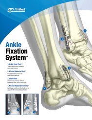
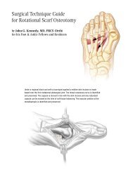
1-3mm osteotomyDorsally at the articular margin of the head begin the osteotomy, staying fairly horizontal, parallel to theground or plane of the foot. Depending on the amount of plantar prominence a second dorsal wedge cutcan be made. Incremental cuts that fine-tune the correction are preferred to a single wedge cut.Slide the bone to the desired shortened and dorsiflexed position. Holdreduction with a 0.7mm K-wire that is placed in the orientation and to thedepth of the eventual screw. Check clinically and fluoroscopically that thecorrect position has been achieved. Measure the screw length with the wiregauge. Insert the 1.7mm self-drilling cannulated screw over the K-wire andtighten. If required insert a second wire and screw. Remove any overhangingbone at the articular margin of the dorsal cortex.The remaining metatarsals are evaluated andcorrected as needed for length and plantarflexion.The wound is closed in layers. In the presence of a proximal interphalangeal (PIP) or distal interphalangeal (DIP)deformity, these may need to be addressed with a tendon lengthening and/or transfers, as well as IP fusions.Because the osteotomy shifts the center of rotation of the metatarsal phalangeal joint, there is a significant risk of adorsiflexion deformity. The joint is therefore taped for 4 weeks post-operatively, and then followed with appropriatephysical therapy.