download (pdf, 455 kb) - Institute of Pharmacology and Toxicology
download (pdf, 455 kb) - Institute of Pharmacology and Toxicology
download (pdf, 455 kb) - Institute of Pharmacology and Toxicology

You also want an ePaper? Increase the reach of your titles
YUMPU automatically turns print PDFs into web optimized ePapers that Google loves.
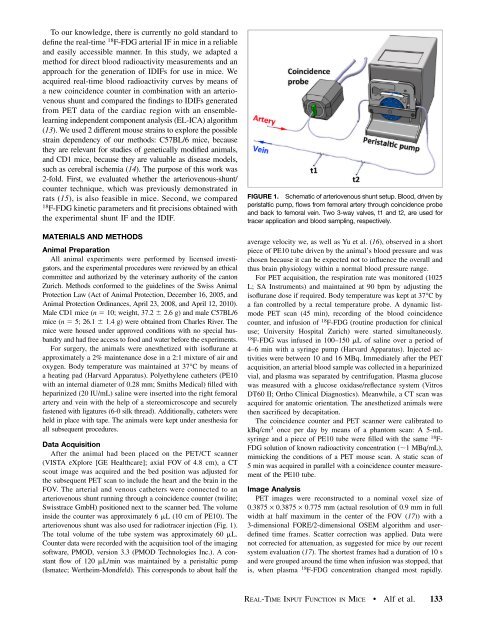
To our knowledge, there is currently no gold st<strong>and</strong>ard todefine the real-time 18 F-FDG arterial IF in mice in a reliable<strong>and</strong> easily accessible manner. In this study, we adapted amethod for direct blood radioactivity measurements <strong>and</strong> anapproach for the generation <strong>of</strong> IDIFs for use in mice. Weacquired real-time blood radioactivity curves by means <strong>of</strong>a new coincidence counter in combination with an arteriovenousshunt <strong>and</strong> compared the findings to IDIFs generatedfrom PET data <strong>of</strong> the cardiac region with an ensemblelearningindependent component analysis (EL-ICA) algorithm(13). We used 2 different mouse strains to explore the possiblestrain dependency <strong>of</strong> our methods: C57BL/6 mice, becausethey are relevant for studies <strong>of</strong> genetically modified animals,<strong>and</strong> CD1 mice, because they are valuable as disease models,such as cerebral ischemia (14). The purpose <strong>of</strong> this work was2-fold. First, we evaluated whether the arteriovenous-shunt/counter technique, which was previously demonstrated inrats (15), is also feasible in mice. Second, we compared18F-FDG kinetic parameters <strong>and</strong> fit precisions obtained withthe experimental shunt IF <strong>and</strong> the IDIF.MATERIALS AND METHODSAnimal PreparationAll animal experiments were performed by licensed investigators,<strong>and</strong> the experimental procedures were reviewed by an ethicalcommittee <strong>and</strong> authorized by the veterinary authority <strong>of</strong> the cantonZurich. Methods conformed to the guidelines <strong>of</strong> the Swiss AnimalProtection Law (Act <strong>of</strong> Animal Protection, December 16, 2005, <strong>and</strong>Animal Protection Ordinances, April 23, 2008, <strong>and</strong> April 12, 2010).Male CD1 mice (n 5 10; weight, 37.2 6 2.6 g) <strong>and</strong> male C57BL/6mice (n 5 5; 26.1 6 1.4 g) were obtained from Charles River. Themice were housed under approved conditions with no special husb<strong>and</strong>ry<strong>and</strong> had free access to food <strong>and</strong> water before the experiments.For surgery, the animals were anesthetized with is<strong>of</strong>lurane atapproximately a 2% maintenance dose in a 2:1 mixture <strong>of</strong> air <strong>and</strong>oxygen. Body temperature was maintained at 37°C by means <strong>of</strong>a heating pad (Harvard Apparatus). Polyethylene catheters (PE10with an internal diameter <strong>of</strong> 0.28 mm; Smiths Medical) filled withheparinized (20 IU/mL) saline were inserted into the right femoralartery <strong>and</strong> vein with the help <strong>of</strong> a stereomicroscope <strong>and</strong> securelyfastened with ligatures (6-0 silk thread). Additionally, catheters wereheld in place with tape. The animals were kept under anesthesia forall subsequent procedures.Data AcquisitionAftertheanimalhadbeenplacedonthePET/CTscanner(VISTA eXplore [GE Healthcare]; axial FOV <strong>of</strong> 4.8 cm), a CTscout image was acquired <strong>and</strong> the bed position was adjusted forthe subsequent PET scan to include the heart <strong>and</strong> the brain in theFOV. The arterial <strong>and</strong> venous catheters were connected to anarteriovenous shunt running through a coincidence counter (twilite;Swisstrace GmbH) positioned next to the scanner bed. The volumeinside the counter was approximately 6 mL (10 cm <strong>of</strong> PE10). Thearteriovenous shunt was also used for radiotracer injection (Fig. 1).The total volume <strong>of</strong> the tube system was approximately 60 mL.Counter data were recorded with the acquisition tool <strong>of</strong> the imagings<strong>of</strong>tware, PMOD, version 3.3 (PMOD Technologies Inc.). A constantflow <strong>of</strong> 120 mL/min was maintained by a peristaltic pump(Ismatec; Wertheim-Mondfeld). This corresponds to about half theFIGURE 1. Schematic <strong>of</strong> arteriovenous shunt setup. Blood, driven byperistaltic pump, flows from femoral artery through coincidence probe<strong>and</strong> back to femoral vein. Two 3-way valves, t1 <strong>and</strong> t2, are used fortracer application <strong>and</strong> blood sampling, respectively.average velocity we, as well as Yu et al. (16), observed in a shortpiece <strong>of</strong> PE10 tube driven by the animal’s blood pressure <strong>and</strong> waschosen because it can be expected not to influence the overall <strong>and</strong>thus brain physiology within a normal blood pressure range.For PET acquisition, the respiration rate was monitored (1025L; SA Instruments) <strong>and</strong> maintained at 90 bpm by adjusting theis<strong>of</strong>lurane dose if required. Body temperature was kept at 37°C bya fan controlled by a rectal temperature probe. A dynamic listmodePET scan (45 min), recording <strong>of</strong> the blood coincidencecounter, <strong>and</strong> infusion <strong>of</strong> 18 F-FDG (routine production for clinicaluse; University Hospital Zurich) were started simultaneously.18F-FDG was infused in 100–150 mL <strong>of</strong> saline over a period <strong>of</strong>4–6 min with a syringe pump (Harvard Apparatus). Injected activitieswere between 10 <strong>and</strong> 16 MBq. Immediately after the PETacquisition, an arterial blood sample was collected in a heparinizedvial, <strong>and</strong> plasma was separated by centrifugation. Plasma glucosewas measured with a glucose oxidase/reflectance system (VitrosDT60 II; Ortho Clinical Diagnostics). Meanwhile, a CT scan wasacquired for anatomic orientation. The anesthetized animals werethen sacrificed by decapitation.The coincidence counter <strong>and</strong> PET scanner were calibrated tokBq/cm 3 once per day by means <strong>of</strong> a phantom scan: A 5-mLsyringe <strong>and</strong> a piece <strong>of</strong> PE10 tube were filled with the same 18 F-FDG solution <strong>of</strong> known radioactivity concentration (;1 MBq/mL),mimicking the conditions <strong>of</strong> a PET mouse scan. A static scan <strong>of</strong>5 min was acquired in parallel with a coincidence counter measurement<strong>of</strong> the PE10 tube.Image AnalysisPET images were reconstructed to a nominal voxel size <strong>of</strong>0.3875 · 0.3875 · 0.775 mm (actual resolution <strong>of</strong> 0.9 mm in fullwidth at half maximum in the center <strong>of</strong> the FOV (17)) with a3-dimensional FORE/2-dimensional OSEM algorithm <strong>and</strong> userdefinedtime frames. Scatter correction was applied. Data werenot corrected for attenuation, as suggested for mice by our recentsystem evaluation (17). The shortest frames had a duration <strong>of</strong> 10 s<strong>and</strong> were grouped around the time when infusion was stopped, thatis, when plasma 18 F-FDG concentration changed most rapidly.REAL-TIME INPUT FUNCTION IN MICE • Alf et al. 133