Management of patients with palpitations: a position paper from the ...
Management of patients with palpitations: a position paper from the ...
Management of patients with palpitations: a position paper from the ...
You also want an ePaper? Increase the reach of your titles
YUMPU automatically turns print PDFs into web optimized ePapers that Google loves.
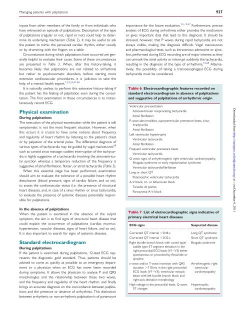
Managing <strong>patients</strong> <strong>with</strong> <strong>palpitations</strong> 927inputs <strong>from</strong> o<strong>the</strong>r members <strong>of</strong> <strong>the</strong> family or <strong>from</strong> individuals whohave witnessed an episode <strong>of</strong> <strong>palpitations</strong>. Description <strong>of</strong> <strong>the</strong> type<strong>of</strong> <strong>palpitations</strong> (regular or not, rapid or not) could help to determineits underlying mechanism (Table 2). It may be useful to ask<strong>the</strong> patient to mimic <strong>the</strong> perceived cardiac rhythm, ei<strong>the</strong>r vocallyor by drumming <strong>with</strong> <strong>the</strong> fingers on a table.Circumstances during which <strong>palpitations</strong> have occurred are generallyhelpful to evaluate <strong>the</strong>ir cause. Some <strong>of</strong> <strong>the</strong>se circumstancesare presented in Table 3. When, after this history-taking, itbecomes likely that <strong>palpitations</strong> are not related to arrhythmiabut ra<strong>the</strong>r to psychosomatic disorders, before starting moreextensive cardiovascular procedures, it is judicious to take <strong>the</strong>help <strong>of</strong> a mental health expert. 1,22,23,47,58It is naturally useless to perform this extensive history-taking if<strong>the</strong> patient has <strong>the</strong> feeling <strong>of</strong> palpitation even during <strong>the</strong> consultation.The first examination in <strong>the</strong>se circumstances is to instantaneouslyrecord ECG.Physical examinationDuring <strong>palpitations</strong>The execution <strong>of</strong> <strong>the</strong> physical examination while <strong>the</strong> patient is stillsymptomatic is not <strong>the</strong> most frequent situation. However, whenthis occurs it is crucial to have some notions about frequencyand regularity <strong>of</strong> heart rhythm by listening to <strong>the</strong> patient’s chestor by palpation <strong>of</strong> <strong>the</strong> arterial pulse. The differential diagnosis <strong>of</strong>various types <strong>of</strong> tachycardia may be guided by vagal manoeuvres 68such as carotid sinus massage: sudden interruption <strong>of</strong> <strong>the</strong> tachycardiais highly suggestive <strong>of</strong> a tachycardia involving <strong>the</strong> atrioventricularjunction whereas a temporary reduction <strong>of</strong> <strong>the</strong> frequency issuggestive <strong>of</strong> atrial fibrillation, flutter, or atrial tachycardia (Table 3).When this essential stage has been performed, examinationshould aim to evaluate <strong>the</strong> tolerance <strong>of</strong> a possible heart rhythmdisturbance (blood pressure, signs <strong>of</strong> cardiac failure, and so on),to assess <strong>the</strong> cardiovascular status (i.e. <strong>the</strong> presence <strong>of</strong> structuralheart disease), and, in case <strong>of</strong> a sinus rhythm or sinus tachycardia,to evaluate <strong>the</strong> presence <strong>of</strong> systemic diseases potentially responsiblefor <strong>palpitations</strong>.In <strong>the</strong> absence <strong>of</strong> <strong>palpitations</strong>When <strong>the</strong> patient is examined in <strong>the</strong> absence <strong>of</strong> <strong>the</strong> culpritsymptom, <strong>the</strong> aim is to find signs <strong>of</strong> structural heart disease thatcould explain <strong>the</strong> occurrence <strong>of</strong> <strong>palpitations</strong> (cardiac murmur,hypertension, vascular diseases, signs <strong>of</strong> heart failure, and so on).It is also important to search for signs <strong>of</strong> systemic diseases.Standard electrocardiogramDuring <strong>palpitations</strong>If <strong>the</strong> patient is examined during <strong>palpitations</strong>, 12-lead ECG represents<strong>the</strong> diagnostic gold standard. Thus, <strong>patients</strong> should beadvised to come as quickly as possible to an emergency departmentor a physician when an ECG has never been recordedduring symptoms. It allows <strong>the</strong> physician to analyse P and QRSmorphologies and <strong>the</strong> relationship between <strong>the</strong>se two waves,and <strong>the</strong> frequency and regularity <strong>of</strong> <strong>the</strong> heart rhythm, and finallybrings an accurate diagnosis on <strong>the</strong> concordance between <strong>palpitations</strong>and <strong>the</strong> presence or absence <strong>of</strong> arrhythmia. This distinctionbetween arrhythmic or non-arrhythmic palpitation is <strong>of</strong> paramountimportance for <strong>the</strong> future evaluation. 1,5 – 9,47 Fur<strong>the</strong>rmore, preciseanalysis <strong>of</strong> ECG during arrhythmia ei<strong>the</strong>r provides <strong>the</strong> mechanismor gives important data that lead to this diagnosis. It should bestressed, however, that P waves during rapid tachycardia are notalways visible, making <strong>the</strong> diagnosis difficult. Vagal manoeuvresand pharmacological tests, such as intravenous adenosine or ajmaline,performed during ECG recording are <strong>of</strong> major interest as <strong>the</strong>ycan unmask <strong>the</strong> atrial activity or interrupt suddenly <strong>the</strong> tachycardia,resulting in <strong>the</strong> diagnosis <strong>of</strong> <strong>the</strong> type <strong>of</strong> arrhythmia. 16,68 Alternatively,<strong>the</strong> possibility <strong>of</strong> taking a transoesophageal ECG duringtachycardia must be considered.Table 6 Electrocardiographic features recorded onstandard electrocardiogram in absence <strong>of</strong> <strong>palpitations</strong>and suggestive <strong>of</strong> <strong>palpitations</strong> <strong>of</strong> arrhythmic originVentricular pre-excitationAtrioventricular reciprocating tachycardiaAtrial fibrillationP-wave abnormalities, supraventricular premature beats, sinusbradycardiaAtrial fibrillationLeft ventricular hypertrophyVentricular tachycardiaAtrial fibrillationFrequent ventricular premature beatsVentricular tachycardiaQ wave, signs <strong>of</strong> arrhythmogenic right ventricular cardiomyopathy,Brugada syndrome or early repolarization syndromeVentricular tachycardia/fibrillationLong or short QTPolymorphic ventricular tachycardiaA-V block, tri- or bifascicular blockTorsades de pointesParoxysmal A-V blockTable 7 List <strong>of</strong> eletrocardiographic signs indicative <strong>of</strong>primary electrical heart diseasesECG signsSuspected disease................................................................................Corrected QT interval .0.46 sLong QT syndromeCorrected QT interval ,0.32 sShort QT syndromeRight bundle branch block <strong>with</strong> coved type/ Brugada syndromesaddle type ST segment elevation in <strong>the</strong>right precordial ECG leads (V1–V3) ei<strong>the</strong>rspontaneous or provoked by flecainide orajmaline1-wave and/or T-wave inversion <strong>with</strong> QRSduration .110 ms in <strong>the</strong> right precordialECG leads (V1–V3); ventricular ectopicbeats <strong>with</strong> left bundle branch block andright-axis deviation morphologyHigh voltage in <strong>the</strong> precordial leads, Q wave,ST changesArrythmogenic rightventricularcardiomyopathyHypertrophiccardiomyopathyDownloaded <strong>from</strong> http://europace.oxfordjournals.org/ at :: on December 6, 2011


