A Methods Manual for the Collection, Preparation and Analysis of ...
A Methods Manual for the Collection, Preparation and Analysis of ...
A Methods Manual for the Collection, Preparation and Analysis of ...

You also want an ePaper? Increase the reach of your titles
YUMPU automatically turns print PDFs into web optimized ePapers that Google loves.
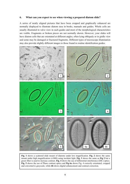
6. What can you expect to see when viewing a prepared diatom slide?A series <strong>of</strong> neatly aligned pictures that have been cropped <strong>and</strong> graphically enhanced arenormally displayed to illustrate diatom taxa in books, manuals <strong>and</strong> guides. Whole cells areusually illustrated in valve view in such guides <strong>and</strong> most <strong>of</strong> <strong>the</strong> morphological characteristicsare visible. Fragments or broken pieces are not normally shown. However, your slides willhave diatom cells that are orientated at different angles, <strong>of</strong>ten lying obliquely or in girdle view<strong>and</strong> some may be damaged or fractured fragments. Different types <strong>of</strong> microscope illuminationmay also provide slightly different images to those found in routine identification guides.1 23 45 6a 6bFig. 1 shows a scattered slide mount <strong>of</strong> diatoms under low magnification. Fig. 2 shows <strong>the</strong> samemount under high magnification (x1000) using incident light. Fig. 3 shows <strong>the</strong> same as Fig 2 but agreen filter is used to increase contrast. Fig. 4 shows <strong>the</strong> use <strong>of</strong> differential interference (DIC) optics.Fig. 5 shows <strong>the</strong> use <strong>of</strong> Phase contrast optics <strong>and</strong> Fig 6a shows Fig. 4 correctly orientated, cropped<strong>and</strong> converted to greyscale, while 6b shows digital enhancement <strong>and</strong> contrast correction.9